SECOND MOSASAUR MEETING - Oceans of Kansasoceansofkansas.com/2ndMosaMtg/Abstracts.pdf · SECOND...
28
SECOND MOSASAUR MEETING May 2-6, 2007 Abstract Booklet Michael J. Everhart, (ed.) Fort Hays State University Sternberg Museum of Natural History 3000 Sternberg Drive Hays, Kansas 67601-2006
Transcript of SECOND MOSASAUR MEETING - Oceans of Kansasoceansofkansas.com/2ndMosaMtg/Abstracts.pdf · SECOND...

SECOND MOSASAUR MEETING
May 2-6, 2007
Abstract BookletMichael J. Everhart, (ed.)
Fort Hays State UniversitySternberg Museum of Natural History
3000 Sternberg DriveHays, Kansas 67601-2006

MEETING SCHEDULEThursday, May 3, 2007: Sessions
8:00 AM Registration opens 9:00 AM Welcome by Dr. Jerry Choate, Director, Sternberg Museum of Natural History
Opening remarks and orientation: Mike Everhart
9:30 AM FIRST SESSION - Moderator: Mike Everhart 9:30 AM THE MOSASAURS OF GEORGE F. STERNBERG, PALEONTOLOGIST
AND FOSSIL PHOTOGRAPHERMichael J. Everhart
10:00 AM FIRST RECORD OF STOMACH CONTENTS IN TYLOSAURUSNEPAEOLICUS AND COMMENTS ON PREDATION AMONGMOSASAURIDAE
Gorden L. Bell, Jr. and Kenneth R. Barnes
10:30 AM BREAK
11:00 AM SECOND SESSION – Moderator: Anne Schulp11:00 AM THE ANATOMY AND SYSTEMATICS OF TYLOSAURINE MOSASAURS
Timon S. Bullard and Michael W. Caldwell
11:30 AM CHRONOSTRATIGRAPHIC DISTRIBUTION OF MOSASAUROIDS FROMTHE BIG BEND REGION OF WEST TEXAS
Gorden L. Bell, Jr., Kenneth R. Barnes, and Michael Polcyn.
NOON LUNCH (Provided)
1:00 PM THIRD SESSION – Moderator: Gorden Bell 1:00 PM TOOTH ATTACHMENT AND TOOTH REPLACEMENT PATTERNS IN
MOSASAURS Michael W. Caldwell
1:30 PM ONTOGENETIC CHANGES IN THE MARGINAL DENTITION OFTYLOSAURUS PRORIGER (SQUAMATA: MOSASAURIDAE):ECOLOGICAL AND EVOLUTIONARY IMPLICATIONS
Takuya Konishi 2:00 PM MORE ON CARINODENS BELGICUS FROM THE MAASTRICHTIAN TYPE
AREAJohn W. M. Jagt, Anne S. Schulp and Eric W. A. Mulder
2:30 PM BREAK
3:00 PM FOURTH SESSION – Moderator: Johan Lindgren 3:00 PM PRELIMINARY OBSERVATIONS ON CAMPANIAN MOSASAURS IN
ARKANSASKelly J. Irwin
2 Second Mosasaur Meeting, Sternberg Museum, Hays, Kansas, May 3-6, 2007
3:30 PM OSTEOLOGY OF THE MIDDLE EAR IN MOSASAURS (SQUAMATA):FROM IMPEDANCE MATCHING TO UNDERWATER HEARING
Michael W. Caldwell, Takuya Konishi, Alexander Dutchak, Gordon L.Bell, Jr., and James Lamb
4:00 PM A CLOSE SHAVE: THE YOUNGEST RECORD TO DATE OF MOSASAURUSHOFFMANNI FROM THE MAASTRICHTIAN TYPE AREA
John W. M. Jagt, Anne S. Schulp, Jacques Severijns, Dirk Cornelissen,Louis Verding, and Eric W. A. Mulder
4:30 PM THE FIRST MOSASAUR (REPTILIA: SQUAMATA) FROM THE LLANOESTACADO OF NORTHWEST TEXAS
Bill D. Mueller, Sankar Chatterjee, William Cornell, David Watkins, andGretchen L. Gurtler
5:00 PM Social Hour: TBD
=================================================================Friday, May 4, 2007: Sessions
8:30 AM Registration opens 9:00 AM FIFTH SESSION - Moderator: John Jagt 9:00 AM CALIFORNIA MOSASAURS II: THE MAASTRICHTIAN MORENO
FORMATION REVISITEDJohan Lindgren
9:30 AM SOUTHERNMOST OCCURRENCE OF PLATECARPUS PLANIFRONS(SQUAMATA: MOSASAURIDAE) FROM THE TOMBIGBEE SANDMEMBER (CA. MIDDLE SANTONIAN) OF ALABAMA, USA
Takuya Konishi10:00 AM A SEA URCHIN BITTEN BY A MOSASAUR - EVIDENCE OF
OPPORTUNISTIC FEEDING?Christian Neumann and Oliver Hampe
10:30 AM BREAK
11:00 AM SIXTH SESSION – Moderator: Louis Jacobs11:00 AM CONVERGENCE BETWEEN MOSASAURS AND THE
HESPERORNITHIFORM BIRDSLarry D. Martin
11:30 AM MOSASAUROID MATERIAL FROM NORTHERN ITALY: APRELIMINARY REPORT
Alessandro Palci, Michael W. Caldwell, and Cesare Papazzoni
NOON LUNCH (Provided)
1:00 PM SEVENTH SESSION (POSTERS)
2:30 PM BREAK
Second Mosasaur Meeting, Sternberg Museum, Hays, Kansas, May 3-6, 2007 3

3:00 PM EIGHTH SESSION – Moderator: Mike Caldwell 3:00 PM THE MOSASAURS OF ANGOLA
Michael Polcyn, Louis L. Jacobs, Anne S. Schulp, and Octávio Mateus 3:30 PM “PACHYOSTOSIS” WITHIN MOSASAUROIDS
Alexandra Houssaye 4:00 PM SKELETOCHRONOLOGY OF THE LIMB ELEMENTS OF MOSASAURS
(SQUAMATA; MOSASAURIDAE)Rodrigo A. Pellegrini
4:30 PM USING ELECTRON MICROSCOPY AND FOCUSED ION BEAM AS A TOOL FOR ANALYSIS OF MOSASAUR BONE
M. Amy Sheldon==================================================================
Saturday, May 4, 2007: Sessions
8:30 AM Registration opens 9:00 AM NINTH SESSION - Moderator: Eric Mulder 9:00 AM MORPHOLOGY AND SYSTEMATIC POSITION OF ANGOLASAURUS
BOCAGEI AND THE EVOLUTION OF THE BRAINCASE INPLIOPLATECARPINE MOSASAURS
Michael Polcyn, Louis L. Jacobs, Anne S. Schulp, and Octávio Mateus 9:30 AM ON DISTRIBUTION, DIVERSITY AND PHYLOGENY OF THE GENUS
PROGNATHODONAnne S. Schulp
10:00 AM VERTEBRAL PATHOLOGY IN MOSASAURS Bruce M. Rothschild
10:30 AM BREAK
11:00 AM TENTH SESSION – Moderator: Mike Polcyn11:00 AM THE TALE OF THE TAIL - TAIL FIN EVOLUTION IN THE
MOSASAUROIDEAJohan Lindgren
11:30 AM MOSASAUR BITE MARKS ON AN AMMONITE. PRESERVATION OF ANABORTED ATTACK?
William R. Wahl
NOON LUNCH (on your own)
1:00 PM ELEVENTH SESSION – Moderator: Gorden Bell 1:00 PM THE DIVERSITY OF AQUATIC LOCOMOTION IN EXTANT VARANOID
LIZARDSBruce A. Young, M. Boetig, A. Fahey, and A. Lawrence
1:30 PM PRELIMINARY ANALYSIS OF STABLE CARBON ISOTOPES INMOSASAURS AND OTHER MARINE AMNIOTES
John A. Robbins, Kurt Ferguson, Michael Polcyn, Louis L. Jacobs, andTorben C. Rick
4 Second Mosasaur Meeting, Sternberg Museum, Hays, Kansas, May 3-6, 2007

2:00 PM REMAINS OF YOUNG MOSASAURS FROM THE SMOKY HILL CHALK(UPPER CONIACIAN-LOWER CAMPANIAN) OF WESTERN KANSAS
Michael J. Everhart 2:30 PM NEW TOOLS TO UNCOVER TRENDS IN MOSASAUR RICHNESS AND
MORPHOLOGY: STRATIGRAPHICALLY CORRELATED ASSEMBLAGESMarcus R. Ross and David E. Fastovsky
3:00 PM BREAK / END OF SESSIONS
6:00 PM Reception and Banquet- Sternberg Museum of Natural History==================================================================Sunday, May 6, 2007: Field Trip (Meet at Museum by 8:30 AM)
------------------------------------------------------------------------------------------------------------
ABSTRACTS
NEW MATERIAL OF CARINODENS FROM THE MAASTRICHTIAN PHOSPHATES OFMOROCCO.
BARDET, Nathalie, UMR 5143 du CNRS, M.N.H.N., 8 rue Buffon, 75005 Paris, France,PEREDA SUBERBIOLA, Xabier, Depto. Estratigrafía y Paleontología, Fac. Ciencia yTecnología, Universidad del País Vasco / EHU, 4 8080 Bilbao, SCHULP, Anne S.,Natuurhistorisch Museum Maastricht, De Bosquetplein 6-7, NL6211 KJ Maastricht, TheNetherlands, and BOUYA, Baâdi, Office Chérifien des Phosphates, Centre Minier deKhouribga, Khouribga, Morocco.
Carinodens has been referred to as a very enigmatic, rare, and certainly the smallest mosasaurid ofthe latest Cretaceous. It is also one of the oldest figured mosasaur taxa (Faujas de Saint-Fond 1799).It is known from a subcomplete partially toothed dentary and an incomplete dentary fragment fromthe Maastrichtian of The Netherlands and some thirty-odd isolated teeth from coeval levelsworldwide (The Netherlands, Belgium, Bulgaria, Ukraine, Russia, Brazil, Morocco, and possiblyJordan) (Schulp et al. 2006). The Moroccan record is based on a currently lost tooth from theMaastrichtian phosphates described by Arambourg (1952). Here we report on the discovery of fivenew teeth found in the Upper Maastrichtian strata of the Oulad Abdoun and the Ganntour phosphaticbasins of Morocco. The crowns are typically low, swollen, laterally compressed, bicarinate, with alarge apical nubbin and are referred to C. belgicus (Woodward, 1891). Most Carinodens specimens,including the new ones from Morocco, are of late Maastrichtian age. Carinodens is mainly knownfrom the northern margin of the Mediterranean Tethys, around palaeolatitudes 40°N. The Brazilianand Moroccan teeth represent the only tropical record and confirm the astonishingly largegeographical distribution of this taxon.
Faujas de Saint Fond, B. 1799. Histoire Naturelle de la Montagne de Saint Pierre de Maëstricht.Paris, 263 pp., 54 pls.
Arambourg, C. 1952. Notes & Mém. Service géologique Maroc. 92: 1-372.Schulp, A. S., A. O. Averianov, A.A. Yarkov, F. A. Trikolide and J. W. M. Jagt. 2006. Russian J.
Herpetology 13: 175-180.
Second Mosasaur Meeting, Sternberg Museum, Hays, Kansas, May 3-6, 2007 5

FIRST RECORD OF STOMACH CONTENTS IN TYLOSAURUS NEPAEOLICUS ANDCOMMENTS ON PREDATION AMONG MOSASAURIDAE
BELL, Gorden L., Jr., Guadalupe Mountains National Park, Salt Flat, Texas, and BARNES,Kenneth R., Mosasaur Ranch Museum, Terlingua, Texas, USA.
An articulated mosasaur specimen identified as Tylosaurus nepaeolicus found in the Big Bend Regionnear Terlingua, TX is from the highest portions of the San Vicente Member of the BoquillasFormation. The specimen was found at a position about 27m (90 ft) below the top of the BoquillasFm. and probably 55m (180 ft) below the zone of Inoceramus undulatoplicatus, based on calculationsfrom local strike and dip. Stratigraphic and biostratigraphic data support an age assignment of LateConiacian.The skeleton is intimately associated with approximately 275 disarticulated bones and teeth fromother, smaller vertebrate taxa. Several factors support a conclusion that the associated bones andteeth of the other taxa represent the remains of prey items consumed by the tylosaur shortly before itsdeath. The densest concentration of disarticulated remains lies in the posterior visceral region of thearticulated tylosaur. Disarticulated bony elements exhibit typical digestive dissolution of cortical boneand exposure of internal spongiosa, the latter being most extensively exposed on protuberant bonestructures. Tooth crowns of mosasaurian prey items are also dissolved approximately to the level ofskeletal bone surrounding the tooth attachment and open pulp cavities are apparent. Lack ofdissolution on the articulated mosasaur skeleton is sufficient evidence to confirm that post-mortemtaphonomic processes or diagenesis are not causes of the observed dissolution.
Even though somewhat modified by digestive erosion, the disarticulated bony elements aresufficiently preserved to determine that a minimum of 3 immature individuals of two size classes ofPlatecarpus af. P. planifrons are present. Also present are numerous teeth of the shark, Ptychodusmortoni, none of which show significant digestive dissolution. No materials of coprolitic texture arepresent. This evidence suggests that the prey items were ingested shortly before the mosasaur died,possibly within a few days, and that the Ptychodus was likely the last item consumed.
A growing body of evidence supports conclusions that mosasaurs actively preyed upon eachother, but that congeneric individuals were not among items in the prey selection. Thereforemosasaurs cannot technically be classed as cannibals. Currently there is no evidence that members ofthe 4 major mosasaur clades dined within their own lineage.
Furthermore, the diversity of prey items found with a majority of tylosaurine remains suggeststhat they were rather opportunistic in their hunting habits. We also report here, from the same areaand stratigraphic interval as the T. nepaeolicus specimen, another mosasaur, Platecarpus af. P.planifrons, that contains several fish vertebrae in relative position consistent with stomach contents.The consistent presence of only fish in the gut contents of two species of Platecarpus correspondswell to plioplatecarpine tooth morphology and it is likely that Platecarpus specialized in eating onlyfish.
Table 1 is a synopsis of mosasaurs with associated materials that can arguably be attributed tothe remains of stomach contents. Massare (1987) previously listed mosasaurian associations withputative stomach contents. Table 1 incorporates new data but does not include potentially accidentalassociations or records not attributable to any particular mosasaur clade, as reported in Massare(1987, table 1). Original sources of data are cited from that reference.
6 Second Mosasaur Meeting, Sternberg Museum, Hays, Kansas, May 3-6, 2007
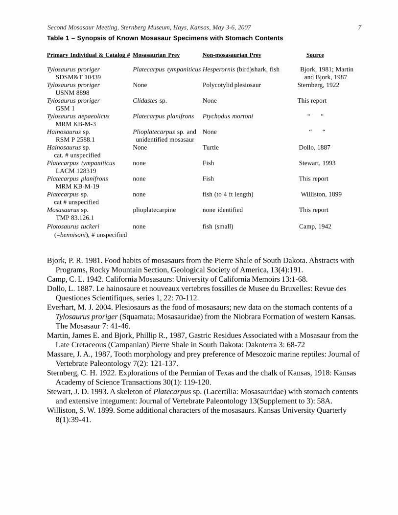
Table 1 – Synopsis of Known Mosasaur Specimens with Stomach Contents
Primary Individual & Catalog # Mosasaurian Prey Non-mosasaurian Prey Source
Tylosaurus proriger Platecarpus tympaniticus Hesperornis (bird)shark, fish Bjork, 1981; Martin SDSM&T 10439 and Bjork, 1987Tylosaurus proriger None Polycotylid plesiosaur Sternberg, 1922 USNM 8898Tylosaurus proriger Clidastes sp. None This report GSM 1Tylosaurus nepaeolicus Platecarpus planifrons Ptychodus mortoni “ “ MRM KB-M-3Hainosaurus sp. Plioplatecarpus sp. and None “ “ RSM P 2588.1 unidentified mosasaurHainosaurus sp. None Turtle Dollo, 1887 cat. # unspecifiedPlatecarpus tympaniticus none Fish Stewart, 1993 LACM 128319Platecarpus planifrons none Fish This report MRM KB-M-19Platecarpus sp. none fish (to 4 ft length) Williston, 1899 cat # unspecifiedMosasaurus sp. plioplatecarpine none identified This report TMP 83.126.1Plotosaurus tuckeri none fish (small) Camp, 1942 (=bennisoni), # unspecified
Bjork, P. R. 1981. Food habits of mosasaurs from the Pierre Shale of South Dakota. Abstracts withPrograms, Rocky Mountain Section, Geological Society of America, 13(4):191.
Camp, C. L. 1942. California Mosasaurs: University of California Memoirs 13:1-68.Dollo, L. 1887. Le hainosaure et nouveaux vertebres fossilles de Musee du Bruxelles: Revue des
Questiones Scientifiques, series 1, 22: 70-112.Everhart, M. J. 2004. Plesiosaurs as the food of mosasaurs; new data on the stomach contents of a
Tylosaurus proriger (Squamata; Mosasauridae) from the Niobrara Formation of western Kansas.The Mosasaur 7: 41-46.
Martin, James E. and Bjork, Phillip R., 1987, Gastric Residues Associated with a Mosasaur from theLate Cretaceous (Campanian) Pierre Shale in South Dakota: Dakoterra 3: 68-72
Massare, J. A., 1987, Tooth morphology and prey preference of Mesozoic marine reptiles: Journal ofVertebrate Paleontology 7(2): 121-137.
Sternberg, C. H. 1922. Explorations of the Permian of Texas and the chalk of Kansas, 1918: KansasAcademy of Science Transactions 30(1): 119-120.
Stewart, J. D. 1993. A skeleton of Platecarpus sp. (Lacertilia: Mosasauridae) with stomach contentsand extensive integument: Journal of Vertebrate Paleontology 13(Supplement to 3): 58A.
Williston, S. W. 1899. Some additional characters of the mosasaurs. Kansas University Quarterly8(1):39-41.
Second Mosasaur Meeting, Sternberg Museum, Hays, Kansas, May 3-6, 2007 7

CHRONOSTRATIGRAPHIC DISTRIBUTION OF MOSASAUROIDS FROM THE BIG BENDREGION OF WEST TEXAS
BELL, Gorden L. Jr., Guadalupe Mountains National Park, Salt Flat, Texas, BARNES,Kenneth R., Mosasaur Ranch Museum, Terlingua, Texas, and POLCYN, Michael J.,Shuler Museum of Paleontology, Southern Methodist University, Dallas, Texas, USA.
Although mostly unpublished, there is a significant succession of mosasauroids from Late Cretaceousmarine deposits of the Big Bend region in far southwest Texas. The oldest known specimens occur inlag deposits and calcarenites that are associated with the ammonoids Prionocyclus hyatti andCoilopoceras springeri in the upper part of the late Middle Turonian P. hyatti Zone approximately 36m above the base of the Ernst Member, Boquillas Formation. All are disarticulated individualelements, some from small plesiopedal forms possibly referable to the basal Russellosaurina clade andsome from larger (probably hydropedal) forms definitely referable to Russellosaurina. A fairly wellpreserved skull and vertebrae of an undescribed tylosaurine is known from a position about 30 metershigher in the Ernst Member also within the P. hyatti Zone. Several specimens of Tylosaurusnepaeolicus and a specimen of a potentially different Tylosaurus species are known from the upperportions of the San Vicente Member, Boquillas Fm., where it is of Late Coniacian age. Also from thissame interval are several specimens of Platecarpus af. P. planifrons, or a closely relatedplioplatecarpine with some similarities to Ectenosaurus.
A specimen each of Tylosaurus sp., Clidastes liodontus, Clidastes sp., and an undescribed speciesof Ectenosaurus are known from the lower portion of the Pen Fm. in the western part of the Big Bendregion. There Inoceramus undulatoplicatus is known to occur in the lower Pen Fm., but the stratigraphicpositions of these mosasaurs have not been accurately determined relative to the position of that zone.Thus, these mosasaurs may be either latest Coniacian or early Santonian age. Additionally, a singlespecimen of halisaurine? mosasaur was recovered from a higher but unconstrained level in the PenFormation. This specimen might range from middle Santonian to early Campanian in age.
BASAL MOSASAUROIDS FROM THE TURONIAN OF VALLECILLO, NUEVO LEÓN, MEXICO BUCHY, Marie-Céline, Museo del Desierto, Saltillo, Coahuila, Mexico; SMITH, Krister T.,
Vertebrate Paleontology Lab, University of Texas, Austin, Texas 78758, U.S.A; IFRIM, Christina, and GIERSCH, Samuel, Staatliches Museum für NaturkundeKarlsruhe, Germany.
The lower Turonian lithographic limestones of Vallecillo, quarried for building materials, represent anopen marine plattenkalk, deposited at the junction of the Atlantic Ocean and Western Interior Seawayduring the Early Turonian. They are known for yielding excellently preserved fish and rare othervertebrate remains. Among the aquatic vertebrates are an aigialosaur-grade mosasauroid, which waspresented at this meeting three years since. In late 2006, additional remains were discovered. Theseinclude matching fragments containing the thoracic region and forelimbs of “Pablo,” the originalaigialosaur specimen, and also “Viktor,” a less well-preserved fragment of a rather larger individual,originally mistaken for a fish. A fragment of a manus as well as “Monika,” a portion of tail, are alsoknown. Additionally, a new and complete skeleton, “Pedro,” represents the smallest individual.Finally, we introduce “Marcelo,” a large and excellently preserved but unfortunately incomplete basalmosasauroid; it preserves the tail, hindlimbs, pelvic girdle and parts of the thoracic region and theskull; soft-tissue is preserved throughout, including gut contents. The taxonomic identity of all thespecimens is not yet certain, pending further preparation. Vallecillo, to date, has yielded the remainsof six basal mosasauroids and so is the most productive site for these animals in the world. Moreover,ongoing scientific excavations and lasting collaboration with quarrymen will surely yield morespecimens from this stratigraphically and paleogeographically propitious site.
8 Second Mosasaur Meeting, Sternberg Museum, Hays, Kansas, May 3-6, 2007

THE ANATOMY AND SYSTEMATICS OF TYLOSAURINE MOSASAURSBULLARD, Timon S.1, and CALDWELL, Michael W.1,2, 1Department of Biological Sciences,
University of Alberta, Edmonton, Alberta, Canada, T6G2E9, 2Department of Earthand Atmospheric Sciences, University of Alberta, Edmonton, Alberta, Canada,T6G2E9.
The type and associated materials of Hainosaurus pembinensis (Reptilia: Squamata) from the MiddleCampanian Pembina Member of the Pierre Shale, Manitoba, are redescribed. The anatomy of thisspecies is clarified with comparison to Hainosaurus bernardi from the Early Maastrichtian ofBelgium, and Hainosaurus pembinensis is referred to the genus Tylosaurus. A new species oftylosaurine mosasaur, from the Late Campanian Bearpaw Formation of Saskatchewan, is recognized.This species represents the youngest occurrence of Tylosaurus. The phylogeny of the mosasaurSubfamily Tylosaurinae is the focus of a cladistic analysis of mosasauroids, expanding the work ofprevious authors to include Hainosaurus bernardi, Taniwhasaurus oweni, Tylosaurus pembinensisand Tylosaurus n. sp. The findings are consistent with previous studies, and show that Hainosaurusbernardi and Taniwhasaurus oweni are the most basal tylosaurines, with the species of Tylosaurusforming a monophyletic clade.
TOOTH ATTACHMENT AND TOOTH REPLACEMENT PATTERNS IN MOSASAURSCALDWELL, Michael W., Department of Earth and Atmospheric Sciences, and Department
of Biological Sciences, University of Alberta,Edmonton, Alberta, Canada, T6G2E9Recently published data on the tooth attachment histology of the Late Cretaceous marine lizardPlatecarpus (Mosasauridae) noted the presence of all currently recognized thecodont attachmenttissues. In mosasaurs, a woven-fiber bone matrix forms the margins and floor of the tooth alveolusand is identified as alveolar bone based on its histologic and topologic similarities to archosaurian andmammalian alveolar bone. Mosasaurs also appear to possess a cribiform-plate, consistent withhistologic evidence indicating the presence of a periodontal ligament. Evidence of the periodontalligament includes parallel fibers present in multiple, non-resorbed generations of alveolar bone thatare similar to mineralized collagen fiber bundles known as Sharpey’s fibers. The mosasaur tooth alsopossesses a dentine root around which is a thin layer of acellular cementum; this tissue is surroundedby a massive amount of cellular cementum which completely fills the alveolar space, increasing in sizeduring tooth replacement. The cementum mass is composed of two histologically distinct tissues: 1)a loosely organized ground matrix; 2) a laminar form surrounding the vascular tissue. Toothreplacement and the ontogeny of these tissues begins with enamel crown development in the dentallamina, posterior and lingual to the tooth position it will later occupy. The crown develops within thelamina in a vertical position and not horizontally as has been recently suggested. Utilizing amechanism not currently understood, the developing crown and its dental papilla migrate to theposterolingual margin of the tooth position where the lamina and papilla appear to initiate resorptionof the cementum mass at the base of the crown. As the resorption pit grows in size, the crown,papilla, and presumably the dental lamina, descend into the pit. Once the resorption pit has achieveda dimension equal to the size of the adult tooth crown, crown growth is truncated, and cementumbegins to develop around the non-enameled portion of the tooth. The resorption pit expands toinclude generalized resorption of the cementum and alveolar bone throughout the alveolus. Growthof cementum tissues appears to be rapid as the mineralized component thickens from the bottomdown. The effect is to erupt the tooth vertically in the alveolus; the replaced tooth likely is removedmechanically during feeding as the replacement tooth rises into the alveolus. The mosasaur form oftooth attachment is certainly thecodont and raises intriguing questions about the evolution ofpleurodonty and acrodonty within lepidosauromorphs. Likewise, the pattern of tooth replacement in
Second Mosasaur Meeting, Sternberg Museum, Hays, Kansas, May 3-6, 2007 9

mosasaurs appears to be unique among squamates specifically, and among thecodont amniotesgenerally. The conclusion that mosasaurs possess the attachment tissues used to diagnose thecodontankylosis are explored further here, and examined in the context of a reassessment of the pattern ofmosasaur tooth replacement.
IMPEDANCE MATCHING TO UNDERWATER HEARINGCALDWELL, Michael W.1,2, KONISHI, Takuya2, DUTCHAK, Alexander3, BELL, Gordon,
Jr.4, and LAMB, James5. 1Department of Earth and Atmospheric Sciences, Universityof Alberta, Edmonton, Alberta, Canada, 2Department of Biological Sciences,University of Alberta, Edmonton, Alberta, Canada, 3Department of GeologicalSciences, University of Colorado at Boulder, Boulder, Colorado, USA,4GuadalupeMountains National Park, Salt Flat, Texas, USA, and 5McWane Center, Birmingham,Alabama, USA.
The anatomical constituents of the mosasaurid middle ear are seldom preserved in their entirety in anyone mosasaur specimen as the elements are represented by three skeletal types: 1) ossifiedendochondral elements; 2) non-ossified/non-calcified cartilaginous elements; 3) calcified cartilageelements. However, by study of many different mosasaur specimens from around the globe,preserved in a variety of sediments and affected by a variety of taphonomic conditions, we havecollected sufficient data to reconstruct the middle ear skeleton in its entirety. Beginning externally,we identify a massive, plate-like element filling the expanse of the quadrate conch, as the expandedand fused pars superior, pars inferior and processus posteriorus of the extracolumella. Internally thiselement presents a short, internally trifurcating process that extends towards the stapedial opening. Inthis regard, we agree in large part with previous identifications as given by Camp (1942). The fusedelements of the extracolumella have continued to be identified, despite Camp (1942), as an ossifiedtympanum, a myth which has persisted for nearly a century. We differ from Camp (1942) in ouridentification of the elements internal to the stapedial notch and medial to the quadrate. As theextracolumella passes through the stapedial notch the shaft does not articulate with the processusinternus, but rather is fused to it so that the processus internus appears to be a middle ear bifurcationof the extracolumellar shaft. The head of the processus internus inserts into the unique mosasauridstapedial pit. The length of the fused calcified cartilaginous shaft of the processus internus +extracolumella is extremely long, tapering medially as it approaches the very delicate, ossified,columella. Our new reconstruction of the middle ear anatomy of mosasaurids necessitates two majorconsiderations: 1) re-evaluation of anatomical nomenclature as applied to the processus internus; 2)re-evaluation of the acoustic capacity of mosasaurs as it is not clear if they retained any capacity forimpedance-matching hearing in such a massive middle ear skeleton. The phylogenetic implications ofthis anatomy will be discussed by comparison to more basal mosasauroids, snakes, and other lizards.
10 Second Mosasaur Meeting, Sternberg Museum, Hays, Kansas, May 3-6, 2007

REMAINS OF YOUNG MOSASAURS FROM THE SMOKY HILL CHALK (UPPERCONIACIAN-LOWER CAMPANIAN) OF WESTERN KANSAS
EVERHART, Michael J., Sternberg Museum of Natural History, Fort Hays State University,Hays, Kansas, 67601, USA.
The remains of young mosasaurs, especially those that could be considered to be newly born, arepoorly represented in the fossil record. This lack of specimens led some earlier workers to suggestthat mosasaur babies were born or hatched in other localities such as distant shores, or along thebanks of rivers or estuaries. However, no such birthing area or concentration of the remains of youngmosasaurs has ever been located. On the other hand, specimens of immature mosasaurs have beenfound within the Smoky Hill Chalk of western Kansas. The chalk was deposited on the eastern shelfof the Western Interior Sea during late Coniacian through Early Campanian time, hundreds ofkilometers from the nearest land. Most of the specimens recovered to date are the isolatedpremaxillae of small tylosaurines, arguably the most resistant part of the skull of a mosasaur at anyage. Most also appear to be the remains of a larger predator’s meal because of the digestedappearance of the bone and the fact that the teeth have been dissolved into their alveoli. Measurementof the specimens and comparisons to complete skulls of tylosaurines for scaling purposes indicatesthat the total skull length of these individuals was 18-25 cm. Assuming that the skull to body lengthproportions (14%) remained approximately equal through life in mosasaurs, the remains representyoung tylosaurines that were 1.3 to 1.8 m in length at the time of death. Some specimens (KUVP5012 and FHSM VP-14845) include additional skull fragments and one specimen (VP-14846 and VP-14847) that includes the partially digested and commingled skull fragments of two smallplioplatecarpines. Another tylosaurine specimen (FHSM VP-14848) represents a 2 m long individualthat apparently reached the sea bottom relatively intact.
THE MOSASAURS OF GEORGE F. STERNBERG, PALEONTOLOGIST AND FOSSILPHOTOGRAPHER
EVERHART, Michael J., Sternberg Museum of Natural History, Fort Hays State University,Hays, Kansas 67601, USA.
George F. Sternberg is a well known as a field paleontologist who collected fossils primarily fromKansas and also from as far away as Canada and Argentina. His abilities as a photographer are not aswell known but were probably developed as teenager while helping his father, Charles H. Sternberg,in the family business. Later, G. F. Sternberg used small versions of his photographs glued totypewritten pages to advertise his fossils for sale to various museums around the country. His wellcomposed and large format black and white photographs represent some of the earliest visualdocumentation of fossil discoveries and collecting techniques. Although most of G. F. Sternberg’sphotographic negatives were destroyed in the basement of his home by a flood in 1951, many of hisphotographs are maintained in the archives of the Forsyth Museum at Fort Hays State University,Hays, Kansas. They include the remains of a Platecarpus ictericus collected in 1909, anotherPlatecarpus skull collected in 1911, a Clidastes propython skull and front paddles (date unknown), acomplete Platecarpus skeleton sent to Wiman in 1921 along with the type specimen of “Halisaurus”sternbergi, a complete skull of Tylosaurus kansasensis collected in 1924 and sent to Harvard,extensive preparation shots of a nearly complete Tylosaurus proriger (VP-3) in 1926, and a 1931 fieldshot on the dig of a Tylosaurus. Sternberg’s other photographs document field work and hispreparatory techniques as well as his efforts to promote fossils for public school and universitycollections.
Second Mosasaur Meeting, Sternberg Museum, Hays, Kansas, May 3-6, 2007 11

“PACHYOSTOSIS” WITHIN MOSASAUROIDSHOUSSAYE, Alexandra, UMR 5143 du CNRS, Département Histoire de la Terre, Muséum
National d’Histoire Naturelle, CP-38, 57 rue Cuvier, 75231 Paris, France, Cedex 5.“Pachyostosis” is displayed by vertebrae and sometimes ribs of various varanoid lizards withestimated reduced swimming abilities adapted to shallow marine environments (Ricqlès & Buffrénil2001). Among mosasauroids, it seems to affect only the plesiopedal form (sensu Bell & Polcyn 2005)Carentonosaurus mineaui Rage & Néraudeau 2004 and a new plesiopedal taxon (Houssaye, inprogress). The histological study of this bone specialization in Carentonosaurus reveals that itcorresponds in fact to pachyosteosclerosis (Houssaye et al. submitted) caused by the combination ofcortical hyperplasy (pachyostosis s.s.) and bone compaction due to an inhibition of the chondroclasticand osteoclastic activities (osteosclerosis). “Pachyostosis” had been observed in part of the vertebraeof Carentonosaurus because pachyostosis s.s. is morphologically observable, but osteosclerosis isnot. Other mosasauroids, displaying no external sign of pachyostosis s.s., may therefore presentosteosclerosis. Several observations suggest that this may be the case for some “mosasaurs”supposed to have lived in near shore environments (Sheldon, 1997). This specialization, which may beregarded as a neotenic process (Ricqlès & Buffrénil 2001), presents a broad polymorphism in the taxa“affected” (Lee et al., 1999) which suggests important functional implications. Moreover, the factthat it occurs only in Pythonomorpha within squamates raises the question of its possible phylogeneticsignificance.
PRELIMINARY OBSERVATIONS ON CAMPANIAN MOSASAURS IN ARKANSASIRWIN, Kelly J., Arkansas Game and Fish Commission, 915 E. Sevier St., Benton, Arkansas
72015, USA.Marine vertebrate fossils were first reported from Upper Cretaceous formations of southwesternArkansas over 150 years ago. However, with the exception of marine turtles, the subsequentliterature on Upper Cretaceous marine reptiles from Arkansas is meager. Two mosasaur taxa havepreviously been reported from the upper Campanian Marlbrook Marl Formation, Mosasaurusconodon and Plioplatecarpus marshi, based on material from historic collections. Recent fieldwork(2002-2006) in isolated exposures of the upper Marlbrook Marl, in Clark, Hempstead, and Howardcounties, has produced material from 32 mosasaurs; allowing for refinement in the determination oftaxonomic composition and measures of relative abundance, in this overlooked Gulf Coast mosasaurcommunity. A community comprised of Mosasaurus conodon, Plioplatecarpus primaevus, andPrognathodon sp. is indicated, based on characters of the teeth, vertebrae, humeri, and quadratemorphology. The Marlbrook Marl correlates to the Bluffport Marl Member, Upper Demopolis Chalkin Alabama, constraining this community to the early-middle Mosasaurus Acme Zone (MAZ) (mid-Campanian-Maastrichtian), as defined by C. R. Kiernan in 2002. The MAZ was originally erected asthe Mosasaurus-Plioplatecarpus Acme Zone by G. L. Bell in 1985, and was modified by K. R. Wrightin 1986. Comparison in the relative abundance of genera reveals a significant difference incomposition between the Alabama and Arkansas communities. Kiernan’s Alabama MAZ (mid-Campanian-Maastrichtian) was based on 30 specimens, with Mosasaurus comprising 63.3% of thesample, Plioplatecarpus 30% and Prognathodon 6.7%. Whereas the upper Campanian Arkansassample (n=34) is composed of Plioplatecarpus 55.9%, Mosasaurus 35.3%, and Prognathodon 8.8%.This preliminary analysis suggests that a more temporally and stratigraphically restricted“Plioplatecarpus Acme Zone” may have existed along the Gulf Coast during the mid- to lateCampanian.
12 Second Mosasaur Meeting, Sternberg Museum, Hays, Kansas, May 3-6, 2007

MORE ON CARINODENS BELGICUS FROM THE MAASTRICHTIAN TYPE AREAJAGT, John W. M., SCHULP, Anne S., Natuurhistorisch Museum Maastricht, de
Bosquetplein 6-7, NL-6211 KJ Maastricht, The Netherlands, MULDER, Eric, W. A.,Museum Natura Docet, Denekamp, The Netherlands.
A single tooth crown (NHMM JJ 13527), assignable to the durophagous mosasaur Carinodensbelgicus, from the basal 0.1 metre of Meerssen Member (Maastricht Fomation) subunit IVf-6, at theENCI-HeidelbergCement Group quarry (Maastricht), is the youngest in situ find of this species in thetype area of the Maastrichtian Stage so far. We have previously considered the extreme rarity ofmaterial of this taxon, now known from two lower jaws (one of which is fragmentary) and two dozenor so of isolated tooth crowns, and even done some tests to obtain data on possible prey items (J.vert. Paleont. 24: 744-747, 2004; Neth. J. Geosci., 84: 345-357, 2005). Here we plot potential foodsources, including hard-shelled prey such as bivalve (predominantly ostreids) and gastropod molluscs,ammonites, nautiloids, belemnitellid coleoids, brachiopods, irregular and regular echinoids as well ascrustaceans, in a chart documenting their distribution ranges and acmes. With this in hand it is evenmore of a mystery why this species is so rare in the area, with such a plethora of food items available.Possibly it lived elsewhere (e.g., either in deeper or shallower waters) and only fed in the area atcertain times. Of note also is that, contrary to finds of more or less articulated skeletons ofmosasaurines (Mosasaurus hoffmanni, Prognathodon saturator) and plioplatecarpines(Plioplatecarpus marshi) in the area, no postcranial elements of C. belgicus are known, or none havebeen recognised as such to date.
A CLOSE SHAVE: THE YOUNGEST RECORD TO DATE OF MOSASAURUS HOFFMANNIFROM THE MAASTRICHTIAN TYPE AREA
JAGT, John W. M., SCHULP, Anne S., SEVERIJNS, Jacques, CORNELISSEN, Dirk,VERDING, Louis, Natuurhistorisch Museum Maastricht, de Bosquetplein 6-7, NL-6211 KJ Maastricht, The Netherlands, MULDER, Eric W. A., Museum Natura Docet,Denekamp, The Netherlands.
A partial skull (left lower and upper jaws, left splenial, ?coronoid, isolated teeth and tooth crowns),found April 16, 2004 in a severely fractured state and scattered through blasting, was recovered byone of us (JS) from the uppermost Meerssen Member (Maastricht Formation) at the Ankerpoort-Curfs quarry, Geulhem (Valkenburg aan de Geul, Netherlands). Coming from the highest metre of thismember (i.e., subunit IVf-6, just below the Berg en Terblijt Horizon = K/Pg boundary), thisconstitutes the highest in situ record of Mosasaurus hoffmanni to date. There is general agreementthat the Meerssen Member represents deposition in (very) shallow, subtropical waters, with anestimated maximum depth of 2-15 meters. However, compared to other subunits, IVf-6 is marked byincreased thickness, possibly related to rapid local subsidence during the latest Maastrichtian. Thusthe sea in which this part of the sequence was laid down could still have supported large-bodiedmosasaurs such as M. hoffmanni. Although no associated skeletal material was discovered at the site,the skull must have belonged to a (semi-)articulated skeleton, rather than a floating carcass. Itsimportance is twofold; first, it represents an in situ record close to the K/Pg boundary (contraSullivan, 1987, Contr. Sci. Nat. Hist. Mus. Los Angeles Cty, 391; and other authors), and second, itenables detailed comparison of skull and dental characters (faceting, enamel structure, etc.) with othermaterial of M. hoffmanni, adding to a better understanding of that species. Previous records from thisclose to the K/Pg boundary comprised isolated tooth crowns only, of a plioplatecarpine and aglobidensine, also known, albeit rarely, from basal lag deposits of the overlying Geulhem Member(Houthem Formation) of early Paleocene age.
Second Mosasaur Meeting, Sternberg Museum, Hays, Kansas, May 3-6, 2007 13

ONTOGENETIC CHANGES IN THE MARGINAL DENTITION OF TYLOSAURUS PRORIGER(SQUAMATA: MOSASAURIDAE): ECOLOGICAL AND EVOLUTIONARY IMPLICATIONS
KONISHI, Takuya, Department of Biological Sciences, University of Alberta, Edmonton,Alberta, Canada T6G 2E9.
Detailed description of juvenile mosasaur (Squamata: Mosasauridae) anatomy has received littleattention in the literature. Here I report on the crown morphology of marginal teeth in a well-preserved juvenile specimen pertaining to one of the largest mosasaur species, Tylosaurus proriger;the specimen was collected from the lower Mooreville Chalk Formation (Upper Santonian-LowerCampanian) of western Alabama, USA. Compared with the adult dentition, which exhibits closely-spaced, conical, stout tooth crowns having bases that are nearly circular in cross section, the marginalteeth of the juvenile are much more slender and posteromedially recurved, and highly compressedlaterally at the bases; there is a proportionally larger gap between the adjacent crowns in the juvenile.The only discernible similarity in the adult and juvenile dental morphology, and thus possibly the onlydental character diagnosable of this taxon, is the intercarinal angle, which is about 117o on the labialside.
The evidence suggests that large T. proriger individuals sometimes consumed other, smallermosasaurs such as Platecarpus, but the estimated body length of this juvenile specimen at less thanfour meters is clearly smaller than a fully grown Platecarpus specimen. Therefore, the ontogeneticchanges in dental morphology reported here in T. proriger seem to be correlated with change(s) indiet through its ontogeny, as is also known in the extant, large-bodied monitor lizard, Varanusniloticus. The general similarity between the tooth crown morphology of juvenile T. proriger and thatof adult Platecarpus is also discussed from a phylogenetic point of view.
A NEW SPECIMEN OF SELMASAURUS SP., CF. S. RUSSELLI (MOSASAURIDAE:PLIOPLATECARPINAE) FROM GREEN COUNTY, WESTERN ALABAMA, USA
KONISHI, Takuya, Department of Biological Sciences, University of Alberta, Edmonton,Alberta, Canada T6G 2E9.
A rare species of plioplatecarpine mosasaur, Selmasaurus russelli Wright and Shannon, 1988, hasbeen only known from the holotype. It mainly consists of partial but otherwise superbly preservedcranial materials collected from the Selma Group of western Alabama, USA. Since its erection morethan 15 years ago, however, the taxon has never been included in any of the published andunpublished mosasaur phylogenetic analyses, and no additional material has been reported to date.Here I present detailed description of a right quadrate that is assignable to Selmasaurus sp., cf. S.russelli, collected from the Tombigbee Sand Member (Santonian) of the Eutaw Formation, GreenCounty, western Alabama. Although the quadrate is about 25% taller than that of the holotype, itshares the following characters with the type material: quadrate shaft distinctively bent medially atmid-height; medial surface of suprastapedial process broadly excavated; suprastapedial process notexpanded distally; infrastapedial process tall (>40% of quadrate height measured from condylarsurface) and pedestal-shaped, with distal end broadly contacting distal portion of suprastapedialprocess ventrolaterally; stapedial pit broadly keyhole-shaped. While the smaller quadrates of theholotype show no indication of fusion between the supra- and infrastapedial processes, the newspecimen preserves a possible cartilaginous remnant filling the small gap between these processes,making them appear to be fused. Although large, identification of the new quadrate as pertaining toPrognathodon from North America is rejected based mainly on the proportional narrowness of theelement and the non-circular stapedial pit morphology. The element differs from that of Ectenosaurusin lacking the distal expansion of the suprastapedial process and the posteriorly overlapping tonguefrom the infrastapedial process.
14 Second Mosasaur Meeting, Sternberg Museum, Hays, Kansas, May 3-6, 2007

SOUTHERNMOST OCCURRENCE OF PLATECARPUS PLANIFRONS (SQUAMATA:MOSASAURIDAE) FROM THE TOMBIGBEE SAND MEMBER (CA. MIDDLE SANTONIAN)OF ALABAMA, USA
KONISHI, Takuya, Department of Biological Sciences, University of Alberta, Edmonton,Alberta, Canada T6G 2E9.
Platecarpus is the most commonly found mosasaur taxon in North America, comprising about 60%of mosasaur specimens from the Smoky Hill Chalk Member (Upper Coniacian to Lower Campanian)of west-central Kansas, USA. Specimens of P. planifrons (Cope, 1874) occur in the lower part of themember (Upper Coniacian to Lower Santonian), but are known in much fewer numbers comparedwith the other congener from the member (= P. ictericus/P. tympaniticus), due partially to the lack ofits formal diagnosis until recently. While P. ictericus/P. tympaniticus is reported from the eastern Gulfregion (e.g., Mississippi and Alabama) as well, materials of P. planifrons have been known to dateonly from the aforementioned member in western Kansas.
Here I report on a single frontal element assignable to Platecarpus planifrons collected fromthe Tombigbee Sand Member (ca. Middle Santonian) of Montgomery County, central Alabama, USA.Although missing the anterior extremity of the premaxillary processes, left ala, and a pair ofposteromedian flanges, the element is well preserved with no distortion, and shows the followingdiagnostic characters for the species: preorbital width greater than interorbital width; dorsal surfaceplanar, lacking median dorsal keel; prefrontal and postorbitofrontal not meeting above orbit; lateralborder thickened above orbit. This specimen documents the first unambiguous occurrence of thispoorly known Platecarpus species outside of Kansas, and extends its geographical range as far southas central Alabama. Contemporary occurrence of P. planifrons during the Santonian in both westernKansas and Alabama is significant in gaining new insights into vertebrate distribution within theWestern Interior Seaway during this interval of Late Cretaceous time.
CALIFORNIA MOSASAURS II: THE MAASTRICHTIAN MORENO FORMATION REVISITEDLINDGREN, Johan. Department of Integrative Biology and Museum of Paleontology,
University of California, 1101 Valley Life Sciences Building, Berkeley, California94720-4780, USA, and Department of Geology, GeoBiosphere Science Centre, Lund University, Sölvegatan 12, SE-223 62 Lund, Sweden.
Even though the occurrence of mosasaur remains in the Maastrichtian part of the Moreno Formation(Great Valley Group), San Joaquin Valley, central California, has been known since the early twentiethcentury, surprisingly little has been done to determine the composition of this important assemblage.In the most comprehensive work on California mosasaurs published so far, Camp (1942) erected anddescribed three new and seemingly endemic taxa, “Kolposaurus” (Plotosaurus) bennisoni, “K.” (P.)tuckeri and Plesiotylosaurus crassidens, from the Tierra Loma Shale and Garzas Sand member of theformation. More recently, Bell (1997) added yet another mosasaurine to this faunal list when heidentified the enigmatic globidensine Prognathodon rapax from two partial skeletons collected in theTumey and Panoche hills, central San Joaquin Valley.
In 2006, a project was initiated with the objective of documenting the diversity andbiogeography of the Moreno mosasaur fauna and to date, five genera have been identified from theformation including Plotosaurus, Plesiotylosaurus, Prognathodon, Mosasaurus, and Halisaurus.Although the material available only permits species determination of Plotosaurus andPlesiotylosaurus, it is still sufficient enough to conclude that the Moreno assemblage is more similarat the generic level to approximately contemporaneous faunas from the Western Interior, Gulf Coastand Eastern Coastal Plain than previously (Nicholls and Meckert 2002) hypothesized. Moreover, onlyone species of Plotosaurus (P. bennisoni), is recognized, as the type materials of P. bennisoni and P.
Second Mosasaur Meeting, Sternberg Museum, Hays, Kansas, May 3-6, 2007 15

tuckeri by all likelihood pertain to a single taxon. A re-examination of the two skeletons purportedlyrepresenting P. rapax failed to verify the presence of this mosasaur in the Moreno Formation. Instead,one of the specimens is a primitive, yet undetermined species of Mosasaurus, whereas the other fossilcan be assigned only to Prognathodon sp.
Bell Jr., G. L. 1997: A phylogenetic revision of North American and Adriatic Mosasauroidea. pp. 293-332 in J. M. Callaway and E. L. Nicholls (eds.). Ancient Marine Reptiles. Academic Press, SanDiego.
Camp, C. L. 1942: California mosasaurs. University of California Memoirs 13:68 pp.Nicholls, E. L., and Meckert, D. 2002: Marine reptiles from the Nanaimo Group (Upper Cretaceous)
of Vancouver Island. Canadian Journal of Earth Sciences 39:1591-1603.
THE TALE OF THE TAIL - TAIL FIN EVOLUTION IN THE MOSASAUROIDEALINDGREN, Johan, Department of Integrative Biology and Museum of Paleontology,
University of California, 1101 Valley Life Sciences Building, Berkeley, California94720-4780, USA, and Department of Geology, GeoBiosphere Science Centre, LundUniversity, Sölvegatan12, SE-223 62 Lund, Sweden.
The Mosasauroidea includes distinctive and specialized diapsid reptiles that originated, diversified,and eventually vanished within the last 35 Ma of the Cretaceous. During their comparatively briefgeological lifespan mosasauroids were nonetheless very successful, and following their firstappearance in the Cenomanian they rapidly became abundant in epicontinental seas worldwide. Oneof the reasons for this global distribution was the ability of mosasauroids to adapt swiftly to aquaticenvironments. Early members of the group radically transformed the skull, limbs and axial skeleton tomeet the demands of marine life. Moreover, locomotion was transferred from the appendages to thetail, resulting in a very specialized sculling organ.
Despite this remarkable evolutionary transition exceedingly little work has hitherto been doneon the different stages of mosasauroid tail fin evolution, and most studies inaccurately assume thatthey were all anguilliform swimmers powered by isocercal caudal flukes (i.e. single-lobed tail finssupported by a centrally located backbone), unsuitable for rapid, sustained cruising. Nonetheless,recent investigations have demonstrated that at least the derived members of the Mosasauroidea werepisciform animals equipped with semilunate tails, making them similar in appearance to moderatelyderived ichthyosaurs (another group of extinct marine reptiles), sharks and whales.
Here, mosasaur tail fin morphology and evolution are reviewed and reconstructed from keyaspects of vertebral osteology, including centrum morphometrics and process orientation. Ageneralized pattern of regional tail anatomy in derived columns with three discrete structural units(stable proximal tail stock, mid-sectional displacement unit, and distal propulsive surface) is definedand described. Three principal evolutionary trends are recognized that helped transform the plianttails of the ancestral mosasauroids into stable, hydrodynamically efficient sculling organs – caudalregionalization, reduced inter-vertebral mobility and increased tail depth. The latter was achieved bythe formation of a hypocercal tail fluke, i.e. an asymmetrical, two-lobed caudal fin where the axialsupport is turned downwards.
16 Second Mosasaur Meeting, Sternberg Museum, Hays, Kansas, May 3-6, 2007

CONVERGENCE BETWEEN MOSASAURS AND THE HESPERORNITHIFORM BIRDSMARTIN, L. D. Biodiversity Research Center and Department of Ecology and
Evolutionary Biology, University of Kansas, Lawrence, Kansas 66045, USA.Although convergent morphology is a common occurrence in widely separated phylogenetic lines,examples are less common when the comparison extends to members in different taxonomic classes.However, giant extinct lizards (mosasaurs), share similar jaw morphology with the toothed, marine,ichthyornithiform and hesperornithiform birds. These features include the general shape of thedentary; teeth with expanded bases; a pseudothecodont tooth implantation, and a distinctiveintermandibular joint. Superficially the similarity is so strong that it resulted in the misidentification ofthe jaws of Ichthyornis as from a mosasaur and caused workers to question the association of thejaws of Hesperornis with its clearly avian skeleton. Finally one researcher actually figured a lowerjaw of a small mosasaur as the only jaw certainly pertaining to Hesperornis. Because adult mosasaursare significantly larger than hesperornithiform birds, all of the putative mosasaur jaws were thought tobe from juveniles. New fossil bird material shows that the jaws of mosasaurs and birds are dissimilarin detail and no reason exists for further confusion. Mosasaurs have pleurodont teeth with horizontaltooth replacement in an inclined row while bird teeth have vertical tooth replacement with the originalimplantation in a groove and sockets forming with maturity. Mosasaurs have coronoid bones whileornithurine birds do not. The intermandibular joint is crossed by a projection of the surangular inHesperornis but not in mosasaurs. The dentaries of Ichthyornis and Hesperornis join a smallpredentary anteriorly. The extreme similarity between the jaws of the two forms results from similar,but not identical solutions to the problems of being a fish predator in the open ocean.
THE FIRST MOSASAUR (REPTILIA: SQUAMATA) FROM THE LLANO ESTACADO OFNORTHWEST TEXAS
MUELLER, Bill D.1, CHATTERJEE, Sankar1, CORNELL, William2, WATKINS, David3, andGURTLER, Gretchen L.1, 1Paleontology Division, Museum of Texas Tech University,Lubbock, Texas 79409, USA, 2Department of Geological Sciences, University ofTexas - El Paso, El Paso, Texas 79912, 3Department of Geosciences, Universityof Nebraska, Lincoln, Nebraska 68588, USA.
The Mosasauridae is believed to have originated in the Cenomanian with Haaisiasaurus, from Israelbeing the oldest well-documented taxon. Here we report on two isolated mosasaur vertebrae foundon a cotton farm in the Llano Estacado of northwest Texas. The youngest Cretaceous strata on theLlano Estacado and adjoining Gypsum Plains are middle Late Albian, but there are no Cretaceousexposures associated with the vertebrae. A fragment of an ammonite, Mortoniceras (Late Albian),was collected near one of the vertebra’s original location. The nearest Late Cretaceous exposures areover 300 km away; however, an analysis of limestone matrix from the neural canal producedcalcareous nannofossils. These nannofossils indicate that the probable age of the vertebrae to beEarly Campanian.
Second Mosasaur Meeting, Sternberg Museum, Hays, Kansas, May 3-6, 2007 17

A SEA URCHIN BITTEN BY A MOSASAUR - EVIDENCE OF OPPORTUNISTIC FEEDING?NEUMANN, Christian and HAMPE, Oliver, Museum für Naturkunde, Humboldt Universität
zu Berlin, 10115 Berlin, Germany.Although mainly coastal, Early Maastrichtian mosasaurs occasionally roamed the open ocean of thenorthwest European chalk sea as evidenced by scarce findings of teeth or skeletons (Stolley, 1892;Gripp, 1964). The chalk sea, however, was a nutrient deficient environment, with low abundances ofsuitable prey items for large predators. At the sea floor inoceramids and large deposit feedingechinoids of the genus Echinocorys dominated the macrobenthos community. Here, we present alarge Echinocorys ovata (Leske) from the Lower Maastrichtian of Hemmoor (NW Germany) withtooth marks produced by a large animal. The tooth incisions are well preserved and were regeneratedby the sea urchin, meaning that the attack was unsuccessful and not lethal. Analyses of the biting traceusing methods of forensic odontology as well as experiments with jaw models and clay dummiessuggest that the sea urchin was bitten by an animal with large pointed teeth arranged in a prognathposition. By exclusion of other probable suspects (i.e., sharks, large teleosts, plesiosaurs, crocodiles)we strongly suggest that the bite was produced by a mosasaur of the genus Prognathodon, mostprobably P. solvayi (Dollo) which has been recorded from the Early Maastrichtian chalk (Dortangs etal., 2002).
The bitten echinoid provides clues for the functioning of the Maastrichtian chalk sea paleo-food web. As the saying goes, “beggars cannot be choosers”: in modern nutrient deficientenvironments, opportunistic feeding is the prevalent feeding strategy of large predators. Our examplesshow that Prognathodon was a feeding opportunist rather than a specialist, being capable of takingadvantage of even hard-shelled benthic organisms when other prey was not available. Additionally,our observation shows that not only mosasaurs like Carinodens and Globidens (which possessed acrushing dentition) were able to feed on armoured prey. Moreover, conical tooth morphologies notnecessarily reflect dietary specialization – as has recently been documented also from plesiosaurswhich possessed pointed conical teeth and were also capable to feed on benthic hard-shelledinvertebrates (McHenry et al., 2005).
Dortangs, R. W., Schulp, A. S., Mulder, E.W.A., Jagt, J.W.M., Peeters, H.H.G. and de Graaf, D. Th.(2002): A large new mosasaur from the upper Cretaceous of the Netherlands.- Netherlands Journalof Geosciences/Geologie en Mijnbouw 81: 1-8.
Gripp, K. (1964): Erdgeschichte von Schleswig-Holstein. Neumünster: Wachhholtz Verlag, 411 pp.McHenry, C.R. Cook, A.G., and Wroe; S. (2005): Bottom-feeding plesiosaurs.- Science 310: 75.Stolley, E. (1892): Die Kreide Schleswig Holsteins. Mitteilungen aus dem Mineralogischen Institut
der Universität Kiel 191-309.
18 Second Mosasaur Meeting, Sternberg Museum, Hays, Kansas, May 3-6, 2007

NEW MOSASAUROID MATERIAL FROM NORTHERN ITALY: A PRELIMINARY REPORTPALCI, Alessandro1, CALDWELL, Michael W.2, and PAPAZZONI, Cesare A.1,
1Dipartimento del Museo di Paleobiologia e dell’Orto Botanico, Università di Modenae Reggio Emilia, via dell’Università n°4, 41100, Modena, Italy, 2Department of Earthand Atmospheric Sciences, and Department of Biological Sciences, University ofAlberta, Edmonton, Alberta T6G2E9, Canada.
New and up to now undescribed mosasauroid material is reported from the Turonian-Coniacian ofNorthern Italy (Lessini Mountains). The mosasaurs were collected from units of the Scaglia RossaVeneta Formation, and more precisely within the lithozone informally known as the “Lastame”, anassemblage of well-bedded, reddish, often nodular marly limestones. The material is curated in twodifferent collections: The Natural History Museum in Verona and the Paleontological Museum inSant’Anna di Alfaedo. The specimen in Verona (MCSNV V7481) includes the right and left maxillae,the right dentary, the frontal, and the posterior end of the lower jaw (surangular + articular complex),while the specimens in Sant’Anna di Alfaedo are represented by a very well preserved butfragmentary skull (IGVR 4224), and by an almost complete but very badly preserved skull andpostcranial skeleton (IGVR 4301). On the basis of the unique combination of anatomical characters(e.g. tooth morphology and number, shape of the frontal, quadrate, maxillary and dentary) thepreserved material cannot be directly referred to any known mosasaur species and may represent twodifferent species and a new genus. Based on currently available anatomical details (additionalpreparation is planned), these Turonian-Coniacian mosasaurs show marked similarities to the recentlydescribed Russellosaurus coheni Polcyn and Bell, 2005. In order to address the taxonomic positionof this Italian material a thorough comparison with all basal mosasauroids is deemed necessary,especially with the known taxa currently assigned to the subfamilies Halisaurinae, Plioplatecarpinae,and Tylosaurinae, and to the genus Russellosaurus.
SKELETOCHRONOLOGY OF THE LIMB ELEMENTS OF MOSASAURS (SQUAMATA;MOSASAURIDAE)
PELLEGRINI, Rodrigo A., New Jersey State Museum, 205 W. State Street, P.O. Box 530,Trenton, New Jersey 08625-0530, USA.
Skeletochronology is a method used to determine the age of an individual from bone histology. Themethod is based on growth lines found in cortical bone, an area until now seldom examined inmosasaurs. For the first time, diaphysial thin sections of Tylosaurus, Platecarpus and Clidastes limbbones are studied using skeletochronological techniques. Results indicate that sexual maturity inmosasaurs was reached between ages five and seven, and that the mosasaurs studied exhibit a typicalsauropsid growth pattern, although their growth rates are interpreted as faster than those of extanttaxa because of fully aquatic adaptations.
A NEW SPECIES OF ECTENOSAURUS FROM TEXAS AND KANSASPOLCYN, Michael, Department of Geological Sciences, Southern Methodist University,
Dallas, Texas, 75275, USA.The mosasaur specimen KUVP 1024 was found by Charles Sternberg in the 1890s and has had acheckered taxonomic history. It was originally named Clidastes stenops by Williston (1902) based onCopes description of Edentosaurus stenops (1872), referred to Mosasaurus ivoensis by Russell(1967) based on the description of a tooth from Sweden by Persson (1963) and identified as possiblya new species of Ectenosaurus by Bell (1993). Bell’s identification was questioned by Lindgren andEverhart (2000) on the basis of the tooth count in the maxilla and other apparent differences. Therecent discovery of a nearly complete skull and partial postcranial skeleton in southwest Texas appear
Second Mosasaur Meeting, Sternberg Museum, Hays, Kansas, May 3-6, 2007 19
to represent the same taxon as KUVP 1024 and confirms Bell’s (1993) identification of the Kansasspecimen as Ectenosaurus. Further, the Texas specimen supports Bell’s (1993) suggestion that KUVP1024 may be a new species.
A PHYLOGENETIC ANALYSIS OF FHSM VP-13910; AN UPDATE ON THE MOSASAURFORMERLY AND INFORMALLY IDENTIFIED AS PLATECARPUS PLANIFRONS
POLCYN, Michael, Department of Geological Sciences, Southern Methodist University,Dallas, TX, 75275, and EVERHART, Michael J., Sternberg Museum of NaturalHistory, Fort Hays State University, Hays, Kansas 67601, USA.
Mosasaur remains discovered in 1997 and currently in the collections of the Sternberg Museum hasbeen informally referred to as Platecarpus planifrons since 1998. The well-preserved specimenincluded a complete but disarticulated skull, vertebrae and ribs, and was collected from the SmokyHill Chalk (lower Santonian) of Gove County, Kansas. Closer examination of the specimen andcomparison with the type material of P planifrons and other plioplatecarpine mosasaurs, however,does not support the earlier identification. The specimen was recently included in a phylogeneticanalysis of plioplatecarpine mosasaurs. The results of this analysis suggests that FHSM VP-13910 isthe sister taxon to a clade comprised of (Angolasaurus (Platecarpus + Plioplatecarpus)) andEctenosaurus is the sister taxon of that clade. FHSM VP-13910 presents a unique mosaic of derivedand plesiomorphic characters. In addition, the specimen also displays a number of autapomorphiesdefying referral to any known genus of mosasaur, and thus represents a new taxon withinPlioplatecarpinae.
MORPHOLOGY AND SYSTEMATIC POSITION OF ANGOLASAURUS BOCAGEI AND THEEVOLUTION OF THE BRAINCASE IN PLIOPLATECARPINE MOSASAURS
POLCYN, Michael J., Department of Geological Sciences, Southern Methodist University,Dallas, TX, 75275, USA, JACOBS, Louis L., Department of Geological Sciences,Southern Methodist University, Dallas, TX, USA, 75275, SCHULP, Anne S.,Natuurhistorisch Museum Maastricht, Maastricht, The Netherlands, MATEUS,Octávio, Museu da Lourinhã, Lourinhã, Portugal.
Recent field work in Angola has led to the discovery of new specimens of the Turonian mosasaurAngolasaurus bocagei providing new details of the anatomy of this taxon and allowing reassessmentof its phylogenetic position. One of the new specimens was encased in a well-cemented sandstone andpreserved in a fashion similar to the type specimen, but is significantly less crushed than the type. Asecond unprepared specimen includes an articulated skull and partial postcrania in a poorly-cementedsandstone matrix, and promises to be the best representative of this taxon known to date. The typespecimen and one of the new specimens were CT scanned to study previously unavailable details ofthe internal surfaces and the skull and braincase. Additionally a series of braincase CT scans ofTuronian through Campanian plioplatecarpine mosasaurs was performed for comparison andassessment of characters such as basalar artery path. Phylogenetic analysis supports a sister-taxonrelationship to the clade Platecarpus + Plioplatecarpus but indicates that the ascription ofAngolasaurus bocagei to the genus Platecarpus is unjustified.
20 Second Mosasaur Meeting, Sternberg Museum, Hays, Kansas, May 3-6, 2007
THE MOSASAURS OF ANGOLAPOLCYN, Michael W, Department of Geological Sciences, Southern Methodist University,
Dallas, TX 75275, USA, JACOBS, Louis L.,Department of Geological Sciences,Southern MethodistcUniversity, Dallas, TX, USA, 75275, SCHULP, Anne S.,Natuurhistorisch Museum Maastricht, Maastricht, The Netherlands, MATEUS,Octávio, Museu da Lourinhã, Lourinhã, Portugal.
Although occurrences of marine reptiles have been previously reported from Angola, with theexception of two Turonian taxa, these reports were based largely on isolated teeth. Fieldwork in 2005and 2006 yielded well-preserved remains of marine reptiles including plesiosaurs, turtles, andmosasaurs. The mosasaurs discussed here were recovered from two field areas: Turonian sediments atIembe along the north coast and Maastrichtian sediments at Bentiaba on the south coast. TheTuronian section near Iembe produced at least two new specimens of Angolasaurus bocagei and onefragmentary specimen of Tylosaurus iembeensis. One of the Angolasaurus specimens is representedby a well preserved, complete and articulated skull and partial postcrania, including portions of theforelimbs and pectoral girdle. The preservation of material from the Bentiaba locality is remarkabledue to the grain support of the entombing sandstone, which preserves fine anatomical details withlittle apparent crushing, and in the number of articulated, semi-articulated, and associated skeletons.Identifications from the field and preliminary preparation show the Bentiaba mosasaur fauna isrepresented by at least five genera including Mosasaurus, Prognathodon, Globidens, Plioplatecarpusand Halisaurus. Collectively, these new specimens greatly expand our knowledge of the anatomy andsystematics of Angolan mosasaurs.
THE POSSIBLE OCCURRENCE OF ANGOLASAURUS IN THE TURONIAN OF NORTH ANDSOUTH AMERICA
POLCYN, Michael, Department of Geological Sciences, Southern Methodist University,Dallas, TX, 75275, LINDGREN, Johan, Department of Integrative Biology andMuseum of Paleontology, University of California, 1101 Valley Life Sciences Building,Berkeley, California 94720-4780, USA, and Department of Geology, GeoBiosphereScience Centre, Lund University, Sölvegatan 12, SE-223 62 Lund, Sweden, andBELL, Gorden L. Jr., Guadalupe Mountains National Park, Salt Flat, TX 79847,USA.
A partial skull of a mosasaur was collected from the Eagle Ford Formation near Waco Texas in about1918 and briefly mentioned by Adkins (1924, 1928). The specimen found its way to Sweden througha donation and was housed in the collections of Lund University until relocated by one of us (GB)and recently prepared for study. The specimen includes the posterior portion of the skull, preserved toapproximately the anterior terminus of the frontals, the atlas, axis and a portion of the firsthypapohysis-bearing cervical vertebra. It is remarkable in both its Turonian age and the preservationof the osseous portion of the hyoid apparatus. The dimensions of the frontal and parietal, the path ofthe frontoparietal suture, the position of the pineal foreman in the center of the triangular parietaltable, and an enlarged posteroventral process of the parietal meeting the supraoccipital allow tentativereferral of the specimen to the genus Angolasaurus. A second specimen consisting of a partialpremaxilla and jaw fragments containing teeth from the Prionocyclus hyatti zone of the Eagle FordFormation south of Dallas, Texas is also likely referable to Angolasaurus. Further, two teeth reportedfrom the Sergipe Basin in Brazil are virtually indistinguishable from those in the type specimen ofAngolasaurus. Thus Angolasaurus appears to be the only Turonian mosasaur genus represented onboth sides of the Atlantic Ocean. Preservation of the hyoid allows comparison with other squamatesand is consistent with the morphology found in varanoid lizards and markedly different from that ofsnakes and thus has bearing on the broader relationships of mosasaurs.
Second Mosasaur Meeting, Sternberg Museum, Hays, Kansas, May 3-6, 2007 21

PRELIMINARY ANALYSIS OF STABLE CARBON ISOTOPES IN MOSASAURS AND OTHERMARINE AMNIOTES
ROBBINS, John A., FERGUSON, Kurt, POLCYN, Michael, and JACOBS, Louis L.Department of Geological Sciences, and TORBEN, C. Rick, Department ofAnthropology, Southern Methodist University, Dallas, Texas 75275, USA.
Little detail is known about the trophic webs of the complicated marine communities of whichmosasaurs were a part. Analyses of stable carbon isotopes have proven useful in dietary studies ofterrestrial organisms and in archaeology. An initial study by Biasatti (2004; Palaeogeography,Palaeoclimatology, Palaeoecology, 206: 203-216) on marine turtles indicates major shift in δ13Cvalues between consumer and food species. Similar offsets are seen in red abalone (Haliotisrufescens) and marine iguanas (Amblyrhynchus cristatus) and their foods. We report here initialcarbon isotope data for the carbonate component of tooth enamel from mosasaurs and some modernmarine amniotes to develop baseline hypotheses for the interpretation of trophic levels in Cretaceousseas. Preliminary values suggest a 15‰ shift in the carbon isotopic value from macroalgae (~-15‰)to the carbonate component of tooth enamel in marine iguanas (-0.55‰), a value comparable to thoseof the carbonate component of humeri in green turtles (Chelonia mydas), which feed on marineangiosperms. Using the same isotopic difference (“δ = 15‰), one value from a Coniasaurus sp.tooth (-2‰) suggests that this species subsisted primarily on bony fish, shrimp, and crab.
NEW TOOLS TO UNCOVER TRENDS IN MOSASAUR RICHNESS AND MORPHOLOGY:STRATIGRAPHICALLY CORRELATED ASSEMBLAGES
ROSS, Marcus R., Dept. of Biology, Liberty University, 1971 University Blvd., Lynchburg,Virginia 23402; and FASTOVSKY, David E., Department of Geoscience, Universityof Rhode Island, Kingston, Rhode Island 02881, USA.
Existing high-resolution marine biostratigraphic systems (e.g., ammonites, belemnites, calcareousnannofossils) offer tremendous precision in dating vertebrate fossils. However, due to thecomparative rarity of vertebrate remains in any particular microfossil or invertebrate biozone, and thepotential for non-recovery of an index taxon due to various environmental, ecological, or humanfactors, individual biozones are ineffective sampling windows. As a result, most analyses of marinevertebrate diversity consist either of local/regional studies with high temporal resolution (e.g.,Sheldon, 1996; Mulder et al., 1998) or studies on far broader scales with concomitantly lowertemporal resolution (e.g., Russell, 1993; Hirayama, 1997; Kriwet and Benton, 2004).Stratigraphically correlated assemblages (SCAs) are informal sub-stage level subdivisions, basedprimarily on grouped ammonite and calcareous nannofossil biozones, sequence stratigraphy, and,where possible, radioisotope-derived dates. The fifteen late Cretaceous SCAs are designed toprovide a framework for correlations among marine shelf and epicontinental sea deposits, particularlyin North America but broadly enough defined to be useful globally. Though not uniform in duration(SCAs range from 1.2-3.0 myr using timescale of Gradstein et al., 2004), SCAs offer a convenientmethod for compiling and organizing marine vertebrate fossils from far-ranging localities with highlevels of biostratigraphic resolution.Following a brief description of the SCAs, we provide examples of their use. Utilizing an onlineglobal database of catalogued mosasaur specimens (MOSABASE), we employ SCAs as samplingbins to track changes in biogeography, diversity, extinction, and intra-clade morphological trendsthroughout the entirety of their distribution during the late Cretaceous. The results of this studyinclude recognition of an initial and statistically significant expansion of mosasaurs during theConiacian and Santonian, followed by stable generic richness levels until their mass extinction at theK-Pg boundary. Surprisingly, despite stable generic richness levels during the Campanian and
22 Second Mosasaur Meeting, Sternberg Museum, Hays, Kansas, May 3-6, 2007
Maastrichtian, mosasaur morphological diversification, as measured by novel tooth forms, expands asnew forms proliferate at the expense of less-derived members.
Gradstein, F. M., Ogg, J. G., and Smith, A.G. (eds.) 2004. A Geologic Time Scale 2004. CambridgeUniversity Press, 589 p.
Hirayama, R. 1997. Distribution and diversity of Cretaceous chelonioids, p. 225-241, in Calloway, J.M., and Nicholls, E. L. (eds.), Ancient Marine Reptiles. Academic Press, San Diego, California.
Kriwet, J., and M. J. Benton. 2004. Neoselachian (Chondrichthyes, Elasmobranchii) diversity acrossthe Cretaceous-Tertiary Boundary. Palaeogeography, Palaeoclimatology, Palaeoecology, 214(3):181-194.
Mulder, E. W. A., J. W. M. Jagt, M. M. M. Kuypers, H. H. G. Peeters, and J. Rompen. 1998.Preliminary observations on the stratigraphic distribution of late Cretaceous marine and terrestrialreptiles from the Maastricht area (SE Netherlands, NE Belgium). Oryctos, 1: 55-64.
Nicholls, E. L., and A. P. Russell. 1990. Paleobiogeography of the Cretaceous Western InteriorSeaway of North America: the vertebrate evidence. Palaeogeography, Palaeoclimatology,Palaeoecology, 79: 149-169.
Russell, D. A. 1993. Vertebrates in the Cretaceous Western Interior Sea, p. 665-680. In Caldwell,W. G. E. and E. G. Kauffman (eds.), Evolution of the Western Interior Basin. GeologicalAssociation of Canada, Special Paper 39.
VERTEBRAL PATHOLOGY IN MOSASAURSROTHSCHILD, Bruce M., University of Kansas Museum of Natural History, Lawrence,
Kansas, 66045, USA.Three forms of pathology are routinely noted in the vertebrae of mosasaurs: shark bites, avascularnecrosis and fusion, in addition to isolated reports of ‘tumors.’
Grooves on vertebrae evidence shark bites, while associated new bone formation allowsrecognition of predation. The former is common; the latter, relatively rare but provides insights tobehavior of both the mosasaur and its attacker.
Recognition of diving disease in mosasaurs was based upon identification of a specificassociated pathology, avascular necrosis. The devitalized bone typically becomes necrotic (and lucentto x-rays), secondary to loss of vascular supply. The resultant loss of mechanical integrity makes thesurface susceptible to compression stresses across the shoulder and hip joint, respectively. Theresultant damaged bone can no longer resist the normal stresses across the joint and partly collapses,producing a visible subsidence zone. Avascular necrosis was invariably present in Platecarpus,Tylosaurus, Mosasaurus, Plioplatecarpus, Prognathodon, Hainosaurus and an Antarctic mosasaur,and invariably absent from Clidastes,Ectenosaurus, Globidens, Halisaurus and Kolposaurus.
Fused mosasaur vertebrae are attributable to reactive bone from trauma and infection(e.g., related to trauma of shark bites) and perhaps to splinting or a disease documented incontemporary varanids. It would appear that fusion through vertebral centra occurs whenevermotion is lost at that segment. However, there is another form of fusion through the outer layers ofwhat is presumed to have been an intervertebral disk, as has been documented in contemporaryVaranus.
Isolated suggestion of tumors is a more complex subject. Moodie’s suggestion ofosteoma must be rejected on the basis of apparent lesion size and location. The only other recognized‘tumor’ has been called an osteoma, but its size complicates histologic examination to assure it is notsimply a hamartoma, as human skull so-called osteoma have been now reclassified.
Second Mosasaur Meeting, Sternberg Museum, Hays, Kansas, May 3-6, 2007 23

ON DISTRIBUTION, DIVERSITY AND PHYLOGENY OF THE GENUS PROGNATHODONSCHULP, Anne S., Natuurhistorisch Museum Maastricht, De Bosquetplein 6-7, NL-6211 KJ
Maastricht, The Netherlands.Globidensine mosasaurs constituted a highly successful, morphologically diverse group which, duringthe latest Cretaceous, was widely distributed geographically and had diversified into a considerablearray of ecological niches. During this time, the globidensine genus Prognathodon achievedworldwide distribution, with the late Maastrichtian Prognathodon saturator being one of the larger,most massively built representatives. The holotype of this species, discovered in 1998 at the ENCI-Heidelberg Cement Group quarry (Maastricht), is compared with material from New Zealand, Israel,Belgium and the USA. Cladistic analysis places P. saturator with the Maastrichtian P. waiparaensisand P. solvayi; contrary to previous interpretations, the Campanian P. currii is considered moreclosely related to the globidensine mosasaur Globidens. The North American species Prognathodonstadtmani does not belong to the genus, rather representing a more basal form, more closely relatedto Mosasaurus. Two new partial mosasaur skulls from the Upper Cretaceous of Angola are brieflydiscussed, and tentatively assigned to the genus Prognathodon, pending excavation of the remainderof the skeleton.
MORPHOLOGY AND FUNCTION OF TAILBENDS IN MOSASAURSSCHUMACHER, Bruce A., Sternberg Museum of Natural History, Hays, Kansas, and
VARNER, D. W., Plainsboro, New Jersey, USA.The existence of a downward bend in mosasaur tails is not yet firmly established, in particular forderived members of the group. Mosasaur skeletal mounts and reconstructions often overlook the tailbend, but as early as 1899 H. F. Osborn confirmed ventral tail flexure in an articulated specimen ofTylosaurus and concluded that the tail possessed a swimming organ. In the early 20th century, J. C.Merriam and C. J. Wiman further discussed tail flexure in specimens of Halisaurus, Clidastes, andPlatecarpus, and compared mosasaur tails to those of ichthyosaurs and metriorhynchid crocodylians.Our studies confirm the presence of downward tail bends in the early mosasaurs Clidastes,Platecarpus, and Tylosaurus. Articulated caudal series of each of these genera reveal slightly wedge-shaped vertebral centra, with dorsal and ventral lengths differing between 0.5 and 1.5 mm. Althoughthis variation is not visibly apparent, it accounts for a cumulative tail flexure of ~30 degrees.Throughout the tail curve, equal spacing of neural spines is maintained by a successive anterior toposterior inclination. The most pronounced anterior inclination of neural spines coincides with themost pronounced downward flexure of the tail, a feature paralleled in early ichthyosaurs and marinecrocodiles. The down-turned segment of mosasaur tails are dorso-ventrally expanded and stiffened‘paddles’, a feature reflected by elongate neural spines and haemal arches, and loss of transverseprocesses coupled with lateral compression of caudal centra.
Unlike the tails of derived ichthyosaurs and marine crocodilians, groups that possess a fish-like dorsal caudal fin, mosasaur tails are not sharply bent and bear no abrupt change in morphology.The hypocercal tails of mosasaurs bear a gentle sinuous curve throughout their length, with a slightupward flexure anterior to a more significant downward flexure. This shape, likely lacking apronounced dorsal caudal fin, would produce downward thrust to compensate for positive buoyancy.Mosasaurs retain elongate neural and haemal spines in the area of the downward bend, heightened inthe manner of primitive ichthyosaurs, which coupled with elongate body plans indicates a similar fromof eel-like swimming in early members of both groups. Future work on mosasaur skeletons withpreserved integument should focus on evidence of the tails outline.
24 Second Mosasaur Meeting, Sternberg Museum, Hays, Kansas, May 3-6, 2007

USING ELECTRON MICROSCOPY AND FOCUSED ION BEAM AS A TOOL FOR ANALYSISOF MOSASAUR BONE
SHELDON, M. Amy, Biology Department, Oklahoma Panhandle States University, 205Hefley Hall, Goodwell, Oklahoma 73939, USA.
Traditionally it has been accepted that elements in biological tissue have been replaced over time byelements from the surrounding sediments. Using standard light microscopy investigators have shownthat soft tissue as well as the expected calcified tissue is preserved. Further work has shown thatbones are either not changed or just premineralized, leaving microstructure in place. As a result,research into fossil bone histology is a growing area of inquiry. Incorporating histological study intothe investigation, present new opportunities for gaining data to understand better the ontogeny,ecology and evolution of vertebrates.
I have restricted the fossil material in this study to ribs and vertebrae. Ribs are not shown tohave important morphological data relating to systematics or evolution therefore their destructiondoes not impede the research of others. Vertebrae were collected as isolated specimens on outcropsbut are identifiable to genus and growth stage.
Histology has given new insight to changes in growth and bone architecture for vertebratesespecially those that secondarily moved to marine environment, Vascular tissue and analysis ofchemical composition are processes that reveal valid data. Preserved structures are visible usingSEM (Scanning Electron Microscopy). Focused Ion Beam (FIB) processes allow the removal ofminute wafers of material to be further analyzed using TEM (Transmitting Electron Microscopy).
KOURISODON SP. FROM THE UPPER CAMPANIAN OF JAPANTANIMOTO, Masahiro, Higashi 3-249, Yurigaoka, Nabari, Mie 518-0473, Japan.
The small mosasaurine mosasaur Kourisodon characterized by having laterally compressed teeth wasfirst described by Nicholls and Meckert (2002) from the upper Santonian Nanaimo Group of theVancouver Island, Canada. They also cited the report of a similar type of teeth from the MaastrichtianIzumi Group of southwest Japan by Tanimoto et al. (1998).
Tanimoto and Kanazawa (2001) published an abstract on the partial skull and mandible of asmall mosasaur specimen (Kanazawa specimen) from the upper Campanian Izumi Group. At thattime, however, the paper by Nicholls and Meckert (2002) was not published yet, and Kanazawaspecimen only remained as Mosasauridae gen. et sp. indet. After the paper by Nicholls and Meckert(2002) was published, Kanazawa specimen was readily identifiable as Kourisodon because it had theunique, laterally compressed tooth morphology (Tanimoto et al, 2004).Thereafter, through the progress of cleaning of this fossil, it became clearer that the shape of theedentulous prow of the left dentary is also similar to that of Kourisodon puntledgensis from Canada,more so than Tanimoto et al (2004) had initially considered. Although the teeth of Kanazawaspecimen are laterally compressed to the same degree as the specimen from Canada, the prismaticornamentation appears to be more clearly defined in Kanazawa specimen than in the other.Nevertheless, the final recognition of such a feature will be pending the direct comparison of bothspecimens. The left maxilla of Kanazawa specimen has distinctly slenderer teeth than those of thedentary: such a tendency is observed in Kourisodon puntledgensis and the similarly small mosasaurineClidastes propython to a lesser extant. Consequently, the Japanese specimen is tentatively treated asKourisodon sp., albeit probably not pertaining to the generic type from Canada.
Second Mosasaur Meeting, Sternberg Museum, Hays, Kansas, May 3-6, 2007 25

From the Izumi Group sediments, many fragmentary mosasaur remains have been discoveredin the last decade. As most of them are anatomically small in size, they may well representKourisodon or related forms.
Nicholls, E. & Meckert, D., 2002. Marine reptiles from the Nanaimo Group (Upper Cretaceous ofVancouver Island. Canadian Journal of Earth Sciences 39: 1591-1603.
MOSASAUR BITE MARKS ON AN AMMONITE. PRESERVATION OF AN ABORTED ATTACK?WAHL, William R. Wyoming Dinosaur Center, 110 Carter Ranch Road, Thermopolis,
Wyoming 82443, USA.Preserved bites are a rare demonstration of predator and prey and give a useful view ofpaleoenvironments. Non-invasive tests were utilized in an investigation of bites in a specimen ofPlacenticeras meeki at the Wyoming Dinosaur Center, Thermopolis, Wyoming. (WDC-Bps-002).This investigation led to a reevaluatition of the limpet home marks vs. mosasaur predation bitehypothesis.
The specimen was collected by Canada Fossils (Government of Alberta, Disposition no. 2-707) from the Bearpaw Shale in Alberta, Canada, and it consists of a complete specimen (49cm by37cm) with what appears to be a single bite mark (14cm by 6cm) across both flanks (FIG.1, FIG.2). Itwas suggested that the specimen might be a composite specimen, mixing a phragmocome of one anda body chamber of another, or a near-complete specimen with a body chamber with a wedge ofanother containing a “bitten portion”. However, a CAT scan indicated a consistent thickness of shell,suggesting both portions are from an original intact but fractured specimen.
This specimen is unique in that it preserves a single bite mark with an opposing tooth markpattern (i.e. on either flank of the ammonite) (G.E.G. Westermann, 2007, personal communication). Inmost specimens with multiple bite marks, the pattern is not obvious making it hard to visualizewhether or not the tooth marks actually match in pattern (Tsujita and Westermann, 2001).
It appears that the specimen has the entire body chamber preserved which is also unusual.Most bitten specimens exhibit more damage often missing a portion of the body chamber (Tsujita andWestermann, 2001). Even though the “bite marks” are within the phragmocone, the CAT scans and3D surface scans suggested that the phragmocone contact at the body chamber was sediment filledbut not collapsed.
The hypothesis that the holes are limpet home marks was reevaluated as mosasaur bite markson shells and mentioned in other studies (Kauffman and Kesling, 1960.Tsujita and Westermann, 2001;Kauffman, 2004). The option of thin-sectioning WDCB-Bps-002 to establish this hypothesis was notavailable. However, the bite marks are aligned through the shell flanks and pierce into the septa. CATscans of this ammonite lined up on these punctures provided non-invasive views within the septasection revealing an inverted “v” opening unlike those that would be produced by the limpethypothesis (Tsujita and Westermann, 2001)(FIG.3).
Equally interesting about this specimen is that the bites are aligned but offset on both flanks.The 3D scanned images of the aligned bites on WDCB-Bps-02 were superimposed together.Measurements taken from this image suggested the alignment was offset by 12 degrees (FIG.4). Arobot set of jaws modeled after a mosasaur was used in a previous study to determine the effect ofbites on cephalopod shells (Kase et al, 1996). As the jaw structure of the robots was dimensionallylinear the effect was uniform punctures across the shell. However it has been suggested that such mayhave neglected the offset flexing of the midline dentary hinge (Kauffman and Kesling, 1960; Tsujitaand Westermann, 2001).
26 Second Mosasaur Meeting, Sternberg Museum, Hays, Kansas, May 3-6, 2007

It is interesting to note that the phragmocone contained the only punctures. The flatplanispiral section of the shell of WDCB-Bps-02 is full of complex structural septa that reactdifferently to the teeth of a mosasaur than that of the open unsupported body chamber (Tsujita andWestermann, 2001). The spacing of holes was preserved by the complex large spaces in betweensepta (Westermann, 1996). Not only would complex septa provide more elasticity to the shell wall(thus inhibiting shattering), but they would support the shell wall more evenly. Also, the spaces withinthe body chamber are analogous to those at the anterior end of the belemnite shells in the JurassicSundance Formation (Wahl, 1998). These open belemnite phragmocones are crimped and smashed bya center point of force; which is what would be expected from a tooth. The Placenticeras meekiphragmocone might instead hold the tooth shape without much crushing as in the open spaces of abody chamber.
The surface puncture exhibits an extra tooth mark that may have been a pathology or artifactof an initial or secondary bite (FIG.5). Mosasaur tooth series occasionally display new teeth growingvertically beside the old teeth (Rieppel and Kearney, 2005). The extra hole may correspond to a large“newer” tooth that is preserved out of alignment. However, it may also represent where only onetooth mark was made in an initial or “mouthing” bite. The odd puncture hole was beyond the extentof the mantle and based on healed bite marks in other species thus would not likely have beenrepaired (Bond and Saunders, 1989). The extra puncture could not have been present before the mainbite marks, as any hole in the phragmocone would have led to the phragmocone flooding andammonite death (Gordon Bell, 2007 personal communication). Interpretation of the bite suggests itwould have gone through parts of the ammonite that would have been exposed outside the bodychamber. However, it has been suggested that ammonites could withdraw the majority of the bodyinto this chamber (Kauffman, 2004).
Finally, why bite the shell if not to feed? The bite may have occurred as an attack on a gas-filled floating shell of a dying or dead ammonite; or the ammonite could have been one of dozens ofammonites bitten in a frenzy and just sank while others were eaten. Likewise it is not beyondexpectation that a mosasaur would have been attracted by fish to rotting ammonite carcass. Ofcourse, if the shell records only one “bite event,” then it might mean that the attack was aborted forsome reason and the ammonite died, sank and was buried without being consumed.
Specimens with corresponding bites on both sides are rare. Single bites occurring relative tothe ammonite phragmocone/body chamber contact are rarer still. Interpretation of predator preyrelationships will benefit from additional finds.
The author wishes to thank the Wyoming Dinosaur Center and Dr. B. Pohl for allowingresearch on specimen WDCB-Bps-002 and the staff, especially Daryl Mattern, of the RadiologyDepartment of the Hot Springs County Memorial Hospital.
Bond, P. N. and W. B. Saunders, 1989. Sublethal injury and shell repair in Upper MississippianAmmonoids. Paleobiology,15.4:414-428
Callison. G. 1967. Intracranial mobility in Kansas mosasaurs. University of Kansas PaleontologicalContributions Paper No. 26:1-15
Kase, T, P. A Johnston, P. A Seilacher, and J.B. Boyce. 1998. Alleged mosasaur bite marks on LateCretaceous ammonites are limpet (patellogastropod) home scars: Geology. 26:947-950.
Kauffman, E. G. and R. V. Kesling. 1960. An Upper Cretaceous ammonite bitten by a mosasaur;University of Michigan Contributions from the Museum of Paleontology, 15:103-248.
Kauffman, E. G. 2004. Mosasaur predation on Upper Cretaceous nautiliods and ammonites from theUnited States Pacific coast. Palios.19:96-100
Second Mosasaur Meeting, Sternberg Museum, Hays, Kansas, May 3-6, 2007 27

Rieppel, O. and M. Kearney, 2005.Tooth replacement in the Late Cretaceous mosasaur Clidastes.Journal of Herpetology. 39.4:688-692.
Tsujita C. J and G. E. G. Westermann, 2001. Were limpets or mosasaurs responsible for theperforations in ammonite Placenticeras? Paleogeography, Palaeleoclimatology, Paleoecology. 169:245-270.
Westermann, G. E. G. 1996. Ammonoid life and habitat, in Ammonoid Paleobiology. Edited by N.Landman et al. p677. Vol.13. Topics in Geobiology. Plenum Press, New York
THE DIVERSITY OF AQUATIC LOCOMOTION IN EXTANT VARANOID LIZARDSYOUNG, Bruce A., BOETIG, M., FAHEY, A., and LAWRENCE, A. Department of Biology,
Washburn University, Topeka, Kansas, 66621, USA.It is generally recognized that there is a close phylogenetic relationship between the aigialosaurs,mosasaurs and the extant varanoid lizards. The aigialosaurs, as traditionally recognized, would appearto represent a functional intermediate in being aquatic forms with limbs similar to those of terrestrialvaranoids. As a first step toward exploring the potential locomotor dynamics of these groups; weexamined the kinematics and mechanics of aquatic locomotion in terrestrial and semi-aquaticmonitors (Varanus). A large swimming tank was constructed and the kinematics of aquaticlocomotion recorded using standard and high-speed digital video. Electromyographic recordings ofaxial, tail, and limb muscles during unrestrained swimming helped identify several discrete modes ofaquatic locomotion. Among the monitors examined there is a sharp difference in both swimmingmode and performance between the terrestrial and semi-aquatic species. Of particular significance isthe diversity of functional roles played by the hindlimbs during swimming.
28 Second Mosasaur Meeting, Sternberg Museum, Hays, Kansas, May 3-6, 2007