Screening Lu
11
Genes involved in protein glycosylation determine the activity and cell internalization of the antifungal peptide PAF26 in Saccharomyces cerevisiae Eleonora Harries a,b , Lourdes Carmona a , Alberto Muñoz b,1 , José I. Ibeas c , Nick D. Read b,1 , Mónica Gandía a , Jose F. Marcos a,⇑ a Food Science Department, Instituto de Agroquímica y Tecnología de Alimentos (IATA), Consejo Superior de Investigaciones Científicas (CSIC), Avda Agustín Escardino 7, 46980 Paterna, Valencia, Spain b Fungal Cell Biology Group, Institute of Cell Biology, University of Edinburgh, Rutherford Building, Edinburgh EH9 3JH, UK c Centro Andaluz de Biología del Desarrollo, Universidad Pablo de Olavide-Consejo Superior de Investigaciones Científicas (CSIC), Carretera de Utrera km. 1, 41013 Sevilla, Spain article info Article history: Received 21 May 2013 Accepted 2 August 2013 Available online 11 August 2013 Keywords: Antimicrobial peptide Histatins Melittin Cell wall Protein glycosylation Cell-penetrating peptide abstract We have previously characterized the synthetic hexapeptide PAF26 as a cell-penetrating and non-lytic antifungal peptide that is active against Saccharomyces cerevisiae and filamentous fungi. Numerous cell wall (CW) proteins are glycosylated in fungi and many of these play important roles in fungal pathogen- esis. In this study, we screened a collection of S. cerevisiae deletion mutants for protein glycosylation genes whose deletion altered the sensitivity to PAF26. Increased tolerance to PAF26 was observed in mutants with the following disrupted genes: PMT1-6, EOS1, ALG5, MNN1, MNN4 and MNN5. Significantly, genes coding for protein O-mannosyltransferase 2 (Pmt2p), which is responsible for the addition of the first mannosyl residue of O-linked carbohydrates, and for Eos1p, an enzyme involved in N-linked glyco- sylation of proteins, showed resistance to PAF26 and defects in CW integrity. Microscopic studies on the S. cerevisiae Deos1 deletion mutant demonstrated a blockage of peptide internalization by cells. Protop- lasts lacking CWs interacted with the peptide, but were more resistant to peptide killing than cells pos- sessing CWs due to a blockage in PAF26 internalization. Interestingly, protoplasts obtained from Deos1 behaved similarly to those of the parental strain. Collectively, these observations demonstrate that the CW is a positive factor that determines the internalization of the PAF26, and that Eos1p exerts its activity through the glycosylation of specific protein(s) involved in peptide internalization. Ó 2013 Elsevier Inc. All rights reserved. 1. Introduction Antimicrobial peptides (AMPs) have been isolated from a vast number of organisms, including bacteria, insects, plants and hu- mans (Zasloff, 2002), and are considered a potential source of alter- native compounds for the control of pathogenic microorganisms (Hancock and Sahl, 2006; Marcos et al., 2008; Peters et al., 2010; Duncan and O’Neil, 2013). Peptides with antifungal activity have not received as much attention as antibacterial peptides, probably as consequence of the impact of bacterial infections on human health. However, a number of AMPs have demonstrated selective and promising activities against fungal pathogens. A detailed understanding of the antimicrobial mechanism is of high priority if peptides are to be considered as useful antimicrobial agents. It also might facilitate the discovery of novel targets in the fungal cell for antifungal therapy. Properties common to AMPs and small anti- microbial proteins are: direct antimicrobial activity, abundance of cationic and hydrophobic residues, and amphipathic conforma- tions. In the past, the killing activity of cationic and amphipathic peptides was considered to be mainly due to disruption of the plas- ma membrane causing cell lysis. However, more recent evidence clearly demonstrated that some AMPs are able to penetrate cells and exert their activity via intracellular targets (Yeaman and You- nt, 2003; Brogden, 2005; Henriques et al., 2006; Marcos and Gandí- a, 2009). PAF26 is a synthetic cationic antifungal hexapeptide that was identified by a combinatorial approach (López-García et al., 2002), and has been proposed as a suitable model to study the mechanism of action of short, cell-penetrating antifungal peptides (Muñoz et al., 2013a). This peptide belongs to the class of cationic, tryptophan-rich AMPs that includes peptides of natural origin such as indolicidin and tritrpticin, peptides derived from proteins such as lactoferricins, and de novo designed peptides of which PAF26 1087-1845/$ - see front matter Ó 2013 Elsevier Inc. All rights reserved. http://dx.doi.org/10.1016/j.fgb.2013.08.004 ⇑ Corresponding author. Fax: +34 963 636 301. E-mail address: [email protected] (J.F. Marcos). 1 Current address: Manchester Fungal Infection Group, Institute of Inflammation and Repair, University of Manchester, Core Technology Facility, Grafton Street, Manchester M13 9NT, UK. Fungal Genetics and Biology 58-59 (2013) 105–115 Contents lists available at ScienceDirect Fungal Genetics and Biology journal homepage: www.elsevier.com/locate/yfgbi
Transcript of Screening Lu

Fungal Genetics and Biology 58-59 (2013) 105–115
Contents lists available at ScienceDirect
Fungal Genetics and Biology
journal homepage: www.elsevier .com/locate /yfgbi
Genes involved in protein glycosylation determine the activity and cellinternalization of the antifungal peptide PAF26 in Saccharomycescerevisiae
1087-1845/$ - see front matter � 2013 Elsevier Inc. All rights reserved.http://dx.doi.org/10.1016/j.fgb.2013.08.004
⇑ Corresponding author. Fax: +34 963 636 301.E-mail address: [email protected] (J.F. Marcos).
1 Current address: Manchester Fungal Infection Group, Institute of Inflammationand Repair, University of Manchester, Core Technology Facility, Grafton Street,Manchester M13 9NT, UK.
Eleonora Harries a,b, Lourdes Carmona a, Alberto Muñoz b,1, José I. Ibeas c, Nick D. Read b,1,Mónica Gandía a, Jose F. Marcos a,⇑a Food Science Department, Instituto de Agroquímica y Tecnología de Alimentos (IATA), Consejo Superior de Investigaciones Científicas (CSIC), Avda Agustín Escardino 7, 46980Paterna, Valencia, Spainb Fungal Cell Biology Group, Institute of Cell Biology, University of Edinburgh, Rutherford Building, Edinburgh EH9 3JH, UKc Centro Andaluz de Biología del Desarrollo, Universidad Pablo de Olavide-Consejo Superior de Investigaciones Científicas (CSIC), Carretera de Utrera km. 1, 41013 Sevilla, Spain
a r t i c l e i n f o a b s t r a c t
Article history:Received 21 May 2013Accepted 2 August 2013Available online 11 August 2013
Keywords:Antimicrobial peptideHistatinsMelittinCell wallProtein glycosylationCell-penetrating peptide
We have previously characterized the synthetic hexapeptide PAF26 as a cell-penetrating and non-lyticantifungal peptide that is active against Saccharomyces cerevisiae and filamentous fungi. Numerous cellwall (CW) proteins are glycosylated in fungi and many of these play important roles in fungal pathogen-esis. In this study, we screened a collection of S. cerevisiae deletion mutants for protein glycosylationgenes whose deletion altered the sensitivity to PAF26. Increased tolerance to PAF26 was observed inmutants with the following disrupted genes: PMT1-6, EOS1, ALG5, MNN1, MNN4 and MNN5. Significantly,genes coding for protein O-mannosyltransferase 2 (Pmt2p), which is responsible for the addition of thefirst mannosyl residue of O-linked carbohydrates, and for Eos1p, an enzyme involved in N-linked glyco-sylation of proteins, showed resistance to PAF26 and defects in CW integrity. Microscopic studies on theS. cerevisiae Deos1 deletion mutant demonstrated a blockage of peptide internalization by cells. Protop-lasts lacking CWs interacted with the peptide, but were more resistant to peptide killing than cells pos-sessing CWs due to a blockage in PAF26 internalization. Interestingly, protoplasts obtained from Deos1behaved similarly to those of the parental strain. Collectively, these observations demonstrate that theCW is a positive factor that determines the internalization of the PAF26, and that Eos1p exerts its activitythrough the glycosylation of specific protein(s) involved in peptide internalization.
� 2013 Elsevier Inc. All rights reserved.
1. Introduction
Antimicrobial peptides (AMPs) have been isolated from a vastnumber of organisms, including bacteria, insects, plants and hu-mans (Zasloff, 2002), and are considered a potential source of alter-native compounds for the control of pathogenic microorganisms(Hancock and Sahl, 2006; Marcos et al., 2008; Peters et al., 2010;Duncan and O’Neil, 2013). Peptides with antifungal activity havenot received as much attention as antibacterial peptides, probablyas consequence of the impact of bacterial infections on humanhealth. However, a number of AMPs have demonstrated selectiveand promising activities against fungal pathogens. A detailedunderstanding of the antimicrobial mechanism is of high priorityif peptides are to be considered as useful antimicrobial agents. It
also might facilitate the discovery of novel targets in the fungal cellfor antifungal therapy. Properties common to AMPs and small anti-microbial proteins are: direct antimicrobial activity, abundance ofcationic and hydrophobic residues, and amphipathic conforma-tions. In the past, the killing activity of cationic and amphipathicpeptides was considered to be mainly due to disruption of the plas-ma membrane causing cell lysis. However, more recent evidenceclearly demonstrated that some AMPs are able to penetrate cellsand exert their activity via intracellular targets (Yeaman and You-nt, 2003; Brogden, 2005; Henriques et al., 2006; Marcos and Gandí-a, 2009).
PAF26 is a synthetic cationic antifungal hexapeptide that wasidentified by a combinatorial approach (López-García et al.,2002), and has been proposed as a suitable model to study themechanism of action of short, cell-penetrating antifungal peptides(Muñoz et al., 2013a). This peptide belongs to the class of cationic,tryptophan-rich AMPs that includes peptides of natural origin suchas indolicidin and tritrpticin, peptides derived from proteins suchas lactoferricins, and de novo designed peptides of which PAF26

Table 1S. cerevisiae strains used in this study.
Strain Genotype Source
BY4741 MATa his3D1 leu2D0 met15D0 uraD0 EuroscarfY07225 (Deos1) Same as BY4741 eos1D::KanMX4 EuroscarfY01065 (Dalg5) Same as BY4741 alg5D::KanMX4 EuroscarfY01239 (Dmnn5) Same as BY4741 mnn5D::KanMX4 EuroscarfY03792 (Dpmt1) Same as BY4741 pmt1D::KanMX4 EuroscarfY00385 (Dpmt2) Same as BY4741 pmt2D::KanMX4 EuroscarfY01750 (Darg1) Same as BY4741 arg1D::KanMX4 EuroscarfSCLC01 (Darg1) Same as BY4741 arg1D:: HphR This studySCLC02 (Deos1Darg1) Same as Y07225 arg1D:: HphR This studySCLC03 (Dpmt2Darg1) Same as Y00385 arg1D:: HphR This studySCLC04 (Dpmt2Dpmt1) Same as Y00385 pmt1D:: HphR This study
106 E. Harries et al. / Fungal Genetics and Biology 58-59 (2013) 105–115
is an example. PAF26 is selective against filamentous fungi havingin vitro inhibitory activity in the low micromolar range, but it isalso active against the budding yeast at higher concentrations. Indifferent studies with Saccharomyces cerevisiae, Penicillium digita-tum, Aspergillus fumigatus and Neurospora crassa, it has been shownthat PAF26 first interacts with the outer layers of the fungal cell, itis then translocated to the cell interior where it finally exerts itsintracellular killing mechanism (Muñoz et al., 2006, 2012, 2013b).
A transcriptomic study conducted with S. cerevisiae demon-strated that genes encoding proteins that are involved in reinforce-ment of the cell wall (CW) participate in the response tosub-inhibitory concentrations of PAF26 (López-García et al.,2010). The yeast CW is an essential and dynamic structure andmay account for up to one third of the dry weight of the cell. Majorcomponents of the yeast CW are glycoproteins (mannoproteins),b-1,3-linked glucans, branched b-1,6-glucans and chitin (Kliset al., 2006). On the outer layer of the CW there are glycoproteinsthat are extensively O- and N-mannosylated. Protein glycosylationis the most universal and structurally diverse form of post-transla-tional modification (Deshpande et al., 2008). It occurs by theattachment of a glycan to a protein either at an asparagine residue(N-glycosylation), or at hydroxylysine, hydroxyproline, serine, orthreonine residues (O-glycosylation). Protein glycosylation playsdiverse biological roles in the secretion, localization and functionof many proteins, as well as in CW integrity, and has been impli-cated as being important in the morphogenesis and virulence offungal pathogens (Girrbach and Strahl, 2003; Munro et al., 2005;Prill et al., 2005; Schirawski et al., 2005; Zhou et al., 2007; Fernán-dez-Álvarez et al., 2009). In addition, several protein glycosylationgenes have been shown to determine the sensitivity of fungi toantifungal peptides and proteins (Lussier et al., 1995; Gentzschand Tanner, 1996; Kimura et al., 1999; Ibeas et al., 2000; Kooet al., 2004; Bai et al., 2006; Harris et al., 2009; Szafranski-Schnei-der et al., 2012).
Following these previous observations, we screened the publiccollection of S. cerevisiae deletion strains for protein glycosylationgenes encoding proteins that alter the sensitivity to PAF26, in orderto gain further insights into the antifungal mechanism of this cell-penetrating peptide. We demonstrated an increased tolerance toPAF26 in mutants with the disrupted protein glycosylation genesPMT1-6, EOS1, ALG5, MNN1, MNN4 and MNN5, revealing the impor-tance of glycosylation pathways for the antifungal action of thispeptide. Evidence is presented that PAF26 requires the presenceof a functional CW to be internalized by yeast cells to exert its kill-ing activity, and that Eos1p is particularly involved in PAF26internalization.
2. Materials and methods
2.1. S. cerevisiae strains and culture conditions
Experiments were performed with S. cerevisiae BY4741 parentalstrain (MATa; his3D1; leu2D0; met15D0; ura3D0), correspondingisogenic deletion strains from the EUROSCARF public collection(http://web.unifrankfurt.de/fb15/mikro/euroscarf), and additional
Table 2Antifungal activity of peptides used in this work.
Peptide Amino acid sequence
PAF26 RKKWFWP113 AKRHHGYKRKFHMelittin GIGAVLKVLTTGLPALISWIKRKRQQ-NH2Cecropin A KWKLFKKIEKVGQNIRDGIIKAGPAVAVVGQATQIAK-NH2
strains obtained in this study, which are detailed in Table 1. S. cere-visiae strains were grown on YPD (yeast peptone dextrose) platesand liquid media. The yeast cells were grown overnight at 30 �Cwith shaking at 200 rpm. The culture cell concentration was ad-justed to an optical density at 600 nm (OD600) of 0.2 and grownat 30 �C for 3 h to exponential phase (OD600 0.4-0.5). Cells werethen collected and processed as described below, depending onthe experiment.
2.2. Peptides
PAF26, P113 (which is a derivative of histatin 5), and cecropin Awere synthesized and provided at >95% purity by Genscript Corpo-ration (Piscataway, NJ, USA) (see peptide amino acid sequences inTable 2). Melittin was purchased from Sigma–Aldrich (ReferenceM2272). PAF26 labelled with tetramethyl-rhodamine (TMR-PAF26) by covalent modification of its N-terminus was alsosynthesized. Stock solutions of peptides were prepared in 10 mM3-(N-morpholino)-propanesulfonic acid (MOPS) pH 7 buffer andstored at �20 �C. TMR-PAF26 was first dissolved in a small volumeof dimethylsulfoxide (DMSO) and then diluted to �200 lM inMOPS buffer, to maintain DMSO at a concentration of less than1% in the stock solution. Peptide concentrations were determinedspectrophotometrically.
2.3. Antimicrobial activity assays
Fungicidal assays with PAF26 were carried out as described pre-viously (López-García et al., 2010). Briefly, serial five-fold dilutionsfrom exponential phase cells were treated either with 64 lMPAF26 or with MOPS buffer (as the non-treated control) at 30 �Cfor 24 h, and then plated onto peptide-free YPD plates to determinecell viability. Additionally, samples of non-treated cells were pla-ted onto YPD plates amended with either the cell wall disruptingfluorophore calcofluor white (CFW) (Sigma–Aldrich, F3543) orthe plasma membrane disrupting detergent sodium dodecyl sul-phate (SDS) (Sigma–Aldrich, L4509).
S. cerevisiae BY4741 strain and selected deletion mutants Deos1,Dalg5, Dmnn5 and Dpmt2 were treated with tunicamycin (TM)(Sigma–Aldrich, T7765) and/or PAF26 as follows. Exponential
MIC (lM)
BY4741 Deos1 Dpmt2
32 64 64128 256 256
32 32 1632 32 16
E. Harries et al. / Fungal Genetics and Biology 58-59 (2013) 105–115 107
phase cells (OD600 0.4–0.5) were adjusted to 4 � 105 cells/mL in20% YPD containing 40 lg/mL chloramphenicol (to avoid bacterialcontamination) and subjected to no treatment (control) or treat-ment with 4 lg/mL TM at 30 �C for 24 h. Subsequently, serialfive-fold dilutions were prepared in either 20% YPD (i), or 20%YPD containing 4 lg/mL TM (ii), and subjected to 64 lM PAF26treatment as described above. After incubation at 30 �C for 24 h,aliquots of 5 lL of each sample were dotted onto YPD agar platesto determine cell viability. The agar plates were incubated at30 �C for two days to allow colony visualization.
Dose response curves of the effect of AMP on yeast strains werecarried out as described previously (López-García et al., 2010). Theviability of cells was determined after treatment for 24 h with thedifferent peptides (PAF26, P113, cecropin A, and melittin) at con-centrations ranging from 4 lM to 256 lM.
2.4. Flow cytometry
Quantification of peptide labeling of cells of S. cerevisiae strainswas carried out as described (López-García et al., 2010) using TMR-PAF26 (5 or 30 lM). Analysis was performed using a FACSCANTOflow cytometer (Beckton Dickinson,USA) equipped with an ar-gon-ion laser emitting a 488 nm beam at 15 mW. Data (10,000cells/sample) were analyzed with the DIVA software included inthe flow cytometry system. Three independent experiments werecarried with triplicate samples in each.
2.5. Fluorescence microscopy analyses
Confocal laser scanning microscopy to analyze differences inthe localization patterns of the fluorescently-labelled PAF26 pep-tide in wild type and mutant strains was performed as describedpreviously (Muñoz et al., 2012, 2013b). Yeast cells at 2.5 � 107 -cells/mL were incubated in sterile water with 2.5 lM TMR-PAF26for 1 h at 30 �C in the dark, and subsequently transferred to andimaged in 8-well slide culture chambers (Nalge Nunc International,Rochester, NY). The confocal microscope used was a Radiance 2100system (Bio-Rad Microscience, Hemel Hempstead, UK) mountedonto a Nikon TE2000U Eclipse inverted microscope equipped withblue diode, argon ion and HeNe lasers. Excitation at 543 nm andRFP filters for emission at 580–700 nm were used to visualizeTMR-PAF26 and propidium iodide (PI) fluorescence. Simultaneousbrightfield images were captured with a transmitted light detector.Confocal images were captured using Lasersharp software (v 5.1,BioRad) and were subsequently processed using ImageJ software(v. 1.44, MacBiophotonics).
2.6. Protoplast preparation
Protoplasts were obtained as described previously (Petit et al.,1994) with modifications. Yeast cells (1 � 108 cells/mL) were
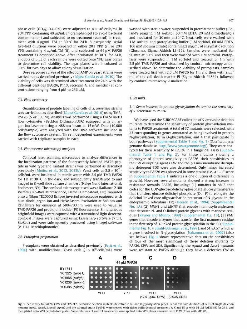
Fig. 1. Sensitivity to PAF26, CFW and SDS of S. cerevisiae deletion mutants defective inmutants Deos1, Dalg5, Dmnn5, Dpmt2 and the parental strain BY4741 were treated withthen plated onto YPD peptide-free plates. Same dilutions of control treatments were ap
washed with sterile water, suspended in pretreatment buffer (Cle-land’s reagent, 1 M sorbitol, 60 mM EDTA, 20 mM dithiothreitol)and incubated for 30 min at 30 �C. Next, cells were washed with1 M sorbitol and protoplasting buffer (1 M sorbitol, 60 mM EDTA,100 mM sodium citrate) containing 2 mg/mL of enzymatic solution(Glucanex, Sigma–Aldrich L1412). Samples were incubated for90 min at 30 �C and then were washed with 1 M sorbitol. Protop-lasts were suspended in 1 M sorbitol and treated for 1 h with2.5 lM TMR-PAF26 and visualized by confocal microscopy as de-scribed above. To determine the viability of protoplasts, sampleswere treated first with 2.5 lM PAF26 for 1 h and then with 2 lg/mL of the cell death marker PI (Sigma-Aldrich P4864), followedby confocal microscopy visualization.
3. Results
3.1. Genes involved in protein glycosylation determine the sensitivityof S. cerevisiae to PAF26
We have used the EUROSCARF collection of S. cerevisiae deletionmutants to determine the sensitivity of protein glycosylation mu-tants to PAF26 treatment. A total of 37 mutants were selected, with23 corresponding to genes annotated as being involved in proteinN-glycosylation, 10 in O-glycosylation, and 4 that participate inboth pathways (Supplemental Table 1 and Fig. 1) (Saccharomycesgenome database, http://www.yeastgenome.org/). They were ana-lyzed for their sensitivity to PAF26 in a fungicidal assay (Supple-mental Table 1 and Fig. 2). For those mutants showing aphenotype of altered sensitivity to PAF26, their sensitivities tothe CW disrupting agent CFW and the plasma membrane disrupt-ing detergent SDS were also determined. Only minor increasedsensitivity to PAF26 was observed in some strains (i.e., a ‘‘�1’’ scorein Supplemental Table 1 indicates a one dilution of difference ingrowth). However, several mutants showed a strong increase inresistance towards PAF26, including: (1) mutants in ALG5 thatcodes for the UDP-glucose:dolichyl-phosphate glucosyltransferasethat transfers glucose dolichyl-phosphate (Dol-P) to elongate thedolichol-linked core oligosaccharide precursor of N-glycans in theendoplasmic reticulum (ER) (Heesen et al., 1994) (SupplementalFig. 1A), (2) MNN1 and MNN5 that encode mannosyltransferasesthat decorate N- and O-linked protein glycans with mannose resi-dues (Rayner and Munro, 1998) (Supplemental Fig. 1B), (3) PMTgenes that encode enzymes that transfer the first mannose residuein the first step of O-linked protein glycosylation in the ER (Supple-mental Fig. 1C) (Strahl-Bolsinger et al., 1999), and (4) EOS1 which isa gene involved in N-glycosylation (Nakamura et al., 2007) (alsosee below). Fig. 1 shows representative data on the sensitivitiesof four of the most significant of these deletion mutants toPAF26, CFW and SDS. Significantly, the Dpmt2 and Deos1 mutantswere resistant to PAF26 although they have a defective CW as
N- and O-glycosylation genes. Serial five-fold dilutions of cells of single deletioneither buffer (control treatment, A, C and D) or with 64 lM PAF26 (B) for 24 h, and
plied onto YPD plates amended with CFW (C) or with SDS (D).

Fig. 2. Analysis of sensitivity to PAF26 and tunicamycin of S. cerevisiae deletion mutants. Cells were first exposed to either buffer (A and B) or tunicamycin (TM) (C and D) for24 h. Subsequently, serial five-fold dilutions of cells were either control-treated (A and C) or treated with PAF26 (B and D), as described in Fig. 1, and dotted onto YPD plates.
108 E. Harries et al. / Fungal Genetics and Biology 58-59 (2013) 105–115
demonstrated by their increased sensitivity to CFW and SDS,respectively.
3.2. The N-glycosylation mutant Deos1 shows tolerance to PAF26 andTunicamycin
Tunicamycin (TM) is an antibiotic inhibitor of N-glycosylationin eukaryotes and is frequently used in the characterization ofdeletion strains with defects in protein glycosylation pathways (El-bein, 1987). It is known that deletion of EOS1 results in tolerance toTM (Nakamura et al., 2007), an observation confirmed in our assaysof TM treatment (4 lg/mL) (Fig. 2). This is significantly different tothe near wild-type sensitivity to TM of the other glycosylation mu-tants analyzed (Fig. 2). Therefore, the sensitivity profile of thestrains analyzed is qualitatively different for PAF26 and TM. TheDeos1 strain was the only one that showed tolerance to bothagents. We therefore questioned whether TM and PAF26 mighthave overlapping activities. Cells were treated first with TM and la-ter with PAF26 and then plated onto plates free of these antifungalagents (Fig. 2). At the tested concentrations of either PAF26 or TM,there was no loss of CFU recovery in the Deos1 mutant; howeverwhen both agents were combined, significant loss of CFU was ob-served. Therefore, our data suggest synergistic effects of both anti-fungal agents acting on the Deos1 mutant, a behaviour also sharedby the other mutants analyzed with deletions of glycosylation-associated genes (Fig. 2).
Fig. 3. Sensitivity to PAF26 of S. cerevisiae single and double deletion mutants inarginine metabolism and glycosylation pathways. Serial five-fold dilutions of cellsof Darg1, Deos1 and Deos1Darg1 (arginine metabolism, top panels) and Dpmt1,Dpmt2, Dpmt2Dpmt1 and Dpmt2Darg1 (protein glycosylation, bottom panels) withthe corresponding parental strain BY4741, were plated onto YPD peptide-free platesafter treatment with control buffer (A) or with 64 lM PAF26 (B) for 24 h.
3.3. The fungicidal activity of PAF26 requires different cellularprocesses
Recently, we presented evidence that arginine metabolism is in-volved in the fungicidal action of PAF26 (López-García et al., 2010;Carmona et al., 2012). The S. cerevisiae deletion mutant of ARG1,which encodes the cytosolic enzyme arginino-succinate synthaseinvolved in arginine metabolism, is also resistant to PAF26(López-García et al., 2010) (see also as control in Fig. 3). Wedecided to analyze the influence on peptide sensitivity of blockingthe arginine and protein glycosylation pathways together. As de-scribed in Supplemental Fig. 3, we generated the double deletionmutants of ARG1, with either PMT1 or EOS1. The double deletionmutants SCLC02 (Deos1-Darg1) and SCLC03 (Dpmt2-Darg1)showed a weakened growth phenotype. However, none of themshowed an enhancement of resistance to the peptide and displayedsensitivity levels similar to those of strains carrying a single genedeletion (Fig. 3). These results indicate that each gene targets dif-ferent steps of the same killing mechanism.
Similarly, we generated a double mutant of PMT2 and PMT1(SCLC04) that also had impaired growth (Fig. 3) and an extremelyweakened CW determined by its sensitivity to CW disruptingagents (data not shown) (Gentzsch and Tanner, 1996). It was alsoresistant to PAF26 and showed no increased resistance as com-pared with the respective single mutants (Fig. 3).
3.4. PMT2 and EOS1 also play roles in the mode of action of anothercell-penetrating antifungal peptide
We investigated whether PMT2 and EOS1 also mediate the sen-sitivity to other AMPs with different modes of action (Table 2).P113 is a 12-amino acid fragment from the human histatin 5,which is also described as a cell-penetrating antifungal peptide(Jang et al., 2008). Cecropin A is a natural peptide which has bacte-rial membrane disrupting activity and is also antifungal (Steineret al., 1981; Brogden, 2005). Melittin is a non-specific antimicrobialpeptide with a cytolytic mode of action (Terwillinger and Eisen-berg, 1982).
Dose–response experiments were carried out for the four pep-tides against strains BY4741, Deos1 and Dpmt2 (Fig. 4). These as-says confirmed the increased resistance to PAF26 in bothmutants (Fig. 4A and Table 2). Increased resistance was also ob-served in the case of P113 although this peptide had lower anti-fungal activity than PAF26 under our assay conditions, and theincreased resistance of the mutants was not so obvious in thedose–response curves (Fig. 4C and Table 2). On the contrary, forthe membrane targeting peptides cecropin A and melittin, themutations did not result in increased resistance (Table 2) butrather in increased sensitivity. This was more evident in the caseof Dpmt2 and melittin (Fig. 4B), probably as consequence of theirweakened CWs. These results provide evidence that PMT2 andEOS1, besides playing a role in the mechanism of action of PAF26,

Fig. 4. Dose–response curves of fungicidal activity of different peptides againstspecific S. cerevisiae deletion mutants. Cell survival after treatment with PAF26 (A),melittin (B) and P113 (C) are shown. Cells of BY4741 (black circles), Dpmt2 (whitetriangles) and Deos1 (white squares) were exposed to different concentrations ofeach peptide for 24 h. Cell survival (CFU/mL) was determined by dilution andplating.
Fig. 5. Differential interaction of S. cerevisiae deletion mutants with TMR-PAF26.Flow cytometry measurements of TMR-PAF26 signal from cells of the deletionmutants and parental strain. The graph shows the percentage of fluorescentlylabelled cells after exposure of 10,000 cells to either 5 (up) or 30 lM (bottom) TMR-PAF26. Mean ± standard deviation from two replicas in each of two independentexperiments are shown for each strain.
E. Harries et al. / Fungal Genetics and Biology 58-59 (2013) 105–115 109
are also important for the antifungal activity of another cell-pene-trating antifungal peptide (P113).
3.5. Subcellular location of PAF26 within S. cerevisiae cellsdemonstrates an inhibition of peptide internalization in the EOS1mutant
Flow cytometry has been used to correlate the amount of fluo-rescently labelled peptides that bind yeast cells of selected resis-tant mutants (Harris et al., 2009; López-García et al., 2010). Weused the same approach to study the Deos1 and Dpmt2 mutantsexposed to two different peptide concentrations (see alsoFig. 4A): a sub-inhibitory concentration of 5 lM and an inhibitoryconcentration of 30 lM (Fig. 5). At low concentrations both mu-tants had a statistically significant higher amount of peptide thatlabelled the cells (p < 0.05), which was somehow unexpected dueto the higher resistance shown by these strains (Figs. 1 and 4A).At high peptide concentration (Fig. 5 and Supplemental Fig. 4)more than 90% of BY4741 cells were labelled with peptide, anamount not significantly different from Dpmt2, while around 75%of Deos1 cells were labelled.
Previously, we reported that FITC-labelled PAF26 is internalizedinto yeast cells at sub-inhibitory concentrations (López-Garcíaet al., 2010). Recently, we have described three sequential steps
during the interaction/internalization of this antifungal peptidein S. cerevisiae using TMR-labelled PAF26 and confocal microscopy(Marcos et al., 2012; Muñoz et al., 2013b). BY4741 cells treatedwith a sub-inhibitory concentration (2.5 lM) of TMR-PAF26 for1 h (e.g. see Fig. 6A) showed three patterns of subcellular localiza-tion within the cell population. A small proportion of cells had thelabelled peptide located at the cell surface (pattern 1 in Fig. 6B);most of the cells had the peptide internalized in intracellular vac-uolar-like organelles (pattern 2); and a small proportion showedTMR-PAF26 labelling throughout the whole cell (pattern 3). Thesesequential steps and subcellular location patterns are similar tothose described in the filamentous model fungus N. crassa (Muñozet al., 2012).
We investigated whether the mutation of genes involved in pro-tein glycosylation affected not only the amount of peptide bound(Fig. 5) but also the distribution and subcellular location of peptidewithin cells. There was no significant variation in the subcellularlocation of PAF26 in any of the mutants disrupted in proteinmannosyltransferase genes (PMT) as compared with the parentalstrain (Supplemental Fig. 5), including the Dpmt2 deletion strainthat is resistant to PAF26. These findings might be partially ex-plained by the functional redundancy of the PMT gene family inS. cerevisiae (Gentzsch and Tanner, 1997; Strahl-Bolsinger et al.,1999; Girrbach and Strahl, 2003). Interestingly, the Deos1 deletionstrain demonstrated an inhibition of TMR-PAF26 internalizationinto the cells (Fig. 6A), showing the labelled PAF26 predominatelylocated at the cell envelope in the majority of cells (pattern 1,Fig. 6B). These results are consistent with the idea that the cellenvelope is critical for peptide interaction and internalization,and also suggest that Eos1p exerts its activity through the glycosyl-ation of cell envelope protein(s) required for the internalization ofPAF26.

Fig. 6. Differential localization of TMR-PAF26 in S. cerevisiae BY4741 and Deos1 strains. (A) Representative bright field and confocal microscopy images from walled yeastcells (top row) and protoplasts (bottom row) for each strain after treatment with 2.5 lM TMR-PAF26 for 1 h. (B) Graph showing the percentages of walled cells (CW) orprotoplasts (Prot.) for each strain (as labelled on the x-axis) that show TMR-PAF26 fluorescence with the three different patterns of localization detailed in the text andillustrated on top as pattern 1 (white bars), pattern 2 (light grey bars), or pattern 3 (black bars). Results represent the means ± standard deviations of three experiments, eachperformed in triplicate. Bar = 10 lm.
110 E. Harries et al. / Fungal Genetics and Biology 58-59 (2013) 105–115
3.6. A functional cell wall is required for internalization and activity ofPAF26
The next logical step was to explore the role of the CW in theinteraction and internalization of PAF26 by yeast cells. We foundthat BY4741 protoplasts exhibited limited internalization of thepeptide (Fig. 6A) and, in fact, mimicked to a large extent the peptidelocalization phenotype of walled Deos1 cells (see also Fig. 6B). Fur-ther characterization of the differential uptake of fluorescent pep-tide was carried out on protoplasts of the Deos1 mutant. Here theamount of fluorescence detected within protoplasts was not signif-icantly different to that detected within BY4741 protoplasts orDeos1 walled cells (Fig. 6A and B). Therefore, protoplasting changedthe distribution of peptide signal in BY4741 but not in Deos1 cells.
It is expected for an AMP, whose proposed mechanism of anti-fungal action is by membrane permeabilization, to be much moreactive against cells lacking a CW. In N. crassa it has been shown thatpeptide internalization is necessary for cell killing (Muñoz et al.,2012). Consistent with this and the above data, we hypothesizedthat inhibition of peptide internalization by removal of the CW
would render cells less sensitive to PAF26. To test this hypothesis,we further compared cell viability in protoplasts and whole cellsof both BY4741 and Deos1 strains after exposure to PAF26 by usingthe PI cell viability assay (Fig. 7). Walled cells of the Deos1 mutantexhibited a lower percentage of cell death (PI staining) as comparedwith the parental strain, in agreement with the resistant phenotypeidentified in this study. On the other hand, protoplasts from boththe BY4741 and Deos1 strains exhibited a similar percentage of celldeath that was lower than those of the corresponding intact walledcells. This is clearly shown in Fig. 7A in which a smaller proportionof the protoplasts in both the BY4741 and Deos1 strains have takenup PI compared with the walled cells of these strains (Fig. 7). Theseresults are thus consistent with a functional CW being importantfor PAF26 internalization and antifungal activity.
4. Discussion
It has long been recognized that differences in CW compositionand structure can influence the selective toxicity of many AMPs

Fig. 7. Killing activity of PAF26 on walled cells and protoplasts of S. cerevisiae BY4741 and Deos1 strains. (A) Representative brightfield and confocal microscopy images fromwalled yeast cells (top row) and protoplasts (bottom row) from each strain after treatment with 2.5 lM PAF26 for 1 h and staining with 2 lg/mL of the death cell marker PI.(B) Graph showing the percentage cell death determined by PI uptake from images as shown in (A). Results represent the mean ± standard deviation of three experiments,each performed in triplicate. The white bars represent walled cells and black bars protoplasts, indicating statistical significance (p value) for each comparison. Bar = 10 lm.
E. Harries et al. / Fungal Genetics and Biology 58-59 (2013) 105–115 111
against microbes (Yeaman and Yount, 2003). The synthetic hexa-peptide PAF26 has been proposed as a simple model to character-ize the mode of action of cationic cell-penetrating antifungalpeptides (Muñoz et al., 2012, 2013a). We have recently describedthe PAF26 kinetics of interaction and killing in two model fungi,N. crassa and S. cerevisiae (Marcos et al., 2012; Muñoz et al.,2012, 2013b). At a relatively low concentration, the peptide inter-acts with the cell envelope and then diffuses to the plasma mem-brane. The peptide is then internalized and reaches theintracellular vacuolar system. In the case of N. crassa, it has beenshown that the internalization at low peptide concentration is en-ergy dependent and involves endocytosis. After reaching a criticalconcentration in the vacuolar network, active peptide transportfrom vacuoles to the cytoplasm is coincident with the peptide fill-ing the cell interior and killing the cell. These three steps of peptideinteraction, internalization and killing are reflected in the threesubcellular peptide localization patterns shown in Fig. 6. Similarobservations have been reported in Candida albicans for the humanpeptide histatin 5 (Mochon and Liu, 2008; Jang et al., 2010) and thedermaseptin DsS3(1-16) (Harris et al., 2009).
Interestingly, enzymatic digestion that removed the CW almostcompletely blocked the internalization of TMR-PAF26 into protop-lasts of BY4741 (Fig. 6), and resulted in lower percentages of celldeath (Fig. 7). The amount of TMR-PAF26 bound to the cell enve-lopes was not significantly affected in BY4741 protoplasts as com-pared to walled cells (Fig. 6A). These data indicate that thepresence of a functional CW determines the internalization of
PAF26 into S. cerevisiae cells and therefore its antifungal activity,and also further supports an intracellular killing mechanism forPAF26. Recently, an inactive PAF26 sequence variant was shownto be retained at the cell envelope without internalization or toxic-ity (Muñoz et al., 2013b). The fungal CW is also required for theantifungal activity of the plant defensins Pn-AMP1 against yeastcells (Koo et al., 2004) and NaD1 against the filamentous fungusFusarium oxysporum (van der Weerden et al., 2010). FITC-conju-gated Pn-AMP1 localized to the surface of the S. cerevisiae BWG7acells but, in contrast to what we have shown with PAF26, removalof the CW impeded the binding of the defensin to the cell and pro-tected the fungus from being killed (Koo et al., 2004). In contrast,yeast protoplasts were more sensitive than walled cells to the anti-fungal tobacco protein osmotin and this higher sensitivity was alsoobserved for protoplasts of the resistant MNN4 mutant (Ibeas et al.,2000). Altogether these data indicate different roles for the fungalCW in the mechanism of antifungal action of PAF26, plant defen-sins and osmotin.
We had previously demonstrated that exposure of S. cerevisiaeto PAF26 or to the cytolytic melittin results in induction of genesthat code for structural CW proteins, and null mutations of specificCW-related genes such as Ecm33p resulted in increased sensitivityto both peptides (López-García et al., 2010). Similarly, deletion ofCW-related genes resulted in an increase in the sensitivity of yeastto other AMPs (Morton et al., 2007). These observations suggestthat the CW broadly also acts as a defence mechanism againstthe effects of AMPs. We therefore postulate a dual role for the

112 E. Harries et al. / Fungal Genetics and Biology 58-59 (2013) 105–115
fungal CW in the interaction and killing mechanism of PAF26 andrelated peptides. The CW is the first fungal cell component thatthe peptide interacts with and, as discussed above, a positivedeterminant of peptide action since the CW is required for peptideinternalization. However, it is also a defence barrier and CWstrengthening protects the cell from the detrimental effects ofthe peptide. It is likely that these two roles are carried out by dif-ferent molecular determinants of the CW and future studies willaim to reveal them.
Most fungal CW proteins, and membrane proteins exposed tothe CW, are glycosylated. Our results indicate that the correct gly-cosylation of proteins is required for the full susceptibility of bud-ding yeast to the fungicidal action of PAF26 (Supplemental Table 1and Figs. 1 and 2). Increased resistance to PAF26 was observed innull mutants of protein glycosylation genes, including the O-glyco-sylation genes PMT1-6, the N-glycosylation EOS1 and ALG5, as wellas MNN1, MNN4 and MNN5 (Supplemental. Table 1 and Fig. 8).These genes (marked in red in Fig. 8) are involved in the additionof sugars at two distinct locations of the glycan structure: eitherat the linkage with the amino acid residues (i.e., PMT genes orEOS1, see below) or at the outer mannose layers that decoratethe N- and O-protein glycans. As an aside, it should be mentionedthat we could not observe any significant changes in the expres-sion of these genes in yeast cells exposed to low concentrationsof PAF26 (data not shown).
It is well known that filamentous fungal and yeast mutantsdefective in protein glycosylation show increased sensitivity tovarious antifungal agents, slow growth rates and CW-related phe-notypes that indicate a compromised CW integrity (Gentzsch andTanner, 1997; Strahl-Bolsinger et al., 1999; Mouyna et al., 2010).Previous reports have found increased tolerance to small AMPsand proteins among protein glycosylation mutants. However, un-iquely the genes MNN1, MNN4 and MNN5 identified in our studyhave been discussed extensively in the context of AMP resistance(Ibeas et al., 2000; Bai et al., 2006; Harris et al., 2009).
S. cerevisiae mutants in genes responsible for the branching ofO-linked mannans confer resistance to plant antifungal proteins,as is the case of MNN10, MNN11 and HOC1 to Pn-AMP1 (Kooet al., 2004), and MNN2, MNN4 or MNN6 to tobacco osmotin (Ibeaset al., 2000). In this latter study, MNN2, MNN4 or MNN6 wererequired for osmotin binding to the fungal CW, while mutation ofMNN1 that exposed negatively charged phosphate groups of phos-phomannans exhibited enhanced binding and sensitivity (Ibeaset al., 2000). In the pathogenic yeast C. albicans, deletion ofMNN4 also resulted in enhanced resistance and reduced bindingto the synthetic peptide DsS3(1-16) (Harris et al., 2009), andMNN5 was required for the antifungal activity of the bovine lacto-ferrin (Bai et al., 2006). Resistance to yeast killer toxins has beenfound in S. cerevisiae mutants in the N-glycosylation genes STT3and ALG3 (Kimura et al., 1999), and in the PMT genes (Lussieret al., 1995; Gentzsch and Tanner, 1996). Therefore, proper proteinglycosylation is required for the activity of a significant number ofantifungal peptides and proteins, including PAF26.
All the above studies imply that there must be glycosylated CWprotein(s) to which the AMPs actually bind, although their identi-fication has not been achieved in most cases, including the presentstudy. The CW protein Ssa2 from C. albicans is a chaperone-likeprotein that binds histatin 5 (Sun et al., 2008) and has potentialglycosylation sites. Msb2p, that is a transmembrane mucin-likeprotein localized at the CW and which is glycosylated by Pmt1p,has also been associated with the antifungal activity of histatin 5and LL-37 against C. albicans (Szafranski-Schneider et al., 2012).In this example, null mutants of Pmt1p were more susceptible toLL-37. The released glycosylated extracellular domain of Msb2 pro-tected cells from AMP killing action by sequestering peptides andpreventing their binding to the cells. In a parallel study, we have
confirmed that flor strains of S. cerevisiae, which naturally formbiofilms on the surface of wine, can bind PAF26 and that a mutantlacking the hyper-glycosylated CW mucin-like Flo11p lost the abil-ity to bind the peptide (Bou Zeidan et al., in press). However, wehave not confirmed these findings with laboratory strains ofS. cerevisiae such as the one used in the present study (data notshown).
An alternative hypothesis to explain the involvement of proteinglycosylation in the mechanism of antifungal peptides is based onthe glycosylated nature of sensor proteins of fungal mitogen-acti-vated protein kinase (MAPK) signaling pathways (de Nadal et al.,2007; Lien et al., 2013). Protein glycosylation is required for theproper function and signal transduction in the MAPK cascades. Thisis the case of the above-mentioned Msb2p, which is a hyper-gly-cosylated sensor protein of the osmotic stress and filamentousgrowth pathways and whose defective glycosylation activatesthem constitutively (Tatebayashi et al., 2007; Yang et al., 2009).Previous studies have shown that yeasts adapt to some AMPs byactivating the Hog1 signaling pathway (Gamberi et al., 2007; Vylk-ova et al., 2007). The possibility that protein glycosylation has arole in the transduction pathways that signal the perception ofPAF26 by fungal cells will be examined in the near future.
We focused our analyses on the characterization of Dpmt2 andDeos1 mutants because they are less susceptible to the cell-pene-trating peptides PAF26 and P113 (a synthetic derivative of histatin5), yet they show hypersensitivity to the CW- and plasma mem-brane-disrupting compounds CFW or SDS, respectively. Moreover,they are also more sensitive to membrane lytic peptides such asmelittin or cecropin A (Figs. 1 and 4, and Table 2). All our currentand previous analyses indicate that these two mutants have aweakened CW that, presumably, should make them broadly moresensitive to most antifungal agents. Therefore, their PAF26 toler-ance phenotype is noteworthy and specific to a sub-class of AMPsthat penetrate cells by mechanisms (e.g. endocytosis) other thanplasma membrane permeabilization.
Tunicamycin (TM) is an antifungal agent that inhibits protein N-glycosylation (Nakamura et al., 2007). PAF26 and TM antifungalactivities are synergistic (Fig. 2). This suggests that although pro-tein glycosylation determines sensitivity to AMPs, the antifungalmechanism of PAF26 does not target protein glycosylation itself.Previous data are consistent with this view. For instance, pharma-cological or genetic inhibition of protein glycosylation resulted inthe activation of genes from the CW integrity signaling pathway(Cantero et al., 2007; Arroyo et al., 2011; Cantero and Ernst,2011), and the inhibition of N-glycosylation by TM triggers theactivation of PMT genes. This suggests a compensatory mechanismto alleviate defects in protein glycosylation. However, the tran-scriptomic analyses of PAF26-treated S. cerevisiae did not identifyany process involved in the induction of the CW integrity pathwayor protein glycosylation genes including PMTs (López-García et al.,2010), as would be expected if PAF26 affected protein glycosyla-tion. On the other hand, recent studies demonstrated the increasedsensitivity of all S. cerevisiae Dpmt1-6 mutants to a specific O-gly-cosylation inhibitor (OGT2468) (Arroyo et al., 2011), while in ourstudy the same mutants are more resistant to PAF26.
Previously, we have shown that disruption of ARG genes in-volved in arginine metabolism also resulted in increased resistanceto PAF26 (López-García et al., 2010). The mutant Darg1 does notproduce the intracellular nitric oxide (NO) associated with expo-sure to PAF26, and an inhibitor of arginine-derived NO synthasepartially protects yeast cells from PAF26 action (Carmona et al.,2012). Moreover, Darg1 showed a block in the transport of PAF26from the vacuole to the cytosol that accompanies cell death(Muñoz et al., 2013b). Therefore ARG1 is likely involved in theintracellular killing phase of the mode of action of PAF26. Conse-quently, none of the generated double deletion strains that

Fig. 8. Schematic representation of the structure of representative S. cerevisiae N-linked (left) and O-linked (right) glycans. Diagram adapted from (Lehle et al., 2006). Squaresindicate N-acetylglucosamine residues, and circles indicate mannose residues. Phosphate residues are indicated by red circles. Open symbols indicate residues added to theglycan at the cytosolic side of the ER membrane, filled symbols indicate residues added at the lumen side of the ER, and striped symbols indicate residues added in the Golgiapparatus. The proteins involved in the addition of each sugar residue are labelled; and those proteins whose genes when deleted are lethal are shown in grey. The proteinswhose genes when deleted resulted in increased tolerance to PAF26 are highlighted in red.
E. Harries et al. / Fungal Genetics and Biology 58-59 (2013) 105–115 113
targeted arginine metabolism or protein glycosylation resulted inenhanced resistance to PAF26 (Fig. 3). These results are consistentwith the involvement of both biological processes (protein glyco-sylation and arginine metabolism) in different steps of the samepathway that results in cell killing by PAF26.
Most of the aforementioned previous studies identified manno-syltransferase genes involved in decorating the outer branches ofthe core N-glycan structure, some of which have been also identi-fied in our study (Fig. 8). Few studies have reported roles for theEOS1 or PMT genes in this context. Significantly, EOS1 (YLN080C)was included in the list of genes that when deleted showed en-hanced resistance to the cyclic lipopeptide caspofungin in twoindependent genome-wide studies (Lesage et al., 2004; Markovichet al., 2004), although the involvement of EOS1 in AMP action wasunexplored and not discussed at that time. The Deos1 mutantshows a pleiotropic phenotype with hypersensitivity to oxidativeand osmotic stress, as well as altered metal homeostasis (Nakam-ura et al., 2007, 2010). One of the most remarkable phenotypes ofDeos1 is its high tolerance to TM (Fig. 2 and also Nakamura et al.,2007), which together with the altered glycosylation pattern of cellproteins and location of Eos1p at the ER justified its annotation asinvolved in protein N-glycosylation (Nakamura et al., 2007). TMinhibits the transfer of N-acetylglucosamine-P to Dol-P in the ERin the first step of the dolichol pathway of protein N-glycosylation,a reaction catalyzed by Alg7p (Fig. 8). The null mutation of ALG7 islethal, and therefore could not be assayed in our study. We specu-late that Eos1p participates in the N-glycosylation of a subset ofcell proteins, by interaction with Alg7p in the first step of the dol-ichol pathway (Fig. 8 and Supplemental Fig. 1A).
Compared with the BY4741 cells, we observed a remarkably dif-ferent localization of TMR-PAF26 in Deos1 cells, in which the la-belled peptide remained at the cell envelope and was notinternalized (Fig. 6A). This was coincident with a lower percentageof cell death (Fig. 7), confirming the resistant phenotype found infungicidal assays (Fig. 1). These observations demonstrate that
EOS1 is necessary for the internalization of this antifungal hexa-peptide. PAF26 localization (Fig. 6) and killing activity (Fig. 7)was similar in protoplasts of BY4741 and Deos1, contrarily toBY4741 in which protoplasting affected PAF26 localization andactivity. Therefore the phenotypic difference between the eos1 mu-tant and the parental strain is lost after removal of the CW. A plau-sible explanation is that EOS1 functions at the level of the CW,through involvement in the glycosylation of a protein(s) requiredfor cell-penetration by PAF26. The killing activity of histatin 5 toC. albicans is determined by the intracellular translocation of thepeptide mediated by the CW located protein Ssa2 (Sun et al.,2008; Jang et al., 2010). In our case, an alternative hypothesis couldbe that compensatory changes in the CW of the eos1 mutant in-crease the content of (glyco)proteins and/or glycans that sequesterthe peptide at the CW layer and avoid its internalization. Finally, itcannot be ruled out that the pleiotropic effect of the EOS1 nullmutation (Nakamura et al., 2010) affects additional (maybe evenoverlapping) cell processes that might explain the blockage of pep-tide cell penetration and resistance of this mutant.
5. Conclusions
In conclusion, our data further confirm the involvement of pro-tein glycosylation genes in the activity of short antifungal peptidesand expand the repertoire of glycosylation genes known in thiscontext. The results are consistent with a mechanism for whichPAF26 internalization is required for killing fungal cells (Muñozet al., 2013a). It is shown that the CW and the N-glycosylation geneEOS1 are required for internalization of PAF26. The EOS1 null muta-tion results in the peptide being located at the cell surface and in-creased tolerance of cells to its antifungal action. Our presentworking hypothesis is that Eos1p participates in the glycosylationof yet unidentified CW protein(s) required for peptide cell penetra-tion. The identification of CW proteins that participate in the

114 E. Harries et al. / Fungal Genetics and Biology 58-59 (2013) 105–115
internalization and antifungal activity of PAF26 is currently beingexplored.
Acknowledgments
EH’s visit to the University of Edinburgh was funded by the JAE-PREDOC Program of CSIC (Spain) and the European FEDER fund.The study was supported by research grants BIO2009-12919 (MIC-INN, Spain) and ACOMP/2012/018 (Generalitat Valenciana, Spain).This article is dedicated to the dearest memory of our friend andcolleague Enrique (Quique) Pérez-Payá, with whom we had a fruit-ful scientific collaboration in the field of antimicrobial peptidesover the last two decades.
Appendix A. Supplementary material
Supplementary data associated with this article can be found, inthe online version, at http://dx.doi.org/10.1016/j.fgb.2013.08.004.
References
Arroyo, J., Hutzler, J., Bermejo, C., Ragni, E., Garcia-Cantalejo, J., Botias, P., Piberger,H., Schott, A., Sanz, A.B., Strahl, S., 2011. Functional and genomic analyses ofblocked protein O-mannosylation in baker’s yeast. Mol. Microbiol. 79, 1529–1546.
Bai, C., Xu, X.L., Chan, F.Y., Lee, R.T.H., Wang, Y., 2006. MNN5 encodes an iron-regulated a-1,2-mannosyltransferase important for protein glycosylation, cellwall integrity, morphogenesis and virulence in Candida albicans. Eukaryot. Cell5, 238–247.
Bou Zeidan, M., Carmona, L., Zara, S., Marcos, J.F., in press. FLO11 gene is involved inthe interaction of flor strains of Saccharomyces cerevisiae with a biofilm-promoting synthetic hexapeptide. Appl. Environ. Microbiol. 79, 2013,doi:10.1128/AEM.01647-13.
Brogden, K.A., 2005. Antimicrobial peptides: pore formers or metabolic inhibitors inbacteria? Nat. Rev. Microbiol. 3, 238–250.
Cantero, P.D., Ernst, J.F., 2011. Damage to the glycoshield activates PMT-directed O-mannosylation via the Msb2-Cek1 pathway in Candida albicans. Mol. Microbiol.80, 715–725.
Cantero, P.D., Lengsfeld, C., Prill, S.K.H., Subanovic, M., Roman, E., Pla, J., Ernst, J.F.,2007. Transcriptional and physiological adaptation to defective protein-O-mannosylation in Candida albicans. Mol. Microbiol. 64, 1115–1128.
Carmona, L., Gandía, M., López-García, B., Marcos, J.F., 2012. Sensitivity ofSaccharomyces cerevisiae to the cell-penetrating antifungal peptide PAF26correlates with endogenous nitric oxide (NO) production. Biochem. Biophys.Res. Commun. 417, 56–61.
de Nadal, E., Real, F.X., Posas, F., 2007. Mucins, osmosensors in eukaryotic cells?Trends Cell Biol. 17, 571–574.
Deshpande, N., Wilkins, M.R., Packer, N., Nevalainen, H., 2008. Protein glycosylationpathways in filamentous fungi. Glycobiology 18, 626–637.
Duncan, V.M.S., O’Neil, D.A., 2013. Commercialization of antifungal peptides. FungalBiol. Rev. 26, 156–165.
Elbein, A.D., 1987. Inhibitors of the biosynthesis and processing of N-linkedoligosaccharide chains. Annu. Rev. Biochem. 56, 497–534.
Fernández-Álvarez, A., Elías-Villalobos, A., Ibeas, J.I., 2009. The O-mannosyltransferase PMT4 is essential for normal appressorium formationand penetration in Ustilago maydis. Plant Cell. 21, 3397–3412.
Gamberi, T., Cavalieri, D., Magherini, F., Mangoni, L., De Filippo, C., Borro, M., Gentile,G., Simmaco, M., Modesti, A., 2007. An integrated analysis of the effects ofEsculentin 1-21 on Saccharomyces cerevisiae. Biochim. Biophys. Acta – Prot.Proteom. 1774, 688–700.
Gentzsch, M., Tanner, W., 1996. The PMT gene family: protein O-glycosylation inSaccharomyces cerevisiae is vital. EMBO J. 15, 5752–5759.
Gentzsch, M., Tanner, W., 1997. Protein-O-glycosylation in yeast: protein-specificmannosyltransferases. Glycobiology 7, 481–486.
Girrbach, V., Strahl, S., 2003. Members of the evolutionarily conserved PMT familyof protein O-mannosyltransferases form distinct protein complexes amongthemselves. J. Biol. Chem. 278, 12554–12562.
Hancock, R.E.W., Sahl, H.G., 2006. Antimicrobial and host-defense peptides as newanti-infective therapeutic strategies. Nat. Biotechnol. 24, 1551–1557.
Harris, M., Mora-Montes, H.M., Gow, N.A.R., Coote, P.J., 2009. Loss ofmannosylphosphate from Candida albicans cell wall proteins results inenhanced resistance to the inhibitory effect of a cationic antimicrobialpeptide via reduced peptide binding to the cell surface. Microbiol. SGM 155,1058–1070.
Heesen, S.t., Lehle, L., Weissmann, A., Aebi, M., 1994. Isolation of the ALG5 locusencoding the UDP-glucose:dolichyl-phosphate glucosyltransferase fromSaccharomyces cerevisiae. Eur. J. Biochem. 224, 71–79.
Henriques, S.T., Melo, M.N., Castanho, M.A.R.B., 2006. Cell-penetrating peptides andantimicrobial peptides: how different are they? Biochem. J. 399, 1–7.
Ibeas, J.I., Lee, H., Damsz, B., Prasad, D.T., Pardo, J.M., Hasegawa, P.M., Bressan, R.A.,Narasimhan, M.L., 2000. Fungal cell wall phosphomannans facilitate the toxicactivity of a plant PR-5 protein. Plant J. 23, 375–383.
Jang, W.S., Li, X.W.S., Sun, J.N.N., Edgerton, M., 2008. The P-113 fragment of Histatin5 requires a specific peptide sequence for intracellular translocation in Candidaalbicans, which is independent of cell wall binding. Antimicrob. AgentsChemother. 52, 497–504.
Jang, W.S., Bajwa, J.S., Sun, J.N., Edgerton, M., 2010. Salivary histatin 5internalization by translocation, but not endocytosis, is required for fungicidalactivity in Candida albicans. Mol. Microbiol. 77, 354–370.
Kimura, T., Komiyama, T., Furuichi, Y., Iimura, Y., Karita, S., Sakka, K., Ohmiya, K.,1999. N-glycosylation is involved in the sensitivity of Saccharomyces cerevisiaeto HM-1 killer toxin secreted from Hansenula mrakii IFO 0895. Appl. Microbiol.Biotechnol. 51, 176–184.
Klis, F.M., Boorsma, A., de Groot, P.W.J., 2006. Cell wall construction inSaccharomyces cerevisiae. Yeast 23, 185–202.
Koo, J.C., Lee, B., Young, M.E., Koo, S.C., Cooper, J.A., Baek, D., Lim, C.O., Lee, S.Y., Yun,D.J., Cho, M.J., 2004. Pn-AMP1, a plant defense protein, induces actindepolarization in yeasts. Plant Cell Physiol. 45, 1669–1680.
Lehle, L., Strahl, S., Tanner, W., 2006. Protein glycosylation, conserved from yeast toman: a model organism helps elucidate congenital human diseases. Angew.Chem. Int. Ed. 45, 6802–6818.
Lesage, G., Sdicu, A.M., Menard, P., Shapiro, J., Hussein, S., Bussey, H., 2004. Analysisof beta-1,3-glucan assembly in Saccharomyces cerevisiae using a syntheticinteraction network and altered sensitivity to caspofungin. Genetics 167, 35–49.
Lien, E.C., Nagiec, M.J., Dohlman, H.G., 2013. Proper protein glycosylation promotesmitogen-activated protein kinase signal fidelity. Biochemistry 52, 115–124.
López-García, B., Pérez-Payá, E., Marcos, J.F., 2002. Identification of novelhexapeptides bioactive against phytopathogenic fungi through screening of asynthetic peptide combinatorial library. Appl. Environ. Microbiol. 68, 2453–2460.
López-García, B., Gandía, M., Muñoz, A., Carmona, L., Marcos, J.F., 2010. A genomicapproach highlights common and diverse effects and determinants ofsusceptibility on the yeast Saccharomyces cerevisiae exposed to distinctantimicrobial peptides. BMC Microbiol. 10, 289.
Lussier, M., Gentzsch, M., Sdicu, A.M., Bussey, H., Tanner, W., 1995. Protein O-glycosylation in yeast. The PMT2 gene specifies a second protein O-mannosyltransferase that functions in addition to the PMT1-encoded activity.J. Biol. Chem. 270, 2770–2775.
Marcos, J.F., Gandía, M., 2009. Antimicrobial peptides: to membranes and beyond.Expert Opin. Drug Discov. 4, 659–671.
Marcos, J.F., Muñoz, A., Pérez-Payá, E., Misra, S., López-García, B., 2008.Identification and rational design of novel antimicrobial peptides for plantprotection. Annu. Rev. Phytopathol. 46, 273–301.
Marcos, J.F., Gandía, M., Harries, E., Carmona, L., Muñoz, A., 2012. Antifungalpeptides: exploiting non-lytic mechanisms and cell penetration properties. In:Rajasekaran, K., Cary, J.W., Jaynes, J., Montesinos, E. (Eds.), Small Wonders:Peptides for Disease Control. American Chemical Society, pp. 337–357.
Markovich, S., Yekutiel, A., Shalit, I., Shadkchan, Y., Osherov, N., 2004. Genomicapproach to identification of mutations affecting caspofungin susceptibility inSaccharomyces cerevisiae. Antimicrob. Agents Chemother. 48, 3871–3876.
Mochon, A.B., Liu, H.P., 2008. The antimicrobial peptide Histatin-5 causes a spatiallyrestricted disruption on the Candida albicans surface, allowing rapid entry of thepeptide into the cytoplasm. PLoS Pathog. 4, e1000190.
Morton, C.O., Hayes, A., Wilson, M., Rash, B.M., Oliver, S.G., Coote, P., 2007. Globalphenotype screening and transcript analysis outlines the inhibitory mode(s) ofaction of two amphibian-derived, a-helical, cationic peptides on Saccharomycescerevisiae. Antimicrob. Agents Chemother. 51, 3948–3959.
Mouyna, I., Kniemeyer, O., Jank, T., Loussert, C., Mellado, E., Aimanianda, V.,Beauvais, A., Wartenberg, D., Sarfati, J., Bayry, J., Prevost, M.C., Brakhage, A.A.,Strahl, S., Huerre, M., Latge, J.P., 2010. Members of protein O-mannosyltransferase family in Aspergillus fumigatus differentially affectgrowth, morphogenesis and viability. Mol. Microbiol. 76, 1205–1221.
Muñoz, A., López-García, B., Marcos, J.F., 2006. Studies on the mode of action of theantifungal hexapeptide PAF26. Antimicrob. Agents Chemother. 50, 3847–3855.
Muñoz, A., Marcos, J.F., Read, N.D., 2012. Concentration-dependent mechanisms ofcell penetration and killing by the de novo designed antifungal hexapeptidePAF26. Mol. Microbiol. 85, 89–106.
Muñoz, A., Gandía, M., Harries, E., Carmona, L., Read, N.D., Marcos, J.F., 2013a.Understanding the mechanism of action of cell-penetrating antifungal peptidesusing the rationally designed hexapeptide PAF26 as a model. Fungal Biol. Rev.26, 146–155.
Muñoz, A., Harries, E., Contreras-Valenzuela, A., Carmona, L., Read, N.D., Marcos, J.F.,2013b. Two functional motifs define the interaction, internalization and toxicityof the cell-penetrating antifungal peptide PAF26 on fungal cells. PLoS ONE 8,e54813.
Munro, C.A., Bates, S., Buurman, E.T., Hughes, H.B., MacCallum, D.M., Bertram, G.,Atrih, A., Ferguson, M.A.J., Bain, J.M., Brand, A., Hamilton, S., Westwater, C.,Thomson, L.M., Brown, A.J.P., Odds, F.C., Gow, N.A.R., 2005. Mnt1p and Mnt2p ofCandida albicans are partially redundant a-1,2-mannosyltransferases thatparticipate in O-linked mannosylation and are required for adhesion andvirulence. J. Biol. Chem. 280, 1051–1060.
Nakamura, T., Ando, A., Takagi, H., Shima, J., 2007. EOS1, whose deletion conferssensitivity to oxidative stress, is involved in N-glycosylation in Saccharomycescerevisiae Biochem. Biophys. Res. Commun. 353, 293–298.

E. Harries et al. / Fungal Genetics and Biology 58-59 (2013) 105–115 115
Nakamura, T., Takahashi, S., Takagi, H., Shima, J., 2010. Multicopy suppression ofoxidant-sensitive eos1 mutation by IZH2 in Saccharomyces cerevisiae and theinvolvement of Eos1in zinc homeostasis. FEMS Yeast Res. 10, 259–269.
Peters, B.M., Shirtliff, M.E., Jabra-Rizk, M.A., 2010. Antimicrobial peptides: primevalmolecules or future drugs? PLoS Pathog. 6, e1001067.
Petit, J., Boisseau, P., Arveiler, B., 1994. Glucanex: a cost-effective yeast lytic enzyme.Trends Genet. 10, 4–5.
Prill, S.K.H., Klinkert, B., Timpel, C., Gale, C.A., Schroppel, K., Ernst, J.F., 2005. PMTfamily of Candida albicans: five protein mannosyltransferase isoforms affectgrowth, morphogenesis and antifungal resistance. Mol. Microbiol. 55, 546–560.
Rayner, J.C., Munro, S., 1998. Identification of the MNN2 and MNN5mannosyltransferases required for forming and extending the mannosebranches of the outer chain mannans of Saccharomyces cerevisiae. J. Biol.Chem. 273, 26836–26843.
Schirawski, J., Böhnert, H.U., Steinberg, G., Snetselaar, K., Adamikowa, L., Kahmann,R., 2005. Endoplasmic reticulum glucosidase II is required for pathogenicity ofUstilago maydis. Plant Cell. 17, 3532–3543.
Steiner, H., Hultmark, D., Engstrom, A., Bennich, H., Boman, H.G., 1981. Sequenceand specificity of 2 anti-bacterial proteins involved in insect immunity. Nature292, 246–248.
Strahl-Bolsinger, S., Gentzsch, M., Tanner, W., 1999. Protein O-mannosylation.Biochim. Biophys. Acta 1426, 297–307.
Sun, J.N.N., Li, W.S., Jang, W.S., Nayyar, N., Sutton, M.D., Edgerton, M., 2008. Uptakeof the antifungal cationic peptide Histatin 5 by Candida albicans Ssa2p requiresbinding to non-conventional sites within the ATPase domain. Mol. Microbiol.70, 1246–1260.
Szafranski-Schneider, E., Swidergall, M., Cottier, F., Tielker, D., Roman, E., Pla, J.,Ernst, J.F., 2012. Msb2 shedding protects Candida albicans against antimicrobialpeptides. PLoS Pathog. 8, e1002501.
Tatebayashi, K., Tanaka, K., Yang, H.Y., Yamamoto, K., Matsushita, Y., Tomida, T.,Imai, M., Saito, H., 2007. Transmembrane mucins Hkr1 and Msb2 are putativeosmosensors in the SHO1 branch of yeast HOG pathway. EMBO J. 26, 3521–3533.
Terwillinger, T.C., Eisenberg, D., 1982. The structure of melittin. II. Interpretation ofthe structure. J. Biol. Chem. 257, 6016–6022.
van der Weerden, N.L., Hancock, R.E.W., Anderson, M.A., 2010. Permeabilization offungal hyphae by the plant defensin NaD1 occurs through a cell wall-dependentprocess. J. Biol. Chem. 285, 37513–37520.
Vylkova, S., Jang, W.S., Li, W.S., Nayyar, N., Edgerton, M., 2007. Histatin 5 initiatesosmotic stress response in Candida albicans via activation of the Hog1 mitogen-activated protein kinase pathway. Eukaryot. Cell 6, 1876–1888.
Yang, H.Y., Tatebayashi, K., Yamamoto, K., Saito, H., 2009. Glycosylation defectsactivate filamentous growth Kss1 MAPK and inhibit osmoregulatory Hog1MAPK. EMBO J. 28, 1380–1391.
Yeaman, M.R., Yount, N.Y., 2003. Mechanisms of antimicrobial peptide action andresistance. Pharmacol. Rev. 55, 27–55.
Zasloff, M., 2002. Antimicrobial peptides of multicellular organisms. Nature 415,389–395.
Zhou, H., Hu, H.Y., Zhang, L.J., Li, R.Y., Ouyang, H.M., Ming, J., Jin, C., 2007. O-mannosyltransferase 1 in Aspergillus fumigatus (AfPmt1p) is crucial for cell wallintegrity and conidium morphology, especially at an elevated temperature.Eukaryot. Cell 6, 2260–2268.