Screening, Identification, Characterization and Production ... Golani, et al.pdf · Screening,...
19
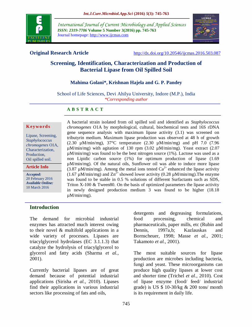
Int.J.Curr.Microbiol.App.Sci (2016) 5(3): 745-763 745 Original Research Article http://dx.doi.org/10.20546/ijcmas.2016.503.087 Screening, Identification, Characterization and Production of Bacterial Lipase from Oil Spilled Soil Mahima Golani*, Krishnan Hajela and G. P. Pandey School of Life Sciences, Devi Ahilya University, Indore (M.P.), India *Corresponding author ABSTRACT Introduction The demand for microbial industrial enzymes has attracted much interest owing to their novel & multifold applications in a wide variety of processes. Lipases are triacylglycerol hydrolases (EC 3.1.1.3) that catalyze the hydrolysis of triacylglycerol to glycerol and fatty acids (Sharma et al., 2001). Currently bacterial lipases are of great demand because of potential industrial applications (Sirisha et al., 2010). Lipases find their applications in various industrial sectors like processing of fats and oils, detergents and degreasing formulations, food processing, chemical and pharmaceuticals, paper mills, etc (Rubin and Dennis, 1997a,b; Kazlauskas and Bornscheuer, 1998; Masse et al., 2001; Takamoto et al., 2001). The most suitable sources for lipase production are microbes including bacteria, fungi and yeast. These microorganisms can produce high quality lipases at lower cost and shorter time (Trichel et al., 2010). Cost of lipase enzyme (food/ feed/ industrial grade) is US $ 10-30/kg & 200 tons/ month is its requirement in daily life. International Journal of Current Microbiology and Applied Sciences ISSN: 2319-7706 Volume 5 Number 3(2016) pp. 745-763 Journal homepage: http://www.ijcmas.com A bacterial strain isolated from oil spilled soil and identified as Staphylococcus chromogenes O1A by morphological, cultural, biochemical tests and 16S rDNA gene sequence analysis with maximum lipase activity (3.1) was screened on tributyrin medium. Maximum lipase production was observed at 48 h of growth (2.30 μM/min/mg), 37°C temperature (2.30 μM/min/mg) and pH 7.0 (7.96 μM/min/mg) with agitation of 130 rpm (3.02 μM/min/mg). Yeast extract (2.07 μM/min/mg) was found to be the best nitrogen source (1%). Lactose was used as a non Lipidic carbon source (1%) for optimum production of lipase (1.69 μM/min/mg). Of the natural oils, Sunflower oil was able to induce more lipase (3.87 μM/min/mg). Among the metal ions tested Ca 2+ enhanced the lipase activity (1.67 μM/min/mg) and Zn 2+ showed lower activity (0.28 μM/min/mg).The enzyme was found to be stable in 0.5 % solutions of different Surfactants such as SDS, Triton X-100 & Tween80. On the basis of optimized parameters the lipase activity in newly designed production medium 3 was found to be higher (18.18 μM/min/mg). Keywords Lipase, Screening, Staphylococcus chromogenes O1A, Characterization, Production, Oil spilled soil. Accepted: 20 February 2016 Available Online: 10 March 2016 Article Info
Transcript of Screening, Identification, Characterization and Production ... Golani, et al.pdf · Screening,...
Int.J.Curr.Microbiol.App.Sci (2016) 5(3): 745-763
745
Original Research Article http://dx.doi.org/10.20546/ijcmas.2016.503.087
Screening, Identification, Characterization and Production of
Bacterial Lipase from Oil Spilled Soil
Mahima Golani*, Krishnan Hajela and G. P. Pandey
School of Life Sciences, Devi Ahilya University, Indore (M.P.), India *Corresponding author
A B S T R A C T
Introduction
The demand for microbial industrial
enzymes has attracted much interest owing
to their novel & multifold applications in a
wide variety of processes. Lipases are
triacylglycerol hydrolases (EC 3.1.1.3) that
catalyze the hydrolysis of triacylglycerol to
glycerol and fatty acids (Sharma et al.,
2001).
Currently bacterial lipases are of great
demand because of potential industrial
applications (Sirisha et al., 2010). Lipases
find their applications in various industrial
sectors like processing of fats and oils,
detergents and degreasing formulations,
food processing, chemical and
pharmaceuticals, paper mills, etc (Rubin and
Dennis, 1997a,b; Kazlauskas and
Bornscheuer, 1998; Masse et al., 2001;
Takamoto et al., 2001).
The most suitable sources for lipase
production are microbes including bacteria,
fungi and yeast. These microorganisms can
produce high quality lipases at lower cost
and shorter time (Trichel et al., 2010). Cost
of lipase enzyme (food/ feed/ industrial
grade) is US $ 10-30/kg & 200 tons/ month
is its requirement in daily life.
International Journal of Current Microbiology and Applied Sciences ISSN: 2319-7706 Volume 5 Number 3(2016) pp. 745-763
Journal homepage: http://www.ijcmas.com
A bacterial strain isolated from oil spilled soil and identified as Staphylococcus chromogenes O1A by morphological, cultural, biochemical tests and 16S rDNA
gene sequence analysis with maximum lipase activity (3.1) was screened on
tributyrin medium. Maximum lipase production was observed at 48 h of growth (2.30 µM/min/mg), 37°C temperature (2.30 µM/min/mg) and pH 7.0 (7.96
µM/min/mg) with agitation of 130 rpm (3.02 µM/min/mg). Yeast extract (2.07
µM/min/mg) was found to be the best nitrogen source (1%). Lactose was used as a
non Lipidic carbon source (1%) for optimum production of lipase (1.69 µM/min/mg). Of the natural oils, Sunflower oil was able to induce more lipase
(3.87 µM/min/mg). Among the metal ions tested Ca2+
enhanced the lipase activity
(1.67 µM/min/mg) and Zn2+
showed lower activity (0.28 µM/min/mg).The enzyme was found to be stable in 0.5 % solutions of different Surfactants such as SDS,
Triton X-100 & Tween80. On the basis of optimized parameters the lipase activity
in newly designed production medium 3 was found to be higher (18.18
µM/min/mg).
K ey wo rd s
Lipase, Screening,
Staphylococcus
chromogenes O1A,
Characterization,
Production,
Oil spilled soil.
Accepted: 20 February 2016
Available Online:
10 March 2016
Article Info
Int.J.Curr.Microbiol.App.Sci (2016) 5(3): 745-763
746
Bacterial lipases are glycoproteins but some
extracellular bacterial lipases are
lipoproteins. Among bacteria,
Achromobacter sp., Alcaligenes sp.,
Arthrobacter sp., Pseudomonas sp.,
Staphylococcus sp. and Chromobacterium
sp. (Godfredson et al., 1990) have been
exploited for the production of lipases.
Staphylococcal lipases are lipoprotein in
nature (Brune et al., 1992). Many
Staphylococci are able to produce
extracellular lipases and some of them
have been purified and their biochemical
properties studied in detail (Oort et al.,
1989; Gotz et al., 1985; Farrell et al., 1993;
Lee and Iandolo, 1986; Talon et al., 1996;
Oh et al., 1999; Simons et al., 1996; Jaeger
et al., 1999; Van-Kampen et al., 2001;
Pandey et al., 1999).
In order to get the highest yields of lipase,
the optimal growth conditions were studied
(Linefield et al., 1990). Bacterial lipases are
mostly extracellular and are greatly
influenced by nutritional and physico-
chemical factors, such as temperature, pH,
nitrogen and carbon sources, presence of
lipids, inorganic salts, agitation and
dissolved oxygen concentration (Brune and
Gotz, 1992; Aires-Barros et al., 1994; Jaeger
et al., 1994; Kim et al., 1996).
Various methods of lipase assay have been
classified as; Titrimetry, Interfacial
tensiometry, Spectroscopy,
Chromatography, Immunochemistry and
Conductimetry (Beisson et al., 2000;
Kulkarni, 2002) of these methods titrimetry
is the simplest method which was used in
our studies also.
The purpose of the present study was to
screen potential lipase producing bacteria
from various samples and optimize the
production of lipase by isolated and
identified strain as Staphylococcus
chromogenes O1A.
Materials and Methods
Screening of Lipase Producing Bacteria
Collection of Samples
Various 17 samples from diverse sources,
such as oil contaminated soils of vegetable
oil processing factories, oil packing
industries & selling shops, auto garage soil,
domestic waste water (sewage), slaughter
house soil, spoiled coconut water, milk &
milk cream etc. were collected in and around
the city of Indore of Madhya Pradesh, India
for the isolation of potential lipase
producing bacteria.
Enrichment and Isolation of Lipolytic
Bacteria
Lipolytic bacteria were screened by
enrichment culture technique from 17
diverse samples. These samples were
enriched by inoculating in Tributyrin broth
medium flask (50 ml) containing 0.5% (w/v)
peptone, 0.3% (w/v) yeast extract, 1% (v/v)
Tributyrin pH 7.0 & 9.0 and incubated at
37˚C for 48 hrs. After incubation, a loopful
of growth obtained from each enriched
culture sample was isolated on Tributyrin
agar medium plates (Lawrence et al., 1967)
(pH 7 & 9) by sector plate method and
incubated at 37˚C for 48 hrs.
Screening of the Isolates for Lipase
Activity
Lipolytic organisms were screened by
qualitative plate assay. The lipolytic activity
of isolated colonies were observed by spot
inoculation on Tributyrin agar medium
plates and incubated at 37˚C for 48 hrs (pH-
7 & 9) and zone of clearance was observed
due to hydrolysis of tributyrin by lipase. The
alkalotolerant nature of isolates was
determined by growing each isolate of pH- 7
Tributyrin agar medium plate to pH- 9
Int.J.Curr.Microbiol.App.Sci (2016) 5(3): 745-763
747
Tributyrin agar medium plate & vice versa
and incubating at 37˚c for 48 hrs. The
organisms producing maximum zone of
hydrolysis around the colony were selected
for further study. Pure cultures of the isolate
were maintained on minimal medium agar
slants containing 0.5% (w/v) peptone, 0.3%
(w/v) yeast extract, 0.5% (w/v) NaCl, 2.0%
(w/v) agar pH 7.0 at refrigerated
temperature and were sub cultured every
month.
Characterization and Identification of
Selected Isolate
The bacterial isolate, O1A (pH-7) isolated
from oil spilled soil from vegetable oil
packing factory of Indore region of Madhya
Pradesh, found to produce maximum zone
of hydrolysis (Table-1) around the colony
was selected and was studied for its
morphological, cultural, physiological,
biochemical characteristics and 16S rDNA
gene sequence analysis. The colonial
characteristics of the isolate were studied on
Tributyrin agar medium plate (pH- 7).
Morphological Characteristics
The size, shape and arrangement of the cells
were studied by Gram’s staining technique.
Standard Bacteriological
Characterization
The physiological characteristics included
the growth experiments to check the effect
of various parameters on growth of bacterial
isolate (O1A). The growth experiments at
pH 4.4-12, growth at various NaCl
concentrations (0.5%-10%), growth at
various Sucrose concentrations (0.5%-10%)
and at various temperatures (15-55˚C) were
performed in Tributyrin broth medium by
inoculating the inoculum (1%) and
incubated at 37˚C for 24 hrs. The growth of
the organism (O.D.) was determined
spectrophotometrically at 660 nm.
Identification
The taxonomic status of the selected
bacterium O1A was identified following the
criteria laid down by Bergey’s Manual of
Determinative Bacteriology (Holt et al.,
1994). The biochemical tests such as indole
production from tryptophan, methyl-red and
Voges–Proskauer tests, Simmons’ citrate
utilization test, urea hydrolysis, production
of H2S from cysteine, various sugar
fermentation tests, catalase and oxidase
activity, Nitrate reduction tests were
examined.
The isolate was further identified up to
species level and confirmed on the basis of
1500bp of 16S rDNA gene sequence
analysis by Merck Millipore, Bangalore,
India.
Molecular Characterization and
Identification of the Bacteria
DNA Preparation and PCR Amplification
Genomic DNA was isolated from the culture
using DNA Extraction Solution, Cat No.
612104680501730. Using consensus
primers, 16S rDNA fragment was amplified
using Taq DNA Polymerase. Primers used
for PCR amplification were the Forward
primer 5’-AGAGTTTGATCMTGGCT
CAG-3’ and Reverse primer 5’-ACGGYTA
CCTTGTTACG ACTT-3’. Amplification
process was carried out in 50 µl of reaction
mixture containing ~20ng Genomic DNA,
1.0µl dNTP mix (2.5mM each), 100ng
Forward Primer, 100ng Reverse Primer, 1X
Taq Buffer A (10X), 3U Taq Polymerase
enzyme and glass distilled water to make up
the volume 50 µl. Thermal cycler was
programmed as denaturation at 94˚C for 5

Int.J.Curr.Microbiol.App.Sci (2016) 5(3): 745-763
748
min followed by subsequent 35 cycles of
denaturation at 94˚C for 30 sec, annealing at
55˚C for 30 sec, extension at 72˚C for 2 min
with final extension at 72˚C for 5 min. The
PCR product was analyzed on 1.0% agarose
gel along with Step UpTM
500bp DNA
ladder.
16 S rDNA Sequencing and Data Analysis
Sequencing analysis was performed on a
1500bp by PCR product. The PCR product
was cloned into TA vector and sequenced
using The T7 forward read, 13BG read
(internal primer) & SP6 (reverse primer) and
were checked for the overlap to get the
similarity and then compile the whole
sequence with results obtained. This
compiled sequence uploaded in NCBI Basic
Local Alignment Search Tool BLAST with
nucleotide filtering option to get the 10
closest homologs. A distance matrix was
generated using the Kimura-2-Parameters.
The phylogenetic analysis was performed
using CLUSTAL W program (Thompson et
al., 1997) and multiple sequence alignment.
The phylogenetic trees made using
Neighbour Joining method (Saitou and Nei,
1987) with alphabet size 4 and length size
1000.
Characterization of the Isolate O1A for
Lipase Activity
The isolate (O1A) showing the maximum
zone of clearance was selected for further
analysis. The extracellular lipase produced
by O1A a soil isolate was characterized for
optimum temperature, optimum pH, and
various carbon and nitrogen sources.
Lipase Assay
The selected bacterium (O1A) was assayed
for extracellular lipase production using
titrimetric method using olive oil as a
substrate. Olive oil (10% v/v) was
emulsified with gum Arabic (5% w/v) in
0.1M Tris-HCl buffer with pH 7.0. 0 .1
ml of cellular extract/partially purified
lipase was added to the emulsion and
incubated for 30 min. at 37˚C. The
reaction was stopped and fatty acids were
extracted by addition of 2.0 ml of acetone.
The amount of fatty acid liberated was
estimated by titrating with 0.05M NaOH
until pH 10.5 using phenophathelin as
indicator. Amount of NaOH required to
achieve end point (colorless to pink) was
recorded (Jensen, 1983). One unit of lipase
activity is defined as the amount of enzyme
required to hydrolyse µmol of fatty acids
from triglycerides.
Lipase Activity
(μM/min/ml) = Volume of alkali consumed × Strength of alkali × 1000
Volume of sample × Time in min
One unit (U) of lipase activity is equal to
one μmol of free fatty acid liberated per min
per ml using the assay condition.
Specific activity was determined as enzyme
unit per mg of total protein concentration.
Protein concentration was determined by
Lowry’s method (Lowry et al., 1951).
Enzyme Unit (μM/min) = Fatty Acids Liberated (μM) / Time of Incubation (in min)
Specific Activity (μM.min-1.mg-1) =
Enzyme Units / Protein Concentration
Optimization of Media Parameters
for Lipase Production by Isolate O1a
Influence of Incubation Period on Lipase
Activity
To study the effect of time course of lipase
production, 500-ml Erlenmeyer flasks each
containing 100 ml of Tributyrin broth
medium containing yeast extract, NaCl,
Peptone and 1% (w/v) olive oil was
Int.J.Curr.Microbiol.App.Sci (2016) 5(3): 745-763
749
inoculated with 1% of inoculum and
incubated at 37˚C in orbital shaker at a
rotary speed of 130 rpm for 5 days (120
hours). The crude broth was harvested,
aseptically, at every 12 hours interval by
high speed cooling centrifugation at 10,000
g for 30 min at 4˚C. The supernatant
collected was used as crude enzyme solution
and was assayed for enzyme activity.
Influence of Temperature on Lipase
Activity
For selecting optimum temperature for
lipase production by isolate O1A, the
incubation temperatures varying from 22˚C
- 42˚C were selected, keeping the
remaining parameters same, except the
incubation period as standardized above.
Influence of pH on Lipase Activity
Effect of pH on lipase action was analyzed
by substituting the buffer in reaction mixture
with the different buffers for different pH.
Acetate buffer for pH – 4, 5; Phosphate
buffer for pH – 6, 7, 8; & pH – 9, 10 was
adjusted by adding Na2CO3. Thus pH
from 4.0 to 10.0 was scanned for
determining the optimal pH for lipase
production by the isolate O1A, keeping
other parameters unchanged except for the
incubation time and temperature, as
optimized.
Influence of Agitation Speed on Lipase
Activity
To determine the optimum agitation speed
for the maximum production of lipase by
isolate O1A, the isolate was cultured in
orbital shaking incubator at varying rotary
speed from 110-160 rpm at 37˚C for 48 hrs.
Influence of Different Lipidic Carbon
Sources (Oils) on Lipase Activity
To evaluate different Lipidic C-sources for
maximum lipase production by the isolate
O1A, olive oil (1% w/v) present in the
culture media was replaced with different
oils like palm oil, sunflower oil, mustard
oil, soybean oil, coconut oil, groundnut oil,
castor oil, tributyrin and ghee, with the
respective final concentration of 1% (w/v).
The other parameters were as per their
respective optimized value.
Influence of Different Non Lipidic
Carbon Sources (Sugars) on Lipase
Activity
Effect of Non Lipidic Carbon sources on the
lipase production was analysed with
different Carbon sources glucose, mannose,
xylose, mannitol, fructose, lactose, sucrose,
maltose, molasses at a concentration of 1%
(w/v) were added into the production
medium on a rotary shaker (130 rpm) and
incubated at 37˚C for 48 hrs and the enzyme
was assayed.
Influence of Different Nitrogen Sources
on Lipase Activity
Different organic nitrogen sources like
peptone, yeast extract, beef extract, gelatin,
casein, soy meal, corn steep liquor, tryptose
and inorganic nitrogen sources like
ammonium sulphate, ammonium hydrogen
phosphate, urea, and sodium nitrate were
added to the broth at a final concentration of
1 % (w/v). Remaining parameters were
unaltered.
Influence of Different Concentration of
Oil on Lipase Activity
To study the effect of different concentration
of olive oil, the culture media flasks with
different percentage of oil 1, 3, 6, 9, 12, 15
% were inoculated with 1% of inoculum and
incubated at 37˚C for 48 hrs in a rotary
shaker (130 rpm) and the enzyme was
assayed.

Int.J.Curr.Microbiol.App.Sci (2016) 5(3): 745-763
750
Influence of Different mineral salts on
Lipase Activity
Screening for the optimum mineral salts was
conducted using the lipase production
medium containing either of the mineral
salts viz., magnesium sulphate (MgSO4),
manganese sulphate (MnSO4), copper
sulphate (CuSO4), zinc sulphate (ZnSO4),
iron sulphate (FeSO4), calcium chloride
(CaCl2), calcium carbonate (CaCO3) at a
concentration of 0.02 % were inoculated
with 1% of inoculum and incubated at 37˚C
for 48 hrs in a rotary shaker (130 rpm) and
the enzyme was assayed.
Influence of Surfactants on Lipase
Activity
Various surfactants viz., Tween 80, and
Triton X-100 (0.5%, v/v); SDS (0.5%, w/v)
were incorporated in the production
medium. Rest of the parameters was kept
unaltered and checked for lipase assay.
Design of Production Media for
maximum yield of Lipase Activity
On the basis of optimization of media
parameters for Lipase production by
Isolate O1A, various media were
designed to get the maximum yield of
lipase enzyme activity.
Composition of Fermentation Media
(gram/100ml) are, Medium 1: Sun
flower Oil–2 ml; Yeast Extract–1gm;
NaCl–0.5gm; Lactose–1gm; MnSO4-
0.02gm; K2HPO4-1.07gm; KH2PO4-0.52gm;
D/W – 100 ml; pH– 7.0 (Phosphate buffer).
Medium 2: Olive Oil–2 ml; Yeast
Extract–0.3gm; NaCl–0.5gm; Peptone-
0.5gm; D/W–100ml; pH–7.0. Medium 3:
Sun flower Oil–1 ml; Yeast Extract–
0.3gm; NaCl–0.5gm; Peptone-0.5gm;
Lactose–1gm; CaCl2-0.02gm; K2HPO4-
1.07gm; KH2PO4-0.52gm; Tween80- 0.5ml;
D/W–100 ml; pH–7.0 (Phosphate buffer).
Medium 4: Sun flower Oil–1 ml; Yeast
Extract–0.3gm; NaCl–0.5gm; Peptone-
0.5gm; Lactose–1gm; CaCl2-0.05gm;
K2HPO4-1.07gm; KH2PO4-0.52gm;
Tween80- 1ml; D/W–100 ml; pH–7.0
(Phosphate buffer).
These media were inoculated with 1% of
inoculum in 500-ml Erlenmeyer flasks each
containing 100 ml of medium and incubated
at 37˚C in orbital shaker at a rotary speed
of 130 rpm for 48 hours. The crude broth
was harvested, aseptically, by high speed
cooling centrifugation at 10,000 g for 30
min at 4˚C. The supernatant collected was
used as crude enzyme solution and was
assayed for enzyme activity.
Results and Discussion
Screening of Lipase Producing Bacteria
Enrichment, Isolation and Screening of
Lipolytic Bacteria
72 Lipolytic bacterial isolates were screened
by enrichment culture technique from 17
diverse samples. Out of these 24 isolates
were found to be growing well at pH 7& 9.
Among the 24 Isolates, O1A (pH-7) showed
maximum zone of hydrolysis around colony
(Fig-1) and was also able to grow at pH-9
with maximum lipase activity (3.12) (Table-
1) which shows its alkali tolerant nature and
was selected for further studies.
Characterization of Selected Bacterial
Isolate
Morphological and Cultural
Characterization of Selected Isolate
The morphological and cultural studies of
selected isolate O1A were performed. The
isolate O1A was found to be gram positive
cocci. The colonial characters of isolate
Int.J.Curr.Microbiol.App.Sci (2016) 5(3): 745-763
751
O1A were medium, round, even, regular,
low convex, smooth, opaque, orange
pigmented.
Physiological and Biochemical
Characterization
Isolate designated as O1A was studied
further for their physiological characters.
The isolate O1A was able to grow up to pH
9.2 which shows its alkali tolerant nature.
Optimum temperature for growth was 37˚C
and was able to tolerate up to 7.5% salt and
5% sucrose concentration. It was negative
towards citrate utilization, indole test, MR-
VP tests, H2S production, urea hydolysis and
oxidase. The strain could reduce nitrate
weakly and was catalase positive.
Molecular Characterization and
Identification of the Bacteria
The strain showing maximum zone of
hydrolysis was designated as O1A. Using
consensus primers, the ~ 1.5 kb 16S rDNA
fragment was amplified using Taq DNA
Polymerase by PCR technique (Fig-2). The
physiological analysis of this strain using its
16S rDNA sequence shows that strain O1A
had highest homology (99.9%) with
Staphylococcus sp. ChDC B592 (accession
no. KF733731.1). The biochemical
characteristics as well as phylogenetic trees
made using Neighbour Joining method
(Saitou and Nei, 1987) suggested that the
isolate O1A was close to Staphylococcus
chromogenes (Hajek et al., 1987) which was
earlier named as Stapylococcus hyicus
subsp. Chromogenes (Devriese et al., 1978).
Hence this strain was identified as
Staphylococcus chromogenes O1A.
Optimization of Media Parameters
for Lipase Production by
Staphylococcus chromogenes O1A
Influence of Incubation Period on Lipase
Activity
The effect of incubation time on lipase
production revealed that maximum lipase
production 2.30 µM/min/mg for
Staphylococcus chromogenes O1A was
found to be at 48 hours of incubation. The
activity gradually decreased after 48 hours
(Fig-3).
Influence of Temperature and pH on
Lipase Activity
The Study of the effect for the optimization
of temperature on lipase production showed
that the bacteria produce lipase in wide
range of temperature from 22°C to 42°C.
The optimum temperature for lipase enzyme
production was at 37°C (2.30 µM/min/mg)
(Fig-4) and the enzyme production was
affected and decreased after increase of
temperature above 37°C to 42 °C. It was
also noted that the lipase enzyme production
was ceased at temperature 22°C.
It was observed from the results that the
bacterium is capable of producing lipase
from initial pH 4.0 to pH 10.0. The enzyme
production varied considerably from 0.181
to 7.96 µM/min/mg. The bacteria
Staphylococcus chromogenes O1A has
optimum lipase production at pH 7.0 (7.96
µM/min/mg) (Fig-5). However it was noted
that the lipase production was declined with
increase in pH from pH 7.0 to pH 10.0 but
was able to produce lipase towards alkaline
pH which shows its alkalotolerant nature.
Influence of Agitation Speed on Lipase
Activity
Agitation at 110 rpm to 130 rpm enhanced
the lipase production. The optimum
agitation speed for the production of lipase
by the bacteria was 130 rpm (3.02

Int.J.Curr.Microbiol.App.Sci (2016) 5(3): 745-763
752
µM/min/mg). The rate of agitation speed
above 130 rpm led to decrease in the
enzyme production ((Fig-6).
Influence of Lipidic (Oils) and Non
Lipidic Carbon Sources (Sugars) on
Lipase Activity
It was inferred from the results that the
maximum lipase production of the natural
oils, sunflower oil was able to induce more
lipase (3.87 µM/min/mg) followed by
mustard oil and olive oil while optimizing
the process for Lipidic C-source (Fig-7).
Among the Non Lipidic C-sources, it was
reported that 1 % lactose was the best
carbon source for lipase production (1.69
µM/min/mg) followed by moloasses (1.488
µM/min/mg) by Staphylococcus
chromogenes O1A (Fig-8).
Influence of Different Nitrogen Sources
on Lipase Activity
Among the different organic nitrogen
sources, yeast extract (2.07 µM/min/mg)
enhanced lipase production followed by beef
extract (1.83 µM/min/mg) by
Staphylococcus chromogenes O1A while
inorganic nitrogen sources were found to be
poor for lipase production (Fig-9).
Influence of Different Concentration of
Oil on Lipase Activity
Production of lipase by Staphylococcus
chromogenes O1A gradually increased from
1%-12% and was found to be maximum at
12% olive oil concentration (2.50
µM/min/mg) in the production medium
while it got reduced at 15% of olive oil
concentration (0.8 µM/min/mg) (Fig-10).
But 1-2% of oil was sufficient to induce
lipase production.
Influence of Different mineral salts on
Lipase Activity
Among the metal ions tested Ca2+
enhanced
the lipase activity (1.67 µM/min/mg)
followed by Mn2+
(0.92 µM/min/mg) & Zn2+
showed lower activity (0.28 µM/min/mg),
while Mg2+
, Cu2+
& Fe2+
inhibited its
activity (Fig-11).
Influence of Surfactants on Lipase
Activity
In order to determine the effect of surfactant
at a conc. of 0.5 % in production medium,
SDS (2.27 µM/min/mg) was shown to
enhance lipase production after 48 hrs of
incubation which was followed by Triton X-
100 (2.0 µM/min/mg) & Tween80 (1.72
µM/min/mg) (Fig-12).
Lipase Activity in designed production
medium of isolate O1A
The specific lipase activity of isolate O1A in
designed production medium 1 was found to
be (3.55 µM/min/mg), in medium 2 (11.89
µM/min/mg), and medium 3 (18.18
µM/min/mg), medium 4 (5.52 µM/min/mg)
(Fig-13). Results indicates that medium 3
was found to increase the lipase activity
maximally which was used further for
production and purification of lipase
enzyme.
Bacterial lipases are mostly extracellular and
are greatly influenced by the type and
concentration of carbon and nitrogen
sources, the culture pH, the growth
temperature, and the dissolved oxygen
concentration (Elibol and Ozer, 2001).
Many strains of staphylococci have been
reported previously which produce
extracellular lipases e.g. Staphylococcus
aureus, S. caseolyticus, S. epidermidis, S.
haemolyticus, S. hyicus, S. warneri, S.

Int.J.Curr.Microbiol.App.Sci (2016) 5(3): 745-763
753
xylosus (Jaeger et al., 1999; Volpato et al.,
2008; Simons et al., 1998; Oh et al., 1999;
Van Kampen et al., 1998; Khoramnia et al.,
2010).
Staphylococcus chromogenes O1A, the
isolate reported in this study, also produces
extracellular lipase in 48 hrs. The results are
in good accordance with Kumar et al., 2012.
Maximum lipase activity was observed at
37°C temperature and pH 7.0 by
Staphylococcus chromogenes O1A. Similar
results were reported for staphylococcus sp.
(Sirisha et al., 2010). Various Pseudomonas
species were found to be mesophilic
(Dharmasthiti and Kuhasuntisuk, 1998,
Dong et al. 1999; Kulkarni and Gadre, 1999,
Rashid et al., 2001, Kanwar et al., 2002).
This finding supports the data by Veerapagu
et al., 2013 that the optimum temperature
for lipase enzyme production by
Pseudomonas gessardii was also at 37°C.
Most bacterial species are able to produce
greater amounts of lipase at pH 6.5 to 7.0
(Dharmsthiti et al., 1998; Gao et al., 2004;
Joseph et al., 2006). S. xylosus lipase
remained active at a pH range of 6-10
(Khoramnia et al., 2010). The bacteria
Pseudomonas gessardii has optimum lipase
production at pH 7.0 (Veerapagu et al.,
2013). Our results were found to be similar
accordingly that is pH 7.0 adjusted by
phosphate buffer during lipase production
by Staphylococcus chromogenes O1A.

Int.J.Curr.Microbiol.App.Sci (2016) 5(3): 745-763
754
Fig.1 Zone of hydrolysis by lipase producing Staphylococus chromogenes O1A on
Tributyrin agar plate
M I
Fig.2 Lane Description
Lane-1 PCR Amplification of – Staphylococcus chromogram O1A
Lane-2 StepUpTM 500bp DNA ladder (Cx#612657970501730)

Int.J.Curr.Microbiol.App.Sci (2016) 5(3): 745-763
755

Int.J.Curr.Microbiol.App.Sci (2016) 5(3): 745-763
756

Int.J.Curr.Microbiol.App.Sci (2016) 5(3): 745-763
757

Int.J.Curr.Microbiol.App.Sci (2016) 5(3): 745-763
758
It was clear from the results that agitation is
required for proper mixing of oil and
medium along with bacterial culture for the
production of lipase (Veerapagu et al.,
2013).
Lipidic C-source that is sunflower oil (1 %)
induced more lipase production by
Staphylococcus chromogenes O1A than Non
Lipidic C-sources (1 % lactose). Lipidic
carbon sources seem to be generally
essential for obtaining a high lipase yield.
Most bacterial lipases are generally induced
in medium that contains the proper fatty
acids and oils (Joseph et al., 2006;
Immanuel et al., 2008; Kiran et al., 2008).
Other carbon sources such as sugars,
polysaccharides, whey, casamino acids and
other complex sources influences its
production significantly (Dharmsthiti and
Kuhasuntisuk 1998; Ghanem et al. 2000;
Rashid et al. 2001). Natural oil like palm oil
was found to be best carbon source for
Staphylococcus sp. (Sirisha et al., 2010) and
mustard oil for Pseudomonas sp.
(Tembhurkar et al., 2012). Among the
different carbon sources used, olive oil was
found to be the most suitable carbon source
(Senthilkumar et al., 2008, Omar et al.,
2010, Mishra et al., 2011, Kumar et al.,
2012).
Besides carbon source, the type of nitrogen
source in the medium also influences the
lipase titers in production broth (Ghosh et
al., 1996). Staphylococcus chromogenes
O1A released maximum lipase when
organic nitrogen sources like Yeast Extract
and Peptone were used in the production
medium 3 and poor yield with inorganic
nitrogen sources. Generally, microorganisms
provide high yields of lipase when organic
nitrogen sources are used, such as peptone
and yeast extract, which have been used for
lipase production by various thermophilic
Bacillus sp. (viz. B. alcalophilus, Bacillus
sp. RSJ1) (Ghanem et al., 2000, Sharma et
al., 2002b), and P. aeruginosa KKA-5
(Sharon et al., 1998) and by
Staphylococcus xylosus also (Khormania et
al., 2010). In some cases meat extract and
yeast extract was found to be the best carbon
source for Staphylococcus spp (Tembhurkar
et al., 2012, Kumar et al., 2012). Sharon et
al., (1998) reported a lipase of P.
aeruginosa KKA-5 that retained its
activity in presence of Ca2+
and Mg2+ but
was slightly inhibited by Mn2+
, Cd2+
, and
Cu2+
. In our results Ca2+
enhanced the
lipase activity of Staphylococcus
chromogenes O1A while Mg2+
, Cu2+
& Fe2+
inhibited its activity. The effect of various
metal ions on S. epidermidis lipase
activity was reported t h a t enzyme
needed calcium as a cofactor for catalytic
activity (Simons et al., 1998). Metal

Int.J.Curr.Microbiol.App.Sci (2016) 5(3): 745-763
759
cations, particularly Ca2+
, play important
roles in influencing the structure and
function of enzymes, and calcium-
stimulated lipases have been reported
(Khattabi et al., 2003). It has been
demonstrated that the activity of
Staphylococcal lipases may depend on the
presence of Ca2+
ions (Rosenstein and Gotz,
2000). The lipase activity of S. xylosus
increased maximum about 3 times at the
Ca2+
concentration of 10 mM however,
Mosbah et al., (2005) reported 1.9 times
increase with 2 mM Ca2+
concentration. It
has been reported that the lipases from
Staphylococcus hyicus (Rosenstein and
Gotz, 2000; Tiesinga et al., 2007) contain a
Ca2+
-binding site which is formed by two
conserved aspartic acid residues near the
active-site, and that binding of the Ca2+
ion
to this site dramatically enhanced the
activities of these enzymes. These data
supports our results that Ca2+
ions enhance
lipase activity.
Among the different lipase inducers tested,
Tween 80 produced a great level of
extracellular lipase (Anbu et al. 2011). The
same results were observed when Tween 80
(0.5%) was used in production medium 3
during the lipase production by
Staphylococcus chromogenes O1A.
The isolate O1A, characterized as
Staphylococcus chromogenes O1A, has
shown a broad range of pH (6-10) and
temperature (25-42˚C). The newly designed
production medium 3 increased the yield of
lipase enzyme. It can be used as a potential
bacterial source of lipase and due to alkali
tolerant nature of bacterium; lipase enzyme
can be used in detergent formulation and
also in various industrial applications.
Acknowledgement
The financial assistance provided by
University Grant Commission Bhopal
(CRO) and facilities provided by the college
and School of Life Sciences, UTD, DAVV,
Indore is great fully acknowledged.
References
Aires-Barros, M.R., Taipa, M.A., Cabral,
J.M.S. 1994. Isolation and
purification of lipases. In: Wooley P,
Petersen SB (eds) Lipases—their
structure, biochemistry and
applicat ion. Cambridge University
Press, Cambridge, pp. 243–270.
Anbu, P., Noh, M.J., Kim, D., Seo, J., Hur,
B.K., Min, K.H. 2011. Screening and
optimization of extracellular lipases
by Acinetobacter species isolated
from oil-contaminated soil in South
Korea. Afri. J. Biotechnol., Vol.
10(20), pp. 4147–4156.
Beisson, F., Tiss, A., Rivie`re, C.,Verger, R.
2000. Methods for lipase detection
and assay: a critical review. Eur. J.
Lipid Sci. Technol., 102: 133–153.
Bisht, S.P.S., Kumari, A., Panda, K.A. 2011.
Isolation and identification of new
lipolytic thermophilic bacteria from
an Indian hot spring. Int. J. Pharm.
Bio. Sci., ISSN 0975-6299
vol2/issue2.
Brune, A.K., Gotz, F. 1992. In Microbial
Degradation of Natural Products (ed.
Winkelmann G), VCH, Weinheim,
pp. 243–263.
Dharmsthiti, S., Kuhasuntisook, B. 1998.
Lipase from Pseudomonas aeruginosa
LP 602: Biochemical properties and
application for wastewater treatment.
J. Ind. Microbiol. Biotechnol., 21: 75–
80.
Dharmsthiti, S., Pratuangdejkul, J.,
Theeragool, G., Luchai, S. 1998.
Lipase activity and gene cloning of
Acinetobacter calcoaceticus LP009. J.
Gen. Appl. Microbiol., 44: 139–145.

Int.J.Curr.Microbiol.App.Sci (2016) 5(3): 745-763
760
Dong, H., Gao, S., Han, S., Cao, S. 1999.
Purification and characterization of a
Pseudomonas sp. lipase and its
properties in non-aqueous media.
Appl. Microbiol. Biotechnol., 30:
251–256.
Elibol, M., Ozer, D. 2000. Influence of
OxygenTransfer on Lipase Production
by Rhizopus arrhizus. Process
Biochem., 36: 325–329.
Farrell, A.M., Foster, T.J., Holland, K.T.
1993. Molecular analysis and
expression of the lipase of
Staphy1ococcus epiderimidis. J.
Gen.Microbiol., 139: 267–277.
Gao, L., Xu, J.H., Li, X.J., Liu, Z.Z. 2004.
Optimization of Serratia marcescens
lipase production for enantioselective
hydrolysis of 3-phenylglycidic acid
ester. J. Ind. Microbiol. Biotechnol.,
31: 525–530.
Ghanem, Essam H., Al-Sayed Hashim, A.,
Saleh Kareema, M. 2000. An
alkalophilic thermostable lipase
produced by a new isolate of Bacillus
alcalophilus. World J. Microbiol.
Biotechnol., 16: 459–464.
Ghosh, P.K, Saxena, R.K., Gupta, R.,
Yadav, R.P., Davidson, W.S. 1996.
Microbial lipases: production and
applications. Sci. Prog., 79: 119–157.
Godfredson, S.E. 1990. In Microbial
Enzymes and Biotechnology (eds
Fogarty WM and Kelly ET), Elsevier.
Appl. Sci., The Netherlands, pp. 255–
273.
Gotz, F., Popp, F., Korn, E., Schleifer, K.H.
1985.Complete nucleotide sequence
of the lipase from Sraphylococcus
hyicus cloned in Staphylococcus
carnosus. Nucleic Acids Res., 13:
5895–5906.
Hajek, V., Devriese, L.A., Mordarski, M.,
Goodfellow, M., Pulverer, G.,
Varaldo, P.E. 1986. Elevation of
Staphylococcus hyicus subsp.
Chromogenes (Devriese et al. 1978)
to species status: Staphylococcus
Chromogenes (Devriese et al. 1978)
comb. nov. Syst. Appl. Microbiol., 8:
169–173.
Holt, J.G., Krieg, N.R., Sneath, P.H.A.,
Staley, J.T., Williams, S.T. 1994.
Bergey’s Manual of Determinative
Bacteriology, Ninth Edition, Williams
and Wilkins, Baltimore. Group 17,
Gram-Positive Cocci, 527–558.
Immanuel, G., Esakkiraj, P., Jebadhas, A.,
Iyapparaj, P., Palavesam, A. 2008.
Investigation of lipase production by
milk isolate Serratia rudidaea. Food
Technol. Biotechnol., Vol.46, No.1,
pp. 60–65.
Jaeger, K.E., Dijkstra, B.W., Reetz, M.T.
1999. Bacterial biocatalysts:
molecular biology, three-dimensional
structures and biotechnological
applications of lipases. Ann. Rev.
Microbiol., 53: 315–351.
Jaeger, K.E., Ransac, S., Dijkstra, B.W.,
Colson, C., Heuvel, M., Van Misset,
O. 1994. Bacterial lipases. FEMS
Microbiol. Rev., 15: 29–63.
Jensen, R.G. 1983. Detection and
determination of lipase (acylglycerpl
hydrolase) activity from various
sources. Lipids, 18: 650–657.
Joseph, B., Ramteke, P.W., Kumar, P.A.
2006. Studies on the enhanced
production of extracellular lipase by
Staphylococcus epidermidis. J. Gen.
Appl. Microbiol., 52: 315–320.
Kanwar, L., Gogoi, B.K., Goswami, P.
2002. Production of a Pseudomonas
lipase in n-alkane substrate and its
isolation using an improved
ammonium sulfate precipitation
technique. Bioresour. Technol., 84:
207–211.
Kazlauskas, R.J., Bornscheuer, U.T. 1998.
Biotransformation with lipases.
Biotechnology,New York. VCH., 8:
Int.J.Curr.Microbiol.App.Sci (2016) 5(3): 745-763
761
37–192.
Khattabi, M.E., Van, G.P., Bitter, W.,
Tommassen, J. 2003. Role of the
calcium ion and the disulfide bond in
the Burkholderia glumae lipase. J.
Mol. Catalysis B: Enzymatic, Vol. 22,
No. 5-6, pp. 329–338.
Khoramnia, A., Lai, O.M., Ebrahimpour, A.,
Tanduba, C.J., Voon, T.S., Mukhlis,
S. 2010. Thermostable lipase from a
newly isolated Staphylococcus
xylosus strain; process optimization
and characterization using RSM and
ANN. Microbial. Biotech. J., Vol. 13
No. 5.
Kim, S.S., Rhee, E.K., J.S. 1996. Effects of
growth rate on the production of
Pseudomonas fluorescens lipase
during the fed-batch cultivation of
Escherichia coli. Biotechnol. Prog.,
12: 718–722.
Kiran, G.S., Shanmughapriya, S.,
Jayalakshmi, J., Selvin, J.,
Gandhimathi, R., Sivaramakrishnan,
S., Arunkumar, M., Thangavelu, T.,
Natarajaseenivasan, K. 2008.
Optimization of extracellular
psychrophilic alkaline lipase produced
by marine Pseudomonas sp. MSI057.
Bioprocess Biosyst. Eng., 31: 483–
492.
Kulkarni, N., Gadre, R.V. 2002. Production
and properties of an alkaline,
thermophilic lipase from
Pseudomonas fluorescens NS2W. J.
Ind. Food Microbiol., 28: 344–348.
Kumar, A., Parihar, S., Batra, N. 2012.
Enrichment, isolation and
optimization of lipase-producing
Staphylococcus sp.from oil mill waste
Oil cake. J. Experimental Sci., 38:
26–30.
Lawerence, R.C., Fryer, T.F., Reiter, B.
1967. Rapid method for the
quantitative estimation of microbial
lipases. Nature. 213: 1264–1265.
Lee, C.Y., Iandolo, J.J. 1986. Lysogenic
conversion of Staphylococcal lipase
is caused by insertion of the
bacteriophage L54a genome into the
lipase structural gene. J. Bacteriol.,
166: 385–391.
Linefield, W.M., Barauskas, R.A., Serota,
S.L., S.R.W. 1990. Stevenson,
Enzymatic fat hydrolysis and
synthesis. JAOCS, 61: 191–195.
Lowry, O.H., Rosebrough, N.J., Farr, N.J.,
Randall, R.J. 1951. Protein
measurement with folin phenol
reagent. J. Biological Chem., 193:
265–275.
Masse, L., Kennedy, K.J., Chou, S.P. 2001.
The effect of an enzymatic pre
treatment on the hydrolysis and size
reduction of fat particles in
slaughterhouse wastewater. J. Chem.
Technol. Biotechnol., 76: 629–35. 17.
Mishra, A., Yaginik, S.K., Pranali, M.,
Yadav, S.K. 2011. Screening and
Temperature Optimization for Lipase-
Producing Bacteria from Waste
Contaminated Water. Asian J.
Biochem. Pharma. Res., 1(1): 62–68.
Mosbah, H., Sayari, A., Mejdoub, H.,
Dhouib, H., Gargouri, Y.T. 2005.
Biochemical and molecular
characterization of Staphylococcus
xylosus lipase. Biochimica et
Biophysica Acta, 1723: 282–291.
Oh, B., Kim, H., Lee, J., Kang, S., Oh, T.
1999. Staphylococcus haemolyticus
lipase: biochemical properties,
substrate speci city and gene cloning.
FEMS Microbiol. Lett., 179: 385–392.
Omar, C.I., Saad, S.B.M. 2010. Isolation
Identification And Characterization
Of Lipase- Producing Bacteria and
Optimization Of Lipase Production.
Faculty Of Agro Industry & Natural
Resources University Malaysia
Kelantan, Locked Bag 36, 16100
Int.J.Curr.Microbiol.App.Sci (2016) 5(3): 745-763
762
Pengkalan Chepa, Kota Bharu,
Kelantan.
Oort, Van, M.G., Deveer, A.M., Dijkman,
R., Tjeenk, M.L., De Haas Verheij,
H.M. 1989. Purification and
substrate specificity of
Staphylococcus hyicus lipase.
Biochem., 28: 9278–9285.
Pandey, A., Benjamin, S., Soccol, C.R.,
Nigam, P., Krieger, N., Soccol, U.T.
1999.The realm of microbial lipases
in biotechnology. Biotechnol. Appl.
Biochem., 29: 119–131.
Rashid, N., Shimada, Y., Ezaki, S., Atomi,
H., Imanaka, T. 2001. Low
temperature lipase from
psychrotrophic Pseudomonas sp.
Strain KB700A. Appl. Environ.
Microbiol., 67: 4064–4069.
Rosenstein, R., Gotz, F. 2000.
Staphylococcal lipases: Biochemical
and molecular characterisation.
Biochimie., 82: 1005–1014.
Rubin, B., Dennis, E.A. 1997. Lipases: Part
B, Biotechnology Methods in
enzymology. New York Academic
Press, 284: 1–408.
Saitou, N., Nei, M. 1987. The neighbour-
joining method: A new method for
reconstructing phylogenetic trees.
Mol. Biol. Evolution, Vol. 4: 406–
425.
Senthilkumar, R., Selvakumar, G. 2008.
Isolation and characterization of an
extracellular lipase producing Bacillus
sp SS-1 from slaughterhouse soil.
Advanced Biotech., pp. 24–25.
Sharma, R., Chisti, Y., Banerjee, U.C. 2001.
Production, purification,
characterization and application of
lipases. Biotech. Adv., 19, pp. 627–
662.
Sharma, R., Soni, S.K., Vohra, R. M.,
Gupta, L.K., Gupta, J.K. 2002b.
Purification and characterisation of a
thermostable alkaline lipase from a
new thermophilic Bacillus sp. RSJ-1.
Process Biochem., 37(10): 1075–
1084.
Sharon, C., Furugoh, S., Yamakido, T.,
Ogawa, H., Kato, Y. 1998.
Purification and characterization of
a lipase from Pseudomonas
aeruginosa KKA-5 and its role in
castor oil hydrolysis. J. Ind.
Microbiol. Biotechnol., 20: 304–7.
Simons, J.W.F.A., Gotz, F., Egmond, M.R.,
Verheij, H.M. 1998. Biochemical
properties of staphylococcal
phospholipases. Chem. Phys. Lipids,
93: 27–37.
Sirisha, E., Rajasekar, N., Narasu, L.M.
2010. Isolation and Optimization of
Lipase Producing Bacteria from Oil
Contaminated Soils. Adv. Biol. Res.,
45: 249–252.
Takamoto, T., Shirasaka, H., Uyama, H.,
Kobayashi, S. 2001. Lipase-catalyzed
hydrolyticdegradation of polyurethane
in organic solvent. Chem. Lett., 6:
492–3.
Talon, R., Marie-Christine, M., Jean-
Louis, B. 1996. Production of flavor
esters by lipases of Staphylococcus
warneri and Staphylococcus xylosus.
Enzyme Microb. Technol., 19: 620–
622
Tembhurkar, V.R., Dama, L.B., Attarde,
N.P., Zope, P.S. 2012. Production and
characterization of extracellular
lipases of Staphylococcus sp. Isolated
from contaminated soil. Trends in
Biotechnol. Res. Int. J., Vol.1, No.1,
pp. 36–41.
Thompson, J.D., Gibson, T.J., Plewniak, F.,
Jeanmougin, F., Higgins, D.G. 1997.
The CLUSTAL_X Windows
interface: flexible strategies for
multiple sequence alignment aided by
quality analysis tools. Nucleic Acids
Res., Vol. 25: 4876–4882.
Int.J.Curr.Microbiol.App.Sci (2016) 5(3): 745-763
763
Trichel, H., Oliveira, D., Mazutti, M.A.,
Luccio, M.D., Oliveira, J.V. 2010. A
review on microbial lipases
production. Food Bioprocess
Technol., 3: 182–196.
VanKampen, M.D., Simons, J.W.,
Dekker, N., Egmond, M.R.,
Verheij, H.M. 1998. The
phospholipase activity of
Staphylococcus hyicus lipase
strongly depends on a single Ser
to Val mutation. Chem. Phys.
Lipids., 93: 39–45.
Veerapagu, M., Narayanan, S.A.,
Ponmurugan, K., Jeya, K.R. 2013.
Screening Selection Identification
Production And Optimization Of
Bacterial Lipase From Oil Spilled
Soil. Asian J. Pharm. Clin. Res., Vol.
6, Suppl. 3: 62–67.
Volpato, G., Rodrigues, R.C., Heck, J.X.,
Ayub, M.A.Z. 2008. Production of
organic solvent tolerant lipase by
Staphylococcus caseolyticus EX17
using raw glycerol as substrate. J.
Chem. Technol. Biotechnol., V ol. 83,
pp. 821–828.
How to cite this article:
Mahima Golani, Krishnan Hajela and Pandey, G. P. 2016. Screening, Identification,
Characterization and Production of Bacterial Lipase from Oil Spilled
Soil.Int.J.Curr.Microbiol.App.Sci. 5(3): 745-763.
doi: http://dx.doi.org/10.20546/ijcmas.2016.503.087