Screening for a low-cost Haematococcus pluvialis medium ... · Screening for a low-cost...
10
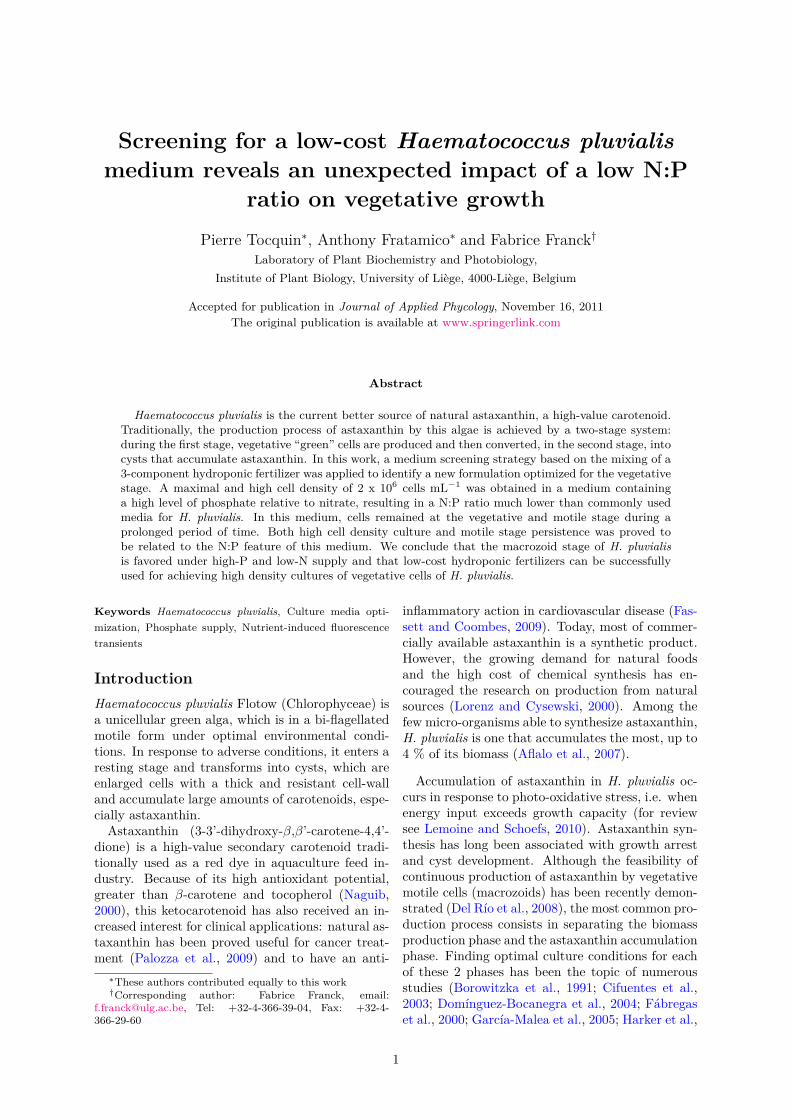
Screening for a low-cost Haematococcus pluvialis medium reveals an unexpected impact of a low N:P ratio on vegetative growth Pierre Tocquin * , Anthony Fratamico * and Fabrice Franck † Laboratory of Plant Biochemistry and Photobiology, Institute of Plant Biology, University of Li` ege, 4000-Li` ege, Belgium Accepted for publication in Journal of Applied Phycology, November 16, 2011 The original publication is available at www.springerlink.com Abstract Haematococcus pluvialis is the current better source of natural astaxanthin, a high-value carotenoid. Traditionally, the production process of astaxanthin by this algae is achieved by a two-stage system: during the first stage, vegetative “green” cells are produced and then converted, in the second stage, into cysts that accumulate astaxanthin. In this work, a medium screening strategy based on the mixing of a 3-component hydroponic fertilizer was applied to identify a new formulation optimized for the vegetative stage. A maximal and high cell density of 2 x 10 6 cells mL -1 was obtained in a medium containing a high level of phosphate relative to nitrate, resulting in a N:P ratio much lower than commonly used media for H. pluvialis. In this medium, cells remained at the vegetative and motile stage during a prolonged period of time. Both high cell density culture and motile stage persistence was proved to be related to the N:P feature of this medium. We conclude that the macrozoid stage of H. pluvialis is favored under high-P and low-N supply and that low-cost hydroponic fertilizers can be successfully used for achieving high density cultures of vegetative cells of H. pluvialis. Keywords Haematococcus pluvialis, Culture media opti- mization, Phosphate supply, Nutrient-induced fluorescence transients Introduction Haematococcus pluvialis Flotow (Chlorophyceae) is a unicellular green alga, which is in a bi-flagellated motile form under optimal environmental condi- tions. In response to adverse conditions, it enters a resting stage and transforms into cysts, which are enlarged cells with a thick and resistant cell-wall and accumulate large amounts of carotenoids, espe- cially astaxanthin. Astaxanthin (3-3’-dihydroxy-β,β’-carotene-4,4’- dione) is a high-value secondary carotenoid tradi- tionally used as a red dye in aquaculture feed in- dustry. Because of its high antioxidant potential, greater than β-carotene and tocopherol (Naguib, 2000), this ketocarotenoid has also received an in- creased interest for clinical applications: natural as- taxanthin has been proved useful for cancer treat- ment (Palozza et al., 2009) and to have an anti- * These authors contributed equally to this work † Corresponding author: Fabrice Franck, email: [email protected], Tel: +32-4-366-39-04, Fax: +32-4- 366-29-60 inflammatory action in cardiovascular disease (Fas- sett and Coombes, 2009). Today, most of commer- cially available astaxanthin is a synthetic product. However, the growing demand for natural foods and the high cost of chemical synthesis has en- couraged the research on production from natural sources (Lorenz and Cysewski, 2000). Among the few micro-organisms able to synthesize astaxanthin, H. pluvialis is one that accumulates the most, up to 4 % of its biomass (Aflalo et al., 2007). Accumulation of astaxanthin in H. pluvialis oc- curs in response to photo-oxidative stress, i.e. when energy input exceeds growth capacity (for review see Lemoine and Schoefs, 2010). Astaxanthin syn- thesis has long been associated with growth arrest and cyst development. Although the feasibility of continuous production of astaxanthin by vegetative motile cells (macrozoids) has been recently demon- strated (Del R´ ıo et al., 2008), the most common pro- duction process consists in separating the biomass production phase and the astaxanthin accumulation phase. Finding optimal culture conditions for each of these 2 phases has been the topic of numerous studies (Borowitzka et al., 1991; Cifuentes et al., 2003; Dom´ ınguez-Bocanegra et al., 2004; F´ abregas et al., 2000; Garc´ ıa-Malea et al., 2005; Harker et al., 1
Transcript of Screening for a low-cost Haematococcus pluvialis medium ... · Screening for a low-cost...
Screening for a low-cost Haematococcus pluvialismedium reveals an unexpected impact of a low N:P
ratio on vegetative growthPierre Tocquin∗, Anthony Fratamico∗ and Fabrice Franck†
Laboratory of Plant Biochemistry and Photobiology,Institute of Plant Biology, University of Liege, 4000-Liege, Belgium
Accepted for publication in Journal of Applied Phycology, November 16, 2011The original publication is available at www.springerlink.com
Abstract
Haematococcus pluvialis is the current better source of natural astaxanthin, a high-value carotenoid.Traditionally, the production process of astaxanthin by this algae is achieved by a two-stage system:during the first stage, vegetative “green” cells are produced and then converted, in the second stage, intocysts that accumulate astaxanthin. In this work, a medium screening strategy based on the mixing of a3-component hydroponic fertilizer was applied to identify a new formulation optimized for the vegetativestage. A maximal and high cell density of 2 x 10⁶ cells mL−1 was obtained in a medium containinga high level of phosphate relative to nitrate, resulting in a N:P ratio much lower than commonly usedmedia for H. pluvialis. In this medium, cells remained at the vegetative and motile stage during aprolonged period of time. Both high cell density culture and motile stage persistence was proved tobe related to the N:P feature of this medium. We conclude that the macrozoid stage of H. pluvialisis favored under high-P and low-N supply and that low-cost hydroponic fertilizers can be successfullyused for achieving high density cultures of vegetative cells of H. pluvialis.
Keywords Haematococcus pluvialis, Culture media opti-mization, Phosphate supply, Nutrient-induced fluorescencetransients
IntroductionHaematococcus pluvialis Flotow (Chlorophyceae) isa unicellular green alga, which is in a bi-flagellatedmotile form under optimal environmental condi-tions. In response to adverse conditions, it enters aresting stage and transforms into cysts, which areenlarged cells with a thick and resistant cell-walland accumulate large amounts of carotenoids, espe-cially astaxanthin.
Astaxanthin (3-3’-dihydroxy-β,β’-carotene-4,4’-dione) is a high-value secondary carotenoid tradi-tionally used as a red dye in aquaculture feed in-dustry. Because of its high antioxidant potential,greater than β-carotene and tocopherol (Naguib,2000), this ketocarotenoid has also received an in-creased interest for clinical applications: natural as-taxanthin has been proved useful for cancer treat-ment (Palozza et al., 2009) and to have an anti-∗These authors contributed equally to this work†Corresponding author: Fabrice Franck, email:
[email protected], Tel: +32-4-366-39-04, Fax: +32-4-366-29-60
inflammatory action in cardiovascular disease (Fas-sett and Coombes, 2009). Today, most of commer-cially available astaxanthin is a synthetic product.However, the growing demand for natural foodsand the high cost of chemical synthesis has en-couraged the research on production from naturalsources (Lorenz and Cysewski, 2000). Among thefew micro-organisms able to synthesize astaxanthin,H. pluvialis is one that accumulates the most, up to4 % of its biomass (Aflalo et al., 2007).
Accumulation of astaxanthin in H. pluvialis oc-curs in response to photo-oxidative stress, i.e. whenenergy input exceeds growth capacity (for reviewsee Lemoine and Schoefs, 2010). Astaxanthin syn-thesis has long been associated with growth arrestand cyst development. Although the feasibility ofcontinuous production of astaxanthin by vegetativemotile cells (macrozoids) has been recently demon-strated (Del Rıo et al., 2008), the most common pro-duction process consists in separating the biomassproduction phase and the astaxanthin accumulationphase. Finding optimal culture conditions for eachof these 2 phases has been the topic of numerousstudies (Borowitzka et al., 1991; Cifuentes et al.,2003; Domınguez-Bocanegra et al., 2004; Fabregaset al., 2000; Garcıa-Malea et al., 2005; Harker et al.,
1

P. Tocquin et al., Screening for a low-cost Haematococcus pluvialis medium [...]Accepted for publication in Journal of Applied Phycology, November 16, 2011
1995; Choi et al., 2002). Even if the exact influenceor efficiency of some specific factors on growth -N-source (Borowitzka et al., 1991; Cifuentes et al.,2003), acetate addition (Cifuentes et al., 2003; Jeonet al., 2006), light level (Borowitzka et al., 1991;Garcıa-Malea et al., 2005; Harker et al., 1995) - andon astaxanthin induction - salt stress, phosphate de-ficiency (Borowitzka et al., 1991; Choi et al., 2002),light requirement (Cifuentes et al., 2003; Choi et al.,2002; Garcıa-Malea et al., 2005) - are still unclear ordebated, the general rule governing the green andred stages is widely accepted: a low C:N ratio isfavorable to the production of green biomass and,conversely, the production of astaxanthin occurspreferentially when C:N is high (Kakizono et al.,1992).
In practice, low C:N ratio for green biomass pro-duction is generally achieved by growing the cells atlow light in media containing saturating levels of ni-trate. The Bold Basal Medium (BBM, Nichols andBold, 1969) has been often used in its initial formu-lation or modified to contain up to 4-fold more ni-trate (Domınguez-Bocanegra et al., 2004; Fabregaset al., 2000). Since biomass accumulation is themajor bottleneck in the 2-stage process of astaxan-thin production, further optimizations of the grow-ing medium have been undertaken (Cifuentes et al.,2003; Fabregas et al., 2000; Harker et al., 1995;Gong and Chen, 1997; Sarada et al., 2002).
A common approach to determine the factorsfor optimal biomass production has been to startfrom a known medium and change relative con-centrations of different macro-elements and micro-elements in the medium. In an attempt to opti-mize the elemental composition of an Haematocco-cus growing medium, Fabregas et al. (2000) evalu-ated the contribution of 18 components on the finalyield of vegetative biomass using a single-variableoptimization strategy. In a semi-continuous culture,the steady-state cell density they obtained withtheir optimized medium (OHM) was three timeshigher than with the often used BBM. However, asnoticed by the authors, this “one-factor-at-a-time”strategy probably lacks to identify positive or neg-ative interactions between nutrients or between nu-trient availability and other environmental condi-tions.
In a comparative study, Dalay et al. (2007)observed, comparing 9 common medium formula-tions, that biomass accumulation was maximal in amedium prepared with a common agricultural fer-tilizer. As a starting point of our study, we lookedfor a low-cost and optimized growing medium for H.pluvialis based on the use of a commercial fertilizer.Our optimization strategy relied on the use of a 3-component hydroponic fertilizer which allowed tovary the composition of the media by using differentratios of the 3 core solutions. By screening 18 dif-
Table 1: Composition of the Floraseries componentsNutrient concentrations (guaranteed minimum concentra-tions) in % (w/v) as described by the producer (GHE, Fleu-rance, France) for each of the 3 components of the FloraSeriesfertilizer. M: FloraMicro, G: FloraGro, B: FloraBloom.
FloraSeries componentsConstituents M G B
Nitrogen 5.0 3.0...Ammonium N 1.0 1.0...Nitrate N 4.0 2.0Phosphate (as P2O5) 1.0 5.0Potassium (as K2O) 1.3 7.0 4.0Magnesium 0.8 3.0Sulfur 1.0 5.0Boron 0.010Calcium 7.000Copper 0.010Iron 0.120Manganese 0.040Molybdenum 0.004Zinc 0.015
ferent combinations for vegetative high density cul-tures, we selected the best-performing formulationand compared growth, nutrients (NO3, PO4) up-take and carotenoid accumulation in batch cultureof H. pluvialis in this medium and two other com-mon formulations. The relationship between theunusual N and P content of the identified mediumand growth of vegetative cells were further investi-gated.
Materials and MethodsAlgal strain and culture conditions Haema-tococcus pluvialis (strain 34/1D) was obtained fromthe Culture Collection of Algae and Protozoa ofthe Center for Hydrology and Ecology, Amble-side, UK. Stock cultures were grown in 100 mLof modified BBM3N in 250 mL cell culture flask(Cellstar Suspension Culture Flasks, Greiner Bio-One, Belgium). Modified BBM3N was preparedfrom a concentrated Bold modified Basal Freshwa-ter Nutrient solution (Sigma-Aldrich, Belgium) towhich NaNO3 was added up to 8.82 mM. Flaskswere placed horizontally to increase the gas ex-change surface and optimize exposition to light.They were gently shaken twice-a-day. Cultureswere not supplied with an extra source of CO2.Temperature was maintained at 25◦C, cultureswere continuously illuminated by Phillips MasterTL5 HO 54W/827 fluorescent lamps. Light in-tensity was either 30 µmol m−2 s−1 (low-light) or150 µmol m−2 s−1 (high-light).
Media used in the screening were prepared bymixing 3 components of the Floraseries hydroponicfertilizer (GHE, Fleurance, France, guaranteed min-imum concentrations, see Table 1). Cells from stockculture were collected at the vegetative stage, cen-
The original publication is available at www.springerlink.com 2

P. Tocquin et al., Screening for a low-cost Haematococcus pluvialis medium [...]Accepted for publication in Journal of Applied Phycology, November 16, 2011
trifuged (3 min, ± 3000 x g, room temperature)and resuspended in 50 mL flasks (Cellstar Suspen-sion Culture Flasks, Greiner Bio-One, Belgium) atthe specified density in 10 mL of each of the mediaformulations tested.
Analytical procedures Cell densities were es-tablished by counting under the microscope, usingan improved Neubauer hematocytometer. For pig-ments analysis, cells were collected by centrifugingculture aliquots (3 min, 16000 x g, room temper-ature). The supernatant was discarded and thepellet was homogenized in 100 % methanol. Theextraction procedure was repeated until cell de-bris was almost colorless. Chlorophylls and totalcarotenoids were quantified with a UV-VIS spec-trophotometer (PerkinElmer UV-Vis spectropho-tometer Lambda 20) by recording absorbance at470, 652 and 665 nm and using the equations ofLichtenthaler (1987).
Investigations of Nutrient Induced FluorescenceTransients (NIFT) involved the recording of chloro-phyll a fluorescence by Pulse Modulated Fluorime-ter (PAM, MFMS, Hansatech). 2 mL of culturesample were placed into a glass cuvette connected tothe PAM and containing a magnetic stirrer. The ac-tinic light (λ=650 nm) was set at 30 µmol m−2 s−1.After the sample had been left to stabilize for10 min, H2O (control), 500 µM of either NaNO3or K2HPO4 were added to the cuvette. Any sig-nificant change in fluorescence measured at 680 nm(the analytic modulated light was provided by light-emitting diodes with a central emission wavelengthat 580 nm and a photon flux of 0.5 µmol m−2 s−1)following nutrient addition was considered as aNIFT.
Nitrate in media was assayed by the colorimetricreaction with salicylic acid as described by Cataldoet al. (1975). Briefly, 20 µL of diluted sampledmedium were incubated with 2.5 % (w/v) salicylicacid in H2SO4 98 % for 20 minutes before neutral-ization with 500 µL NaOH 3.8 M. Concentrationin NO3 was then assayed by measuring absorbanceat 405 nm with a spectrophotometer (Victor X3,Multilabel Plate Reader, Perkin-Elmer) and com-parison with a NaNO3 standard curve.
Phosphate was assayed by reaction with ascor-bic acid as described by Chen et al. (1956).Briefly, 50 µL of diluted sampled medium weremixed with 50µL of a freshly prepared reac-tion solution containing ascorbic acid 2 % (w/v),(NH4)6Mo7O24.4H2O 0.5 % (w/v) and H2SO40.6 M. Concentration in PO4 was then assayed bymeasuring absorbance at 750 nm with a spectropho-tometer and comparison with a KH2PO4 standardcurve.
Figure 1: 9-day-old cultures of a screening experi-ment Representative picture showing 6 of the 18 culturesof a screening experiment. On the left-hand side, from thetop to the bottom, cultures M1G1, M1G2 and M1G5 and,on the right-hand side, M1B1, M1B2 and M1B5 (see Table 2for details). The reddening observed from M1G5 to M1G1and M1B5 to M1B1 is a typical illustration of the devel-opmental shift from the vegetative stage to the resting andastaxanthin accumulating stage. The M1B5 culture showsthe distinguishing reticulated-pattern of motile cells group-ing together.
Results
Screening for a medium formulation leadingto high vegetative cell density A screeningfor an optimized Haematococcus pluvialis mediumwas carried out using a blind approach: 18 mediawere prepared by mixing the 3 components of theFloraseries hydroponic fertilizer (GHE, Fleurance,France) following the experimental design of Ta-ble 2. A pre-culture in modified Bold Basal medium(BBM3N) was used to inoculate 10 mL of each cul-ture media at 2 x 10⁴ cells mL−1. Cells were grownin continuous light at 30 µmol m−2 s−1 during 9days.
Maximum cell densities are presented in Table 2:among the 18 screened media, a maximum cell den-sity of 1 x 10⁶ cells mL−1, corresponding to amean growth rate of 0.41 day−1, was recorded inM1B5 medium (i.e. 1 µL mL−1 FloraMicro and5 µL mL−1 FloraBloom) on day 9 while for all othermedia, except M5B5, the maximum cell density wasreached earlier and was 2 to 10-fold lower. M1B5was the only medium allowing to keep the cells inthe green and motile stage after day 9 (Fig. 1) andto sustain growth during 14 days, up to a cell den-sity of 1.73 x 10⁶ cells mL−1 (data not shown).
One specificity of M1B5 medium, and other ’B5media, is to have a very high content in phos-phate (P), magnesium (Mg) and sulfur (S) whichare specifically provided at a high level by the Bcomponent (Tables 1 and 2). As for potassium (K),it is provided at highest level in the media preparedwith the G component and was thus not supposedto contribute in M1B5 efficiency. Since the B com-ponent does not contain a nitrogen (N) source, an-
The original publication is available at www.springerlink.com 3

P. Tocquin et al., Screening for a low-cost Haematococcus pluvialis medium [...]Accepted for publication in Journal of Applied Phycology, November 16, 2011
Table 2: Maximal cell densities obtained in the 18 media Maximal cell densities (mean ± standard deviation,x 105 cells mL−1, n=16) measured in a 9-day screening experiment of 18 media obtained by mixing concentrated FloraSeriesstock solutions.
G B
µL.mL−1 1 2 5 1 2 5
1 1.9 ± 0.6 1.4 ± 0.5 1.4 ± 0.5 1.6 ± 0.6 1.6 ± 0.7 10.0 ± 1.6M 2 1.2 ± 0.6 1.2 ± 0.5 5.0 ± 1.0 1.9 ± 0.8 5.2 ± 1.5 5.4 ± 1.4
5 2.3 ± 0.3 2.9 ± 0.7 5.3 ± 1.1 4.2 ± 0.8 3.5 ± 0.9 6.9 ± 1.4
other feature of M1B5 is to have a basal contentin N. We thus evaluated which of these 4 nutrients(P, Mg, S and N) could account, alone or inter-acting with others, for the high macrozoid densityobtained in M1B5. Starting from the BBM3N for-mulation, we decreased the N content by a factor 3,increased P content by a factor 3, and the Mg andS content (as MgSO4) by a factor 5 or 10. Twelvemedia were generated and used for the culture ofH. pluvialis in the same experimental conditions asfor the previous screening experiment. As shown inFig. 2a, the highest cell density at day 6 was mea-sured in the media in which P was increased 3 times,other nutrients remaining at their basal BBM3Nlevel. Increasing Mg/S supply was found to have anegative impact on cell growth, independently of Nand P supply, while increasing N concentration didnot stimulate vegetative cell production and even,in some experiments (data not shown), counterbal-ance the positive effect of high P supply.
This observation suggested that N and P areinteracting in controlling production of vegetativecells. When modifying BBM3N formulation tomimic N and P content of M1B5 (M1B5-like), weindeed observed that it was sufficient to prevent pre-cocious encystment and to stimulate green cells pro-duction (Fig. 2b). Moreover, when plotting the 18screened media on a graph as a function of theirinitial NO3 and PO4 content, the best performingmedium, M1B5, appeared as the one with the low-est N:P ratio (± 0.6, Fig. 3). We further observed,by analyzing nitrate and phosphate contents of themedia during growth, that there was a very strongrelationship between the phosphate consumptionby the algae and initial phosphate content of themedium (Fig. 4a). This correlation was not ob-served for nitrate (Fig. 4b) for which measured de-pletions in media were low and not related to theinitial content. Note that ammonium was not as-sayed but could act as an N-source at the beginningof the culture.
Evaluation of nutrient limitations in screenedmedia by chlorophyll a fluorescence analyzesThe role of phosphate as a growth-limiting factorwas further evaluated by a non-invasive method
based on chlorophyll a fluorescence measurement.It is known that, if a nutrient such as phosphate, ni-trate or ammonium is added to algal culture limitedin that particular nutrient, a change in fluorescenceis observable within minutes. This phenomenonhas been referred to as “nutrient-induced fluores-cence transients” (NIFT) (Beardall et al., 2001) andproved useful to detect true, in situ, nutrient lim-itations (Holland et al., 2004). In a preliminaryexperiment, we checked for NIFT occurrence in a3-week old culture of H. pluvialis in BBM3N andobserved phosphate-induced NIFT but no nitrate-induced NIFT (Fig. 5a).
The chlorophyll a fluorescence analysis at day 6and day 9 in all 18 screened media revealed thatNIFT were induced by phosphate addition in al-most all media at day 6 and/or day 9 (data notshown). Conversely, as observed in the preliminaryexperiment, no NIFT were induced by nitrate ad-dition. When results were analyzed independentlyof media and age but rather plotted against eitherrelative phosphate uptake or remaining phosphateconcentration in medium, the NIFT were observedto occur preferentially when phosphate uptake wasgreater than 25% of initial content (Fig. 5b).
Evaluation of growth in selected mediumM1B5 and in two other media To the bestof our knowledge, high-phosphate containing me-dia, such as M1B5, were never used for growing H.pluvialis. To evaluate the benefit of such a media onthe growth of H. pluvialis, we compared growth, interms of cell density, in M1B5 and two other com-mon media, BBM3N and OHM, containing respec-tively about 2.5 and 25 times less phosphate thanM1B5 and whose NO3/PO4 ratio are 4.6 and 23.9,respectively (Fig. 3). Cultures were initiated in100 ml fresh media at a density of 5 x 10⁴ cells mL−1
and were run at 30 µmol m−2 s−1 (low light) and150 µmol m−2 s−1 (high light). We observed that,in both BBM3N and OHM, the cells shifted rapidly(around days 4-6) to the palmelloid stage while, inM1B5, they remained in the macrozoid form duringthe whole experimental period. The developmentalshift observed in BBM3N and OHM occurred atboth light intensities and coincided with a signif-
The original publication is available at www.springerlink.com 4

P. Tocquin et al., Screening for a low-cost Haematococcus pluvialis medium [...]Accepted for publication in Journal of Applied Phycology, November 16, 2011
Figure 2: Growth of H. pluvialis in modified BBM3N-based media (a) H. pluvialis was grown in BBM3N mediummodified to contain 3 fold less nitrate (2.9 mM vs 8.8 mM),3 fold more phosphate (5.1 mM vs 1.7 mM), 5 or 10 fold moreMgSO4 (1.5 and 3 mM vs 0.3 mM), either separately or incombination. (b) Starting from the BBM3N formulation,NO3, NH4 and PO4 concentrations (respectively 8.7 mM,0 mM and 1.7 mM in BBM3N) were modified to mimicM1B5. The resulting medium M1B5-like contained 2.7 mMNO3, 0.7 mM NH4 and 4.6 mM PO4. The observed de-crease of cell density in BBM3N is due to adsorption of cyststo walls of flask. These figures are representative results of3 independent experiments.
icant increase in the carotenoid-to-chlorophyll ra-tio above 0.5 which is a maximum for H. pluvi-alis green cells (this study, see also Solovchenkoet al. (2011)). These observations correlated wellwith growth measurements: as shown on Fig. 6,a significant increase in cell density was only ob-served in M1B5 medium. A maximum of 1.4 and2.1 x 106 cells mL−1 was measured after 14 days atlow and high light, respectively.
Figure 3: Nitrate-to-phosphate content and relativeN:P ratio of the 18 screened media 2D-graph of the ini-tial measured nitrate (y-axis) and phosphate (x-axis) contentof 18 screened media. The 2 other common media (OHM andBBM3N) used in this study are also plotted. The plot areaswhere N:P ratio are below 1, between 1 and 10, and above 10are filled with dark-grey, light-grey and white, respectively.
Effect of N & P deficiencies on M1B5 cul-tures The above observations tend to show thatthe M1B5 medium has specific impacts on growthand development of H. pluvialis by stimulating celldivision and preventing precocious cysts formation.Nutrient uptake measurements and NIFT assays in-dicated that the high-phosphate content and/or thelow NO3/PO4 ratio of M1B5 could be at the originof these responses. To further test this hypothesis,we transferred M1B5 exponentially growing cellsto media containing lower phosphate (high N:P ra-tio) or lower nitrate (low N:P ratio) and evaluatedthe quantitative (cell density) and developmental(cell types, carotenoid-to-chlorophyll ratio) conse-quences of these treatments.
Independently of light intensity, transfer toBBM3N or phosphate-deprived BBM3N led to adramatic decrease in cell densities due to inducedcell death as observed under the microscope (Fig. 7a& b). In high light, cell death occurred rapidly andwas almost complete after 2 days while, in low light,the surviving cells shifted to the palmelloid stage:95% of them were palmella at the end of the ex-periment. Conversely, when cells are transferred tofresh M1B5 or nitrate-deprived BBM3N, cell den-sity still increased. Furthermore, more than 90%of the cells stayed in motile macrozoid stage at ei-ther low or high light. Strikingly, the maximumcell density was observed in the nitrate-deprivedBBM3N media in high light. In these conditions,cells accumulated carotenoids, they became red andcarotenoid-to-chlorophyll ratio increased up to 0.8,
The original publication is available at www.springerlink.com 5

P. Tocquin et al., Screening for a low-cost Haematococcus pluvialis medium [...]Accepted for publication in Journal of Applied Phycology, November 16, 2011
Figure 4: Phosphate and nitrate uptake at day 6in relation with initial content Phosphate and nitrateconcentrations in culture media were measured in fresh me-dia (initial content) and at day 6. Phosphate and nitratedepletion were respectively plotted against initial phosphatecontent (a) and initial nitrate content (b) of screened media.
while remaining at the macrozoid stage (Fig. 7b).In this specific case, carotenoids accumulation didnot occur simultaneously with growth arrest as itis the case in the BBM3N and phosphate-deprivedmedia at high light.
DiscussionIn this study, medium optimization was performedby screening three-component hydroponic fertilizerbased media. This strategy allowed us to evaluate,on a limited number of media (18), the effect of alarge range of individual nutrient supplies togetherwith a large range of macro-nutrients ratios. For
Figure 5: NIFT occurrence at day 6 or day 9 inscreened media in relation with nutrient uptake (a)Representative result of a preliminary experiment whereNIFT occurrence was analyzed in 3-week old BBM3N stockculture following addition of either 20 µL H2O, 500 µMNaNO3 (NO3) or 500 µM K2HPO4 (PO4). (b) Phosphate-induced NIFTs plotted as 2-D graph of actual phosphate con-tent vs phosphate uptake. Empty dots: no NIFT observed.Filled dots: phosphate-induced NIFT.
example, nitrate and phosphate ranges were 2.20-28.85 mM and 0.18-4.33 mM, respectively, whiletheir relative N:P ratio ranged from 0.6 to 111.8.With this experimental set-up, the relative supplyof micro-nutrients was not allowed to be optimizedsince micro-elements are provided by only one ofthe three fertilizer components. However, accord-ing to the work of Fabregas et al. (2000) micro-elements do not contribute much in optimizationand even their omission has been shown to havea limited growth-inhibitory impact on H. pluvialis(Domınguez-Bocanegra et al., 2004; Hagen et al.,2001).
Our screening clearly led to the identification
The original publication is available at www.springerlink.com 6

P. Tocquin et al., Screening for a low-cost Haematococcus pluvialis medium [...]Accepted for publication in Journal of Applied Phycology, November 16, 2011
Figure 6: Growth of H. pluvialis in M1B5, BBM3Nand OHM media at two light intensities Cell den-sities, chlorophyll and carotenoids content were measuredat 2 days interval during a 14 days culture in M1B5,BBM3N and OHM media either at 30 µmol m−2 s−1 (a)or 150 µmol m−2 s−1. The carotenoid-to-chlorophyll ratioof each sample is represented on a color-coded scale fromlight-green (0) to red (3). A non-linear scale was used to vi-sualize “green” stage (car:chl < 0.5) as green. This figure is arepresentative result of two independent experiments. Dataare means ± standard deviations (n=8). The fluctuating celldensities measured in BBM3N and OHM from day 6 are dueto partial adsorption of cyst to walls of flask and productionof macrozoids from cyst (concomitant to a drop in Car/Chlratio).of one optimal formulation, which is composed of1 µL ml−1 FloraMicro and 5 µL ml−1 FloraBloom(see Materials & Methods for composition) andwas labeled “M1B5”. During 9 days screening ex-periments, growth was indeed sustained until day9 in only 2 media (M1B5 and M5B5, data notshown), but cell density was the highest in M1B5(1 x 10⁶ cells mL−1, Table 2). When we com-pared growth in M1B5 with BBM3N and OHM,we observed that vegetative growth was sustainedat least during 2 weeks and a cell density of
Figure 7: Impact of nitrate or phosphate starvationon the growth of H. pluvialis Cell densities, chlorophylland carotenoid contents were measured at 24-hours intervalduring 3 days following the transfer of M1B5 growing cells toBold Basal medium (BBM3N), N-depleted BBM3N (-N) andP-depleted BBM3N (-P) at either at 30 µmol m−2 s−1 (a) or150 µmol m−2 s−1 (b). The carotenoid-to-chlorophyll ratioof each sample is represented on a color-coded scale fromlight-green (0) to red (3). A non-linear scale was used tovisualize “green” stage (car:chl < 0.5) as green. This figureis a representative result of 2 independent experiments. Dataare means ± standard deviations (n=8).
2 x 10⁶ cells mL−1 was reached under high lightintensity. These results represent a minimum 5-fold improvement compared with previous stud-ies in similar batch-mode conditions (Borowitzkaet al., 1991; Cifuentes et al., 2003; Domınguez-Bocanegra et al., 2004; Kaewpintong et al., 2007)and with the maximal cell densities of 1.8 and3.2 x 106 cells mL−1 obtained in this study withBBM3N and OHM, respectively. For BBM3N,recorded maximal cell density was in the range ofpublished data obtained in similar growing condi-
The original publication is available at www.springerlink.com 7

P. Tocquin et al., Screening for a low-cost Haematococcus pluvialis medium [...]Accepted for publication in Journal of Applied Phycology, November 16, 2011
tions (Harker et al., 1996; Tripathi et al., 1999;Domınguez-Bocanegra et al., 2004), while for OHM,it correspond to steady-state density obtained byFabregas et al. (2000) in a semi-continuous modewith daily addition of fresh medium. The efficiencyof M1B5 can not be explained by absolute availabil-ity of nutrients since, in our screening, other formu-lations contained as much nutrients, like nitrate orphosphate, or even more. Rather, we observed thatM1B5 had a specific physiological effect by delayingthe shift to encystment. This effect was neither ob-served for any of the other screened media, nor forBBM3N or OHM, where a maximum in cell densityobserved after 3 or 6 days preceded a developmentalshift to the red resting stage.
Although phosphate is not the only nutrient thatis at high level in M1B5, our results indicate thathigh phosphate is the key feature leading to its ben-eficial effect on cell growth. Firstly, we showed thatamong nutrients that are present in higher amountin M1B5, phosphate accounted, alone, for the in-crease in cell density. Secondly, we observed thatphosphate uptake increased with available phos-phate in medium and, finally, we also demonstrated,using the NIFT approach, that cells are rapidlybehaving as phosphate-limited whatever the initialphosphate content of the medium. Compared to ni-trate, phosphate has received less attention in opti-mization approaches and has long been consideredto promote growth at moderate or low concentra-tion (± 0.5 mM, Borowitzka et al. (1991); Fabregaset al. (2000)) while it could promote carotenogenesisat higher concentration (up to 0.9 mM, Borowitzkaet al. (1991)). However, in other reports, carotenoidaccumulation has been shown to be reduced whenphosphate supply was increased above 0.85 mM (upto 3.4 mM, Harker et al. (1996)). Harker et al.(1996) even reported a positive impact of a highphosphate concentration (>3 mM) on the growthof H. pluvialis.
In addition to have a high phosphate content,M1B5 also is remarkable for having the low-est nitrate-to-phosphate ratio (0.6) among the 18screened media. Both features are rarely found inalready known H. pluvialis media: most of themhave a moderate phosphate content (± 0.2-2 mM)and a N:P ratio largely in favor of N (± 7-20)(Domınguez-Bocanegra et al., 2004; Fabregas et al.,2000). There are only a few reports on H. pluvi-alis growing in N:P ratio close or below one but,in all cases that we identified, the low N:P condi-tions were beneficial to growth (Dalay et al., 2007;Harker et al., 1996; Hagen et al., 2001). In thepresent study, we showed that the transfer fromthe M1B5 medium to low-phosphate containingBBM3N medium led to a rapid cell death while itwas not the case if nitrate was omitted in media. Inthis case where N:P ratio was null, growth was not
inhibited during the time-course of the experimentand the cells stayed at the vegetative motile stageand accumulated carotenoids under increased irra-diance without shifting to resting stage as it was al-ready observed in similar conditions by Hagen et al.(2001). Moreover, we observed that increasing N-supply together with P-supply reduced the positiveeffect of high P on macrozoids production. The lowN:P ratio thus appeared to be a critical parameterthat maintains actively dividing cells at the macro-zoid stage.
In most organisms, phosphate accumulates in-side the cell as a linear polymer forming intra-cellular polyphosphate granules (PolyP) (Kulaevand Kulakovskaya, 2000). In a preliminary experi-ment, we looked for the presence of PolyP granulesin H. pluvialis by microscopy with the fluorescentdye 4’,6-diamidino-2-phenylindole (DAPI), follow-ing the method by Tijssen et al. (1982) and weobserved that granules were visible in M1B5 grow-ing macrozoids (data not shown). The function ofPolyP, which has been essentially studied in pro-caryotes, are numerous, including stress response,quorum sensing, motility, etc... (for review, seeRao et al.). In microalgae, roles in osmotic stressresponse, in heavy metal tolerance or phosphatestorage have been already proposed but the iden-tification of more functions will undoubtedly occuras the interest for PolyP in eucaryote increases.In this context, the relation between phosphate-supply, PolyP accumulation and cell cycle controlin H. pluvialis is of particular interest and requiresdeeper investigations.
In conclusion, by a medium screening strategybased on a 3-component commercial fertilizer, weobtained a new medium formulation that proved tobe efficient, at least in batch culture, in promotinghigh density culture of H. pluvialis macrozoids andin post-poning the development shift to the restingstage. We demonstrated that these effects were aresponse to both the unusual phosphate content andthe below-one N:P ratio of this medium. Even if theexact impact of these parameters on the physiologyof H. pluvialis remains to be determined, achieve-ment of high vegetative cell density culture in thiseasy to prepare and low-cost media could be evalu-ated for the production of astaxanthin in either 1-or 2-step strategies.
Acknowledgements
This research was funded by WINNOMAT2 program, ServicePublic de Wallonie, DGO6, Grant N◦71/6662. A.F. is grate-ful to F.R.I.A. for the award of research fellowships. F.F.is a Senior Research Associate of the Fonds de la RechercheScientifique, F.R.S-FNRS. Authors are grateful to Dr. P.Cardol and anonymous reviewers for helpful comments onan earlier draft of this manuscript.
The original publication is available at www.springerlink.com 8

P. Tocquin et al., Screening for a low-cost Haematococcus pluvialis medium [...]Accepted for publication in Journal of Applied Phycology, November 16, 2011
ReferencesC. Aflalo, Y. Meshulam, A. Zarka, and S. Boussiba. On
the relative efficiency of two- vs. one-stage production ofastaxanthin by the green alga Haematococcus pluvialis.Biotechnology and Bioengineering, 98(1):300–305, 2007.
J. Beardall, T. Berman, P. Heraud, M. Omo Kadiri, B. R.Light, G. Patterson, S. Roberts, B. Sulzberger, E. Sa-han, U. Uehlinger, and B. Wood. A comparison of meth-ods for detection of phosphate limitation in microalgae.Aquatic Sciences - Research Across Boundaries, 63(1):107–121, 2001.
M. Borowitzka, J. Huisman, and A. Osborn. Culture of theastaxanthin-producing green alga Haematococcus pluvi-alis 1. Effects of nutrients on growth and cell type. Journalof Applied Phycology, 3(4):295–304, 1991.
D. A. Cataldo, M. Maroon, L. E. Schrader, and V. L. Youngs.Rapid colorimetric determination of nitrate in plant tissueby nitration of salicylic acid. Communications in SoilScience and Plant Analysis, 6(1):71, 1975.
P. S. Chen, T. Y. Toribara, and Huber. Warner. Microde-termination of phosphorus. Analytical Chemistry, 28(11):1756–1758, 1956.
Y. E Choi, Y. S Yun, and J. M Park. Evaluation of factorspromoting astaxanthin production by a unicellular greenalga, Haematococcus pluvialis, with fractional factorial de-sign. Biotechnology Progress, 18(6):1170–1175, 2002.
A. S. Cifuentes, M. A. Gonzalez, S. Vargas, M. Hoeneisen,and N. Gonzalez. Optimization of biomass, totalcarotenoids and astaxanthin production in Haematococ-cus pluvialis flotow strain steptoe (Nevada, USA) underlaboratory conditions. Biological Research, 36(3-4):343–57, 2003.
M. C. Dalay, E. Imamoglu, and Z. Demirel. Agricultural fer-tilizers as economical alternative for cultivation of Haema-tococcus pluvialis. Journal of Microbiology and Biotech-nology, 17(3):393–7, 2007.
E. Del Rıo, F. G. Acien, M. C. Garcıa-Malea, J. Rivas,E. Molina-Grima, and M. G. Guerrero. Efficiency assess-ment of the one-step production of astaxanthin by the mi-croalga Haematococcus pluvialis. Biotechnology and Bio-engineering, 100(2):397–402, 2008.
A. R. Domınguez-Bocanegra, I. Guerrero Legarreta, F. Mar-tinez Jeronimo, and A. Tomasini Campocosio. Influenceof environmental and nutritional factors in the productionof astaxanthin from Haematococcus pluvialis. BioresourceTechnology, 92(2):209–214, 2004.
R. G. Fassett and J. S. Coombes. Astaxanthin, oxidativestress, inflammation and cardiovascular disease. FutureCardiology, 5:333–342, 2009.
J. Fabregas, A. Dominguez, M. Regueiro, A. Maseda, andA. Otero. Optimization of culture medium for the contin-uous cultivation of the microalga Haematococcus pluvialis.Applied Microbiology and Biotechnology, 53(5):530–535,2000.
M.C. Garcıa-Malea, C. Brindley, E. Del Rıo, F.G. Acien,J.M. Fernandez, and E. Molina. Modelling of growth andaccumulation of carotenoids in Haematococcus pluvialis asa function of irradiance and nutrients supply. BiochemicalEngineering Journal, 26(2-3):107–114, 2005.
X. Gong and F. Chen. Optimization of culture medium forgrowth of Haematococcus pluvialis. Journal of AppliedPhycology, 9(5):437–444, 1997.
C. Hagen, K. Grunewald, M. Xylander, and E. Rothe. Effectof cultivation parameters on growth and pigment biosyn-thesis in flagellated cells of Haematococcus pluvialis. Jour-nal of Applied Phycology, 13(1):79–87, 2001.
M. Harker, A. J. Tsavalos, and A. J. Young. Use of responsesurface methodology to optimise carotenogenesis in themicroalga, Haematococcus pluvialis. Journal of AppliedPhycology, 7(4):399–406, 1995.
M. Harker, A. J. Tsavalos, and A. J. Young. Factors re-sponsible for astaxanthin formation in the ChlorophyteHaematococcus pluvialis. Bioresource Technology, 55(3):207–214, 1996.
D. Holland, S. Roberts, and J. Beardall. Assessment of thenutrient status of phytoplankton: a comparison betweenconventional bioassays and nutrient-induced fluorescencetransients (NIFTs). Ecological Indicators, (3):149–159,2004.
Y-C. Jeon, C-W. Cho, and Y-S. Yun. Combined effects oflight intensity and acetate concentration on the growthof unicellular microalga Haematococcus pluvialis. Enzymeand Microbial Technology, 39(3):490–495, 2006.
K. Kaewpintong, A. Shotipruk, S. Powtongsook, andP. Pavasant. Photoautotrophic high-density cultivation ofvegetative cells of Haematococcus pluvialis in airlift biore-actor. Bioresource Technology, 98(2):288–295, 2007.
T. Kakizono, M. Kobayashi, and S. Nagai. Effect of car-bon/nitrogen ratio on encystment accompanied with as-taxanthin formation in a green alga, Haematococcus plu-vialis. Journal of Fermentation and Bioengineering, 74(6):403–405, 1992.
I. Kulaev and T. Kulakovskaya. Polyphosphate and phos-phate pump. Annual Review of Microbiology, 54(1):709–734, 2000.
Y. Lemoine and B. Schoefs. Secondary ketocarotenoid astax-anthin biosynthesis in algae: a multifunctional response tostress. Photosynthesis Research, 106:155–177, 2010.
H. K. Lichtenthaler. Chlorophylls and carotenoids: Pigmentsof photosynthetic biomembranes. Methods in Enzymology,pages 350–382, 1987.
R. T. Lorenz and G. R. Cysewski. Commercial potential forHaematococcus microalgae as a natural source of astaxan-thin. Trends in Biotechnology, 18(4):160–7, 2000.
Y. M. A. Naguib. Antioxidant activities of astaxanthin andrelated carotenoids. Journal of Agricultural and FoodChemistry, 48(4):1150–1154, 2000.
H.W. Nichols and H.C. Bold. Trichsarcina polyinorpha gen.et sp. nov. J. Phycol., pages 34–38, 1969.
P. Palozza, C. Torelli, A. Boninsegna, R. Simone, A. Cata-lano, M. C. Mele, and N. Picci. Growth-inhibitory effectsof the astaxanthin-rich alga Haematococcus pluvialis inhuman colon cancer cells. Cancer Letters, 283(1):108–117,2009.
N. N. Rao, M. R. Gomez-Garcıa, and A. Kornberg. Inorganicpolyphosphate: Essential for growth and survival. AnnualReview of Biochemistry, 78(1):605–647.
R. Sarada, S. Bhattacharya, and G.A. Ravishankar. Opti-mization of culture conditions for growth of the green algaHaematococcus pluvialis. World Journal of Microbiologyand Biotechnology, 18(6):517–521, 2002.
The original publication is available at www.springerlink.com 9

P. Tocquin et al., Screening for a low-cost Haematococcus pluvialis medium [...]Accepted for publication in Journal of Applied Phycology, November 16, 2011
A. E. Solovchenko, O. B. Chivkunova, and I. P. Maslova.Pigment composition, optical properties, and resistanceto photodamage of the microalga Haematococcus pluvi-alis cultivated under high light. Russian Journal of PlantPhysiology, 58(1):9–17, 2011.
J.P.F. Tijssen, H.W. Beekes, and J. Van Steveninck. Lo-calization of polyphosphates in Saccharomyces fragilis,as revealed by 4’,6-diamidino-2-phenylindole fluorescence.Biochimica et Biophysica Acta (BBA) - Molecular CellResearch, 721(4):394–398, 1982.
U. Tripathi, R. Sarada, S.R. Rao, and G.A. Ravishankar.Production of astaxanthin in Haematococcus pluvialis cul-tured in various media. Bioresource Technology, 68(2):197–199, 1999.
The original publication is available at www.springerlink.com 10