Scottish Stroke AHP Forum - ssahpf.scot · Scottish Stroke AHP Forum ... • Electrical stimulation...
87
Scottish Stroke AHP Forum Scottish Stroke Allied Health Professionals Forum Use of Electrical Stimulation Following Stroke A Consensus Statement August 2014
Transcript of Scottish Stroke AHP Forum - ssahpf.scot · Scottish Stroke AHP Forum ... • Electrical stimulation...
Scottish Stroke AHP Forum
Scottish Stroke Allied Health Professionals Forum
Use of Electrical Stimulation Following StrokeA Consensus Statement August 2014

Scottish Stroke Allied Health Professionals Forum
Use of Electrical Stimulation Following StrokeA Consensus Statement

Use of Electrical Stimulation Following StrokeA Consensus Statement
Scottish Stroke Allied Health Professionals Forum Scottish Stroke Allied Health Professionals Forum
3Use of Electrical Stimulation Following StrokeA Consensus Statement
Contents
Section Page
Introduction
Background _______________________________________________________________ 4
Intended Readers __________________________________________________________ 5
Purpose of this Consensus Statement _________________________________________ 5
Methodology______________________________________________________________ 6
National Survey of Allied Heath Professionals __________________________________ 8
Summaries of audits on acute and rehabilitation stroke units_____________________ 9
Summary of evidence including parameters of treatment ______________________ 13
Contraindications _________________________________________________________ 19
Electrical Stimulation Device Review _________________________________________ 20
Conclusion _______________________________________________________________ 22
Appendices:
A. National survey of Allied Heath Professionals _______________________________ 23
B. Audit of incidence of shoulder subluxation _________________________________ 30
C. Audit of service delivery of electrical stimulation ____________________________ 46
D. Literature review _______________________________________________________ 59
Glossary/Abbreviations ____________________________________________________ 80
References _______________________________________________________________ 83
Who was involved in developing the statement _______________________________ 87
Scottish Stroke Allied Health Professionals Forum
4Use of Electrical Stimulation Following StrokeA Consensus Statement
IntroductionThere is an increasing body of evidence supporting the use of electrical stimulation (ES) for patients affected by stroke. However, the available guidance is limited and practice is varied. As a result of this, the Scottish Stroke Allied Health Professionals Forum (SSAHPF) wished to explore the evidence base, particularly with respect to details of ES interventions, and to consult with clinicians about current practice in Scotland. The aim then was to establish a consensus of opinion, based on the available evidence, and agreement on best practice for the use of ES following stroke.
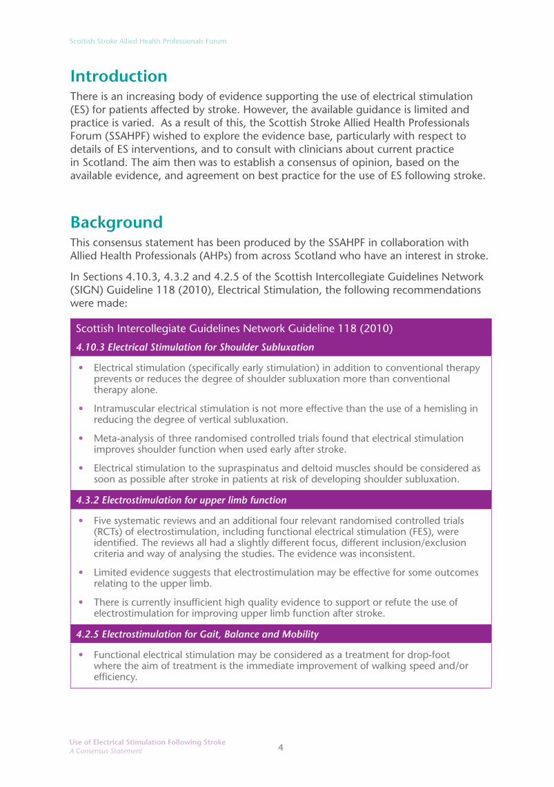
BackgroundThis consensus statement has been produced by the SSAHPF in collaboration with Allied Health Professionals (AHPs) from across Scotland who have an interest in stroke.
In Sections 4.10.3, 4.3.2 and 4.2.5 of the Scottish Intercollegiate Guidelines Network (SIGN) Guideline 118 (2010), Electrical Stimulation, the following recommendations were made:
Scottish Intercollegiate Guidelines Network Guideline 118 (2010)
4.10.3 Electrical Stimulation for Shoulder Subluxation
• Electrical stimulation (specifically early stimulation) in addition to conventional therapy prevents or reduces the degree of shoulder subluxation more than conventional therapy alone.
• Intramuscular electrical stimulation is not more effective than the use of a hemisling in reducing the degree of vertical subluxation.
• Meta-analysis of three randomised controlled trials found that electrical stimulation improves shoulder function when used early after stroke.
• Electrical stimulation to the supraspinatus and deltoid muscles should be considered as soon as possible after stroke in patients at risk of developing shoulder subluxation.
4.3.2 Electrostimulation for upper limb function
• Five systematic reviews and an additional four relevant randomised controlled trials (RCTs) of electrostimulation, including functional electrical stimulation (FES), were identified. The reviews all had a slightly different focus, different inclusion/exclusion criteria and way of analysing the studies. The evidence was inconsistent.
• Limited evidence suggests that electrostimulation may be effective for some outcomes relating to the upper limb.
• There is currently insufficient high quality evidence to support or refute the use of electrostimulation for improving upper limb function after stroke.
4.2.5 Electrostimulation for Gait, Balance and Mobility
• Functional electrical stimulation may be considered as a treatment for drop-foot where the aim of treatment is the immediate improvement of walking speed and/or efficiency.
Use of Electrical Stimulation Following StrokeA Consensus Statement
Scottish Stroke Allied Health Professionals Forum Scottish Stroke Allied Health Professionals Forum
5Use of Electrical Stimulation Following StrokeA Consensus Statement
In the Royal College of Physicians (RCP) National Clinical Guideline for Stroke (2012), Sections 6.19.2.1 and 6.13.1, the following recommendations were made:
Royal College of Physicians National Clinical Guideline for Stroke (2012)
6.19.2.1 Shoulder pain and subluxation
• Any patient who has developed, or is developing, shoulder subluxation should be considered for functional electrical stimulation of the supraspinatus and deltoid muscles.
6.13.1 Neuromuscular electrical stimulation (including FES)
• Functional electrical stimulation can be used for drop foot of central neurological origin provided normal arrangements are in place for clinical governance, consent and audit.
• Therapeutic electrical stimulation for treatment of the upper and lower limbs following stroke should only be used in the context of a clinical trial.
Following a Scotland-wide survey of the use of electrical stimulation by AHPs with stroke patients (Appendix A), the SSAHPF saw the need to establish a consensus of opinion and seek agreement on best practice for the use of ES following stroke across Scotland.
Intended ReadersThis statement is intended for healthcare professionals involved in the care of adults with a diagnosis of stroke. The statement defines adults as people who are 16 years and older.
Purpose of Consensus StatementA consensus statement may be defined as:
“A statement of the advised course of action in relation to a particular clinical topic, based on the collective views of a body of experts”
National Centre for Biotechnology Information, www.ncbi.nlm.nih.gov accessed 01 August 2014.
This consensus statement provides practical guidance for the use of ES following stroke. The contents of the document are based on the best available evidence at the time of publication and expert opinion.
In the context of this work, we have attempted to reflect the uncertainty and challenges faced by clinicians working with stroke patients with regard to the clinical application of ES. For this reason we have elected to exclude certain elements of treatment modalities from our consideration of the literature which either do not reflect mainstream practice in Scotland or which have already been well researched. These include percutaneous and implanted electrodes for the delivery of ES, which at the time of writing was primarily a research intervention, ES for facial weakness and swallowing difficulties, and FES as an orthosis to improve gait, as this has recently

Scottish Stroke Allied Health Professionals Forum
6Use of Electrical Stimulation Following StrokeA Consensus Statement
been the subject of an evidence note (Intercollegiate Stroke Working Party 2012) which summarised the evidence robustly.
It is acknowledged that the extent to which particular interventions are appropriate and can be implemented will be dependent on patients’ physical and mental health status at the time of assessment and treatment.
This statement provides practical guidance on the following:
• ES for motor control
• ES to the shoulder to help prevent shoulder subluxation
It will consider:
• parameters of treatment
• contraindications
• ES devices available
MethodologyThe membership of the SSAHPF was invited to take part in this work via their professional representatives. All interested parties at that time then had the opportunity to join the working group. The working group was formed in May 2013 and included physiotherapists, and an occupational therapist working in stroke who actively used and/or had an interest in ES. The final document was then subjected to critical appraisal by a group of expert readers. The methodology and rationale are presented in this section.
AimThe main aim of developing the consensus statement was to address the following questions:
• How is ES currently used for stroke patients in Scotland?
• What are the facilitators and barriers to using ES?
• What does the high level evidence conclude about the effectiveness of using ES?
• What parameters of ES intervention are used in the research studies?
• What parameters should ES devices be capable of delivering?

Use of Electrical Stimulation Following StrokeA Consensus Statement
Scottish Stroke Allied Health Professionals Forum Scottish Stroke Allied Health Professionals Forum
7Use of Electrical Stimulation Following StrokeA Consensus Statement
Data collection and literature reviewsWe divided the working group into subgroups to undertake the following tasks concurrently:
1. A national survey to obtain the status of ES use in Scotland (Appendix A, Page, summary on page)
2. A prospective audit to find the incidence of shoulder subluxation, the pattern of onset and the number of patients eligible for ES in one department (Appendix B, summary on page )
3. A prospective audit to find the extent to which ES is administered to prevent shoulder subluxation in a department equipped with ES equipment (Appendix C, summary on page )
4. A three stage literature review to find high quality evidence of the effectiveness of ES, any promising research findings from recent studies and examples of treatment parameters and device settings from the research trials (Appendix D, summary on page). We included evidence from the Cochrane Library and Database of Abstracts of Reviews of Effects (DARE), more recent ES trials from the Medline and CINAHL databases (2005 – February 2014), and ES studies with explicit treatment parameters and ES device settings.
5. A review of ES devices currently on the UK market.
6. Production of a glossary to clarify ES associated terminology (Appendix D).
Consensus statement reviewA review group was established comprising clinicians and academics from the United Kingdom. The consensus statement was e-mailed to the review group members in July 2014 with a feedback sheet for completion. Suggested revisions and clarifications were undertaken by the working group.
Structure of the consensus statementSummaries were produced and integrated into the consensus statement to provide clinicians with an efficient means to access the key points of the document in addition to having access to the details in the main body of the text.

Scottish Stroke Allied Health Professionals Forum
8Use of Electrical Stimulation Following StrokeA Consensus Statement
Electrical Stimulation Scotland-wide Survey November 2013
IntroductionThere is an increasing body of evidence supporting the use of ES for patients affected by stroke (SIGN 118, 2010, RCP, 2102). However, the practical guidance available to clinicians is limited and practice is varied. As a result of this, the SSAHPF wished to explore the evidence base, particularly with respect to details of ES interventions, and to consult with clinicians about current practice in Scotland. The aim then was to establish a consensus of opinion, based on the available evidence, and agreement on best practice for the use of ES following stroke.
A survey was conducted through stroke Managed Clinical Networks (MCNs) in all 14 Scottish Health Board areas, using Surveymonkey® via the SSAHPF. It was aimed at physiotherapists (PT), occupational therapists (OT) and orthotists with an interest in stroke. Each question in the survey referred to using electrical stimulation with stroke patients.
One hundred and thirty-seven AHPs responded, representing every Health Board in Scotland (53% PT, 31% OT and 16% orthotist).
ConclusionsFrom this survey, 28% of respondents used ES with their stroke patients.
It was clear to see from the results that the biggest barriers to using ES were:
• a lack of knowledge, skills and experience
• a lack of equipment
• funding and cost
The majority of respondents (85%) said they would consider using ES (or use it more) if these barriers could be overcome.
It was also interesting to note that those therapists who used ES were more confident in choosing appropriate patients than in selecting treatment parameters.
The results from this survey became the driver for the consensus statement. The full report of the survey may be viewed as Appendix A, Page 20.
Use of Electrical Stimulation Following StrokeA Consensus Statement
Scottish Stroke Allied Health Professionals Forum Scottish Stroke Allied Health Professionals Forum
9Use of Electrical Stimulation Following StrokeA Consensus Statement
An audit of shoulder subluxation in patients within NHS Greater Glasgow and Clyde stroke units. Examining incidence, predictive factors, and potential numbers eligible for electrical stimulation treatment. December 2013.
Author: Julie Macdonald, Stroke Specialist Physiotherapist
IntroductionGlenohumeral subluxation in stroke has been the subject of investigation for many years. SIGN 118, point 4.10.3 advocates electrical stimulation as an appropriate intervention for this. Treating patients with ES over the supraspinatus or posterior deltoid muscle has been advocated in the literature for prevention of shoulder subluxation. In view of this, an audit was undertaken in NHS Greater Glasgow and Clyde Stroke Units, with the exception of Inverclyde Royal Hospital, to answer the following key questions:
1. What percentage of new stroke patients within NHS Greater Glasgow and Clyde Stroke Units develop shoulder subluxation within a four week time frame and what is their time of onset?
2. Is it possible to predict patients at risk of developing shoulder subluxation using pre determined criteria and if so what criteria would be the most useful in determining risk?
3. What proportion of new stroke patients within a one month period would potentially benefit from ES as a treatment modality for shoulder subluxation?
MethodThe audit was conducted over a one month period over nine separate wards. Information was collected on all patients admitted with a confirmed stroke or being treated clinically as a new stroke (n=110). The incidence of subluxation was recorded for all patients from initial assessment until discharge or until the end of the audit. Further information was collected on a subset of patients deemed to be at risk of subluxation (n=39) using pre determined criteria based on a small literature search. Patients were excluded if they had a pre-existing shoulder subluxation from a stroke or other neurological condition on the newly affected side. Ten of those predicted as being at risk were ultimately excluded in the audit due to insufficient information.
Scottish Stroke Allied Health Professionals Forum
10Use of Electrical Stimulation Following StrokeA Consensus Statement
ResultsResults revealed a 14.5% incidence in shoulder subluxation in new stroke patients admitted within a four week period, with the majority developing the subluxation within the first week of stroke onset. Shoulder pain was also present in 34.58% of patients from initial assessment and increased to 57.21% by the end of the audit period with a trend towards greater numbers in the subluxation group. If patients presented with low tone, flaccidity or reduced voluntary movement, scoring ≤4 on the Brunnström Scale of Motor Recovery, then there appeared to be a trend towards developing shoulder subluxation. Impaired sensation, proprioception and haemorrhagic type of stroke appeared to be less predictive of shoulder subluxation, although the lack of sophisticated statistical testing does limit these results. Of the 110 new patients admitted across Glasgow and Clyde, during the audit phase, 35.4% (n=39) were identified as being at risk of developing shoulder subluxation and 48.2% of those (17.1% of the 110 patients identified) were deemed eligible for treatment using ES (see criteria in Appendix B2). However, it should be noted that only 41% (14% of the 110 patients reviewed) of those at risk were recorded as actually having developed shoulder subluxation. These numbers would be higher still if reduced sensation and ability to consent were not deemed, in this study, to be absolute contraindications.
ConclusionSelection of patients requires consideration in the context of staffing levels and caseload demands as there could be the potential to over treat. Development of a care pathway for this is recommended.
The full description of this audit may be viewed as Appendix B, Page.
Use of Electrical Stimulation Following StrokeA Consensus Statement
Scottish Stroke Allied Health Professionals Forum Scottish Stroke Allied Health Professionals Forum
11Use of Electrical Stimulation Following StrokeA Consensus Statement
Barriers to delivering electrical stimulation for the prevention of post-stroke shoulder subluxation in suitable patients: an audit of service provision at University Hospital, Ayr. June 2014 Author: Iain Larkin, Stroke Specialist Physiotherapist
IntroductionGlenohumeral subluxation is one of the common sequelae of acute stroke. Scottish Intercollegiate Guidelines Network (SIGN) 118 Section 4.10.3 recommends: “electrical stimulation to the supraspinatus and deltoid muscles should be considered as soon as possible after stroke in patients at risk of developing shoulder subluxation”.
To determine if this was being achieved an audit of local service delivery by physiotherapy staff in one general hospital site with acute and rehabilitation stroke wards was conducted. The following audit questions were generated:
1. What is the demand for electrical stimulation (ES) within patients following acute stroke within this hospital?
2. What is the average length of time from admission to provision of electrical stimulation in suitable patients?
3. In circumstances where patients suitable for electrical stimulation of the shoulder do not receive it, what are the reported reasons for this?
Methods and ResultsThe audit was conducted on weekdays for 35 days between 24th February and 11th April 2014. The audit sample included 84 patients. Based on the common predisposing factors for post-stroke shoulder subluxation from the literature, it was determined that on average 35% of patients with an initial diagnosis of stroke were suitable for consideration of ES. Of these patients 46% of them had no contraindications for ES. On average, 16% of patients with initial diagnosis of stroke were deemed suitable for ES to prevent shoulder subluxation. The mean time from admission to first use of ES was seven calendar days. It is not clear from the literature whether or not this was timely enough to prevent glenohumeral subluxation. On a daily basis, clinicians were asked to select the most appropriate barrier from a predetermined list (or give their own reason if this was more pertinent) for each patient who did not receive ES despite being indicated for this. The largest barriers to providing ES during the audit period were patients being unfit for treatment (48%) and a lack of time to deliver or assess patients for ES (29%). Other time related issues accounted for a further 11% of non-delivery of ES to appropriate patients. When there was lack of time to deliver ES, this always coincided with personnel shortage due to leave, meetings or training. It must be made clear that patients who did not receive ES due to any of the barriers highlighted above still received other forms of appropriate physiotherapy.

Scottish Stroke Allied Health Professionals Forum
12Use of Electrical Stimulation Following StrokeA Consensus Statement
ConclusionsPhysiotherapy staff may have little or no impact on the (largely medical) factors which make patients unfit for ES. However, time and resource related issues are factors that may be influenced. This may require change to working practices and/or staffing levels. The results of this audit reflect the local situation and may not mirror the barriers experienced elsewhere or at a national level. However, the audit process used here could easily be employed at other sites. An algorithm for screening patients for ES suitability is proposed, this may help to ensure equity of service.
The full description of this audit may be viewed as Appendix C, Page 46.
Use of Electrical Stimulation Following StrokeA Consensus Statement
Scottish Stroke Allied Health Professionals Forum Scottish Stroke Allied Health Professionals Forum
13Use of Electrical Stimulation Following StrokeA Consensus Statement
Summary of reported Electrical Stimulation treatment parameters.The literature review supporting this statement was conducted in two parts. Firstly, a search of the higher level evidence was performed within the Cochrane Library and the Database for Reviews and Dissemination (DARE) in order to identify the studies which had contributed to the evidence informing the stroke guidelines. Within these reviews, studies which described the ES intervention were consulted. The second part of the literature review involved identifying more recent studies which may not yet have been included in the systematic reviews but could potentially contribute evidence and rationale for specific treatment parameters.
We searched the Medline and CINAHL databases to January 2014 using search terms which included stroke, hemiplegia, shoulder subluxation, electrical stimulation, ES, EMS, FES, NMES,TES, TENS
The following sections contain summaries of the available evidence for parameters of ES treatment strategies for the recovery of motor control and shoulder subluxation reported in the full literature review which may be viewed as Appendix D, Page.
Summary of reported treatment parameters for the use of ES to restore motor controlThe lowest frequency possible required to achieve a fused muscle response may minimise patient intolerance and fatigue whilst maximising clinical benefits. Usual minimal stimulation frequency reported to achieve this is 12.5Hz (Scheffler and Chae 2007) although others recommend somewhere between 20-50Hz (de Kroon et. al. 2005, Sijuth 2008). This may vary depending on the limb treated, with lower frequencies required for the upper limb (Scheffler and Chae 2007).
Pulse amplitude (intensity) and pulse width (usually 200-400μsec) may be adjusted to achieve greater muscle force generation through recruitment of neurons increasingly further from the electrode (Scheffler and Chae 2007, Shu-Shyuan 2102). In a review of ES studies, de Kroon et. al. (2005) reported ranges of amplitudes from 0-100mA and as narrow as 30-40mA. Most used a fixed pulse duration of 200-300μsec. Recommendations suggest that the intensity frequency and pulse width of the electrical current should be adjusted in order to produce a visible contraction. Whilst there are broad areas of agreement, there is still considerable variability in application and the ultimate clinical decision may fall to the therapist with respect to the individual patient.
Common doses and duration of treatments delivered range from 30 minutes once per day to one hour three times per day for two weeks to three months (de Kroon et. al. 2005) although this was not substantiated or justified by the original authors. Hsu (2012) randomised 95 participants to dosages of 0, 15, 30, 60 minutes of ES five times per week for four weeks and reported improved recovery in the upper limb with more intensive ES. However, de Kroon et. al. (2005) suggested that the particular treatment parameters may not in fact be the critical element in the efficacy of ES

Scottish Stroke Allied Health Professionals Forum
14Use of Electrical Stimulation Following StrokeA Consensus Statement
within their study so it may be that individual patient treatment approaches may be sufficient.
Most authors do not justify choice of ramp times, stimulation wave forms or on/off cycle times so recommendations regarding these are difficult to make. However, Hsu (2012) reported cycles of 10 seconds on 10 seconds off in the first two weeks and 10 seconds on and 5 seconds off in the second two weeks. Descriptions of the common ES treatment parameters reported in the literature with recommended ranges are synthesised in Table 1 below.
Table 1: Electrical Stimulation Treatment Parameters reported for Motor Recovery
ES Parameter Description Reported treatment parameters
Consideration
Frequency Pulses per second (Hz) 12-35 Hz Needs to be sufficiently high to achieve a smooth contraction but not so high as to cause fatigue or a tetanic contraction
Pulse width Length of individual pulses (μsec)
200-400 μsec Increasing pulse width and/or amplitude increases the area and strength of activation. So these parameters may need to be adjusted with respect to one another.
Intensity Wave amplitude (mA) 0-100 mA
Duration Individual treatment time (minutes)
60 minutes Consider patient tolerance/compliance, response, feasibility and situation.
Dosage Number of treatments per day/week/total treatments
Daily 4 weeks
Ramp/ramp down
Time to reach chosen treatment intensity and then return to rest after selected stimulation
No recommendation can be made
2 seconds up and down
Adjust to obtain a comfortable near normally graded movement.
Stimulation wave form
May be Monophasic (repetitive unidirectional pulse) or Biphasic (pulses with current flow in both directions) which may be Symmetrical or Asymmetrical
No recommendation can be made
These parameters may affect skin irritation and patient comfort.

Use of Electrical Stimulation Following StrokeA Consensus Statement
Scottish Stroke Allied Health Professionals Forum Scottish Stroke Allied Health Professionals Forum
15Use of Electrical Stimulation Following StrokeA Consensus Statement
ES Parameter Description Reported treatment parameters
Consideration
On/off cycle time
Work/rest time (seconds) 10 seconds on /10 seconds off
Adjust in order to obtain balance between rest and fatigue.
Time since stroke
Acute or chronic phase No recommendation can be made
There is a lack of differentiation within studies and further research is required.
Additional considerations
+/- EMG trigger
Percutaneous/implantable electrodes
No recommendation can be made
These additional parameters may need to be delivered in a specialist setting.
Summary of reported treatment parameters for the use of ES for shoulder subluxation following strokeVarious authors have demonstrated that subluxation appears to occur during the flaccid period in the first three weeks post-stroke and is less likely to appear after the supraspinatus muscle has been shown to develop activity, recorded by EMG. It has also been suggested that once the shoulder joint capsule has been stretched, subluxation can persist, even if supraspinatus becomes active or spasticity develops (Wang, Chan et. al.. 2000, Linn, Granat et. al.. 1999, Chaco, Wolf 1971, Griffin 1986).
Although some authors have reported a positive trend towards a reduction in subluxation using ES, in more chronic stages this was not statistically significant and so cannot be recommended (Ada & Foongchomcheay 2002). However, evidence would suggest that the early application of ES post stroke, ideally within 48 hours, is important to attain positive results in preventing shoulder subluxation (Linn et. al. 1999, Fil et. al. 2011). Some authors demonstrated that a positive treatment effect was also maintained following ES application at longer time intervals of up to two to three weeks post stroke, albeit to a lesser extent (Faghri, Rodgers et al 1994, Chantraine, Baribeault et al 1999 and Wang, Chan et al, 2000). Church et al. (2006) recommended caution in the use of ES to the shoulder in patients with more severe paresis of the arm as they found a trend towards poorer recovery of motor control in these patients, but this would need to be balanced against the potential positive effect of reducing subluxation.
Ada & Foongchomcheay (2002) and Linn et. al. (1999) both observed a positive correlation between the development of subluxation and a lower score on Item Six of the Motor Assessment Scale (MAS). The former authors proposed that ES should be applied to those patients with a score of less than four and the latter, a score of less than or equal to two.
Kobayashi et. al. (1999) reported that supraspinatus activity alone is insufficient to maintain humeral alignment in the hemiplegic shoulder. Most authors stimulated
Scottish Stroke Allied Health Professionals Forum
16Use of Electrical Stimulation Following StrokeA Consensus Statement
supraspinatus in combination with posterior and/or middle deltoid. More recently, Manigandan et. al. (2014) reported that in addition to this, the stimulation of long head of biceps had an improved impact on reducing subluxation.
Although a wide variety of treatment frequencies were used, the range being 10-60Hz, 20-30Hz was most common. It is important that the choice of frequency is sufficient to elicit a motor response. In addition to frequency, pulse width and amplitude (intensity) can be adjusted to produce a visible, but comfortable or tolerable contraction. Common pulse widths ranged from 100-350μsec, but amplitudes were not often documented.
A large range of treatment durations and overall dosages is reported in the literature for the treatment of shoulder subluxation with ES. These ranged from five minutes to seven hours per day, five to seven days per week, for four to six weeks. Ada & Foongchomcheay (2002) suggested discontinuing treatment once patients scored more than four on Item Six of the MAS, whilst Linn et. al. (1999) suggested that a score of more than two may be sufficient.
Ada & Foongchomcheay (2002) synthesised the evidence available at the time to recommend 1 hour per day as a starting point, progressing to six hours per day. Most authors do not justify their choice of ramp up/down times, on:off ratios or waveform choice but do report a variety of applications.
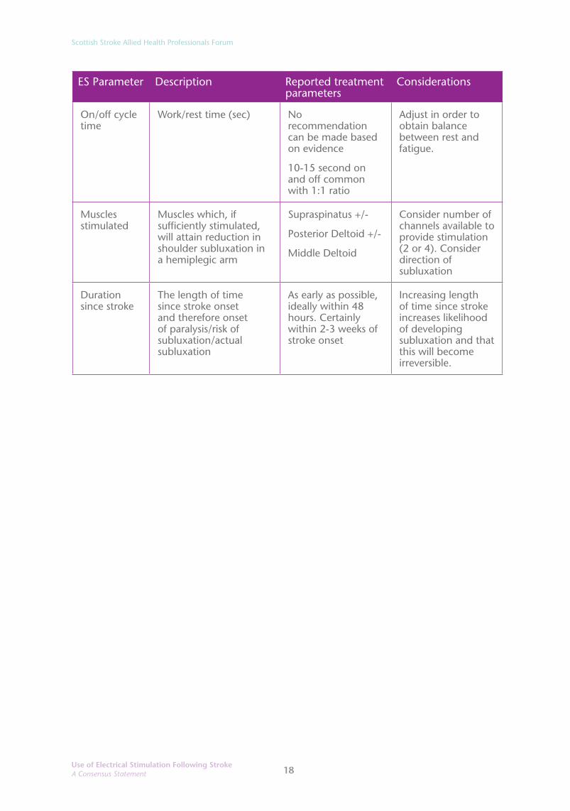
The common ES treatment parameters considered in the literature for use in shoulder subluxation with recommended ranges are synthesised in Table 2 below.

Use of Electrical Stimulation Following StrokeA Consensus Statement
Scottish Stroke Allied Health Professionals Forum Scottish Stroke Allied Health Professionals Forum
17Use of Electrical Stimulation Following StrokeA Consensus Statement
Table 2: Electrical stimulation treatment parameters reported for reduction of shoulder subluxation
ES Parameter Description Reported treatment parameters
Considerations
Frequency Pulses per second (Hz) 10-60Hz Needs to be sufficiently high to achieve a smooth contraction but not so high as to cause fatigue. Many studies aimed to produce tetanised contraction.
Pulse width Length of individual pulses (μsec)
100-350μs Increasing pulse width and/or amplitude increases the area and strength of activation. So these parameters may need to be adjusted with respect to one another.
Intensity Wave amplitude (mA) No recommendation can be made. Aim to produce painless contraction
Duration Individual treatment time (minutes)
5 minutes to 7 hours per session, generally 1 hour per day
Consider patient tolerance/compliance, response, feasibility and situation.
Dosage Number of treatments per day/week/total treatments
5-7 days per week
4-6 weeks or until sufficient voluntary muscle activity/reduction of subluxation without stimulation
Ramp/ramp down
Time to reach chosen treatment intensity and then return to rest after selected stimulation
No recommendation can be made
2-3 seconds up and down
Adjust to obtain a comfortable near normally graded movement.
Stimulation wave form
May be Monophasic (repetitive unidirectional pulse) or Biphasic (pulses with current flow in both directions) which may be Symmetrical or Asymmetrical
No recommendation can be made
These parameters may affect skin irritation and patient comfort
Scottish Stroke Allied Health Professionals Forum
18Use of Electrical Stimulation Following StrokeA Consensus Statement
ES Parameter Description Reported treatment parameters
Considerations
On/off cycle time
Work/rest time (sec) No recommendation can be made based on evidence
10-15 second on and off common with 1:1 ratio
Adjust in order to obtain balance between rest and fatigue.
Muscles stimulated
Muscles which, if sufficiently stimulated, will attain reduction in shoulder subluxation in a hemiplegic arm
Supraspinatus +/-
Posterior Deltoid +/-
Middle Deltoid
Consider number of channels available to provide stimulation (2 or 4). Consider direction of subluxation
Duration since stroke
The length of time since stroke onset and therefore onset of paralysis/risk of subluxation/actual subluxation
As early as possible, ideally within 48 hours. Certainly within 2-3 weeks of stroke onset
Increasing length of time since stroke increases likelihood of developing subluxation and that this will become irreversible.

Use of Electrical Stimulation Following StrokeA Consensus Statement
Scottish Stroke Allied Health Professionals Forum Scottish Stroke Allied Health Professionals Forum
19Use of Electrical Stimulation Following StrokeA Consensus Statement
Contraindications and cautions in the use of ESA number of contraindications and recommendations for caution are reported in the literature irrespective of the purpose for which ES is being applied. Clinical judgement around the care of individual patients should be applied in all cases where ES is being considered as a treatment. These are summarised in Table 3 below. This list may not be fully comprehensive and we recommend that clinicians read and observe manufacturers guidance provided with individual devices.
Table 3: Commonly reported contraindications, cautions and reasons to stop ES treatment.
Contraindications Cautions
Cardiac demand pacemaker Poor skin condition
Pregnancy, application directly over trunk Excessive tissue swelling
Poorly controlled epilepsy Excessive adipose tissue
Acute DVT (over site) DVT post anticoagulation
Complete peripheral nerve lesion Avoid stimulation over carotid sinus
Uncontrolled hyper/hypotension Avoid stimulation over thoracic region
Neoplastic tissue Avoid stimulation over phrenic nerve
Active infection Peripheral vascular disease
Implanted devices
Reasons to stop stimulation
Patient cannot tolerate (e.g. pain, agitation)
Electrode intolerance (skin irritation/allergy)
Benefits outweighed by practical difficulties
The full detail of this literature review may be viewed as Appendix D, Page.

Scottish Stroke Allied Health Professionals Forum
20Use of Electrical Stimulation Following StrokeA Consensus Statement
ES Device Review (2013)The Clinical Physics and Bioengineering Medical Device Unit (Software) Department at NHS Greater Glasgow and Clyde reviewed a short list of potential ES devices for home use in stroke rehabilitation. This list of devices is not exhaustive and they were chosen as those easily available and in clinical use at the time of writing.
The list of ES devices given to the Department by the ES working group was broken down into three sub-categories:
• Category 1: Multipurpose devices, readily available for home and clinical use (prices in 2013 ranged from £50 to £77)
• Category 2: Devices provided by physiotherapists, occupational therapists and orthotists, have stronger currents and more complex features (prices in 2013 ranged from £275 to £3500)
• Category 3: Devices specifically designed for FES (prices in 2013 ranged from £995 to over £20,000)
List of Requirements for an ES device for home use/self management:
1. Current ramp (at beginning and end)
2. Dual channels for stimulation
3. Easy to use
4. Inexpensive
5. Uses standard electrodes
6. Not for single person use only
7. Suitable for patients to use unsupervised
8. Easy to charge
9. Lightweight and compact
10. Easily cleaned
11. Range of frequency: 10 – 50Hz (normally 20 - 40Hz)
12. Pulse width: 100 –450μs
13. Input current: 10 – 15mA
14. Output current: 70 – 100mA
15. CE marked medical device

Use of Electrical Stimulation Following StrokeA Consensus Statement
Scottish Stroke Allied Health Professionals Forum Scottish Stroke Allied Health Professionals Forum
21Use of Electrical Stimulation Following StrokeA Consensus Statement
Device ReviewBased on the list of requirements it was decided to focus primarily on Category 1 devices due to the cost of the other systems and because therapists are keen to know the efficacy of using a cheaper alternative, particularly for self-management. A thorough review of all associated documentation for each device was performed to check if they matched the requirements. When the information required was not available the manufacturers were contacted direct.
Certification as a Medical DeviceTo check that each system is a registered, Conformité Européenne (CE) marked medical device, the manufacturers were asked to provide a declaration of conformity. The notified body on the declaration was then contacted to ensure that this was a valid certification. It was decided to focus on European Union (EU) based manufacturers.
SummaryThe three EU based manufactured devices reviewed all met the key requirements for home use in stroke rehabilitation and had documentation proving that they are certified medical devices.
The SSAHPF cannot endorse a particular product; however, this review should help inform therapists as in what to look for when purchasing ES devices, particularly for home use and self-management.
Scottish Stroke Allied Health Professionals Forum
22Use of Electrical Stimulation Following StrokeA Consensus Statement
Conclusion – use of Electrical Stimulation following strokeThere is an increasing body of evidence supporting the use of ES for patients affected by stroke. However, the practical guidance available is limited and clinical practice is varied.
In a survey carried out by the SSAHPF of 137 AHPs only 28% of respondents used ES as a treatment after stroke. The main barriers to the use of ES were lack of knowledge, skills and experience, lack of equipment and funding/cost issues. Two clinical audits based in Scotland identified an ongoing need for better management of the hemiplegic shoulder, using ES, by identifying the continuing incidence of shoulder subluxation and illustrating the challenges of delivering this in clinical practice.
An extensive literature review was undertaken and the main findings have been presented within this review. There is good quality evidence for the use of FES to enhance walking ability, however it was beyond the scope of this review to discuss suitable parameters for treatment using FES as it was felt that the most commonly used devices in the UK require specific expertise and training in their use by the manufacturers of that device.
There is good quality evidence supporting the recommendation that ES should be considered as soon as possible after stroke to prevent the development of subluxation of the shoulder. The practicalities of device and parameter selections are not so clear from the literature. This review attempts to synthesise the evidence from the most commonly cited robust trials to provide some guidance on this for the clinician and this is tabulated in the main body of this review, with supporting literature in the appendices.
The evidence for the use of ES to improve motor recovery is not as robust as that available for the prevention of the subluxed shoulder, but as new research is emerging this supports the current findings of a trend towards support for the use of ES. Again the particular parameters and dosages which clinicians should employ are not as clear. This review attempts to synthesise the evidence from the most commonly cited robust trials to provide some guidance on this for the clinician and this is tabulated in the main body of this review, with supporting literature in the appendices.
With the emergence of more robust trials and reviews, the evidence and recommendations within this review may develop and change. However, it is hoped that this consensus statement will assist the clinician in providing evidence-based therapy to the stroke population as well as stimulating discussion and ideas for future research.

Use of Electrical Stimulation Following StrokeA Consensus Statement
Scottish Stroke Allied Health Professionals Forum Scottish Stroke Allied Health Professionals Forum
23Use of Electrical Stimulation Following StrokeA Consensus Statement
Appendix A
Scottish Stroke Allied Health Professionals Forum (SSAHPF)
Electrical Stimulation SurveyNovember 2013
This survey was conducted Scotland-wide using Surveymonkey® and was aimed at physiotherapists (PT), occupational therapists (OT) and orthotists with an interest in stroke.
Each question in this survey referred to using electrical stimulation (ES) with stroke patients.
Results137 respondents representing every Health Board in Scotland
Question 1
Scottish Stroke Allied Health Professionals Forum
24Use of Electrical Stimulation Following StrokeA Consensus Statement
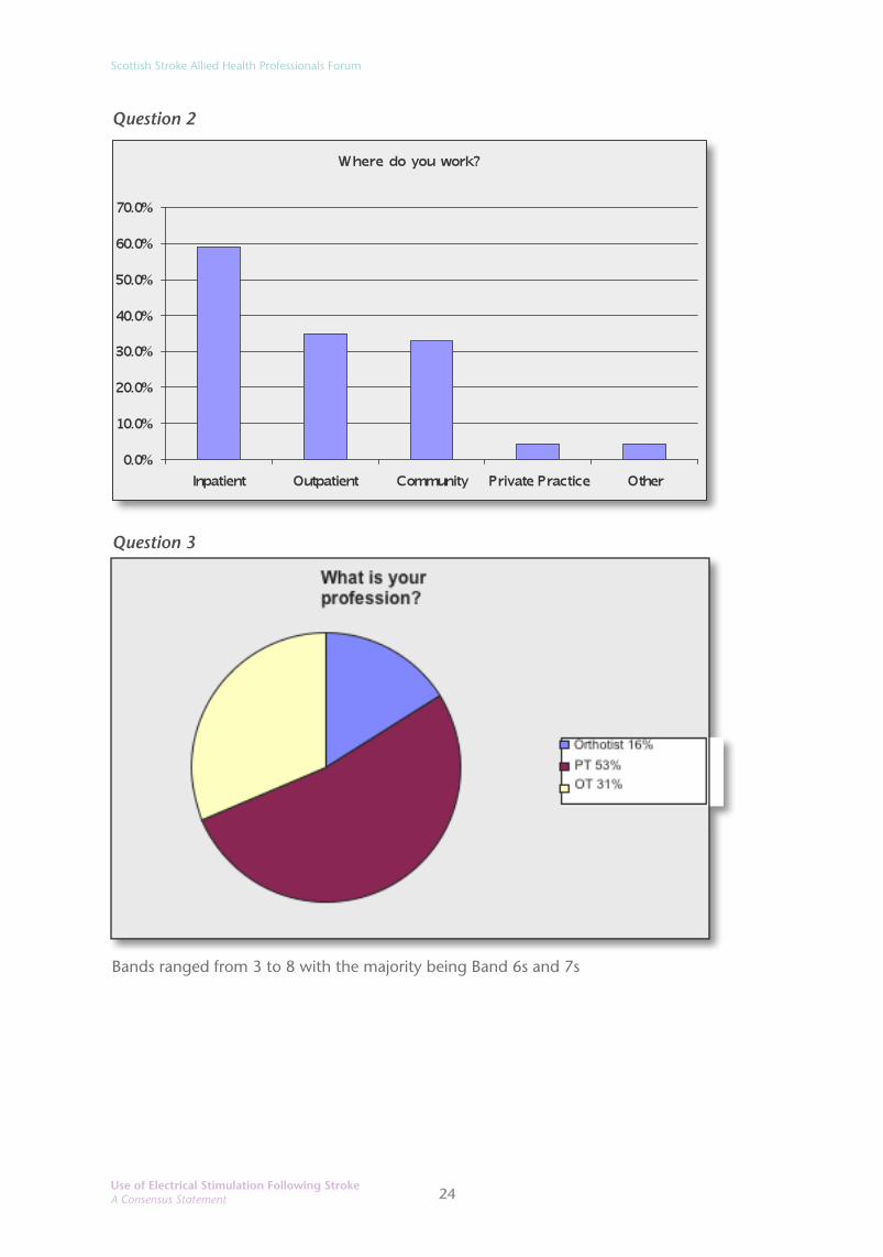
Question 2
Question 3
Bands ranged from 3 to 8 with the majority being Band 6s and 7s

Use of Electrical Stimulation Following StrokeA Consensus Statement
Scottish Stroke Allied Health Professionals Forum Scottish Stroke Allied Health Professionals Forum
25Use of Electrical Stimulation Following StrokeA Consensus Statement
Question 4
YES: Total=28% of those surveyed Breakdown by discipline: (23%, n=32 (44%) PT), (3%, n=4 (9.5%) OT), (2%, n=2 (9%) Orthotists)
NO: 72% Those who responded NO to Question 4 moved directly to Question 12 Those who responded YES continued to Question 5
Question 5Please state how often you use ES with stroke patients to treat the following? (1=rarely; 4=frequently)
Answer Options 1 2 3 4 Not used
Foot drop 6 8 7 4 15
Shoulder subluxation 4 7 3 6 17
Upper limb muscle recovery 6 7 13 3 12
Lower limb muscle recovery 6 5 6 2 17
Other 1 1 0 0 18
Other (please specify):- facial nerve palsy, scapular stability (lower/mid trapezius)
Of the 137 respondents only four (3%) frequently use ES for foot drop and only six (4%) frequently use ES for shoulder subluxation
Scottish Stroke Allied Health Professionals Forum
26Use of Electrical Stimulation Following StrokeA Consensus Statement
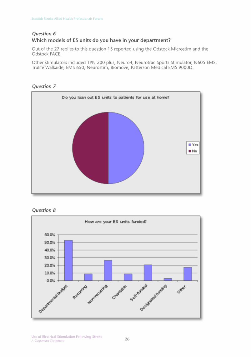
Question 6Which models of ES units do you have in your department?
Out of the 27 replies to this question 15 reported using the Odstock Microstim and the Odstock PACE.
Other stimulators included TPN 200 plus, Neuro4, Neurotrac Sports Stimulator, N605 EMS, Trulife Walkaide, EMS 650, Neurostim, Biomove, Patterson Medical EMS 9000D.
Question 7
Question 8

Use of Electrical Stimulation Following StrokeA Consensus Statement
Scottish Stroke Allied Health Professionals Forum Scottish Stroke Allied Health Professionals Forum
27Use of Electrical Stimulation Following StrokeA Consensus Statement
Question 9Please rate how effective you have personally found ES in treating the following? (1=not effective; 4=very effective)
Answer Options 1 2 3 4 Not used
Foot drop 1 5 6 12 16
Shoulder subluxation 3 7 5 3 17
Upper limb muscle recovery 1 8 12 5 12
Lower limb muscle recovery 1 4 10 1 18
Other 0 0 2 0 19
Other (please specify): facial nerve palsy
Question 10How confident do you feel in the following? (1=not confident; 4=very confident)
Answer Options 1 2 3 4
Selecting appropriate patients for ES 9 4 18 12
Selecting treatment parameters for ES 12 11 12 7
Question 11
From the 46 responses, 26 said they had not received any postgraduate training

Scottish Stroke Allied Health Professionals Forum
28Use of Electrical Stimulation Following StrokeA Consensus Statement
From the 22 comments:
• 13 had received training from Odstock (Salisbury)
• six had received in-service training
• two had attended the Walkaide Training Course (Trulife)
• one had received product training on MyGait (Ottobock)
Question 12
Question 13
85% responded YES

Use of Electrical Stimulation Following StrokeA Consensus Statement
Scottish Stroke Allied Health Professionals Forum Scottish Stroke Allied Health Professionals Forum
29Use of Electrical Stimulation Following StrokeA Consensus Statement
The editors acknowledge that in hindsight it would have been useful to have asked all the respondents if they had received any postgraduate training in ES rather than just those who use ES. The Editors also acknowledge some discrepancies in the numbers who continued to answer questions 4 to 11.
Please note – the SSAHPF does not endorse any particular ES product
Scottish Stroke Allied Health Professionals Forum
30Use of Electrical Stimulation Following StrokeA Consensus Statement
Appendix B
Scottish Stroke Allied Health Professionals Forum (SSAHPF)
An audit of shoulder subluxation in patients within NHS Greater Glasgow and Clyde (NHS GG&C) stroke units, examining incidence, predictive factors, and potential numbers eligible for Electrical Stimulation treatment. December 2013
Author: Julie Macdonald, Stroke Specialist Physiotherapist
In view of the evidence supporting the early use of electrical stimulation (ES) in the prevention of post-stroke shoulder subluxation (Ada and Foongchomcheay 2002), an audit was undertaken for the purpose of answering the following key questions;
1. What percentage of new stroke patients within NHS Greater Glasgow and Clyde Stroke Units develop shoulder subluxation within a four week time frame and what is the time of onset?
2. Is it possible to predict patients at risk of developing shoulder subluxation using pre determined criteria and if so what criteria would be the most useful in determining risk?
3. What proportion of new stroke patients within a one month period would potentially benefit from ES as a treatment modality for shoulder subluxation?
BackgroundGlenohumeral subluxation in stroke has been the subject of investigation for many years and yet there is still a paucity of literature on the matter. In order to prevent and treat it there must first be an understanding of what causes it. Several authors have attempted to examine the risks and it was theorised by Basmajian and Bazant (1959) that tonal changes both flaccidity and hypertonus could cause the scapula to be rotated downwards, thus causing a subluxing shoulder. However this has not been substantiated in the literature as Prevost et al (1987) and Culham et al (1995) found no correlation between the orientation of the scapula and glenohumeral subluxation.
Kumar et al (2010) suggest that risks for subluxation include complete loss of motor function and severity of arm impairment, as well as the absence of supraspinatus contraction, sensory impairment, loss of proprioception and haemorrhagic type of stroke. One of the difficulties in synthesising the data from their review is that there seems to be no standardised assessment method or tool evaluating the subluxation. Despite this, Kumar and colleagues confidently report that the complete loss of arm function in the hemiplegic side is a significant risk factor (Kumar et. al. 2010).

Use of Electrical Stimulation Following StrokeA Consensus Statement
Scottish Stroke Allied Health Professionals Forum Scottish Stroke Allied Health Professionals Forum
31Use of Electrical Stimulation Following StrokeA Consensus Statement
Based on these findings an audit tool was designed to explore whether these factors could be used to identify patients at risk of subluxation so as to provide selection criteria for treatment with ES (Appendix B1).
MethodsAn audit was carried out from week beginning Monday 25th November until Friday 20th December 2013. Data were collected over a four week period weekdays only. Data were collected in nine separate wards over seven different hospitals. These included acute stroke units at Glasgow Royal Infirmary, Western Infirmary and Institute of Neurological Sciences as well as acute/ rehabilitation wards in the Southern General Hospital, Royal Alexandria Hospital, Mansionhouse Unit, and acute and rehabilitation wards at Stobhill Hospital and Gartnavel General Hospital. Inverclyde Royal Hospital was omitted from the audit mainly due to its geographical location from the author’s base and time restriction in terms of training staff in the use of the audit tool.
Two separate audit sheets were used for collection purposes (See Appendix 1 and 2). The first audit tool was designed to capture information about all confirmed stroke cases and determine whether those patients actually developed a subluxation during the audit period. Subluxation was assessed clinically by palpation only as this was deemed to be the most clinically reproducible method. Patients were categorised from day one into “At risk” or “No risk” of subluxation using a predetermined state of criteria. For those deemed to be “at risk” then a second audit tool was completed following the patient’s journey until the end of the audit period noting specific risk factors and eligibility for treatment using ES. Data were collected only up until the point of discharge from therapy or until the end of the audit period.
Other additional information collated included the presence of shoulder pain, the presence of pre existing limitations in shoulder function of the affected arm and whether or not the affected arm was previously used for weight bearing when walking.
Inclusion CriteriaPatients were included in the audit if they were confirmed as having had a new stroke event or being treated clinically as a new stroke. Patients who presented with one or more of the following features were deemed to be “at risk” and subsequently included in the second part of the audit:
1. Flaccidity2. Low Tone3. Reduced Voluntary Movement (Should be considered to be insufficient in
maintaining Glenohumeral stability or could be Brunströmm Motor recovery Stage of 4 or less.
4. Sensory Impairment5. Proprioceptive Impairment6. Haemorrhagic Stroke

Scottish Stroke Allied Health Professionals Forum
32Use of Electrical Stimulation Following StrokeA Consensus Statement
Exclusion Criteria Patients were excluded if they had pre–existing shoulder subluxation from a stroke or other neurological condition on the newly affected side.
Data AnalysisData were analysed manually by the author.
ResultsOver a four week period a total of 110 patients were admitted through the stroke units across NHS GG&C with either a confirmed stroke or being treated clinically as a new stroke and were included in the first part of the audit. Twenty nine of these patients were transferred from their original hospital to one of the main rehabilitation units during this time. Of these only sixteen (14.5%) patients were identified as having developed shoulder subluxation during this period. All patients with a subluxation had been accurately identified as at risk using the predetermined criteria. No subluxation was detected in those not deemed to be at risk.
Using the predetermined criteria, 39 patients were identified as being at risk for shoulder subluxation. Seventeen (41%) of these developed subluxation during the audit period whilst 22 (59%) did not. The time of onset of subluxation during the audit period ranged from one to 21 days. An average time of onset cannot be given as patients were not followed up for an equal amount of time. There was also insufficient data from one of the nine hospitals and so data for this unit has been excluded. The majority however of those detected, did seem to present earlier as Figure 1 illustrates.
Figure 1 Time to onset of Subluxation
Use of Electrical Stimulation Following StrokeA Consensus Statement
Scottish Stroke Allied Health Professionals Forum Scottish Stroke Allied Health Professionals Forum
33Use of Electrical Stimulation Following StrokeA Consensus Statement
Of the 39 patients identified as at risk only a complete data set was available for 29 patients, 13 in the subluxation group and 16 in the non subluxation group. Using only the complete data set the mean time for follow up was 17 days in the subluxation group and 11 days in the non subluxation group suggesting there could still be scope for greater numbers of patients to develop subluxation at a later stage. However, these numbers would still appear to indicate that the overwhelming majority of patients present with subluxation within the first week of their hospital stay.
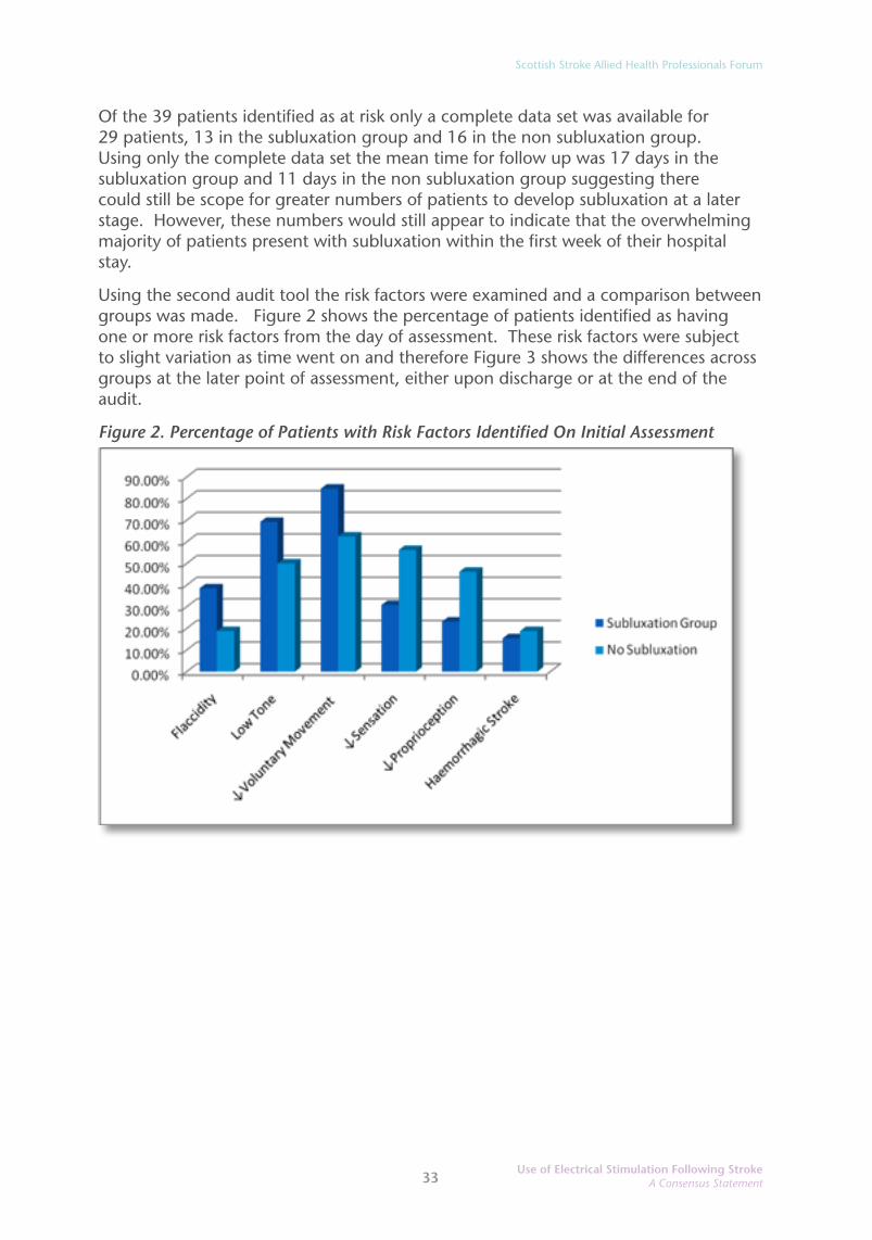
Using the second audit tool the risk factors were examined and a comparison between groups was made. Figure 2 shows the percentage of patients identified as having one or more risk factors from the day of assessment. These risk factors were subject to slight variation as time went on and therefore Figure 3 shows the differences across groups at the later point of assessment, either upon discharge or at the end of the audit.
Figure 2. Percentage of Patients with Risk Factors Identified On Initial Assessment

Scottish Stroke Allied Health Professionals Forum
34Use of Electrical Stimulation Following StrokeA Consensus Statement
Figure 3. Percentage of Patients with Risk Factors Identified at Final Assessment
Most significant is that every person with a subluxation at the end of the audit period had a degree of low tone (100%), but interestingly there was only a 19.2% difference between the subluxation group and the non subluxation group at initial assessment. Similarly there was only a 22.1% difference between the two groups for “Reduced Voluntary Movement” and a 19.5% difference between the two groups for flaccidity.
A higher proportion of patients who did not develop subluxation exhibited more decreased sensation and proprioception compared with those who did develop shoulder subluxation at initial assessment although by the end of the audit period there were little differences between these two groups for both these risk factors. Reduced sensation appeared to worsen in the subluxation group. It is not known whether any patients had an extension to their stroke which may have explained the increase. Alternatively it is plausible that as communication improves so might the detection of sensory symptoms although this cannot be substantiated. There appears to be little difference between the two groups for haemorrhagic type of stroke and in fact numbers of this type of stroke were low overall. No subluxation was detected in any of the patients deemed not to be at risk during the length of their stay in hospital. This suggests the criteria were robust enough in determining risk; however there does not appear to be any one single factor that determines which at risk patients go on to develop subluxation.

Use of Electrical Stimulation Following StrokeA Consensus Statement
Scottish Stroke Allied Health Professionals Forum Scottish Stroke Allied Health Professionals Forum
35Use of Electrical Stimulation Following StrokeA Consensus Statement
The incidence of shoulder pain was also recorded for those identified as at risk. From Table 1, 15.83% of patients who developed shoulder subluxation were assessed to have shoulder pain from day one of assessment. This increased to 38.46% by the end of the audit. In comparison 18.75% of patients who did not develop shoulder subluxation were assessed to have shoulder pain at the outset although this appeared to remain static to the end of the audit period. In the latter group two patients were noted to have pre-exisiting limitations in their upper limb function compared with zero in the subluxation group. Interestingly three patients in the no subluxation group used their upper limb for mobility purposes and again zero in the subluxation group. However data was missing for two of these patients on this section. It is reasonable to question whether pre existing limitation in upper limb function or previous weight bearing through the affected limb may have had any bearing on the presence of shoulder pain. Numbers however are too small to draw any real conclusions. An increase in shoulder pain in the subluxation group is suggestive of a trend towards the development of pain with subluxation although it cannot be said to be a cause given similar numbers of patients had shoulder pain at initial assessment who did not subsequently develop shoulder subluxation.
Table 1. Percentages of Patients with Shoulder Pain
Subluxation Group No Subluxation
Shoulder Pain
No shoulder
PainUnknown Shoulder
Pain
No shoulder
PainUnknown
Initial Assessment 15.83 76.92 7.7 18.75 81.25 0
Final Assessment 38.46 53.85 7.7 18.75 68.75 12.5
Patients’ suitability for ES was reported for all patients identified as at risk. Based on the completed assessments a total of 48.2% of patients (n=29) would have been deemed eligible for this treatment by meeting all necessary criteria. Of those eligible, 46.15 % (n=6) were in the subluxation group and 43.75 % (n= 7) were in the no subluxation group. By the end of the assessment period these figures rose to 61.54% (n= 8) and 50% (n= 8) in the subluxation and no subluxation groups respectively. It is worth mentioning that these figures would be higher still if the full data had been available on all 39 patients identified as at risk on audit form number one. In proportion to the overall number of new patients admitted this would therefore mean a maximum of 14.5% (n=16) patients would have been eligible for treatment with ES for prevention of shoulder subluxation.
Scottish Stroke Allied Health Professionals Forum
36Use of Electrical Stimulation Following StrokeA Consensus Statement
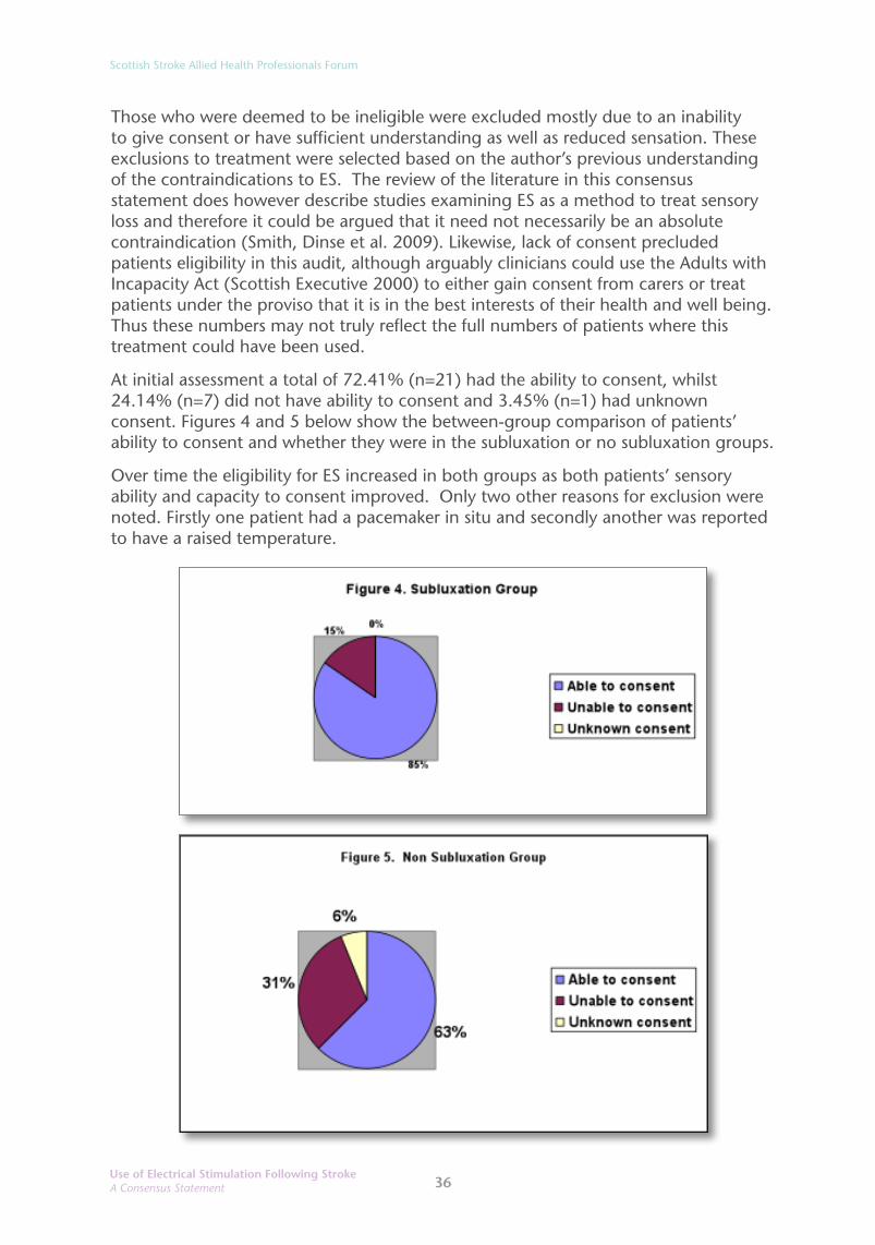
Those who were deemed to be ineligible were excluded mostly due to an inability to give consent or have sufficient understanding as well as reduced sensation. These exclusions to treatment were selected based on the author’s previous understanding of the contraindications to ES. The review of the literature in this consensus statement does however describe studies examining ES as a method to treat sensory loss and therefore it could be argued that it need not necessarily be an absolute contraindication (Smith, Dinse et al. 2009). Likewise, lack of consent precluded patients eligibility in this audit, although arguably clinicians could use the Adults with Incapacity Act (Scottish Executive 2000) to either gain consent from carers or treat patients under the proviso that it is in the best interests of their health and well being. Thus these numbers may not truly reflect the full numbers of patients where this treatment could have been used.
At initial assessment a total of 72.41% (n=21) had the ability to consent, whilst 24.14% (n=7) did not have ability to consent and 3.45% (n=1) had unknown consent. Figures 4 and 5 below show the between-group comparison of patients’ ability to consent and whether they were in the subluxation or no subluxation groups.
Over time the eligibility for ES increased in both groups as both patients’ sensory ability and capacity to consent improved. Only two other reasons for exclusion were noted. Firstly one patient had a pacemaker in situ and secondly another was reported to have a raised temperature.

Use of Electrical Stimulation Following StrokeA Consensus Statement
Scottish Stroke Allied Health Professionals Forum Scottish Stroke Allied Health Professionals Forum
37Use of Electrical Stimulation Following StrokeA Consensus Statement
DiscussionIn order to postulate how many stimulation units would be required to treat all eligible patients in the prevention of shoulder subluxation then a further audit or series of audits would be required capturing both new and existing patients. In Glasgow this should be done on a local basis to reflect the various care pathways and patient groups in each ward.
Based on the results of this audit shoulder subluxation remains a significant problem. The incidence reported here is 14.5%. This figure is relatively low as Kumar et al (2010) reported an incidence range of 31-81% in their review, whereas Paci et. al. (2007) reported incidences of 17-66%. Reasons why figures could be lower in this audit include; the short follow up periods for several patients, incomplete data from one of the stoke units and the possibility that a small degree of subluxation may be difficult to detect on palpation and thus may be insensitive to early changes.
Like previous studies this audit found the majority of patients developed subluxation early. Chaco & Wolf (1971) as cited in Linn et al (2013) clearly showed subluxation to occur in the first three weeks. Within this audit the majority of patients presented in the first week and thus any treatment intervention aimed at prevention must be considered and actioned as early as possible. Various treatment strategies have been suggested for the treatment of subluxation and hemiplegic shoulder pain such as the use of slings, supports and strapping, although the effectiveness of these remains questionable with various drawbacks (Linn et al 1999). Electrical Stimulation on the other hand has been reported as being significantly beneficial in the prevention and treatment of shoulder subluxation when used early after stroke (Ada and Foongchomcheay 2002). Selection of appropriate patients at the appropriate stage is therefore crucial.
One way to identify appropriate patients is to identify those most at risk. From the results of this audit low tone was present in 100% of patients who developed subluxation. However it was also present in 56.25% of patients who did not develop subluxation. One could argue again that the presence of subluxation could have been underestimated through palpation assessment and follow up time was limited. Reduced voluntary movement of ≤4 on the Brunnström Motor Recovery Scale was also highly correlated with the development of subluxation in this audit with 92.3% of patients reported as having this problem. These findings are similar to those reported by Chang et al (1995), Culham et al (1995) and Suethanapornkul (2008) as cited in Kumar et al (2010). However there was also a fairly significant proportion (62.5%) of patients who did not develop subluxation who were reported to having this. Unfortunately this audit did not reveal one specific factor or combination of factors that could differentiate between who might develop subluxation and who may not. One could argue therefore that all patients with either flaccidity, low tone around the shoulder and reduced voluntary movement should be considered at risk and subject therefore to intervention and those most at risk are likely to be those with a dense weakness with little or no movement. Certainly all the factors mentioned in combination seem robust enough to identify all at risk. Sensation, proprioception and haemorrhagic type of stroke were however found to be insensitive as independent factors with little between group variations.

Scottish Stroke Allied Health Professionals Forum
38Use of Electrical Stimulation Following StrokeA Consensus Statement
Limitations of this audit include the relatively small sample size and the possibility of bias as the results collated were examined and calculated independently by the author. This relates to the fact this was an unfunded piece of work and supported by physiotherapists during clinical time. For this reason background information was kept to a minimum for ease of completion and the audit period did not extend beyond one month. It is not therefore possible to make any assumptions about severity of stroke and degree of risk or add any statistical significance to these results.
Therapists should expect that almost 50% of patients with the risk factors mentioned could be suitable for ES. This would inevitably require time and the cost of appropriate stimulation units and electrodes. Patients’ prognoses and general health should undoubtedly play an integral part in the justification for such treatments and therefore therapists should use their own clinical reasoning and judgement to select appropriate individuals. It is likely that patients could be treated with this modality for a problem they might not have otherwise incurred as this audit suggests that at least 59% of patients identified as at risk did not develop shoulder subluxation during the audit. Therapists therefore must decide who should receive ES as part of their routine treatment. Staffing levels and overall caseload will inevitably be factors in this decision making process. Development of patient pathways is one way to streamline this process.
Conclusion Glenohumeral subluxation and shoulder pain are common sequelae of stroke. NHS Greater Glasgow and Clyde Stroke Units demonstrated a 14.5% incidence in subluxation in new stroke patients admitted within a four week period. The majority (88%) of those who developed subluxation did so within the first week of stroke onset. This highlights the importance of early identification and intervention. Shoulder pain was present in 34.58% of patients at initial assessment and this increased to 57.21% by the end of the audit period, with a trend towards greater numbers of patients with shoulder pain also having shoulder subluxation. Patients can be accurately predicted as potentially having shoulder subluxation if they present with low tone, flaccidity or reduced voluntary movement scoring ≤4 on the Brunnström Scale of Motor Recovery. Impaired sensation, proprioception and haemorrhagic type of stroke were less sensitive to detection of this. Treating patients with ES over the supraspinatus or posterior deltoid muscle has been advocated in the literature for prevention of shoulder subluxation. Of the 110 new patients admitted across Glasgow, during the audit phase, 35.4% (n=39) were identified as being at risk of developing shoulder subluxation and 48.2% of those (17.1% of the 110 patients identified) were deemed eligible for treatment using ES (see criteria in Appendix B2). However, it should be noted that only 41% (14% of the 110 patients reviewed) of those at risk were recorded as actually having developed shoulder subluxation. These numbers would be higher still if reduced sensation and ability to consent were not deemed to be absolute contraindications in this audit. Selection of patients requires further consideration in the context of staffing levels and caseload demands as there is potential to over treat. Development of a care pathway for this is recommended.

Use of Electrical Stimulation Following StrokeA Consensus Statement
Scottish Stroke Allied Health Professionals Forum Scottish Stroke Allied Health Professionals Forum
39Use of Electrical Stimulation Following StrokeA Consensus Statement
AcknowledgementsThe author greatly acknowledges the support of the Physiotherapy Staff of NHS Greater Glasgow and Clyde Stroke Units.
References (Audit 1)1. Ada, L. and Foongchomcheay, A. (2002). Efficacy of electrical stimulation in preventing
or reducing subluxation of the shoulder after stroke: A meta-analysis. Australian Journal of Physiotherapy. 48, pp257 -267.
2. Basmajian, J.V. and Bazant, F.J. (1959) Factors preventing downward dislocation of the adducted shoulder joint. Journal of Bone and Joint Surgery. American Volume 41. pp1182 -1186.
3. Culham, E., Noce, R. and Bagg, S. (1995) Shoulder complex position and glenohumeral subluxation in hemiplegia. Arch Phys Med Rehabil. 76. pp857-64.
4. Linn, S.L., Granat, M.H. and Kennedy, R. L. (1999) Prevention of Shoulder Subluxation After Stroke With Electrical Stimulation. Stroke. 30, pp963-968.
5. Kumar, P., Kassam, J., Denton, C., Taylor, E. and Chatterley, A. (2010) Risk factors for inferior shoulder subluxation in patients with stroke. Physical Therapy Reviews. 15 (1).pp3-11.
6. Paci, M., Nannetti, L., Taiti, P., Baccinnni, M., Rinaldi, L. (2007) Shoulder subluxation after stroke: relationships with pain and motor recovery. Physiother Res Int, 12, pp37-50. Scottish Executive. (2000). Explanatory notes to the adults with incapacity (Scotland) Act 2000. Edinburgh: HMSO.
7. Smith, P.S., Dinse, H.R., Kalisch, T., Johnson, M. and Walker- Batson, D., (2009) Effects of repetitive electrical stimulation to treat sensory loss in persons poststroke. Archives of Physical Medicine & Rehabilitation, 90(12), pp. 2108-2111.
Bibliography• Blennerhassett, J., Gyngell, K. and Crean, R. (2010) Reduced active control and passive range
at the shoulder increase risk of shoulder pain during inpatient rehabilitation post-stroke: an observational study. Australian Journal of Physiotherapy, 56. pp195-199.
• Paci, M. (2010) Risk factors for shoulder subluxation after stroke. Physical Therapy Reviews. 15. (2) pp119-120.
• Paci, M., Nannetti, L. and Rinaldi, L.A. (2005) Glenohumeral subluxation in hemiplegia : An overview. Journal of Rehabilitation Research and Development. 42 (4) pp557-568.
• Price, C.I.M. and Pandyan, A.D. (2008) Electrical stimulation for preventing and treating post-stroke shoulder pain. The Cochrane Library, Issue 4., John Wiley & Sons Ltd.
• Scottish Intercollegiate Guidelines Network (SIGN) Management of patients with stroke: Rehabilitation, prevention and management of complications, and discharge planning. Edinburgh: SIGN; 2010, (SIGN Guideline 118).
• Zorowitz, R.D. (2001) Recovery Patterns of Shoulder Subluxation After Stroke: A Six Month Follow-UP Study. Top Stroke Rehabil. 8. (2) pp1-9.

Scottish Stroke Allied Health Professionals Forum
40Use of Electrical Stimulation Following StrokeA Consensus Statement
Appendices
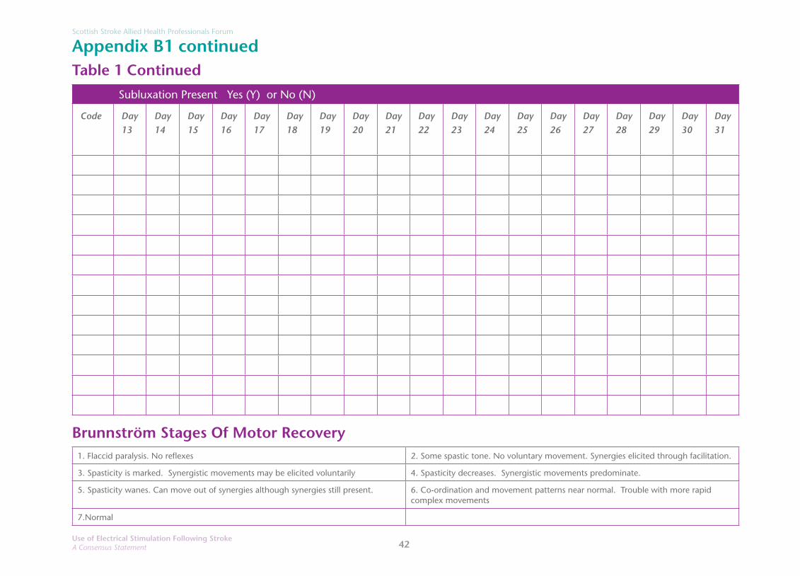
Appendix B1. Daily Record of Subluxation Audit Form , pages
Appendix B2. Patients At Risk Of Developing New Shoulder Subluxation - Daily Audit of Potential use for EMS

Use of Electrical Stimulation Following StrokeA Consensus Statement
Scottish Stroke Allied Health Professionals Forum
41
Appendix B1 Subluxation AuditMonth: _____________ Hospital _____________________ Ward_______________________
Please complete for all New Patients with a confirmed new stroke event.
Subluxation Present Yes (Y) or No (N)
CodeEg.
GRI.1
Name CHI: No
Date of Stroke
At Risk of Subluxation?
Y N
Day1
Day2
Day3
Day4
Day5
Day6
Day7
Day8
Day9
Day10
Day11
Day12
• Code : please use acute site code eg . GRI, WIG, SGH. When a patient is transferred across the city please continue to use the same code where possible.
• Patients will be deemed to be at risk of developing shoulder subluxation if they present with one or more of the following defecits in relation to the shoulder: 1. Flaccidity, 2. Low Tone, 3. Reduced Voluntary movement, 4. Sensory Impairment, 5. Proprioceptive Impairment, 6. Haemorrhagic Event
• Reduced Voluntary Movement should be considered to be insufficient in maintaining glenohumeral stability or could be Brunstromm’s Motor Recovery Stage of 4 or less.
Scottish Stroke Allied Health Professionals Forum
42Use of Electrical Stimulation Following StrokeA Consensus Statement
Appendix B1 continuedTable 1 Continued
Subluxation Present Yes (Y) or No (N)
Code Day13
Day14
Day15
Day16
Day17
Day18
Day19
Day20
Day21
Day22
Day23
Day24
Day25
Day26
Day27
Day28
Day29
Day30
Day31
Brunnström Stages Of Motor Recovery1. Flaccid paralysis. No reflexes 2. Some spastic tone. No voluntary movement. Synergies elicited through facilitation.
3. Spasticity is marked. Synergistic movements may be elicited voluntarily 4. Spasticity decreases. Synergistic movements predominate.
5. Spasticity wanes. Can move out of synergies although synergies still present. 6. Co-ordination and movement patterns near normal. Trouble with more rapid complex movements
7.Normal

Use of Electrical Stimulation Following StrokeA Consensus Statement
Scottish Stroke Allied Health Professionals Forum
43
Appendix B1 continued Table 1 Continued
Code Did the person develop new subluxation within the audit
period?Y N
Date of Discharge Discharge Location Further Physiotherapy Treatment Required For
Subluxation upon dischargeY N
Scottish Stroke Allied Health Professionals Forum
44Use of Electrical Stimulation Following StrokeA Consensus Statement
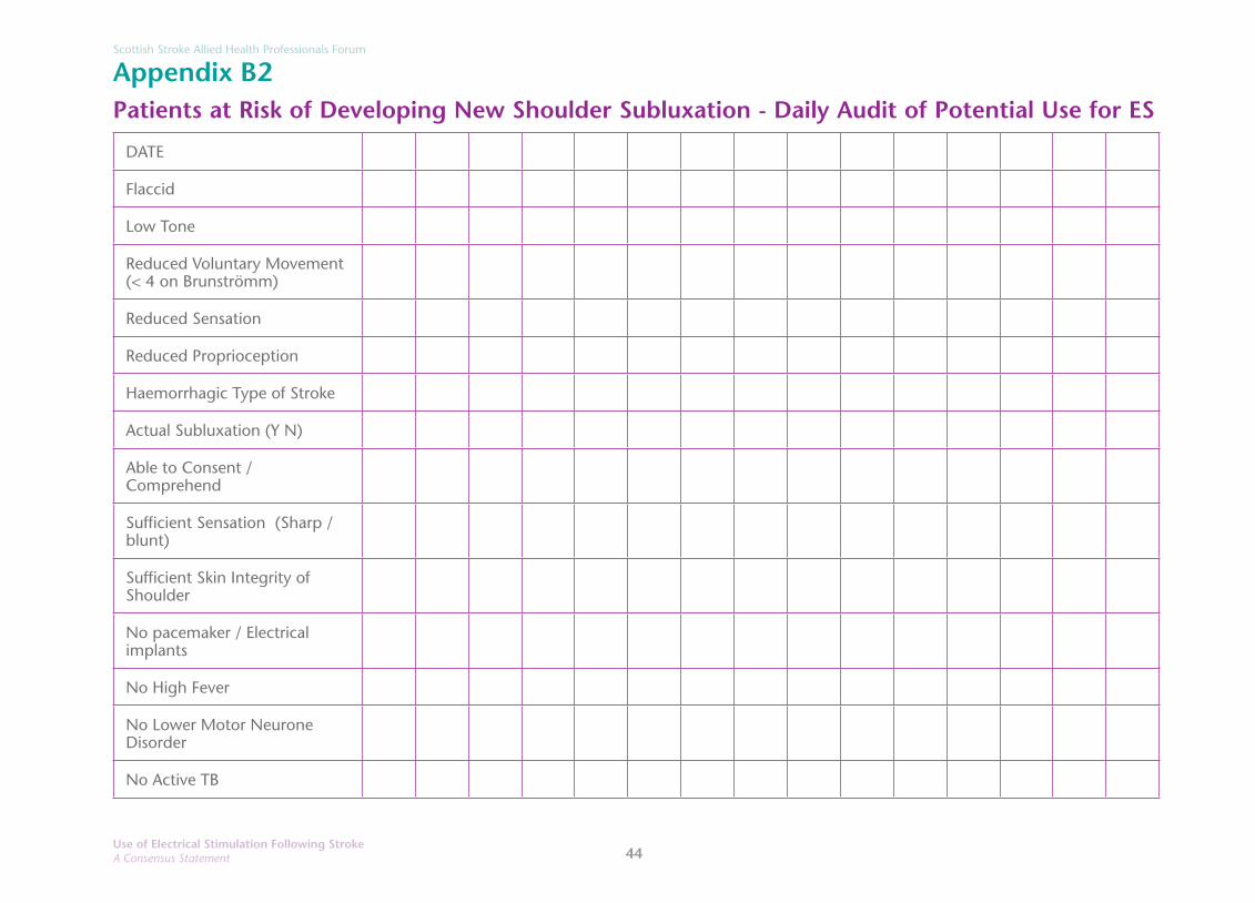
Appendix B2Patients at Risk of Developing New Shoulder Subluxation - Daily Audit of Potential Use for ES
DATE
Flaccid
Low Tone
Reduced Voluntary Movement (< 4 on Brunströmm)
Reduced Sensation
Reduced Proprioception
Haemorrhagic Type of Stroke
Actual Subluxation (Y N)
Able to Consent / Comprehend
Sufficient Sensation (Sharp / blunt)
Sufficient Skin Integrity of Shoulder
No pacemaker / Electrical implants
No High Fever
No Lower Motor Neurone Disorder
No Active TB

Use of Electrical Stimulation Following StrokeA Consensus Statement
Scottish Stroke Allied Health Professionals Forum
45
No Tumour or Suspected Tumour in Region of Shoulder
No Recent Haemorrhage in area
No Blood Clots
Appropriate For EMS (Y N )
Any Previous Limitation in Function of Affected limb (Y N)
Pre Mobility Status of affected U/L - Independent / Dependant
Shoulder Pain (Y N)
Scottish Stroke Allied Health Professionals Forum
46Use of Electrical Stimulation Following StrokeA Consensus Statement
Appendix CBarriers to delivering Electrical Stimulation for the prevention of post-stroke shoulder subluxation in suitable patients: An audit of service provision at University Hospital Ayr. Date 2/6/2014
Author: Iain Larkin, Stroke Specialist Physiotherapist
AbstractElectrical stimulation (ES) to the supraspinatus and deltoid muscles should be considered as soon as possible after stroke in patients at risk of developing shoulder subluxation (SIGN 118, 2010). To determine if this was being achieved an audit of local service delivery on one general hospital site with acute and rehabilitation stroke wards was conducted. The audit was conducted on weekdays for 35 days and included 84 patients. Based on the common predisposing factors for post-stroke shoulder subluxation from the literature, it was determined that on average 35% of patients with an initial diagnosis of stroke were suitable for consideration of ES. Of these patients 46% of them had no contraindications for ES. The average time from admission to first use of ES was seven calendar days. It is not clear from the literature whether this was timely enough to be effective in preventing glenohumeral subluxation. On a daily basis, in the case of patients who were deemed appropriate for ES but did not receive this, clinicians were asked to select the most appropriate barrier from a predetermined list or to give their own reason if this was more pertinent. The largest barriers to providing ES during the audit period were patients being unfit for treatment (48%) and a lack of time to deliver or assess patients for ES (29%). There was a strong link between lack of time to deliver ES and personnel shortage due to leave, meetings or training. Although medical unsuitability for ES may be an unavoidable barrier, time issues can be viewed as moveable barriers. This may require change to working practices or staffing levels. The results of this local audit may not reflect the prominent barriers in other areas or nationally. However, the audit process used herein could be employed at other sites. An algorithm for screening patients for ES suitability is proposed.
IntroductionGlenohumeral subluxation is one of the common sequelae of acute Stroke. Guideline 118 of the Scottish Intercollegiate Guidelines Network (SIGN) was published in June 2010. Within this guideline section 4.10.3 recommends: “electrical stimulation to the supraspinatus and deltoid muscles should be considered as soon as possible after stroke in patients at risk of developing shoulder subluxation”.
In November 2013 the Scottish Stroke Allied Health Professionals Forum (SSAHPF) conducted an electrical muscle stimulation survey. Only 4% of all respondents

Use of Electrical Stimulation Following StrokeA Consensus Statement
Scottish Stroke Allied Health Professionals Forum Scottish Stroke Allied Health Professionals Forum
47Use of Electrical Stimulation Following StrokeA Consensus Statement
reported using ES frequently to treat or prevent shoulder subluxation after stroke. Also, of those practitioners using ES with patients post-stroke only 16% of these were using it to treat the shoulder on a regular basis. Question 8 from the survey showed that of the sub-group of practitioners using ES for the shoulder (28% of respondents), 39% of these felt that it is of moderate benefit or better in treating shoulder subluxation and 66% felt that it is moderately to highly beneficial for upper limb activity. The discrepancy between the proportion of therapists who find ES useful and those who actually use it warrants exploration. Question 12 from the survey revealed that respondents cited several barriers to providing ES although no barrier had overwhelming prominence over any other. Barriers included lack of training or experience, several funding related issues, perceived lack of evidence, time and unrealistic patient expectations. Eighty-five percent of respondents stated that they would consider using ES more if these barriers were addressed.
This author of this paper conducted an audit in 2011 to determine the number of ES units which would be required to provide an ES service for patients following stroke with the aim of treating or preventing glenohumeral subluxation. Subsequently, a small number of ES units were purchased and training was provided. ES for the prevention of post-stroke shoulder subluxation then became an available treatment option using existing physiotherapy staff and funding. A limited (not dedicated) ES service was commenced using existing staff and funding. It was of interest to this author to determine whether patients who were suitable for post-stroke ES within our hospital consistently had the opportunity to receive this treatment. It was also important to establish what the specific barriers to provision of this treatment were in our hospital. Within this local general hospital there is an acute stroke ward and a stroke rehabilitation ward. The following audit questions were generated:
1. What is the demand for electrical stimulation to prevent shoulder subluxation within patients following acute stroke within this hospital?
2. What is the average length of time from admission to provision of electrical stimulation in patients suitable for this treatment modality?
3. In circumstances where patients suitable for electrical stimulation of the shoulder do not receive it, what are the reported reasons for this?
Risks factors for subluxationA systematic review by Kumar et al. (2010) highlighted complete loss of motor function, severe arm paralysis, impairment in proprioception, sensory loss and haemorrhagic stroke aetiology as risk factors for post stroke shoulder subluxation. Furthermore, Suethanapornkul et al. (2008) found that haemorrhagic type stroke, decreased proprioception and reduced Brunnström’s motor recovery stage of the hemiplegic shoulder score were significantly associated with increased risk of post stroke shoulder subluxation. Similarly, Huang et al. (2010) and Pong et al. (2009) found that a decreased score on Brunnström’s recovery stages is significantly associated with higher incidence of shoulder subluxation. This information can be used by clinicians to guide them in selecting patients suitable to be considered for ES.

Scottish Stroke Allied Health Professionals Forum
48Use of Electrical Stimulation Following StrokeA Consensus Statement
Indications and contraindications for ESPatients must have sufficient skin integrity to apply the electrodes. If ES is to be applied over the trunk the intended recipient must not be pregnant. Patients must not have a cardiac pacemaker, electrical implants or monitors. Recipients should be apyrexial. ES is not suitable to treat contracture or lower motor neurone disorder. ES should not be employed in close proximity to a tumour, recent haemorrhage or blood clots or in patients with active tuberculosis. Poorly controlled epilepsy also contraindicates ES. The capacity to comprehend and consent to the proposed treatment is desirable if not essential. ES should be used with caution if the ability to distinguish between sharp and blunt in the affected area is diminished. The contraindications and cautions detailed in this section were cited on a training course administrated by a commercial provider of ES units (Odstock Medical Limited, (OML) 2012).
MethodsAn audit tool was designed to capture the desired information. This tool was developed in collaboration with a physiotherapy colleague, Julie Macdonald and evolved from a tool designed for a previous audit undertaken in 2011. On a daily basis, all patients admitted to University Hospital Ayr with an initial diagnosis of acute stroke (within the first 12 weeks) were assessed against a set of criteria to determine their risk of developing subluxation and suitability for consideration for ES. These criteria are listed in Table 1 and justifications for them were discussed in the introduction. To ensure ES was targeted at patients with newly acquired upper limb motor or functional deficits, those patients with significant pre-morbid impairment of the affected upper limb were deemed unsuitable to be considered for ES.
Table 1: Criteria to determine who should be considered for ES
• Flaccid upper limb
• Reduced tone
• Insufficient active movement in the affected limb
• Reduced proprioception
• Haemorrhagic stroke
• No significant pre-morbid impairment in the affected upper limb
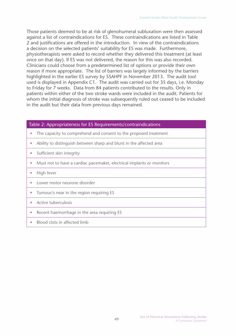
Use of Electrical Stimulation Following StrokeA Consensus Statement
Scottish Stroke Allied Health Professionals Forum Scottish Stroke Allied Health Professionals Forum
49Use of Electrical Stimulation Following StrokeA Consensus Statement
Those patients deemed to be at risk of glenohumeral subluxation were then assessed against a list of contraindications for ES. These contraindications are listed in Table 2 and justifications are offered in the introduction. In view of the contraindications a decision on the selected patients’ suitability for ES was made. Furthermore, physiotherapists were asked to record whether they delivered this treatment (at least once on that day). If ES was not delivered, the reason for this was also recorded. Clinicians could choose from a predetermined list of options or provide their own reason if more appropriate. The list of barriers was largely informed by the barriers highlighted in the earlier ES survey by SSAHPF in November 2013. The audit tool used is displayed in Appendix C1. The audit was carried out for 35 days, i.e. Monday to Friday for 7 weeks. Data from 84 patients contributed to the results. Only in patients within either of the two stroke wards were included in the audit. Patients for whom the initial diagnosis of stroke was subsequently ruled out ceased to be included in the audit but their data from previous days remained.
Table 2: Appropriateness for ES Requirements/contraindications
• The capacity to comprehend and consent to the proposed treatment
• Ability to distinguish between sharp and blunt in the affected area
• Sufficient skin integrity
• Must not to have a cardiac pacemaker, electrical implants or monitors
• High fever
• Lower motor neurone disorder
• Tumour/s near in the region requiring ES
• Active tuberculosis
• Recent haemorrhage in the area requiring ES
• Blood clots in affected limb
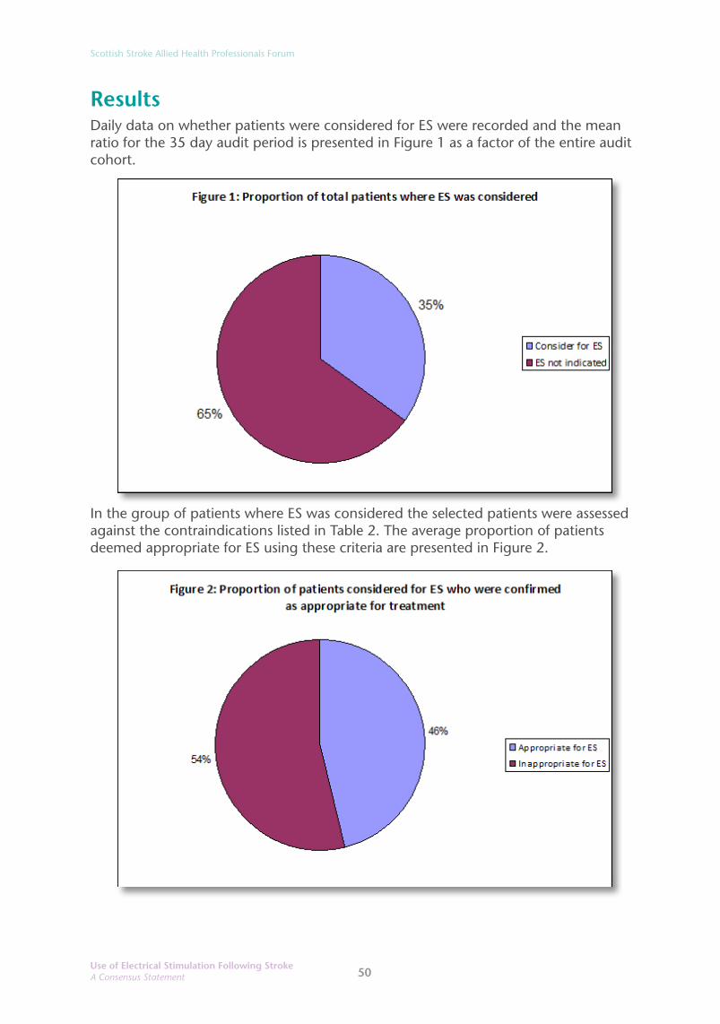
Scottish Stroke Allied Health Professionals Forum
50Use of Electrical Stimulation Following StrokeA Consensus Statement
ResultsDaily data on whether patients were considered for ES were recorded and the mean ratio for the 35 day audit period is presented in Figure 1 as a factor of the entire audit cohort.
In the group of patients where ES was considered the selected patients were assessed against the contraindications listed in Table 2. The average proportion of patients deemed appropriate for ES using these criteria are presented in Figure 2.

Use of Electrical Stimulation Following StrokeA Consensus Statement
Scottish Stroke Allied Health Professionals Forum Scottish Stroke Allied Health Professionals Forum
51Use of Electrical Stimulation Following StrokeA Consensus Statement
Otherwise stated, 16% of patients with an initial diagnosis of stroke were ultimately appropriate for ES. The average incidence of contraindications recorded over the audit period is shown in Figure 3.
Not all of the patients who were confirmed as appropriate for treatment with ES received the treatment. The average ratio of ES delivery versus non-delivery is shown in Figure 4.

Scottish Stroke Allied Health Professionals Forum
52Use of Electrical Stimulation Following StrokeA Consensus Statement
The barriers to delivery of ES were recorded daily in the patients who were deemed appropriate for ES but did not receive it. The average values are presented in Figure 5.
Three patients were reported to have shoulder subluxation; these were all diagnosed by non-invasive means by the physiotherapy staff. Radiological imaging was not used to confirm the diagnosis in any of the cases. Two out of the three patients were suitable for and received ES. Nevertheless, despite this they developed subluxation. The other patient was contraindicated for ES due to malignancy. In all three cases the patients were identified by the “subluxation risks” as being suitable for consideration for ES.
One of the aims of the current audit was to determine how long it took between admission and commencement of ES with suitable patients. In the three patients where the data were available, the mean time from admission to first ES treatment was seven calendar days, with a maximum of 10 days and a minimum of five days. Time was a barrier to earlier ES provision.
It may be spurious to draw any conclusions from such a small sub-group of patients.
DiscussionOn average, 35% of patients in the audit were suitable for consideration of ES. In contrast, a relatively small number of these patients (46%) were deemed appropriate for ES. Otherwise stated, 16% of the whole cohort was appropriate for ES. In the small number of patients in the current audit the most common contraindications for ES were reduced capacity to consent or comprehend ES and malignancy near the proposed site of ES.
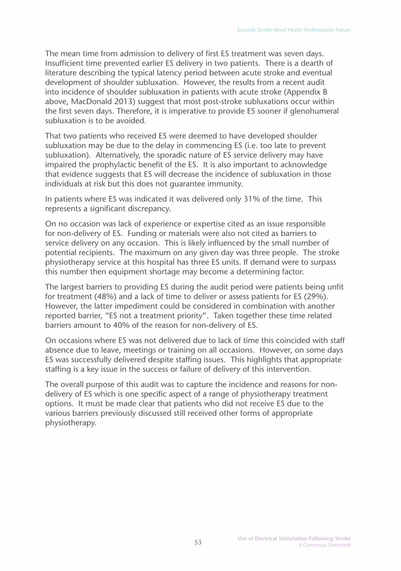
Use of Electrical Stimulation Following StrokeA Consensus Statement
Scottish Stroke Allied Health Professionals Forum Scottish Stroke Allied Health Professionals Forum
53Use of Electrical Stimulation Following StrokeA Consensus Statement
The mean time from admission to delivery of first ES treatment was seven days. Insufficient time prevented earlier ES delivery in two patients. There is a dearth of literature describing the typical latency period between acute stroke and eventual development of shoulder subluxation. However, the results from a recent audit into incidence of shoulder subluxation in patients with acute stroke (Appendix B above, MacDonald 2013) suggest that most post-stroke subluxations occur within the first seven days. Therefore, it is imperative to provide ES sooner if glenohumeral subluxation is to be avoided.
That two patients who received ES were deemed to have developed shoulder subluxation may be due to the delay in commencing ES (i.e. too late to prevent subluxation). Alternatively, the sporadic nature of ES service delivery may have impaired the prophylactic benefit of the ES. It is also important to acknowledge that evidence suggests that ES will decrease the incidence of subluxation in those individuals at risk but this does not guarantee immunity.
In patients where ES was indicated it was delivered only 31% of the time. This represents a significant discrepancy.
On no occasion was lack of experience or expertise cited as an issue responsible for non-delivery of ES. Funding or materials were also not cited as barriers to service delivery on any occasion. This is likely influenced by the small number of potential recipients. The maximum on any given day was three people. The stroke physiotherapy service at this hospital has three ES units. If demand were to surpass this number then equipment shortage may become a determining factor.
The largest barriers to providing ES during the audit period were patients being unfit for treatment (48%) and a lack of time to deliver or assess patients for ES (29%). However, the latter impediment could be considered in combination with another reported barrier, “ES not a treatment priority”. Taken together these time related barriers amount to 40% of the reason for non-delivery of ES.
On occasions where ES was not delivered due to lack of time this coincided with staff absence due to leave, meetings or training on all occasions. However, on some days ES was successfully delivered despite staffing issues. This highlights that appropriate staffing is a key issue in the success or failure of delivery of this intervention.
The overall purpose of this audit was to capture the incidence and reasons for non-delivery of ES which is one specific aspect of a range of physiotherapy treatment options. It must be made clear that patients who did not receive ES due to the various barriers previously discussed still received other forms of appropriate physiotherapy.

Scottish Stroke Allied Health Professionals Forum
54Use of Electrical Stimulation Following StrokeA Consensus Statement
Limitations of the current auditThe personnel responsible for collecting the data in this audit were also responsible for delivering the physiotherapy interventions. Therefore, it must be acknowledged that this method of data collection is vulnerable to observer bias. This must be put in the context of the small scale of the current audit and the lack of any funding. The methods employed remain the only practical way to collect the amount of data required efficiently.
A more useable version of the audit tool used is shown in Appendix C2. This follows the same pattern as the suggested algorithm. The reader is welcomed to use or adapt the audit tool to monitor their own service provision.
The audit included all patients admitted with an initial diagnosis of stroke. Many patients later had their diagnosis revised with stroke being ruled out. The current audit aimed to analyse the assessment process and time taken from admission to delivery of ES in appropriate patients. Therefore, it seems appropriate to include data from all patients with an initial diagnosis of stroke as this diagnosis triggered therapist activity.
The suitability and exclusion criteria for ES were suggested by a limited literature review and informed by this author’s own judgment and experience. The SSAHPF has outlined a far more extensive review of the literature on the subject. In particular, reduced capacity to consent/comprehend, or reduced sensation are not universally accepted as a contraindications for ES, though some training courses state that these exclusion criteria are considered relative not absolute exclusions (OML, 2012). On balance, it felt important to include these criteria in the audit. If these exclusions had not been applied there would have been significantly more patients deemed suitable for ES. This may have influenced the results of the current audit.
The audit was conducted in a small general hospital. It is likely to have been influenced by local factors such as staffing levels and the low number of patients indicated for ES mean that there are many potential causes of bias.
ConclusionDuring the audit period the related factors of time pressure and staffing were the most significant avoidable barriers to delivery of ES to patients in whom this treatment was indicated. Improvement in ES delivery is likely to be achieved by changes to working practices or staffing levels. The audit process has suggested that a simple algorithm could be developed to ensure all patients are given equal access to this treatment. The suggested algorithm follows below:

Use of Electrical Stimulation Following StrokeA Consensus Statement
Scottish Stroke Allied Health Professionals Forum Scottish Stroke Allied Health Professionals Forum
55Use of Electrical Stimulation Following StrokeA Consensus Statement
Selecting Patients Appropriate for Electrical Stimulation to Prevent Shoulder Subluxation after Acute Stroke
The author, Iain Larkin MSc, BSc (Hons.) permits the reprinting and use of this algorithm for clinical purposes.

Scottish Stroke Allied Health Professionals Forum
56Use of Electrical Stimulation Following StrokeA Consensus Statement
References (Audit 2)
1. Functional Electrical Stimulation for the Upper Limb. Training course delivered by Odstock Medical LTD (2012).
2. Huang, Y.-C., Liang, P.-J., Pong, Y.-P., Leong, C.-P., and Tseng, C.-H. (2010). Physical Findings and Sonography of Hemiplegic Shoulder in Patients After Acute Stroke During Rehabilitation. Journal of Rehabilitation Medicine. 42 (1): 21-26.
3. Kumar, P., Kassam, J., Denton, C., Taylor, E. and Chatterley, A. (2010). Risk factors for inferior shoulder subluxation in patients with stroke. Physical Therapy Reviews. 15 (1): 3-11.
4. Pong, Y.-P., Wang, L.-Y., Wang, L., Leong, C.-P., Huang, Y.-C. and Chen, Y.-K. (2009). Sonography of the shoulder in hemiplegic patients undergoing rehabilitation after a recent stroke. Journal of Clinical Ultrasound, 37: 199–205.
5. Scottish Intercollegiate Guidelines Network (SIGN) Management of patients with stroke: Rehabilitation, prevention and management of complications, and discharge planning. Edinburgh: SIGN; 2010, (SIGN Guideline 118).
6. Suethanapornkul, S., Srissa-an Kuptniratsaikul, P., Kuptniratsaikul, V., Uthensut, P., Dajpratha, P., and Wongwisethkam. (2008). Post Stroke Shoulder Subluxation and Shoulder Pain: A Cohort Multicenter Study. Journal of the Medical Associationof Thailand. 91 (12): 1885-93.

Use of Electrical Stimulation Following StrokeA Consensus Statement
Scottish Stroke Allied Health Professionals Forum
57
Appendix C1: Daily Post-Stroke Shoulder Electrical Stimulation (ES) AuditWard: Date: Assess all patients with diagnosis of Stroke against subluxation risk factors and ES inclusion criteria:
Patient CHIDate of Admission
Subl
uxat
ion
risks
Flaccid upper limbReduced Tone Insufficient active movement (<4 on Brunnström*)Reduced proprioceptionHemorrhagic StrokeWithout pre-morbid shoulder pain or impairment Consider for ES Subluxation (yes/no)
Ap
pro
pria
tene
ss fo
r ES
Able to consent/comprehendSufficient sensation (sharp/blunt)Sufficient skin integrityNo pacemaker, electrical implants/monitorsNo high feverNo lower motor neurone disorderNo tumour or suspected tumour in regionNo active TBNo recent haemorrhage in regionNo blood clots in affected limbAppropriate for ES (yes/no)Was patient given ES (yes/no)
Reas
ons
for
with
hold
ing
ES
Insufficient time to deliver ESInsufficient training to deliver ESES not a treatment priorityInsufficient equipment to deliver ESPatient not available due to tests etcPatient not fit/not for active treatmentAny other reason please state:Assessor initials
*See separate guidance on Brunnström’s motor recovery stages

Scottish Stroke Allied Health Professionals Forum
58Use of Electrical Stimulation Following StrokeA Consensus Statement
Appendix C2: Modified Daily Post-Stroke Shoulder Electrical Stimulation (ES) AuditWard: Date: Assess all patients with diagnosis of Stroke against subluxation risk factors and ES inclusion criteria:
Patient NamePatient CHI/identifierDate of Admission with Acute StrokeLacks pre-morbid shoulder pain or impairment (yes/no)
Subl
uxat
ion
risks
Flaccid upper limbReduced Tone Insufficient active movement Reduced proprioceptionHemorrhagic Stroke
Consider for ES (yes/no)Subluxation (yes/no)
Ap
pro
pria
tene
ss fo
r ES
Reduced ability to consent/comprehend/complyReduced sensation (sharp/blunt)Reduced skin integrityPacemaker, electrical implants/monitorsHigh feverLower motor neurone disorderTumour or suspected tumour in regionActive tuberculosisRecent haemorrhage in regionBlood clots in affected limbPoorly controlled epilepsy
Appropriate for ES (yes/no)Patient Consent to ES (yes/no)Was patient given ES (yes/no)
Reas
ons
for
with
hold
ing
ES
Insufficient time to deliver ESInsufficient training to deliver ESES not a treatment priorityInsufficient equipment to deliver ESPatient not available due to tests etcPatient not fit/not for active treatmentAny other reason please state
Assessor initials
The author, Iain Larkin MSc, BSc (Hons) permits the reprinting and use of this audit tool for clinical purposes.

Use of Electrical Stimulation Following StrokeA Consensus Statement
Scottish Stroke Allied Health Professionals Forum Scottish Stroke Allied Health Professionals Forum
59Use of Electrical Stimulation Following StrokeA Consensus Statement
Appendix DElectrical Stimulation used for treating stroke – a literature review of efficacy and application.
IntroductionElectrical stimulation (ES) in humans involves the application of electrical impulses to muscle or nerve either cutaneously through electrodes applied to the skin, or subcutaneously, through implanted electrodes. These methods have long been applied as elements of stroke rehabilitation as a means of eliciting and re-educating movement. It has also been used in other neurological conditions such as traumatic brain injury, spinal cord injury and multiple sclerosis. ES has also become established as having an orthotic function, known as Functional Electrical Stimulation (FES), mainly used in the treatment of dropped foot in hemiplegic gait. This can allow a more normal gait pattern through eliciting contraction of ankle dorsiflexors at the toe-off phase of walking reducing the tendency to trip.
A number of systematic literature reviews have been published and included in the Cochrane Library and in the Database of Abstracts of Reviews of Effects (DARE). There have been a number of subsequent clinical trials published in this area. This paper aims to:
1. Synthesise the published critical appraisals of these existing reviews as of July 2013 so as to summarise the high level evidence around the efficacy of ES as an intervention for eliciting and re-educating movement after stroke. The reviews fall broadly into two categories – application in the upper limb and in the lower limb and will be considered in this sequence.
2. Examine specific studies, published up to January 2014, which contributed most to the evidence and/or gave clear indications of the techniques used.
3. Consider the ES treatment parameters utilised in studies relating to the recovery of motor control and shoulder subluxation.
In the context of this work, we have attempted to reflect the uncertainty and challenges faced by clinicians working with stroke patients with regard to the clinical application of ES. For this reason we have elected to exclude certain elements of treatment modalities from our consideration of the literature which either do not reflect mainstream practice in Scotland or which have already been well researched. These include percutaneous and implanted electrodes for the delivery of ES, which at the current time of writing was primarily a research intervention, ES for facial weakness and swallowing difficulties, and FES as an orthosis to improve gait, as this has recently been the subject of an evidence note (Intercollegiate Stroke Working Party 2012) which summarised the evidence robustly.

Scottish Stroke Allied Health Professionals Forum
60Use of Electrical Stimulation Following StrokeA Consensus Statement
Literature reviews of ES applied to the upper limbElectrical stimulation for promoting recovery of movement or functional ability after stroke was considered in a Cochrane systematic review (Pomeroy, King et al. 2006). These authors included 24 randomised controlled trials (RCT), involving 888 participants, in their review from a pool of 2077 potential references. Their literature search was concluded in August 2005. They found that ES improved some aspects of functional motor ability and motor impairment. However there was also evidence which favoured no treatment control over ES for an aspect of functional motor activity. Benefits of ES over placebo treatment or no treatment control could have been as a result of delivering higher intensities of treatment to those in the ES groups. Although they uncovered positive trends in the effects of ES in improving motor ability, the authors recommended caution as there were small numbers of trials in each sub-analysis and small numbers of participants in those trials. There was also a great deal of variation in the doses of ES administered in the studies. Further research in this area was recommended.
Another Cochrane review considered ES for preventing and treating post-stroke shoulder pain (Price, Pandyan 2001). The authors found four RCTs of ES around the shoulder involving 170 participants which met the Cochrane inclusion criteria. They concluded that while ES could reduce muscle stiffness around the shoulder, there was insufficient evidence to recommend it for shoulder pain. ES was shown to reduce the severity of glenohumeral subluxation but there was no significant improvement in upper limb recovery that could be attributed to ES. Reported increased passive range of external rotation at the shoulder could have a secondary benefit of reducing the risk of pain, but this could not be verified.
A review of randomised, or quasi-randomised, studies reporting the efficacy of ES in preventing or reducing subluxation of the shoulder used late, or early, after stroke included a meta-analysis of the data from the studies reviewed (Ada, Foongchomcheay 2002). These authors only included studies using surface application of ES and the validity of the individual studies was assessed using the Physiotherapy Evidence Database (PEDro) scale to assign a quality score. Seven trials (n=183) were included in the review. The mean PEDro Score was 5.8 (4-9) in the early trials and 4.3 (4 – 5) in the later trials. The authors concluded that the evidence supported the use of ES early after stroke for the prevention of shoulder subluxation (WMD 6.5, 95% CI: 4.4, 8.6, p<0.001), but not late after stroke, for the reduction of established shoulder subluxation. DARE reported that the authors had used a robust methodology and considered the recommendations from this review to be broadly based on the evidence, although this evidence was sparse.
Studies examining “therapeutic” electrical stimulation (TES) intended to improve motor control and functional abilities of the upper extremity after stroke were included in a review which was subsequently critically appraised by DARE (de Kroon, van der Lee et al. 2002). Stimulation was delivered through surface electrodes only and was defined as TES. The stimulation was variously described as neuromuscular electrical stimulation, EMG-triggered electrical stimulation, positional feedback stimulation/training or transcutaneous electrical nerve stimulation; each applied by different devices. This highlights the issues around variation in terminology around the delivery of ES. Six RCTs involving 207 participants were included in this review

Use of Electrical Stimulation Following StrokeA Consensus Statement
Scottish Stroke Allied Health Professionals Forum Scottish Stroke Allied Health Professionals Forum
61Use of Electrical Stimulation Following StrokeA Consensus Statement
according to clear quality criteria. The studies were combined narratively according to whether there was a positive or negative effect on motor control and functional abilities. A pooled analysis was not performed owing to the heterogeneity of the included studies. The results suggested that ES had a positive effect on motor control, although it is not known if this improvement was clinically relevant. They concluded that no definite conclusions could be drawn concerning the effects of electrical stimulation on functional abilities in the upper limb after stroke and that no one particular method could be considered superior to any other. Their conclusion was replicated within the DARE appraisal of this review which stated that the review methodology overall was clear and that the authors' conclusions seemed appropriately cautious given the limitations of the data presented.
DARE considered a meta-analysis of RCTs designed to test the effectiveness of ES in improving functional use of the upper limb in stroke patients (Handy, Salinas et al. 2003). The original review included five RCTs (n=229) but detailed inclusion criteria and selection processes for the review were not provided. A small but statistically significant effect of ES on overall recovery of the upper limb after stroke was reported by the authors (d=0.21, 95% CI: 0.04, 0.38, p<0.05). They then concluded that ES produced a positive effect in patient recovery from stroke-related incidences. On considering this review, DARE stated that the authors' choice of a quantitative synthesis might not have been appropriate given the variation between studies. In addition, statistical homogeneity was not assessed and some studies contributed more than one effect size to the meta-analysis. Given these considerations and the small data set on which the review was based, DARE stated that the authors' conclusion, that ES produced functional benefit in upper limb recovery, should be viewed with some caution.
Reviews of ES applied to the lower limbThirty studies (n=1159) were included in a systematic review of surface-applied functional electrical stimulation (FES) for orthotic and therapeutic treatment of dropped foot after stroke which was subsequently appraised by DARE (Roche, Laighin et al. 2009). Studies employed a variety of designs and sample sizes ranged from one to 291 participants with 25 studies having sample sizes of less than 50. The authors concluded that FES can have a positive orthotic effect, particularly for gait speed and physiological cost index, in patients who were in the chronic stage of stroke recovery although evidence for a therapeutic effect was less conclusive. DARE stated that the reliability of these findings remained uncertain. The original authors did not suggest any implications for clinical practice or research but made many recommendations that included the need for a large RCT of surface FES versus ankle foot orthosis. They suggested that studies should have more standardisation of protocols, employ reliable quantitative outcome measures and be more collaborative between engineers, researchers, clinicians and users.
The efficacy and cost-effectiveness of FES as an orthosis to improve gait has recently been explored in an evidence note (Healthcare Improvement Scotland August 2012). This review concluded that there was moderate evidence that the use of FES could improve walking speed and reduce walking effort for patients with dropped foot as a result of a central nervous system lesion. However, this evidence came mainly from
Scottish Stroke Allied Health Professionals Forum
62Use of Electrical Stimulation Following StrokeA Consensus Statement
uncontrolled observational studies and recommended the execution of large RCTs to develop a more robust evidence base.
A “position review” was undertaken of the health and fitness benefits of FES-evoked leg exercise for spinal-cord-injured individuals (Hamzaid, Davis 2009). Thirty-two studies were included in the review (n=644, range 4-90), one RCT and 31 randomised or controlled studies. The authors concluded that FES-evoked leg exercise promoted certain health and fitness benefits such as changes to skeletal muscle morphology and biochemistry, increased aerobic fitness or positive metabolic responses, positive changes in indicators of functional exercise capacity and decreased depression levels. However, given the heterogeneity of the studies, lack of critical appraisal detail of the primary studies and small participant numbers in individual studies, DARE recommended that caution in the interpretation of the results of this review be exercised. This review, although not stroke specific, was included as potentially clinically relevant.
A recent systematic review of “cyclical electrical stimulation” was carried out to determine the effects of ES on strength and activity after stroke (Nascimento et.al. 2014). Sixteen trials representing 17 different comparisons met the inclusion criteria and the authors reported that ES may increase muscle strength by a standardised mean difference of 0.47 (95% CI 0.26-0.68). They also reported that ES increased physical activity (SMD 0.38, 95% CI 0.05-0.56). It should be noted that the quality of the trials was variable as they included some non-randomised controlled trials.
Summary of systematic reviewsThe high level evidence suggests that ES can reduce glenohumeral subluxation if used early after stroke; may have a positive influence on motor recovery but this cannot be verified; while FES may increase walking speed and reduce physiological cost when worn as an orthosis there was no strong evidence for a role in recovery of lower limb movement.
A number of more recent studies have been completed since the DARE reviews were published and these have provided examples of some other applications of ES after stroke. In addition, they provide further evidence behind ES for the aforementioned applications of upper limb recovery and the prevention of glenohumeral subluxation and shoulder pain. It is beyond the scope of this consensus statement to fully critically appraise this research and they are summarised here under appropriate headings for information:
Improving motor and functional recoveryFour RCTs compared ES to a control condition. Two of the studies included electromyography (EMG)-triggered ES. In one study, 31 participants with subacute and chronic stroke were randomised to one of three groups: EMG-triggered ES, passive ES or sham stimulation (Boyaci, Topuz et al. 2013). The control group (n=10) received the same duration of intervention but with placebo ES. The EMG-triggered group was found to have significantly improved active range of motion, grip strength
Use of Electrical Stimulation Following StrokeA Consensus Statement
Scottish Stroke Allied Health Professionals Forum Scottish Stroke Allied Health Professionals Forum
63Use of Electrical Stimulation Following StrokeA Consensus Statement
and functional ability than the other two groups (p<0.05). In the other EMG-triggered ES study, 33 participants in the acute phase of stroke with upper limb strength below grade three were randomised to the EMG-triggered ES plus usual therapy group, or to the usual therapy group (Dorsch, Ada et al. 2013). In contrast to the first study, there were no differences found between the groups in relation to strength or activity. Although the differences in findings between these two studies could be attributable to the participants being in the acute or subacute phase of stroke, an appraisal of the methodology of these studies is recommended to identify other sources of bias.
A RCT of participants in the sub-acute phase of stroke (six months post-stroke) compared self-triggered ES using an accelerometer during bilateral activities to placebo stimulation (Chan, Tong et al. 2009). After 15 treatment sessions, the ES group (n=10) was found to have achieved significantly greater upper limb movement than the control group (n=10) on three outcome measures (p<0.05). Another RCT of participants in the subacute phase of stroke found no statistically significant differences in outcome between a group receiving ES and stretch arm positioning (n=23) and a control group receiving sham ES and sham stretch positioning (n=23) (de Jong, Dijkstra et al. 2013). Both studies included participants at the subacute phase of stroke but other factors may have contributed to the difference of findings. The second study included participants with poor initial arm control and applied passive ES to non-active participants. Again, there are differences between the studies which may have influenced outcome and a more detailed appraisal is recommended.
Preventing glenohumeral subluxationA RCT of shoulder subluxation prevention compared Bobath techniques in combination with ES to a control group receiving Bobath techniques only (Fil, Armutlu et al. 2011). Forty-eight participants with acute stroke were randomised into the two groups (n=24 in each group). In addition to the Bobath exercises, the experimental group received ES to the supraspinatus and to the mid and posterior regions of the deltoid muscle. The published paper did not state the duration or frequency of interventions but found that nine participants in the control group developed subluxation whereas, no participants in the experimental group experienced subluxation (p<0.05) suggesting that the combined Bobath and ES intervention was statistically significantly more effective for preventing subluxation than Bobath techniques alone.
A more recent non-randomised trial compared the ES of supraspinatus and posterior deltoid to ES of the long head of biceps, supraspinatus and posterior deltoid on reducing subluxation (Manigandan, Ganesh et al. 2014). Twenty-four participants with stroke were assigned consecutively to each group and intervention was provided over a five week period. The authors reported that although improvement was detected in both groups, the group with ES to the long head of biceps experienced a significantly greater improvement of subluxation. To date, the more robust evidence has related to subluxation prevention (Price, Pandyan 2001) but the findings of this study suggest that ES could reduce subluxation. However, the non-randomised design is a key source of potential bias.
Scottish Stroke Allied Health Professionals Forum
64Use of Electrical Stimulation Following StrokeA Consensus Statement
The following sections refer to published material relating to other potential clinical applications of ES in the management of spasticity, sensory loss and unilateral neglect. We have not included these areas in the overall guidance and have restricted this to management of shoulder subluxation and recovery of motor control.
Reducing spasticity in the affected limb using ES in combination with botulinum toxin or physiotherapyElectrical stimulation combined with either botulinum toxin to spastic muscles or physiotherapy to reduce spasticity after stroke has been the topic of several small studies. The evidence from current studies is not strong enough to recommend either combination (Bakheit 2013) despite there being pathophysiological reasons to support their effectiveness (Wilkenfeld 2013). Stretching for stroke related wrist flexor spasticity was compared with stretching and ES of the wrist extensors in a RCT of 44 (n=22 in each group) participants (Sahin, Ugurlu et al. 2012). Both groups demonstrated a significant improvement of spasticity after one month of intervention, with the ES group demonstrating a greater improvement in spasticity (p=0.001), wrist movement and functional ability (p=0.001). Although providing a positive result, the authors acknowledged that the main limitation of this study was that the outcome was measured immediately after treatment, making the longer term outcome uncertain.
Two RCTs of ES combined with botulinum toxin injection for lower limb spasticity after stroke found that the combination treatment was more effective than botulinum toxin alone (Hesse, Jahnke et al. 1995) or physiotherapy alone (p<0.05) (Johnson, Burridge et al. 2004). However, the sample sizes were very small with only five and 10 participants in the experimental groups of each study and the replication of these results with fully powered studies is required before a more conclusive conclusion of effectiveness can be reached.
The treatment of chronic upper limb spasticity after stroke using the ES and botulinum toxin injection combination has also been investigated and found to have uncertain effectiveness. A RCT of four groups; 1) ES and botulinum toxin, 2) botulinum toxin only, 3) ES and placebo, and 4) placebo only, found that the combined treatment (group one) was more effective than botulinum toxin alone, or placebo, but not ES alone (p<0.01) (Hesse, Reiter et al. 1998). This may suggest that combining ES with botulinum toxin is no more effective that simply using ES. However, this was a very small study with only six participants in each group and the general conclusion that ES and botulinum toxin reduces chronic upper limb spasticity more than the individual treatments cannot be reached. Another RCT comparing the combination of ES and botulinum toxin with ES combined with extracorporeal shockwave therapy (ESWT) for upper limb spasticity after stroke found that the ESWT and botulinum toxin group (n=16) experienced a statistically significant reduction on upper limb spasticity than the ES and botulinum toxin group (n=16) (Santamato, Notarnicola et al. 2013). This was also a small study but the findings of both studies emphasise the uncertain effectiveness of ES and botulinum toxin for upper limb spasticity after stroke.

Use of Electrical Stimulation Following StrokeA Consensus Statement
Scottish Stroke Allied Health Professionals Forum Scottish Stroke Allied Health Professionals Forum
65Use of Electrical Stimulation Following StrokeA Consensus Statement
Reducing the impact of sensory loss in the affected upper limbTwo studies investigated ES to treat sensory loss. In one study, a pre-experimental one-group design (n=4) was used to investigate the potential of ES to improve sensory discrimination of the involved hand in patients six months post-stroke with sensory loss (Smith, Dinse et al. 2009). The ES intervention was applied for 90 minutes, four days per week for six weeks. Sensory discrimination was found to have improved in all of the participants and was maintained at four weeks follow-up. In the second study, a case study was used to investigate the potential for ES feedback to improve pinch pressure control (Kita, Takeda et al. 2011). A participant with stroke and sensory loss affecting grip strength awareness used the ES system where ES was modulated by the pinch pressure applied. After two months of intervention, the authors stated that the participant was able to maintain a stable grip pressure without the ES system. Although it is not possible to determine the effectiveness of these interventions for treating sensory loss with ES due to the limitations in study design, the two studies could contribute to the justification for undertaking future RCTs.
Alleviation of unilateral neglectTwo studies investigated the impact of left hand ES in combination with scanning training to improve unilateral neglect (UN). In the earlier study, a number of experiments were conducted with nine participants with UN resulting from stroke (Eskes, Butler 2006). The experiments included ES to their neglected hand to produce digit extension in combination with scanning tasks of near and far space. The results were variable and the authors concluded that their findings supported further investigations. The second study was an RCT published in 2009 comparing ES with scanning to scanning with sham ES (Polanowska, Seniów et al. 2009). Forty participants between two and 12 weeks post-right hemisphere stroke with UN were randomised into the two groups (n=20 in each group). The 45 minute interventions were provided five days per week for four weeks. After the interventions, the experimental group was found to have a statistically significantly greater scanning score than the control group (p=0.01) suggesting the effectiveness of combined scanning and ES of the involved hand on the alleviation of UN.
Efficacy of Electrical Stimulation in Stroke - Conclusion The continued research into ES for a variety of applications suggests that the interest in the use of ES for treatment after stroke remains strong. However, the poor methodological quality and small sample sizes of the reported RCTs limit any additional conclusions of effectiveness of ES in both the applications reviewed by DARE and to the other specific applications. In light of the limited evidence, other non-systematic reviews were also considered, in order to gain information about specific ES treatment parameters which could guide clinicians. These are discussed below.
Scottish Stroke Allied Health Professionals Forum
66Use of Electrical Stimulation Following StrokeA Consensus Statement
Electrical Stimulation Treatment ParametersThe synthesis of the high level evidence identified three main areas in which ES or FES is used – as an orthosis to aid foot drop and improve gait; as a therapeutic modality to aid motor recovery of the upper or lower limb; and to reduce glenohumeral joint subluxation after stroke. This section will present elements of therapeutic ES interventions reported in the literature with a view to providing some clarity on potentially useful treatment regimens.
Not all ES devices will allow the delivery and adjustment of all the parameters considered below and may provide a limited choice of a series of set programmes of stimulation and channels for delivery.
Reported electrical stimulation treatment parameters used to aid motor recoveryA wide variety of stimulation parameters have been reported in the published literature.
Electrical stimulation has been described as:
“...a waveform of electrical current characterised by stimulus frequency, amplitude and pulse width” (Sheffler, Chae 2007) p.563
It has been suggested in a review of ES, that it is the adjustment of these parameters which determines the nature of the evoked action potential response and thus impacts on the amount of muscle force generated as well as patient comfort and safety (de Kroon, Ijzerman et al. 2005b). In this review, the authors suggest that different combinations of settings produce differing effects on the radial nerve. Many devices allow for adjustment of pulse frequency and use a constant frequency train of equally spaced pulses at a set frequency.
FrequencyA variety of pulse frequencies have been described in the literature, mostly ranging between 20-50Hz, although frequencies higher than 35Hz may produce a tetanised contraction (Boyaci, Topuz et al. 2013, Chan, Tong et al. 2009, de Kroon, Ijzerman et al. 2005b). The minimum frequency reported to achieve a fused muscle response has been reported as 12.5Hz, with the further suggestion that 12-16Hz and 18-25Hz may be ideal stimulation frequencies for the upper and lower limbs respectively (Sheffler, Chae 2007).
Artificially stimulated muscles can fatigue rapidly (Sujith 2008) and while higher frequency choices may increase muscle contraction they can also accelerate fatigue (Sheffler, Chae 2007). Ideally, electrical stimulators would recruit fatigue-resistant Type I muscle fibres initially, but they may actually recruit Type II fibres preferentially due to lower stimulation thresholds (Sheffler, Chae 2007).

Use of Electrical Stimulation Following StrokeA Consensus Statement
Scottish Stroke Allied Health Professionals Forum Scottish Stroke Allied Health Professionals Forum
67Use of Electrical Stimulation Following StrokeA Consensus Statement
Intensity and pulse widthPulse amplitude/intensity (mA) and pulse width (μs) may be adjusted to achieve greater muscle force production through recruitment of neurons increasingly further from the electrode (Sheffler, Chae 2007). Pulse widths as low as 50μs have been reported (Hara 2008) but most studies described using a range of 200-400μs (Boyaci, Topuz et al. 2013, Sheffler, Chae 2007, de Kroon, Ijzerman et al. 2005a, Hsu, Hu et al. 2012) One study was reported in which employed a pulse width of 500μs (de Kroon, Ijzerman et al. 2005a) . In this review, ranges of amplitudes from 0-100mA were reported. In another study, a range of 0-60mA was used (Chae, Bethoux et al. 1998). Whilst these may have been the intended treatment parameters, many authors adjusted stimulation characteristics in different combinations to achieve patient comfort. Maximum tolerated intensity (amplitude) was used in one study (Chae, Bethoux et al. 1998) and others used sufficient intensity to produce a visible limb movement whilst maintaining patient comfort (Hsu, Hu et al. 2012).
Intensity/amplitude delivered at insufficient levels will only produce a sensory reaction without motor contraction therefore stimulation intensity should be high enough to exceed motor threshold and evoke muscle contraction (de Kroon, Ijzerman et al. 2005a). It was suggested that the production of repetitive movements facilitated motor recovery by stimulating the sensory and motor cortices (Au-Yeung, Hui-Chan 2014). However, care must be taken with the clinical application of ES as the authors warned that increasing intensity beyond motor threshold may excite small diameter unmyelinated C fibres causing pain, or cause tissue damage.
The review by de Kroon et. al. (2005), which included 19 trials (n=578) of stroke patients, was designed to explore whether stimulation parameters were important for outcomes with electrical stimulation. Although they proposed that data from neurophysiological studies would suggest that they are important, their review did not find this. They observed that the many limitations of the studies included subject heterogeneity both within and across groups which could have diluted potential relationships and their study may also have been biased by the use of some particular outcome measures. They recommended that the important factor in the choice of these three parameters (frequency, intensity and pulse width) may be their potential for adjustment to produce a muscle contraction and joint movement.
Duration and DosageWith regard to treatment duration and dosage, de Kroon et. al. (2005) reported a wide variety of treatment regimens in their review with interventions lasting from as little as 30 minutes once a day, to one hour three times a day and durations over a period of two weeks to three months. With respect to the effect of treatment dosage, a positive effect of ES was found with as small a dosage as 2.5 hours per week (Wong, Su et al. 1999). A randomised trial comparing two types of ES found that as many as 21 hours of ES per week did not guarantee a positive effect (de Kroon, IJzerman et al. 2004). They suggested that weak study methodologies might not have allowed a positive effect to be shown. In contrast, higher dosages of ES were associated with better upper limb function which was maintained at two months in a prospective predictive study which compared four ES dosages (Hsu, Hu et al. 2012).

Scottish Stroke Allied Health Professionals Forum
68Use of Electrical Stimulation Following StrokeA Consensus Statement
They recruited 95 patients less than three months after stroke to receive 0, 15, 30 or 60 minutes of ES, five times per week for four weeks (maximum dosage of 20 hours over the time period). Hara et. al. (2008) and Hara and Yukihiro (2008) reported dose dependant effects of ES in the upper limb were reported in two other trials (Hara 2008, Hara, Ogawa et al. 2008). Hara et. al. (2008) described using 30-60 minute FES sessions six days a week at home and followed up participants for five months. Hara and Yukihiro (2008) argued that six months of stimulation may be required to achieve a significant improvement in upper extremity grip speed. However, they did not describe what the treatment durations were over that time period.
A RCT was conducted of ES, in the form of electroacupuncture to various points on the upper limb in less than 46 hours after stroke (Au-Yeung, Hui-Chan 2014). They provided 60 minutes of treatment per session, five times a week for four consecutive weeks in addition to conventional therapy, and reported that this statistically significantly improved hand grip and index pinch grip compared to conventional therapy alone. They suggested that the difference in outcome in acute stroke between the studies may have been because their treatment dosage was much higher and they argued that the previous studies may have used suboptimal dosages, thereby suggesting that treatment dosage was an important factor.
Chae et. al. (1998) delivered treatment durations of 60 minutes per day over four weeks for a maximum of 15 dosages and also reported a positive effect on upper extremity motor recovery. In a study by Chan, Tong et. al. (2009) the experimental group (N=10) received 15 sessions in combination with 10 minutes of upper limb stretch, 20 minutes of ES during bilateral activities and 60 minutes of occupational therapy. Interventions were applied for 45 minutes, five times per week for three weeks by Boyaci, Topuz et. al. (2013). Dorsch, Ada et. al. (2013) reported interventions which were provided five times per week for four weeks. In a further study (de Jong, Dijkstra et. al.), forty-six stroke participants with severe upper limb weakness were randomised to receive a 45 minute session, five days per week for eight weeks.
Wave form, ramping, electrode placement and on/off cycle time.Most authors did not justify their choices of waveform, ramp times or on/off cycles reported in their studies. Sheffler and Chae (2007) discussed electrode placement. They described ES using two electrodes placed in either a monopolar or bipolar configuration. In a monopolar configuration the active electrode was placed over a motor point or peripheral nerve and the indifferent electrode placed either on fascia or a tendinous junction. In a bipolar configuration the active electrode was placed similarly, and the inactive electrode placed near the active one. They suggested that a bipolar configuration created a more localised electrical field thus resulting in greater selectivity of muscles.
Hara (2008) described the use of trains of biphasic, rectangular impulses and Hsu et. al. (2012) used a symmetrical biphasic waveform, but provided no other detail to suggest if, or why, this was an important parameter. It may have been that the device employed to provide stimulation governed the type of waveform used but this
Use of Electrical Stimulation Following StrokeA Consensus Statement
Scottish Stroke Allied Health Professionals Forum Scottish Stroke Allied Health Professionals Forum
69Use of Electrical Stimulation Following StrokeA Consensus Statement
was not made explicit. They also described an on/off cycle of 10 seconds on and 10 seconds off in the first two weeks of their study and 10 seconds on and five seconds off in the second two weeks. Chae et. al. (1998) used a 10 second on, 10 second off cycle with ramp up and ramp down times of two seconds each but again these were not described or justified further. Boyaci, Topuz et. al. (2013) described a two second ramp-up phase with 10 seconds of symmetric biphasic stimulation followed by a two second ramp-down.
Additional considerations regarding electrical stimulation for improving motor controlAs well as the variables above, there are others which authors have suggested may warrant further examination to determine their importance in improving motor control with ES. These include stroke severity, with de Kroon et. al. (2005) suggesting that milder initial impairment may produce better results from ES, although Hsu et. al. (2012) found no relationship with stroke severity and outcome of ES. Au-Yeung and Hui-Chan (2014) found that the participants in their study had generally mild-moderate stroke and so their results could not be generalised to patients with severe stroke. Stroke severity, time since stroke, stroke location, electrode placement and the number of channels and muscles targeted are among several interesting areas which could warrant further research.
There is another notable incidental finding in the de Kroon (2005) review, which may hold some clinical interest which also requires further research. The authors found that the positive effects of ES were enhanced by using electromyographically (EMG)-triggered stimulation and suggested that the additional cognitive component required by the need to voluntarily activate muscle in response to the stimulation enhanced the effect. However, they noted that the evidence was not from RCTs and therefore proposed that this was an interesting area for further research. They further suggested that neuroplasticity may be enhanced by activities which are more important, meaningful to the individual and which require cognitive investment.
Summary of electrical stimulation treatment parameters for recovery of motor control.Descriptions of the common ES treatment parameters reported in the literature with recommended ranges are synthesised in Table 1 below.
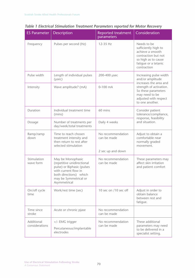
Scottish Stroke Allied Health Professionals Forum
70Use of Electrical Stimulation Following StrokeA Consensus Statement
Table 1 Electrical Stimulation Treatment Parameters reported for Motor Recovery
ES Parameter Description Reported treatment parameters
Consideration
Frequency Pulses per second (Hz) 12-35 Hz Needs to be sufficiently high to achieve a smooth contraction but not so high as to cause fatigue or a tetanic contraction
Pulse width Length of individual pulses (μsec)
200-400 μsec Increasing pulse width and/or amplitude increases the area and strength of activation. So these parameters may need to be adjusted with respect to one another.
Intensity Wave amplitude? (mA) 0-100 mA
Duration Individual treatment time (mins)
60 mins Consider patient tolerance/compliance, response, feasibility and situation.Dosage Number of treatments per
day/week/total treatmentsDaily 4 weeks
Ramp/ramp down
Time to reach chosen treatment intensity and then return to rest after selected stimulation
No recommendation can be made
2 sec up and down
Adjust to obtain a comfortable near normally graded movement.
Stimulation wave form
May be Monophasic (repetitive unidirectional pulse) or Biphasic (pulses with current flow in both directions) which may be Symmetrical or Asymmetrical
No recommendation can be made
These parameters may affect skin irritation and patient comfort
On/off cycle time
Work/rest time (sec) 10 sec on /10 sec off Adjust in order to obtain balance between rest and fatigue.
Time since stroke
Acute or chronic pjase No recommendation can be made
Additional considerations
+/- EMG trigger
Percutaneous/implantable electrodes
No recommendation can be made
These additional parameters may need to be delivered in a specialist setting.
Use of Electrical Stimulation Following StrokeA Consensus Statement
Scottish Stroke Allied Health Professionals Forum Scottish Stroke Allied Health Professionals Forum
71Use of Electrical Stimulation Following StrokeA Consensus Statement
Conclusion of evidence for treatment parameters of ES to aid motor controlOverall, there is no absolutely definitive guidance which can be given on specific treatment parameters for the application of ES to aid motor recovery. However, we recommend adjusting treatment parameters within the limits suggested above (Table 1), which will be particular to the device used, and using clinical judgement to produce as smooth, graded and natural a visible movement as possible whilst maintaining patient comfort.
We would recommend that future research in the form of RCTs focuses on well described interventions with adequate power to demonstrate the efficacy of differing ES treatment protocols.

Scottish Stroke Allied Health Professionals Forum
72Use of Electrical Stimulation Following StrokeA Consensus Statement
Recommended treatment parameters from studies of electrical stimulation to reduce shoulder subluxation after strokeWe observed that the reported clinical application parameters of ES, for the purpose of preventing or reducing shoulder subluxation after stroke, were different from those suggested as a means of recovering motor control. For this reason, we have reported ES applications to the shoulder aimed at the treatment of subluxation separately in this section.
Timing of ES interventionThe use of ES as an adjunct to conventional therapy early after stroke for those who have developed, or are at risk of developing, shoulder subluxation is recommended by the national guidelines in both England and Scotland (Intercollegiate Stroke Working Party 2012, Scottish Intercollegiate Guidelines Network (SIGN) June 2010). In a meta-analysis of studies assessing the efficacy of ES to prevent or reduce shoulder subluxation (Ada, Foongchomcheay 2002) studies of participants with a short duration (less than two months) were compared to studies of long duration (more than two months) since stroke. They reported that early application of ES in addition to conventional therapy was superior to conventional therapy alone and prevented shoulder subluxation by 6.5mm. Two months was used as an arbitrary time frame of short duration since stroke and when the individual studies within this review are examined along with more recent studies there was considerable variation in time since stroke before treatment was commenced.
Participants included in an experimental group receiving ES to the shoulder muscles were on average 49 days post stroke (Baker, Parker 1986). The authors of this study reported that the application of ES produced a significant reduction in subluxation and suggested that had they applied the ES earlier the reduction in subluxation may have been greater. Other studies included participants who were, on average, 16.5 days post stroke and two to four weeks post brain injury (including stroke) respectively (Faghri, Rodgers et al. 1994; Chantraine, Baribeault et al. 1999). An RCT compared the effects of the use of ES on acute (less than 21 days; average 15.9 days) and chronic (more than 1 year; average 427.1 days) on subluxation (Wang, Chan et al. 2000). The authors observed that early use of ES resulted in a statistically significant reduction in shoulder subluxation, whereas only marginal improvements were found in the chronic condition when compared to control.
In two separate RCTs, ES was applied within the first 48 hours post stroke and appeared to prevent the development of subluxation when compared to the control groups (Fil, Armutlu et al. 2011, Linn, Granat et al. 1999). Both authors noted that in other trials where ES was applied slightly later after stroke, the participants had already developed subluxation. In the study by Baker and Parker (1986) a minimum of 5mm of subluxation was an inclusion criterion for the study. Further, Baker and Parker (1986) did not achieve full reduction in subluxation, even with ES, and suggested that the degree of subluxation at the beginning of the study may have been a contributing factor.
Use of Electrical Stimulation Following StrokeA Consensus Statement
Scottish Stroke Allied Health Professionals Forum Scottish Stroke Allied Health Professionals Forum
73Use of Electrical Stimulation Following StrokeA Consensus Statement
Various authors have demonstrated that subluxation appears to occur during the flaccid period in the first three weeks post-stroke and is less likely to appear after the supraspinatus muscle has been shown to develop activity, recorded by EMG (Wang, Chan et al. 2000, Linn, Granat et al. 1999, Chaco, Wolf 1971, Griffin 1986). They also suggested that once the shoulder joint capsule had been stretched, subluxation would persist, even if supraspinatus became active or spasticity developed. Linn et. al. (1999) also observed, that patients who had a score of less than two on the Motor Assessment Scale (MAS) (Carr, Shepherd et al. 1985) did not develop subluxation. Therefore Linn et. al. (1999) and Fil et. al. (2011) suggested that the early application of ES (less than 48 hours after stroke) to prevent subluxation was important to attain positive results for the use of ES, as both found in their studies. Fil et. al. (2011) even applied ES to the unconscious patient after obtaining informed consent from relatives. Church et al. (2006) however, recommended caution in the use of ES to the shoulder in patients with more severe paresis of the arm as they found a trend towards poorer recovery of motor control in these patients, but this would need to be balanced against the potential positive effect of reducing subluxation.
Muscles targeted for electrical stimulation in reported studiesAda and Foongchomcheay (2002) reviewed EMG studies which suggested that it was supraspinatus and to a lesser extent posterior fibres of deltoid which were key muscles in the prevention of shoulder subluxation. Kobayashi et. al. (1999) reported that in hemiparetic patients, supraspinatus activity alone was reported to be insufficient to maintain humeral alignment (Kobayashi, Onishi et al. 1999). In addition to this, Fil et. al. (2011) suggested that the likelihood of anterior subluxation, as well as inferior subluxation, was important to consider. In a study which compared clinical assessment techniques with radiographic measurement of shoulder subluxation, seven of 20 participants in their study presented with anterior subluxation after stroke suggesting that more posterior muscle activity may be required to maintain normal anatomical shoulder joint alignment (Hall, Dudgeon et al. 1995).
Fil et. al. (2011) successfully applied ES to the middle fibres of deltoid in addition to supraspinatus and posterior fibres of deltoid, to reduce anterior and inferior subluxation. This was in response to reported evidence that ES to the mid portion of deltoid in healthy participants reduced anterior instability of the shoulder (Kido, Itoi et al. 2003).
Most researchers have applied ES to both supraspinatus and posterior deltoid (Baker, Parker 1986, Wang, Chan et al. 2000, Linn, Granat et al. 1999, Koyuncu, Nakipoglu-Yüzer et al. 2010) However, Kobayashi et. al. (1999) studied the effects separately of ES to supraspinatus and middle deltoid versus a control group. They found that stimulation of either muscle reduced subluxation and improved EMG muscle activity and the ability to produce abduction muscle force. Whilst the effect on subluxation was similar whichever muscle was stimulated, there was a tendency for stimulation of middle deltoid to be more effective at increasing the maximum abduction muscle force produced by participants at the end of the study. It has been reported that the additional stimulation of the long head of biceps, along with supraspinatus and posterior deltoid, had an improved impact on reducing subluxation (Manigandan, Ganesh et al. 2014).

Scottish Stroke Allied Health Professionals Forum
74Use of Electrical Stimulation Following StrokeA Consensus Statement
Patient selection for electrical stimulation for shoulder subluxationAda and Foongchomcheay (2002) suggested that the rationale for applying ES for shoulder subluxation should be loss of function as a result of paralysis of shoulder muscles after stroke. It appears that subluxation is related to the absence of muscle activity rather than pain (Smith, Dinse et al. 2009, Miglietta, Lewitan et al. 1959, Miglietta, Lewitan et al. 1959, Najenson, Yacubovich et al. 1971, Smith, Cruikshank et al. 1982, Bohannon, Andrews 1990, Van Langenberghe, Hogan 1988, Zorowitz, Hughes et al. 1996). Ada and Foongchomcheay (2002) therefore proposed that ES should be applied to those patients with a score of less than four on item six of the MAS.
Linn et. al. (1999) found a correlation between motor recovery and subluxation but not between pain and subluxation and that no participants who had a score of less than or equal to two on the upper arm section of the MAS developed a subluxation.
Although it has been discussed that prevention of subluxation very early after stroke may be preferable, it is interesting to note that the studies by Baker and Parker (1986), Wang et. al. (2000), Chantraine et. al. (1999) and Faghri et. al. (1994), in which participants may already have had shoulder subluxation, all reported positive effects in favour of ES for the treatment of subluxation. So it could be proposed that the presence of subluxation in the first few weeks after stroke should not preclude the use of ES for the treatment of this. The studies for subluxation of more chronic duration showed less favourable results, therefore ES is not recommended to treat established shoulder subluxation (Ada, Foongchomcheay 2002, Wang, Chan et al. 2000).
Use of Electrical Stimulation Following StrokeA Consensus Statement
Scottish Stroke Allied Health Professionals Forum Scottish Stroke Allied Health Professionals Forum
75Use of Electrical Stimulation Following StrokeA Consensus Statement
FrequencyA wide variety of frequencies were described in the literature aimed at preventing or reducing shoulder subluxation. Chantraine et. al. (1999) used a three sequence treatment programme where 8Hz was used in the first and longest treatment period, 40Hz in the second period and 1Hz in the final, and shortest treatment period. This was the only study we reviewed which used frequencies of less than 10Hz and still aimed to produce a visible muscle contraction rather than TENS which generally produces only a sensory stimulus.
Most authors used frequencies between 10 and 60Hz but most commonly ranging from 20-30Hz. Some authors (Baker, Parker 1986, Wang, Chan et al. 2000) used a variety of frequencies individualised to each participant with the aim of producing a tetanised contraction as did Koyuncu et. al. (2010) who used a fixed frequency of 36Hz. In the trials which were included in their meta-analysis, Ada and Foongchomcheay (2002) observed that only frequencies greater than 30Hz were utilised, or that sufficient to elicit a muscle response to electrical stimulation in order to produce enough force to counteract the inferior subluxation. As mentioned in the previous section, it is the interaction between pulse frequency, pulse width and pulse amplitude/intensity in addition to frequency choice which determines the amount of muscle activity generated.
Pulse width and pulse amplitude (intensity)Many authors did not state the pulse width used and those that did, did not justify the choice. Reported values varied between 100μs and 350μs (Fil, Armutlu et al. 2011, Chantraine, Baribeault et al. 1999, Linn, Granat et al. 1999, Kobayashi, Onishi et al. 1999, Koyuncu, Nakipoglu-Yüzer et al. 2010). In relation to pulse amplitude, the studies did not necessarily use quantitative measures. However Linn et. al. (1999) and Kobayashi et. al. (1999) used X-rays to confirm that the muscle contraction produced was sufficient to reduce the subluxation which would not be feasible in the clinical setting. Kobayashi et. al. (1999) reported that the intensity was adjusted within the limits of the participants’ pain tolerance and Fil et. al. (2011) adjusted it to produce a visible contraction without causing any distress.
Factors such as muscle fatigue and patient comfort should be considered as we have previously discussed in the preceding section on motor recovery but, as suggested by de Kroon et. al. (2005), it may be that it is the adjustment of all three variables in relation to each other in order to produce a visible muscle contraction which may be the most important factor. However, the full effects of varying parameter settings on the efficacy of ES for shoulder subluxation have not been fully investigated.

Scottish Stroke Allied Health Professionals Forum
76Use of Electrical Stimulation Following StrokeA Consensus Statement
Duration and dosageThere was significant variability in the individual treatment durations and overall dosages reported. In their study with acute stroke patients, Fil et. al. (2011) used 10 minute sessions, twice per day on five days of the week for a total of 25 sessions on average (range 20-30 sessions). They suggested that at least 25 sessions were required for the stimulation to show its strengthening effect, although they did not expand on the evidence base for this suggestion. Linn et. al. (1999) applied ES for increasing session times from 30-60 minutes, providing four sessions per day for four weeks. Chantraine et. al. (1999) provided one session per day of a three sequence treatment session lasting a total of 130-150 minutes, daily for four weeks.
Two of the studies (Baker, Parker 1986, Wang, Chan et al. 2000) provided increasingly longer individual sessions, fewer times per day. They started with 30 minutes per session, three times per day to one 6-7 hour session per day, five days per week for six weeks. Faghri et. al. (1994) used an increasingly longer single session starting at 1.5 hours increasing to six hours per day, seven days per week for six weeks. Ada and Foongchomcheay (2002) synthesised the evidence in order to produce a recommendation that ES be applied from one hour per day initially, increasing to six hours per day.
As previously discussed, improved levels of motor activity appear to lessen the risk of shoulder subluxation occurring in hemiplegia post-stroke. This led Ada and Foongchomcheay (2002) to recommend that the application of ES should continue until patients scored more than four on Item six of the MAS. Similarly, Linn et. al. (1999) suggested that as they found that no patients who scored more than two on the upper arm section of the MAS went on to develop shoulder subluxation, this might provide useful information on when treatment could be discontinued without the risk of subluxation occurring.
In the case where subluxation has already occurred the evidence is less clear. Wang et. al. (2000) found that, whilst six weeks of ES produced a significant reduction in shoulder subluxation when compared to the control group, when the treatment was withdrawn and conventional therapy alone continued for another six weeks both the control group and experimental groups showed an increase in shoulder subluxation, although this was a milder trend in the experimental group. Baker and Parker (1986) demonstrated a one to two millimetre loss of the subluxation reduction achieved in their experimental group at the three month follow up.
In contrast, Chantraine et. al. (1999), who provided the longest follow up period of 24 months, reported that the maximum improvement in shoulder subluxation, utilising a five week ES treatment programme, occurred in the first six months. There was a smaller number of patients who had a Grade I or no subluxation at 12 months. After this the results did not change at 24 month follow up. This suggests that the improvement in subluxation in the treatment group following ES occurred soon after the five week duration of treatment and that there was no more improvement after 12 months. However, as the improvements were maintained at 12 and 24 months this might be beneficial for those who already have subluxation.
Use of Electrical Stimulation Following StrokeA Consensus Statement
Scottish Stroke Allied Health Professionals Forum Scottish Stroke Allied Health Professionals Forum
77Use of Electrical Stimulation Following StrokeA Consensus Statement
Waveform, ramp times and on/off cycle timeMany authors described the waveform delivered but most did not justify its choice. Only Baker and Parker (1986) described in detail the factors which they considered in waveform choice. They suggested that both monopolar and bipolar electrode placements were appropriate for use in shoulder subluxation treatment programmes. They also postulated that stimulation with symmetrical biphasic waveforms, usually combined with bipolar electrode placements, was often perceived as being more comfortable than stimulation with a compensated monophasic waveform. They argued that a symmetrical biphasic waveform reduces the amount of amplitude needed to achieve muscle contraction (Baker and Parker 1986).
Baker and Parker (1986) used a compensated monophasic waveform. Wang et. al. (2000) and Chantraine et. al. (1999) described using asymmetrical biphasic waveforms. However, it may be that the device employed to provide stimulation governs the type of waveform used.
Ramp up/down times were varied, if described, from 1sec ramp up and down to 3 sec ramp up and down.
The on/off cycle times varied again but were not explained or justified further. Linn et. al. (1999) used 15 seconds in a 1:1 ratio, Chantraine et (1999) used a 1:5 ratio, Fil et. al. five seconds on and off and both Baker and Parker (1986) and Wang et. al. (2000) increased the on time initially, then reduced the off time, in response to the muscles ability to be stimulated for six to seven hours without fatigue, to a maximum setting of 24 seconds on and two seconds off.
Overall, there is no absolutely definitive evidence on which to base guidance on specific treatment parameters for preventing or reducing shoulder subluxation. However, what does seem to be important and has subsequently been recommended in national guidelines is that ES should be considered early after stroke as an adjunct to conventional therapy. Early appears to be certainly within the first few weeks after stroke in the “flaccid” period but potentially and even more favourably in the first few days after stroke for those in whose shoulder subluxation is at high risk due to significant muscle weakness around the shoulder. The muscles to be considered for stimulation are supraspinatus, posterior deltoid and middle deltoid.
Ada and Foongchomcheay (2002) synthesised the data available at the time to make the following recommendation:
“… (for) patients with a score <4 on Item 6 of the Motor Assessment Scale early after stroke ES should be applied daily to posterior deltoid and supraspinatus at more than 30Hz, beginning at 1hr/day, progressing to 6hr/day and continuing until the score on Item 6 of the Motor Assessment Scale is >4” (Ada, Foongchomcheay 2002)p. 265
We recommend adjusting treatment parameters within the limits suggested above (Table 2), which will be particular to the device used and the use of clinical judgement to produce as smooth, graded and natural a visible contraction as possible whilst maintaining patient comfort.
We would recommend that future research in the form of RCTs focuses on well described interventions with adequate power to demonstrate the efficacy of differing

Scottish Stroke Allied Health Professionals Forum
78Use of Electrical Stimulation Following StrokeA Consensus Statement
ES treatment protocols for the treatment of the subluxed shoulder after stroke.
The common ES treatment parameters considered in the literature for use in shoulder subluxation with recommended ranges are synthesised in Table 2 below.
Table 2 Electrical stimulation treatment parameters reported for reduction of shoulder subluxation
ES Parameter Description Reported treatment parameters
Considerations
Frequency Pulses per second (Hz) 10-60Hz Needs to be sufficiently high to achieve a smooth contraction but not so high as to cause fatigue. Many studies aimed to produce tetanised contraction.
Pulse width Length of individual pulses (μsec)
100-350μs Increasing pulse width and/or amplitude increases the area and strength of activation. So these parameters may need to be adjusted with respect to one another.
Intensity Wave amplitude (mA) No recommendation can be made. Aim to produce painless contraction
Duration Individual treatment time (mins) 5mins to 7 hours per session, generally 1 hour per day
Consider patient tolerance/compliance, response, feasibility and situation.
Dosage Number of treatments per day/week/total treatments
5-7 days per week
4-6 weeks or until sufficient voluntary muscle activity/reduction of subluxation without stimulation
Ramp/ramp down Time to reach chosen treatment intensity and then return to rest after selected stimulation
No recommendation can be made
2-3 seconds up and down
Adjust to obtain a comfortable near normally graded movement.
Stimulation wave form
May be Monophasic (repetitive unidirectional pulse) or Biphasic (pulses with current flow in both directions) which may be Symmetrical or Asymmetrical
No recommendation can be made
These parameters may affect skin irritation and patient comfort
On/off cycle time Work/rest time (sec) No recommendation can be made based on evidence
10-15 second on and off common with 1:1 ratio
Adjust in order to obtain balance between rest and fatigue.
Muscles stimulated
Muscles which, if sufficiently stimulated, will attain reduction in shoulder subluxation in a hemiplegic arm
Supraspinatus +/-
Posterior Deltoid +/-
Middle Deltoid
Consider number of channels available to provide stimulation (2 or 4). Consider direction of subluxation
Duration since stroke
The length of time since stroke onset and therefore onset of paralysis/risk of subluxation/actual subluxation
As early as possible, ideally within 48 hours. Certainly within 2-3 weeks of stroke onset
Increasing length of time since stroke increases likelihood of developing subluxation and that this will become irreversible.

Use of Electrical Stimulation Following StrokeA Consensus Statement
Scottish Stroke Allied Health Professionals Forum Scottish Stroke Allied Health Professionals Forum
79Use of Electrical Stimulation Following StrokeA Consensus Statement
ConclusionThis review has considered the high level evidence from Cochrane, DARE, other systematic reviews and more recent higher quality studies to report the likely efficacy of ES interventions in stroke rehabilitation and management. Guidance on treatment parameters for both the potential recovery of motor control and the prevention and possible treatment of shoulder subluxation has also been presented. This is an area of considerable uncertainty due to variation in the quality of research studies available but also the variety of specific treatment parameters used in study designs. Further high quality research in this area is recommended to better inform treatment.
Scottish Stroke Allied Health Professionals Forum
80Use of Electrical Stimulation Following StrokeA Consensus Statement
Glossary of Terms
• Asymmetrical Biphasic Waveform - a waveform is referred to as asymmetrical if the way in which the current amplitude varies in the first phase of a biphasic pulse is not the mirror image of the second phase.
• Biphasic Waveform - a pulse that deviates from the zero current (baseline) first in one direction and then in the opposite direction.
• Bipolar Electrode Placement- both surface electrodes are placed over the target area.
• Electrical Stimulation (ES) Or Neuromuscular Electrical Stimulation (NMES) - the electrical stimulation of an intact lower motor neurone (LMN) to activate weak or parietic muscles. The initiation of stimulation does not require voluntary contraction of target muscles. A pre-set programme is delivered once the stimulation device is started. May also be used as an umbrella term to describe all forms of electrical stimulation.
• Electroacupuncture - the application of ES to acupuncture points. May be transcutaneous delivery or via acupuncture needles inserted into specific meridian points.
• EMG-Triggered Electrical Stimulation - the use of ES to activate weak or parietic muscles where the stimulation is only delivered when EMG-signal of the voluntary muscle contraction reaches a pre-determined threshold.
• Extracorporeal Shockwave Therapy (ESWT) - is a non-invasive procedure using sound waves to stimulate healing.
• Functional Electrical Stimulation - is the use of electrical stimulation (ES) to activate paralysed muscles in a precise sequence and magnitude in order to directly accomplish or carry out a functional task i.e. walking, reaching to pick up an object.
• Glenohumeral Joint - is a multiaxial synovial ball and socket joint with articulation between the glenoid fossa and the head of the humeral bone.
• Hemiplegia - paralysis of one side of the body.
• Implantable Electrode - electrodes and stimulator are implanted. Electrodes are implanted on or in the muscle, beside or around a nerve.
• Lower Motor Neurone - motor neurones located in either the ventral horn of the spinal cord and anterior nerve roots or the cranial nerve nuclei of the brainstem and cranial nerves with motor function.
• Monophasic Waveform - a pulse that deviates from the zero current (baseline) in one direction only.
• Monopolar Electrode Placement - the active (black) electrode is placed over the target muscle and the indifferent (red) electrode is placed away from the target area.

Use of Electrical Stimulation Following StrokeA Consensus Statement
Scottish Stroke Allied Health Professionals Forum Scottish Stroke Allied Health Professionals Forum
81Use of Electrical Stimulation Following StrokeA Consensus Statement
• Motor Assessment Scale (MAS) - The Motor Assessment Scale (MAS) is a performance-based scale that was developed as a means of assessing everyday motor function in patients with stroke.
• Motor Electrical Stimulation - the use of electrical stimulation to evoke a motor response.
• Motor Point - a point at which a motor nerve enters a muscle; a point on the skin overlying a muscle at which electrical stimulation (via electrode) causes contraction of the muscle.
• Multi-Channel Stimulation - allows multiple sets of parameters to be delivered to separate target areas either simultaneously or alternatively or in a precise pattern to produce a specific movement or set of movements.
• Negative Electrode/Cathode/Active Electrode - positive ions are attracted and negative ions are repelled. Usually coloured black.
• Neuroprosthesis - is a device or system that provides FES.
• On:Off Ratio/Time - time which the signal or pulse is on or off. The percentage of time which the cycle is on versus off is known as the on:off ratio. Usually indicated in seconds (s).
• Orthosis - a brace, splint, or other artificial external device serving to support the limbs or spine or to prevent or assist relative movement.
• Percutaneous Electrode - electrode inserted into an isolated muscle with a hypodermic needle.
• Positive Electrode/Anode/Indifferent Electrode - positive ions at the interface are repelled while negative ions are attracted. Usually coloured red.
• Pulse Amplitude/Intensity - the level of output current produced by a unit. The amplitude of a pulse is a measurement of how far the medium is displaced momentarily from a position of rest. Usually indicated in milliamps (mA).
• Pulse Frequency – the rate at which pulses are emitted, normally described as pulses per second (Hertz, Hz).
• Pulse Width - the measure of the time duration of an individual pulse, usually indicated in microseconds (μs).
• Ramp Down Time - the time for the trailing edge of the phase to return to zero baseline from the peak amplitude.
• Ramp Up Time - the time for the leading edge of the phase to increase in amplitude from zero to peak amplitude.
• Sensory Electrical Stimulation - electrical stimulation sufficient to cause sensory stimulation but which does not cause a motor response.
• Single Channel Stimulation - a single set of parameters is delivered to one target area.
Scottish Stroke Allied Health Professionals Forum
82Use of Electrical Stimulation Following StrokeA Consensus Statement
• Subluxation - incomplete or partial dislocation; the atypical anatomic positioning of any joint that exceeds the physiologic but not the anatomic limit.
• Surface Electrode - electrodes placed on the surface of the skin over nerves, muscles or motor points.
• Symmetrical Biphasic Waveform – if the way in which current amplitude varies over time for the first phase of biphasic waveform is identical in nature but opposite in direction to that in the second phase, the biphasic waveform is described as symmetrical.
• Therapeutic Electrical Stimulation (TES) - May be used to describe the use of electrical stimulation as a therapeutic tool i.e. for motor recovery or strengthening. Or may be used as an alternative term to mean the same as ES or NMES.
• Transcutaneous Electrical Nerve Stimulation (TENS)- use of electrical stimulation, via electrodes placed on the skin, which does not produce a motor response. Generally used for the relief of pain. In some literature TENS is used to describe ES with a motor response.
• Treatment Dosage - the total treatment time. The number of individual treatment sessions per day, per week or total number of sessions delivered over a specified period of time. Usually indicated in minutes (mins), hours (hr), days and weeks.
• Treatment Duration - the time for which an individual treatment is applied. Usually indicated in minutes (mins) or hours (hr).
• Upper Motor Neurone - motor neurones that originate in the motor region of the cerebral cortex or the brainstem and carry motor information down to the lower motor neurones.
• Waveform - the shape of a single pulse or AC cycle on a current versus time plot.
• Waveform Shape - describes the shape of the waveform or pulse as it appears on a plot of current versus time. Shapes commonly described are rectangular, square, triangular, saw-tooth and spike.
Use of Electrical Stimulation Following StrokeA Consensus Statement
Scottish Stroke Allied Health Professionals Forum Scottish Stroke Allied Health Professionals Forum
83Use of Electrical Stimulation Following StrokeA Consensus Statement
Master References1. ADA, L. and FOONGCHOMCHEAY, A., 2002. Efficacy of electrical stimulation in preventing
or reducing subluxation of the shoulder after stroke: a meta-analysis. Australian Journal of Physiotherapy, 48(4), pp. 257-267.
2. AU-YEUNG, S. and HUI-CHAN, C., 2014. Electrical acupoint stimulation of the affected arm in acute stroke: a placebo-controlled randomized clinical trial. England: SAGE Publications.
3. BAKER, L.L. and PARKER, K., 1986. Neuromuscular electrical stimulation of the muscles surrounding the shoulder. UNITED STATES: American Physical Therapy Association.
4. BOHANNON, R.W. and ANDREWS, A.W., 1990. Shoulder subluxation and pain in stroke patients. The American Journal Of Occupational Therapy: Official Publication Of The American Occupational Therapy Association, 44(6), pp. 507-509.
5. BOYACI, A., TOPUZ, O., ALKAN, H., OZGEN, M., SARSAN, A., YILDIZ, N. and ARDIC, F., 2013. Comparison of the effectiveness of active and passive neuromuscular electrical stimulation of hemiplegic upper extremities: a randomized, controlled trial. International Journal Of Rehabilitation Research.Internationale Zeitschrift Für Rehabilitationsforschung.Revue Internationale De Recherches De Réadaptation, 36(4), pp. 315-322.
6. CARR, J.H., SHEPHERD, R.B., NORDHOLM, L. and LYNNE, D., 1985. Investigation of a new motor assessment scale for stroke patients. UNITED STATES: American Physical Therapy Association.
7. CHACO, J. and WOLF, E., 1971. Subluxation of the glenohumeral joint in hemiplegia. American Journal of Physical Medicine, 50(3), pp. 139-143.
8. CHAE, J., BETHOUX, F., BOHINE, T., DOBOS, L., DAVIS, T. and FRIEDL, A., 1998. Neuromuscular stimulation for upper extremity motor and functional recovery in acute hemiplegia. Stroke; a journal of cerebral circulation, 29(5), pp. 975-979.
9. CHAN, M.K., TONG, R.K. and CHUNG, K.Y., 2009. Bilateral upper limb training with functional electric stimulation in patients with chronic stroke. Neurorehabilitation & Neural Repair, 23(4), pp. 357-365.
10. CHANTRAINE, A., BARIBEAULT, A., UEBELHART, D. and GREMION, G., 1999. Shoulder pain and dysfunction in hemiplegia: effects of functional electrical stimulation. Archives of Physical Medicine and Rehabilitation, 80(3), pp. 328-331.
11. CHURCH, C., PRICE, C., PANDYAN, A., HUNTLEY, S., CURLESS, R., RODGERS, H., 2006. Randomized controlled trial to evaluate the effect of surface neuromuscular stimulation to the shoulder after acute stroke. Stroke, 37(10), pp. 2995-3001.
12. DE JONG, L., D., DIJKSTRA, P.U., GERRITSEN, J., GEURTS, A.C.H. and POSTEMA, K., 2013. Combined arm stretch positioning and neuromuscular electrical stimulation during rehabilitation does not improve range of motion, shoulder pain or function in patients after stroke: a randomised trial. Journal Of Physiotherapy, 59(4), pp. 245-254.
13. DE KROON, J.,R., IJZERMAN, M.J., CHAE, J., LANKHORST, G.J. and ZILVOLD, G., 2005a. Relation between stimulation characteristics and clinical outcome in studies using electrical stimulation to improve motor control of the upper extremity in stroke. Journal Of Rehabilitation Medicine: Official Journal Of The UEMS European Board Of Physical And Rehabilitation Medicine, 37(2), pp. 65-74.
14. DE KROON, J.,R., IJZERMAN, M.J., CHAE, J., LANKHORST, G.J. and ZILVOLD, G., 2005b. Relation between stimulation characteristics and clinical outcome in studies using electrical stimulation to improve motor control of the upper extremity in stroke. Journal Of Rehabilitation Medicine: Official Journal Of The UEMS European Board Of Physical And Rehabilitation Medicine, 37(2), pp. 65-74.
Scottish Stroke Allied Health Professionals Forum
84Use of Electrical Stimulation Following StrokeA Consensus Statement
15. DE KROON, ,J.R., IJZERMAN, M.J., LANKHORST, G.J. and ZILVOLD, G., 2004. Electrical stimulation of the upper limb in stroke: stimulation of the extensors of the hand vs. alternate stimulation of flexors and extensors. American Journal of Physical Medicine & Rehabilitation / Association of Academic Physiatrists, 83(8), pp. 592-600.
16. DE KROON, ,J.R., VAN DER LEE, ,J.H., IJZERMAN, M.J. and LANKHORST, G.J., 2002. Therapeutic electrical stimulation to improve motor control and functional abilities of the upper extremity after stroke: a systematic review. Clinical rehabilitation, 16(4), pp. 350-360.
17. DORSCH, S., ADA, L. and CANNING, C.G., 2013. EMG-triggered electrical stimulation is a feasible intervention to apply to multiple arm muscles in people early after stroke, but does not improve strength and activity more than usual therapy: a randomized feasibility trial. Clinical rehabilitation, .
18. ESKES, G.A. and BUTLER, B., 2006. Using limb movements to improve spatial neglect: the role of functional electrical stimulation. Restorative Neurology and Neuroscience, 24(4-6), pp. 385-398.
19. FAGHRI, P.D., RODGERS, M.M., GLASER, R.M., BORS, J.G., HO, C. and AKUTHOTA, P., 1994. The effects of functional electrical stimulation on shoulder subluxation, arm function recovery, and shoulder pain in hemiplegic stroke patients. UNITED STATES: W.B. Saunders.
20. FIL, A., ARMUTLU, K., ATAY, A.O., KERIMOGLU, U. and ELIBOL, B., 2011. The effect of electrical stimulation in combination with Bobath techniques in the prevention of shoulder subluxation in acute stroke patients. Clinical rehabilitation, 25(1), pp. 51-59.
21. GRIFFIN, J.W., 1986. Hemiplegic shoulder pain. Physical Therapy, 66(12), pp. 1884-1893.
22. HALL, J., DUDGEON, B. and GUTHRIE, M., 1995. Validity of clinical measures of shoulder subluxation in adults with post-stroke hemiplegia. The American Journal Of Occupational Therapy: Official Publication Of The American Occupational Therapy Association, 49(6), pp. 526-533.
23. HAMZAID, N.A. and DAVIS, G.M., 2009. Health and fitness benefits of functional electrical stimulation-evoked leg exercise for spinal cord-injured individuals: a position review. Topics in Spinal Cord Injury Rehabilitation, 14(4), pp. 88-121.
24. HANDY, J., SALINAS, S., BLANCHARD, S.A. and AITKEN, M.J., 2003. Meta-analysis examining the effectiveness of electrical stimulation in improving functional use of the upper limb in stroke patients. Physical & Occupational Therapy in Geriatrics, 21(4), pp. 67-78.
25. HARA, Y., OGAWA, S., TSUJIUCHI, K. and MURAOKA, Y., 2008. A home-based rehabilitation program for the hemiplegic upper extremity by power-assisted functional electrical stimulation. Disability & Rehabilitation, 30(4), pp. 296-304.
26. HARA, Y., 2008. Neurorehabilitation with new functional electrical stimulation for hemiparetic upper extremity in stroke patients. Journal Of Nippon Medical School = Nippon Ika Daigaku Zasshi, 75(1), pp. 4-14.
27. HEALTHCARE IMPROVEMENT SCOTLAND, August 2012-last update, The Use of Functional Electrical Stimulation (FES) in Adults with Dropped Foot [Homepage of Healthcare Improvement Scotland], [Online]. Available: http://www.healthcareimprovementscotland.org/our_work/technologies_and_medicines/shtg_-_evidence_notes/evidence_note_46.aspx.
28. HESSE, S., JAHNKE, M.T., LUECKE, D. and MAURITZ, K.H., 1995. Short-term electrical stimulation enhances the effectiveness of Botulinum toxin in the treatment of lower limb spasticity in hemiparetic patients. Neuroscience letters, 201(1), pp. 37-40.
29. HESSE, S., REITER, F., KONRAD, M. and JAHNKE, M.T., 1998. Botulinum toxin type A and short-term electrical stimulation in the treatment of upper limb flexor spasticity after stroke: a randomized, double-blind, placebo-controlled trial. ENGLAND: SAGE Publications.
Use of Electrical Stimulation Following StrokeA Consensus Statement
Scottish Stroke Allied Health Professionals Forum Scottish Stroke Allied Health Professionals Forum
85Use of Electrical Stimulation Following StrokeA Consensus Statement
30. HSU, S., HU, M., LUH, J., WANG, Y., YIP, P. and HSIEH, C., 2012. Dosage of neuromuscular electrical stimulation: is it a determinant of upper limb functional improvement in stroke patients? Journal of Rehabilitation Medicine (Stiftelsen Rehabiliteringsinformation), 44(2), pp. 125-130.
31. INTERCOLLEGIATE STROKE WORKING PARTY, 2012-last update, National Clinical Guideline for Stroke Fourth Edition [Homepage of Royal College of Physicians], [Online]. Available: http://www.rcplondon.ac.uk/sites/default/files/national-clinical-guidelines-for-stroke-fourth-edition.pdf.
32. JOHNSON, C.A., BURRIDGE, J.H., STRIKE, P.W., WOOD, D.E. and SWAIN, I.D., 2004. The effect of combined use of botulinum toxin type A and functional electric stimulation in the treatment of spastic drop foot after stroke: a preliminary investigation. Archives of Physical Medicine & Rehabilitation, 85(6), pp. 902-909.
33. KIDO, T., ITOI, E., LEE, S., NEALE, P.G. and AN, K., 2003. Dynamic stabilizing function of the deltoid muscle in shoulders with anterior instability. The American Journal of Sports Medicine, 31(3), pp. 399-403.
34. KITA, K., TAKEDA, K., OSU, R., USHIBA, J., SAKATA, S. and OTAKA, Y., 2011. A sensory feedback system utilizing cutaneous electrical stimulation for stroke patients with sensory loss. IEEE ...International Conference On Rehabilitation Robotics: [Proceedings], 2011, pp. 5975489-5975489.
35. KOBAYASHI, H., ONISHI, H., IHASHI, K., YAGI, R. and HANDA, Y., 1999. Reduction in subluxation and improved muscle function of the hemiplegic shoulder joint after therapeutic electrical stimulation. ENGLAND: Elsevier.
36. KOYUNCU, E., NAKIPOGLU-YÜZER, G., DOGAN, A. and ÖZGIRGIN, N., 2010. The effectiveness of functional electrical stimulation for the treatment of shoulder subluxation and shoulder pain in hemiplegic patients: a randomized controlled trial. Disability & Rehabilitation, 32(7), pp. 560-566.
37. LINN, S.L., GRANAT, M.H. and LEES, K.R., 1999. Prevention of shoulder subluxation after stroke with electrical stimulation. Stroke; a journal of cerebral circulation, 30(5), pp. 963-968.
38. MacDonald, J. (2013) An Audit of Shoulder Subluxation in Patients within NHS Greater Glasgow and Clyde Stroke Units: Examining Incidence, Predictive Factors, and Potential Numbers Eligible for Electrical Muscle Stimulation Treatment NHS Greater Glasgow and Clyde December 2013
39. MANIGANDAN, J.B., GANESH, G.S., PATTNAIK, M. and MOHANTY, P., 2014. Effect of electrical stimulation to long head of biceps in reducing gleno humeral subluxation after stroke. Neurorehabilitation, .
40. MIGLIETTA, O., LEWITAN, A. and ROGOFF, J.B., 1959. Subluxation of the shoulder in hemiplegic patients. New York state journal of medicine, 59(3), pp. 457-460.
41. NASCIMENTO, L.R., MICHAELSEN, S.M., ADA, L., POLESE, J.C. and TEIXEIRA-SALMELA, L., 2014. Cyclical electrical stimulation increases strength and improves activity after stroke: a systematic review. Journal Of Physiotherapy, 60(1), pp. 22-30.
42. NAJENSON, T., YACUBOVICH, E. and PIKIELNI, S.S., 1971. Rotator cuff injury in shoulder joints of hemiplegic patients. Scandinavian journal of rehabilitation medicine, 3(3), pp. 131-137.
43. POLANOWSKA, K., SENIÓW, J., PAPROT, E., LEŚNIAK, M. and CZŁONKOWSKA, A., 2009. Left-hand somatosensory stimulation combined with visual scanning training in rehabilitation for post-stroke hemineglect: a randomised, double-blind study. Neuropsychological Rehabilitation, 19(3), pp. 364-382.
44. POMEROY, V.M., KING, L., POLLOCK, A., BAILY-HALLAM, A. and LANGHORNE, P., 2006. Electrostimulation for promoting recovery of movement or functional ability after stroke. The Cochrane Database Of Systematic Reviews, (2), pp. CD003241.

Scottish Stroke Allied Health Professionals Forum
86Use of Electrical Stimulation Following StrokeA Consensus Statement
45. PRICE, C.I. and PANDYAN, A.D., 2001. Electrical stimulation for preventing and treating post-stroke shoulder pain: a systematic Cochrane review. Clinical rehabilitation, 15(1), pp. 5-19.
46. ROCHE, A., LAIGHIN, G. and COOTE, S., 2009. Surface-applied functional electrical stimulation for orthotic and therapeutic treatment of drop-foot after stroke -- a systematic review. Physical Therapy Reviews, 14(2), pp. 63-80.
47. SAHIN, N., UGURLU, H. and ALBAYRAK, I., 2012. The efficacy of electrical stimulation in reducing the post-stroke spasticity: a randomized controlled study. Disability and rehabilitation, 34(2), pp. 151-156.
48. SANTAMATO, A., NOTARNICOLA, A., PANZA, F., RANIERI, M., MICELLO, M.F., MANGANOTTI, P., MORETTI, B., FORTUNATO, F., FILONI, S. and FIORE, P., 2013. SBOTE study: extracorporeal shock wave therapy versus electrical stimulation after botulinum toxin type a injection for post-stroke spasticity-a prospective randomized trial. Ultrasound in medicine & biology, 39(2), pp. 283-291.
49. Scottish Executive. (2000). Explanatory notes to the adults with incapacity (Scotland) Act 2000. Edinburgh: HMSO.
50. SCOTTISH INTERCOLLEGIATE GUIDELINES NETWORK (SIGN), June 2010-last update, SIGN 118 Management of Patients With Stroke: Rehabilitation, Prevention and Management of Complications and Discharge Planning. A National Clinical Guideline. [Homepage of NHS Quality Improvement Scotland], [Online]. Available: http://www.sign.ac.uk/pdf/qrg118.pdf.
51. SHEFFLER, L.R. and CHAE, J., 2007. Neuromuscular electrical stimulation in neurorehabilitation. Muscle & nerve, 35(5), pp. 562-590.
52. SMITH, P.S., DINSE, H.R., KALISCH, T., JOHNSON, M. and WALKER-BATSON, D., 2009. Effects of repetitive electrical stimulation to treat sensory loss in persons poststroke. Archives of Physical Medicine & Rehabilitation, 90(12), pp. 2108-2111.
53. SMITH, R.G., CRUIKSHANK, J.G., DUNBAR, S. and AKHTAR, A.J., 1982. Malalignment of the shoulder after stroke. British medical journal (Clinical research ed.), 284(6324), pp. 1224-1226.
54. SUJITH, O.K., 2008. Functional electrical stimulation in neurological disorders. European Journal Of Neurology: The Official Journal Of The European Federation Of Neurological Societies, 15(5), pp. 437-444.
55. VAN LANGENBERGHE, H. and HOGAN, B.M., 1988. Degree of pain and grade of subluxation in the painful hemiplegic shoulder.
56. WANG, R.Y., CHAN, R.C. and TSAI, M.W., 2000. Functional electrical stimulation on chronic and acute hemiplegic shoulder subluxation. UNITED STATES: Lippincott Williams & Wilkins.
57. WILKENFELD, A.J.L., 2013. Review of electrical stimulation, botulinum toxin, and their combination for spastic drop foot. Journal Of Rehabilitation Research And Development, 50(3), pp. 315-326.
58. WONG, A., SU, T., TANG, F., CHENG, P. and LIAW, M., 1999. Clinical trial of electrical acupuncture on hemiplegic stroke patients. American Journal of Physical Medicine & Rehabilitation, 78(2), pp. 117-122.
59. ZOROWITZ, R.D., HUGHES, M.B., IDANK, D., IKAI, T. and JOHNSTON, M.V., 1996. Shoulder pain and subluxation after stroke: correlation or coincidence? The American Journal Of Occupational Therapy: Official Publication Of The American Occupational Therapy Association, 50(3), pp. 194-201.
Use of Electrical Stimulation Following StrokeA Consensus Statement
Scottish Stroke Allied Health Professionals Forum Scottish Stroke Allied Health Professionals Forum
87Use of Electrical Stimulation Following StrokeA Consensus Statement
Who was involved in developing this consensus statement?Membership of the ES Working group
• Carolyn Agnew Lead Physiotherapist, Fife Rehabilitation Services
• Gillian Alexander AHP Consultant in Stroke, NHS Greater Glasgow and Clyde
• Charlie Chung Occupational Therapy Manager, NHS Fife
• Iain Larkin Stroke Specialist Physiotherapist, NHS Ayrshire and Arran
• Julie Macdonald Stroke Specialist Physiotherapist, NHS Greater Glasgow and Clyde
• Gillian McClure Senior Physiotherapist, NHS Lothian
• Craig Paterson Clinical Scientist, NHS Greater Glasgow and Clyde
• Lynn Robertson Clinical Specialist Physiotherapist, NHS Dumfries and Galloway
• Mark Smith Consultant Physiotherapist in Stroke, NHS Lothian
Membership of the Critical Review Group• Carolyn Agnew Lead Physiotherapist, Fife Rehabilitation Service
• Dr Gillian Baer Senior Lecturer, Physiotherapy, Queen Margaret University, Edinburgh
• Fiona Dodson Clyde Stroke AHP Team Lead, Royal Alexandra Hospital, NHS Greater Glasgow and Clyde
• Dr Sybil Farmer Institute for Science and Technology in Medicine, Keele University
• Dr Nicola Hancock Lecturer in Physiotherapy and Associate Director Enterprise, University of East Anglia
• Thérèse Jackson Consultant Occupational Therapist in Stroke, NHS Grampian
• Wendy Juner Team Lead Physiotherapist, NHS Lothian
• Audrey Lambe South Service Lead Occupational Therapist, University Hospital Ayr, Ayrshire & Arran NHS
• Clare McDonald Advanced Occupational Therapist, Fife Rehabilitation Service
The SSAHPF acknowledge the ongoing support of Chest Heart & Stroke Scotland in hosting this document on their website: http://www.chss.org.uk/ssahpf/ecs-statement.pdf
The Quick Reference Guide may be viewed at: http://www.chss.org.uk/ssahpf/ecs-guide.pdf
The SSAHPF would also like to thank NHS GG&C Medical Illustration department for their help in producing this document.
272508