Scleroderma FAQ™ · 1 Scleroderma FAQ™ About this Document The Scleroderma FAQ* is a...
42
1 Scleroderma FAQ™ About this Document The Scleroderma FAQ * is a comprehensive document that covers systemic scleroderma diagnosis and treatment. All information contained in the FAQ is based on current medical research and includes up-to-date information on new diagnostic criteria and treatments for systemic scleroderma. Here is what is included in the Scleroderma FAQ: • General Description – This initial section gives a general description of the scleroderma family of diseases. • Differential Diagnosis – This section of the FAQ discusses localized forms of scleroderma that don’t have systemic involvement and other diseases that have similar symptoms but are not in the scleroderma family of diseases. It discusses in detail a new diagnostic criteria for systemic scleroderma that was adopted in 2013. It also discusses a controversial special diagnosis that is sometimes given to patients who have internal organ involvement but no skin changes. • Affected Population – This section of the FAQ describes the incidence (number of new cases per year) and prevalence (number of patients with a diagnosis) of patients with a systemic scleroderma diagnosis. It also talks about age and gender distribution of systemic scleroderma patients. • Causes – Systemic scleroderma is considered to be a disease that requires genetic susceptibility and exposure to some type of trigger event, for example exposure to organic solvents or silica dust. • Symptoms – Systemic scleroderma affects many internal organs in addition to the skin. This section of the FAQ discusses affected organs, including skin, musculoskeletal (muscles and joints), pulmonary (lungs), gastrointestinal, cardiac (heart), renal (kidney), sexual dysfunction, and other symptoms. • Scleroderma Antibodies and Clinical Relevance – There are currently about 10 known scleroderma specific antibodies, each of which has a different clinical profile. In * When the Scleroderma FAQ was first published online in 1995, it was formatted as a F.A.Q (Frequently Asked Questions) style document. Over the years, the format of the FAQ has changed, but we decided to maintain the original "Scleroderma FAQ" name for consistency.
Transcript of Scleroderma FAQ™ · 1 Scleroderma FAQ™ About this Document The Scleroderma FAQ* is a...

1
Scleroderma FAQ™
About this Document
The Scleroderma FAQ* is a comprehensive document that covers systemic scleroderma
diagnosis and treatment. All information contained in the FAQ is based on current medical
research and includes up-to-date information on new diagnostic criteria and treatments for
systemic scleroderma.
Here is what is included in the Scleroderma FAQ:
• General Description – This initial section gives a general description of the
scleroderma family of diseases.
• Differential Diagnosis – This section of the FAQ discusses localized forms of
scleroderma that don’t have systemic involvement and other diseases that have similar
symptoms but are not in the scleroderma family of diseases. It discusses in detail a new
diagnostic criteria for systemic scleroderma that was adopted in 2013. It also discusses
a controversial special diagnosis that is sometimes given to patients who have internal
organ involvement but no skin changes.
• Affected Population – This section of the FAQ describes the incidence (number of
new cases per year) and prevalence (number of patients with a diagnosis) of patients
with a systemic scleroderma diagnosis. It also talks about age and gender distribution
of systemic scleroderma patients.
• Causes – Systemic scleroderma is considered to be a disease that requires genetic
susceptibility and exposure to some type of trigger event, for example exposure to
organic solvents or silica dust.
• Symptoms – Systemic scleroderma affects many internal organs in addition to the
skin. This section of the FAQ discusses affected organs, including skin, musculoskeletal
(muscles and joints), pulmonary (lungs), gastrointestinal, cardiac (heart), renal
(kidney), sexual dysfunction, and other symptoms.
• Scleroderma Antibodies and Clinical Relevance – There are currently about 10
known scleroderma specific antibodies, each of which has a different clinical profile. In
* When the Scleroderma FAQ was first published online in 1995, it was formatted as a
F.A.Q (Frequently Asked Questions) style document. Over the years, the format of the
FAQ has changed, but we decided to maintain the original "Scleroderma FAQ" name
for consistency.
2
addition, a small percentage of patients diagnosed with systemic scleroderma test
negative for antibodies. This section of the FAQ lists the known antibodies and general
classification and risk profile.
• Pregnancy and Scleroderma – Since about 80% of diagnosed systemic scleroderma
patients are female and middle aged, the FAQ includes a discussion on the effects of
systemic scleroderma on fertility, and pregnancy. It also includes a discussion on how
pregnancy can affect scleroderma symptoms.
• Treatments - General: Standard / Multi-Symptoms – This section of the FAQ
focuses on systemic level treatments and includes a list of the most common drugs used
in scleroderma treatment, potential side effects, and other issues related to each of
these drugs.
• Treatments - General: Research-Based Experimental / Alternative – This
section of the FAQ discusses two experimental systemic-level research-based
treatments that are sometimes used to treat patients with systemic scleroderma: 1)
autologous stem cell transplants, and 2) therapeutic plasma exchange.
• Treatments: Specific Symptoms – In addition to systemic level treatments
discussed previously, much of the treatment focus is on dealing with individual
symptoms. This section of the FAQ covers treatments focused on individual symptoms
such as Raynaud’s, skin changes, muscles and joints, lungs, gastrointestinal, heart,
kidney, and other symptoms including sexual dysfunction and depression.
• About Scleroderma Research – This section of the FAQ gives information about
scleroderma research as well as information about how to better interpret published
research studies.
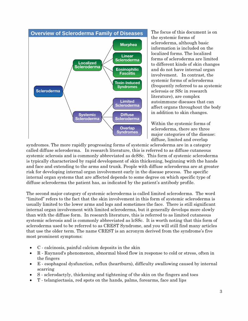
General Description
Scleroderma (literally "hard skin") is an umbrella term for a family of rare diseases with the
common factor being abnormal thickening (fibrosis) of the skin. However, not everyone with
scleroderma develops skin changes. With some variants of the disease, skin changes usually
occur early in the disease process and can develop very rapidly. With other forms of
scleroderma, skin changes may not occur for many years after the development of other
symptoms and in rare cases may never be a significant symptom of the disease.
There are two main groupings of the scleroderma family of diseases: Localized and Systemic,
as shown in the diagram below:
3
The focus of this document is on
the systemic forms of
scleroderma, although basic
information is included on the
localized forms. The localized
forms of scleroderma are limited
to different kinds of skin changes
and do not have internal organ
involvement. In contrast, the
systemic forms of scleroderma
(frequently referred to as systemic
sclerosis or SSc in research
literature), are complex
autoimmune diseases that can
affect organs throughout the body
in addition to skin changes.
Within the systemic forms of
scleroderma, there are three
major categories of the disease:
diffuse, limited and overlap
syndromes. The more rapidly progressing forms of systemic scleroderma are in a category
called diffuse scleroderma. In research literature, this is referred to as diffuse cutaneous
systemic sclerosis and is commonly abbreviated as dcSSc. This form of systemic scleroderma
is typically characterized by rapid development of skin thickening, beginning with the hands
and face and extending to the arms and trunk. People with diffuse scleroderma are at greater
risk for developing internal organ involvement early in the disease process. The specific
internal organ systems that are affected depends to some degree on which specific type of
diffuse scleroderma the patient has, as indicated by the patient’s antibody profile.
The second major category of systemic scleroderma is called limited scleroderma. The word
“limited” refers to the fact that the skin involvement in this form of systemic scleroderma is
usually limited to the lower arms and legs and sometimes the face. There is still significant
internal organ involvement with limited scleroderma, but it generally develops more slowly
than with the diffuse form. In research literature, this is referred to as limited cutaneous
systemic sclerosis and is commonly abbreviated as lcSSc. It is worth noting that this form of
scleroderma used to be referred to as CREST Syndrome, and you will still find many articles
that use the older term. The name CREST is an acronym derived from the syndrome’s five
most prominent symptoms:
• C - calcinosis, painful calcium deposits in the skin
• R - Raynaud's phenomenon, abnormal blood flow in response to cold or stress, often in
the fingers
• E - esophageal dysfunction, reflux (heartburn), difficulty swallowing caused by internal
scarring
• S - sclerodactyly, thickening and tightening of the skin on the fingers and toes
• T - telangiectasia, red spots on the hands, palms, forearms, face and lips
Overview of Scleroderma Family of Diseases
4
While limited scleroderma progresses more slowly and has a better overall prognosis than
diffuse scleroderma, different variants of limited scleroderma (based on antibody profile) have
different complication risks over the long term.
The third category of systemic scleroderma is a diverse group that is generally referred to as
scleroderma overlap syndromes. With overlap syndromes, while patients have clear
scleroderma specific symptoms, they also have symptoms that overlap with other autoimmune
diseases, including lupus and myositis (muscle inflammation). An example is Mixed
Connective Tissue Disorder, which includes symptoms that are common in scleroderma, lupus,
and myositis. The specific antibody determines the nature of the overlap syndrome.
Differential Diagnosis
Localized Scleroderma and Scleroderma-Like Disorders
Morphea, or localized scleroderma, can affect all ages and is more common in women. It
typically presents as patches of yellowish or ivory-colored rigid, dry skin. These are followed by
the appearance of firm, hard, oval-shaped plaques with ivory centers that are encircled by a
violet ring. These spots generally appear on the trunk, face, and/or extremities. Many patients
with localized morphea improve without treatment. Generalized morphea is more rare and
serious and involves the skin but not the internal organs.
Linear scleroderma appears as a band-like thickening of skin on the arms or legs. This type of
scleroderma is most likely to be on one side of the body but may be on both sides. Linear
scleroderma generally appears before age 20. When it occurs in young children, it may result
in the failure of one limb (e.g., an arm or leg) to grow as rapidly as its counterpart.
Diffuse fasciitis with eosinophilia (DFE, also called eosinophilic fasciitis or Shulman’s
syndrome) is a rare condition that mimics scleroderma with swelling, stiffness, and decreased
flexibility of the limbs associated with skin thickening. Although the symptoms can be
widespread and involve the trunk and limbs, in contrast to scleroderma, the fingers, hands,
and face are usually not affected. In addition, there is no occurrence of Raynaud’s or GI
involvement.
Eosinophilia-myalgia syndrome (EMS) is a rare condition that was first described after 3
patients in New Mexico were found to have an illness with significant myalgia (muscle pain)
and an increase in the number of eosinophils (a type of white blood cell). All three patients
had taken supplements containing L-tryptophan, which may have been contaminated. All
told, about 1500 people were affected. A similar outbreak occurred in Spain in 1981 and
affected almost 20,000 people. As it may have been the result of consuming contaminated
rapeseed oil, it was known as toxic oil syndrome (TOS). About 60% of the patients developed
skin thickening that look like skin changes typical for scleroderma patients, although the
affected areas were different than what is normally seen with scleroderma, and there is no
associated Raynaud’s phenomenon.

5
Scleroderma-like skin changes have also been associated with insulin-dependent diabetes,
carcinoid syndrome, myeloma, scleromyxedema, chronic graft-versus-host disease, porphyria
cutanea tarda, Werner’s syndrome, progeria, phenylketonuria, bleomycin exposure, local
lipodystrophies, nephrogenic fibrosing dermopathy, and POEMS syndrome.
Systemic Scleroderma
Systemic scleroderma diagnosis is often a challenging and lengthy process. It is not
uncommon for a person who ultimately is diagnosed with one of the forms of systemic
scleroderma to be initially misdiagnosed with many different disorders. Part of the reason for
this is that some early scleroderma symptoms are non-specific, and unless the physician
suspects scleroderma, s/he may not order the appropriate tests to diagnose the condition.
Scleroderma and ANA (Anti-nuclear Antibody) Testing
In almost all cases of systemic scleroderma, the patient will have a positive anti-nuclear
antibody (ANA) test result. However, even this test can be problematic. There are now
several different ways of testing for ANA. The long-term “gold standard” is a method called
indirect immunofluorescence (commonly abbreviated as IFA or IIF). This has very high
reliability and is the best way to test for the presence of anti-nuclear antibodies. However, it
is a complex and time consuming test that depends on highly trained laboratory personnel.
Recently, many commercial laboratories and some larger hospital laboratories have switched
their routine ANA testing to solid phase immunoassays (ELISA or EIA) or a related technique
known as a Multiplex platform. These new techniques can handle high testing volumes since
they are not labor intensive like IFA testing and are, therefore, less expensive than IFA.
However, these new methods of testing can only detect a limited subset of the specific
antibodies that are targeted by the tests (typically 8-10) in contrast to IFA that can detect 100
to 150 different possible antibodies. As a result, these alternate testing methods are more
likely to miss relevant autoantibodies yielding false negative ANA results. For example, a
recent study (Shanmugam et al. 2011) reported that up to 43% of scleroderma patients with
positive ANA results by IFA yielded negative ANA results using the Multiplex method. This
can have major impact on scleroderma diagnosis. If the results of an initial ANA screening
come back negative to the doctor who ordered the ANA test without knowing this data, this
can be the start of (in some cases) years of diagnostic limbo for patients. By the time they are
finally retested for ANA by the more comprehensive IFA method, their symptoms will have
progressed and may be more difficult to treat.
If a physician orders just an ANA test in a setting where there is a local laboratory, there is
still a reasonably good chance that the ANA test will be done by IFA. However, if the ANA
test is sent to an outside lab, it is more likely that the default method of testing will be ELISA
or Multiplex. Even more problematic, in order to save time and money, many physicians tend
to order an ANA test with reflex antibody testing. This initial test will almost always be done
using ELISA or Multiplex methodology. If the result is positive, then the ANA test is
automatically re-run using IFA in order to get the titer and staining pattern, which can be
useful diagnostic information. In addition, an antibody panel is also run to determine which
specific antibodies are present, potentially directing the clinician to more quickly reach a
correct diagnosis. However, given the potential for a false negative ANA result with

6
scleroderma patients, this new “improved” method of testing is significantly more likely to give
an incorrect result than if the initial ANA testing was done by IFA. Ironically, had the same
ANA plus reflex antibody panel been ordered 15 years ago, the initial ANA test would have
been done by IFA, yielding a significantly more accurate result. This raises a serious question
as to whether modern scleroderma diagnosis is being compromised by using these new, less
expensive, testing methods.
Unfortunately, many primary care physicians (and probably some rheumatologists as well) are
unaware of these methodological problems with ANA testing, especially about the potential for
false negative result. The American College of Rheumatology in a 2011 Position Paper
discusses these problems and recommends that testing by IFA “should remain the gold
standard for ANA testing”. While it is true that ELISA and Multiplex ANA testing usually is
consistent with IFA ANA testing, if an initial ANA result done by ELISA or Multiplex testing
is negative, it is very important that the test be re-run by IFA to confirm the negative results.
However, between 2% and 10% of patients (depending on the study) with systemic
scleroderma symptoms are ANA negative, even when done by IFA. In some cases, the ANA
does change to positive over time. It is worth noting that ANA level is generally stable over
time and there is no evidence that the actual tested ANA level is correlated with disease
severity.
Once a potential scleroderma patient shows a positive ANA, the next step in diagnosis is to
test for specific antibodies that can be used to help determine which form of systemic
scleroderma the patient has or may develop in the future. Most systemic scleroderma patients
will test positive for anti-Scl-70 antibodies (anti-Topoisomerase I, also sometimes listed as
“Scleroderma IgG” on lab tests), anti-Centromere antibodies, or anti-RNA polymerase III. The
anti-Scl-70 antibody is highly specific for one of the diffuse forms of systemic scleroderma, and
the anti-Centromere antibody is highly correlated with a limited scleroderma variant.
Historically, only the anti-Scl-70 and the anti-Centromere antibodies were strongly associated
with the two general categories of systemic scleroderma: diffuse or limited. The anti-RNA
polymerase III antibody is now recognized as a third major scleroderma-related antibody.
Patients with anti-RNA polymerase III antibodies are considered to be in the diffuse category,
but the specific clinical manifestations are different from the typical clinical manifestations
shown by patients with anti-Scl-70 antibodies. In addition to these three main antibodies,
several other antibodies have been associated with different variants of systemic scleroderma,
although these other antibodies are detected much less frequently than the three main
antibody types listed above, and commercial testing for some of these antibodies is not
currently widely available. This topic is discussed in more detail later in this document.
It is very rare (about 2%) for a patient to have more than one scleroderma-related antibody.
Antibody status does not change over time.
New Formal Diagnostic Criteria for Systemic Scleroderma
In late 2013, the American College of Rheumatology (ACR) and the European League Against
Rheumatism (EULAR) approved a new set of diagnostic criteria for systemic scleroderma,
replacing the older 1980 diagnostic criteria (van den Hoogen et al. 2013). These new
7
standards will improve clinical diagnosis of systemic scleroderma, but it is very important to
understand that the reason for developing these new diagnostic standards was “to develop a
set of criteria that would enable identification of individuals with SSc for inclusion
in clinical studies,” not for normal diagnosis of patients in a clinical setting. The authors of
the special report that formally introduces the new criteria note that many symptoms that are
used for clinical diagnosis are not included in these formal research criteria, including
common symptoms such as tendon friction rubs, calcinosis, difficulty swallowing, as well as
less common but more serious complications such as renal crisis.
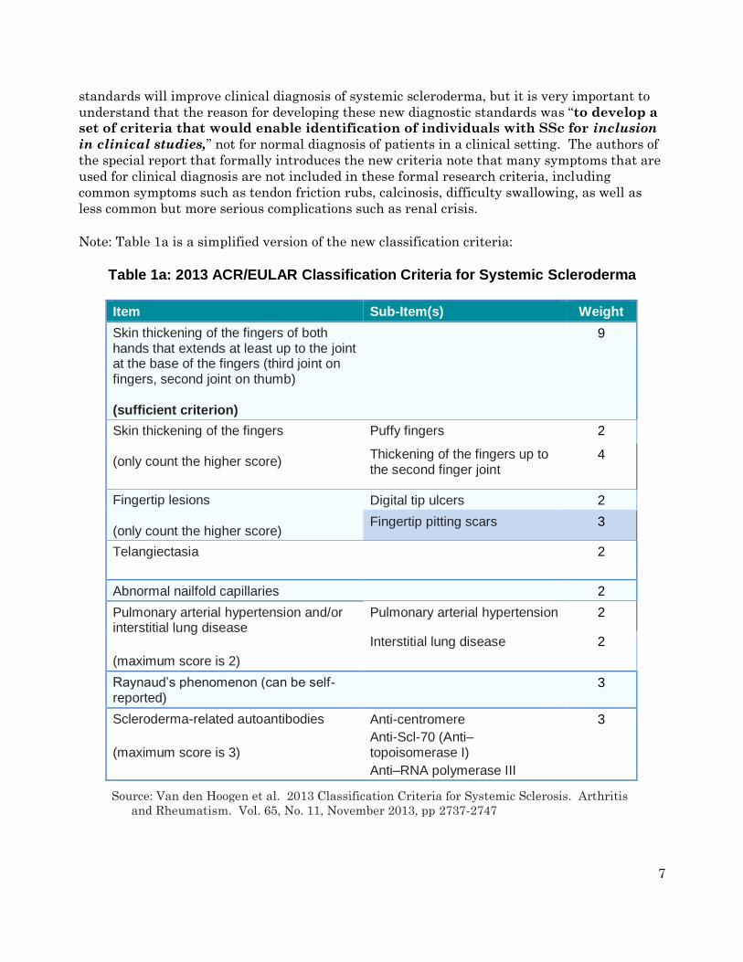
Note: Table 1a is a simplified version of the new classification criteria:
Table 1a: 2013 ACR/EULAR Classification Criteria for Systemic Scleroderma
Item Sub-Item(s) Weight
Skin thickening of the fingers of both hands that extends at least up to the joint at the base of the fingers (third joint on fingers, second joint on thumb) (sufficient criterion)
9
Skin thickening of the fingers (only count the higher score)
Puffy fingers 2
Thickening of the fingers up to the second finger joint
4
Fingertip lesions (only count the higher score)
Digital tip ulcers 2
Fingertip pitting scars 3
Telangiectasia
2
Abnormal nailfold capillaries 2
Pulmonary arterial hypertension and/or interstitial lung disease
(maximum score is 2)
Pulmonary arterial hypertension 2
Interstitial lung disease 2
Raynaud’s phenomenon (can be self-reported)
3
Scleroderma-related autoantibodies
(maximum score is 3)
Anti-centromere
Anti-Scl-70 (Anti–topoisomerase I)
Anti–RNA polymerase III
3
Source: Van den Hoogen et al. 2013 Classification Criteria for Systemic Sclerosis. Arthritis
and Rheumatism. Vol. 65, No. 11, November 2013, pp 2737-2747

8
The total score is determined by adding the maximum weight (score) in each category.
Patients with a total score of 9 or greater are classified as having definite systemic
scleroderma. For example, a patient with definite skin thickening on both hands all the
way to the base of the fingers receives a score of 9 just for that single symptom and is
automatically classified as having definite systemic scleroderma. For the other
categories, you receive points based on the highest scoring symptom in that category. To
illustrate, a patient that has Raynaud’s (3), fingertip lesions with pitting scars (3), anti-
centromere antibodies (3), and abnormal nailfold capillaries (2) would receive a total
weighted score of 11 and would also be diagnosed with systemic scleroderma. Note that
within a general category, e.g., “Skin thickening of the fingers”, you would “earn” 4
points for skin thickening up to the second finger joint OR 2 points if you just had puffy
fingers, but not 6 points for both.
There is no question that these new diagnostic criteria will be helpful to clinicians as well as
researchers, but there are a number of issues that will arise in clinical diagnosis because of
the way these criteria were developed. For example, you will note that there is nothing in
these criteria that includes any GI involvement, which is very common with all forms of
systemic scleroderma. There is also no mention of renal (kidney) problems, which are rare but
a strong clinical complication that occurs with some forms of systemic scleroderma.
These were excluded from these research criteria for different reasons. In the case of GI
symptoms such as GERD (reflux), from a research classification perspective they are not
specific enough to just systemic scleroderma to be useful in patient classification, since they
can occur with many other different diseases, e.g., lupus. On the other hand, while renal crisis
associated with some of the other symptoms is very specific to systemic scleroderma, it is
actually so rare that it didn’t reach the level of significance in doing the classification research,
so there was no benefit to including it in the classification criteria.
It is also very noteworthy that the “Scleroderma-related autoantibodies” category adds anti-
RNA polymerase III to the standard anti-centromere and anti-Scl-70 antibodies that have
been associated with systemic scleroderma for many years. As mentioned above, the anti-
RNA polymerase III antibody is associated with one of the diffuse variants of scleroderma and
has a different typical clinical symptom profile than diffuse patients with the anti-Scl70
antibody (see Table 2 below). Also, the paper discussed additional antibodies indicating that
they are likely to be added to the table in the future as more research is done to allow better
understanding of the clinical significance of these less common antibodies. However, it is
worth noting that the new criteria only result in a diagnosis of systemic scleroderma, but does
not directly indicate which form of scleroderma, even at the general level of limited or diffuse,
despite directly including three specific antibodies in the table.
Scleroderma diagnosis will remain a clinical challenge in many cases, notwithstanding the
new diagnostic criteria. For example, clinicians still need to consider clinical symptoms that
support a diagnosis of systemic scleroderma that are not included in the new 2013 ACR
criteria, e.g., GI symptoms such as GERD (reflux), difficulty swallowing, muscle pain, etc. An
additional challenge for physicians is the switch to a new ICD10 diagnostic coding system that
occurred in October 2015 (see note below). This will require more specific diagnosis than is
currently required.

9
The reality is that in most cases, when patients start developing symptoms such as Raynaud’s,
heartburn, puffy fingers, muscle pain and weakness, their first visit will be to their primary
care physician, who is likely to be an internist, family medicine doctor, or a nurse practitioner.
In most cases, these physicians will have rarely, if ever, encountered a patient with
scleroderma and may not have read anything about the disease since they were in medical
school 20 years earlier! Because of the rarity of systemic scleroderma, many primary care
physicians may not initially think of autoimmune diseases. However, once the patient or
physician starts to consider a potential autoimmune disease as the cause of the patient’s
symptoms, it is almost always the best course of action to bring a rheumatologist into the
diagnostic loop since s/he will be trained in diagnosing and treating autoimmune diseases. It
is still important to realize that, especially in a small community, most rheumatologists may
have never seen a patient with scleroderma, but at least they are much more likely to have the
training needed to correctly diagnosis scleroderma and work with the patient to determine the
best treatment options for his/her particular situation.
At a final level, there are now a number of clinics (at least in the US) that specialize in
scleroderma diagnosis and treatment. The Scleroderma Foundation (www.scleroderma.org) is
a good resource for locating scleroderma specialty clinics. The list of research and treatment
centers is located under the tab “Healthcare Professional”.
“Sine” Scleroderma
"Sine scleroderma" is a term that is used to describe cases of systemic scleroderma where
there is internal organ involvement that is characteristic of scleroderma, but with no skin
thickening. It is described as a rare variant of scleroderma in several online articles about
scleroderma, but the term almost never appears in scleroderma research literature. In some
of the few studies that have looked at the characteristics of patients with sine scleroderma, it
is mostly associated with limited forms of scleroderma rather than diffuse forms, and is
generally considered to have a good prognosis. While there can be skin abnormalities, such as
telangiectasias and abnormal nailfold capillaries, the skin thickening which is the hallmark
symptom of all forms of scleroderma is not present in these patients.
A number of researchers have commented that sine scleroderma is really nothing more than a
symptom variant of either the limited or diffuse forms of scleroderma, in the same way that
lung involvement is a symptom variant in both forms of the disease (Diab et al. 2014).
Classically, with limited scleroderma it is very common for patients to have a symptom
progression that begins with Raynaud’s, is followed by “puffiness” of the fingers, especially in
the morning, abnormal nailfold capillaries, and GI symptoms (primarily reflux) for many years
before actual skin thickening is noted. Internal organ damage is typically later with limited
scleroderma as well, but can sometimes occur early in the disease process, creating the
potential for the “sine” condition. In most cases, skin changes do eventually occur even with
limited scleroderma, but in other cases they may never reach diagnostic significance during
the overall course of the disease.
With the more rapidly progressing diffuse forms of scleroderma, skin changes typically occur
earlier and progress more rapidly. However, internal organ damage typically appears much
earlier than with limited scleroderma, sometimes before even Raynaud’s symptoms or skin

10
changes are evident, so the sine state is possible here as well, although less often than with
limited scleroderma.
From a diagnostic standpoint, having a cluster of symptoms that can be associated with
systemic scleroderma but without visible skin changes can be a major problem. When skin
thickening is evident, especially when accompanied by Raynaud’s and reflux, many primary
care physicians will have the training to recognize that this is likely to be an autoimmune
disease and either order the appropriate diagnostic tests to try to determine this, or
alternatively, refer the patient to a rheumatologist. However, if someone comes in
complaining of muscle and joint pain, reflux, and shortness of breath, for example, most
physicians will not automatically think about an autoimmune disease, much less scleroderma.
Even if the patient also has mild Raynaud’s, they may not think of this as being anything
more than that they have cold hands and, thus, may not even mention it to their physician.
This makes diagnosis very challenging, and, unfortunately, especially in cases with no visible
skin involvement, getting a proper diagnosis can sometimes take literally years and be very
frustrating for the patient (as well as their physicians).
A Note About Scleroderma Diagnostic Coding
Until October 2015, hospitals and physicians used a diagnostic coding system called ICD9 for
billing. ICD9 had only one code for systemic scleroderma – 710.1. All variants of systemic
scleroderma, including Progressive Systemic Sclerosis (old name for diffuse scleroderma) and
CREST syndrome (old name for limited scleroderma) were lumped together under this single
billing code. From a practical standpoint, it meant that Medicare and private insurance
companies did not distinguish between the two general categories of systemic scleroderma
when deciding what medications and treatment options would be covered.
On October 1, 2015, ICD9 was replaced with ICD10. Under ICD10, all variants of systemic
scleroderma are grouped under a general billing code of M34. However, for the first time,
there are specific scleroderma subcategories under the general M34 classification. Even
though the old names are still being used, there are now separate diagnostic categories for
diffuse scleroderma (M34.0) and limited scleroderma (M34.1). There is also a specific code for
systemic scleroderma caused by known exposure to drugs or toxic chemicals (M34.2).
However, there are also two other new subcategories that are unfortunately going to create a
lot of confusion for physicians. Table 1b shows the new ICD10 classification codes:
Table 1b: ICD10 Coding for Systemic Scleroderma
Code Description Notes
M34.0 Progressive systemic sclerosis Diffuse scleroderma
M34.1 CR(E)ST syndrome Limited scleroderma
M34.2 Systemic sclerosis induced by drug or chemicals
M34.8
• M34.81
• M34.82
Other forms of systemic sclerosis Systemic sclerosis with lung involvement
Systemic sclerosis with myopathy
Muscle pain and weakness

11
• M34.83
• M34.89
Systemic sclerosis with polyneuropathy
Other systemic sclerosis
Nerve damage and weakness
M34.9 System sclerosis, unspecified
Where the confusion is likely to occur is with the M34.8 subcategory codes, e.g., M34.81 for
scleroderma with lung involvement. Since lung involvement is possible with both limited and
diffuse scleroderma, it is not clear how clinicians will decide on a diagnostic code for patients
that are known to be diffuse or limited, based on antibody profile and symptoms, but also have
lung involvement. It is also not clear how physicians will distinguish between M34.89 and
M34.9 coding, since there are currently no clear guidelines for these subcategories.
Presumably the ICD10 codes will become better defined over time. Hopefully, at some point in
the future, insurance companies will potentially decide which kinds of treatments will be
covered based on the specific scleroderma type with which the patient is formally diagnosed.
Affected Population
Estimates of incidence (number of new cases per year) and prevalence (total number of active
cases) of systemic scleroderma vary widely depending on geographic location and classification
criteria. However, recent studies (Mayes 2003) estimate that in the US the incidence of new
cases is about 20 per million adults (about 4800 new cases per year based on current US
population estimates) and that the current prevalence is about 240 cases per million adults
(about 60,000 diagnosed cases). The American College of Rheumatology estimates that the
number may be as high as 100,000 people in the US. Recent international studies suggest
that systemic scleroderma occurs at about the same rates in the United States and most of
Europe. Other places in the world appear to have somewhat lower rates. These regional
differences may be a consequence of differential genetic susceptibility to scleroderma, different
exposure to possible environmental triggers, different diagnostic criteria, or a combination of
all of these factors.
Systemic scleroderma may occur at any age, but the symptoms most frequently begin in mid-
life (25-45). The diffuse and limited forms of scleroderma are very rare in children. The disease
is about 4 times more common in women than men for diffuse variants of scleroderma with a
slightly higher female-male ration in the limited variants. There is some evidence that black
women have a significantly greater risk than white women. In addition, diffuse scleroderma
appears to occur more frequently among black women and starts at an earlier age. Native
Americans of the Choctaw tribe have especially high rates of scleroderma.
There seems to be a relatively weak genetic link with scleroderma. Close order relatives of an
affected individual are more likely to have some type of autoimmune condition but this is more
likely to be a different disease, such as rheumatoid arthritis, Hashimoto's (autoimmune
hypothyroidism), Graves (autoimmune hyperthyroidism), or lupus. Also, close order relatives
of affected patients may have elevated ANA levels as compared to the normal population, but
without any symptoms of any autoimmune disease.
12
Causes
The exact cause of scleroderma is unknown. There are a number of environmental factors that
appear to be related to scleroderma or scleroderma-like illnesses, including exposure to silica
dust, vinyl chloride, epoxy resins, and other organic solvents. Several studies have shown
some evidence of geographic clustering, which is also consistent with possible environmental
risk factors. Scleroderma is best thought of as a disease with two components: genetic
susceptibility and a trigger event, for example, exposure to silica dust.
A number of researchers have investigated the possible link between scleroderma and silicone
breast implants (e.g., Lipworth et al. 2011). To date, all of these studies have shown no causal
link. While there are certainly many reported cases of scleroderma and other auto-immune
disorders among women who have had breast implants, this is the same population
demographic which is most likely to develop auto-immune disorders such as scleroderma in
any case, and the incidence of auto-immune disorders among these women is consistent with
the expected incidence in this mid-life female population.
There is some research support for the idea that a subset of scleroderma patients may have
mycoplasma or bacterial infections as a possible trigger for their scleroderma. It also appears
that a significant percentage of Lyme disease patients may also have mycoplasma or other co-
infections (Berghoff 2012). While there have not been any studies directly linking Lyme
disease to scleroderma, the linkage between Lyme disease and mycoplasma co-infections
suggest this may be a possible trigger for scleroderma in susceptible patients.
Symptoms
This section includes a list of possible symptoms that can occur with different forms of
systemic scleroderma. No patient will experience all of these symptoms and, even among
patients who have the same specific subtype of systemic scleroderma, there is a tremendous
variability in terms of which symptoms ultimately will occur and in what order.
Clinical Features - General
Scleroderma often begins with Raynaud's phenomenon (see below) - the fingers and sometimes
the toes lose circulation and turn white upon exposure to cold. Raynaud's phenomenon usually
(but not always) precedes skin changes by several months with diffuse scleroderma and often
precedes skin changes by several years with limited scleroderma. Other early symptoms may
be painful joints, morning stiffness, red swollen hands, fatigue, and/or weight loss. It is
important to note, however, that Raynaud’s phenomenon without any underlying disease is
not uncommon in the general population, especially among young women. This form of
Raynaud’s is called "primary Raynaud’s." A key distinguishing characteristic is that with
primary Raynaud’s, the anti-nuclear antibody (ANA) will normally be negative, while with

13
Raynaud’s which accompanies scleroderma or other auto-immune disorders (secondary
Raynaud’s), ANA is usually positive. The clear majority of young women with Raynaud’s
symptoms that appear in their teenage years never develop a positive ANA or any systemic
damage or skin changes. However, in a small percentage of this population, the early
appearance of Raynaud’s symptoms will be followed years later by ANA becoming positive and
additional scleroderma symptoms developing over time.
The first specific clinical symptom to suggest a diagnosis of scleroderma is skin thickening
that begins as swelling or "puffiness" of the fingers and hands. The puffiness is usually worse
in the morning and reduces later in the day, especially in early stages of the disease. Later
the skin becomes hard, shiny, and leathery. With diffuse scleroderma, these areas of hardness
are widespread and typically appear on both sides of the body. In the more limited form, skin
thickening is often restricted to the hands and face. Eventually, tissue loss occurs and the skin
becomes more highly colored.
People with limited scleroderma usually have Raynaud’s symptoms for years (often 5 to 10
years) before other signs of scleroderma are noted. However, even the limited form can, in rare
cases, present with internal organ involvement without being preceded by Raynaud’s
symptoms. Patients with limited scleroderma are less likely to develop severe lung, heart, or
kidney involvement than patients with diffuse disease, although these complications can occur
late in the disease process. (The likelihood of developing specific complications based on
antibody type is shown in Table 2.) Many patients with limited scleroderma eventually
develop a cluster of symptoms that are listed using the acronym CREST. CREST is an
acronym for calcinosis, Raynaud's phenomenon, esophageal dysfunction, sclerodactyly and
telangiectasia. Calcinosis is the abnormal accumulation of calcium salts under the skin and in
many other organs. It presents as small, localized, hard masses on fingers, forearms, or other
pressure points. Raynaud's phenomenon is characterized by the intermittent loss of blood to
various parts of the body - particularly the fingers, toes, nose, and/or ears after exposure to
cold and causes tingling sensations, numbness, and/or pain. This can result in ulceration and
necrosis of the fingertips and in some severe cases, lead to amputation of the affected digits.
Dysfunction of the lower esophagus results in chronic heartburn and possible esophageal
scarring. If the heartburn symptoms are not well controlled, the repeated acid exposure can
eventually lead to a condition known as Barrett’s esophagus, a pre-cancerous condition. The
esophagus may eventually have areas that are narrowed and swallowing may become difficult.
The small intestine may also lose the ability to push food through to the large intestine
leading to malabsorption and increased bacterial growth in the small intestine. Sclerodactyly,
a condition in which the skin becomes thin, shiny, and bright, results in decreased function of
the fingers and toes. Telangiectasia, the appearance of small blood vessels near the surface of
the skin, usually on the face, hands, and in the mouth, is unsightly but not debilitating.
Depending on the antibody profile for patients diagnosed with limited scleroderma, they can
be at increased risk of developing kidney failure, lung fibrosis, and pulmonary artery
hypertension, but these complications usually occur at a much later date than with diffuse
scleroderma.
With diffuse scleroderma, there is usually a short interval (weeks or months) between the
development of Raynaud’s and significant additional symptoms, and, in some cases, Raynaud’s
will not be the first symptom. Relatively rapid skin changes often occur in the first few months

14
of the disease and continue to progress over the next 2 to 3 years. This is often followed by a
remission of the skin changes, and the skin either thins or sometimes returns toward normal
thickness. The severe fibrosis of the skin, especially in the fingers and hands, can cause
significant disability. Diffuse scleroderma can also include a wide range of potential
complications, including inflammation of the muscles, swelling of the fingers and/or hands,
microvascular abnormalities, gastrointestinal malfunction, lung fibrosis, pulmonary artery
hypertension, progressive kidney failure, and cardiovascular problems. Internal organ
involvement often occurs early in diffuse scleroderma and can be the initial presenting
symptom.
Raynaud’s Phenomenon
Raynaud’s phenomenon is characterized by cold sensations and color changes in the hands and
feet. Upon exposure to cold or emotional stress, the fingers and/or toes (sometimes the nose),
lose circulation and turn white (blanch). Once the digits are re-warmed the blood flow returns,
commonly 10 to 15 minutes later. The affected portion of the digits will often turn a bluish
color or will appear mottled before returning to normal appearance.
Several studies have reported that between 4% and 15% of the general adult population,
primarily women, have symptoms of Raynaud’s phenomenon. These symptoms are usually
quite mild and are not associated with any underlying disease. This form of Raynaud’s is
known as "primary Raynaud’s." It is also associated with a negative ANA. It generally first
appears at a much younger age than secondary Raynaud’s, often before the age of 20. When
Raynaud’s attacks are intense or long lasting or first occur after the age of 20, there is an
increased likelihood that the Raynaud’s is secondary to an underlying autoimmune disorder.
Note that in addition to scleroderma, Raynaud’s can be associated with a number of other
disorders, for example, lupus, mixed connective tissue disorder, polymyositis,
dermatomyositis, cold agglutinin disease, or hypothyroidism.
With secondary Raynaud’s there will usually be enlargement of the blood vessels at the base of
the fingernails (nail bed capillary enlargement), although this is not always the case.
Skin Changes
In the earliest stages of scleroderma, the skin appears mildly inflamed with swelling and often
redness. The skin gradually thickens (more rapidly in the diffuse form) and the patient feels
progressive "tightening" of the skin with decreased flexibility. The skin changes are more
widespread in the diffuse form, and the skin can become "hyperpigmented," giving the skin a
salt and pepper appearance.
The pattern of skin changes is different for limited scleroderma and diffuse scleroderma. With
limited scleroderma, the skin changes are typically limited to the fingers and lower arms, toes
and lower legs, and the face. With diffuse scleroderma, the changes can cover more of the
body including upper arms and legs and the trunk area.
As the skin changes progress, the skin becomes thicker and can become severely dried with
intense itching. This stage can progress for a long period, up to several years. With diffuse
scleroderma, the inflammation and further thickening stops as the skin begins to thin,
15
although the skin will usually bind with underlying structures. Painful ulcerations can occur
at joints.
With the limited form of the disease calcium deposits may form under the skin. These can
appear as white spots or ulcerations and may be quite painful. Spider veins (telangiectasia)
often appear on the fingers, chest, face, lips, and tongue.
Musculoskeletal (Muscles and Joints)
Nonspecific muscle pain and stiffness are often some of the earliest symptoms of scleroderma.
While arthritis can also occur, the pain and stiffness over the joints is greater than would
normally be expected based on the degree of inflammation visible. Pain can also occur along
tendons and into muscles of the arms and legs. This can occur with movement of the ankles,
wrists, knees, or elbows. These symptoms are more common in the diffuse form of the disease.
Often, a grating sound can be heard as the inflamed tissues move over each other, particularly
at and below the knees. With diffuse scleroderma, the fingers, wrists, and elbows can become
fixed in flexed positions because of the scarring of the skin. In the limited form, this is usually
limited to the fingers.
In later stages of the disease, muscle loss and weakness are the main problems. In some cases,
however, some of the symptoms may be caused by some of the drugs commonly used to treat
scleroderma, such as steroids.
Recent research has isolated which subtypes of scleroderma are more likely to have muscle
problems. This is covered in Table 2, later in this document.
Pulmonary (Lungs)
Some reduction of lung functioning occurs in almost all cases of scleroderma, both in the
limited and diffuse forms. However, unless closely monitored, there may be no symptoms until
later stages of the disease, at which point lung problems can become a major cause of death.
The most common initial symptom is shortness of breath after exercise or other exertion.
Later, a persistent non-productive cough can develop. Usually, there is no chest pain caused
by the lung involvement, although chest pain can occur from other causes such as muscle pain
or heartburn.
There are two different lung complications that are associated with scleroderma – interstitial
lung disease (ILD) and pulmonary artery hypertension (PAH). While these can both occur,
there is a definite association between different subtypes of scleroderma and the likelihood of
developing these specific complications (see Table 2).
Interstitial lung disease (ILD) is basically inflammation and scarring of the lung tissue caused
by progressive fibrosis of the lungs. Pulmonary artery hypertension means high blood
pressure in the lungs. Patients with limited scleroderma have the greater risk of developing
progressive blood vessel narrowing in the lungs even in the absence of lung scarring and
inflammation.

16
The main diagnostic tool for lung problems is a pulmonary function test (PFT), often done on a
yearly basis. Two different measures are specifically looked at when diagnosing lung
problems: forced vital capacity (FVC) and diffusion capacity of the lung for carbon (DLCO).
With ILD, these two measures tend to decline simultaneously while with PAH the DLCO
measure declines more rapidly. High-resolution computerized axial tomography (CAT scan) is
more useful for diagnosis of ILD than a standard x-ray. For detection of PAH, Doppler
echocardiography is useful and should also be done on an annual basis for patients who are
likely to develop this complication (based on specific antibody subtypes). An additional
method of evaluating the severity of PAH, as well as the effectiveness of PAH treatments is a
simple test called the 6-minute walk test (6MWT). Basically, this test measures the distance
that a patient can walk on a flat, hard surface in a period of 6 minutes. This test is
inexpensive and easy to perform and assesses distance walked, shortness of breath, and O2
saturation levels. Several studies have shown that the score on the 6MWT is correlated with
mortality rates over a one to three-year period.
While the course of lung involvement is highly variable, most patients have an early but
limited decline in lung functioning and then either stabilize or improve. About one third of
patients have a more severe progression for several years before stabilizing. While other lung
problems can develop secondary to other complications, these are much less common. In
addition, there is an increased risk of lung cancer with scleroderma.
Gastrointestinal
Some of the most common symptoms of scleroderma are various difficulties with the
gastrointestinal tract. This occurs with both systemic forms of the disease. As fibrosis develops
in the upper part of the gastrointestinal tract, moderate to severe heartburn commonly
develops. At later stages, the muscles that propel food from the mouth to the stomach function
less efficiently leading to difficulty in swallowing. In addition, the stomach may empty more
slowly adding to heartburn symptoms and causing bloating, nausea, and vomiting. Some
scleroderma patients develop what is called “watermelon stomach” (GAVE syndrome), in
which the stomach develops red streaked areas from widened blood vessels. This can increase
the risk of stomach cancer and can lead to anemia (low red blood cell counts).
There is some research that supports the idea that patients with severe reflux disease may
breathe in tiny amounts of stomach acid that in turn may contribute to lung scarring and
fibrosis. Untreated or undertreated reflux can lead to erosion of the esophagus resulting in
bleeding, strictures with narrowing of the esophageal opening, and Barrett’s esophagus, a pre-
cancerous condition.
In the lower part of the GI tract, movement of the food through the intestines can be slowed,
sometimes resulting in an increase of bacterial levels in the intestines and reduction in food
absorption. This can cause weight loss, cramping, constipation or diarrhea, and in severe
cases, malnutrition. An under-reported symptom that can develop with severe scleroderma is
fecal incontinence (leakage) because of fibrosis and reduced muscle tone of the internal anal
sphincter.

17
Two tests are often used to investigate the extent and severity of upper GI symptoms related
to systemic scleroderma: upper endoscopy and esophageal manometry.
• Upper endoscopy involves using a thin scope with a tiny light and camera that is
inserted through the mouth to examine the condition of the esophagus, stomach, and the
first part of the small intestine. This is done under sedation in a hospital setting and
allows the physician to detect several potential scleroderma related complications,
including esophageal erosion from frequent reflux, Barrett’s esophagus, and watermelon
stomach.
• Esophageal manometry is a procedure for measuring how well the muscles of the
esophagus work, especially the strength of the lower esophageal sphincter. The
procedure is performed by passing a thin plastic tube through one nostril, down the
throat, and into the esophagus. Once the tube is inserted, pressure readings can be done
when the esophagus is resting or when the patient is swallowing. The strength of
contractions while the patient is swallowing can help to diagnosis a number of
swallowing related problems, and the measure of LES pressure can be useful in
determining appropriate treatments for the patient’s reflux problems.
Cardiac (Heart)
Most systemic scleroderma patients have limited heart problems that may be detectable but
are not clinically significant. Even with diffuse scleroderma, serious heart complications are
uncommon and occur in only 10% to 15% of patients, usually within the first few years after
the disease begins. The complication rate with limited scleroderma is even lower. When more
severe heart problems develop, they can be difficult to manage and may be associated with
poor prognosis. The most direct effect on the heart is scarring, which increases the risk of
heart rhythm problems. Also, a condition called pericarditis (inflammation of the membrane
around the heart) can occur.
Renal (Kidney)
Kidney involvement is common in scleroderma, although there may be no obvious clinical
problems. Kidney problems tend to be more serious and more common in the diffuse form of
the disease, especially with RNA Polymerase III antibodies, with life-threatening scleroderma
renal crisis occurring in 10% to 20% of diffuse scleroderma patients. Scleroderma renal crisis
is much less common in limited scleroderma although it can occur, often early in the disease.
Approximately 80% of all major kidney problems occur within the first 4 to 5 years of the
disease. For unknown reasons, serious kidney problems are more common in men and with
patients who had an older age of disease onset. Note that treatment with high dose
corticosteroids can increase the chances of developing major kidney problems and should
generally be avoided in patients with early diffuse scleroderma.
Since systemic scleroderma patients tend to have relatively low blood pressure compared to
the general population, any sudden increase in blood pressure is of concern with scleroderma
patients. For this reason, frequent monitoring of blood pressure is important, especially for
diffuse scleroderma patients for the first few years of the disease.

18
Sexual Dysfunction
Sexual dysfunction is very common in patients with systemic scleroderma. A recent study
(Schouffoer et al. 2009) found that women with systemic sclerosis reported significantly
impaired sexual functioning and more sexual distress than healthy controls, often leading to
marital distress and depressive symptoms. Major problems were increased vaginal dryness,
skin tightness and decreased lubrication resulting in painful intercourse, heartburn and reflux
during intercourse, and reduced frequency and intensity of orgasms.
Men with systemic scleroderma are much more likely to have problems with erectile
dysfunction (ED) than men with rheumatoid arthritis (Hong et al. 2004). Onset of ED
averaged about three years after disease onset. In the Hong study, about 81% of men reported
problems with ED compared to 48% with rheumatoid arthritis.
Other Symptoms
While scleroderma does not appear to cause major central nervous system dysfunction, recent
studies have shown that more than 50% of all scleroderma patients develop moderate to major
depression (Thombs et al. 2007). Patients also frequently have difficulty with altered self-
image because scleroderma can be disfiguring in some cases. The incidence of depression is
somewhat higher than would be expected in a population of patients with a severe, chronic
disease. However, in almost all cases the depression is responsive to treatment with
medications commonly used to treat depression.
It is very common for patients with both limited and diffuse forms of scleroderma to have
severe, sometimes debilitating fatigue. It is not clear what the specific mechanism of action is
for this fatigue, but anemia can often develop with scleroderma, which may contribute to the
severity of this symptom.
A significant number of scleroderma patients also suffer from Sjögren’s syndrome (also called
Sicca syndrome). The primary symptoms are dry mouth and eyes. This can result in dental
complications and the need to use lubricating eye drops to prevent eye problems.
Hypothyroidism (reduced function of the thyroid) is very common in systemic scleroderma
because of either fibrosis of the thyroid or thyroid autoimmune disorder. Hypothyroidism
causes many bodily functions to slow down. Some of the more common symptoms include:
hoarse voice, slowed speech, eye and face puffiness, weight gain, cold intolerance, dry skin,
carpal tunnel syndrome, and coarse, dry, sparse hair.
Sleep disturbance is also common with scleroderma patients (Frech et al. 2011). There is a
variety of reasons for this, including muscle and other pain (e.g., digital ulceration), difficulty
breathing, and reflux symptoms.
Other symptoms that have been linked to scleroderma include: severe chronic chilling even in
the absence of hypothyroidism, trigeminal neuralgia (sudden painful spasms in the lower
portion of the face radiating to the neck), osteoporosis, increased occurrence of vertigo
(dizziness), and liver damage. However, in some cases, the linkage may not necessarily be a
direct result from the underlying disease process in scleroderma. For example, while an

19
increased risk of osteoporosis is often listed as a potential complication of scleroderma, other
co-factors such as early menopause, usage of corticosteroids, or malabsorption may in fact be
the causal agent, rather than the disease process itself.
Scleroderma Antibodies and Clinical Relevance
Historically, systemic scleroderma was diagnosed as either diffuse or limited. The presence of
anti-SCL-70 (anti-topoisomerase) antibodies is highly specific to the diagnosis of diffuse
scleroderma, while the presence of anti-centromere antibodies is highly specific to the
diagnosis of limited scleroderma. Over the past 35 years, however, several additional
antibodies have been isolated that are related to the scleroderma family of diseases. Some of
these more recently isolated antibodies are specific to scleroderma, for example, anti-RNA
polymerase III and anti-Th/To. Others are found in other autoimmune disorders and include
symptoms of scleroderma as well as other disorders (e.g., anti-PM-Scl).
Several studies have shown that there is clear clinical relevance based on the specific antibody
type. Different antibodies have very different risk profiles. For example, with RNA
Polymerase III antibodies, there is a significantly increased risk of kidney involvement early
in the disease process. With centromere antibodies, pulmonary artery hypertension is a
significant risk, but usually later in the disease process.
As indicated in Table 2 below, the three most common antibodies found in patients with
systemic scleroderma are Scl-70, centromere, and RNA Polymerase III. While relatively
complete scleroderma antibody panels are available from some commercial reference labs (e.g.,
RDL Reference Laboratory and ARUP Laboratories), individual antibody testing can be done
at most other labs. Since most patients with systemic scleroderma will have one of these three
common antibodies, many clinicians will start with testing for these antibodies before doing
additional testing for rarer antibodies.
One cautionary note about Scl-70 testing: there is some data that suggests a significant false
positive error rate when testing for Scl-70 antibodies using newer solid-phase Multiplex
testing methods (Meier et al. 2011), primarily when results are in the low positive range.
Because of this potential issue, RDL Reference Lab confirms all positive Scl-70 results initially
done by the ELISA method using a more accurate method called immunodiffusion. Another
reference lab, ARUP Laboratories, notes that if more than one scleroderma-specific antibody
tests positive in their full scleroderma antibody panel, the Scl-70 is probably a false positive
and should be ignored. Research shows that fewer than 2% of systemic scleroderma patients
have more than one positive scleroderma specific antibody when testing problems are
eliminated.
Table 2 lists all generally accepted scleroderma-related antibodies along with some general
information on risks and other clinical associations.

20
Table 2: Scleroderma-Related Antibodies
Antibody
Estimated Prevalence
Classifi- cation*
Testing Available
Clinical Associations
Notes
Anti-centromere (ACA)
20 to 30% Limited Yes CREST, PAH Skin changes often delayed for many years
Anti-Scl-70 (Topoisomerase)
15 to 20% Diffuse Yes ILD Rapid skin thickening, early internal organ involvement
Anti-RNA Polymerase III
~ 20% Diffuse Yes PAH, cardiac, kidney
Increased mortality
Anti-Th/To 2 to 5% Limited Yes PAH, ILD Worse prognosis than ACA
Anti-PM-Scl 2 to 3% Overlap Yes Myositis (muscle) Good prognosis, often responsive to steroids
Anti-U3-RNP (Fibrillarin)
~ 4% Diffuse Yes Myositis, PAH, kidney, cardiac
Seen in younger patients with greater internal organ involvement
Anti-U1-RNP ~ 8% Overlap Yes Myositis, ILD, joint
MCTD. More benign, often responsive to steroids
Anti-Ku
~ 2% Overlap Yes Myositis, ILD Limited cutaneous involvement
Anti-U11/U12-RNP ~ 3% Diffuse / Limited
No ILD Severe lung fibrosis
Anti-RuvBL1/2 ~ 2% Overlap No Myositis Diffuse cutaneous involvement
ANA/antibody negative†
~6% Diffuse more common
Yes GI Reduced vascular and lung involvement
* classification as diffuse or limited refers to the skin fibrosis pattern seen with the antibody. Overlap variants include symptoms seen in other disease.
† patients are ANA negative when tested by indirect immunofluorescence and have no detectable scleroderma-specific antibodies (Salazar et al. 2015)
Pregnancy and Scleroderma
Since most newly diagnosed systemic scleroderma patients are women of child-bearing age,
the issue of pregnancy and childbirth is an important topic for many scleroderma patients.
Historically, pregnancy in scleroderma patients was considered high risk, and physicians

21
typically recommended that scleroderma patients avoid pregnancy or consider elective
abortions if pregnancy occurs.
However, it is now clear that, while still high risk compared to a normal pregnancy, most
scleroderma patients can have successful pregnancies if closely monitored and carefully
managed.
Fertility and Overall Outcome
Scleroderma appears to have little effect on fertility. There does appear to be an increased
frequency of premature deliveries and lower weight infants as compared to the normal
population.
Miscarriage risk in scleroderma appears to be associated with the presence of antiphospholipid
antibodies (APS). While APS antibodies are associated with several diseases and are
sometimes found in healthy patients as well, several studies have looked at the prevalence of
APS antibodies and have found these antibodies are present in scleroderma patients at a
much higher rate than are found in the general population - up to 57% in some studies but
typically in the 30% to 42% range vs. 2% to 4% in the general population (Mubarak et al.
2013). APS antibodies cause blood to flow improperly and can lead to clotting problems, which
can be especially problematic during pregnancy. APS antibodies, when present, are a major
cause of recurrent miscarriages and pregnancy complications. While the complications of APS
syndrome can usually be managed effectively, it is important that patients with scleroderma
be tested for APS antibodies so appropriate interventions can be started at the beginning of
pregnancy to minimize later complications.
Effects of Scleroderma on Pregnancy
Raynaud’s symptoms usually improve during pregnancy, especially in the later stages when
there is increased blood flow to support the developing fetus. While reflux is common in all
pregnancies, since reflux disease is common in scleroderma, the severity may be worse than
usual during a scleroderma pregnancy.
The greatest danger during a scleroderma pregnancy is the occurrence of renal crisis that can
be life-threatening. Any pregnant scleroderma patient must be closely monitored to detect
this. Normally, ACE inhibitors (standard treatment for scleroderma renal crisis) would not be
recommended during pregnancy because of an increased risk of fetal abnormalities. However,
in this case the risk to the mother may require their usage in the event of renal crisis.
Generally, it is recommended that pregnancy be avoided during the early stages of rapidly
progressing diffuse scleroderma because of increased risk of renal and cardiac problems that
are common even without pregnancy. Once the disease has stabilized after this initial rapid
progression, pregnancy risk is lowered. However, in all cases, scleroderma pregnancies should
be considered high risk and should involve a multidisciplinary team in the management of the
pregnancy.

22
Effects of Pregnancy on Scleroderma
Following a successful scleroderma pregnancy, Raynaud’s symptoms and reflux symptoms
generally return to pre-pregnancy levels. For limited scleroderma patients, there appears to
be little or no overall post-partum effect on scleroderma symptoms. However, for diffuse
patients, there are a few reports of increased blood pressure causing worsening of kidney
disease and increased lung problems. However, given that diffuse scleroderma tends to be
progressive at a significantly faster rate than limited scleroderma, it is difficult to determine if
this is directly related to the pregnancy or is more a manifestation of normal progression of
the disease.
Treatments - General: Standard
This section of the Scleroderma FAQ is focused on standard treatment approaches that are
designed either to target the overall disease process or to modify the disease in a way that can
potentially improve more than one symptom, for example, bosentan (Tracleer) for skin ulcers
and PAH. It is important to understand that no current conventional treatment is effective in
stopping or reversing the overall course of systemic scleroderma. A number of medications
have been demonstrated in well-designed scientific studies either to slow down the
progression of specific existing symptoms or to reduce the development of new symptoms, at
least in the short term.
However, while studies that have looked at changes in long-term survival rates for
scleroderma patients over the past few decades show significant improvement over
this time period, they do not directly demonstrate that any of these standard
treatments are significantly improving long-term scleroderma patient
survival. Instead, the improvements in patient longevity may be more a result of
overall improvements in longevity in the general population, presumably as a result of
improved health care and nutrition.
Immunosuppressant / Disease Modifying Medications
In addition to medications that are used to treat individual symptoms (thes are covered
below), a number of different medications used in treating systemic scleroderma patients are
designed to interrupt the disease process in a variety of ways. Since systemic scleroderma is
considered an autoimmune disease, some of these drugs are designed to suppress the entire
immune system, thereby (hopefully) reducing the disease level and slowing or stopping disease
progression, for example, cyclophosphamide (Cytoxan). Others target specific aspects of the
disease, such as the mechanisms involved in skin fibrosis. An example would be imatinib
mesylate (Gleevec). A third category involves medications that are used to “regulate” the
immune system, such as hydroxychloroquine (Plaquenil).

23
Potential Side Effects of Scleroderma Medications
It is very important for scleroderma patients who are exploring treatment options with their
physicians to understand that many of these treatments are themselves toxic or have the
potential of leading to serious side effects, either short-term or long-term. There is a clear
trade-off about which patients need to be aware in order to make an informed decision as to
whether or not to start a particular medication.
Also, even for patients with the same formal diagnosis, for example, anti-SCL70 positive
diffuse scleroderma, there is wide variation in disease symptoms and progressions within that
subset of patients. This means that it is critical for scleroderma patients to work with
physicians who are knowledgeable about using these medications before starting treatment.
At a minimum, many of these medications require close monitoring for potential side effects to
prevent the development of problems that may be difficult to treat.
One issue that patients will discover if they review the current literature on standard
scleroderma medications is that there is little consistency on how these medications are
described and categorized. For example, in many articles, methotrexate is classified as an
immunosuppressant drug. In other articles, it is put into a category called DMARD (disease-
modifying anti-rheumatic drugs). This distinction is important for researchers but for patients
it is best to understand what these medications are supposed to do, how this fits into the
overall treatment plan that the patient and their physicians are using, and how to balance the
potential gains from using these medications against the potential for (in some cases very
significant) side effects and risks.
Table 3 below lists many of the currently used mainline medications for treating systemic
scleroderma. The information presented varies widely in the literature and represents the
author’s best effort to summarize current research literature.
Table 3: Immunosuppressant / Disease Modifying Medications
Generic Name
Brand Names
Targeted Symptoms
Potential Side Effects
Notes
Azathioprine Imuran Azasan
ILD Serious: increased susceptibility to infections and lymphoma. Patients need to be closely monitored.
Primary usage is to suppress the immune system to help prevent transplanted organ rejection. Considered less effective than cyclophosphamide, often combined with low doses of corticosteroids.

24
Cyclophosphamide Cytoxan Neosar
ILD Severe: including hair loss, high blood pressure, kidney and liver problems, reduced ability to fight infections, increased risk of some forms of cancer. Patients need to be closely monitored.
Anti-cancer drug, suppresses the immune system. Studies show modest improvement in lung functioning.
Cyclosporine Neoral Sandimmune Restasis Gengraf
Skin fibrosis Severe: requires close monitoring for high blood pressure and potential major kidney problems
Immunosuppressant that is commonly used for treating rheumatoid arthritis. Limited effectiveness in scleroderma.
D-penicillamine Cuprimine Depen
Skin fibrosis Moderate: many drug interactions. Can cause serious birth defects if taken during pregnancy. Close monitoring is needed.
Classified as a disease modifying anti-rheumatic drug (DMARD) used primarily to treat patients with rheumatoid arthritis. Research suggests limited benefit.
Hydroxychloroquine Plaquenil Fatigue, joint pain
Mild: mostly GI symptoms, except for serious eye problems with chronic use at high dosages
Antimalarial drug, frequently used to treat lupus and rheumatoid arthritis. Limited research on specific effectiveness in scleroderma.
Imatinib mesylate Gleevec Skin fibrosis, pulmonary
Moderate Anti-cancer drug. Research results are mixed. Recent well-controlled study failed to show any improvement in skin fibrosis for diffuse scleroderma patients.
IV immunoglobulin Privigen Gammagard Gamunex Carimune
Joint pain, skin fibrosis, pulmonary function
Mild A well-designed study is now underway.

25
Methotrexate Rheumatrex Trexall Amethopterin
Joint stiffness, pain, and inflammation, skin fibrosis
Serious: patients should be closely monitored for potential liver damage. Can cause serious birth defects if taken during pregnancy.
Research suggests limited effectiveness in treatment of scleroderma. Commonly used to treat rheumatoid arthritis and lupus. Not for use by women able to get pregnant unless using two forms of contraception.
Mycophenolate mofetil
CellCept
Pulmonary fibrosis (ILD), skin fibrosis
Serious: increased susceptibility to infections and lymphoma. Patients need to be closely monitored.
Primary usage is to suppress the immune system to help prevent transplanted organ rejection. Considered less toxic than cyclophosphamide or azathioprine.
Prednisone Coltran Orasone Deltasone Sterapred Rayos
ILD Severe: risk of kidney damage, pneumonia, cataracts, diabetes, and infections.
While glucocorticoids are generally useful with lupus and rheumatoid arthritis, they appear to have little benefit in most types of scleroderma.
Rituximab Rituxan ILD Severe: can have severe life threatening reactions when first administered. Also, for patients with certain (potentially undiagnosed) viral infections, rituximab can trigger life-threatening problems, including PML – a progressive brain infection.
Suppresses B-cells, a form of white blood cells that generate antibodies that are assumed to trigger the development of scleroderma symptoms. This drug is normally used for treating non-Hodgkin’s lymphoma and other white blood cell related cancers
Bosentan Tracleer
Skin ulcers, pulmonary artery hypertension (PAH)
Serious: including potential liver damage. Can cause serious birth defects if taken during pregnancy.
Not for use by women able to get pregnant unless using two forms of contraception.

26
Treatments - General: Research-Based Experimental / Alternative
Autologous Stem Cell Transplant
There are currently a number of trials around the world of autologous stem cell transplants
(sometimes called hematopoietic stem cell transplants and abbreviated HSCT) for treating
scleroderma. In this procedure, the patient’s immune system is essentially destroyed using
powerful immunosuppressive drugs. The patient then receives a transplant of his/her own
previously saved hematopoietic stem cells. (These are the blood cells that give rise to all types
of blood cells.)
In essence, this procedure "restarts" the patient’s immune system – hopefully without the
immune system malfunction that led previously to the development of an autoimmune
disease. It is important to note that this technique is being tried for many different
autoimmune diseases, including lupus, multiple sclerosis, and Crohn's disease, in addition to
scleroderma.
Initial studies using this technique for treating scleroderma patients had a high mortality
rate, since these early studies were mostly done on late-stage diffuse scleroderma patients
with significant organ damage. The newer studies are primarily focused on early stage
rapidly progressing diffuse scleroderma patients, who appear to tolerate this treatment with
fewer complications.
As of December 2016, four major studies testing autologous stem cell transplants for treating
scleroderma have been completed or are underway:
• The Autologous Stem Cell Transplantation International Scleroderma (ASTIS)
trial began patient enrollment in 2001 and ended patient recruitment in 2008. 156
patients were enrolled in this European-based study. The patients in this study were
primarily early stage diffuse scleroderma patients with disease duration of four years or
less with evidence of internal organ involvement. The control group was treated with
cyclophosphamide (Cytoxan), a commonly used immunosuppressant. The initial results,
reported in 2012, concluded that, "[D]espite a 10% treatment-related mortality, long term
event-free survival and overall survival were better in the HSCT group than in the group
treated with iv pulse cyclophosphamide." In other words, about 10% of the patients
given this treatment died directly from complications arising from the treatment itself.
However, for the rest of the patients who received the treatment, they lived longer and in
better health than the patients who just received immunosuppressant therapy.
• The Cyclophosphamide or Transplantation (SCOT) trial is similar to the ASTIS
study. The SCOT study is a US-based multi-center study that began in 2005 with about
115 patients. Enrollment closed for this study in 2011, and the study is ongoing. The
initial results of this study were released in November 2016 (Assassi
• et al. 2016). At that point a total of 75 patients with diagnosed diffuse systemic
scleroderma and high-risk lung and/or renal involvement were randomized to receive

27
either 12 monthly cyclophosphamide (Cytoxan) treatments or HSCT. Patients were
followed for 54 months. At the endpoint assessment, patients in the HSCT group had
significantly better overall survival and event free survival rates. There were more
adverse events in the HSCT group, including one treatment-related death. These results
suggest that HSCT may be a significant advance over treatment with cyclophosphamide
for patients with aggressive diffuse systemic scleroderma.
• The Autologous Stem Cell Systemic Sclerosis Immune Suppression (ASSIST) II
trial is currently being conducted at Northwestern University (Chicago, IL). This study
began recruiting patients in 2011 and is still recruiting patients (target study population
is 160 patients). The first ASSIST trial was a small open-label study that showed that
almost all patients who received standard cyclophosphamide (Cytoxan) treatment over a
one-year period showed disease progression. In contrast, patients that received
autologous stem cell transplants showed no disease progression, and, in addition, some of
the patients receiving HSCT had some symptom improvement. The new ASSIST II
study will compare treatment with the ASSIST I HSCT treatment protocol against a
modified treatment protocol that is expected to be less toxic to the heart than the original
treatment protocol. This study will be completed in 2018 with initial data gathering in
2016.
• The Scleroderma Treatment With Autologous Transplant (STAT) study is a
multi-center small study (30 patients) that is looking at the effects of treating patients
with mycophenolate mofetil (Cellcept) as maintenance therapy following HSCT. The
study began in 2011, and enrollment is now closed. It will be completed in 2019.
Autologous stem cell transplant is a complex procedure, and there is definite risk associated
with the procedure itself. However, mortality rates are now much lower than in the initial
studies as researchers have learned to screen patients more effectively for HSCT. One of the
primary risk factors for treatment mortality is heart involvement, so patients who receive
HSCT as part of these studies are screened carefully for potential existing cardiac problems
before being accepted into the study.
It is far too early to know how long the positive results of HSCT therapy will last, even if
successful in the short-term. However, the preliminary data suggest that this treatment
approach may result in initial symptom improvement and improved five-year survival rates
for patients with diffuse scleroderma. This suggests that for some patients with early-stage,
rapidly-progressing diffuse scleroderma, enrolling in an ongoing HSCT research study may be
an appropriate option to consider.
Therapeutic Plasma Exchange
Over the past 51 years, more than 20 published studies have consistently documented that
blood rheology is abnormal in patients with systemic scleroderma. Individual studies have
focused on differing aspects of this abnormal rheology, including elevated whole blood and
plasma viscosity as well as abnormal red blood cell aggregation. Abnormal rheology in
autoimmune diseases is not uncommon – it has been documented in rheumatoid arthritis
(Gudmundsson et al. 1993) and systemic lupus erythematosus (Rosenson et al. 2001). While
28
the significance of this abnormal rheology is not yet fully understood, a recently published
review (Harris et al. 2018) of 46 published studies on the use of therapeutic plasma exchange
(TPE) to treat patients with systemic scleroderma indicates that this treatment approach
alone has a striking effect on clinical symptoms, such as Raynaud's and digital ulcers, and also
leads to significant improvements in blood rheology, suggesting the presence of a plasma
related pathogenic factor in SSc. In contrast, while patients with RA showed improvements in
blood rheology following TPE, there were no significant improvements in clinical symptoms
(Dwosh et al. 1983), suggesting a different mechanism of action in RA pathogenesis.
Therapeutic plasma exchange, also sometimes (incorrectly) called plasmapheresis, is a
procedure where the patient's red blood cells, white blood cells, and most of their platelets are
separated from the blood plasma, and the separated cells are then remixed with new donated
plasma or sterilized albumin and returned to the patient in a continuous process that takes
about one and one-half hours to perform. Several studies on the use of TPE to treat systemic
scleroderma have documented that a series of four weekly TPE treatments eliminated the red
blood cell clumping in all the patients and eliminated Raynaud's symptoms in almost all the
patients as well. The studies also reported significant improvement in other scleroderma
related symptoms, including healing of digital ulcers. Patients were monitored for up to three
years following this single course of treatments. After a varying number of months following
the end of the TPE treatments, red blood cell aggregation returned to elevated pre-treatment
levels and Raynaud’s symptoms redeveloped, but none of the patients developed skin ulcers
during the three-year follow up period. A recently published case report (Harris et al. 2017)
suggests that TPE may be an effective sole treatment for patients with limited systemic
scleroderma, as long as treatments are continued on a permanent, regular basis.
Even if TPE is beneficial for scleroderma patients, as suggested by the published research, a
number of factors make this other than an ideal treatment option for most scleroderma
patients, including relatively high cost (comparable to the cost of biologics used to treat
patients with rheumatoid arthritis), the need for consistent good venous access, access to a
hospital that has the necessary equipment, etc. Nevertheless, the results of these early
studies suggest that more research on the possible significance of scleroderma-related blood
hyperviscosity is justified and might lead to alternative ways of treating scleroderma that are
not currently being explored.
See the Research section of this website for more information on scleroderma related
hyperviscosity and the use of therapeutic plasma exchange as a treatment option.
Treatments: Specific Symptoms
While there is currently no definitive overall treatment for scleroderma, specific treatments
can often relieve symptoms and improve functioning. This section of the Scleroderma FAQ
focuses on treatments targeting specific symptoms of systemic scleroderma. Note that the
drugs used for overall treatment of scleroderma discussed earlier in this document often have
specific beneficial effects on some of these symptoms.

29
Raynaud’s Phenomenon and Digital Ulceration
Raynaud's phenomenon is almost universal with systemic scleroderma, although with diffuse
scleroderma other symptoms may develop before Raynaud’s. Prevention is very important --
patients should dress warmly and limit outdoor activities in cold weather. If it is possible, it
may make sense for patients in cold climates to move to a warmer climate! (However, the
reduction of Raynaud’s attacks that may result from moving to a warmer climate does not
appear to alter the overall progression of the disease.)
Medications to Treat Raynaud’s
Over time, severe Raynaud’s can lead to digital ulcerations that can be very painful and
difficult to heal. Medications that are used to treat Raynaud’s by relaxing blood vessels and
improving blood flow also help to prevent the development of digital ulcerations. A number of
medications can be prescribed to reduce Raynaud's symptoms, including calcium channel
blockers such as nifedipine (Procardia). Table 4 below lists many of the drugs commonly used
to treat Raynaud’s and digital ulcerations.
Table 4: Common Drug Treatments for Raynaud’s and Digital Ulceration
Type of Drug
Generic Name (Brand Name)
Side Effects
Notes
Calcium Channel Blockers
(Relaxes blood vessels and reduces blood pressure)
Nifedipine (Procardia, Adalat) Amlodipine (Norvasc) Verapamil (Calan) Diltiazem (Cardizem) Felodipine (Plendil)
Generally well tolerated. Dizziness, headache, constipation, flushing, rapid heartbeat, nausea, swelling of feet/lower legs.
Can reduce lower esophageal sphincter (LES) pressure and can make reflux (heartburn) worse.
Reduces duration, frequency, and severity of Raynaud’s attacks. Improves digital ulcer healing.
“Gold Standard” for Raynaud’s drug treatments.
Phosphodiesterase Type 5 (PDE5) inhibitors
(Improves blood flow)
Sildenafil (Viagra)
Tadalafil (Cialis)
Vardenafil (Levitra)
Headache, dizziness, flushing, upper GI pain, nasal congestion.
(Normally used for erectile dysfunction.)
Reduces duration, frequency, and severity of Raynaud’s attacks. Improves digital ulcer healing.
May be an effective add-on therapy to calcium channel blockers. Patients using nitrates in any form cannot take PDE5 inhibitors.

30
Endothelin Receptor Antagonists
(Reduces blood vessel constriction)
Bosentan (Tracleer)
Macitentan (Opsumit)
Serious, including potential liver damage. Can cause serious birth defects if taken during pregnancy.
Reduces development of new digital ulcers, appears useful for treatment of pulmonary arterial hypertension.
Not for use by women able to get pregnant unless using two forms of contraception.
Angiotensin II Receptor Antagonists
(Blocks narrowing of blood vessels)
Losartan (Cozaar) Valsartan (Diovan)
Olmesartan (Benicar)
Generally well tolerated. Headache, cough, fever/sore throat, dizziness, back and leg pain, diarrhea.
Limited studies as to effectiveness vs. calcium channel blockers.
Prostacyclin Analogs
(Relaxes blood vessels)
Iloprost (Ventavis)
Epoprostenol (Flolan)
Treprostinil (Remodulin)
Only IV form appears to be effective. Usually used for severe cases only.
Reduces duration, frequency, and severity of Raynaud’s attacks. Improves digital ulcer healing.
Topical Nitroglycerine
(Nitro Bid)
MQX-503
Decreased blood pressure, dizziness, slow heartbeat.
Reduces severity but not duration or frequency of Raynaud’s attacks. Does not reduce new digital ulcer frequency.
Drug interactions with many medications including calcium channel blockers and PDE5 inhibitors.
Non-Drug Approaches to Treating Raynaud’s and Digital Ulceration
• When Raynaud’s symptoms and digital ulceration become severe, a surgical option called
digital sympathectomy is sometimes used to reduce these symptoms. This procedure,
performed by an expert hand surgeon, cuts the tiny nerves that constrict the blood
vessels in the hands and removes scar tissue around the blood vessels. Research shows
long term benefit is mixed, although there is often significant improvement immediately
following surgery. This procedure can reduce the likelihood of Raynaud’s progressing to
the point where gangrene develops and digital amputations may be required.
• Hyperbaric chambers were originally developed to treat divers suffering from
decompression sickness (the bends) from rising to sea level too rapidly to remove gas
bubbles from the blood stream. Patients in a hyperbaric chamber inhale pure oxygen
while lying in the pressurized chamber. Recent preliminary studies have shown that
hyperbaric chambers appear to help healing of foot ulcers in patients with diabetes by
promoting development of new blood vessels. There are now also anecdotal reports
indicating that hyperbaric chambers may also be helpful for digital ulcers that occur with
severe Raynaud’s, although controlled studies are lacking at this point.

31
• Recent studies have looked at treating skin ulcerations using stem cells extracted from
the patient’s own fat cells. While the positive results are preliminary, this may be a way
to help treat digital ulcerations resulting from severe chronic Raynaud’s.
• A recent review poster on the use of therapeutic plasma exchange (TPE) to treat
Raynaud's and digital ulcers in systemic scleroderma (Harris et al. 2016) noted that
several studies have indicated that a small number of TPE treatments, typically three or
four weekly treatments, eliminates or significantly reduces Raynaud's symptoms and
also leads to long-lasting healing of digital ulcers.
Skin Changes (Fibrosis)
Many medications have been tried for reducing skin thickening and for delaying internal
organ involvement. D-penicillamine and methotrexate (see Table 3) appear to have some
limited effectiveness in improving skin thickening, as well as improved 5-year survival rates.
However, both medications can have serious side effects, and patients need to be monitored
closely when using these drugs. Both can cause serious birth defects, and women who could
potentially become pregnant need to use two forms of birth control to prevent pregnancy.
While cyclosporine may have some limited effect on skin fibrosis, its potential for major kidney
problems general precludes its use with scleroderma. A newer medication, imatinib mesylate
(Gleevec), normally used as an anti-cancer drug, showed some promise in early studies as a
potential anti-fibrosis drug, but a recent well-designed double-blind controlled study failed to
confirm any benefit.
Steroids such as prednisone do not appear to be effective in most subtypes of systemic
scleroderma but may be more appropriate for scleroderma overlap syndromes which include
more arthritic symptoms such as muscle weakness and pain.
Dryness of the skin may be reduced by frequent use of lubricating creams. Regular exercise
can help to maintain flexibility of joints and pliability of skin.
Musculoskeletal (Muscles and Joints)
Joint and tendon related pain is more common with some of the subtypes of scleroderma than
others (see Table 2). These symptoms are usually helped by nonsteroidal anti-inflammatory
drugs (NSAIDS), but relief is usually more difficult to achieve than in other auto-immune
disorders. Celebrex, a COX-2 inhibitor, is sometimes used instead of an NSAID. Note that
some studies suggest that using an NSAID or a COX-2 inhibitor can reduce the effectiveness
of ACE inhibitors, which are used to treat kidney problems. In some cases, low-dose
corticosteroids, such as prednisone, may be necessary to control musculoskeletal pain.
A number of small studies have demonstrated that regular exercise with an emphasis on
stretching and range of motion can be beneficial in helping improve joint motion in
scleroderma patients. Patients should work with occupational or physical therapists in
developing suitable exercise programs for their individual condition and limitations.

32
Pulmonary (Lungs)
As was discussed earlier in this document, there are two different lung complications that are
associated with scleroderma – interstitial lung disease (ILD) and pulmonary artery
hypertension (PAH). These conditions are correlated to some extent with specific scleroderma
antibody subtypes (see Table 2).
Most patients with lung involvement have a mild, non-progressive course that does not require
treatment. However, when serious lung complications develop, they can be difficult to treat
and are a leading cause of death. This makes it very important to do regular screening for
lung complications.
Some of the systemic medications, such as immunosuppressant drugs, used for treating
scleroderma are targeting either ILD or PAH. Table 3 above lists a number of medications
that target lung related complications. Medications that mostly help with Raynaud’s
symptoms, such as calcium channel blockers (e.g., nifedipine), prostacyclin analogs (e.g.,
iliprost), endothelin receptor antagonists (e.g., bosetan), and PDE5 inhibitors (e.g., sildenafil),
are thought to be helpful for PAH as well.
In October 2013, the FDA approved two new drugs for treatment of PAH: macitentan
(Opsumit), an endothelin receptor antagonist, and riociguat (Adempas), a new class of drug
called a guanylate cyclase (sGC) stimulator that works by relaxing blood vessels. Both drugs
are generally well tolerated with relatively minor side effects.
Pulmonary infections require prompt treatment with antibiotics. Supplemental oxygen may
also be needed. With advanced lung fibrosis or pulmonary hypertension, the only option may
be a single or double lung transplant, sometimes including a heart transplant if heart
problems are also severe.
Gastrointestinal
As was discussed earlier, there can be several different gastrointestinal tract symptoms with
systemic scleroderma. Treatment approaches to these various symptoms are discussed below:
• Heartburn (Reflux) / Acid Indigestion – Treatment for heartburn caused by
scleroderma is no different from treatment of heartburn caused by other issues.
Heartburn develops in scleroderma patients because the lower esophageal sphincter
(LES) muscle that separates the esophagus from the stomach does not work normally,
allowing acid from the stomach to flow back up into the esophagus, causing pain and,
over a prolonged period, damage to the esophagus that can lead to Barrett’s Esophagus, a
pre-cancerous condition. In addition to the pain of heartburn and the potential for direct
damage to the esophagus, recent research suggests that minor quantities of stomach acid
can be inhaled into the lungs, thereby increasing fibrosis problems.
Some foods are known to aggravate reflux symptoms, but their effects vary widely on an
individual basis. These include fatty foods, coffee, alcohol, mint, and chocolate. Eating
smaller meals, avoiding eating late at night, sleeping on the left side, and elevating the
head of the bed are simple measures that can reduce heartburn symptoms, especially at

33
bedtime.
In addition to the direct effects of scleroderma on the lower esophageal sphincter, there
are also many drugs that can reduce LES pressure and, thus, make heartburn even
worse. Unfortunately, the most common type of drugs that reduce LES pressure are
calcium channel blockers, such as nifidepine (Procardia), which are also the most
effective drugs for treating Raynaud’s symptoms.
Here is a partial list of classes of drugs that can reduce LES pressure and thereby
potentially worsen heartburn symptoms. It is important to discuss the tradeoffs of using
or not using these drugs with your clinicians:
o Calcium channel blockers – used to treat blood pressure and Raynaud’s. Examples
include nifedipine (Procardia), amlodipine (Norvasc) and many others. However, it
is worth noting that some calcium channel blockers appear to reduce LES pressure
more than others. For example, nifedipine causes a very significant drop in LES
pressure when studied in healthy volunteers; Verapamil (Calan, Verelan) causes a
moderate reduction in LES pressure; and diltiazem (Cardizem, Dilacor) showed
almost no drop in LES pressure in one study. Since this study was done with
healthy volunteers rather than scleroderma patients, the study results may not be
applicable for these patients. However, it does suggest that it may be worthwhile
for some patients to try switching to a different calcium channel blocker if they are
having a lot of reflux symptoms when taking nifedipine, in particular.
o Beta-Blockers – used to treat high blood pressure, glaucoma, and migraines. There
are many drugs in this class. Some examples include metoprolol (Lopressor),
propranolol (Inderal), atenolol (Temormin) and many others.
o Alpha blockers – used to treat a variety of conditions, such as high blood pressure,
benign prostatic hyperplasia, and Raynaud’s. Examples include alfuzosin
(Uroxatral), doxazosin (Cardura), tamsulosin (Flomax) and others.
o Nitrates – used to treat angina and Raynauds. Examples include isosorbide
dinitrate (Dilatrate) and nitroclycerin (Nitro-Dur)
o Anticholinergics – used to treat a variety of disorders, e.g., asthma, depression, and
sleep disorders. Examples include propantheline (ProBanthine), oxybutynin
(Ditropan), imipramine (Tofranil ), tolterodine (Detrol) and MANY others.
o Theophylline – used to treat asthma and COPD. Examples include elixophyllin
(TheoCap), norphyl (Theochron), and others.
o Iron supplements – used to treat anemia.
o Other classes of drugs that reduce LES pressure include morphine, meperidine,
benzodiazepines, and barbiturates.
There are also a few drugs that increase LES pressure and can potentially be tried to
help control reflux symptoms:
o Bethanechol (Urecholine) – normally used to treat urinary problems by helping to
empty the bladder.

34
o Buspirone (Buspar) – anti-anxiety drug that is generally well tolerated with few
serious side effects.
Drug treatments for reflux usually start with antacids that work by neutralizing excess
stomach acid. These are considered very safe and can be used in conjunction with other,
stronger drug treatment approaches to reducing reflux symptoms. Typical brand names
are Maalox, Mylanta, Gaviscon, Rolaids, and Tums, as well as generic equivalents.
The second, stronger class of acid-reducing drugs is called H2 blockers. These reduce
stomach acid for several hours but take 30 to 90 minutes to work. They are often taken
preventatively before meals and at bedtime. These are generally well tolerated with
minor side effects such as headache. Examples include Pepcid, Zantac, Tagamet, and
Axid and their generic equivalents.
The strongest class of standard drugs used to treat reflux is called proton pump
inhibitors (PPIs). These drugs inhibit stomach acid production for much longer than H2
blockers and are taken daily. Generally, PPIs are very effective for reducing stomach
acid and reflux. They are usually well tolerated with few side effects. Many scleroderma
patients need to be on relatively high doses of PPIs on a long-term basis to help keep
reflux symptoms under control. There are concerns that long-term usage of PPIs can
lead to increased risk of osteoporosis, and PPIs can interact with some classes of drugs,
but for scleroderma patients with significant reflux symptoms the long-term risks are
probably a reasonable trade-off considering the effectiveness of PPIs to treat reflux
symptoms. However, a recent study showing that PPIs can potentially cause
methotrexate toxicity requires close monitoring for patients who are concurrently using
PPIs and taking methotrexate. This problem does not seem to occur when using H2
blockers.
Also, long-term use of PPIs has been linked to increased likelihood of bone fractures,
pneumonia, C. difficile infection, and vitamin B12 deficiency. Since PPIs are the most
effective standard drug treatment for reflux disease, their usage must be weighed
against possible side effects. However, in the case of severe reflux disease, for most
patients the benefits in using PPIs to treat reflux symptoms probably outweigh the risks.
But it is also important to be aware of these potential side effects and monitor for these
side effects, especially one like vitamin B12 deficiency, which can be easily monitored
and corrected with vitamin supplements.
In addition to drug treatments for reflux, in severe cases anti-reflux surgery is used to
tighten the lower esophageal sphincter. There are several different surgical approaches
that are used, and several new surgical approaches are currently in trial. The “gold
standard” surgical approach is laparoscopic Nissen fundoplication in which the upper
curve of the stomach is wrapped around the esophagus and sewn into place to strengthen
the lower esophageal sphincter. An alternative approach that is less invasive is the
Stretta procedure, which uses radio frequency energy to tighten the lower esophageal
sphincter. These procedures are done in a hospital setting and, for best results, should
be done by gastroenterologists who have performed many of these procedures. Recently
(2012), the FDA approved a system called LINX™ for treatment of GERD. The LINX

35
system consists of a series of titanium beads, each with a magnetic core, connected with
titanium wires to form a ring shape. The LINX device restricts the flow of stomach
contents back into the esophagus, but when the patient swallows, the pressure in the
esophagus increases, and the magnetic beads move apart on the titanium wires, allowing
food and liquids to pass normally into the stomach. Once the food has moved past the
esophagus, the magnetic beads return to the closed position, reducing reflux symptoms.
Research on this device is generally positive, but it may not be suitable for patients with
difficulty swallowing because of reduced peristaltic action (sequential contraction of
esophageal muscles that moves food to the stomach).
• Difficulty swallowing (dysphagia) – this symptom occurs because the tissues of the
esophagus become less flexible, and the normal muscle contractions that propel food from
the mouth to the stomach do not work as well (esophageal hypomotility). This is
normally a side effect of the reflux problem since the continuous cycle of damage and
healing causes scarring of the esophageal tissues. It can also cause the esophagus to
become narrower. Getting the reflux symptoms under control is very important to help
reduce swallowing problems.
The most common treatment for this condition is called dilation. In this treatment, a
device is placed down the esophagus to carefully stretch it out. This may need to be done
on a periodic basis.
Several pilot studies have shown that regular use of TENS (Transcutaneous Electrical
Nerve Stimulation) applied at GI acupoints P6 and ST36 can significantly reduce
dysphagia symptoms in patients with scleroderma. The presumed mechanism of action,
based on animal studies, is an increase in and regulation of esophageal muscle
contractions.
• Impaired stomach emptying (gastroparesis) – The primary drug treatment for
delayed stomach emptying and reduced intestinal food movement is metoclopramide
(Reglan). Metoclopramide can also benefit heartburn symptoms by helping to empty the
stomach more quickly, thereby relieving pressure on the lower esophageal sphincter.
There are many potential drug interactions with metoclopramide, so patients should
carefully review their medication list with their physicians before starting treatments
with metoclopramide. There is a risk of a serious complication from using
metoclopramide – tardive dyskinesia –a disorder that involves involuntary movements,
especially of the lower face. This can be permanent in some cases. Risk of tardive
dyskinesia increases with how long it is used, and, therefore, the general
recommendation is not to use metoclopramide for more than 12 weeks.
Erythromycin (E-Mycin, etc.) is a commonly used antibiotic that at low doses stimulates
contractions of the muscles of the stomach and small intestine. Probiotics, readily
available at grocery stores and pharmacies, can be added to the diet to reduce antibiotic
side effects when in an antibiotic treatment cycle. It is always best to use a refrigerated
probiotic instead of a shelf stable probiotic that does not require refrigeration.
Domperidone (Motilium, etc.) is not generally available in the US but studies have shown

36
that it can be effective in treating gastroparesis. It is widely used in Canada and Europe
and approved for treatment for this purpose. However, there are recent safety concerns
about potential heart problems that should be considered before taking this drug.
• Constipation – with delayed stomach emptying and impaired functioning of the lower
intestinal symptom, constipation is a common problem. Aside from normal treatments
for constipation, any of the medications used to treat delayed stomach emptying might
help with constipation problems.
• Malabsorption – a potential serious GI complication of scleroderma is the reduced
ability to absorb nutrients from food (malabsorption syndrome). Malabsorption is a poor
prognostic sign and is associated with increased death rates. Patients with
malabsorption problems are at serious risk of fat soluble vitamin deficiencies,
particularly A, D, E, K, and B12. These patients must be closely monitored for symptoms
of vitamin deficiency, including easy bruising, weakness, bleeding, and bone loss. If
bacterial overgrowth is the cause of the malabsorption problem, this can usually be
treated effectively with antibiotics, such as tetracycline, doxycycline, minocycline,
amoxicillin, ciprofloxacin, and others. In addition, if needed, octreotide can be used to
stimulate small bowel activity. Since this problem can re-occur over time, it is generally
recommended that the antibiotics be rotated – that is, a different antibiotic should be
used for repeated occurrences of bacterial overgrowth.
• Bacterial Overgrowth - reduced contractions of the intestinal system can also lead to
bacterial overgrowth that in turn can lead to chronic diarrhea. As indicated above,
normal treatments for bacterial overgrowth focus on using rotating antibiotics and
octreotide.
Also, working with a dietician to make suitable dietary changes may be helpful. While
there is no research evidence suggesting that gluten-free or other specific diets have any
direct impact on scleroderma related GI symptoms, there have been some anecdotal
reports of successful results, so it is worth trying dietary modifications to see if they can
help with symptoms such as constipation or diarrhea.
• Fecal incontinence – this is a condition that can occur because scleroderma fibrosis can
lead to reduced anal sphincter pressure in the same way it can lead to reduced lower
esophageal sphincter pressure. The possibility of diarrhea symptoms with bacterial
overgrowth can worsen this problem. Drug treatments include loperamide (Lomotil) or
other antidiarrheal medications when that is the main problem. A procedure called
sacral nerve stimulation, normally used for treating urinary incontinence, has been
shown to be effective in reducing fecal incontinence symptoms related to scleroderma.
This procedure involves implanting a small device that sends low-level electrical
impulses continuously to the nerves that control the muscles in the bladder and bowel.
In severe cases, surgery can also be used to deal with this problem.

37
Cardiac (Heart)
As was mentioned earlier in this document, for most scleroderma patients, although there are
often detectable changes to the heart, most of these changes do not create major problems.
When more severe heart problems do develop -- usually associated with diffuse scleroderma --
they typically develop within the first 3 years of the disease. Generally, heart complications in
scleroderma patients are treated the same as for non-scleroderma patients.
Renal (Kidney)
Since kidney failure can be life threatening, close monitoring of kidney function is necessary.
All patients with scleroderma should monitor their blood pressure on a regular basis, since
reduction in kidney function is often accompanied by a sudden increase in blood pressure. In
addition, periodic blood testing of kidney functioning is important, especially in cases of recent
onset diffuse scleroderma, rapidly progressing scleroderma, or patients with anti-RNA
Polymerase III antibodies.
If detected early, treatments are usually very effective, which in part accounts for the
significant improvement in scleroderma survival rates in recent decades. Current drugs of
choice to treat kidney disease associated with scleroderma are ACE inhibitors, such as
captopril (Capoten) or enalapril (Vasotec). The best success rates in treating scleroderma renal
crisis occur by adjusting drug doses to achieve a gradual reduction of blood pressure until
blood pressure returns to close to the normal range, as rapid decreases in blood pressure can
result in increased kidney damage.
If kidney damage is not caught early, then dialysis and eventual kidney transplants may be
the only remaining treatment options.
Other Symptoms / Issues
• Hypothyroidism – Hypothyroidism is readily treated by replacing the deficient thyroid
hormone, using one of several oral medications. The preferred form is synthetic thyroid
hormone, T4.
• Dryness of eyes and mouth - Many systemic scleroderma patients suffer from
excessive dryness of the mouth and eyes (Sjogren's Syndrome / Sicca Syndrome).
Lubricating drops and ointments as well as artificial saliva products can relieve these
symptoms. In addition to over-the-counter products, several prescription drugs are used
to help control Sjogren’s symptoms, including: cyclosporine eye drops (Restatis), and
pilocarpine hydrochloride (Salogen) or cevimeline (Exovac) to increase saliva flow. Good
oral hygiene is important because gum disease is common in scleroderma resulting from
this dryness problem. However, some patients with systemic scleroderma may have
difficulty with maintaining good oral health because of extensive hand involvement that
can make brushing and flossing difficult. Many dentists can provide adaptive devices
and tools that can help, and frequent dental checkups are very important for scleroderma
patients.
• Sexual dysfunction – Painful intercourse from skin tightness and decreased vaginal
lubrication is common in women with systemic scleroderma. External lubrication can be

38
helpful. Post-menopausal women should consult with their physicians about whether the
use of prescription local estrogen releasing products such as creams, suppositories, or
Estring are appropriate options to help with lubrication and vaginal dryness problems.
Erectile dysfunction (ED) is very common in male patients with systemic sclerosis.
PDE5 inhibitors such as sildenafil (Viagra), tadalafil (Cialis), and vardenafil (Levitra),
that are commonly used to treat Raynaud's symptoms in systemic scleroderma, are also
often effective in treating systemic scleroderma related ED.
• Depression - Moderate to severe depression is common in systemic scleroderma, even
beyond the expected incidence that is common with having a chronic, serious medical
condition. Scleroderma related depression seems to respond well to standard medical
treatments using medications such as Prozac, Wellbutrin, Zoloft, and Paxil.
About Scleroderma Research
It is very challenging to do research on treatments for scleroderma, as is the case with most
rare disorders. The small number of patients means that it is difficult to locate enough
patients in a single geographical area to do appropriate, well controlled research. This usually
means that multi-center research is required, which is costly and difficult to coordinate.
An additional complication for scleroderma research is the fact that scleroderma is a general
term for a cluster of related diseases. It is quite possible that some treatments may be effective
for some forms of scleroderma, but ineffective or less effective in other forms. And, since each
subset of scleroderma affects an even smaller population than the total scleroderma patient
population, research on subsets is even harder to do. Historically, most scleroderma research
performed over the past 20 years has not been done on specific disease subsets, but usually on
a heterogeneous population that includes both diffuse and limited scleroderma patients. Even
the research that breaks down results into diffuse and limited scleroderma subsets does not
address the fact that each of these broader categories includes patients with different specific
antibodies.
By conducting research on an overall population of scleroderma patients without considering
antibody subtypes, results may appear to be negative overall, yet still might be positive for a
subset of the patients. For example, since limited forms of scleroderma typically progress
much more slowly than the diffuse forms of the disease, it is quite conceivable that certain
treatments which show minimal results for diffuse patients may be much more effective for
limited scleroderma patients. Also, the slower progression means that it may take much
longer to show positive results for experimental therapies than might be the case for the more
rapidly progressing diffuse form. Unfortunately, little research has been done that takes this
into consideration.
When considering investigational or alternative therapies, it is important to keep these
limitations in mind. Since there are no generally accepted systemic treatments for

39
scleroderma (although specific treatments for various symptoms may be quite effective), some
physicians may decide to try alternative or experimental treatment approaches on an
individual basis, even though there may be no clear research support for these treatments.
When positive results follow such treatments, it is very easy to believe that these treatments
caused the improvements. In some cases, this conclusion may be valid; however, it is
important to realize that it is difficult to establish a cause and effect relationship between a
treatment and a result with a single patient. In some cases, it may be desirable to withdraw
an experimental treatment from a patient to see if improved symptoms worsen. If this occurs,
and the symptoms again improve when the treatment is resumed, this makes it much more
likely that the treatment may be causing the improvement. Nevertheless, without well-
controlled, double-blind research, cause and effect cannot be clearly established.
A Note About “Significant” Results in Research Studies
Several times a year, we see headlines in the popular press or in online discussion boards with
titles like “New Drug Significantly Improves Lifespan of Late Stage Colon Cancer Patients!”
If you locate and read the original article (often very difficult to do), you might find that the
article abstract says something like “in a pool of 87 stage V colon cancer patients, xiziphinate
hexachloride increased average survival time from 4 months to 5 months.”
The article headline is correct but misleading. In the world of research, the word “significant”
is a statistical term with a precise meaning. Typically, it means that there was only a 5%
chance that the results of the study were not due to chance. To make it a bit clearer, if this
same study was done a group of 10 patients, then it is much more likely that the one-month
extension of survival time is just from chance, and if you picked another group of 10 patients,
then you might not see the same results. On the other hand, if the study was done on 1000
patients, then it is extremely unlikely that this one-month increase in survival time was just
from the chance makeup of the subject group.
Basically, the word “significant” in many research studies has nothing to do with clinical
significance, but rather statistical significance. Is the one-month extension of lifespan
“significant” to a stage V colon cancer patient? It may well be, but that is a judgment on the
part of the patient and has little to do with the usage of the word “significant” in the research
article. It is important to keep this in mind when you hear about research that talks about
“significant” improvement from a treatment or medication.
To illustrate, if we look at the world of scleroderma research, a typical, well-designed research
study on the effects of cyclophosphamide (Cytoxan) on lung function and other health-related
symptoms (Tashkin et al. 2006) showed significant improvement in two measures of lung
functioning at 12 months vs. a placebo group (control group that did not receive the study
drug), although a third measure did not show significant improvement. On one of the
measures (total lung capacity - % of predicted), in the Cytoxan group the value stayed the
same as baseline at 12 months (around 70% of normal). In contrast, the placebo group dropped
from 68% to 65%. Believe it or not, that difference is statistically significant from the research
perspective! However, it really isn't clinically significant. The only other significant change on
40
another measure was even closer between the treatment and control group but still reached
the less than 5% significance criteria.
Also, the researchers didn’t look at potential long-term harm to the patients from suppressing
their immune system for a year. While the article abstract talked about the “significant but
modest beneficial effect on lung function…” and mentioned that the effects “were maintained
through the 24 months of the study,” it was only in the last sentence of the article itself that
the authors noted that, “Caution regarding the use of cyclophosphamide is still warranted,
since potential long-term consequences were not evaluated.” Unfortunately, this is very
typical in scholarly articles like this, and many clinicians never look beyond the article
abstract to see the cautionary note at the end of the actual article. Patients need to be aware
of this problem when they are working with their physicians in devising treatment options. It
is always important to understand the potential for long-term problems with treatments that
are being considered, especially when a treatment might provide modest real-world benefits,
even if statistically “significant.”
Author’s Note
When I originally wrote this Scleroderma FAQ in 1995, the Internet was in its infancy and
there was no useful online information about systemic scleroderma available for patients who
wanted to learn more about their disease than they could in a typical brief doctor visit. From
the very beginning, the goal of this document has been to provide detailed, unbiased
information about scleroderma diagnosis and treatment that incorporates the latest in medical
research but is also written in a manner that can be understood by scleroderma patients so
they can work with their clinicians to make better informed care decisions.
It is important to understand that research in scleroderma diagnosis and treatment is ongoing
and thus, information changes rapidly. Unfortunately, as is the case in all medical research,
many preliminary and pilot studies that look promising (and are sometimes reported in the
press as the next major advance in treatment) turn out to be ineffective or to have major side
effects when carefully controlled follow-up studies are done.
I am always open to suggestions for improving this document and welcome your feedback.
Edward S. Harris, MS
Honorary Associate (Rheumatology)
Dept. of Medicine
University of Wisconsin, Madison

41
References
1. Assassi S, Mayes M, Pedroza C, et al. Immunoablation Followed By Autologous Stem Cell
Transplantation in Systemic Sclerosis Patients Decreases Significantly the Interferon
Signatrue [abstact]. Arthritis Rheumatol. 2016;67 (suppl.)
2. Berghoff W. Chronic Lyme Disease and Co-infections: Differential Diagnosis. Open Neurol J.
2012;6:158-178. doi:10.2174/1874205X01206010158.
3. Diab S, Dostrovsky N, Hudson M, et al. Systemic sclerosis sine scleroderma: a multicenter
study of 1417 subjects. J Rheumatol. 2014;41(11):2179-2185. doi:10.3899/jrheum.140236.
4. Frech T, Hays RD, Maranian P, Clements PJ, Furst DE, Khanna D. Prevalence and correlates
of sleep disturbance in systemic sclerosis--results from the UCLA scleroderma quality of life
study. Rheumatology (Oxford). 2011;50(7):1280-1287. doi:10.1093/rheumatology/ker020.
5. Gudmundsson M, Bjelle A. Viscosity of plasma and blood in rheumatoid arthritis. Br J
Rheumatol. 1993;32(9):774-779. http://www.ncbi.nlm.nih.gov/pubmed/8369887.
6. Rosenson RS, Shott S, Katz R. Elevated blood viscosity in systemic lupus erythematosus.
Semin Arthritis Rheum. 2001;31(1):52-57. doi:10.1053/sarh.2001.24876.
7. Harris E, Meiselman H, Moriarty P, Metzger A. Therapeutic Plasma Exchange for the
Treatment of Raynaud’s and Digital Ulcers in Systemic Sclerosis: A Systematic Review
[abstract]. Arthritis Rheumatol. 2016;68 (suppl.)
8. Harris E, Meiselman H, Moriarty P, Weiss J. Successful long-term (22 Year) treatment of
limited scleroderma using therapeutic plasma exchange: Is blood rheology the key? Clin
Hemorheol Microcirc. 2017;65:131-136. doi:10.3233/CH-16140.
9. Harris ES, Meiselman HJ, Moriarty PM, Metzger A, Malkovsky M. Therapeutic plasma
exchange for the treatment of systemic sclerosis: A comprehensive review and analysis. J
scleroderma relat disord. 2018;0(0):1-21. doi:10.1177/2397198318758606.
10. Hong P, Pope JE, Ouimet JM, Rullan E, Seibold JR. Erectile dysfunction associated with
scleroderma: a case-control study of men with scleroderma and rheumatoid arthritis. J
Rheumatol. 2004;31(3):508-513. http://www.ncbi.nlm.nih.gov/pubmed/14994396.
11. Lipworth L, Holmich LR, McLaughlin JK. Silicone breast implants and connective tissue
disease: no association. Semin Immunopathol. 2011;33(3):287-294. doi:10.1007/s00281-010-
0238-4.
12. Mayes MD. Scleroderma epidemiology. Rheum Dis Clin North Am. 2003;29(2):239-254.
http://www.ncbi.nlm.nih.gov/pubmed/12841293.
13. Meier S, Mikuls TR. Positive Predictive Value of Anti-Centromere and Anti-Scl-70 Antibody
Multiplex Assays in a Rheumatology Practice Setting. [abstract]. Arthritis Rheum.
2011;63(Suppl 10):694.
14. Mubarak M, Nasri H. Antiphospholipid antibodies and systemic scleroderma. Turkish J
Haematol Off J Turkish Soc Haematol. 2013;30(4):429-430. doi:10.4274/Tjh.2013.0127.
15. Salazar GA, Assassi S, Wigley F, et al. Antinuclear antibody-negative systemic sclerosis. Semin
Arthritis Rheum. 2015;44(6):680-686. doi:10.1016/j.semarthrit.2014.11.006.

42
16. Schouffoer AA, van der Marel J, ter Kuile MM, et al. Impaired sexual function in women with
systemic sclerosis: A cross-sectional study. Arthritis Rheum. 2009;61(11):1601-1608.
doi:10.1002/art.24728.
17. Shanmugam VK, Swistowski DR, Saddic N, Wang H, Steen VD. Comparison of indirect
immunofluorescence and multiplex antinuclear antibody screening in systemic sclerosis. Clin
Rheumatol. 2011;30(10):1363-1368. doi:10.1007/s10067-011-1766-6.
18. Tashkin DP, Elashoff R, Clements PJ, et al. Cyclophosphamide versus Placebo in Scleroderma
Lung Disease. N Engl J Med. 2006;354(25):2655-2666. doi:10.1056/NEJMoa055120.
19. Thombs BD, Taillefer SS, Hudson M, Baron M. Depression in patients with systemic sclerosis:
A systematic review of the evidence. Arthritis Rheum. 2007;57(6):1089-1097.
doi:10.1002/art.22910.
20. van den Hoogen F, Khanna D, Fransen J, et al. 2013 classification criteria for systemic
sclerosis: an American College of Rheumatology/European League against Rheumatism
collaborative initiative. Arthritis Rheum. 2013;65(11):2737-2747. doi:10.1002/art.38098.
© Copyright 1995 - 2018 • Scleroderma Education Project Ltd • All Rights Reserved