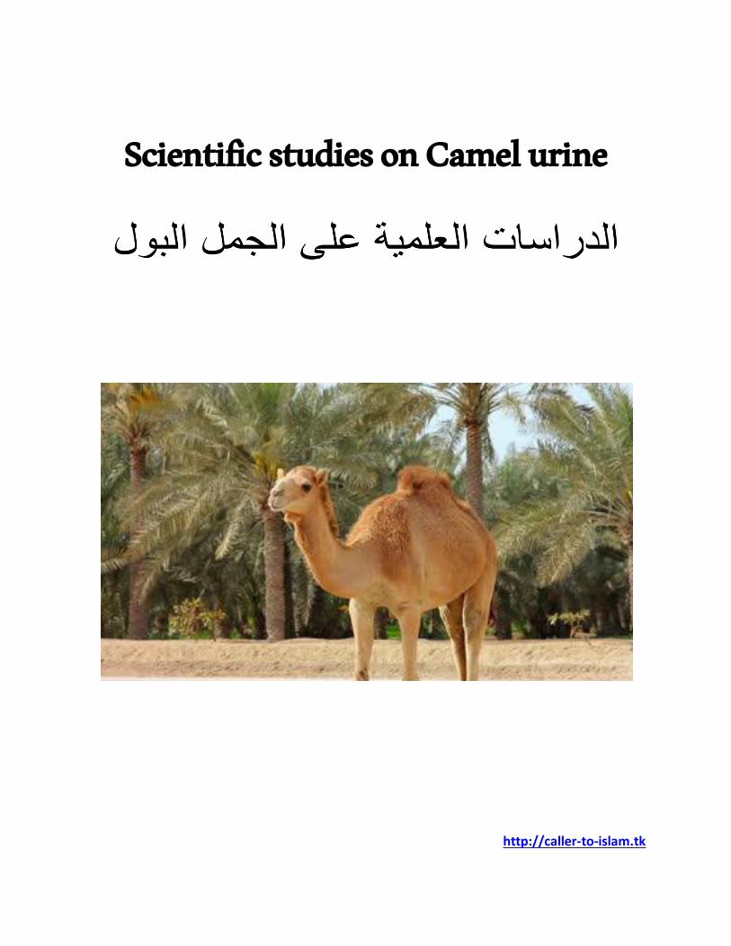

Scientific studies on Camel urine
180
Scientific studies on Camel urine http://caller-to-islam.tk
-
Upload
caller-to-islam- -
Category
Education
-
view
285 -
download
29
Transcript of Scientific studies on Camel urine
Journal of Ethnopharmacology 143 (2012) 819–825
Contents lists available at SciVerse ScienceDirect
Journal of Ethnopharmacology
0378-87
http://d
n Corr
Hospita
Arabia.
E-m
journal homepage: www.elsevier.com/locate/jep
Camel urine components display anti-cancer properties in vitro
Nujoud Al-Yousef a, Ameera Gaafar b, Basem Al-Otaibi c, Ibrahim Al-Jammaz c,Khaled Al-Hussein b, Abdelilah Aboussekhra a,n
a Department of Molecular Oncology, King Faisal Specialist Hospital and Research Center, MBC # 03, PO BOX 3354, Riyadh 11211, Saudi Arabiab Histocompatibility & Immunogenetics Research Unit, Stem Cell Therapy Program, King Faisal Specialist Hospital and Research Center, MBC # 03, PO BOX 3354,
Riyadh 11211, Saudi Arabiac Department of Cyclotron and Radiopharmaceuticals, King Faisal Specialist Hospital and Research Center, MBC # 03, PO BOX 3354, Riyadh 11211, Saudi Arabia
a r t i c l e i n f o
Article history:
Received 17 March 2012
Received in revised form
23 July 2012
Accepted 27 July 2012Available online 16 August 2012
Keywords:
Camel urine
Cancer
Apoptosis
Immune response
41/$ - see front matter & 2012 Elsevier Irelan
x.doi.org/10.1016/j.jep.2012.07.042
espondence to: Department of Molecular On
l and Research Center, MBC # 03-66, PO BOX
Tel.: þ966 1 464 7272x32840; fax: þ966 1 4
ail address: [email protected] (A. Ab
a b s t r a c t
Ethnopharmacological relevance: While camel urine (CU) is widely used in the Arabian Peninsula to
treat various diseases, including cancer, its exact mechanism of action is still not defined. The objective
of the present study is to investigate whether camel urine has anti-cancer effect on human cells in vitro.
Materials and methods: The annexinV/PI assay was used to assess apoptosis, and immunoblotting
analysis determined the effect of CU on different apoptotic and oncogenic proteins. Furthermore, flow
cytometry and Elispot were utilized to investigate cytotoxicity and the effect on the cell cycle as well as
the production of cytokines, respectively.
Results: Camel urine showed cytotoxicity against various, but not all, human cancer cell lines, with only
marginal effect on non-tumorigenic epithelial and normal fibroblast cells epithelial and fibroblast cells.
Interestingly, 216 mg/ml of lyophilized CU inhibited cell proliferation and triggered more than 80% of
apoptosis in different cancer cells, including breast carcinomas and medulloblastomas. Apoptosis was
induced in these cells through the intrinsic pathway via Bcl-2 decrease. Furthermore, CU down-
regulated the cancer-promoting proteins survivin, b-catenin and cyclin D1 and increased the level of
the cyclin-dependent kinase inhibitor p21. In addition, we have shown that CU has no cytotoxic effect
against peripheral blood mononuclear cells and has strong immuno-inducer activity through inducing
IFN-g and inhibiting the Th2 cytokines IL-4, IL-6 and IL-10.
Conclusions: CU has specific and efficient anti-cancer and potent immune-modulator properties in vitro.
& 2012 Elsevier Ireland Ltd. All rights reserved.
1. Introduction
Cancer remains a worldwide public health concern. Althoughcancer incidence has increased over the past four decades, mortalityhas remained stable. This is probably reflecting the improvement intreatment options. Chemotherapy is a core modality for the treat-ment of a wide range of cancer types at different stages. However,most of the currently used chemotherapeutic regimens are highlytoxic with long term side effects, morbidity and lethality (Rood et al.,2004; Rossi et al., 2008). Of 121 prescription drugs in use for cancertreatment, 90 are derived from plant species and 74% of these drugswere discovered by investigating a folklore claim (Craig, 1997;Craig and Beck, 1999). Among the natural products in the Arabicpeninsula that are used for the treatment of various diseases iscamel urine. Patients drink camel urine (�100 mL/day) either aloneor mixed with milk. This prompted us to ask whether the urine of
d Ltd. All rights reserved.
cology, King Faisal Specialist
3354, Riyadh 11211, Saudi
42 7858.
oussekhra).
this extraordinary animal has anticancer properties? Camel urineurine has an unusual and unique biochemical composition. Indeed,Dr. Bernard Read published in 1925 a paper describing the chemicalconstituents of camel (Camelus bactrinus) urine (Read, 1925). Hehas reported that unlike all the other animals, including humans,camels excrete no ammonia and only very slight trace of urea, andthese molecules are responsible for bad smell and toxicity of urine.However, a significant amount of creatine and creatinine wasdetected. Further studies have shown that camel urine containsabout 10 folds more mineral salts than human urine. Furthermore,while human urine is acidic, camel urine is basic with a pHZ7.8(Read, 1925). In a recent report, Alhaidar et al. have shown thatcamel urine has potent antiplatelet activity against ADP-induced(clopidogrel-like) and AA-induced (aspirin-like) platelet aggregation(Alhaidar et al., 2011). Several have claimed anti-cancer effects ofcamel urine. However, no clear scientific evidence has been pub-lished so far to confirm or refute these claims. Recently, it has beenshown that camel urine inhibits the induction of Cyp1a1, a canceractivating gene, in Hepa 1c1c7 cell line (Alhaider et al., 2011). In thepresent report we have shown that camel urine has indeed severalanti-cancer properties in vitro.
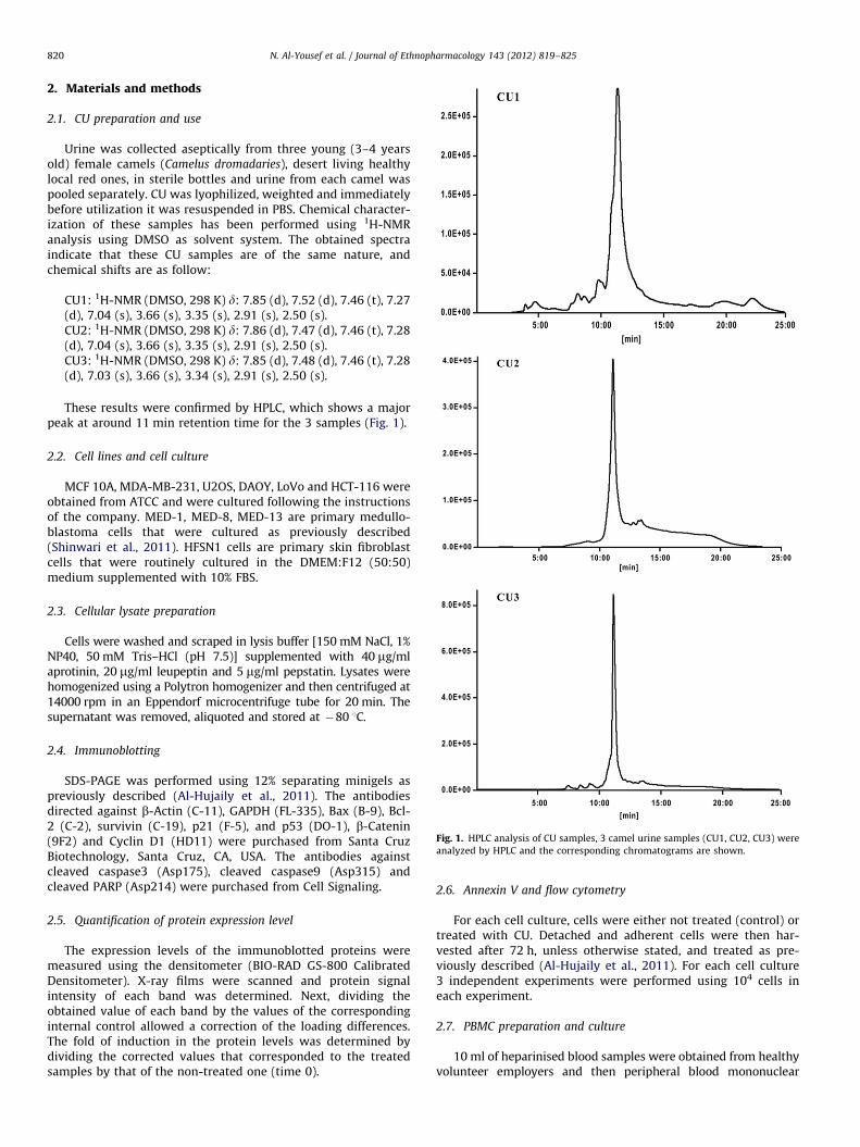
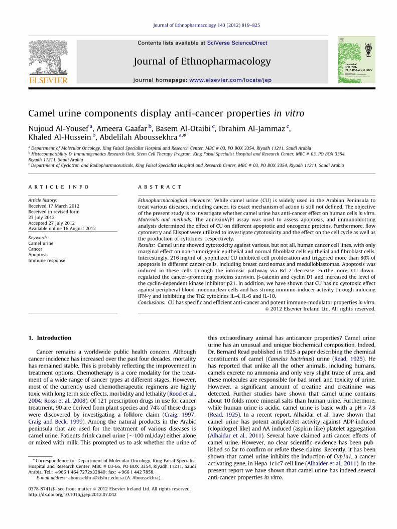
Fig. 1. HPLC analysis of CU samples, 3 camel urine samples (CU1, CU2, CU3) were
analyzed by HPLC and the corresponding chromatograms are shown.
N. Al-Yousef et al. / Journal of Ethnopharmacology 143 (2012) 819–825820
2. Materials and methods
2.1. CU preparation and use
Urine was collected aseptically from three young (3–4 yearsold) female camels (Camelus dromadaries), desert living healthylocal red ones, in sterile bottles and urine from each camel waspooled separately. CU was lyophilized, weighted and immediatelybefore utilization it was resuspended in PBS. Chemical character-ization of these samples has been performed using 1H-NMRanalysis using DMSO as solvent system. The obtained spectraindicate that these CU samples are of the same nature, andchemical shifts are as follow:
CU1: 1H-NMR (DMSO, 298 K) d: 7.85 (d), 7.52 (d), 7.46 (t), 7.27(d), 7.04 (s), 3.66 (s), 3.35 (s), 2.91 (s), 2.50 (s).CU2: 1H-NMR (DMSO, 298 K) d: 7.86 (d), 7.47 (d), 7.46 (t), 7.28(d), 7.04 (s), 3.66 (s), 3.35 (s), 2.91 (s), 2.50 (s).CU3: 1H-NMR (DMSO, 298 K) d: 7.85 (d), 7.48 (d), 7.46 (t), 7.28(d), 7.03 (s), 3.66 (s), 3.34 (s), 2.91 (s), 2.50 (s).
These results were confirmed by HPLC, which shows a majorpeak at around 11 min retention time for the 3 samples (Fig. 1).
2.2. Cell lines and cell culture
MCF 10A, MDA-MB-231, U2OS, DAOY, LoVo and HCT-116 wereobtained from ATCC and were cultured following the instructionsof the company. MED-1, MED-8, MED-13 are primary medullo-blastoma cells that were cultured as previously described(Shinwari et al., 2011). HFSN1 cells are primary skin fibroblastcells that were routinely cultured in the DMEM:F12 (50:50)medium supplemented with 10% FBS.
2.3. Cellular lysate preparation
Cells were washed and scraped in lysis buffer [150 mM NaCl, 1%NP40, 50 mM Tris–HCl (pH 7.5)] supplemented with 40 mg/mlaprotinin, 20 mg/ml leupeptin and 5 mg/ml pepstatin. Lysates werehomogenized using a Polytron homogenizer and then centrifuged at14000 rpm in an Eppendorf microcentrifuge tube for 20 min. Thesupernatant was removed, aliquoted and stored at �80 1C.
2.4. Immunoblotting
SDS-PAGE was performed using 12% separating minigels aspreviously described (Al-Hujaily et al., 2011). The antibodiesdirected against b-Actin (C-11), GAPDH (FL-335), Bax (B-9), Bcl-2 (C-2), survivin (C-19), p21 (F-5), and p53 (DO-1), b-Catenin(9F2) and Cyclin D1 (HD11) were purchased from Santa CruzBiotechnology, Santa Cruz, CA, USA. The antibodies againstcleaved caspase3 (Asp175), cleaved caspase9 (Asp315) andcleaved PARP (Asp214) were purchased from Cell Signaling.
2.5. Quantification of protein expression level
The expression levels of the immunoblotted proteins weremeasured using the densitometer (BIO-RAD GS-800 CalibratedDensitometer). X-ray films were scanned and protein signalintensity of each band was determined. Next, dividing theobtained value of each band by the values of the correspondinginternal control allowed a correction of the loading differences.The fold of induction in the protein levels was determined bydividing the corrected values that corresponded to the treatedsamples by that of the non-treated one (time 0).
2.6. Annexin V and flow cytometry
For each cell culture, cells were either not treated (control) ortreated with CU. Detached and adherent cells were then har-vested after 72 h, unless otherwise stated, and treated as pre-viously described (Al-Hujaily et al., 2011). For each cell culture3 independent experiments were performed using 104 cells ineach experiment.
2.7. PBMC preparation and culture
10 ml of heparinised blood samples were obtained from healthyvolunteer employers and then peripheral blood mononuclear
N. Al-Yousef et al. / Journal of Ethnopharmacology 143 (2012) 819–825 821
cells (PBMCs) were obtained by centrifugation over ficol-hypaquegradients (Pharmacia, Uppsala, Sweden). 106 cells were cultured inRPMI-1640 medium supplemented with 10% FCS (Gibco, Island NY,PBS) and complements and incubated at 37 1C in 5% CO2 incubator.
2.8. PBMC cytotoxicity
PBMCs were treated with different concentrations of CU for 3 daysand cell death was measured with annexinV/PI-flow cytometry.
2.9. Elispot assay
Elispot assay (Diaclone Research, France) was used as recom-mended by the manufacturer. 5.103 cells suspended in 100 mlcomplete media containing CU (16 mg/mL) and IL-2 and wereincubated for 10 days in 96-well microtiter plates pre-coated withthe appropriate antibody. Subsequently, cells were either treatedwith PHA (10 mg/ml) or LPS (1 mg/ml), for stimulating the produc-tion of IFN-g, IL-4 and IL-10 and IL-6, respectively. The plateswere incubated at 37 1C in humidified atmosphere containing 5%CO2 for appropriate period of time according to the differentcytokine kinetics. After washing, the plates were read using theElispot AID reader version 3.0.
Fig. 2. CU is cytotoxic against cancer cells but not against normal fibroblasts and non-tumo
was assessed by flow cytometry following PI staining. (A) Cells were treated with the indicate
of time. (C and D) The indicated cells were treated with CU (16 mg/mL) for 72 h. (C) Flow
2.10. Cell proliferation analysis
2–4.103 cells were seeded in 96 well plate and 100 ml ofcomplete medium was loaded in each well. The plate wasincubated for at least 30 min in a humidified, 37 1C, 5% CO2
incubator, and then was inserted into the Real-Time Cell Electro-nic Sensing System (RT-CES system) (ACEA Biosciences Inc.,San Diego, CA) for 16 h. Cells were then either PBS-treated ortreated with CU (16 mg/mL) and cell proliferation was monitoredfor 24 h.
2.11. HPLC analysis
HPLC analysis was carried out on Econosil C-18 reversed phasecolumn (analytical, 250 mm�4.6 mm). The solvent system usedwas non-linear gradient (eluent A, water with 0.1% TFA; eluent B,ACN; gradient, 0–10% B, 10–90% B, 90–90% B and 90–0% B over5 min each at flow rate of 1.0 mL/min). A Jasco chromatographicsystem equipped with a variable wavelength ultraviolet monitorand in tandem with a Canberra flow through radioactivitydetector was used. Ultraviolet absorption was monitored at254 nm. Chromatograms were acquired and analyzed usingBORWIN software.
rigenic epithelial cells. Cells were treated with CU as indicated and the cytotoxic effect
d CU doses for 72 h. (B) Cells were treated with CU (16 mg/mL) for the indicated periods
cytometry charts. (D) Histogram, Error bars represent means7S.D. *: p valueo0.05.

N. Al-Yousef et al. / Journal of Ethnopharmacology 143 (2012) 819–825822
2.12. Statistical analysis
Statistical analysis was performed by student’s t-test and p
values of 0.05 and less were considered as statistically significant.
3. Results
3.1. Camel urine is cytotoxic against various cancer cells
We started this study by investigating the cytotoxic effect of CUagainst the MDA-MB-231 breast cancer cells as well as the non-tumorigenic breast epithelial cells (MCF 10A), using the PropidiumIodide (PI)/flow cytometry technique. Cells were treated with increas-ing concentrations of CU for 72 h. Fig. 2A shows dose-dependentincrease in the proportion of death cells among MDA-MB-231 cells,more than 80% died in response to 16 mg/ml (�800 ml of urine). Onthe other hand, the same concentrations of CU had no effect on MCF10A cells. Next, we investigated the effect of CU over time using16 mg/ml. Fig. 2B shows that the effect of CU on breast cancer cellsincreased with time reaching its maximum at 72 h of treatment,while no effect was observed on MCF 10A cells. This shows that CU iscytotoxic, but with specific effect on breast cancer cells.
Fig. 3. CU induces apoptosis through the mitochondrial pathway. Cells were treated eit
annexin V/PI in association with flow cytometry. (A) Charts, the numbers into the boxe
apoptotic (higher, right) and necrotic (higher, left) cells. (B) Histograms presenting the p
represent standard deviations of three different experiments, *: p valueo0.05. (C) MDA-
indicated periods of time. 50 mg of extracted proteins were used for western blot analy
corresponding expression levels as compared to time 0 and after normalization against
the Bax/Bcl-2 ratios after normalization against b-actin. Error bars represent standard
Next, we investigated the specific cytotoxic effect of CU onother cancer cell lines. Fig. 1C shows that based on the responseto CU, these cells can be grouped into 2 different sub-groups.Group 1 contains CU-resistant cells including MCF 10A and thenormal fibroblast (HFSN-1) cells, as well as the osteosarcoma(U2OS), the breast cancer (MCF-7), the medulloblastoma MED-8and the colon cancer (LoVo and HCT-116) cells. The second groupis composed of CU-sensitive cells, with more than 50% cell death,and includes the breast cancer (MDA-MB-231) and the medullo-blastoma (DAOY, MED-4 and MED-13) cells (Figs. 2C, 1D). Inter-estingly, these effects were obtained with similar doses of urinecollected from 2 other female camels (data not shown), indicatingthat these CU samples collected from different animals living indifferent regions and receiving different foods have similarchemical characteristics (materials and methods) and cytotoxiceffect against cancer cells. Therefore, CU powders from thesecamels were pooled and used in the next experiments.
3.2. Camel urine triggers apoptosis in cancer cells
In order to identify the cell death pathway that CU triggers incancer cells, we first made use of the AnnexinV-PI/flow cytometrytechnique that can detect both apoptotic and necrotic cells. Fig. 3
her with PBS or with CU (16 mg/mL) for 72 h, and then cell death was assessed by
s indicate the proportion of normal (lower, left), early apoptotic (lower, right), late
roportions of induced apoptosis in the indicated normal and cancer cells. Error bars
MB-231 cells were treated with CU (16 mg/mL), and then were harvested after the
sis utilizing the indicated antibodies. The numbers below the bands represent the
GAPDH. D. As in C, b-actin was used as internal control and the graph is showing
deviations of three different experiments.
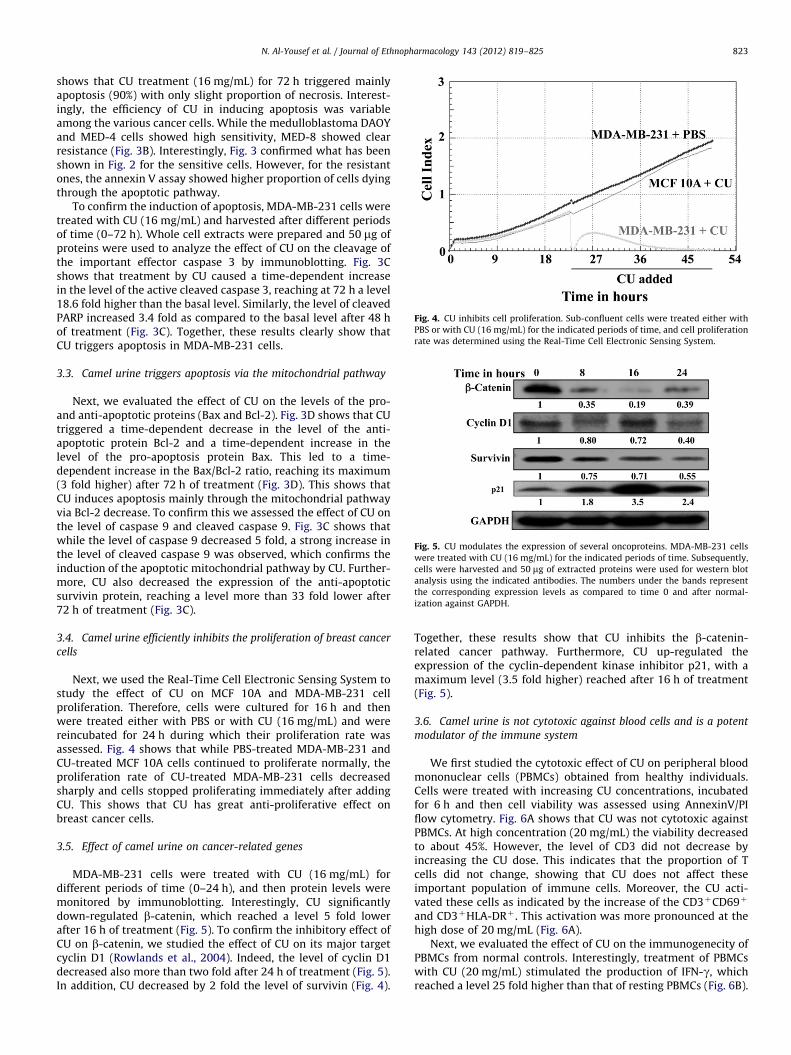
Fig. 4. CU inhibits cell proliferation. Sub-confluent cells were treated either with
PBS or with CU (16 mg/mL) for the indicated periods of time, and cell proliferation
rate was determined using the Real-Time Cell Electronic Sensing System.
Fig. 5. CU modulates the expression of several oncoproteins. MDA-MB-231 cells
were treated with CU (16 mg/mL) for the indicated periods of time. Subsequently,
cells were harvested and 50 mg of extracted proteins were used for western blot
analysis using the indicated antibodies. The numbers under the bands represent
the corresponding expression levels as compared to time 0 and after normal-
ization against GAPDH.
N. Al-Yousef et al. / Journal of Ethnopharmacology 143 (2012) 819–825 823
shows that CU treatment (16 mg/mL) for 72 h triggered mainlyapoptosis (90%) with only slight proportion of necrosis. Interest-ingly, the efficiency of CU in inducing apoptosis was variableamong the various cancer cells. While the medulloblastoma DAOYand MED-4 cells showed high sensitivity, MED-8 showed clearresistance (Fig. 3B). Interestingly, Fig. 3 confirmed what has beenshown in Fig. 2 for the sensitive cells. However, for the resistantones, the annexin V assay showed higher proportion of cells dyingthrough the apoptotic pathway.
To confirm the induction of apoptosis, MDA-MB-231 cells weretreated with CU (16 mg/mL) and harvested after different periodsof time (0–72 h). Whole cell extracts were prepared and 50 mg ofproteins were used to analyze the effect of CU on the cleavage ofthe important effector caspase 3 by immunoblotting. Fig. 3Cshows that treatment by CU caused a time-dependent increasein the level of the active cleaved caspase 3, reaching at 72 h a level18.6 fold higher than the basal level. Similarly, the level of cleavedPARP increased 3.4 fold as compared to the basal level after 48 hof treatment (Fig. 3C). Together, these results clearly show thatCU triggers apoptosis in MDA-MB-231 cells.
3.3. Camel urine triggers apoptosis via the mitochondrial pathway
Next, we evaluated the effect of CU on the levels of the pro-and anti-apoptotic proteins (Bax and Bcl-2). Fig. 3D shows that CUtriggered a time-dependent decrease in the level of the anti-apoptotic protein Bcl-2 and a time-dependent increase in thelevel of the pro-apoptosis protein Bax. This led to a time-dependent increase in the Bax/Bcl-2 ratio, reaching its maximum(3 fold higher) after 72 h of treatment (Fig. 3D). This shows thatCU induces apoptosis mainly through the mitochondrial pathwayvia Bcl-2 decrease. To confirm this we assessed the effect of CU onthe level of caspase 9 and cleaved caspase 9. Fig. 3C shows thatwhile the level of caspase 9 decreased 5 fold, a strong increase inthe level of cleaved caspase 9 was observed, which confirms theinduction of the apoptotic mitochondrial pathway by CU. Further-more, CU also decreased the expression of the anti-apoptoticsurvivin protein, reaching a level more than 33 fold lower after72 h of treatment (Fig. 3C).
3.4. Camel urine efficiently inhibits the proliferation of breast cancer
cells
Next, we used the Real-Time Cell Electronic Sensing System tostudy the effect of CU on MCF 10A and MDA-MB-231 cellproliferation. Therefore, cells were cultured for 16 h and thenwere treated either with PBS or with CU (16 mg/mL) and werereincubated for 24 h during which their proliferation rate wasassessed. Fig. 4 shows that while PBS-treated MDA-MB-231 andCU-treated MCF 10A cells continued to proliferate normally, theproliferation rate of CU-treated MDA-MB-231 cells decreasedsharply and cells stopped proliferating immediately after addingCU. This shows that CU has great anti-proliferative effect onbreast cancer cells.
3.5. Effect of camel urine on cancer-related genes
MDA-MB-231 cells were treated with CU (16 mg/mL) fordifferent periods of time (0–24 h), and then protein levels weremonitored by immunoblotting. Interestingly, CU significantlydown-regulated b-catenin, which reached a level 5 fold lowerafter 16 h of treatment (Fig. 5). To confirm the inhibitory effect ofCU on b-catenin, we studied the effect of CU on its major targetcyclin D1 (Rowlands et al., 2004). Indeed, the level of cyclin D1decreased also more than two fold after 24 h of treatment (Fig. 5).In addition, CU decreased by 2 fold the level of survivin (Fig. 4).
Together, these results show that CU inhibits the b-catenin-related cancer pathway. Furthermore, CU up-regulated theexpression of the cyclin-dependent kinase inhibitor p21, with amaximum level (3.5 fold higher) reached after 16 h of treatment(Fig. 5).
3.6. Camel urine is not cytotoxic against blood cells and is a potent
modulator of the immune system
We first studied the cytotoxic effect of CU on peripheral bloodmononuclear cells (PBMCs) obtained from healthy individuals.Cells were treated with increasing CU concentrations, incubatedfor 6 h and then cell viability was assessed using AnnexinV/PIflow cytometry. Fig. 6A shows that CU was not cytotoxic againstPBMCs. At high concentration (20 mg/mL) the viability decreasedto about 45%. However, the level of CD3 did not decrease byincreasing the CU dose. This indicates that the proportion of Tcells did not change, showing that CU does not affect theseimportant population of immune cells. Moreover, the CU acti-vated these cells as indicated by the increase of the CD3þCD69þ
and CD3þHLA-DRþ . This activation was more pronounced at thehigh dose of 20 mg/mL (Fig. 6A).
Next, we evaluated the effect of CU on the immunogenecity ofPBMCs from normal controls. Interestingly, treatment of PBMCswith CU (20 mg/mL) stimulated the production of IFN-g, whichreached a level 25 fold higher than that of resting PBMCs (Fig. 6B).
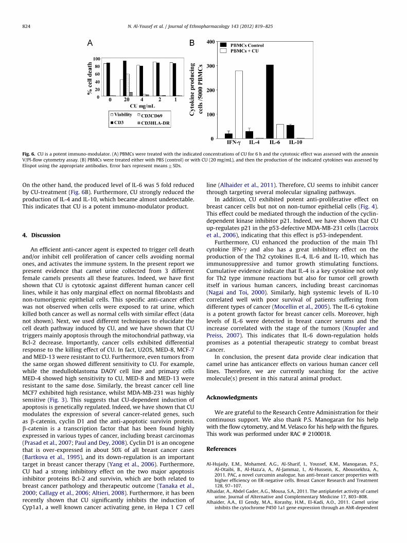
Fig. 6. CU is a potent immuno-modulator. (A) PBMCs were treated with the indicated concentrations of CU for 6 h and the cytotoxic effect was assessed with the annexin
V/PI-flow cytometry assay. (B) PBMCs were treated either with PBS (control) or with CU (20 mg/mL), and then the production of the indicated cytokines was assessed by
Elispot using the appropriate antibodies. Error bars represent means7SDs.
N. Al-Yousef et al. / Journal of Ethnopharmacology 143 (2012) 819–825824
On the other hand, the produced level of IL-6 was 5 fold reducedby CU-treatment (Fig. 6B). Furthermore, CU strongly reduced theproduction of IL-4 and IL-10, which became almost undetectable.This indicates that CU is a potent immuno-modulator product.
4. Discussion
An efficient anti-cancer agent is expected to trigger cell deathand/or inhibit cell proliferation of cancer cells avoiding normalones, and activates the immune system. In the present report wepresent evidence that camel urine collected from 3 differentfemale camels presents all these features. Indeed, we have firstshown that CU is cytotoxic against different human cancer celllines, while it has only marginal effect on normal fibroblasts andnon-tumorigenic epithelial cells. This specific anti-cancer effectwas not observed when cells were exposed to rat urine, whichkilled both cancer as well as normal cells with similar effect (datanot shown). Next, we used different techniques to elucidate thecell death pathway induced by CU, and we have shown that CUtriggers mainly apoptosis through the mitochondrial pathway, viaBcl-2 decrease. Importantly, cancer cells exhibited differentialresponse to the killing effect of CU. In fact, U2OS, MED-8, MCF-7and MED-13 were resistant to CU. Furthermore, even tumors fromthe same organ showed different sensitivity to CU. For example,while the medulloblastoma DAOY cell line and primary cellsMED-4 showed high sensitivity to CU, MED-8 and MED-13 wereresistant to the same dose. Similarly, the breast cancer cell lineMCF7 exhibited high resistance, whilst MDA-MB-231 was highlysensitive (Fig. 3). This suggests that CU-dependent induction ofapoptosis is genetically regulated. Indeed, we have shown that CUmodulates the expression of several cancer-related genes, suchas b-catenin, cyclin D1 and the anti-apoptotic survivin protein.b-catenin is a transcription factor that has been found highlyexpressed in various types of cancer, including breast carcinomas(Prasad et al., 2007; Paul and Dey, 2008). Cyclin D1 is an oncogenethat is over-expressed in about 50% of all breast cancer cases(Bartkova et al., 1995), and its down-regulation is an importanttarget in breast cancer therapy (Yang et al., 2006). Furthermore,CU had a strong inhibitory effect on the two major apoptosisinhibitor proteins Bcl-2 and survivin, which are both related tobreast cancer pathology and therapeutic outcome (Tanaka et al.,2000; Callagy et al., 2006; Altieri, 2008). Furthermore, it has beenrecently shown that CU significantly inhibits the induction ofCyp1a1, a well known cancer activating gene, in Hepa 1 C7 cell
line (Alhaider et al., 2011). Therefore, CU seems to inhibit cancerthrough targeting several molecular signaling pathways.
In addition, CU exhibited potent anti-proliferative effect onbreast cancer cells but not on non-tumor epithelial cells (Fig. 4).This effect could be mediated through the induction of the cyclin-dependent kinase inhibitor p21. Indeed, we have shown that CUup-regulates p21 in the p53-defective MDA-MB-231 cells (Lacroixet al., 2006), indicating that this effect is p53-independent.
Furthermore, CU enhanced the production of the main Th1cytokine IFN-g and also has a great inhibitory effect on theproduction of the Th2 cytokines IL-4, IL-6 and IL-10, which hasimmunosuppressive and tumor growth stimulating functions.Cumulative evidence indicate that IL-4 is a key cytokine not onlyfor Th2 type immune reactions but also for tumor cell growthitself in various human cancers, including breast carcinomas(Nagai and Toi, 2000). Similarly, high systemic levels of IL-10correlated well with poor survival of patients suffering fromdifferent types of cancer (Mocellin et al., 2005). The IL-6 cytokineis a potent growth factor for breast cancer cells. Moreover, highlevels of IL-6 were detected in breast cancer serums and theincrease correlated with the stage of the tumors (Knupfer andPreiss, 2007). This indicates that IL-6 down-regulation holdspromises as a potential therapeutic strategy to combat breastcancer.
In conclusion, the present data provide clear indication thatcamel urine has anticancer effects on various human cancer celllines. Therefore, we are currently searching for the activemolecule(s) present in this natural animal product.
Acknowledgments
We are grateful to the Research Centre Administration for theircontinuous support. We also thank P.S. Manogaran for his helpwith the flow cytometry, and M. Velasco for his help with the figures.This work was performed under RAC # 2100018.
References
Al-Hujaily, E.M., Mohamed, A.G., Al-Sharif, I., Youssef, K.M., Manogaran, P.S.,Al-Otaibi, B., Al-Haza’a, A., Al-Jammaz, I., Al-Hussein, K., Aboussekhra, A.,2011. PAC, a novel curcumin analogue, has anti-breast cancer properties withhigher efficiency on ER-negative cells. Breast Cancer Research and Treatment128, 97–107.
Alhaidar, A., Abdel Gader, A.G., Mousa, S.A., 2011. The antiplatelet activity of camelurine. Journal of Alternative and Complementary Medicine 17, 803–808.
Alhaider, A.A., El Gendy, M.A., Korashy, H.M., El-Kadi, A.O., 2011. Camel urineinhibits the cytochrome P450 1a1 gene expression through an AhR-dependent
N. Al-Yousef et al. / Journal of Ethnopharmacology 143 (2012) 819–825 825
mechanism in Hepa 1c1c7 cell line. Journal of Ethnopharmacology 133,184–190.
Altieri, D.C., 2008. Survivin, cancer networks and pathway-directed drug discovery.Nature Reviews Cancer 8, 61–70.
Bartkova, J., Lukas, J., Strauss, M., Bartek, J., 1995. Cyclin D1 oncoprotein aberrantlyaccumulates in malignancies of diverse histogenesis. Oncogene 10, 775–778.
Callagy, G.M., Pharoah, P.D., Pinder, S.E., Hsu, F.D., Nielsen, T.O., Ragaz, J., Ellis, I.O.,Huntsman, D., Caldas, C., 2006. Bcl-2 is a prognostic marker in breast cancerindependently of the Nottingham Prognostic Index. Clinical Cancer Research12, 2468–2475.
Craig, W., Beck, L., 1999. Phytochemicals: health protective Effects. CanadianJournal of Dietetic Practice and Research 60, 78–84.
Craig, W.J., 1997. Phytochemicals: guardians of our health. Journal of the AmericanDietetic Association 97, S199–204.
Knupfer, H., Preiss, R., 2007. Significance of interleukin-6 (IL-6) in breast cancer(review). Breast Cancer Research and Treatment 102, 129–135.
Lacroix, M., Toillon, R.A., Leclercq, G., 2006. p53 and breast cancer, an update.Endocrine-Related Cancer 13, 293–325.
Mocellin, S., Marincola, F.M., Young, H.A., 2005. Interleukin-10 and the immuneresponse against cancer: a counterpoint. Journal of Leukocyte Biology 78,1043–1051.
Nagai, S., Toi, M., 2000. Interleukin-4 and breast cancer. Breast Cancer 7, 181–186.Paul, S., Dey, A., 2008. Wnt signaling and cancer development: therapeutic
implication. Neoplasma 55, 165–176.
Prasad, C.P., Gupta, S.D., Rath, G., Ralhan, R., 2007. Wnt signaling pathway ininvasive ductal carcinoma of the breast: relationship between beta-catenin,dishevelled and cyclin D1 expression. Oncology 73, 112–117.
Read, B.E., 1925. Chemical constituents of camel’s urine. Journal of BiologicalChemistry 64, 615–617.
Rood, B.R., Macdonald, T.J., Packer, R.J., 2004. Current treatment of medulloblas-toma: recent advances and future challenges. Seminars in Oncology 31,
666–675.Rossi, A., Caracciolo, V., Russo, G., Reiss, K., Giordano, A., 2008. Medulloblastoma:
from molecular pathology to therapy. Clinical Cancer Research 14, 971–976.Rowlands, T.M., Pechenkina, I.V., Hatsell, S., Cowin, P., 2004. Beta-catenin and
cyclin D1: connecting development to breast cancer. Cell Cycle 3, 145–148.Shinwari, Z., Al-Hindi, H., Al-Shail, E., Khafaga, Y., Al-Kofide, A., Al-Kum, N.,
Aboussekhra, A., 2011. Response of medulloblastoma cells to vincristine and
lomustine: role of TRKC, CTNNB1 and STK15. Anticancer Research 31,1721–1734.
Tanaka, K., Iwamoto, S., Gon, G., Nohara, T., Iwamoto, M., Tanigawa, N., 2000.Expression of survivin and its relationship to loss of apoptosis in breastcarcinomas. Clinical Cancer Research 6, 127–134.
Yang, C., Trent, S., Ionescu-Tiba, V., Lan, L., Shioda, T., Sgroi, D., Schmidt, E.V., 2006.Identification of cyclin D1- and estrogen-regulated genes contributing to
breast carcinogenesis and progression. Cancer Research 66, 11649–11658.
Original Articles
The Antiplatelet Activity of Camel Urine
Abdulqader Alhaidar, BPharm, MSc, PhD,1 Abdel Galil M. Abdel Gader, MD, PhD,1
and Shaker A. Mousa, PhD, MBA1,2
Abstract
Background: For centuries, camel urine has been used for medicinal purposes and anecdotally proclaimed as acure for a wide range of diseases. However, the apparent therapeutic actions of camel urine have yet to besubjected to rigorous scientific scrutiny. Recent preliminary studies from the authors’ laboratory have indicatedthat camel urine possesses potent antiplatelet activity, not found in human or bovine urines, suggesting apossible role for camel urine in inhibiting platelet function. The goal of the current study was to characterize theantiplatelet activity of camel urine against normal human platelets based on agonist-induced aggregation andplatelet function analyzer (PFA-100) closure time.Materials and methods: Urine was collected from healthy virgin, pregnant, and lactating camels aged 2–10years. Platelet-rich plasma (PRP) was prepared from blood collected from healthy individuals’ blood into ci-trated anticoagulant. Agonist-induced aggregometry using donor PRP and PFA-100 closure times in wholeblood were carried out in the presence and absence of added camel urine. The responses of platelets to multipledoses of camel urine were also assessed. The experimental procedure was repeated in human and bovine urines.Results: Camel urine completely inhibited arachidonic acid (AA) and adnosine diphosphate (ADP)–inducedaggregation of human platelets in a dose-dependent manner. PFA-100 closure time using human whole bloodwas prolonged following the addition of camel urine in a dose-dependent manner. Virgin camel urine was lesseffective in inhibiting ADP-induced aggregation as compared to urine from lactating and pregnant camels; however,all three showed comparable inhibitory activity. Neither human nor bovine urine exhibited antiplatelet activity.Conclusions: Camel urine has potent antiplatelet activity against ADP-induced (clopidogrel-like) and AA-induced (aspirin-like) platelet aggregation; neither human nor bovine urine exhibited such properties. These novelresults provide the first scientific evidence of the mechanism of the presumed therapeutic properties of camel urine.
Introduction
The one-humped camel (Camelus dromedaries) survivesand reproduces under conditions of extreme drought and
heat that are unsustainable to most other species of domesticmammal. Desert dwellers have used the camel for transpor-tation and as a source of food, but just as importantly, its milkand urine have been used as medicines for centuries.1,2 Camelmilk and urine, for example, have been used to treat variousailments such as cancer,3,4 chronic hepatitis,5 hepatitis C,6,7
and peptic ulcers.8 More recently, it has been reported thatcamel milk can be used to successfully treat severe food al-lergies in children who are unresponsive to more conven-tional treatments.9
Most of the claimed therapeutic benefits of camel milkand urine are attributed variously to anti-infective, anti-
inflammatory, and anticancer properties; by comparison,very little information is available on the efficacy of camelurine and/or milk in treating cardiovascular diseases. Short-chain peptides prepared from bovine milk have been shownto have potent antihypertensive angiotensin-converting en-zyme inhibitory action, because they can significantly reduceblood pressure after intravenous or oral administration, butthey show little or no effect in normotensive subjects.10,11 Bycontrast, none of the claims of therapeutic benefit of camelurine or milk have been subjected to rigorous scientificscrutiny, and as a result, skepticism about camel urine, inparticular as a form of alternative therapy, is strong. Alongwith this, there is a severe shortage of information on theconstituents of camel milk and urine.
The authors’ interest in this area stems from recent workin our laboratory characterizing camel platelets, in which it
1The Coagulation Research Laboratory, Department of Physiology, College of Medicine and King Khalid University Hospital, King SaudUniversity, Riyadh, Saudi Arabia.
2The Pharmaceutical Research Institute, Albany College of Pharmacy and Health Sciences, Rensselaer, NY.
THE JOURNAL OF ALTERNATIVE AND COMPLEMENTARY MEDICINEVolume 17, Number 9, 2011, pp. 803–808ª Mary Ann Liebert, Inc.DOI: 10.1089/acm.2010.0473
803
was demonstrated that the ultrastructure and function ofcamel platelets bears a high degree of dissimilarity as com-pared to human platelets.12,13 In addition, camels exhibitedmarkedly inhibited platelet function in terms of agonist-induced aggregation responses and platelet function ana-lyzer (PFA-100) closure times.12 Notably, the addition of camelplatelet–poor plasma to packed human erythrocytes resultedin a prolongation of PFA-100 closure time in human bloodsamples (Abdul Gader, unpublished observations). These re-sults suggest that camel plasma has antiplatelet properties.The authors set out to investigate whether camel urine hassimilar antiplatelet activity, perhaps lending credence to theclaims of the therapeutic benefit of camel urine.
The aim of the current study was to characterize the an-tiplatelet actions of camel urine on normal human plateletsbased on agonist-induced aggregation responses and PFA-100 closure times. Camel urine exhibited potent platelet in-hibitory activity, blocking both the prostaglandin pathway(aspirin-like activity) as well as the adenosine diphosphate(ADP) receptor–mediated pathway (clopidogrel-like activity).Neither type of activity was detectable in human or bovineurine. These novel findings offer the first scientific evidence insupport of the putative therapeutic properties of camel urine.
Materials and Methods
Animals and urine collection
Urine was collected from healthy virgin, pregnant, andlactating domesticated camels (Camelus dromedaries). Allcamels were females, aged 2–10 years. The camels wereraised on a private farm, were disease-free, and had freeaccess to water and camel feed. The collection of urine wasusually carried out during feeding and was performed byexperienced camel attendants. Urine was allowed to flowdirectly into stainless steel containers and then transferred toglass vials. Urine samples were transported to the laboratoryas soon as practical ( < 4 hours) and were stored at - 80�Cuntil use. Human and bovine urine was collected and storedin a similar manner.
Collection of human blood for platelet aggregationand PFA-100 studies
Healthy volunteers were recruited from among blooddonors, staff, medical students, and residents of our institu-tion. Specific inquiry was made about the ingestion of aspi-rin, nonsteroidal anti-inflammatory drugs (NSAIDs), andany form of cold therapy, at least 2 weeks before bloodcollection. Whole blood was drawn by clean venipuncturedirectly into vacutainer plastic tubes (Terumu Co., Japan)containing 3.8% (0.129 M) or 3.2% (0.105 M) buffered sodiumcitrate to yield a blood:anti-coagulant ratio of 9:1.
Preparation of platelet-rich plasmaand platelet-poor plasma
Platelet-rich plasma (PRP) was obtained by centrifugationof citrated whole blood at 800–1000 rpm for 5 minutes. PRPwas removed and the remaining sample was subjected to asecond round of centrifugation at 3000 rpm for 10 minutes toobtain platelet-poor plasma (PPP). The platelet count of PRPwas in the range of 200–300 · 09/L. PRP was adjusted to aconcentration of 250,000 – 50,000 with PPP.
Platelet aggregometry
The processing of blood samples and agonist-inducedplatelet aggregation technique were carried out as previouslydescribed using a Platelet Aggregation Profile� (PAP-4)system (BioData, Horsham, PA).14,15 Arachidonic acid (AA)(BioData) was reconstituted from a lyophilized preparationof sodium arachidonate using distilled water to yield aworking concentration of 5 mg/mL. ADP (BioData) was re-constituted from a lyophilized preparation with distilledwater to yield a working concentration of 2 · 10 - 4 M. Specialmacrocuvettes (8.75 · 50 mm) were used for all experiments.Briefly, using plastic tips, 0.45 mL of PRP were pipetted intothe cuvette. Raw camel urine (0.05 mL) was added and themixture was stirred with a plastic-coated magnetic stirrer for2 minutes, after which 0.05 mL of the aggregating agent wasadded and the recording was started. Aggregation parame-ters of maximum aggregation (%) versus control (PPP) andthe slope of the aggregation curve were recorded.
PFA-100 closure time
The PFA-100 assay (PFA-100�; Dade Behring, USA) wascarried out as previously described.12 The PFA100 is a devicethat measures platelet-related primary hemostasis in citratedwhole blood specimens. It uses two disposable cartridgesfitted with a membrane with central aperture (147 lm)coated with aggregation agonists (collagen and epinephrineand collegen and ADP), through which platelets are passedat high shear rates (5000–6000 s - 1). The PFA-100� deter-mines in whole blood the time (in seconds) elapsed from thestart of the test until a platelet plug occludes the aperture.This time interval is referred to as closure time (CT), and isan indicator of platelet function (adhesion and aggregation).The system was programmed to stop recording when the CTreached ‡ 300 seconds.
Preparation of the test cartridges. The pouch containingthe test cartridges was allowed to warm up to room tem-perature prior to opening (approximately 15 minutes). Afterremoval of the cartridges, the pouch was immediately closedusing the reclosable seal. The top foil seal was removed fromthe test cartridge and discarded, and then the test cartridge(s)was placed in the cassette of the PFA-100 and snapped se-curely into place.
Sample loading. The following steps were performed insequence without interruption.
1. The blood sample was mixed by inverting the collectiontube gently by hand 3–4 times. While the cassette con-taining the test cartridge was held on a flat surface,800 lL of blood was pipette into the sample reservoir bydispensing slowly along one of the inside corners. Thisreduces the risk of air entrapment in the sample reser-voir.
2. The cassette with test cartridge containing sample wasplaced into the incubation well(s) of the instrumentsuch that the cassette was flush with the carousel sur-face, and then recording was started.
The system was programmed to stop recording when theaperture closed, or 300 seconds, whichever came first.
804 ALHAIDAR ET AL.
Statistical analysis
Data were analyzed using the SSPS program (Version 15).Differences in means between groups were compared usingthe Mann–Whitney test. Analysis of variance was conductedusing the Kruskal–Wallis test. Proportions from two or moreindependent groups were compared using either the v2 testor Fisher’s Exact test, as appropriate. A p-value £ 0.05 wasconsidered statistically significant.
Results
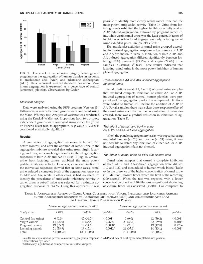
A comparison of aggregation responses of human PRPbefore (control) and after the addition of camel urine to theaggregation mixture revealed that urine from virgin, lactat-ing, and pregnant camels significantly inhibited aggregationresponses to both ADP and AA ( p < 0.001) (Fig. 1). Overall,urine from lactating camels exhibited the most potentplatelet inhibitory activity. However, close examination ofthe individual responses showed that in some cases, camelurine induced a complete block of the aggregation responsesto ADP and AA, while in other cases, it had no effect. Toidentify the prevalence of antiplatelet inhibitory activity incamel urine, a cut-off value was selected for maximum ag-gregation response of £ 40%. Using this approach, it was
possible to identify more clearly which camel urine had themost potent antiplatelet activity (Table 1). Urine from lac-tating camels exhibited the highest inhibitory activity againstADP-induced aggregation, followed by pregnant camel ur-ine, while virgin camel urine was the least potent. In terms ofinhibition of AA-induced aggregation, only lactating camelurine exhibited potent antiplatelet effects.
The antiplatelet activities of camel urine grouped accord-ing to maximal aggregation response in the presence of ADPand AA are shown in Table 2. Inhibition of both ADP- andAA-induced aggregation differed significantly between lac-tating (50%), pregnant (29.7%), and virgin (22.4%) urinesamples ( p = 0.0151; v2 test). These results indicated thatlactating camel urine is the most potent inhibitor of humanplatelet aggregation.
Dose–response AA and ADP-induced aggregationby camel urine
Serial dilutions (neat, 1:2, 1:4, 1:8) of camel urine samplesthat exhibited complete inhibition of either AA- or ADP-induced aggregation of normal human platelets were pre-pared and the aggregation protocol was repeated. Dilutionswere added to human PRP before the addition of ADP orAA. For all samples, there was a clear dose–response effect ofthe camel urine such that as the concentration of urine de-creased, there was a gradual reduction in inhibition of ag-gregation (Table 3).
The effect of human and bovine urineon ADP- and AA-induced aggregation
When the platelet aggregometry assay was repeated usingundiluted human (n = 20) and bovine (n = 24) urine, it wasnot possible to detect any inhibition of either AA- or ADP-induced aggregation (data not shown).
The effect of camel urine on PFA-100 closure time
Camel urine samples that caused a complete inhibitionof both ADP- and AA-induced aggregation were diluted1:10 and 1:20, and then added to human whole blood (Table4). In the presence of the higher concentration of camel urine(1:10 dilution), closure times exceed the limit of the recording(300 second). When the test was repeated with a lowerconcentration of urine (1:20 dilution), a significant shorteningof closure times was observed ( p < 0.001) as compared to
FIG. 1. The effect of camel urine (virgin, lactating, andpregnant) on the aggregation of human platelets in responseto arachidonic acid (Arch) and adenosine diphosphate(ADP). Data represent means – standard deviation. Max-imum aggregation is expressed as a percentage of control(untreated) platelets. Observations by Gader.
Table 1. Antiplatelet Action of Camel Urine Collected from Virgin, Pregnant, and Lactating Animals
on the Aggregation Responses to Adenosine Diphosphate (ADP) and Arachidonic Acid (AA)of Healthy Human Platelet-Rich Plasma
Maximum aggregation response to ADP Maximum aggregation response to AA
Study group £ 40% > 40% p-Value £ 40% > 40% p-Value
Control (no urine) 0 (0.0) 42 (34.2) < 0.001* 0 (0.0) 42 (39.2) < 0.001*Virgin camels 14 (25.9) 44 (35.8) 0.2665 26 (37.1) 32 (29.9) 0.4014Pregnant camels 19 (35.2) 18 (14.6) 0.0038* 18 (25.8) 19 (17.8) 0.2784Lactating camels 21 (38.9) 19 (15.4) 0.0012* 26 (37.1) 14 (13.1) < 0.001*Total 54 (100.0) 123 (100.0) 70 (100.0) 107 (100.0)
Results are expressed as percent maximum aggregation response to ADP and AA of healthy human platelet-rich plasma.Observations by Gader.*Statistically significant as compared to untreated samples.
ANTIPLATELET ACTIVITY OF CAMEL URINE 805
samples treated with the lower dilutions (mean of < 300)(Table 4).
Discussion
Urine therapy, or urotherapy, has been in practice sinceearly historic times. A search of multiple electronic literaturedatabases yields a plethora of information on the use of ur-ine, particularly human urine, with claims of successfultreatment of a wide range of human ailments. However, al-most all the available information can be categorized as al-ternative medical practice by healers in many countries,particularly those where the practice of alternative medicineis prevalent such as India and China, with scant reportingfrom the United States, United Kingdom, and other Euro-pean countries. The perceived success of such therapeuticefforts by those who believe in the efficacy of urine therapy,whether through practice or personal experience, hasprompted several books on urine therapy that have foundwide readership.16–18 Many of the books and reports on ur-otherapy advocate the use of human urine therapy, partic-ularly using the individual’s own urine.
Despite numerous claims of efficacy, the practice of ur-otherapy has yet to be subjected to scientific research, andeven in situations where this form of therapy was prescribedor advised by qualified physicians, there are no studies thatoffer scientific support of such a practice. Therefore, atpresent, the practice of urine therapy should be viewed as
unorthodox medical practice based primarily on trial anderror, and not a field that has been subjected to rigorousscientific scrutiny.
There have been a few isolated references to the use ofbovine urine in Tibet and India, and the use of llama urine (amember of the Camelidae family) in Tibet, Mongolia, andChina.18 The use of camel urine for therapeutic purposes ispracticed widely among tribes that raise camels, both in Asiaand Africa. In the Middle East, there is credible evidence thatProphet Mohamed advised the use of camel urine for thetreatment of a wide range of disease conditions.1,2 There arenumerous claims of the success of camel urine therapy in themanagement of a range of diseases from liver cirrhosis toskin and hair ailments.17 Cancer is prominent among thediseases that are reportedly treatable by urine (human andcamel). Recent studies have shown both in vitro (tissue cul-ture) and in vivo in humans and animals that a componentisolated from camel urine inhibits the growth of cancer cells,and reduces the size of both primary tumors and secondarymetastases.19,20
To date, there are no reports in the literature of the use ofcamel urine to treat cardiovascular disease. The authors wereencouraged to investigate this possibility by recent resultsfrom their laboratory on the structure and function of camelplatelets.12,13 An important finding of this earlier work was
Table 2. Antiplatelet Action of Camel Urine from Virgin, Pregnant, and Lactating Animals Grouped
According to Percent Maximum Aggregation Response to Adenosine Diphosphate (ADP)and Arachidonic Acid (AA) on Healthy Human Platelet-Rich Plasma
Study groupsMaximum aggregationresponse to ADP
Maximum Aggregationresponse to AA Virgin camels Pregnant camels Lactating camels
£ 40 £ 40 13 (22.4%) 11 (29.7%) 20 (50.0%)£ 40 > 40 1 (1.7%) 8 (21%) 1 (2.5%)> 40 £ 40 13 (22.4%) 7 (18.9%) 6 (15.0%)> 40 > 40 31 (53.5%) 11 (29.7%) 13 (32.5%)
Total 58 (100.0%) 37 (100.0%) 40 (100.0%)
Results are expressed as percent maximum aggregation response to ADP or AA of healthy human platelet-rich plasma and are grouped toshow inhibition of aggregation in response to a single agent, or both aggregation agents.
Observations by Gader.
Table 3. The Effect of Different Concentrations
of Camel Urine (Neat and Serial Dilutions)
on Adenosine Diphosphate (ADP)– and Arachidonic
Acid–Induced Platelet Aggregation (Expressed
as Maximum Aggregation %) of Healthy
Human Platelet-Rich Plasma
ADP (10 lg) Arachidonic acid
Neat 1:2 Neat 1:2 1:4 1:8
N 18 18 24 24 24 4Mean 21.2 58.0* 10.4 28.6* 47.9* 58.1*SD 9.5 9.0 11.3 22.1 20.7 29.1
Observations by Gader.*p < 0.001 as compared to neat (Wilcoxon rank sum test).SD, standard deviation.
Table 4. Summary of PFA-100 Closure Times of Human
Whole Blood After the Addition of Camel Urine
(1/10 and 1/20 Dilutions of Camel Urine Samples
that Caused Complete Inhibition of Adenosine
Diphosphate (ADP)– and Arachidonic
Acid–Induced Aggregation)
PFA-ADP-1/10
PFA-ADP-1/20
PFA-EPI-1/10
PFA-EPI-1/20
Number 3 3 3 3Mean 276.7 131.3 300 227.3SD 29.1 27 0 63Min 244 102 300 188Max 300 155 300 300
Observations by Gader.PFA-100, platelet function analyzer; PFA-ADP, collagen/ADP
cartridge; PFA-EPI, collagen/epinephrine cartridge; SD, standarddeviation.
806 ALHAIDAR ET AL.
the putative antiplatelet properties of camel blood. Analysisof platelet function using the PFA-100 platelet function an-alyzer demonstrated that camel blood induces a prolonga-tion of closure time of human blood.12 These resultssuggested that camel plasma may have a platelet inhibitoryactivity, and that this activity may be recoverable in urine.
In the present study, camel urine displayed significantplatelet inhibitory activity against human blood collectedfrom healthy volunteers, blocking the aggregation responsesof human platelets to ADP and AA, and inducing a pro-longation of PFA-100 closure time. A major advantage ofaggregation studies is that they provide information aboutthe mechanism of action of agents that modulate plateletaggregation. Thus, inhibition of ADP-induced aggregationby camel urine can be assumed to occur mostly at the levelof ADP receptors (P2Y12 and P1Y1).21,22 This assumptionis supported by the result of the present authors’ dose–response studies. The ADP inhibitory action of camel urine,therefore, resembles that of the widely used antiplatelettheinopyridine drugs, particularly clopidogrel, which selec-tively blocks the P2Y12 receptor. However, the possibilitycannot be excluded that camel urine also blocks the secondP2Y1 receptor as well.
The inhibition of AA-induced aggregation by camel urineresembles that of aspirin, which blocks the prostaglandinpathway of platelet activation by irreversibly acetylating theenzyme cycloxygenase.23,24 Whether the action of camel ur-ine mimics that of aspirin or whether it acts at other sitesalong the prostaglandin pathway (e.g., thromboxane A2 re-ceptors) is open to speculation.
Conclusions
The current results are the first demonstration of the an-tiplatelet actions of camel urine and provide an importantfoundation of scientific evidence for the exploration of camelurine as a therapeutic antiplatelet agent. There is also theinteresting possibility that the aspirin-like and clopidogrel-like actions of camel urine may be responsible for some of itsother widely claimed therapeutic benefits. Clearly, continuedstudy is needed to uncover the chemical nature of the anti-platelet effects of camel urine. For example, the current re-sults do not elucidate why the urine of some camels hadsignificant antiplatelet effects while that of others did not orelicited only a partial response. The authors’ recent investi-gations of the proteome of camel urine (unpublished data)resulted in the identification of three compounds withknown antiplatelet effects: syndecan-4, an antithrombin-binding cell surface heparan sulphate proteoglycan25; a-1-antichymotrypsin26; and lactoferrin.27 Whether these pro-teins constitute the platelet inhibitory action of camel urineremains to be elucidated.
Lastly, the demonstration that camel urine is endowedwith potent antiplatelet activity lends support to the claimedanticancer effects of camel urine. Numerous studies haveshown that aspirin has growth-inhibitory action againstcancer cells.28–31 This effect of aspirin is hypothesized to bethrough the inhibition of tumor angiogenesis, promotion ofapoptosis, or other possible mechanisms. The potent anti-platelet activity of camel urine demonstrated in the currentstudy suggests a putative mechanism for the claimed anti-cancer properties of camel urine.
Acknowledgments
We thank Lugman Gasmel Sid and Mohamed A. Hamidfor technical assistance.
Disclosure Statement
No competing financial interests exist.
References
1. Al-Azraq I. The Facilitation of Benefits in Medicineand Wisdom [in Arabic]. Online document at: http://hadithexegesis.blogspot.com/2009/05/camels-urine-itscure.html Accessed May 25, 2009.
2. Ali J. In: Details of Arab History Before Islam [in Arabic].Buirur, Lebanon: Dar Alsaqi, 1957.
3. Gauthier-Pilters H, Dagg I. The Camel. London: Universityof Chicago Press, 1981.
4. Kabarity A, Mazroee S, Gendi A. Camel urine as a possibleanticarcinogenic agent. Arab Gulf J Sci Research Agric BiolSci 1988;6:55–63.
5. Sharmanov T, Zhangabylov AK, Zhaksylykova RD. Me-chanism of the therapeutic action of whole mare’s andcamel’s milk in chronic hepatitis [in Russian]. Vopr Pitan1982;1:17–23.
6. Ikeda M, Nozaki A, Sugiyama K, et al. Characterization ofantiviral activity of lactoferrin against hepatitis C virus in-fection in human cultured cells. Virus Res 2000;66:51–63.
7. Redwan el-RM, Tabll A. Camel lactoferrin markedly inhibitshepatitis C virus genotype 4 infection of human peripheralblood leukocytes. J Immunoassay Immunochem 2007;28:267–277.
8. Sharmanov T, Kadyrova R, Salkhanov BA. Effectiveness ofpeptic ulcer diet therapy using rations containing whole ma-re’s and camel’s milk [in Russian]. Vopr Pitan 1981;3:10–14.
9. Shabo Y, Barzel R, Margoulis M, Yagil R. Camel milk forfood allergies in children. Isr Med Assoc J 2005;7:796–798.
10. FitzGerald RJ, Meisel H. Milk protein-derived peptide in-hibitors of angiotensin-I-converting enzyme. Br J Nutr 2000;84(suppl 1):S33–S37.
11. Saito T. Antihypertensive peptides derived from bovine ca-sein and whey proteins. Adv Exp Med Biol 2008;606:295–317.
12. Gader A, Ghumlas A, Hussain M, Al-Haidary A. Plateletaggregation and platelet function analyser 100 (PFA-100)closure time in camels: A comparative study with humans.Comp Clin Pathol 2006;15:31–37.
13. Gader AG, Ghumlas AK, Hussain MF, et al. The ultra-structure of camel blood platelets: A comparative study withhuman, bovine, and equine cells. Platelets 2008;19:51–58.
14. Gader A, Bahakim H, Awadalla S, Malaika S. Ethnic varia-tions in the haemostatic system: Comparison between Ar-abs, Westerners (Europeans and Americans), Asians andAfricans. Blood Coagul Fibrinolysis 1995;6:537–542.
15. Gader A, Bahakim H, Malaika S. A study of the normalpattern of platelet aggregation in healthy Saudis: A popu-lation-based study. Platelets 1990;1:139–143.
16. Armstrong J. Water of Life. Varanasi, India: Pilgrims Pub-lishing, 2004.
17. Christy M. Your Perfect Medicine. Mesa, AZ: WishlandPublishing, 2000.
18. van der Kreoon N. The Golden Fountain. Mesa, AZ: Wish-land Publishing, 2005.
19. Khorshid F. Potential anticancer natural product againsthuman lung cancer cells. Trends Med Res 2009;4:9–15.
ANTIPLATELET ACTIVITY OF CAMEL URINE 807
20. Khorshid F, Moshref S, Heffny N. An ideal selective anti-cancer agent in vitro, I: Tissue culture study of human lungcancer cells A549. JKAU Med Sci 2005;12:3–18.
21. Maree AO, Fitzgerald DJ. Variable platelet response to as-pirin and clopidogrel in atherothrombotic disease. Circula-tion 2007;115:2196–2207.
22. Weerakkody GJ, Brandt JT, Payne CD, et al. Clopidogrelpoor responders: An objective definition based on Bayesianclassification. Platelets 2007;18:428–435.
23. Bhatt DL, Topol EJ. Scientific and therapeutic advances inantiplatelet therapy. Nat Rev Drug Discov 2003;2:15–28.
24. Shantsila E, Watson T, Lip GY. Aspirin resistance: What,why and when? Thromb Res 2007;119:551–554.
25. Kaneider NC, Feistritzer C, Gritti D, et al. Expression andfunction of syndecan-4 in human platelets. Thromb Haemost2005;93:1120–1127.
26. Renesto P, Chignard M. Tumor necrosis factor-alpha en-hances platelet activation via cathepsin G released fromneutrophils. J Immunol 1991;146:2305–2309.
27. Leveugle B, Mazurier J, Legrand D, et al. Lactotransferrinbinding to its platelet receptor inhibits platelet aggregation.Eur J Biochem 1993;213:1205–1211.
28. Borthwick GM, Johnson AS, Partington M, et al. Therapeuticlevels of aspirin and salicylate directly inhibit a model of
angiogenesis through a Cox-independent mechanism. FAS-EB J 2006;20:2009–2016.
29. Flossmann E, Rothwell PM. Effect of aspirin on long-termrisk of colorectal cancer: Consistent evidence from rando-mised and observational studies. Lancet 2007;369:1603–1613.
30. Ou YQ, Zhu W, Li Y, et al. Aspirin inhibits proliferation ofgemcitabine-resistant human pancreatic cancer cells andaugments gemcitabine-induced cytotoxicity. Acta Pharma-col Sin 2010;31:73–80.
31. Schreinemachers DM, Everson RB. Aspirin use and lung,colon, and breast cancer incidence in a prospective study.Epidemiology 1994;5:138–146.
Address correspondence to:Abdel Galil M. Abdel Gader, MD, PhD
The Coagulation Research LaboratoryDepartment of Physiology
College of Medicine and King Khalid University HospitalKing Saud University
Riyadh 11461Saudi Arabia
E-mail: [email protected]
808 ALHAIDAR ET AL.


379
RESEARCH OPINIONS IN ANIMAL & VETERINARY SCIENCES PRINT ISSN 2221-1896, ONLINE ISSN 2223-0343
www.roavs.com
Preliminary pharmacological investigations on camel urine (Camelus dromedarius)
Salwa M.E. Khogali1, Samia .H. Abdrahman1 Baragob, A. E. A. 2 and Elhassan A. M3
1Department of Biochemistry - Central Veterinary Research Lab - Khartoum, Sudan; 2Department of
pharmaceutics, Karai University, Omdurman, Sudan; 3Department of Pharmacology - Alrabat University - Khartoum, Sudan
Abstract
Pharmacological effects of camel urine (CU), its protein precipitate (PP), diluted urine (DU) and chloroformic
extract (CE) were investigated. The PP inhibited the spontaneous movements of the isolated rat duodenum at a dose rate of 0.1ml/bath. Diluted female camel urine (0.4 ml/bath) or its protein precipitate (0.8 ml/bath) on rat fundus and rabbit jejunum revealed serotonin like effect which was antagonized by serotonin blocker cypohyptadine (0.2 ml /bath). In addition crude female camel urine produced transient relaxation on rabbit jejunum followed by increased contraction on first washing. chloroformic extract produced no effect on rat duodenum, fundus and rabbit jejunum, whereas rabbit and chick rectum showed slight changes in the frequency and amplitude contractions.
Key words: Pharmacological, Investigation, Camel, Urine Introduction
Arabian camel urine was standard prescription in Arab medicine and remains stable for Bedouin natural remedies to this day, both as diuretic snuff and delousing hair detergent (Mona, 1989; Kabariti, 1988). The percentage of use of camel urine among five nomadic tribes in eastern Sudan were as follows: 72% use camel urine for internal problems in general, while 52%, 32%, 20% and 32% used it for malaria, ascitis, dental problems and hair shampoo respectively. Regarding the sex of the animal, 88% use female urine whereas only 12% use male urine. Seventy two percent drink it as pure urine, whereas twenty eight percent mix it with milk (Ohaj, 1993, 1998). Therapeutic uses of animal’s urine have a long history as that of human.
Most of the earlier and current studies deal with pharmacological and therapeutic effects of human urine (Bersnyski, 1986; Kabariti, 1988; Kroon, 1996; Martha, 2000; Natalie, 2002). No detailed studies were done on the pharmacology and/or the possible mechanism(s) of action of animals urine, especially the dromedary. Regarding the positive results obtained from the experimental studies (antibacterial, antifungal, anticarcinogenic, antiparasitic and hepatoprotective), as reported by Ohaj, 1998; Wisal, 2002; Mona, 2003 and Salwa, 2005 respectively, necessitate its pharmaco- logical investigations. In this study the pharmacological
effect of female camel urine (different extracts) were performed utilizing laboratory animals isolated strips.
Materials and Methods
Camel urine was collected from naturally grazing animals (normal urination/or by tashweel technique). Physiological saline solutions (Tyroid’s & Kerb’s) were prepared according to the method of Kitchen (1984), CE, PP of she-camel urine: native protein precipitate was performed by salt saturation using ammonium sulphate (40%) w/v and DU was obtained by adding distilled water to the urine in ratio 3:1. Bioassay of isolated tissues was prepared according to the method described by Kitchen (1984). Using duodenum and fundus strips from a Wister albino rats, jejunum and rectum strips from local rabbits and rectum strips from 15 day old chicks. Results
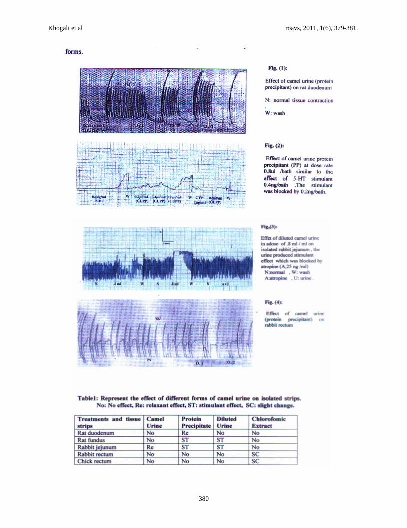
A dose of 0.1 ml/bath of camel urine PP abolished the spontaneous contractions of rat duodenum as shown in Fig. (1). Female CU and PP at a dose rate of 0.4 and 0.8 ml/bath, respectively however, stimulated the rat fundus and rabbit jejunum as shown in Fig. 2 and 3. The stimulant effects were blocked by cyproheptadine and atropine at a dose of 0.2 and 0.25ml/bath, respectively.
Khogali et al roavs, 2011, 1(6), 379-381.
380

Khogali et al roavs, 2011, 1(6), 379-381.
381
CU at 0.1 ml/bath completely abolished the spontaneous contractions of rabbit jejunum. However, the inhibitory effect was followed by transient contraction on first washing. The CE showed slight effect on rabbit and chick rectum strips Fig.4.
Discussion
This study showed that the inherited knowledge of traditional usage of camel urine for treating various ailments in Sudan could be a guide for the discovery of important biological activities which might be of useful therapeutic effects. Moreover, the scientific evaluation and identification of the mechanism (s) of action of camel urine is important for justification of its employment in modern medicine, in view of its wide uses in different parts of Sudan and other Arab countries. The results of the present study demonstrate important biological activities of the CU, PP, CE and DU. DU and CU exerted dual effects on the rabbit jejunum isolated strips. DU stimulated the organ while CU abolished the spontaneous rhythmicity of the same organ. Similar findings were reported by Rodenburg (1937) using human urine. The stimulant effect appeared to be mediated via muscarinic receptors stimulation as the effect was blocked by atropine sulphate (0.25 ml/bath). This is in agreement with Vicher (1983) and Ali et al. (1991) findings using extracts of medicinal plants. The addition of PP directly stimulated rabbit jejunum at 0.8 µl/bath the effect was blocked by atropine sulphate (0.2 ml/bath) which suggests acetylcholine-like action. Rat fundus was markedly stimulated with PP and DU as did serotonin. The abolishment of the stimulant effects of both urine forms and 5-Hydroxytryptamine (5-HT) by the addition of the non-selective serotonin blocker, cyproheptadine, demonstrated the 5-HT like activity of PP and DU. This high sensitivity might be due to the fact that rat fundus was found to be enriched with the 5-HT2B receptors (Vane, 1957). This has been recently verified as subtype of the 5-HT2 receptor family by Cox et al. (1996). The addition of PP to rat duodenum directly inhibited the myogenic contractions, which may suggest a direct musclotropic relaxation of smooth muscles. Similar findings were reported by Guddum (1955) and Horton (1959) using human urine. CE produced slight changes on rabbit and chick rectum rhythm city, however, no effects were observed on other strips. It can concluded that camel urine (indifferent forms) can penetrate subepithelially and induce generation of mast cells with release of chemical
mediators, followed by forceful peristaltic contractions caused by 5-HT and other newly formed mediators.
References Ali, M.B., Mohamed, A.H., Salih, W.M. and Homeida,
A.H. 1991. Effect of an aqueous extract of Hibiscus sabdariffa calyces on the gastrointestinal tract. Fitoterapia Voi. 1. XII. No. 6 Pp: 475-479.
Berzynski, S.R. 1986. Anti neoplaston in cancer therapy. History of the research drugs. Experimental & Clinical Research, Supply 11: 1-9.
Guddum, J.H. 1955 .Polypeptides which stimulates plain muscle. London, Livingstone. P:130.
Horton, E.W. 1959. Human Urinary Kinin Excretion. Brit. J. Pharmacol., 14:125-132.
Kabariti, A. Mazruai, S. and Elgendi, A. 1988. Camel’s urine: A possible anticarcinogenic agent. Arab Gulf Journal of Science and Research Agrc.
Kitchen, L. 1984. Text Book of Experimental Pharmacology, Isolated small intestine, 102-103.
Kroon, C.V. 1996. The Golden fountain, Autourine therapy. Gate Way Books, ISBNO 73:2:244-256.
Martha, C. 2000. Clinically tested medicinal proved book. Your Own Perfect Medicine.
Mona, A.K. 2003. Antibacterial effect of camel urine (Camelus dromedaries) M.V.Sc. Faculty of Vet. Medicine University of Khartoum, Sudan.
Mona, S. 1989. Camel urine as a hair detergent. B.Sc. Dissertation, Ahfad University, Khartoum, Sudan.
Natalie, B. 2002. Urine Therapy (Drinking urine). Journal of Berkeley medicine. www.ocf.berkele. edu.
Ohaj, H.M. 1998. Clinical trial for treatment of ascitis with camel urine M.Sc. University of the Gezira, Sudan.
Ohaj, H.M. 1993. Clinical urine as a medicament in Sudan. B.Sc. Dissertation, University Gezira, Sudan.
Rang, H.P., Dale, M.M. and Ritter, J.M. 1995. Pharmacology. 5th (ed.) Churchill Livingstone, London.
Rodenburg, G.L. and Nagy, S.M. 1937. Growth stimulating and inhibiting substances in human urine. American Journal of Cancer, 29:66.
Salwa, M.E.K. 2005. Hepatoprotective and antiparasitic effect of female camel urine. PhD Thesis. University of Khartoum, Sudan.
Vane, J.R. 1957. A sensitive method for the assay of 5-HT. British Journal of Pharmacology, 12:344-349.
Wisal, G.A. 2002. Antibacterial and antifungal effect of camel urine (Camelus dromedaries) M.V.Sc. University of Khartoum, Sudan.
Journal of Natural Sciences Research www.iiste.org
ISSN 2224-3186 (Paper) ISSN 2225-0921 (Online)
Vol.2, No.5, 2012
9
Cytotoxicity of the Urine of Different Camel Breeds on the
Proliferation of Lung Cancer Cells, A549
Zahraa Alghamdi1*
Faten Khorshid2
1. Biology Department, Dammam University, PO box 1982, Dammam 31441, Kingdom of Saudi Arabia.
2. Biology Department, King Abdulaziz University, PO box 80216, Jeddah 21589, Kingdom of Saudi
Arabia.
* E-mail of the corresponding author: [email protected]
Abstract
Objective: Cancer is a disease characterized by uncontrolled cellular proliferation and differentiation. Nearly all
conventional cancer treatments have undesirable negative impacts, and safer chemotherapeutics would be
advantageous. Consequently, the goal of current study was to evaluate and compare the effects of urine derived
from two different camel breeds on proliferation of cultured human cancer cells. Human lung adenocarcinoma
cells (A549) were cultured in the presence or absence of varied dilutions of urine obtained from two different
camel breeds (Magateer and Majaheem). Within breeds, we compared the effects of sex and age of donor camels
on urine cytotoxicity to A549 cells. After 48 hrs, surviving A549 cells were enumerated using the
sulfarhodamine assay. A549 cell survival was lower using urine from Magateer versus Majaheem camels (84.8%
versus 94.2% of starting cell number, respectively; n=20 for both groups, p<0.001). When evaluating the effect
of camel age, urine from older Magateer camels was significantly more effective in inhibiting A549 proliferation
than was urine from younger camels of this breed. An age-related effect was not observed for Majaheem camels.
When comparing sex-effects on camel urine inhibition of A549 proliferation (n=10 in each group), we observed
a trend towards more A549 inhibition using female versus male urine, in both camel breeds; however, this
difference did not reach statistical significance. The present study confirms previous studies that showed that
camel urine can inhibit the growth of cancer cells. It also provides the first evidence that there are slight
differences in the cancer cell growth-inhibitory effect of camel urine depending on the camel breed, age, and,
possibly, sex.
Keywords: Camel breeds, Urine, Cancer cells, Cytotoxicity.
1. Introduction
Cancer is a disease characterized by uncontrolled cellular proliferation and differentiation. Nowadays, cancer is a
very common disease with a high annual incidence rate (Parkin, et al ; 1999]. Ferlay et al. (2000) reported that
worldwide more than 5 million people are diagnosed with cancer and more than 3.5 million people die from
cancer each year. Managing human malignancies still constitutes a major challenge for contemporary medicine
(Coufal et al., 2007 and Widodo et al., 2007). Although with progress in understanding cancer biology, many
new antineoplastic therapies have been developed that rely primarily on surgery, chemotherapy, radiotherapy,
hormone therapy, and immunotherapeutic approaches (Khorshid et al., 2010). However, all available therapies
are still far from ideal, in which treatment would selectively kill the malignant cells while sparing healthy tissues
and vital organ function (Grever and Charbner, 1997 and Moshref, 2007). chemotherapy resulted in an overall
increase in the survival rate and longevity of patients with life-threatening tumors, On the other hand also mean
increased exposure to toxic substances and harmful effects on different tissues ( Maino, et al.,2000).
Natural products play an important role in our healthcare system (Pezzuto, 1997 and Schwartsmann, 2000).
They offer a valuable source of potent compounds with a wide variety of biological activities and novel chemical
structures, many of which might be important for novel drug development (Vuorela, et al., 2004). Animal studies
have shown that green tea is a potent inhibitor of lung tumor development (Zhang et al., 2000). PM 701 is
another natural product readily available, cheap, and non-toxic (Khorshid, 2008). PM 701 was proven to be an
anticancer substrate (Khorshid et al., 2005, 2008, Moshref et al., 2006 and El-Shahawy et al., 2010), and was
found to be effective in limiting the metastatic spread of leukemia cells in an animal model (Moshref et al.,
2006). PM 701 is considered safe as a potential anti-cancer agent, and exerts negligible effects on vital organs
(Khorshid, 2009).
Camel urine, also a natural product, has been used traditionally in the treatment of many diseases in Arabic
countries. Drinking camel urine was shown to be effective in treating numerous cancer cases (Alhaider et al.,
Journal of Natural Sciences Research www.iiste.org
ISSN 2224-3186 (Paper) ISSN 2225-0921 (Online)
Vol.2, No.5, 2012
10
2011). Moreover, according to Saudi Gazette.com, Dr. F.A. Khorshid has a potential cure for cancer based on
camel urine. After 8 years of research she has announced that nano-particles in camel urine can be used to fight
cancer. Moreover, The Saudi Center for Medical Research added that there is a tendency to start in the
production of a medical capsule containing camel’s urine for use in the treatment of cancer. In the same respect,
Alhaider et al. (2011) examined the ability of three different camel urine samples (virgin, lactating, and pregnant
sources) to modulate a well-known cancer-activating enzyme, cytochrome P 450 1a1 (Cyp 1a1) in the murine
hepatoma Hepa 1c1c7 cell line. They found that all types of camel urine, but not bovine urine, differentially
inhibited the induction of Cyp 1a1 expression by TCDD, a potent Cyp 1a1 inducer and a known carcinogen.
Virgin camel urine showed the highest degree of Cyp 1a1 inhibition, followed by lactating and pregnant camel
urine.
Khorshid (2001) stated that in vitro approaches are the best way to initially evaluate the effect of novel
biological compounds, utilizing growing mammalian cells in tissue culture. Consequently, the main goals of
current study were to: 1) evaluate the inhibitory effect of urine obtained from two different camel breeds on the
growth of lung cancer cells (A549),in vitro; and 2) study whether urine’s effect is changed according to
differences in the camel’s breed, age, or sex.
2. Materials and Methods
2.1. Study area:
The main part of this study was carried out at yebreen region located in the southern west of the eastern region at
the periphery of The Rub' alkali (Empty Quarter) included in Kingdom of Saudi Arabia.
2.2. Animals:
This study was conducted on 40 camels from two different breeds (Magateer and Majaheem). Ten males and 10
females were selected from each breed. The males ranged between 1-8 years old, whereas the females ranged
from 3 to 9 years old.
2.3. Urine sampling and storage:
Twenty milliters of urine were collected from each camel, kept in insulated boxes using freezing packs, and
transferred to the laboratory (Tissue Culture Unit, King Fahd Medical Research Center (KFMRC), King Abdul
Aziz University in Jeddah, Saudi Arabia).
2.4. Methods:
Human non-small-cell adenocarcinoma cells (A549) were obtained from the American Type Culture Collection
(ATCC) and were stored in the cell bank of tissue culture laboratory, where cytotoxicity assays were also
conducted, as pioneered by a research team working in the medical center (Khorshid et al.,2005; Khorshid and
Alameri, 2011). Different concentrations of PM 701 were used (1.0, 2.5, 5.0, 7.5, and 10 Lg/ml) and were
added to A549 cell monolayers. The control group of A549 cells was not treated with PM 701 and is indicated
as 0 concentration.
Cytotoxicity assays were performed using the method of Skehan et al. (1990). Cancer cells were suspended in
DMEM medium and plated in 96-well plates (104 cells/well) for 24h in a 5% CO2 incubator adjusted at 37°C
before treatment with PM701, to allow cell attachment to the bottom of the plate. Different concentrations of the
test substance (0, 1, 2.5, 5, and 10 Lg/ml) were then added to the cells monolayer. Triplicate wells were prepared
for each individual concentration. Cell monolayers were incubated with PM701 for 48 h at 37°C and in
atmosphere of 5% C02. After 48 h, cells were fixed using 50 µl/well trichloroacetic acid, refrigerated at 8°C for
1 hour, washed with distilled water, and then stained with Sulforhodamine B (SRB) (50 µl/well) for 30 min.
Excess stain was washed with off with acetic acid and remaining attached stain was recovered with Tris EDTA
buffer (100 µl/well). Color intensity was measured immediately in an ELISA reader at wavelength 570 nm. The
relation between surviving cells and drug concentration was plotted to get the survival curve of each cell line
after the specified period.
Journal of Natural Sciences Research www.iiste.org
ISSN 2224-3186 (Paper) ISSN 2225-0921 (Online)
Vol.2, No.5, 2012
11
2.5. Statistical analysis
Statistical analysis of the data was performed with SPSS for Windows (Version 17.0.0). Data were calculated as
follows: The different urine samples were collected from the two camel breeds from both sexes. Five
concentrations of urine were tested from each individual camel (1, 2.5, 5, 7.5, 10), with 0 concentration used as
controls. Each experimental concentration was added to six tissue culture wells containing cancer cells. Forty
total urine samples were collected from each camel with their detected concentrations mentioned above, so 40
camels × 5 concentrations equals 200 urine samples. Urine specimens at the listed concentrations were directly
applied to the six wells of cultured cancer cells, so the total wells assayed equaled 1200.
3.Results and Discussion:
3.1.Differences between two camel breeds:
Data shown in Table 1 revealed that, camel urine reduced lung cancer cells to 84.75% and 92.81%, in Magateer
and Majaheem breeds, respectively, versus untreated controls (100%). Highly significant differences were
noticed between treated and control cultures when comparing urine activity within each breed and between the
different breeds (P=0.000 and 0.001, respectively). Magateer urine significantly reduced cancer cell numbers
more than did Majaheem urine.
These results are in accordance with those of Alhaider et al. (2011) who reported that drinking camel urine has
been used traditionally to treat numerous cases of cancer. The authors attributed this anticancer effect to the
ability of camel urine to modulate the well-known cancer-activating enzyme, Cyp 1a1. They found that all types
of camel urine differentially inhibited the induction of Cyp 1a1 gene expression by TCDD, the most potent Cyp
1a1 inducer and a known carcinogenic chemical. In the same respect, Eldor (1997) hypothesized that because
some cancer cell antigens are transferred through urine, through oral autourotherapy, these antigens could be
introduced to the immune system that might then create antibodies.
3.2.Camel age effects on cancer cell proliferation:
3.2.1. In the same strain:
Table 2 clarifies the effects of urine obtained from young and adult Magateer and Majaheem camels on the
growth of lung cancer cells (A549) in vitro. Urine obtained from adult Magateer camels induced a highly
significant reduction in A549 cell survival ( P≤0.004) than that obtained from the same younger breed
(81.538% versus 87.947%, respectively), while urine obtained from adult Majaheem breed induced a non-
significant (P≤ 0.179) reduction in cancer cells when compared to younger camels of the same breed (93.486%
versus 96.974%, respectively).
No available literature could be found regarding the influence of age on the anti-cancer effect of camel urine.
However, Alhaider et al. (2011) studied the ability of three different camel urines (virgin, lactating and pregnant)
to modulate the cancer-activating enzyme CyP 1a1. They found that virgin camel urine showed the highest
degree of inhibition at the activity level, followed by lactating and pregnant camel urine.
3.2.2.Age effects between the different camel breeds:
Table 3 shows a comparison between the anti-cancer effect of urine obtained from the two young camel breeds
as well as the anti-cancer effect of that obtained from the two adult camel breeds. The results revealed that urine
from young Magateer camels induced a significant (P≤0.01) reduction in the growth of cancer cells versus that
obtained from young Majaheem camels (87.947% versus 96.974%, respectively). In addition, urine obtained
from adult Magateer camels induced a significant higher reduction (P=000) of cancer cells versus that obtained
with adult Majaheem camels (81.536% versus 93.486%, respectively).
The reason for the variability in the anti-cancer efficacy of camel urine obtained from Magateer and Majaheen
breeds is not yet known. Further study is needed to determine the specific differences in the urine constituents of
each breed, to know which compound(s) is responsible for this variable effect.
Journal of Natural Sciences Research www.iiste.org
ISSN 2224-3186 (Paper) ISSN 2225-0921 (Online)
Vol.2, No.5, 2012
12
3.2.3.. Sex affects camel urine-mediated cancer cell proliferation:
3.2.3.1 In the same breed:
Table 4 represents the effect of sex on the ability of camel urine to inhibit the growth of lung cancer cells in
vitro. It appears that the sex of camels within the same breed did not significantly affect camel urine-inhibition of
A549 cancer cell proliferation. However, urine of males induced a slight, though insignificant inhibition in
cancer cell proliferation versus that of females of the same breed.
3.2.3.2. In the different breeds:
Table 5 shows a comparison between the anti-cancer effect of urine obtained from males and females of the two
different camel breeds. Urine from male Magateer camels caused a significantly greater reduction in cancer cells
when compared to that induced by urine of male Majaheem camels (86.568 versus 94.014, respectively; P=.000).
Urine of female Magateer camels also induced a significantly greater reduction in cancer cells compared to that
induced by urine of female Majaheem camels (82.935 versus 91.368; P=.000). Urine from male and female
Magateer camels were more efficient in reducing lung cancer cell numbers compared with that observed using
Majaheem camel urine.
5. Conclusion
The present study confirms the findings of previous studies that camel urine can inhibit the growth of cancer
cells. It also provides the first evidence that there are differences in the cancer-inhibiting effect of camel urine
depending on the camel breed, age, and sex.
6. Acknowledgements
The authors gratefully thank King Faisal University, represented by Prof. Dr. AbdelGader Homeida and
Mr.Khalid Borsais who helped in obtaining samples. The authors also appreciate the kind help of Prof. Dr.
Hodallah Hatem, Head of the Physiology Department, Faculty of Veterinary Medicine, Cairo University, Egypt.
References
1. Alhaider, A.A., El Gendy, M.A., Korashy, H.M. & El-Kadi, A.O.(2011). Camel Urine Inhibits the
Cytochrome P 405 1a1 Gene Expression through an AhR- Dependent Mechanism in Hepa 1c1c7 cell line.
Journal of Ethnopharmacology, 133 (1),184-190.
2. Coufal, M., Maxwell, M.M., Russel, D.E., Amore, A.M., Altmann, S.M., Hollingsworth, Z.R., Young,
A.B., Housman, D.E. & Kazantsev, A.G. (2007). Discovery of Novel Small-Molecule Targeting Selective
Clearance of Mutant Huntingtin Fragments. Journal of Biomolecular Screening, 12,351-360.
3. Eldor, J. (1997). Urotherapy for Patients with Cancer. Medical Hypothesis. 48(4), 309-315.
4. El-Shahawy, A., Elsawi, N.M., Baker, W.S., Khorshid, F. & Geweely, N.S. (2010). Spectral Analysis,
Molecular Orbital Calculations and Antimicrobial Activity of PMF-G Fraction Extracted from PM-701.
International Journal Pharma Bioscience, 1(2): 1-19.
5. Ferlay, J., Bray, F., Pisani, P. & Parkin, D.M. GLOBOCAN (2000): Cancer Incidence, Mortality and
Prevalence Worldwide, Version 1.0. IARC Cancer Base No. 5. Lyon: IARC Press, 2001.
6. Grever. M. & Chabner, B. (1997): Cancer Drug Discovery and Development in Cancer Propels and
Practice of oncology. Cytotoxicity of Some Medical Plant Extract Used in Tanzanian Traditional Medicine.
Journal of Ethnopharmacology,70, 143-149.
7. Khorshid, F.A. (2001). The Effect of the Viscosity of the Medium in the Reaction of Cells to Topography.
PhD Thesis, Glasgow University, UK.
8. Khorshid, F., Alshazly, H., Al Jefery, A. & Osman, A.M.M. (2010). Dose Escalation Phase I Study in
Healthy Volunteers to Evaluate the Safety of a Natural Product PM 701. Journal Pharmacology Toxicology, 5(3),
91-97.
9. Khorshid, F.A., Rahimaldeen, S.A. & Alameri, J.S. ( 2011).Apoptosis Study on the Effect of PMF on
Journal of Natural Sciences Research www.iiste.org
ISSN 2224-3186 (Paper) ISSN 2225-0921 (Online)
Vol.2, No.5, 2012
13
Different Cancer Cells, International Journal of Biological Chemistry, 5(2),150-155.
10. Khorshid, F.A.( 2005). Comparative study of Keloids Formation in Human and Laboratory Animals.
Medical science monitor, 11(7), BR212-219
11. Khorshid, F.A.( 2008). Preclinical Evaluation of PM 701 in Experimental Animals. International
Journal of Pharmaceutics, 4(6), 443-451.
12. Khorshid, F.A.(2009). Potential Anticancer Natural Product Against Human Lung Cancer Cells. Trends
in Medical Research, 4(1), 8-15.
13. Khorshid, F.A., Moshref, S.S. & Heffny, N.(2005). An Ideal Selective Anticancer Agent In Vitro, I-
Tissue Culture Study of Human Lung Cancer Cells A549. JKAU- Medical Sciences, 12, 3- 18.
14. Khorshid, F.A., Rahimaldeen, S.A. & AL-Amri, J.S. (2011). Apoptosis Study on the Effect of PMF on
Different Cancer Cells. International Journal of Biological Chemistry, 5, 150-155.
15. Maino, D.M., Tran, S. & Mehta, F.(2000). Side Effects of Chemotherapeutic Oculo-Toxic Agents: A
Review. Clinical Eye and Vision Care, 12(3-4),113-117.
16. Moshref, S.S. (2007). PM 701 A Highly Selective Anti-Cancerous Against L1210 Leukemic Cells :II-
In Vivo Clinical and Histopathological Study. JKAU. Medical Sciences, 14(1), 85-99.
17. Moshref, S.S., Khorshid, F.A.& Jamal, Y.( 2006). The Effect of PM 701 on Mice Leukemic Cells: I -
Tissue Culture Study of L1210 (In Vitro) II - In Vivo Study on Mice, JKAU- Medical Sciences , 13 (1), 3-19.
18. Parkin, D., Pisani, P. & Ferlay, J. (1999). Global Cancer Statistics. A Cancer Journal for Clinicians,
49: 33-64.
19. Pezzuto, J.M. (1997). Plant-Derived Anticancer Agents. Biochemical Pharmacology, 53, 121-133.
20. Schwartsmann, G. (2000). Marine Organisms and Other Novel Natural Sources of New Cancer Drugs.
Annals of Oncology, 11, 235-243.
21. Skehan, P., Storeng, R., Scudiero, D., Monks, A., McMahon, J., Vistica, D., Warren, J.T,. Bokesch, H.,
Kenney, S. & Boyd, M.R. (1990). New Colorimetric Cytotoxicity assay for Anticancer-Drug Screening. Journal
of the National Cancer Institute, 82,1107–1112.
22. Vuorela, P., Leinonen, M., Saikku, P., Tammela, P., Rauha, J-P, Wennberg, T.& Vuorela,
H.( 2004).Natural Products in the Process of Finding New Drug Candidates. Current Medicinal Chemistry,
11,1375–1389. PMid,15180572.
23. Widodo, N., Kaur, K., Shrestha, B.G., Takagi, Y., Ishii, T., Wadhwa R. & Kaul, S.C. (2007). Selective killing
of Cancer Cells by Leaf Extract of Ashwagandha: Identification of a Tumor-Inhibitory Factor and the First
Molecular Insights to its Effect. Clinical Cancer Research, 13, 2298-2306.
24. Zhang, Z., Liu, Q., Lantry, L.E., Wang, Y., Kelloff, G.J., Anderson, M.W., Wiseman, R.W., Lubet, R.A.
& You, M. (2000). A Germ-Line P53 Mutation Accelerates Pulmonary Tumorigenesis: P53-Independent Efficacy
of Chemopreventive Agents Green Tea or Dexamethasone/Myo-inositol and Chemotherapeutic Agents Taxol or
Adriamycin. Cancer Research, 60(4), 901-907.
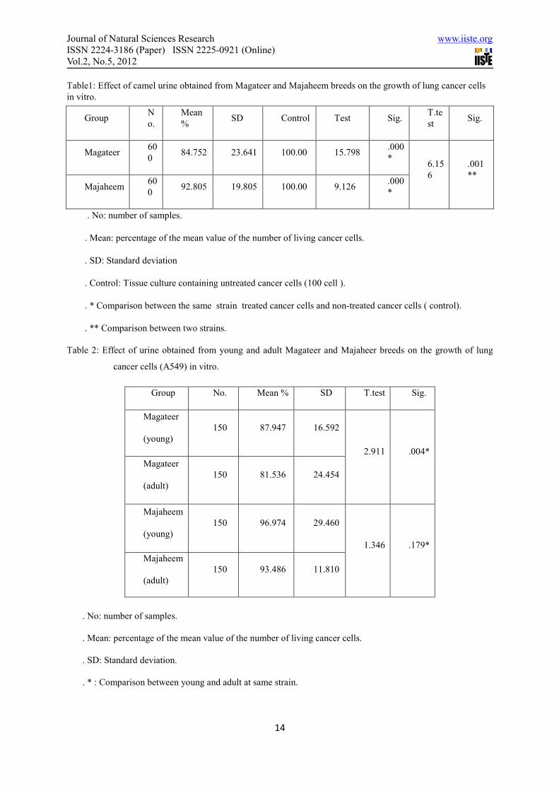
Journal of Natural Sciences Research www.iiste.org
ISSN 2224-3186 (Paper) ISSN 2225-0921 (Online)
Vol.2, No.5, 2012
14
Table1: Effect of camel urine obtained from Magateer and Majaheem breeds on the growth of lung cancer cells
in vitro.
Group N
o.
Mean
% SD Control Test Sig.
T.te
st Sig.
Magateer 60
0 84.752 23.641 100.00 15.798
.000
* 6.15
6
.001
**
Majaheem 60
0 92.805 19.805 100.00 9.126
.000
*
. No: number of samples.
. Mean: percentage of the mean value of the number of living cancer cells.
. SD: Standard deviation
. Control: Tissue culture containing untreated cancer cells (100 cell ).
. * Comparison between the same strain treated cancer cells and non-treated cancer cells ( control).
. ** Comparison between two strains.
Table 2: Effect of urine obtained from young and adult Magateer and Majaheer breeds on the growth of lung
cancer cells (A549) in vitro.
Group No. Mean % SD T.test Sig.
Magateer
(young)
150 87.947 16.592
2.911 .004*
Magateer
(adult)
150 81.536 24.454
Majaheem
(young)
150 96.974 29.460
1.346 .179*
Majaheem
(adult)
150 93.486 11.810
. No: number of samples.
. Mean: percentage of the mean value of the number of living cancer cells.
. SD: Standard deviation.
. * : Comparison between young and adult at same strain.
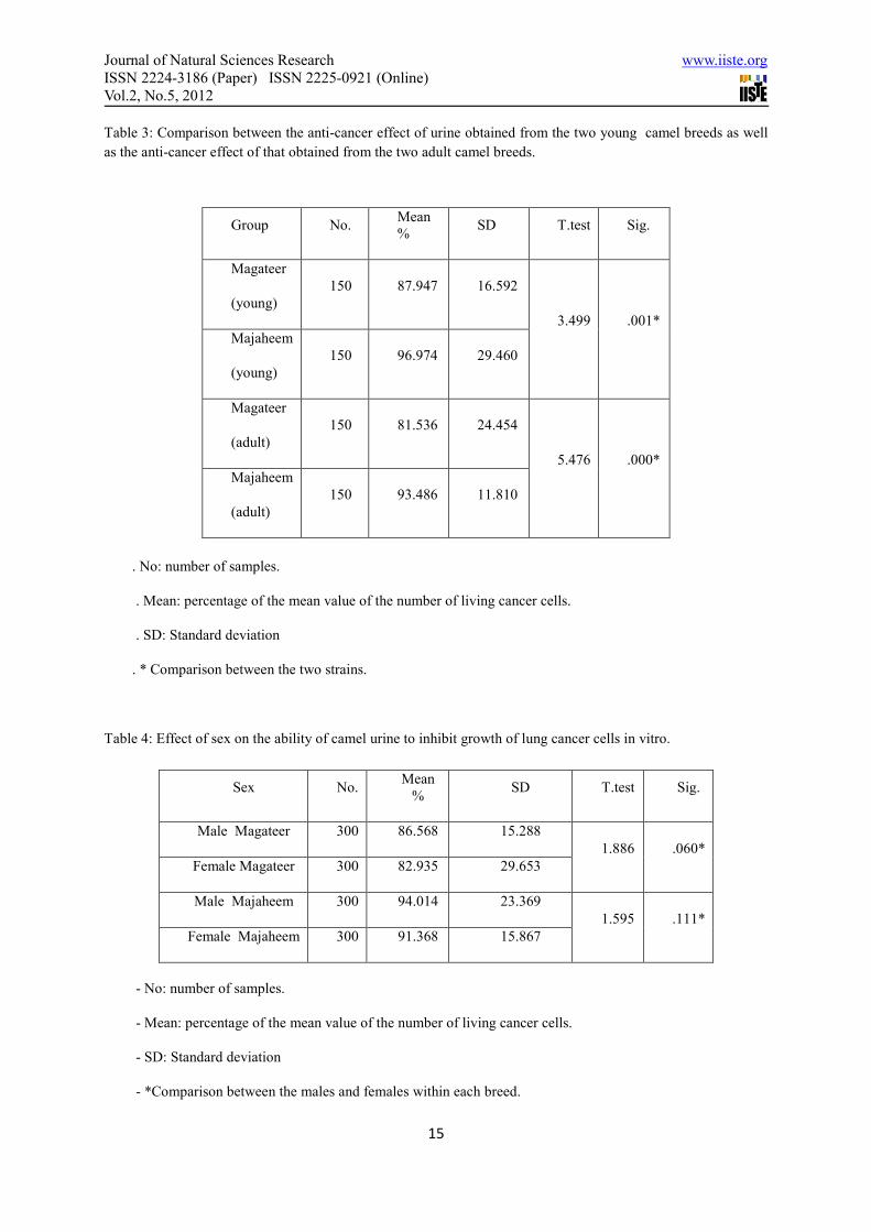
Journal of Natural Sciences Research www.iiste.org
ISSN 2224-3186 (Paper) ISSN 2225-0921 (Online)
Vol.2, No.5, 2012
15
Table 3: Comparison between the anti-cancer effect of urine obtained from the two young camel breeds as well
as the anti-cancer effect of that obtained from the two adult camel breeds.
Group No. Mean
% SD T.test Sig.
Magateer
(young)
150 87.947 16.592
3.499 .001*
Majaheem
(young)
150 96.974 29.460
Magateer
(adult)
150 81.536 24.454
5.476 .000*
Majaheem
(adult)
150 93.486 11.810
. No: number of samples.
. Mean: percentage of the mean value of the number of living cancer cells.
. SD: Standard deviation
. * Comparison between the two strains.
Table 4: Effect of sex on the ability of camel urine to inhibit growth of lung cancer cells in vitro.
Sex No. Mean
% SD T.test Sig.
Male Magateer 300 86.568 15.288
1.886 .060*
Female Magateer 300 82.935 29.653
Male Majaheem 300 94.014 23.369
1.595 .111*
Female Majaheem 300 91.368 15.867
- No: number of samples.
- Mean: percentage of the mean value of the number of living cancer cells.
- SD: Standard deviation
- *Comparison between the males and females within each breed.
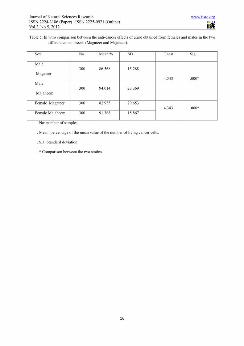
Journal of Natural Sciences Research www.iiste.org
ISSN 2224-3186 (Paper) ISSN 2225-0921 (Online)
Vol.2, No.5, 2012
16
Table 5: In vitro comparison between the anti-cancer effects of urine obtained from females and males in the two
different camel breeds (Magateer and Majaheer).
Sex No. Mean % SD T.test Sig.
Male
Magateer
300 86.568 15.288
4.543 .000*
Male
Majaheem
300 94.014 23.369
Female Magateer 300 82.935 29.653
4.343 .000*
Female Majaheem 300 91.368 15.867
. No: number of samples.
. Mean: percentage of the mean value of the number of living cancer cells.
. SD: Standard deviation
. * Comparison between the two strains.
This academic article was published by The International Institute for Science,
Technology and Education (IISTE). The IISTE is a pioneer in the Open Access
Publishing service based in the U.S. and Europe. The aim of the institute is
Accelerating Global Knowledge Sharing.
More information about the publisher can be found in the IISTE’s homepage:
http://www.iiste.org
The IISTE is currently hosting more than 30 peer-reviewed academic journals and
collaborating with academic institutions around the world. Prospective authors of
IISTE journals can find the submission instruction on the following page:
http://www.iiste.org/Journals/
The IISTE editorial team promises to the review and publish all the qualified
submissions in a fast manner. All the journals articles are available online to the
readers all over the world without financial, legal, or technical barriers other than
those inseparable from gaining access to the internet itself. Printed version of the
journals is also available upon request of readers and authors.
IISTE Knowledge Sharing Partners
EBSCO, Index Copernicus, Ulrich's Periodicals Directory, JournalTOCS, PKP Open
Archives Harvester, Bielefeld Academic Search Engine, Elektronische
Zeitschriftenbibliothek EZB, Open J-Gate, OCLC WorldCat, Universe Digtial
Library , NewJour, Google Scholar
POSTER PRESENTATION Open Access
The effect of camel urine on islet morphologyand CCL4-induced liver cirrhosis in ratS Al Neyadi*, R Al Jaberi, R Hameed, J Shafarin, E Adeghate
From International Conference for Healthcare and Medical Students 2011Dublin, Ireland. 4-5 November 2011
IntroductionCamel urine has been used for decades as a medicationfor several ailments in the Middle East. Folklore medi-cine of the Middle East has shown that, camel urine hasa beneficial effect in conditions such as liver cirrhosis.
MethodsCamel urine was given as a drink daily to normal and trea-ted rats for 4 weeks. Glucose tolerance test was performedat the end of the experiment. Immunohistochemistry wasused to determine the percentage distribution of insulinand glucagon immunoreactive cells. H & E stain was usedto access liver cirrhosis in control and urine-treated rats.
ResultsThe administration of camel urine significantly increasedthe number of insulin-positive cells in pancreatic islets.CCL4-treated rats did not have impaired glucose toler-ance. CCL4 caused vacuolarization of hepatic cells. Ratstreated with camel urine have improved hepatic morphol-ogy compared to untreated controls.
ConclusionsThe study shows that camel urine may contain bioactiveagents capable of preventing CCL4-induced hepatic andpancreatic islet lesions.
Published: 9 July 2012
doi:10.1186/1753-6561-6-S4-P42Cite this article as: Al Neyadi et al.: The effect of camel urine on isletmorphology and CCL4-induced liver cirrhosis in rat. BMC Proceedings2012 6(Suppl 4):P42.
Submit your next manuscript to BioMed Centraland take full advantage of:
• Convenient online submission
• Thorough peer review
• No space constraints or color figure charges
• Immediate publication on acceptance
• Inclusion in PubMed, CAS, Scopus and Google Scholar
• Research which is freely available for redistribution
Submit your manuscript at www.biomedcentral.com/submitDepartment of Anatomy, Faculty of Medicine & Health Sciences, United Arab
Emirates
Al Neyadi et al. BMC Proceedings 2012, 6(Suppl 4):P42http://www.biomedcentral.com/1753-6561/6/S4/P42
© 2012 Al Neyadi et al; licensee BioMed Central Ltd. This is an Open Access article distributed under the terms of the CreativeCommons Attribution License (http://creativecommons.org/licenses/by/2.0), which permits unrestricted use, distribution, andreproduction in any medium, provided the original work is properly cited.
African Journal of Agricultural Research Vol. 5(11), pp. 1331-1337, 4 June, 2010 Available online at http://www.academicjournals.org/AJAR DOI: 10.5897/AJAR09.686 ISSN 1991-637X © 2010 Academic Journals
Full Length Research Paper
The inhibitory effect of camel's urine on mycotoxins and fungal growth
Amira Hassan Abdullah Al-Abdalall
Department of Botany and Microbiology, Faculty of Science for Girls, King Faisal University, El-Dammam, Kingdom of Saudi Arabia. E -mail: [email protected].
Accepted 8 January, 2010
The effect of urine and camel milk in the inhibition of biological effects of mycotoxins produced by nine isolates of Aspergillus flavus and one isolate of Aspergillus niger isolated from pulse seeds was studied. Where these toxins lost their ability to inhibit Bacillus subtilus growth, milk could not. Also, our study records the effect of camel urine on mycelial growth of some roots rot fungi isolated from seeds of pulses like Rhizoctonia solani, Fusarium moliniform, Aschocayta sp., Pythium aphanidermatum, Sclerotinia sclerotiorum studies, also included are some storage fungi (Aspergillus sp) isolated from coffee beans. Results proved that camel urine at low concentrations has no significant inhibitory effect on fungal growth, while inhibition can be obviously recorded after using high concentrations. Key words: Camel urine, mycotoxins, mycelial growth, inhibitory effect on fungal growth.
INTRODUCTION It is mentioned in Islam online that camel's milk and urine have medical effects, so Islam encourages and permits the drinking of camel milk, and camel urine is permitted in case of necessary medical treatment (Al-Bukhhari). The Saheeh Hadeeth says that some people came to Madeenah and fell sick. The Prophet (peace and blessings of Allaah be upon him) told them to drink the milk and urine of camels, and when they drank it they recovered and grew fat. This was narrated by Al-Bukhaari. There are many well known health benefits, with regard to drinking the milk and urine of camels, to the earlier generations of medical science and they have been proven by modern scientific researches. For example swollen abdomen, which may indicate oedema and liver disease (jaundice), or cancer, and thin bodies which indicate extreme weakness, and which often accompanies hepatitis or cancer. This may be due to the effectiveness of camel's urine, as against all other cattle to the active substances contained in desert plants which benefited more of them; this was summed up by the Prophet (peace be upon him). Many researches have been conducted on a variety of desert plants and a strong
effect against bacteria, yeast and fungi has been found. Kaul et al. (1976) and Zaki et al. (1984) have conducted researches on the wormwood plant, and results have shown strong effectiveness against bacteria, yeast and fungi.
The chemical composition and nutritional quality of camel milk was studied. Results showed 11.7% total solids, 3.0% protein, 3.6% fat, 0.8% ash, 4.4% lactose, 0.13% acidity and a pH of 6.5. It contains low level of cholesterol and sugar and is rich in the levels of Na, K, Zn, Fe, Cu, Mn, niacin and vitamin C (Knoess, 1979). Besides, camel milk contains low level of protein and high concentration of insulin, and could be safely taken by people who have high sensitivity to lactose and have immune deficiency (Gast, 1969). Camel milk is pure white and sugary. Camels who feed on certain diets may produce salty milk when feed on desert weeds. There are physiological and genetical factors affecting milk production. Percentage of water in camel milk varies according to the doses of water which camel drinks; it may reach 89% in the milk if camels drink water every day, or 91% if camels drink one hour weekly. It seems
1332 Afr. J. Agric. Res. that camel loose more water during its deficiency in nature for the benefits of babies or human beings in generally (Farah, 1993; Abu-Lehia, 1989). Camel milk is also used in Kazakhstan as an adjunct to chemotherapy for some cancer treatments, especially those of the digestive tract. Good results were reported in autoimmune diseases such as Crohn’s disease and multiple sclerosis (Yagil and van Creveld, 2000). The positive effect of camel milk on diabetic patients has been studied in India (Agrawal et al., 2003). With the consumption of 0.5 l of camel milk per day, the insulin demand decreased in diabetic patients and glycaemia was better balanced. Consumers appreciate camel milk for its medicinal properties: it is reputed to be anti-infectious, anti-cancerous and antidiabetic. More generally, it is regarded as an energy-giving product for convalescents. Camel milk is commonly used to help treat infectious diseases such as tuberculosis in humans. Shubat is commonly used as a cure in sanatoriums (Urazakov and Bainazarov, 1974). The camel milk works as a laxative on people unaccustomed to drinking this milk (Rao, 1970). A series of metabolic and autoimmune diseases are successfully being treated with camel milk. In India, camel milk is used therapeutically against dropsy, jaundice, problems of the spleen, tuberculosis, asthma, anemia, piles and diabetes (Rao, 1970).
Urine, although a waste product of the body, nonetheless has many medical practitioners, and as such is used both internally and externally as medicine. The urines of animals such as goat, sheep, buffalo, elephant, horse, camel, donkey etc. were also very much in use as remedies for the treatment of worms, dropsy, abdominal enlargements, flatulence, colic, anaemia, abdominal tumor, loss of appetite, tuberculosis, poison, haemorrhoids, amenorrhea, leucoderma, leprosy, aggravation of kapha and vat and in several other mental diseases (Thakur, 2004). When Amer and Al-hendi (1996) analyzed urine of mature camels of between 5 -10 years old, they found that its relative density ranged from 1.022 to 1.07, while pH values varied to be either acidic or alkaline. Urea level ranged from 18-36 gm/dL. Keratin recorded 0.2 - 0.5 gm/L. Microscopical analysis proved the presence of phosphorus and calcium oxalate and ammonium urate; some epithelial and granular cells appeared. Al-Attas (2008), using neutron activation analysis, estimated some essential elements within milk and urine of camels, and discovered that it contains large amount of Na and K substituting the loss of such elements in the case of diarrhea. Also it contains large amount of Zn which assists in the cure of the infection due to diarrhea. Mycotoxins are diverse range of molecules that are harmful to animals and humans. They are secondary metabolites secreted by moulds, mostly Penicillium and Fusarium. They are produced in cereal grains as well as forages before, during and after harvest in various environmental conditions. Due to the diversity of their toxic effects and their synergetic properties,
mycotoxins are considered risky to the consumers of contaminated foods and feeds (Yiannikouris and Jonany, 2002). Mycotoxins are metabolized in the liver and the kidneys and also by microorganisms in the digestive tract. Therefore, the chemical structure and associated toxicity of mycotoxin residues often excreted by animals or found in their tissues are different from the parent molecule (Ratcliff, 2002). Storage fungi belonging to genus Penicillium and Aspergillus play an important role in spoiling stored seeds with increasing humidity. Such fungi produce toxins that lead to human liver disfunction, cancer and undesirable mutations (Pereyra et al., 2008). From these fungi, we mentioned the genera, Colletotrichum, Sclerotinia, Alternaria, Fusarium, Rhzoctonia, Pythium, Ascochyta and Botrytis (Sweetingham, 1989; Mackie et al., 1999; Zhang and Yang, 2000; Elmer et al., 2001; Wen et al., 2005). AL-awadi and AL-Jedabi (2000) proved an inhibitory and antibiotic activity of camel urine against the growth of Candida albicans (yeast), Aspergillus niger, Fusarium oxysporum even after it’s boiling to 100°C. Our search was aimed at tracing the effect of camel urine on the growth properties of such fungi. The effect of such products (urine and milk) on efficiency of aflatoxins as inhibitors to Bacillus subtilus growth, is seen as a primary step fined away to get rid of fungal toxins. MATERIAL AND METHODS
Samples of Camelus dromedaries urine and milk were collected from females feed on wild weeds at the west of Dammam. Samples were collected in sterile bottles and kept at 4°C for not more than 2
weeks for urine. Fungal isolates Used fungi isolates (Rhizoctonia solani, Fusarium moliniform, Pythium aphanidermatum, Aschocayta sp., Sclerotinia sclerotiorum, Aspergillus flavus and A. niger) were extracted from pulse seeds as lupine (Lupinus albus L.), cow pea and mung bean (Vigna radiata L.), faba bean and field bean (Vicia faba L.) and lentil (Lens
culinaris), chickpea (Cicer judaicum), kidney beans (Phaseolus
vulgaris) (Al-Abdalall, 2008), and five isolates of A. niger from coffee beans (Coffia arabica). .
Effect of milk and urine on aflatoxins
Filtrates of nine fungal isolates belonging to A. flavus and one belonging to A. niger were obtained from seed of pulses (mung bean, faba bean, field bean, lupine, and lentil) (Al-Abdalall, 2009). All isolates were individually grown on SMKY liquid medium described by Diener and Davis (1966) containing:200 gm sucrose; 0.5 gm magnesium sulphate; 3 gm potassium nitrate; and 7 gm yeast extract for 10 days at 25°C to obtain the culture filtrate. Then, the filtrate of each isolate was extracted three times with equal volumes of ethyl acetate. The ethyl acetate was removed by evaporation and the residue was brought up in sterilized distilled water. The method described by Lenz et al. (1986) was used as follows: A species of specific bacteria, that is, B. subtilis, was
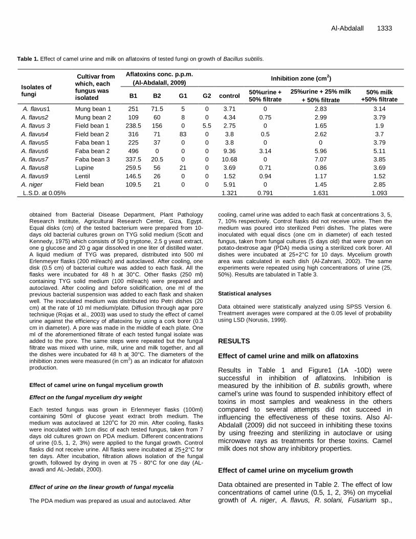
Al-Abdalall 1333
Table 1. Effect of camel urine and milk on aflatoxins of tested fungi on growth of Bacillus subtilis.
Isolates of fungi
Cultivar from which, each fungus was isolated
Aflatoxins conc. p.p.m.
(Al-Abdalall, 2009) Inhibition zone (cm
2)
B1 B2 G1 G2 control 50%urine + 50% filtrate
25%urine + 25% milk
+ 50% filtrate
50% milk +50% filtrate
A. flavus1 Mung bean 1 251 71.5 5 0 3.71 0 2.83 3.14
A. flavus2 Mung bean 2 109 60 8 0 4.34 0.75 2.99 3.79
A. flavus 3 Field bean 1 238.5 156 0 5.5 2.75 0 1.65 1.9
A. flavus4 Field bean 2 316 71 83 0 3.8 0.5 2.62 3.7
A. flavus5 Faba bean 1 225 37 0 0 3.8 0 0 3.79
A. flavus6 Faba bean 2 496 0 0 0 9.36 3.14 5.96 5.11
A. flavus7 Faba bean 3 337.5 20.5 0 0 10.68 0 7.07 3.85
A. flavus8 Lupine 259.5 56 21 0 3.69 0.71 0.86 3.69
A. flavus9 Lentil 146.5 26 0 0 1.52 0.94 1.17 1.52
A. niger Field bean 109.5 21 0 0 5.91 0 1.45 2.85
L.S.D. at 0.05% 1.321 0.791 1.631 1.093
obtained from Bacterial Disease Department, Plant Pathology Research Institute, Agricultural Research Center, Giza, Egypt. Equal disks (cm) of the tested bacterium were prepared from 10-days old bacterial cultures grown on TYG solid medium (Scott and Kennedy, 1975) which consists of 50 g tryptone, 2.5 g yeast extract, one g glucose and 20 g agar dissolved in one liter of distilled water.
A liquid medium of TYG was prepared, distributed into 500 ml Erlenmeyer flasks (200 ml/each) and autoclaved. After cooling, one disk (0.5 cm) of bacterial culture was added to each flask. All the flasks were incubated for 48 h at 30°C. Other flasks (250 ml) containing TYG solid medium (100 ml/each) were prepared and autoclaved. After cooling and before solidification, one ml of the previous bacterial suspension was added to each flask and shaken well. The inoculated medium was distributed into Petri dishes (20 cm) at the rate of 10 ml medium/plate. Diffusion through agar pore
technique (Rojas et al., 2003) was used to study the effect of camel urine against the efficiency of aflatoxins by using a cork borer (0.3 cm in diameter). A pore was made in the middle of each plate. One ml of the aforementioned filtrate of each tested fungal isolate was added to the pore. The same steps were repeated but the fungal filtrate was mixed with urine, milk, urine and milk together, and all the dishes were incubated for 48 h at 30°C. The diameters of the inhibition zones were measured (in cm
2) as an indicator for aflatoxin
production.
Effect of camel urine on fungal mycelium growth
Effect on the fungal mycelium dry weight
Each tested fungus was grown in Erlenmeyer flasks (100ml) containing 50ml of glucose yeast extract broth medium. The medium was autoclaved at 120
oC for 20 min. After cooling, flasks
were inoculated with 1cm disc of each tested fungus, taken from 7 days old cultures grown on PDA medium. Different concentrations of urine (0.5, 1, 2, 3%) were applied to the fungal growth. Control flasks did not receive urine. All flasks were incubated at 25+2°C for ten days. After incubation, filtration allows isolation of the fungal growth, followed by drying in oven at 75 - 80°C for one day (AL-awadi and AL-Jedabi, 2000).
Effect of urine on the linear growth of fungal mycelia
The PDA medium was prepared as usual and autoclaved. After
cooling, camel urine was added to each flask at concentrations 3, 5, 7, 10% respectively. Control flasks did not receive urine. Then the medium was poured into sterilized Petri dishes. The plates were inoculated with equal discs (one cm in diameter) of each tested fungus, taken from fungal cultures (5 days old) that were grown on potato-dextrose agar (PDA) media using a sterilized cork borer. All
dishes were incubated at 25+2°C for 10 days. Mycelium growth area was calculated in each dish (Al-Zahrani, 2002). The same experiments were repeated using high concentrations of urine (25, 50%). Results are tabulated in Table 3.
Statistical analyses
Data obtained were statistically analyzed using SPSS Version 6. Treatment averages were compared at the 0.05 level of probability using LSD (Norusis, 1999).
RESULTS
Effect of camel urine and milk on aflatoxins
Results in Table 1 and Figure1 (1A -10D) were successful in inhibition of aflatoxins. Inhibition is measured by the inhibition of B. subtilis growth, where camel's urine was found to suspended inhibitory effect of toxins in most samples and weakness in the others compared to several attempts did not succeed in influencing the effectiveness of these toxins. Also Al-Abdalall (2009) did not succeed in inhibiting these toxins by using freezing and sterilizing in autoclave or using microwave rays as treatments for these toxins. Camel milk does not show any inhibitory properties.
Effect of camel urine on mycelium growth
Data obtained are presented in Table 2. The effect of low concentrations of camel urine (0.5, 1, 2, 3%) on mycelial growth of A. niger, A. flavus, R. solani, Fusarium sp.,

1334 Afr. J. Agric. Res.
Table 2. Effect of low concentrations of camel urine on dry weight of fungal mycelium.
Tested fungi Sources of isolates concentrations of camel urine
0 0.25 0.50 1 3
A. niger1 Coffee beans 0.46 0.38 0.34 0.31 0.28
A. niger2 Coffee beans 0.37 0.26 0.25 0.25 0.21
A. niger3 Coffee beans 0.40 0.30 0.24 0.23 0.17
A. niger4 Coffee beans 0.30 0.25 0.25 0.21 0.17
A. niger5 Coffee beans 0.40 0.28 0.27 0.26 0.22
R. solani Faba beans 0.07 0.07 0.07 0.07 0.085
F. moliniform Kidney beans 0.07 0.07 0.05 0.075 0.10
Aschocayta sp. Chickpea 0.07 0.11 0.09 0.09 0.09
S. sclerotiorum Mung beans 0.08 0.085 0.09 0.09 0.095
P. aphanidermatum Mung beans 0.08 0.07 0.07 0.06 0.09
L.S.D 1.05 0.87 0.83 0.798 0.69
Table 3. Effect of low concentrations of camel urine on the mycelial growth of test fungi.
Tested fungi Sources of isolates concentrations of camel urine%
0 3 5 7 10
A. niger1 Coffee beans 44.36 41.32 41.32 15.49 7.9
A. niger2 Coffee beans 47.6 31.9 16.1 12.56 12.76
A. niger3 Coffee beans 53.48 33.37 27.19 18.44 2.46
A. niger4 Coffee beans 38.67 21.69 16.1 14.23 14.52
A. niger5 Coffee beans 53.48 23.95 15.9 18.09 7.81
A. niger6 Field beans 53.48 28.46 26.01 16.62 4.91
A. flavus Cowpea 42.49 14.23 10.56 9.09 8.35
R. solani Faba beans 56.72 56.72 56.72 51.03 25.96
F. moliniform Kidney beans 56.72 56.72 56.72 56.72 16.62
Aschocayta sp. Chickpea 56.72 56.72 53.48 45.35 43
S. sclerotiorum Mung beans 56.72 56.06 54.08 42.995 41.83
P. aphanidermatum Mung beans 38.67 36.34 35.82 26.88 18.49
L.S.D 2.35 3.1 3.27 3.18 2.81
Pythium aphanidermatum, Aschocayta sp., and Sclerotinia sclerotiorum in liquid medium, as well as concentrations (3, 5, 7, 10%) on solid medium (Table 3 and Figure 1 (1-12)) was noticed. There is no significant differences in dry weight in treated or control flasks, although there is a decrease in the dry weight of the mycelial growth with increasing urine concentrations. After using concentrations of urine (25, 50%) a significant decrease in fungal growth (on Petri dishes) was recorded (Table 4 and Figure 1 (1-12). Some treatments showed complete inhibition of the fungal growth, Aschocayta sp. is totally inhibited to grow at 25% concentration of camel urine. While complete inhibition of Aschocayta sp., Rhizoctonia solani, Pythium aphanidermatum is recorded after application of urine at 50%.
DISCUSSION
Abdel Magjeed (2005) mentioned that camel milk can
improve some biological aspects after toxication withaflatoxins, including improvement in the level of Keratin, Haemoglobin, triglycrides and blood properties of toxicated rates. Results of the therapeutic groups were compared with other groups treated with either the anticarcinogenic drug or treated with milk camel mixed with small amounts of urine. EL-Elyani and Khalifa (2006) noted that no histopathological changes could be recorded when studying the effect of camel milk or urine on stomach rate, while AL-Kabarity et al. (1988) mentioned that camel urine is used commonly, in alternative medicine, against cancer and respiratory tractinfections. Khalifa, (1999) EL-Elyani (1999) and Khalifa et al. (2005) noted that no pathological signs appeared on liver or kidney tissues after using camel milk or urine. The high salt concentration of the urine causes plasmosis and analysis of mycelium hyphae; this agrees with AL-awadi and AL-Jedabi (2000). Similar results were obtained by Al-Zahrany (2002) who recorded growth
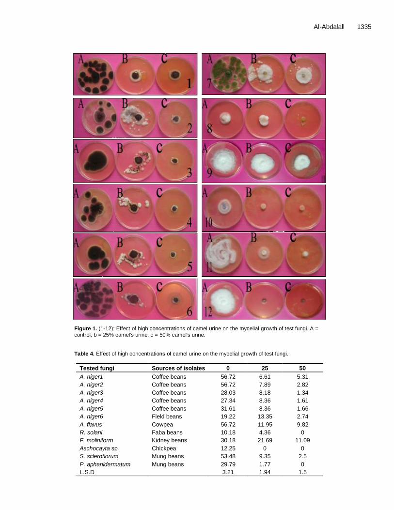
Al-Abdalall 1335
Figure 1. (1-12): Effect of high concentrations of camel urine on the mycelial growth of test fungi. A = control, b = 25% camel's urine, c = 50% camel's urine. Table 4. Effect of high concentrations of camel urine on the mycelial growth of test fungi.
Tested fungi Sources of isolates 0 25 50
A. niger1 Coffee beans 56.72 6.61 5.31
A. niger2 Coffee beans 56.72 7.89 2.82
A. niger3 Coffee beans 28.03 8.18 1.34
A. niger4 Coffee beans 27.34 8.36 1.61
A. niger5 Coffee beans 31.61 8.36 1.66
A. niger6 Field beans 19.22 13.35 2.74
A. flavus Cowpea 56.72 11.95 9.82
R. solani Faba beans 10.18 4.36 0
F. moliniform Kidney beans 30.18 21.69 11.09
Aschocayta sp. Chickpea 12.25 0 0
S. sclerotiorum Mung beans 53.48 9.35 2.5
P. aphanidermatum Mung beans 29.79 1.77 0
L.S.D 3.21 1.94 1.5
1336 Afr. J. Agric. Res. inhibition of A. niger after its treatment with camel urine for 14 - 18 months.
AL-awadi and AL-Jedabi (2000) recorded inhibitory effect on the dry weight of the yeast and fungi. Shoeib and Ba-hatheq (2008) proved through electro microscopic studies, the effect of urine on the morphological properties of some human pathogenic bacteria. The chemical and organic constituents of urine proved to have inhibitory properties against fungal and bacterial growth (Ghosal et al., 1974; Varley et al., 1980; Mura et al., 1987; Amer and Hendi, 1996). Shoeib and Ba-hatheq (2007) mentioned that there is no effect on the deadly bacterial cells for each of the E.coli and P. aeruginosa when treated with fresh urine effect was two fold, firstly, to stop the proliferation of bacterial cells, plasmids, leading to the production of cells of any kind of cured cells in order to be free of plasmids. This agrees with Rose and Barron (1983), and the continued exposure of cells camel's urine, which led to a second effect: the impact of killer cells. This also resulted in non-disintegration of bacterial cells, bacteriolysis after death and this agrees with Höltje (1998), with regards to the appearance of bacterial chromosome without plasmids. REFERENCES
Abdel Magjeed NA (2005). Corrective effect of milk camel on some
cancer biomarkers in blood of rats intoxicated with aflatoxin B1. J.
Saudi Chem. Soc., 9(2): 253-264. Abu-Lehia I (1989). Medical and Chemical Characteristics of Camel Milk
– fat and its fractions. Food chem., 34: 261-268.
Agrawal RP, Swami SC, Beniwal R, Kochar DK, Sahani MS, Tuteja FC, Ghouri SK (2003). Effect of camel milk on glycemic control, risk factors and diabetes quality of life in type-1 diabetes: A randomised
prospective controlled. Al-Abdalall A (2008). Pathological studies of fungi associated with pulse
seeds during storage in Dammam province, kingdom of Saudi Arabia.
Middle Eastern and Russian J. Plant Sci. Biotechnol., 2(2): 71-77. Al-Abdalall A (2009). Production of aflatoxins by Aspergillus flavus and
Aspergillus niger strains isolated from seeds of pulses. J. Food,
Agric. Environ., 7(2) Al-Attas AS (2008). Determination of Essential Elements in Milk and
Urine of Camel and in Nigella sativa Seeds. Arabian J. Chem. 1(2):
123-129. AL-awadi A, AL-Jedabi A (2000). Antimicrobial agents in cemel's Urine.
( 9B) Microbiol. Viruses. 8(11): 265-281.
Al-Bukhari (2009). Encyclopedia of the orient. Narrated Anas. 8(82): 784-794.
Al-Kabarity AM, Al-Mazroee S, Al-Gendi A (1988). Camel urine as a
possible anticarcinogenic agent. Arab. Gulf J. Scient. Res. Agric. Boil. Sci., 6(1): 55-63.
Al-Zahrani SM (2002). Study on the effect of female camels urine (virgin and fertilized) on the Aspergillus niger fungus. Arab Gulf J. Sci. Res.
20(2): 115-122. (English abstract). Amer HA, Al-Hendi AB (1996). Physical, biochemical and
microscopically analysis of camel urine. J. Camel Practice Res., 3 (1): 17-21.
Diener UL, Davis ND (1966). Aflatoxin production by isolates of
Aspergillus flavus. Phytopathol., 56: 1390-1393. EL-Elyani RAA, Khalifa SAM (2006). Histological Studies on the Effect
of Camel`s Urine and Milk on Stomach of Albino Mice. Saudi J. Biolo.
Sci., pp. 13, 2. El-Elyani RA (1999). Some directories on the scientific miracles in
prophetic mesicine effect of Camels urine and milk on the histological
structure of kidney mice. J. biolo. Arabs, Cairo University.18(1). Elmer WH, Yang HA, Sweetingham MW (2001). Characterization of
Colletotrichum gloeosporioides Isolates from ornamental lupines in
Connecticut. Plant Dis. 85:216-219. Farah Z (1993). Composition and Characteristics of Camel Milk. A
review. J. Dairy Res . 60: 603- 626.
Gast M, Mauboisj L, Adda J (1969). Le lait et les produits laitiersen Ahaggar. Centre Rech. Anthr. Prehist. Ethn.
Ghosal AK, Appanna TC, Dwaraknath PK (1974). Seasonal variations
in water compartment of the Indian camel. Brit. Vet. J. pp.130: 2. Höltje JV (1998). Growth of the stress-bearing and shapemaintaining
murein sacculus of Escherichia coli. Ame. Soc. Microbiol. 62 (1): 181-
203. Kaul VK, Nigam S, Sandhar KL (1976). Antimicrobial activities of the
essential oils of Artemisia absinthium Lin,A.vestita wall,and
vulgarislinn. The Indian J. pharmacy. 33:21-22
Khalifa SAM, AL-Elyani RAA, AL- Alwani ADA (2005). Histological. Cytological and Histochemical Studies on the Effect of Camel's Urine
on Liver of Rabbits Infected By Escherichia coli. Saudi J. Biolo. Sci. 12(2): 66-80.
Khalifa SA (1999). Some directories on the scientific miracles in
prophetic mesicine effect of Camels urine and milk on the histological structure of liver mice. J. biolo. Arabs, Cairo University.pp.18(1). (Arabic).
Knoess KH (1979). Milk production of the dromedary. In: Camels. IFS
Symposium, Sudan . 201-14. Lenz P, Sussmuth R, Seibel E (1986). Development of sensitive
bacterial tests, exemplified by two mycotoxins. Toxicol. 40:199- 205. Mackie JM, Ryley MJ, Lloyd DL, Irwin JAG (1999). Fungal diseases of
temperate annual pasture legumes in southern Queensland.
Australian J. Experiment. Agric., 39(6): 699 – 707. Mura U, Osman AM, Mohamed AS, Di-Martino D, Ipata PL (1987).
Purine salvage as metabolite and energy saving mechanism in Camelus dromedarius: the recovery of guanine. Comparative Biochemistry and Physiology Part B: Biochem. Molecular Biol.,
87:157-160.
Norusis MJ (1990). SPSS/PC + Statistics 6.0 for the IBM PC/XT/AT and PS/2. Library of Congress, USA.
Pereyra GM, Pereyra CM, Ramirez ML, Rosa CA, Dalcero AM,
Cavaglieri LR (2008). Determination of mycobiota and mycotoxins in pig feed in central Argentina. Letters in Appl. Microbiol. 46, 555-561.
Rao MB, Gupta RC, Dastur NN (1970). Camel’s milk and milk products.
Indian J. Dairy Sci. 23: 71-8. Ratcliff J (2002). The role of mycotoxins in food and feed safety.
Presented at AFMA (Animal Feed Manufacturers Association) on 16th
August, 2002. Rojas R, Bustamante B, Baure J, Fernandez I, Alban J, Lock O (2003).
Anti-microbial activity of selected Peruvian medical plants. J.
Ethnopharmacol., 88(2-3): 199-204. Rose NR, Barron AL (1983). Microbiology. Basic Principles and Clinical
Application. Macmillan Publishing Company, a division of Macmillan,
Inc., p. 647. Scott PM, Kennedy BP (1975). The analysis of spices and herbs for
aflatoxins. Can. Inst. Food Sci. Technol. J., 8:124-125.
Shoeib AA, Ba-hatheq (2007). Effect of Camel´s Urine on .Pathogenic P.aeruginosa and E.coli Isolates, towards its Maintains to Their
Antibiotic(s) Resistance and the Presence of Plasmid(s). Saudi J.
Bio. Sci. 14(2):177-184. Shoeib AA, Ba-hatheq AM (2008). Electromicroscopic Study of Camel
Urine Effect on the Morphology of Some Human Pathogenic Bacteria,
in Comparison with the Antibiotic Cefuroxime. Saudi J. Biolo. Sci., 15(3): 119-125.
Sweetingham MW (1989). Fungi associated with root and hypocotyl
diseases of seedling lupines in Western Australia. Australian J. Agric. Res., 40(4): 781 – 789.
Thakur AN (2004). Therapeutic use of urine in early Indian medicine.
Indian J. History Sci., 39(4): 415-427. Urazakov NU, Bainazarov SH (1974). Problemy Tuberkuleza, 2: 89-90. Varley H, Gowenlock AH, Bell M (1980). Practical Clinical Biochemistry.
5th ed. Vol.1, William Heinemann Medical Books, Ltd., London. Wen K, Seguim P, St-Arnaud M, Jabaji-Hare S (2005). Real-time

quantitative RT-PCR of defense- associated gene transcripts of
Rhizoctonia solani infected bean seedlings in response to inoculation with a nonpathogenic binucleate Rhizoctonia isolate. Phytopathology,
95: 345-353. Yagil R, Van Creveld C (2000). Medicinal use of camel milk – Fact or
fancy? Proc. Int. Camelid Conf., Almaty, Kazakhstan, p.100.
Yiannikouris A, Jonany J (2002). Mycotoxins in feeds and their fate in animals: A review. Anim. Res., 51:81-99.
Al-Abdalall 1337 Zaki D, Abdul-EL-Aziz M, EL-Gengeihy S, Morsi N (1984). Antimicrobial
potentialities of some Egyptian desert plant. Herba Hungarica., 23: 73-84.
Zhang BQ, Yang XB (2000). Pathogenicity of Pythium populations from
corn-soybean rotation fields. Plant Dis., 84: 94-99.

B
O
Ci
ASa
b
c
ARAA
KCVMNM
1
niIicpoiraata
(
2h
ARTICLE IN PRESSG ModelIOMAG-110; No. of Pages 8
Biomedicine & Aging Pathology xxx (2013) xxx–xxx
Available online at
ScienceDirectwww.sciencedirect.com
riginal article
amel urine inhibits inflammatory angiogenesis in murine spongemplant angiogenesis model
bdulqader A. Alhaidera, Abdel Galil M. Abdel Gadera, Nawaf Almeshalb,arita Saraswati c,∗
Department of Physiology, College of Medicine, King Saud University, Riyadh, Kingdom of Saudi ArabiaDepartment of Medicine, College of Medicine, King Saud University, Riyadh, Kingdom of Saudi ArabiaCamel Biomedical Research Unit, College of Pharmacy and Medicine, King Saud University, Riyadh, Kingdom of Saudi Arabia
a r t i c l e i n f o
rticle history:eceived 10 September 2013ccepted 2 October 2013vailable online xxx
eywords:amel urineEGFicrovessel density-acetylglucosaminidaseyeloperoxidase
a b s t r a c t
Camel urine has traditionally been used to treat cancer, but this practice awaits scientific scrutiny, in par-ticular its role in tumor angiogenesis, the key step involved in tumor growth and metastasis. We aimedto investigate the effects of camel urine on key components of inflammatory angiogenesis in the murinecannulated sponge implant angiogenesis model. Polyester-polyurethane sponges, used as a frameworkfor fibrovascular tissue growth, were implanted in Swiss albino mice and camel urine (25, 50 and100 mg/kg/day) was administered for 14 days through installed cannula. The implants, collected at day 14post-implantation, were processed for the assessment of hemoglobin (Hb), myeloperoxidase (MPO),N-acetylglucosaminidase (NAG) and collagen, which were used as indices for angiogenesis, neutrophiland macrophage accumulation and extracellular matrix deposition, respectively. Relevant inflammatory,angiogenic and fibrogenic cytokines were also determined. Camel urine treatment attenuated the main
components of the fibrovascular tissue, wet weight, vascularization (Hb content), macrophage recruit-ment (NAG activity), collagen deposition and the levels of vascular endothelial growth factor (VEGF),interleukin (IL)-1�, IL-6, IL-17, tumor necrosis factor (TNF)-� and transforming growth factor (TGF-�).A regulatory function of camel urine on multiple parameters of the main components of inflamma-tory angiogenesis has been revealed giving insight into the potential therapeutic benefit underlying theanti-cancer actions of camel urine.. Introduction
Recent clinical data have affirmed that angiogenesis, a processecessary for solid tumor growth and dissemination, is a key clin-
cal target that has its potential to improve therapeutic outcomes.n addition to angiogenesis, it has become increasingly clear thatnflammation is a key component in cancer insurgence that, in turn,an promote tumor angiogenesis and that these are tightly linkedrocesses [1]. At the turn of this century, the seminal “hallmarksf cancer” were identified as self-sufficiency in growth signals,nsensitivity to anti-growth signals, invasion of apoptosis, limitlesseplicative potential, sustained angiogenesis and tissue invasionnd metastasis [2]. These six hallmarks are paramount to the onset
Please cite this article in press as: Alhaider AA, et al. Camel urine
angiogenesis model. Biomed Aging Pathol (2013), http://dx.doi.org/10
nd progression of cancer, but have been expanded and refined inhe last 10 years to include cancer-related inflammation and inter-ction with the immune system [3]. Additionally, the mechanisms
∗ Corresponding author. Tel.: +966534992397.E-mail addresses: [email protected], [email protected]
S. Saraswati).
210-5220/$ – see front matter © 2013 Elsevier Masson SAS. All rights reserved.ttp://dx.doi.org/10.1016/j.biomag.2013.10.003
© 2013 Elsevier Masson SAS. All rights reserved.
of inflammatory angiogenesis provide new approaches to target,cure and prevent tumor angiogenesis by synthetic or natural agentswith anti-inflammatory properties [1].
The camel has played a crucial role in desert dwellers forthousands of years, not only as a means of transportation and food,but also its milk and urine have been used traditionally for themaintenance of good health and in the treatment of diverse dis-eases [4–6]. The medicinal use of camel urine dates back to thetime of the famous scholar known as Avicenna (980–1037), authorof al-Qanoon (The Canon). Until recently, it is traditionally claimedthat drinking camel urine has cured numerous cases of cancer, butthis claim has never been exposed to scientific scrutiny and investi-gation. Preliminary studies suggested an anti-carcinogenic activityof camel urine [7,8]. Camel urine inhibited cell proliferation andinduced of apoptosis via downregulation of Bcl-2 [9]. We previ-ously reported that camel urine inhibited the induction of Cyp1a1,a cancer-activating gene in murine hepatoma (Hepa 1c1c7) cells
inhibits inflammatory angiogenesis in murine sponge implant.1016/j.biomag.2013.10.003
[10]. Since angiogenesis and inflammation co-exist in a varietyof pathological conditions therefore, we hypothesized that camelurine might prevent inflammatory angiogenesis. To our knowledge,there is no study exploring the effect of camel on inflammatory

ING ModelB
2 & Agin
astlp
2
2tRtwuoI
3
cadasw
4
dfetdwi1baUeileeepysa(tcv
5
bDocw
ARTICLEIOMAG-110; No. of Pages 8
A.A. Alhaider et al. / Biomedicine
ngiogenesis. The present study, therefore, is testing the hypothe-is that the camel urine may prevent inflammatory angiogenesis inhe sponge implant model by determining its effect on neovascu-arization, granulation together with relevant pro-angiogenic andro-inflammatory cytokines.
. Materials and methods
Male Swiss albino mice (n = 10) aged 5 to 6 weeks and weighing0–30 g body weight were used and were provided by the Cen-ral Animal Facility at Colleges of Medicine, King Saud University,iyadh, Saudi Arabia. The animals were housed individually in plas-ic cages and allowed access to a normal diet and water ad libitum,ith a light/dark cycle of 12:12 h. Measures were taken to avoid allnnecessary distress to the animals. Housing, anesthesia and post-perative care concurred with the guidelines established by ournstitutional Animal Ethics Committee.
. Camel urine sample collections
Urine was collected aseptically from female healthy domesticamels (Camelus dromedaries). The urine was collected from farmnd desert living animals. The collection of urine was usually con-ucted during the feeding time and was performed by experiencedttendants. Urine was allowed to flow directly into sterile stainlessteel containers and then transferred to glass vials. Urine samplesere lyophilized and stored at −80 ◦C.
. Preparation of cannulated sponge discs and implantation
Polyester-polyurethane sponge discs, 5-mm thick and 1-cmiameter (Vitafoam Ltd., Manchester, UK), were used as the matrixor host tumour cells and to monitor angiogenesis [11–15]. Onend of the polyvinyl tubing 1.2 cm long × 1.2 mm internal diame-er (Portex Ltd., Hythe, Kent, UK) was secured to the centre of eachisc with two 5/0 silk sutures (Ethicon Ltd., UK) so that the tubeas perpendicular to the disc face. Sponges were soaked overnight
n 70% v/v ethanol and sterilized by boiling in distilled water for5 minutes and irradiated with ultraviolet light for 20 minutesefore implantation. Animals were anaesthetized by light ethernesthesia along with 2,2,2-tribromoethanol (1 mg kg/; i.p. Aldrich,SA). The dorsal hair was shaved and the skin wiped with 70%thanol. The cannulated sponge discs were implanted asepticallynto a subcutaneous (s.c.) pouch through a 1 cm long dorsal mid-ine incision made with curved artery forceps. The cannula wasxteriorized through a small incision in dorsal region. A 1.2 cm poly-thylene cannula that was installed inside each sponge disc wasxteriolized through needle puncture in the skin and secured inlace by a 5-0 silk suture, and then plugged with a sterile polyeth-lene stopper. Postoperatively, the animals were monitored for anyigns of infection at the operative site, discomfort or distress; anynimal showing such signs was immediately sacrificed. Camel urine25, 50 and 0.5 mg/kg/bw; 50 �L), including PBS, were adminis-ered daily 24 h post-sponge implantation through each of installedannulas from day 1 to day 14. The control group of mice receivedehicle (PBS).
. Vascularization of implanted sponges
The extent of vascularization of sponge implants was assessedy the amount of hemoglobin detected in the tissue, using the
Please cite this article in press as: Alhaider AA, et al. Camel urine
angiogenesis model. Biomed Aging Pathol (2013), http://dx.doi.org/10
rabkin method [16,17]. At the 14th day post-implantation, groupsf animals were euthanized and the sponge implants were excisedarefully, released from the cannula, and weighed. Each implantas homogenized in 2 mL of Drabkin reagent and centrifuged at
PRESSg Pathology xxx (2013) xxx–xxx
12 000 × g for 20 minutes. The supernatants were filtered througha 0.22 mm Millipore filter. The hemoglobin concentration of thesamples was determined spectrophotometrically by measuringabsorbance at 540 nm using an ELISA plate reader and wascompared against a standard hemoglobin curve. The content ofhemoglobin in the implant was expressed as micrograms of Hb permilligram wet tissue.
6. Measurement of VEGF, TNF-�, IL-1�, IL-6, IL-10, TGF-�and MCP-1 production in the sponge implants
The implants were removed at day nine post implantation,homogenized in PBS pH 7.4 (2 mL) containing 0.05% Tween 20 andcentrifuged at 10 000 × g for 30 minutes. The levels of cytokinesVEGF, TNF-�, IL-1�, IL-6, IL-10, TGF-� and MCP-1 in the supernatantfrom each implant were measured in 50 �L of the supernatant usingBio-Plex Pro Mouse Cytokine 23-Plex Assay (Bio-Rad) according tothe manufacturer’s instructions. Cell-free supernatants (15 �L) anduniversal sample diluents in the kit (45 �L) were mixed and loadedonto a 96-well plate containing beads (each 50 �L). The raw datawere first statistically compared by two-way analysis of variance(ANOVA). The results are expressed as �g cytokine per milligramwet tissue.
7. Tissue extraction and determination of myeloperoxidaseand N-acetylglucosaminidase activities
The extent of neutrophil accumulation in the implants wasmeasured by assaying myeloperoxidase (MPO) activity as pre-viously described [18,19]. After determining the hemoglobinconcentration in the supernatant of the implants, a small partof the corresponding pellet was weighed, homogenized in 2 mLbuffer pH 4.7 (0.1 M NaCl, 0.02 M NaH2PO4, 0.015 M Na-EDTA)and centrifuged at 12 000 × g for 10 minutes. The pellets werethen resuspended in 0.05 M NaH2PO4 buffer (pH 5.4) containing0.5% hexadecyltrimethylammonium bromide (HTAB). MPO activ-ity in the supernatant samples was assayed by the change inabsorbance (optical density; OD) at 450 nm using tetramethylben-zidine (1.6 mM) and H2O2 (0.3 mM). The reaction was terminatedby the addition of 50 �L of H2SO4 (4 M). Results were expressed aschange in OD per gram of wet tissue.
The infiltration of mononuclear cells into the implants wasquantified by measuring the levels of the lysosomal enzymeN-acetylglucosaminidase (NAG), which is present in high lev-els in activated macrophages [15,16]. Part of the pellet thatremained after the hemoglobin measurement was kept for thisassay. These pellets were weighed, homogenized in NaCl solu-tion (0.9% w/v) containing 0.1% v/v Triton X-100 (Promega), andcentrifuged (3000 × g; 10 minutes at 4 ◦C). The resulting super-natant (100 �L) was incubated for 10 minutes with 100 �l ofP-nitrophenyl-N-acetyl-beta-d-glucosaminide (Sigma) prepared incitrate/phosphate buffer (0.1 M citric acid, 0.1 M Na2HPO4; pH 4.5)to yield a final concentration of 2.24 mM. The reaction was stoppedby the addition of 100 �L of 0.2 M glycine buffer (pH 10.6). Hydrol-ysis of the substrate was determined by measuring the absorptionat 400 nm. The readings were interpolated on a standard curve con-structed with p-nitrophenol (0–500 nmol/m) (SigmaAldrich). Dataare reported as nanomole of products formed per milligram of wettissue (implant).
8. Collagen measurement
inhibits inflammatory angiogenesis in murine sponge implant.1016/j.biomag.2013.10.003
Total soluble collagen was measured in whole tissuehomogenates by the Sirius Red reagent based-assay [19,20].The implants were homogenized in 1 mL of PBS and 50 mL of
ING ModelB
& Agin
swpswmptcaw
9
tawl
9
oafpAeTwpctmCcaut(cedtBcc
1
battf
1
1
1l
in IL-1� (Fig. 4A), IL-6 levels (Fig. 4B) and IL-10 levels (Fig. 4C) ina dose-dependent manner. Besides, camel urine treatment causedsignificant decrease in the TNF-� level (Fig. 4D; P < 0.001). Neu-
ARTICLEIOMAG-110; No. of Pages 8
A.A. Alhaider et al. / Biomedicine
ample were mixed with 50 mL of Sirius Red reagent. Samplesere mixed by gentle inversion. The collagen-dye complex wasrecipitated by centrifugation at 5000 × g for 10 minutes. Theupernatants were drained off and discarded and the pelletashed with 500 �L of ethanol (99% pure and methanol free). Oneillilitre of a 0.5 M NaOH solution was added to the remaining
ellet of collagen-bound dye. After solubilization, samples wereransferred to a 96-well plate and read at 540 nm. A calibrationurve was set up on the basis of a gelatin standard (Merck, USA)nd the results are expressed as microgram collagen per milligramet tissue.
. Histological analysis
Three implants from the control (saline) and camel urine-reated mice were excised carefully, dissected from adherent tissuend fixed in formalin (10% w/v in isotonic saline). Sections (∼5 mm)ere stained with hematoxylin and eosin (H&E) and processed for
ight microscopic studies.
.1. Morphometric analysis and blood vessel quantification
To examine the degree of neovascularization in the implantsf control (saline) and lyophilized camel urine treated (25, 50nd 100 mg/kg/day) mice, a total of nine sponge discs (threeor each group) were harvested and immediately fixed with 4%araformaldehyde in 0.1 M phosphate buffer solution (pH 7.4).fter fixation, the tissues were dehydrated with graded series ofthanol solutions and embedded in paraffin (Belo et al., 2004).he sections (∼5 �m) prepared from the paraffin-embedded tissuesere mounted on glass slides, deparaffinized with xylene, and thenlaced in cold (4 ◦C) acetone for immunostaining. The staining pro-edure for the dehydrated sections was according to the protocol ofhe Vectastain ABC Kit (Vector Lab., Burlingame, CA, USA) for VEGF
onoclonal antibody R & D Systems, USA to assess angiogenesis andD31 (platelet endothelial cell adhesion molecule, PECAM-1) poly-lonal antibody (Santa Cruz Biotechnology, Santa Cruz, CA, USA) tossess the microvessel density (MVD). The sections were scannednder an inverted microscope (Eclipse TS100, Nikon, Japan). Twoo three systematically spread areas of high-density microvesselsdefined as any brown-stained endothelial cell or cellular clusterlearly separated from adjacent blood vessels and connective tissuelements) per sample were selected. To calculate the microvesselensity, the area occupied by CD31 positive microvessels and totalissue area per section were quantified using Image J software (NIH,ethesda, MD). Microvessel density was then calculated as a per-entage of CD31 stained per section [21]. Microvessel density wasompared between the treated and control groups.
0. Statistical analysis
Results are presented as mean ± SEM. Statistical comparisonsetween groups (n = 10) of mice were carried out using one-waynalysis of variance (ANOVA) followed by Newman–Keuls correc-ion factor for multiple comparisons as a post-test. A P-value lesshan 0.05 were considered significant. Statistical analysis was per-ormed using SigmaStat 4.0 (San Jose, California).
1. Results
1.1. General assessment
Please cite this article in press as: Alhaider AA, et al. Camel urine
angiogenesis model. Biomed Aging Pathol (2013), http://dx.doi.org/10
The administration of camel urine (25, 50 and 100 mg/kg) during4 days was not associated with any signs of toxicity, such as weight
oss, lacrimation, salivation or sedation in animals. The surgical
PRESSg Pathology xxx (2013) xxx–xxx 3
procedure, the sponge matrix and the treatment were well-tolerated by all animals.
11.2. Changes in the weights of sponge granuloma tissues
After 14-day experimental period, the sponge was excised,photographed (Fig. 1A) and weighed (Fig. 1B). Over 14-day exper-imental period, the weight of the sponge granuloma tissuesincreased gradually in sponge bearing the control group as com-pared to the untreated sponge at day 0. Camel urine treatmentcaused significant reduction of sponge-wet weight, in a dose-dependent manner, as compared to the sponge-bearing mice at day14 (Fig. 1B).
11.3. Histological studies
There was no sign of infection or rejection in the implantcompartment during the 14-day period of the experiment. Sub-cutaneous implantation of sponge discs in mice induced aninflammatory angiogenesis response, causing the synthetic matrixto be filled with fibrovascular stroma (Fig. 1C). This tissue wasvascularized, containing inflammatory cells, multinucleated giantcells, spindle-shaped fibroblast-like cells interspersed with theimplant matrix. Systemic treatment with camel urine caused sig-nificant inhibition of the fibrovascular tissue and the cellularcomponents in the implants.
12. Anti-angiogenic effect of camel urine
Daily administration of camel urine into the sponge implantscaused marked decrease in angiogenesis as evident by the reduc-tion in hemoglobin concentration (Fig. 2A), and in the levels ofVEGF (a marker for angiogenesis) of the implants. VEGF is thebest-characterized and measurable angiogenic factor [22] and isthe main driving force behind, not only tumour angiogenesis, butall blood vessel formation. VEGF assayed in the implants showedthat camel urine treatment resulted in a significant decrease in thelevels of VEGF in the treated implants (Fig. 2B) and this was fur-ther supported by the lowered expression of VEGF, as evident inby immunohistochemistry sections (Fig. 2C). To validate this effectfurther, we did immunostaining of sponge granuloma tissue for theendothelial cell marker, PECAM/CD31. In the camel urine treatedgroup, there was significant reduction in CD31 positive cells ascompared to the control group (Fig. 3A). Camel urine treatmentwas associated with significant decrease in the %MVD (Fig. 3B), ascompared to the control group, which confirms the antiangiogenicactivity of camel urine.
12.1. Measurement of proangiogenic and proinflammatorycytokines
The production of cytokines within the sponge tissue is the maindrive to the angiogenic and inflammatory processes. Accordingly,we measured the concentrations of following proangiogenic medi-ators: IL-1�, IL-6, IL-10, TNF-� and CCL2 [MCP-1/JE] on 14 days afterimplantation. Camel urine treatment caused significant decrease
inhibits inflammatory angiogenesis in murine sponge implant.1016/j.biomag.2013.10.003
trophil numbers (MPO activity) were not affected by camel urinetreatment (Fig. 5A). NAG activity (macrophages number) (Fig. 5B)and CCL2 (MCP-1/JE) (Fig. 5C) levels were found to decrease withcamel urine treatment, in a dose dependent manner.

ARTICLE IN PRESSG ModelBIOMAG-110; No. of Pages 8
4 A.A. Alhaider et al. / Biomedicine & Aging Pathology xxx (2013) xxx–xxx
Fig. 1. (A) The sponge tissues were excised photographed and weighed. (A) Representative image showing sponge tissue after treatment. (B) The wet weight of the implantswas heavier in the control group, compared to the weight of the camel urine treated-groups. Values shown are the means ± SEM (n = 10). ***P < 0.001 versus control group.(C) Representative histological sections (5 mm, stained with H&E) and values of wet weight in sponge implants. The pores of the sponge matrix are filled with inflammatorycells, spindle-shaped fibroblasts and blood vessels. The fibrovascular tissue was denser and more vascularized in the control group as compared to the camel urine-treatedgroups, at the dose of 50 and 100 mg/kg camel urine. Arrow shows the presence of blood vessels. Scale bar 100 �m.
F conteC are tI and 1
1
usovot
ig. 2. Effects of camel urine on angiogenesis in sponge implants. (A) Hemoglobinamel urine administration for 14 days decreased the levels of VEGF. Values shown
mmunohistochemical staining with VEGF was decreased with camel urine (25, 50
3. Measurement of TGF-� and collagen deposition
Transforming growth factor-� (TGF-�) has been shown to stim-late basement membrane production [23] in providing structuralupport for the newly-formed vessels. Collagen forms a critical part
Please cite this article in press as: Alhaider AA, et al. Camel urine
angiogenesis model. Biomed Aging Pathol (2013), http://dx.doi.org/10
f the vascular basement membrane and is vital for new bloodessel formation [24]. It is relevant therefore to study the effectf camel urine on levels of these factors. As shown in Fig. 6A inhe supernatant of the implants, there was a clear decrease in the
nt of the tissue decreased after camel urine treatment (25, 50 and 100 mg/kg). (B)he means ± SEM (n = 10). *P < 0.05; **P < 0.01; ***P < 0.001 versus control group. (C)00 mg/kg) (n = 3). Scale bars: 100 �m.
TGF-� levels (P < 0.001) and in collagen deposition (Fig. 6B;P < 0.001) in the sponge at 100 mg/kg dose.
14. Discuss
inhibits inflammatory angiogenesis in murine sponge implant.1016/j.biomag.2013.10.003
In the present study, we utilized cannulated sponge implantangiogenesis model to investigate the possible effect of camel urineon sequential development of angiogenesis, inflammation and
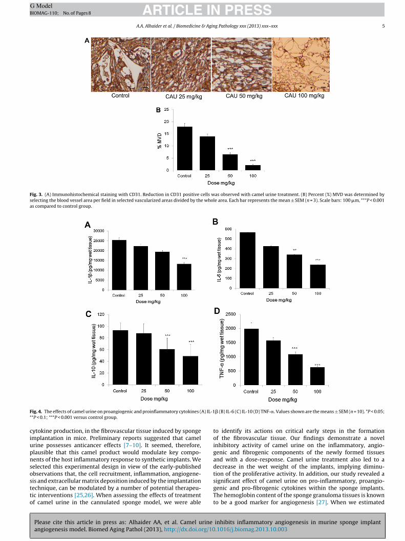
ARTICLE IN PRESSG ModelBIOMAG-110; No. of Pages 8
A.A. Alhaider et al. / Biomedicine & Aging Pathology xxx (2013) xxx–xxx 5
Fig. 3. (A) Immunohistochemical staining with CD31. Reduction in CD31 positive cells was observed with camel urine treatment. (B) Percent (%) MVD was determined byselecting the blood vessel area per field in selected vascularized areas divided by the whole area. Each bar represents the mean ± SEM (n = 3). Scale bars: 100 �m, ***P < 0.001as compared to control group.
F A) IL-1*
ciupnsostto
ig. 4. The effects of camel urine on proangiogenic and proinflammatory cytokines (*P < 0.1; ***P < 0.001 versus control group.
ytokine production, in the fibrovascular tissue induced by spongemplantation in mice. Preliminary reports suggested that camelrine possesses anticancer effects [7–10]. It seemed, therefore,lausible that this camel product would modulate key compo-ents of the host inflammatory response to synthetic implants. Weelected this experimental design in view of the early-publishedbservations that, the cell recruitment, inflammation, angiogene-
Please cite this article in press as: Alhaider AA, et al. Camel urine
angiogenesis model. Biomed Aging Pathol (2013), http://dx.doi.org/10
is and extracellular matrix deposition induced by the implantationechnique, can be modulated by a number of potential therapeu-ic interventions [25,26]. When assessing the effects of treatmentf camel urine in the cannulated sponge model, we were able
� (B) IL-6 (C) IL-10 (D) TNF-�. Values shown are the means ± SEM (n = 10). *P < 0.05;
to identify its actions on critical early steps in the formationof the fibrovascular tissue. Our findings demonstrate a novelinhibitory activity of camel urine on the inflammatory, angio-genic and fibrogenic components of the newly formed tissuesand with a dose-response. Camel urine treatment also led to adecrease in the wet weight of the implants, implying diminu-tion of the proliferative activity. In addition, our study revealed a
inhibits inflammatory angiogenesis in murine sponge implant.1016/j.biomag.2013.10.003
significant effect of camel urine on pro-inflammatory, proangio-genic and pro-fibrogenic cytokines within the sponge implants.The hemoglobin content of the sponge granuloma tissues is knownto be a good marker for angiogenesis [27]. When we estimated
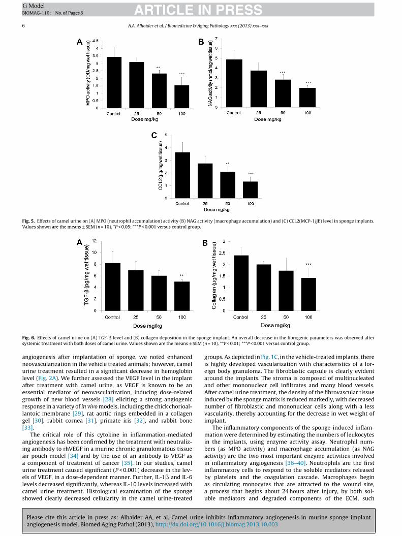
ARTICLE IN PRESSG ModelBIOMAG-110; No. of Pages 8
6 A.A. Alhaider et al. / Biomedicine & Aging Pathology xxx (2013) xxx–xxx
Fig. 5. Effects of camel urine on (A) MPO (neutrophil accumulation) activity (B) NAG activity (macrophage accumulation) and (C) CCL2(MCP-1/JE) level in sponge implants.Values shown are the means ± SEM (n = 10). *P < 0.05; ***P < 0.001 versus control group.
F e spons SEM (
anulaegrlg[
aiaauelcs
ig. 6. Effects of camel urine on (A) TGF-� level and (B) collagen deposition in thystemic treatment with both doses of camel urine. Values shown are the means ±
ngiogenesis after implantation of sponge, we noted enhancedeovascularization in the vehicle treated animals; however, camelrine treatment resulted in a significant decrease in hemoglobin
evel (Fig. 2A). We further assessed the VEGF level in the implantfter treatment with camel urine, as VEGF is known to be anssential mediator of neovascularization, inducing dose-relatedrowth of new blood vessels [28] eliciting a strong angiogenicesponse in a variety of in vivo models, including the chick chorioal-antoic membrane [29], rat aortic rings embedded in a collagenel [30], rabbit cornea [31], primate iris [32], and rabbit bone33].
The critical role of this cytokine in inflammation-mediatedngiogenesis has been confirmed by the treatment with neutraliz-ng antibody to rhVEGF in a murine chronic granulomatous tissueir pouch model [34] and by the use of an antibody to VEGF as
component of treatment of cancer [35]. In our studies, camelrine treatment caused significant (P < 0.001) decrease in the lev-
Please cite this article in press as: Alhaider AA, et al. Camel urine
angiogenesis model. Biomed Aging Pathol (2013), http://dx.doi.org/10
ls of VEGF, in a dose-dependent manner. Further, IL-1� and IL-6evels decreased significantly, whereas IL-10 levels increased withamel urine treatment. Histological examination of the spongehowed clearly decreased cellularity in the camel urine-treated
ge implant. An overall decrease in the fibrogenic parameters was observed aftern = 10). **P < 0.01; ***P < 0.001 versus control group.
groups. As depicted in Fig. 1C, in the vehicle-treated implants, thereis highly developed vascularization with characteristics of a for-eign body granuloma. The fibroblastic capsule is clearly evidentaround the implants. The stroma is composed of multinucleatedand other mononuclear cell infiltrates and many blood vessels.After camel urine treatment, the density of the fibrovascular tissueinduced by the sponge matrix is reduced markedly, with decreasednumber of fibroblastic and mononuclear cells along with a lessvascularity, thereby accounting for the decrease in wet weight ofimplant.
The inflammatory components of the sponge-induced inflam-mation were determined by estimating the numbers of leukocytesin the implants, using enzyme activity assay. Neutrophil num-bers (as MPO activity) and macrophage accumulation (as NAGactivity) are the two most important enzyme activities involvedin inflammatory angiogenesis [36–40]. Neutrophils are the firstinflammatory cells to respond to the soluble mediators released
inhibits inflammatory angiogenesis in murine sponge implant.1016/j.biomag.2013.10.003
by platelets and the coagulation cascade. Macrophages beginas circulating monocytes that are attracted to the wound site,a process that begins about 24 hours after injury, by both sol-uble mediators and degraded components of the ECM, such

ING ModelB
& Agin
atinicmuc
rkescmItcapdat
oocpapi
D
c
A
PK
R
[
[
[
[
[
[
[
[
[
[
[
[
[
[
[
[
[
[
[
[
[
[
[
[
[
[
[
[
[
[
[
ARTICLEIOMAG-110; No. of Pages 8
A.A. Alhaider et al. / Biomedicine
s fragments of collagen and fibronectin. Monocytes bind tohe ECM via integrin receptors and immediately differentiatento tissue macrophages. MPO activity (representing activatedeutrophils) was not affected by the camel urine treatment, reflect-
ng a lack of effect of the camel urine on this inflammatoryell population; however, NAG activity (representing activatedacrophages/monocytes) was significantly decreased in the camel
rine-treated groups, suggesting a degree of selectivity of action ofamel urine.
As expected the levels of TNF-� and CCL2 (MCP-1/JE) wereeduced in the implants of the camel urine-treated groups. It isnown that angiogenesis requires the deposition of collagen byndothelial cells into the basement membrane of new blood ves-els [24]. Deposition of collagen is also essential for endothelialell migration as it forms the scaffold for new blood vessel for-ation. The deposition of various collagens, including collagen I, II,
II, V, and IX, is increased during tumor formation [41–43]. Besides,hickening and linearization of collagen fibers are common in can-ers, and they are often found in areas where active tissue invasionnd tumor vasculature are observed [44,45], suggesting that theylay an active role in facilitating the invasion by cancer cells. Theecreased collagen deposition in the camel urine treated groupsnd the lower level of the pro-fibrogenic cytokine TGF-� were fur-her signs of reduced inflammation observed in our model.
To our knowledge, this is the first report that assessed the effectsf camel urine on multiple parameters of the main componentsf inflammatory angiogenesis. Moreover, a regulatory function ofamel urine on pro-inflammatory and pro-fibrogenic cytokinesroduction has been revealed. Altogether, our study revealed
potent inhibitory action of camel urine on pro-inflammatory,ro-angiogenic and profibrogenic cytokines, within the sponge
mplants.
isclosure of interest
The authors declare that they have no conflicts of interest con-erning this article.
cknowledgement
The authors especially wish to thank the College of Medicine andharmacy Research Centres and Deanship of Scientific Research,ing Saud University, Riyadh, for funding this work.
eferences
[1] Albini A, Tosetti F, Benelli R, et al. Tumor inflammatory angiogenesis and itschemoprevention. Cancer Res 2005;65:10637–41.
[2] Hanahan D, Weinberg RA. The hallmarks of cancer. Cell 2000;100:57–70.[3] Colotta F, Allavena P, Sica A, et al. Cancer-related inflammation, the
seventh hallmark of cancer: links to genetic instability. Carcinogenesis2009;30:1073–81.
[4] Conesa C, Sanchez L, Rota C, et al. Isolation of lactoferrin from milk of differ-ent species: calorimetric and antimicrobial studies. Comp Biochem Physiol B:Biochem Mol Biol 2008;150:131–9.
[5] Agrawal RP, Dogra R, Mohta N, et al. Beneficial effect of camel milk in diabeticnephropathy. Acta bio-medica: Atenei Parmensis 2009;80:131–4.
[6] Redwan el RM, Tabll A. Camel lactoferrin markedly inhibits hepatitis C virusgenotype 4 infection of human peripheral blood leukocytes. J ImmunoassayImmunochem 2007;28:267–77.
[7] al-Harbi MM, Qureshi S, Ahmed MM, et al. Effect of camel urine on the cyto-logical and biochemical changes induced by cyclophosphamide in mice. JEthnoparmacol 1996;52:129–37.
[8] Khorshid FA, Moshref SS. In vitro anticancer agent. I–Tissue culture study ofhuman lung cancer cells A549. II–Tissue culture study of mice leukemia cellsL1210. Int J Cancer Res 2006;2:330–44.
Please cite this article in press as: Alhaider AA, et al. Camel urine
angiogenesis model. Biomed Aging Pathol (2013), http://dx.doi.org/10
[9] Al-Yousef N, Gaafar A, Al-Otaibi B, et al. Camel urine components display anti-cancer properties in vitro. J Ethnoparmacol 2012;143:819–25.
10] Alhaider AA, El Gendy MA, Korashy HM, et al. Camel urine inhibits thecytochrome P450 1a1 gene expression through an AhR-dependent mechanismin Hepa 1c1c7 cell line. J Ethnopharmacol 2010;133:184–90.
[
PRESSg Pathology xxx (2013) xxx–xxx 7
11] Saraswati S, Pandey M, Mathur R, et al. Boswellic acid inhibits inflammatoryangiogenesis in a murine sponge model. Microvasc Res 2011;82:263–8.
12] Saraswati S, Agarwal SS. Strychnine inhibits inflammatory angiogenesis inmice via down regulation of VEGF, TNF-� and TGF-�. Microvasc Res 2013;87:7–13.
13] Agrawal SS, Saraswati S, Mathur R, et al. Brucine, a plant-derived alkaloidinhibits inflammatory angiogenesis in a murine sponge model. Biomed PrevNut 2011;1:180–5.
14] Saraswati S, Alhaider AA, Abdel Gader AGM, et al. Camel milk inhibits inflamma-tory angiogenesis via downregulation of proangiogenic and proinflammatorycytokines in mice. APMIS 2013:12199.
15] Saraswati S, Kanuajia PK, Kumar S, et al. Tylophorine, a phenanthraindolizidinealkaloid isolated from Tylophora indica exerts antiangiogenic and antitumoractivity by targeting vascular endothelial growth factor receptor 2-mediatedangiogenesis. Molecular Cancer 2013;12:82.
16] Passaniti A, Taylor AM, Pili R, et al. A simple, quantitative method for assessingangiogenesis and antiangiogenic agents using reconstituted basement mem-brane, heparin, and fibroblast growth factor. Lab Invest 1992;67:519–28.
17] Lage AP, Andrade SP. Assessment of angiogenesis and tumor growth in con-scious mice by a fluorimetric method. Microvasc Res 2000;59:278–85.
18] Ferreira MA, Barcelos LS, Teixeira MM, et al. Tumor growth, angiogenesis andinflammation in mice lacking receptors for platelet activating factor (PAF). LifeSci 2007;81:210–7.
19] Phillips RJ, Burdick MD, Hong K, et al. Circulating fibrocytes traffic to the lungsin response to CXCL12 and mediate fibrosis. J Clin Invest 2004;114:438–46.
20] Campos PP, Bakhle YS, Andrade SP. Mechanisms of wound healingresponses in lupus-prone New Zealand White mouse strain. Wound Rep Reg2008;16:416–24.
21] Hlkatky L, Hahnfeldt P, Folkman J. Clinical application of antiangiogenic ther-apy: microvessel density, what it does and doesn’t tell us. J Natl Cancer Inst2002;94:883–93.
22] Leung DW, Cachianes G, Kuang WJ, et al. Vascular endothelial growth factor isa secreted angiogenic mitogen. Science 1989;246:1306–9.
23] Holderfield MT, Hughes CC. Crosstalk between vascular endothelial growth fac-tor, notch, and transforming growth factor-beta in vascular morphogenesis.Circ Res 2008;102:637–52.
24] Bonanno E, Iurlaro M, Madri JA, et al., Type IV. collagen modulates angiogen-esis and neovessel survival in the rat aorta model. In Vitro Cell Dev Biol Anim2000;36:336–40.
25] Belo AV, Barcelos LS, Ferreira MAND, et al. Inhibition of inflammatory angio-genesis by distant subcutaneous tumor in mice. Life Sci 2004;74:2827–37.
26] Bergheim I, Luyendyk JP, Steele C, et al. Metformin prevents endotoxininduced liver injury after partial hepatectomy. J Pharmacol Exp Ther 2006;316:1053–61.
27] Yamaoka M, Masaki T, Sudo K, et al. Acceleration of wound healing by recombi-nant human basic fibroblast growth factor in a rat sponge-implantation model.Jpn J Inflamm 1991;11:65–72.
28] Dvorak HF. Vascular permeability factor/vascular endothelial growth factor: acritical cytokine in tumor angiogenesis and a potential target for diagnosis andtherapy. J Clin Oncol 2002;21:4368–80.
29] Plouet J, Schilling J, Gospodarowicz D. Isolation and characterization of anewly identified endothelial cell mitogen produced by AtT20 cells. EMBO J1989;8:3801–6.
30] Nicosia RF, Nicosia SV, Smith M. Vascular endothelial growth factor, platelet-derived growth factor and insulin-like growth factor-1 promote rat aorticangiogenesis in vitro. Am J Pathol 1994;145:1023–9.
31] Phillips GD, Stone AM, Jones BD. Vascular endothelial growth factor(rhVEGF165) stimulates direct angiogenesis in the rabbit cornea. In Vivo1995;8:961–5.
32] Tolentino MJ, Miller JW, Gragoudas ES, et al. VEGF is sufficient to produce irisneovascularization and neovascular glaucoma in a non-human primate. ArchOphthalmol 1996;114:964–70.
33] Connolly D, Olander J, Heuvelman D, et al. Human vascular permeability factor.Isolation from U937 cells. J Biol Chem 1989;264:20017–24.
34] Appleton I, Brown NJ, Willis D, et al. The role of vascular endothelial growth fac-tor in a murine chronic granulomatous tissue air pouch model of angiogenesis.J Pathol 1996;180:90–4.
35] Chen HX, Gore-Langton RE, Cheson BD. Clinical trials referral resource: currentclinical trials of the anti-VEGF monoclonal antibody bevacizumab. Oncology2001;15:1023–6.
36] Edward RH, Sarmenta SS, Hass GM. Stimulation of granulation tissue growthby tissue extracts; study by intramuscular wounds in rabbits. Arch Path1960;69:286–302.
37] Andrade SP, Bakhle YS, Hart I, et al. Effects of tumor cells and vasoconstrictorresponses in sponge implants in mice. Br J Cancer 1992;66:821–6.
38] Andrade SP, Fan TPD, Lewis GP. Quantitative in vivo studies on angiogenesis inrat sponge model. Br J Exp Path 1987;68:755–6.
39] Barcelos LS, Coelho AM, Russo RC, et al. Role of the chemokines. CCL3/MIP-1� and CCL5/RANTES in sponge-induced inflammatory angiogenesis in mice.Microvasc Res 2009;78:148–54.
40] Mendes JB, Rocha MA, Araújo FA, et al. Differential effects of rolipram on chronic
inhibits inflammatory angiogenesis in murine sponge implant.1016/j.biomag.2013.10.003
subcutaneous inflammatory angiogenesis and on peritoneal adhesion in mice.Microvasc Res 2009;78:265–71.
41] Zhu GG, Risteli L, Mäkinen M, et al. Immunohistochemical study of type I colla-gen and type I pN-collagen in benign and malignant ovarian neoplasms. Cancer1995;75:1010–7.
ING ModelB
8 & Agin
[
[
ARTICLEIOMAG-110; No. of Pages 8
A.A. Alhaider et al. / Biomedicine
Please cite this article in press as: Alhaider AA, et al. Camel urine
angiogenesis model. Biomed Aging Pathol (2013), http://dx.doi.org/10
42] Kauppila S, Stenbäck F, Risteli J, et al. Aberrant type I and type III collagen geneexpression in human breast cancer in vivo. J Pathol 1998;186:262–8.
43] Huijbers IJ, Iravani M, Popov S, et al. A role for fibrillar collagen deposition andthe collagen internalization receptor endo180 in glioma invasion. PLoS ONE2010;5:9808.
[
[
PRESSg Pathology xxx (2013) xxx–xxx
inhibits inflammatory angiogenesis in murine sponge implant.1016/j.biomag.2013.10.003
44] Condeelis J, Segall JE. Intravital imaging of cell movement in tumours. Nat RevCancer 2003;3:921–30.
45] Levental KR, Yu H, Kass L, et al. Matrix crosslinking forcestumor progression by enhancing integrin signaling. Cell 2009;139:891–906.

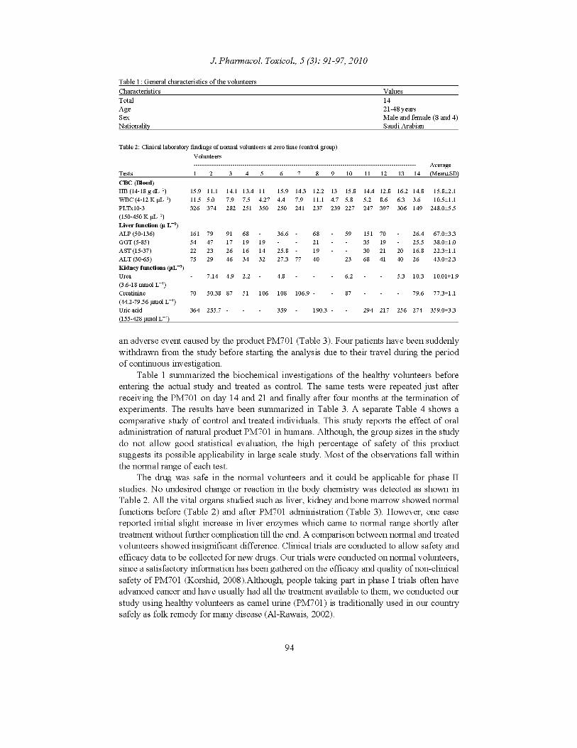
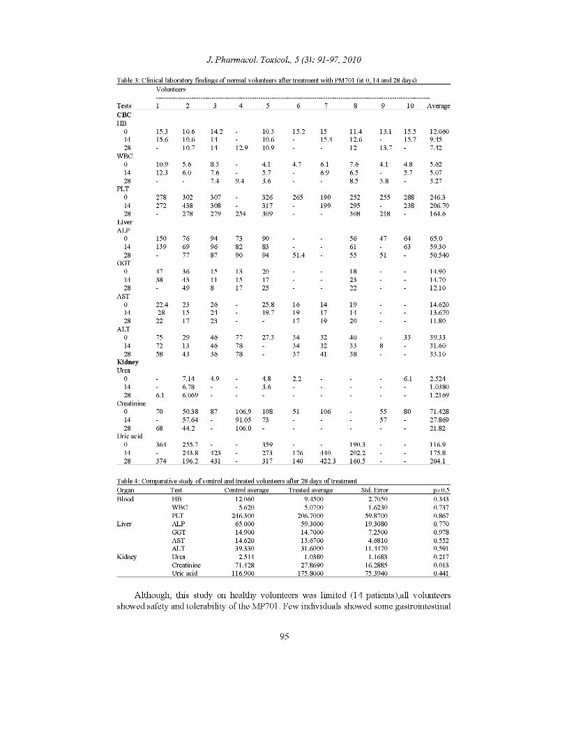

95
Gas chromatography mass spectrophotometry (gc-ms) analysis of
female camel urine extracts
Salwa M.E. Khogali*; Samia H. Abdalrahman*; Esraa M. Musa* and Abdalla M. El
Hassan**
Abstract
In this study the chemical composition of female camel urine extracts (chloroformic,
ethanolic and lyophilized) were analyzed by GC-MS: Agilent technologies 5973N.
Seventeen bioactive organic compounds were detected. The degraded compounds in
all extracts were comparable to each other. The results obtained verify that female
camel urine extracts are an excellent poll of bioactive compounds which are
extremely valuable for detection and manufacture of new drugs of natural origin.
Introduction:
The use of human urine and urine extracts for medical purpose has been known for
centuries (Armstrong, 1937; Burzynski, 1988). Recently the medical science of
human and animal urine has identified profound medical uses (Christy, 2000: Peroni,
2001: Natalie, 2002).The use of urinary remedies to deal with illness has been
gaining high popularity in Asia (Read, 1979: Lai et al, 1999). Use of animal urine is
endorsed in mainstream modern medicine. Mare urine is the source of conjugated
equine estrogens and has been marketed for over fifty years as the pharmaceutical
brand Premarin, “an estrogen treatment for menopausal and pre-menopausal
women”, and especially postpartum – one of the most prescribed drugs in the United
States (Christy 2000). It was very recently discovered that adding distilled cow urine
to medicaments increases their effectiveness while decreasing their side-effects,
making anti-cancer and anti-tubercular drug twenty times more effective and anti-
bacterial eighty times more effective (Natalie, 2002). The reliable therapeutic
efficacy obtained from clinical studies on camel urine is recorded by (Ohag, 1993,
1998); Kabariti,1988; Burziski,1977); (Mona, 2003); (Wisal, G. 2002); (Salwa et al,
2006). These experiments showed that camel urine contains many complex bioactive
compounds which can act against bacterial, fungal, viral, parasitic and carcinogenic
agents, and it has the ability to protect the liver against toxic agents (Salwa et al,
2009). Ethnopharmacology of camel urine as a folk medicine has been gaining
popularity for a variety of ailments, particularly in the light of significant advances
that have been made in recent years. The methods of pharmacology-
pharmochemistry and chromatography offer insights in the etiology and discovery of
new drugs and compounds. However there is a continuing need for a vast
improvement in our knowledge and understanding of discovering new compounds
and drugs. In what follows I identify and summarise significant advances with regard
to medicinal compound in female camel urine.
Objectives:
The known therapeutic efficacy of female camel urine and its extracts led us to
analyze and investigate its bioactive chemical components.
96
Materials and methods:
Sample collection:
Female camel urine was collected by natural urination or by tashweel technique.
Using sterile containers.
Sample preparation:
Chloroformic Extract:
Equal volumes of female camel’s urine (FCU) and chloroform were shaken for three
hours and left to separate; the lower chloroformic layer was then displaced and
analyzed by GC-MS
Ethanolic Extract
Ten grams of lyophilized female camel urine were refluxed in 80% ethanol for 30
mins, then filtered; the filtrate was further analyzed by GC-MS.
Lyophilized Female Camel Urine
Two ml of female camel urine were poured into piqué bottles and freeze dried by
freeze drier machine.
Analytical methods:
Gas chromatography – mass spectrometry (GC-MS) was performed using Agilent
6890 N Net work GC system interfaced with 5973 N Net work, mass selective
detector (MSD). The GC-MS was fitted with a 60 m Agilent fused capillary column,
DB-5ms 0.25mm 1-D, 0.25 mm Film – initial temp 100c◦, hold 2 min, then
programmed at 2c◦/min to 300c◦ min; isothermal temperature was held for 10 min.
Helium carrier gas, head pressure 9.30 psi, column flow 1ml/min. injection temp.
300C◦ El source 230c◦, total scan mode was cycled at 2 seconds. 1 ml of the given
sample was diluted with 10 ml of diethyl chloromethane (DCM) and 1µ was injected
using split less mode.
IR apparatus
A Perkin Elmer 2 Lambda Spectra, 580 infra red spectrophotometer Neel fur was
used for detecting the functional groups in LU & CE of female camel urine using kBr
and NaCl respectively.
Results and discussion
Identification of the degraded compounds was conducted by comparison with
published NIST Library retention time of the chromatogram. Corrected areas
percentage obtained by base line subtraction were used to calculate the percentage of
the compound within the injected amount. Figure 1 and table 1 represent the GC-MS
chromatogram of lyophilized urine. Figure 2 and table 2 for ethanolic extract
degraded compound; Figure 3 and table 3 showed the chromatogram and degraded compounds of chloroformic extract. Figures 4 and 5 represents the infrared (IR)
analysis of lyophilized and chloroformic extract of female camel urine. Tables 4 and
Elbashir et al. – Analysis of female camel urine extracts
97
5 showed the obtained functional groups of (LU) and the medicinal uses of some
degraded compounds respectively.
The GC-MS analysis of ethanolic extract, lyophilized and chloroformic extract of
female camel urine revealed comparable degraded compounds. These compounds
contain aliphatic hydrocarbon chains (3 up to 27 carbon atoms) with oxygen,
nitrogen, silicon, alkyl and phosphorus. Benzene rings, phenolic, Omiga 6 & 9
compounds and some novel compounds such as titanium, oxirane and heptasiloxane
were obtained. These results suggested that these chemicals may have widespread
distribution in the grazing plants of camels. Some of these compounds are medically
used for cancer. This was in agreement with the records of (Khorshid et al 2005);
(Ohag, 2010). The uses of camel urine as antibacterial, antifungal, antiparasitic, and
as an ingredient for cosmetics were reported by (Christy,2000; Natalie,2002);
degraded compounds were confirmed with that in *Merck Index (1968; 1998; 2006).
The presence of hydroxyl (OH), carboxylic (COOH), aromatic (C_C), amine (NH),
thiol (S=O) and chlore (CL) in female camel urine may enable camel urine and its
extracts to act via different chemical pathways.
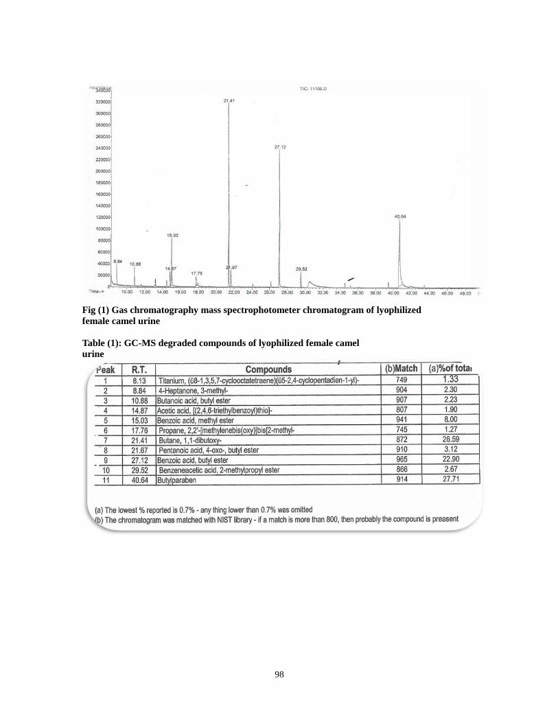
98
Fig (1) Gas chromatography mass spectrophotometer chromatogram of lyophilized
female camel urine
Table (1): GC-MS degraded compounds of lyophilized female camel
urine
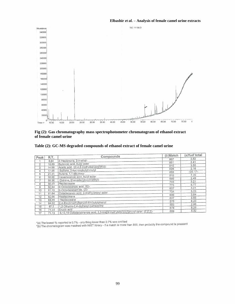
Elbashir et al. – Analysis of female camel urine extracts
99
Fig (2): Gas chromatography mass spectrophotometer chromatogram of ethanol extract
of female camel urine
Table (2): GC-MS degraded compounds of ethanol extract of female camel urine
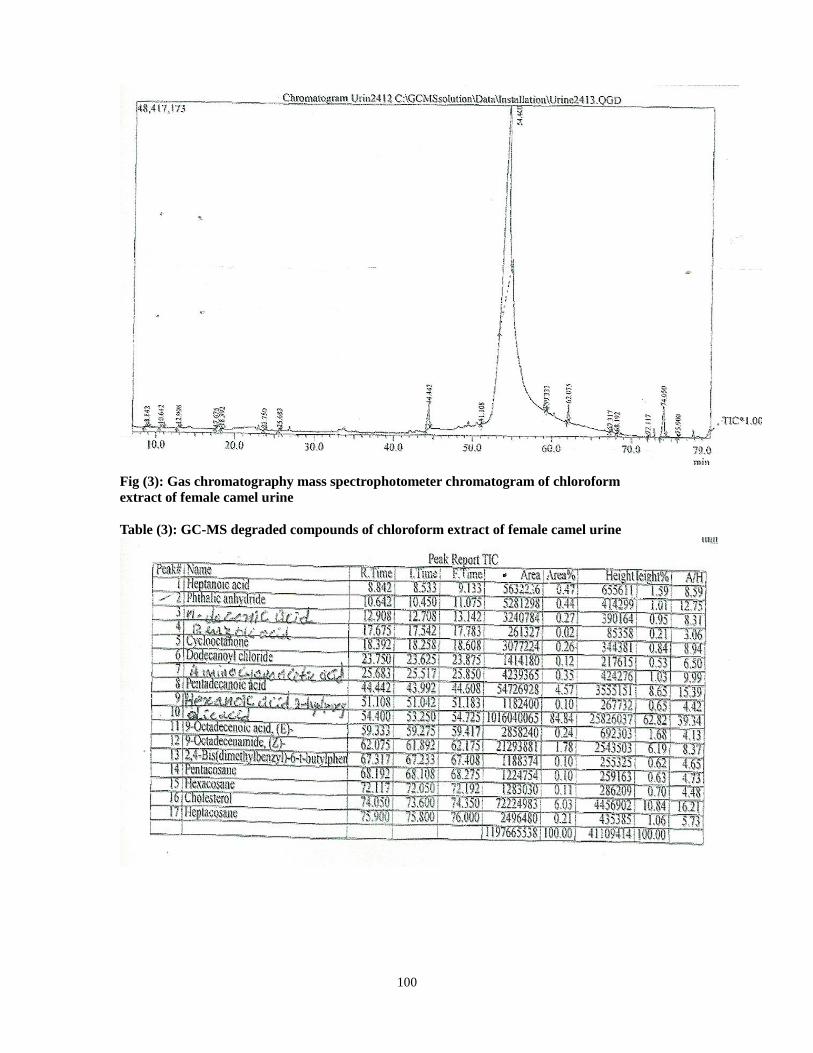
100
Fig (3): Gas chromatography mass spectrophotometer chromatogram of chloroform
extract of female camel urine
Table (3): GC-MS degraded compounds of chloroform extract of female camel urine
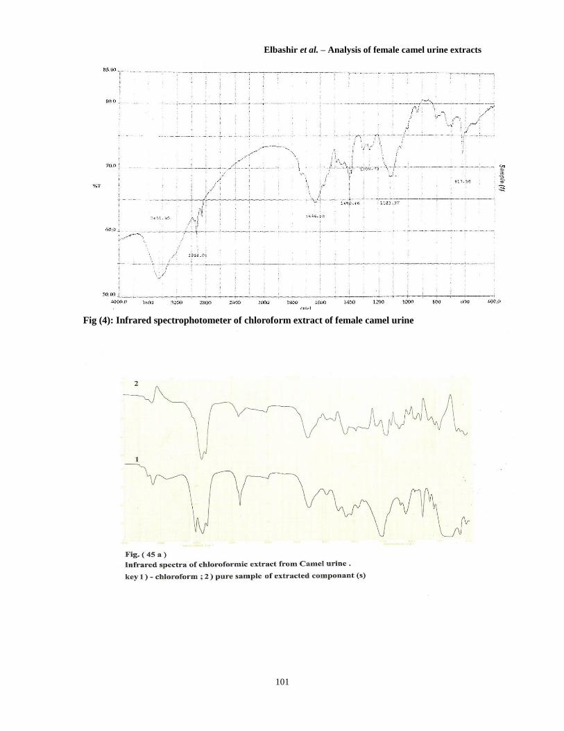
Elbashir et al. – Analysis of female camel urine extracts
101
Fig (4): Infrared spectrophotometer of chloroform extract of female camel urine
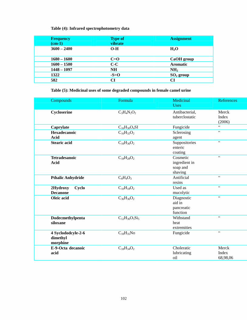
102
Table (4): Infrared spectrophotometry data
Frequency
(cm-1)
Type of
vibrate
Assignment
3600 – 2400 O-H H2O
1680 – 1600 C=O CaOH group
1600 – 1500 C-C Aromatic
1448 – 1097 NH NH2
1322 -S=O SO2 group
582 CI CI
Table (5): Medicinal uses of some degraded compounds in female camel urine
Compounds Formula Medicinal
Uses
References
Cycloserine C3H4N2O2 Antibacterial,
tuberclostatic
Merck
Index
(2006)
Caprylate C16H30O4SI Fungicide “
Hexadecanoic
Acid
C16H32O2 Sclerosing
agent
“
Stearic acid C18H36O2 Suppositories
enteric
coating
“
Tetradesanoic
Acid
C18H28O2 Cosmetic
ingredient in
soap and
shaving
“
Pthalic Anhydride C8H4O3 Antificial
resins
“
2Hydroxy Cyclo
Decanone
C10H18O2 Used as
mucolytic
“
Oleic acid C36H36O2 Diagnostic
aid in
pancreatic
function
“
Dodecmethylpenta
siloxane
C12H36O5Si5 Withstand
heat
extremities
“
4 Syclododcyle-2-6
dimethyl
morphine
C18H35No Fungicide “
E-9-Octa decanoic
acid
C18H34O2 Choleratic
lubricating
oil
Merck
Index
68,98,06
Elbashir et al. – Analysis of female camel urine extracts
103
Conclusion
Medicinal uses of some of these compounds, confirm the therapeutic effects of
female camel urine in our previous clinical studies. To enhance the utility and
convenience of the degraded compounds, each compound should be fractionated and
monitored to know its bioactivity against the actual disease.
ACKNOWLEDGMENTS
We thank the CPL technical staff for their excellent technical assistance; thanks also
to Dr. Safa Omer for her co-operation and computerization of the manuscript.
REFERENCES:
Armstrong, J.W. (1971) The Water Of Life: A Treatise on Urine Therapy, Health
Science Press, Rustington, UK.
Burzynski, Stanislaw. R. et al (1977) "Anti neoplaston A in cancer therapy",
Physiology, Chemistry and Physics, vol. 9, 485.
Christy, M. Martha, (2000) Your Own Perfect Medicine
Kabariti, A., Mazruai, S. and Elgendi, A. (1988) "Camel urine: A possible anti
carcinogenic agent", Arab Gulf. J.Sci, Res.Agric, Biol.Sci.
Mona, A. Khalifa (2003) "Antibacterial effects of camel urine (Camelus dromedarius)", MVSc dissertation, University of Khartoum, Sudan.
Natalie, B. (2002) "Urine therapy (drinking urine)". J. of Berkeley Medicine.
Ohaj, H.M. (1993)"Camel urine as medicament in Sudan", BSc dissertation,
University of the Gezira, Sudan.
Ohag, H.M. (1998) "Clinical trials for treatment of ascites with camel urine", MSc.
University of Gezira, Sudan.
Pieroni A., A.Grazzini and M.E.Giusti (2002) "From the sources of knowledge to the
medicines of the future", Proceedings of the 4th European Colloquium on
Ethnopharmacology, IRD Editions, Paris, France, pp. 371-5.
Salwa, M/E., Khogali; O.Y. Mohamed; A.M. Elhassan and A.M.A. Magid (2006)
"Therapeutic applications of she-camel urine: pathological changes in cattle infected
with fasciolosis". Albuhuth, vol 10 (1):109-122.
Salwa, M/E., Khogali; O.Y. Mohamed; A.M. Elhassan; A.M. Shammat and A.M.A.
Magid (2009) "Hepatoprotective effect against carbontetrachloride induced
hepatotoxicity in rats", J.SCi.and Techn., vol.10 (2):128-34.
Khorshid, F.A., Moshref, S.S.; Heffny, N. (2005)"An ideal selective anticancer agent
in vitro, 1-tissue culture study of human lung cancer cells A590", JKAU-Medical Sciences, vol. 12, pp. 3-18.
Wisal, G.A. (2002) "Antibacterial and antifungal effect of camel urine (Camelus dromedarius)", MVSc dissertation, University of Khartoum, Sudan. __________
* Department of Biochemistry, Toxicology and Pharmacology, Central Veterinary
Research Laboratories, Khartoum, Sudan.
** Department of Pharmacognacy, Faculty of Pharmacy, Al/Rabat University,
Khartoum, Sudan.
E-mail: [email protected]

104

Effects of Heating and Storage on the Antifungal Activity of Camel UrineAhlam Al-Awadi and Awatif Al-Judaibi*
Department of Biological Science, Microbiology Section, King Abdulaziz University, Jeddah, KSA, Saudi Arabia*Corresponding author: Awatif Al-Judaibi, Department of Biological Science, Microbiology Section, King Abdulaziz University, Jeddah, KSA, Saudi Arabia, Tel:966505660345; E-mail: [email protected]
Rec date: Oct 09, 2014, Acc date: Nov 28, 2014, Pub date: Dec 21, 2014
Copyright: © 2014 Al-Awadi A, et al. This is an open-access article distributed under the terms of the Creative Commons Attribution License, which permits unrestricteduse, distribution, and reproduction in any medium, provided the original author and source are credited.
Abstract
Camel urine, considered a ‘miraculous’ drug used in Prophetic Medicine, since the pre-Islamic era camel milk andurine were used as drinking medicine for different health problems. In addition, camel urine has proven to beeffective as an antimicrobial agent, and may not have side effects for humans. Furthermore, camel urine may beresistant to factors such as high temperatures and an extensive waiting period in laboratory conditions, which canreduce the effectiveness of antibiotics. The aim of our study was to examine the effectiveness of camel urine as anantifungal agent following exposure to high temperatures and long time periods in laboratory conditions. Aftermaintaining camel urine in natural laboratory conditions for 6 weeks at temperatures of up to 100°C, we tested camelurine on the fungi Aspergillus niger and Fusarium oxysporum, and on the yeast Candida albicans. We thenmeasured the dry weight of each microorganism, and determined their minimum inhibitory and fungicidalconcentrations. Our results showed that after maintained for 6 weeks, camel urine did not lose its antifungal activity;dry weights following treatment were decreased 100% of the dry weight prior to treatment for Aspergillus niger andCandida albicans, and 53.33% for Fusarium oxysporum. Our study demonstrates that camel urine is a highlyeffective and resilient antifungal agent for treating human and plant fungal diseases.
Keywords: Camel urine; Antifungal; Candida albicans; Aspergillusniger; Fusarium oxysporum
IntroductionThe camel is mentioned in the Holy Qur'an as a particularly
important animal1, and is referred to by other names such as al-ibil, al-nagah, al-jamal, al-ishar and al-him [1]. Ccamel urine is considered a‘miraculous’ drug used in Prophetic Medicine since the pre-Islamicera2 [2], which has been used as traditional and folk medicine forwomen's hair; gums and teeth; skin injuries; snake bites; stomach pain;tumors; the common cold; diarrhea and nausea; diabetes; jaundice;scabies; and eye, skin, liver and nail infections [1-5]. Camel urine isalso commonly used against cancer and respiratory tract infections inalternative medicine [6].
Camel urine has been proven to be effective as an antimicrobialagent, and may not have any side effects for humans [7]. Muhammad(1998) reported that patients who were given camel urine to treatdigestion problems recovered after two months of treatment [8]. Al-Yousef et al. (2012) found that camel urine has no cytotoxic effectagainst mononuclear cells, and has strong immune activity byinducing IFN-γ and inhibiting Th2 cytokines IL-4, IL-6 and IL-10.Kidney, liver and stomach tissues infected with Escherichia coli inmice recovered with no histopathological effects after treatment withcamel urine of concentrations up to 100% [9-12]. Studies have testedthe antimicrobial activity of camel urine against pathogenicmicroorganisms including the fungi Aspergillus niger, A. flavus,Fusarium oxysporum, Rhizoctonia solani, Aschocayta sp., Pythium
aphanidermatum, Sclerotinia sclerotiorum, Candida albicans; and thebacteria Staphylococcus aureus, Streptococci, E. coli, Pseudomonasaeroginosa and Klebsiella pneomoniae. The results of these studiesshowed high antimicrobial activity against the tested microorganisms,even when accompanied by changes in anions and cations [4,13-18].
Antimicrobial activity of camel urine is due to factors such as highsalt concentrations, alkalinity, natural bioactive compounds from theplants camels eat, resident bacteria, and excreted antimicrobial agents.Compared with other cattle, camel urine is alkaline due to highconcentrations of potassium, magnesium and albuminous proteins,and low concentrations of uric acid, sodium and creatine [19-20]. Thedifferent composition of camel urine compared to other cattle andgoats is due to the type of plants they consume and their feedinghabits; camels prefer browse with high concentrations of minerals thatdecline more slowly when they dry instead of other types of foragesuch as grasses [21-23]. Further, camels eat a variety of types ofvegetation including thorny bushes, halophytes, salty and sour plants,shrubs and aromatic species that are avoided by cattle and goat (e.g.,Haloxylon aphyllum, H. persieum, Salsola gemmaseens, S. orientabs,Astragalus, Aristida karelinii and A. pinnate) [17,18,20,24].
The aim of our study was to investigate the resistance of camelurine to heating at high temperatures and storage for extensive waitingperiods in laboratory conditions, which can reduce the effectiveness ofantibiotics.
1 ‘Do they not look at the camel, how it was created?’ (Surah Number 88: Al-Ghâshiyah).2 Several Hadith in Sunnah talked about using camel urine and milk as medicine (the Saying of the prophet Muhammad, Volume 8, Book
82, Number 794: Narrated Anas): ‘Some people from the tribe of 'Ukl came to the Prophet and embraced Islam. The climate of Medina didnot suit them, so the Prophet ordered them to go to the (herd of milk) camels of charity, and to drink their milk and urine (as a medicine).’
Clinical Microbiology: OpenAccess Al-Awadi and Al-Judaibi, Clin Microbiol 2014, 3:6
http://dx.doi.org/10.4172/2327-5073.1000179
Research Article Open Access
Clin MicrobiolISSN:2327-5073 CMO, an open access journal
Volume 3 • Issue 6 • 1000179

Materials and Methods
Study materialsThe molds Aspergillus niger and Fusarum oxysporium were
isolated and identified at the Cairo MIRCEN, Ain Shams University,Cairo, Egypt. Tested fungi were incubated at 28 ± 2°C. Candidaalbicans ATCC CA 10231 was incubated at 30 ± 2°C. Camel urine wascollected from north Jeddah from live camel in the desert in sterilizeddark bottles that were taken directly to the laboratory.
To investigate the effect of storage time and heating on camel urineantifungal activity, collected camel urine was divided into two majorgroups. The first group was further subdivided into three portions thatwere heated at 60, 80 and 100°C for 60 min. The second group wasfurther subdivided into three portions that were stored for 3, 6 and 9months before laboratory analyses. The positive control was freshcamel urine at 4°C.
Laboratory analysesThe antimicrobial activity of camel urine was determined in vitro in
response to A. niger, F. oxysporum and C. albicans. Activity levelswere measured using disc diffusion and broth dilution, methodspreviously described by the Clinical and Laboratory StandardsInstitute (CLSI; formerly known as the National Committee forClinical Laboratory Standards) [25,26]. For disc diffusion we usedfilter paper discs (1 mm diameter impregnated with 100 μL), whichwere placed on the pre-inoculated agar surface. Negative controls wereprepared with sterilized discs. Plates were then incubated at 28°C forA. niger and F. oxysporum for 7 days, and at 30°C for C. albicans for48 h. The inhibitory zones of each disc were measured. All tests wereperformed in triplicate.
The Minimum Inhibitory Concentration (MIC) and MinimumFungicidal Concentration (MFC) of camel urine that inhibited thegrowth of fungi were investigated using a broth-microdilutionmethod. C. albicans, A. niger and F. oxysporum were cultured andresuspended in 1 mL mueller-hinton broth (OXOID) to obtain a finalconcentration of 100 cfu mL-1. Camel urine was serially diluted withMueller-Hinton broth using methods approved by the NationalCommittee for Clinical Laboratory Standards (M27- A) [27]. Afterincubation, the MIC was determined as the lowest concentration ofextract for which there was no visible growth compared with thecontrol [28,29]. The MFC was determined by inoculating 0.1 mL of
negative growth at the MIC onto sterile Sabouraud Dextrose AgarSDA for C. albicans and Potato Dextrose Agar PDA for A. niger and F.oxysporum (OXOID) plates(Table 1). The plates were incubated at30°C for 48 h for C. albicans, and at 28°C for 7 days for A. niger and F.oxysporum.
The lowest concentration of camel urine that did not demonstrategrowth of the tested fungi was considered the MFC; the negativecontrol was a plate grown with media only [30,31].
The dry weight of the tested fungi was measured to determine theeffects of recommended doses in Arab folk-medicine. 1 mL samples ofA. niger and F. oxysporum spores, and C. albicans suspension (108 cfumL-1) were inoculated into 5, 10 and 15 mL samples of treated camelurine with SDB/PDB in 250 mL Erlenmeyer flasks. Flasks wereincubated with shaking (180 rpm) at 30°C for 7 days for A. niger andF. oxysporum, and for 48 hours for C. albicans. Afterwards, sampleswere collected and centrifuged at 10,000 rpm for 10 min. Fungalmycelia and yeast cells were collected. Growth was estimated as dryweight by washing with triple-distilled water and drying at 80°C onWhatman no. 1 filter paper until constant weight [32].
The lowest MICs of 1 μL mL-1 were obtained with untreated camelurine; with urine treated at 60°C and 80°C, and stored for 2 months forC. albicans; and with all treatments except for urine stored for 6months for A. niger (Table 2). The most resistant fungus was F.oxysporum with MIC values ranging from 2 to 8 μL mL-1. MFC valuesranged from 4 to 32 μL mL-1, and were lowest for A. niger andgreatest for F. oxysporum (Table 3).
Statistical AnalysisThe results were analyzed by paired-samples t-test using the IBM
SPSS 20 statistical software to compare the mean values of eachtreatment. The results are expressed as means ± SE. Probability levelsof less than 0.01 were considered highly significant.
ResultsWe observed high inhibitory growth of C. albicans, A. niger and F.
oxysporum after treatment with fresh camel urine, which providedevidence for camel urine as an active antifungal agent (Table 1). Themost sensitive tested fungi were C. albicans and A. niger, while theinhibition of F. oxysporum only decreased by 22% when camel urinewas stored for 6 months.
Incubation temperature Storage time (months)
Fresh 60°C 80°C 100°C 2 4 6
C. albicans 45 ± 0.891** 42 ± 0.895** 39 ± 0.891** 36 ± 0.895** 44 ± 0.589** 40 ± 0.895** 37 ± 0.566**
A. niger 43 ± 1.166** 38 ± 0.895** 35 ± 1.166** 30 ± 0.566** 41 ± 0.589** 37 ± 0.873** 35 ± 0.589**
F. oxysporum 39 ± 0.895** 36 ± 0.895** 34 ± 0.589** 29 ± 0.589** 37 ± 0.891** 35 ± 0.566** 32 ± 0.566**
Table 1: Inhibition of C. albicans, A. niger and F. oxysporium growth after incubation with 100 μL of camel urine.
Incubation temperature Storage time (months)
Fresh 60°C 80°C 100°C 2 4 6
Citation: Al-Awadi A and Al-Judaibi A (2014) Effects of Heating and Storage on the Antifungal Activity of Camel Urine. Clin Microbiol 3: 179. doi:10.4172/2327-5073.1000179
Page 2 of 6
Clin MicrobiolISSN:2327-5073 CMO, an open access journal
Volume 3 • Issue 6 • 1000179
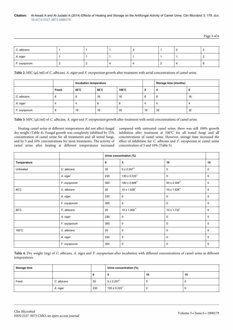
C. albicans 1 1 1 2 1 2 2
A. niger 1 1 1 1 1 1 2
F. oxysporum 2 2 4 4 2 4 8
Table 2: MIC (μL/ml) of C. albicans, A. niger and F. oxysporium growth after treatment with serial concentrations of camel urine.
Incubation temperature Storage time (months)
Fresh 60°C 80°C 100°C 2 4 6
C. albicans 8 8 16 16 8 8 16
A. niger 4 4 8 8 4 4 4
F. oxysporum 8 16 16 32 16 32 32
Table 3: MFC (μL/ml) of C. albicans, A. niger and F. oxysporium growth after treatment with serial concentrations of camel urine.
Heating camel urine at different temperatures did not affect fungaldry weight (Table 4). Fungal growth was completely inhibited by 15%concentration of camel urine for all treatments and all tested fungi,and by 5 and 10% concentrations for most treatments. The activity ofcamel urine after heating at different temperatures increased
compared with untreated camel urine; there was still 100% growthinhibition after treatment at 100°C for all tested fungi and allconcentrations of camel urine. However, storage time increased theeffect of inhibition for C. albicans and F. oxysporum at camel urineconcentration of 5 and 10% (Table 5).
Urine concentration (%)
Temperature 0 5 10 15
Untreated C. albicans 20 5 ± 2.207** 0 0
A. niger 230 130 ± 0.333** 0 0
F. oxysporum 300 180 ± 2.848** 93 ± 2.309** 0
60°C C. albicans 20 10 ± 1.528* 10 ± 1.528** 0
A. niger 230 0 0 0
F. oxysporum 300 0 0 0
80°C C. albicans 20 10 ± 1.000** 10 ± 1.732* 0
A. niger 230 0 0 0
F. oxysporum 300 0 0 0
100°C C. albicans 20 0 0 0
A. niger 230 0 0 0
F. oxysporum 300 0 0 0
Table 4: Dry weight (mg) of C. albicans, A. niger and F. oxysporium after incubation with different concentrations of camel urine at differenttemperatures.
Storage time Urine concentration (%)
0 5 10 15
Fresh C. albicans 20 5 ± 2.207** 0 0
A. niger 230 130 ± 0.333** 0 0
Citation: Al-Awadi A and Al-Judaibi A (2014) Effects of Heating and Storage on the Antifungal Activity of Camel Urine. Clin Microbiol 3: 179. doi:10.4172/2327-5073.1000179
Page 3 of 6
Clin MicrobiolISSN:2327-5073 CMO, an open access journal
Volume 3 • Issue 6 • 1000179

F. oxysporum 300 180 ± 2.848** 93 ± 2.309** 0
2 months C. albicans 20 10 ± 0.333** 10 ± 2.028* 10 ± 0.333**
A. niger 230 110 ± 0.667** 0 0
F. oxysporum 300 290 ± 2.028** 210 ± 0.333** 0
4 months C. albicans 20 10 ± 0.333** 10 ± 0.333** 0
A. niger 230 0 0 0
F. oxysporum 300 240 ± 0.333** 140 ± 2.309** 0
6 months C. albicans 20 10 ± 2.646* 0 0
A. niger 230 0 0 0
F. oxysporum 300 200 ± 0.333** 100 ± 0.333** 0
Table 5: Dry weight (mg) of C. albicans, A. niger and F. oxysporium after incubation with different concentrations of camel urine for differentperiods of time.
DiscussionCamel urine is an efficient antimicrobial compound, particularly
against Aspergillus sp., as demonstrated by our study and others[13,15-17]. Our results on the effects of heating and storage time onthe antimicrobial activity of camel urine were consistent with theresults of several other studies [33,34]. High inhibitory growth of thetested fungi, which were grown in an acidic environment, was due tothe high alkalinity of camel urine as a result of high concentrations ofK, Mg, Ca and proteins, and low concentrations of carbohydrate andcellulose [13,19-21].
Active compounds from plants that camels eat are excreted into theurine and increase its antimicrobial activity; these desert plants includeHaloxylon aphyllum, H. persieum, Salsola gemmaseens, S. orientabs,Astragalus, Aristida karelinii, A. pennate, Citrullus colocynlhis schrad,Acacia eherenbergiana hayne, Dipterygium glaucum, Convolvulushystrix vahl, Rhyzya stricta, Decne and Anabasis setifera Mog[5,21,35]. Camels spend more than 80% of their total feeding time ondicotyledons [21,36], which have more extracellular compoundscompared to plants eaten by cattle, goat and sheep. Camels also grazeon a variety of plants including thorny shrubs, halophytes andaromatic species that are avoided by cattle, goat and sheep [24], whichensures that active compounds such as flavonoids, alkaloids, terpenes,volatile and essential oils, anthraquinones, and phenolics are excretedin the urine [37-41].
Inhibited growth of C. albicans, A. niger and F. oxysporum revealsthat the antimicrobial activity of camel urine was not affected byheating or storage time, perhaps because it was a high dose 100 µl;these results are reflected in the MIC and MFC. There was more of aneffect of heating and storage time on the recommended dose of camelurine in Arab folk-medicine, which may be due to changes in thecamel urine structure and composition as a result of treatment. Al-Awade and Al-Judaibi (1999) explain that camel urine is very effectiveagainst microorganisms because of several components includingbacteria that can survive under extreme conditions. These bacteriahave special characteristics that enable them to live in conditions withhigh osmotic concentrations and alkalinity, and without nutrition.Further, these bacteria stay highly motile even after incubation at lowtemperatures. Our results show that the antimicrobial activity of camel
urine increases after storage and heating up to 100°C, whichcompletely inhibited the growth of C. albicans, A. niger and F.oxysporum. Heating may increase the concentration of activecompounds in urine by lysis of the bacterial cells, which in turn secreteenzymes and antibiotics. Storage time had no effect on the 15%concentration of camel urine. At high concentrations, more antibioticsare secreted by the bacteria, alkaline concentrations are higher andthere are more active compounds from the plants.
The increased inhibitory effects on C. albicans and F. oxysporum atconcentrations of 5 and 10% may be due to low concentrations ofactive compounds in the urine, which may allow the fungal cells tobecome more permeable to antibiotics and active compounds[14,42,43].
The high antifungal activity of camel's urine reflected on theinhibition of the tested fungi and the results agreed with Al-Judaibi'sresults of camel's urine on A. niger and C.albicans compared with theantifungal agents Mycostatin, Pevaryl and Nizoral [44]. Several studiesdetermined the effect of camel's urine on the cells and the resultsshowed the efficient as repaired to the damaged cells, including thetumor cells and can be used as anticancer and antiplatelet activityagainst ADP-induced agent [8-11,45-47].
ConclusionIn conclusion, camel urine is a highly effective and resilient
antifungal agent for treating human and plant fungal diseases. Ourresults confirm the traditional uses of camel urine as an antimicrobialagent, and may not have side effects for humans. In addition, heatingand storage of camel urine did not alter the main fungicidal effects.
References1. Bakhsh AA, WM EL-Deeb, A Al-Judaibi (2012) Camel Urine and Milk in
the Arab Heritage (Folk Medicine): A Review. Camels in Asia and NorthAfrica; Interdisciplinary perspectives on their significance in past andpresent. The Austrian Academy of Sciences for publication: 187- 192.
2. Baesmel S (2004) The milk and urine of the camel between the heritageand science. Journal of science and technology 18: 17-23.
Citation: Al-Awadi A and Al-Judaibi A (2014) Effects of Heating and Storage on the Antifungal Activity of Camel Urine. Clin Microbiol 3: 179. doi:10.4172/2327-5073.1000179
Page 4 of 6
Clin MicrobiolISSN:2327-5073 CMO, an open access journal
Volume 3 • Issue 6 • 1000179
3. Mehari Y, Mekuriaw Z, Gebru G (2007) Potentials of camel productionin Babilie and Kebribeyah woredas of the Jijiga Zone, Somali Region,Ethiopia. Livestock Research for Rural Development 19.
4. Al-Awadi A (1998) Features of the scientific miracles of the camel urineactivity against the pathogenic Candida albicans and treatment of theskin diseases. The 2nd conference of woman and scientific research atSouth Egypt. Assiut University.
5. Al-Awadi A, Al-Judaibi A (1999) Effect of camel's urine inhibitorygrowth of some pathogenic fungi and yeast. J Union Arab Biol 8:335-363.
6. El-Elyani RA (1999) Some directories on the scientific miracles inprophetic mesicine effect of Camels urine and milk on the histologicalstructure of kidney mice. J biolo Arabs, Cairo University 18.
7. Al-Kabarity AM, Al-Mazroee S, Al-Gendi A (1988) Camel urine as apossible anticarcinogenic agent. Arab Gulf J Scient Res Agric Boil Sci., 6:55-63.
8. Muhammad AM (1998) A study of the chemical composition and somemedical uses of the urine of Arabian camels, Al-Jazeerah University,thesis.
9. Al-Yousef N, Gaafar A, Al-Otaibi B, Al-Jammaz I, Al-Hussein K,Aboussekhra A (2012) Camel urine components display anti-cancerproperties in vitro. Journal of Ethnopharmacology 143: 819–825.
10. Khalifa SA (1999) Some directories on the scientific miracles in propheticmesicine effect of Camels urine and milk on the histological structure ofliver mice. J Biolo Arabs, Cairo University 18.
11. EL-Elyani RAA, Khalifa SAM (2006) Histological Studies on the Effect ofCamel`s Urine and Milk on Stomach of Albino Mice. Saudi J Biolo Sci13: 2.
12. Khalifa S, AL-Elyani R, AL- Alwani A (2005) Histological, Cytologicaland Histochemical Studies on the Effect of Camel's Urine on Liver ofRabbits Infected By Escherichia coli. Saudi J Biolo Sci 12: 66-80.
13. AL-awadi A, AL-Judaibi A (2000) Antimicrobial agents in Camel's Urine.(9B) Microbiol Viruses 8: 265-281.
14. Al-Judaibi A (2010) Effect of mixed camel's urine and milk on dry weightand some metabolic activities of tested fungi. Proc. of the 3rd AnimalWealth Research Conf. in the Middle East & North Africa: 414 – 420.
15. Al-Judaibi A (2010) Effect of chemically treated Camel's Urine on growthand glucose uptake of some pathogenic fungi. Saudi Journal of Biologicalscience 17: 177-186.
16. Al-Abdalall AHA (2010) The inhibitory effect of camel's urine onmycotoxins and fungal growth. African Journal of Agricultural Research5: 1331-1337.
17. Al-Zahrani S (2002) Study on the effect of female camel's urine (virginand fertilized) on the Aspergillus niger fungus. Arab Gulf J Sci Res 20:115-122.
18. Al-Bashan MM (2011) In vitro assessment of the antimicrobial activityand biochemical properties of camel's urine against some humanpathogenic microbes. Middle-East journal of scientific research 7:947-958.
19. Kamalu TN, Okpe GC, Williams A (2004) Mineral contents ofExtracellular Fluids in Camel and Cattle in the North East Sahel Regionof Nigeria. Nigerian Veterinary Journal 24: 13-20.
20. Amer HA, Al-Hendi AB (1996) Physical, biochemical andmicroscopically analysis of camel urine. J Camel Practice Res. 3: 17-21.
21. Rutagwenda T, Lechner-Doll M, Schwartz HJ, Schultka W, vonEngelhardt W (1990) Dietary preference and degradability of forage on asemiarid thornbush savannah by indigenous ruminants, camels anddonkeys. Animal Feed Science and Technology 31: 179–192.
22. Kapu MM (1976) Cobalt and Iron contents of nineteen range foragespecies at two different growth periods in Northern Nigeria. Nig J AnimProd 1:92.
23. Kapu MM, Lawal L (1976) Natural forages of Northern Nigeria. 2.Mineral status of some grasses and browse species from the NorthernGuinea Savanna during the raining season. Nig J Amin Prod 1: 93.
24. Iqbal A, Khan BB (2001) Feeding Behavior of Camel, Review. Pak J AgriSei 38: 58-63.
25. Fothergill AW (2011) Antifungal Susceptibility Testing: ClinicalLaboratory and Standards Institute (CLSI) Methods. In: Intraction ofYeast, Moulds and Antifungal Agents, Hall, G.S. (Edn), Humana Press,New York: 65-74.
26. NCCLS (2004) Reference method for antifungal disk diffusionsusceptibility testing of yeasts; approved guideline. NCCLS documentM44-A. National Committee for Clinical Laboratory Standards, Wayne.
27. NCCLS (1997) Reference method for broth dilution antifungalsusceptibility testing of yeasts. Approved standard M27-A. NationalCommittee for Clinical Laboratory Standards,Wayne, Pa.
28. CLSI (2007) Performance standards for antimicrobial susceptibilitytesting. Proceedings of the 17th Information Supplement. CLSIDocument M100- S17 (M2-A7 and M7-A7) 27. Clinical and LaboratoryStandards Institute, Wayne, Pa.
29. CLSI (2008) Reference Method for Broth Dilution AntifungalSusceptibility Testing of Yeasts. (3rd Edn), Clinical and LaboratoryStandards Institute, Wayne.
30. Ernst EJ, Roling EE, Petzold CR, Keele DJ, Klepser ME (2002) In vitroactivity of micafungin (FK-463) against Candida spp.: Microdilution,timekill and postantifungal-effect studies. Antimicrobial AgentsChemotherapy 46: 3846-3853.
31. Wiegand I, Hilpert K, Hancock RE (2008) Agar and broth dilutionmethods to determine the minimal inhibitory concentration (MIC) ofantimicrobial substances. Nat Protoc 3: 163-175.
32. Babu AG, Shim J, Bang KS, Shea PJ, Oh BT (2014) Trichoderma virensPDR-28: a heavy metal-tolerant and plant growth-promoting fungus forremediation and bioenergy crop production on mine tailing soil. JEnviron Manage 132: 129-134.
33. Al-Awade AA, Heikal N (1997) Effect of camel's urine on growth andsporulation of Aspergillus niger. 1st Arab conference of appliedchemistry, Cairo Egypt 1: 181-211.
34. Al-Awadi A, Al-Judaibi A (2001) Effect of antifungal camel's urine on thegrowth and some other metabolic activities of Aspergillus niger Gulf. JSci 19: 44-51.
35. AL-Yahya MA, Hifnawy MS, Mossa J, AL-Meshal IA, Mekkawi A (1985)Aromatic plants of Saudi Arabia. Part 7: Essential oil of Plectranthustenuiflorus Proc.8 sump. Saudi Biol Soc (AL-Hassa): 147-153.
36. Mukasa-Mugerwa E (1981) The Camel (Camelus dromedarius), abibiographical review. The international Live Stock Center for Africa.ILCA Monograph. No 5 Addis Ababa.
37. Murthy GS, Bagyaraj DJ (1981) Flavonol and alkaloid content of pigeonpea cultivars resistance and susceptible to Fusarium udum. Indianphytopthol 33: 633-634.
38. O'Neil TM, Mansfield JW (1982) Antifungal activity of hydroxy flavonsand other flavonoids. Trans Br Mycol Soc 79: 229- 237.
39. Sangwan RS, Agarwal K, Luthra R, Thakur RS, Neelam SS (1993)Biotransformation of arteannuic acid into arteannuin-B and artemisininin Artemisia annua. Phyto Chemistry 34: 1301-1302.
40. Saxena VK, Shahai A, Samaiya G (1984) Studies on antimicrobial efficacyof essential oils of the leaves of Anaphalis contorta. Indian Perfumer 28:177-178.
41. Tsankova ET, Trendafilova AB, Kujumgiev AI, Galabov AS, Robeva PR(1994) Xanthanolides of Xanthium italicum Moretti and their biologicalactivity. Z Naturforsch C 49: 154-155.
42. Brooks GF, Butel JS, Ornston LN (1995) Medical microbiology. AIANGEmedical book Appleton and LANGE 20: 137.
43. Madigan MT, Martinko JM, Parkev J (1997) Brock Biology ofmicroorganisms. Prentic Hall Inc 8: 421-422.
44. Al-Judaibi A (1999) Studies on the Antifungal Activity of Camel's Urineon Some Pathogenic Fungi Showing the Scientific Miracles in Sunnah.King Abdulaziz University, KSA, thesis.
45. Alhaidar A, Abdel Gader AM, Mousa SA (2011) The Journal ofAlternative and Complementary Medicine 17: 803-808.
Citation: Al-Awadi A and Al-Judaibi A (2014) Effects of Heating and Storage on the Antifungal Activity of Camel Urine. Clin Microbiol 3: 179. doi:10.4172/2327-5073.1000179
Page 5 of 6
Clin MicrobiolISSN:2327-5073 CMO, an open access journal
Volume 3 • Issue 6 • 1000179
46. Alghamdi Z, Khorshid F (2012) Cytotoxicity of the Urine of DifferentCamel Breeds on the Proliferation of Lung Cancer Cells, A549. Journal ofNatural Sciences Research 2: 9-16.
47. Alhaider A, Sarwati S, Korashy H (2014) Camel milk and urine inhibitsinflammatory angiogenesis in mice via downregulation of proangiogenicand proinflammatory cytokines (LB499), the FASEB journal, 28.
Citation: Al-Awadi A and Al-Judaibi A (2014) Effects of Heating and Storage on the Antifungal Activity of Camel Urine. Clin Microbiol 3: 179. doi:10.4172/2327-5073.1000179
Page 6 of 6
Clin MicrobiolISSN:2327-5073 CMO, an open access journal
Volume 3 • Issue 6 • 1000179
CA
Aa
b
c
a
ARRAA
KCACTH
1
evai
D73tt
D2
0d
Journal of Ethnopharmacology 133 (2011) 184–190
Contents lists available at ScienceDirect
Journal of Ethnopharmacology
journa l homepage: www.e lsev ier .com/ locate / je thpharm
amel urine inhibits the cytochrome P450 1a1 gene expression through anhR-dependent mechanism in Hepa 1c1c7 cell line
bdulqader A. Alhaidera, Mohamed A.M. El Gendyb, Hesham M. Korashyc, Ayman O.S. El-Kadib,∗
Department of Pharmacology, College of Medicine, King Saud University, Riyadh, Saudi ArabiaFaculty of Pharmacy & Pharmaceutical Sciences, University of Alberta, Edmonton, CanadaDepartment of Pharmacology and Toxicology, College of Pharmacy, King Saud University, Riyadh, Saudi Arabia
r t i c l e i n f o
rticle history:eceived 7 April 2010eceived in revised form 3 September 2010ccepted 17 September 2010vailable online 29 September 2010
eywords:amel urineryl hydrocarbon receptoryp1a1CDDepa 1c1c7
a b s t r a c t
Aim of the study: Drinking camel urine has been used traditionally to treat numerous cases of cancer yet,the exact mechanism was not investigated. Therefore, we examined the ability of three different camelurines (virgin, lactating, and pregnant source) to modulate a well-known cancer-activating enzyme, thecytochrome P450 1a1 (Cyp1a1) in murine hepatoma Hepa 1c1c7 cell line.Materials and methods: The effect of different camel urines, compared to bovine urines, on Cyp1a1 mRNAwas determined using real-time polymerase chain reaction. Cyp1a1 protein and catalytic activity levelswere determined using Western blot analysis and 7-ethoxyresorufin as a substrate, respectively. Therole of aryl hydrocarbon receptor (AhR)-dependent mechanism was determined using electrophoreticmobility shift assay (EMSA) and the AhR-dependent luciferase reporter gene.Results: All types of camel, but not bovine, urines differentially inhibited the induction of Cyp1a1 geneexpression by TCDD, the most potent Cyp1a1 inducer and known carcinogenic chemical. Importantly,virgin camel urine showed the highest degree of inhibition at the activity level, followed by lactatingand pregnant camel urines. Furthermore, we have shown that virgin camel urine significantly inhibitedthe TCDD-mediated induction of Cyp1a1 at the mRNA and protein expression levels. Mechanistically,
the ability of virgin camel urine to inhibit Cyp1a1 was strongly correlated with its ability to inhibit AhR-dependent luciferase activity and DNA binding as determined by EMSA, suggesting that AhR-dependentmechanism is involved.Conclusions: The present work provides the first evidence that camel urine but not that of bovine inhibitsthe TCDD-mediated toxic effect by inhibiting the expression of Cyp1a1, at both transcriptional and post-transcriptional levels through an AhR-dependent mechanism.. Introduction
The Arabian (or one-humped) camel (Camelus dromedarius) is
xceptionally well-adapted to drought and heat, and is able to sur-ive and reproduce in conditions not tolerated by other domesticnimals (Abdalla et al., 1988). The camel has played a crucial rolen desert dwellers for thousands of years. Not only the camel hasAbbreviations: AhR, aryl hydrocarbon receptor; CYP, cytochrome P450;MSO, dimethyl sulfoxide; EMSA, electrophoretic mobility shift assay; 7ER,-ethoxyresorufin; EROD, 7-ethoxyresorufin O-deethylase; Gapdh, glyceraldehyde--phosphate dehydrogenase; MTT, (3-(4,5-dimethylthiazol-2-yl)-2,5-diphenyletrazolium bromide); PAHs, polycyclic aromatic hydrocarbons; TCDD, 2,3,7,8-etrachlorodibenzo-p-dioxin; XRE, xenobiotic responsive element.∗ Corresponding author at: Faculty of Pharmacy & Pharmaceutical Sciences, 3126entistry/Pharmacy Centre, University of Alberta, Edmonton, Alberta, Canada T6GN8. Tel.: +1 780 492 3071; fax: +1 780 492 1217.
E-mail address: [email protected] (A.O.S. El-Kadi).
378-8741/$ – see front matter © 2010 Elsevier Ireland Ltd. All rights reserved.oi:10.1016/j.jep.2010.09.012
© 2010 Elsevier Ireland Ltd. All rights reserved.
provided transportation and food, but also its milk and urine havebeen used traditionally for the maintenance of good health andin the treatment of diverse diseases (Redwan el and Tabll, 2007;Conesa et al., 2008; Agrawal et al., 2009). The medicinal use of camelurine is dated back to the time of the famous Persian scholar knownas Avicenna (980-1037), author of al-Qanoon (The Canon). For theBedouin people, camel urine remains an important natural remedyfor different diseases.
Until recently, it is traditionally claimed that drinking camelurine has cured and treated numerous cases of cancer, but thisclaim has never been exposed to scientific scrutiny investigation. Avery few studies have been published in the literature regarding themedicinal properties of camel urine, with just one report describing
a possible anti-carcinogenic activity (al-Harbi et al., 1996). Fur-thermore, Khorshid and Moshref (2006) have recently reported theanti-carcinogenic effect of camel urine in different cancer types inrats. However, these studies did not investigate the mechanisms bywhich camel urines exhibit anti-carcinogenic effect.
nopha
ea(1eeg(Cueeaclt
bmst(gtw((t
unueu
2
2
(2oSpLmQfw(kAWScpdppLWC
A.A. Alhaider et al. / Journal of Eth
Strategies for protecting human cells and tissues from the toxicffects of carcinogenic and cytotoxic metabolites generally includettenuation of the carcinogen-activating genes signaling pathwaysKorashy et al., 2007a). Among those genes, the cytochrome P450A1 (CYP1A1) is strongly correlated with increased incidence of sev-ral human cancers such as colon, rectal, and lung cancers (Slatteryt al., 2004; Shah et al., 2009). In this context, studies on the carcino-enicity and mutagenicity of the polycyclic aromatic hydrocarbonsPAHs) have demonstrated a significant role for the induction ofYP1A1 in bio-activating these environmental toxicants into theirltimate carcinogenic forms (Korashy and El-Kadi, 2005). It is wellstablished that CYP1A1 bio-activates PAHs to epoxide and diol-poxide intermediates that subsequently lead to DNA and proteindducts formation which eventually causes different types of can-ers (Shimada and Fujii-Kuriyama, 2004). Therefore, the expressionevel of CYP1A1 is considered to be a useful biomarker of exposureo carcinogenic substances (Williams et al., 2000).
The current knowledge of the mechanism of CYP1A1 inductiony PAHs such as 2,3,7,8-tetrachlorodibenzo-p-dioxin (TCDD), theost potent CYP1A1 inducer tested to date (Wei et al., 2002), clearly
uggests a transcriptional regulation, in which the binding of TCDDo a cytosolic transcription factor, the aryl hydrocarbon receptorAhR), is the first step in a series of cellular events leading to carcino-enesis and mutagenesis (Whitlock, 1999). TCDD–AhR complexhereafter translocates to the nucleus where it heterodimerizesith another transcription factor, the AhR nuclear translocator
ARNT). This complex then binds to xenobiotic responsive elementXRE) located in the enhancer region of CYP1A1 gene to activate itsranscription (Whitlock, 1999; Song and Pollenz, 2002).
The possibility that the claimed anti-carcinogenic effect of camelrine is attributed to inhibiting the expression of CYP1A1 gene hasot been examined before. Therefore, we hypothesize that camelrine prevents the toxic effect of TCDD through inhibiting thexpression of Cyp1a1 gene at the activity, mRNA and protein levelssing the murine hepatoma (Hepa 1c1c7) cell line as a model.
. Materials and methods
.1. Materials
7-Ethoxyresorufin (7ER), Dulbecco’s Modified Eagle’s MediumDMEM), protease inhibitor cocktail, 3-(4,5-dimethylthiazol-2-yl)-,5-diphenyltetrazolium bromide (MTT), rabbit anti-goat IgG sec-ndary antibody, and resveratrol (99% pure) were purchased fromigma Chemical Co. (St. Louis, MO). 2,3,7,8-Tetrachlorodibenzo--dioxin, >99% pure, was purchased from Cambridge Isotopeaboratories (Woburn, MA). Resorufin and 100× vitamin supple-ents were purchased from ICN Biomedicals Canada (Montreal,C). TRIzol and T4 polynucleotide kinase reagents were purchased
rom Invitrogen Co. (Grand Island, NY). [�-32P]-ATP (3000 Ci/mmol)as supplied by DNA Core Services Laboratory University of Alberta
Edmonton, AB). The High-Capacity cDNA reverse transcriptionit and SYBR® Green PCR Master Mix were purchased frompplied Biosystems (Foster City, CA, USA). Chemiluminescenceestern blotting detection reagents were from GE Healthcare Life
ciences (Piscataway, NJ, USA). Nitrocellulose membrane was pur-hased from Bio-Rad Laboratories (Hercules, CA, USA). Cyp1a1 goatolyclonal primary antibody, glyceraldehyde-3-phosphate dehy-rogenase (Gapdh) rabbit polyclonal antibody, anti-rabbit IgG
eroxidase secondary antibody and goat anti-ARNT antibody wereurchased from Santa Cruz Biotechnology (Santa Cruz, CA, USA).uciferase assay reagents were obtained from Promega (Madison,I, USA). All other chemicals were purchased from Fisher Scientifico. (Toronto, ON).
rmacology 133 (2011) 184–190 185
2.2. Urine sample collections
Urine was collected aseptically from female virgin, pregnantand lactating healthy domestic camels (Camelus dromedaries) orbovines (Bos primigenius), five of each. The urine was collected fromfarm and desert living animals. The collection of urine was usuallyconducted during the feeding time and was performed by experi-enced attendants. Urine was allowed to flow directly into stainlesssteel containers and then transferred to glass vials. Urine samplesare transported to the laboratory as soon as practical (<4 h) andwere frozen at −80 ◦C. Camel and bovine urines were collected andkept in the frozen state in a similar manner. Frozen urine sam-ples are shipped from Riyadh, Saudi Arabia, to Edmonton, Alberta,Canada on dry ice.
2.3. Cell culture and treatments
Murine hepatoma Hepa 1c1c7 cells (American Type Culture Col-lection, Manassas, VA) were maintained in DMEM, without phenolred supplemented with 10% heat-inactivated fetal bovine serum,20 �M l-glutamine, 100 IU/ml penicillin G, 10 �g/ml streptomycin,0.1 mM non-essential amino acids, and vitamin supplement solu-tion. Cells were grown in 75 cm2 tissue culture flasks at 37 ◦C undera 5% CO2 humidified environment.
Hepa 1c1c7 cells were plated onto 96- and 6-well cell cultureplates in DMEM culture media for Cyp1a1 enzyme activity, andRNA and protein assays, respectively. In all experiments, the cellswere pretreated for indicated time interval in serum-free mediawith various volumes of pregnant, lactating and virgin camel urinein the presence of TCDD as indicated. Stock solutions of TCDD wereprepared in dimethyl sulfoxide (DMSO) and stored at −20 ◦C, inwhich the concentration of DMSO did not exceed 0.05% (v/v).
2.4. Cytotoxicity of camel urine
The effects of different urines on Hepa 1c1c7 cell viabilitywere determined by measuring the capacity of reducing enzymespresent in viable cells to convert MTT salt to formazan crystalsas described previously (Korashy and El-Kadi, 2006). Twenty-fourhours after incubating the cells with the tested urines in a 96-wellcell culture plate at 37 ◦C under a 5% CO2 humidified incubator, themedia were removed and a 100 �l of serum-free medium contain-ing 1.2 mM of MTT dissolved in phosphate-buffered-saline (PBS),pH 7.4, was added to each well. The plate was then incubated in aCO2 incubator at 37 ◦C for 2 h. The media were then decanted offby inverting the plate; and a 100 �l of isopropyl alcohol was addedto each well, with shaking for 1 h to dissolve the formazan crys-tals. The color intensity in each well was measured at wavelengthof 550 using BIO-TEK Instruments EL 312e microplate reader, Bio-Tek Instruments (Winooski, VT). The percentage of cell viabilitywas calculated relative to control wells designated as 100% viablecells.
2.5. Determination of Cyp1a1 enzymatic activity
Cyp1a1-dependent 7-ethoxyresorufin (7ER) O-deethylase(EROD) activity was performed on intact living Hepa 1c1c7 cellsusing 7ER as a substrate (Kennedy et al., 1993). After incubation ofthe cells with different urines and TCDD for 24 h, media were aspi-rated and the cell monolayers were rinsed with PBS. Thereafter,100 �l of 2 �M 7ER in assay buffer (0.05 M Tris, 0.1 M NaCl, pH 7.8)
was then added to each well. Immediately, an initial fluorescencemeasurement (t = 0) at excitation/emission (545 nm/575 nm)was recorded from each well using Baxter 96-well fluorometer(Deerfield, IL). The plates were then replaced in the incubator,and additional set of fluorescence measurements of the wells
1 nopha
wrwsaAwmo
2
ambsktadpn1fi
2c
fa7tpG1cwInrettnw�(
2
uc0tw14lpCg(g
86 A.A. Alhaider et al. / Journal of Eth
ere recorded every 5 min for 20 min interval. The amount ofesorufin formed in each well was determined by comparisonith a standard curve of known concentrations. The working
olution was then aspirated, the cells were rinsed twice with PBS,nd 50 �l of double de-ionized water was added to lyse the cells.fter placing of the cell plates at −80 ◦C for 30 min, the cell lysatesere allowed to thaw, and protein levels were determined using aodified fluorescent assay (Lorenzen and Kennedy, 1993). The rate
f resorufin formation was expressed as pmol/min/mg protein.
.6. RNA extraction and cDNA synthesis
Total RNA was isolated using TRIzol reagent (Invitrogen®)ccording to the manufacturer’s instructions and quantified byeasuring the absorbance at 260 nm. RNA quality was determined
y measuring the 260/280 ratio. Thereafter, first strand cDNA wasynthesized using the High-Capacity cDNA reverse transcriptionit (Applied Biosystems®) according to the manufacturer’s instruc-ions. Briefly, 1 �g of total RNA from each sample was added tomix of 2.0 �l of 10× reverse transcriptase buffer, 0.8 �l of 25×
NTP mix (100 mM), 2.0 �l of 10× reverse transcriptase randomrimers, 1.0 �l of MultiScribe reverse transcriptase, and 3.2 �l ofuclease-free water. The final reaction mix was kept at 25 ◦C for0 min, heated to 37 ◦C for 120 min, heated for 85 ◦C for 5 s, andnally cooled to 4 ◦C (Zordoky et al., 2008).
.7. Quantification of mRNA expression by real-time polymerasehain reaction (RT-PCR)
Quantitative analysis of specific mRNA expression was per-ormed by RT-PCR by subjecting the resulting cDNA to PCRmplification using 96-well optical reaction plates in the ABI Prism500 System (Applied Biosystems®). The 25-�l reaction mix con-ained 0.1 �l of 10 �M forward primer and 0.1 �l of 10 �M reverserimer (40 nM final concentration of each primer), 12.5 �l of SYBRreen Universal Master Mix, 11.05 �l of nuclease-free water, and.25 �l of cDNA sample. The primers used in the current study werehosen from previously published study (El Gendy et al., 2010) andere purchased from Integrated DNA Technologies (IDT, Coralville,
A). Assay controls were incorporated onto the same plate, namely,o-template controls to test for the contamination of any assayeagents. The RT-PCR data were analyzed using the relative genexpression (i.e. ��Ct) method, as described in Applied Biosys-ems User Bulletin No. 2 (Livak and Schmittgen, 2001). Briefly,he data are presented as the fold change in gene expressionormalized to the endogenous housekeeping gene (�-actin) andas determined using the equation fold change = 2−�(�Ct), whereCt = Ct (target) − Ct (�-actin) and �(�Ct) = �Ct (treated) − �Ct
untreated).
.8. Protein extraction and Western blot analysis
Twenty-four hours after incubating the cells with differentrines and TCDD, the cells were washed once with cold PBS andollected by scraping in 100 �l of lysis buffer (50 mM HEPES,.5 M NaCl, 1.5 mM MgCl2, 1 mM EDTA, 10% (v/v) glycerol, 1% Tri-on X-100, and 5 �l/ml of protease inhibitor cocktail). The lysatesere incubated on ice for 1 h with intermittent vortexing every
0 min, followed by centrifugation at 12,000 × g for 10 min at◦C. The supernatant was then stored at a −80 ◦C freezer for
ater use in the Western blot analysis. Western blot analysis was
erformed as described previously (Sambrook et al., 1989). Foryp1a1 immunodetection, 30 �g of proteins from each treatmentroup were diluted with same amount (1:1) of 2× loading buffer0.1 M Tris–HCl, pH 6.8, 4% SDS, 1.5% bromophenol blue, 20%lycerol, 5% �-mercaptoethanol), boiled and loaded onto a 10%rmacology 133 (2011) 184–190
SDS-polyacrylamide gel. Samples were electrophoresed at 120 Vfor 2 h, and the separated proteins were transferred to Trans-Blotnitrocellulose membrane (0.45 �m) in a buffer containing 25 mMTris–HCl, 192 mM glycine, and 20% (v/v) methanol. Protein blotswere blocked overnight at 4 ◦C in a solution containing 5% skimmilk powder, 2% bovine serum albumin and 0.5% Tween 20 in TBSsolution (0.15 M NaCl, 3 mM KCl, 25 mM Tris-base). Thereafter, theblocking solution was removed and the blots were rinsed threetimes in a wash buffer (0.1% Tween 20 in TBS). Proteins weredetected by incubation with a primary polyclonal goat anti-mouseCyp1a1 antibody for 2 h at 4 ◦C in TBS containing 0.01% sodiumazide and 0.05% Tween 20. The primary antibody solution wasremoved and blots were rinsed three times with a wash buffer, fol-lowed by incubation with horseradish peroxidase-conjugate rabbitanti-goat secondary antibody for 1 h at room temperature fol-lowed by washing as previously described. Antibody detectionwas performed using the enhanced chemiluminescence method.The intensity of Cyp1a1 bands was quantified, relative to thesignals obtained for Gapdh, using Java-based image-processingsoftware, ImageJ® (W. Rasband [2005] National Institutes of Health,Bethesda, MD, http://rsb.info.nih.gov/ij).
2.9. Electrophoretic mobility shift assay (EMSA)
XRE complementary oligonucleotides, 5’-GAT CTG GCT CTT CTCACG CAA CTC CG-3’ and 5’-GAT CCG GAG TTG CGT GAG AAG AGCCA-3’, were synthesized, then annealed by heating to 70 ◦C for7 min, then allowed to cool to room temperature. The double-stranded XRE was then labelled with [�-32P]-ATP at the 5’-endusing T4 polynucleotide kinase (Invitrogen®), according to themanufacturer’s instructions, and used as a probe for EMSA reac-tions. EMSA was performed as described previously (Rogers andDenison, 2002). Briefly, aliquots of guinea pig cytosolic protein(2 mg) were incubated for 15 min at room temperature in a reac-tion mixture (20 �l) containing 25 mM HEPES, pH 7.9, 80 mM KCl,1 mM EDTA, 1 mM DTT, 10% glycerol (v/v), and 400 ng poly(dI.dC).Thereafter, ∼1 ng (100,000 cpm) [�-32P]-labelled XRE was incu-bated with the mixture for another 15 min before being separatedthrough a 4% non-denaturing PAGE. The specificity of binding wasconfirmed by competition experiments; cytosolic extracts werepre-incubated at room temperature for 20 min with a 100-foldmolar excess of unlabelled XRE or 0.6 �g of anti-ARNT antibody(Santa Cruz Biotechnology, Inc.) before the addition of the labelledXRE. The gel was dried at 80 ◦C for 1 h, and AhR-XRE complexesformed are visualized by autoradiography (Gharavi and El-Kadi,2005).
2.10. Transient transfection and luciferase assay
Hepa 1c1c7 cells were plated onto 12-well cell culture plates.Each well of cells was transfected with 1.6 �g of the XRE-drivenluciferase reporter plasmid pGudLuc 1.1, generously provided byDr. M.S. Denison (University of California at Davis), using Lipofec-tamine 2000 reagent according to the manufacturer’s instructions(Invitrogen®). Luciferase assay was performed according to themanufacturer’s instructions (Promega®) as described previously(Korashy et al., 2007b). Briefly, after incubation with urine andTCDD for 24 h, the cells were washed with PBS and 200 �l of 1×lysis buffer was added to each well with continuous shaking for atleast 20 min, then the content of each well was collected separately
in 1.5-ml micro-centrifuge tubes. The tubes were then centrifugedto precipitate cellular waste, and 100 �l of cell lysate was incu-bated with 100 �l of luciferase assay buffer. The luciferase activitywas quantified using a TD-20/20 luminometer (Turner BioSystems),and was reported as relative light unit.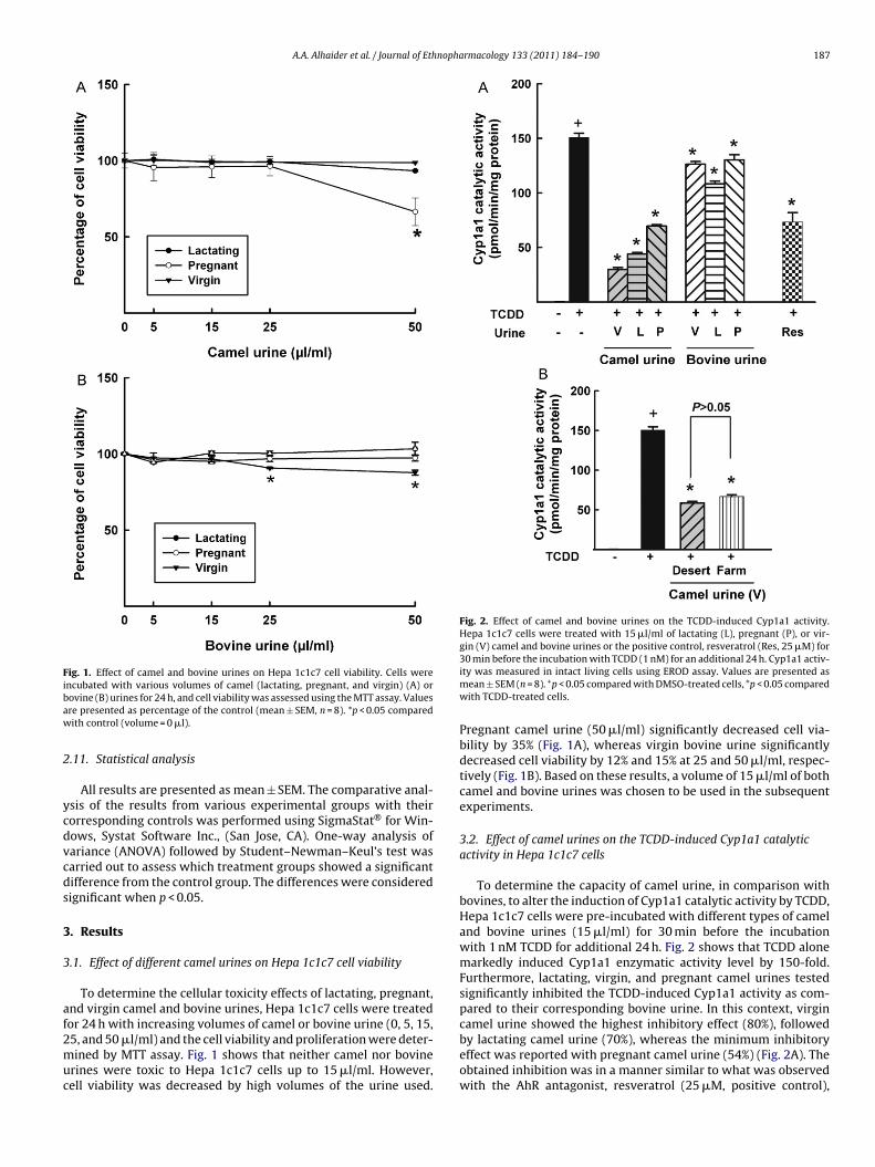
A.A. Alhaider et al. / Journal of Ethnopharmacology 133 (2011) 184–190 187
Fig. 1. Effect of camel and bovine urines on Hepa 1c1c7 cell viability. Cells wereibaw
2
ycdvcds
3
3
af2muc
Fig. 2. Effect of camel and bovine urines on the TCDD-induced Cyp1a1 activity.Hepa 1c1c7 cells were treated with 15 �l/ml of lactating (L), pregnant (P), or vir-gin (V) camel and bovine urines or the positive control, resveratrol (Res, 25 �M) for
ncubated with various volumes of camel (lactating, pregnant, and virgin) (A) orovine (B) urines for 24 h, and cell viability was assessed using the MTT assay. Valuesre presented as percentage of the control (mean ± SEM, n = 8). *p < 0.05 comparedith control (volume = 0 �l).
.11. Statistical analysis
All results are presented as mean ± SEM. The comparative anal-sis of the results from various experimental groups with theirorresponding controls was performed using SigmaStat® for Win-ows, Systat Software Inc., (San Jose, CA). One-way analysis ofariance (ANOVA) followed by Student–Newman–Keul’s test wasarried out to assess which treatment groups showed a significantifference from the control group. The differences were consideredignificant when p < 0.05.
. Results
.1. Effect of different camel urines on Hepa 1c1c7 cell viability
To determine the cellular toxicity effects of lactating, pregnant,nd virgin camel and bovine urines, Hepa 1c1c7 cells were treated
or 24 h with increasing volumes of camel or bovine urine (0, 5, 15,5, and 50 �l/ml) and the cell viability and proliferation were deter-ined by MTT assay. Fig. 1 shows that neither camel nor bovinerines were toxic to Hepa 1c1c7 cells up to 15 �l/ml. However,ell viability was decreased by high volumes of the urine used.
30 min before the incubation with TCDD (1 nM) for an additional 24 h. Cyp1a1 activ-ity was measured in intact living cells using EROD assay. Values are presented asmean ± SEM (n = 8). +p < 0.05 compared with DMSO-treated cells, *p < 0.05 comparedwith TCDD-treated cells.
Pregnant camel urine (50 �l/ml) significantly decreased cell via-bility by 35% (Fig. 1A), whereas virgin bovine urine significantlydecreased cell viability by 12% and 15% at 25 and 50 �l/ml, respec-tively (Fig. 1B). Based on these results, a volume of 15 �l/ml of bothcamel and bovine urines was chosen to be used in the subsequentexperiments.
3.2. Effect of camel urines on the TCDD-induced Cyp1a1 catalyticactivity in Hepa 1c1c7 cells
To determine the capacity of camel urine, in comparison withbovines, to alter the induction of Cyp1a1 catalytic activity by TCDD,Hepa 1c1c7 cells were pre-incubated with different types of cameland bovine urines (15 �l/ml) for 30 min before the incubationwith 1 nM TCDD for additional 24 h. Fig. 2 shows that TCDD alonemarkedly induced Cyp1a1 enzymatic activity level by 150-fold.Furthermore, lactating, virgin, and pregnant camel urines testedsignificantly inhibited the TCDD-induced Cyp1a1 activity as com-pared to their corresponding bovine urine. In this context, virgin
camel urine showed the highest inhibitory effect (80%), followedby lactating camel urine (70%), whereas the minimum inhibitoryeffect was reported with pregnant camel urine (54%) (Fig. 2A). Theobtained inhibition was in a manner similar to what was observedwith the AhR antagonist, resveratrol (25 �M, positive control),
188 A.A. Alhaider et al. / Journal of Ethnopharmacology 133 (2011) 184–190
Fig. 3. Effect of camel and bovine urines on the TCDD-induced Cyp1a1 mRNAexpression. Hepa 1c1c7 cells were treated with 15 �l/ml of lactating (L), pregnant(P), or virgin (V) camel and bovine urines or the positive control, resveratrol (Res,25 �M) for 30 min before the incubation with TCDD (1 nM) for an additional 6 h.The amount of Cyp1a1 mRNA was quantified using real-time PCR and normalizedto �-actin housekeeping gene. Values represent mean of fold change ± SEM. (n = 4).+
c
wic
beeTvi5otp
3c
ueewflaftrb(iui(2
Fig. 4. Effect of camel and bovine urines on the TCDD-induced Cyp1a1 protein. Hepa1c1c7 cells were treated with 15 �l/ml of lactating (L), pregnant (P), or virgin (V)camel and bovine urines for 30 min before the incubation with TCDD (1 nM) for andadditional 24 h. Protein (30 �g) was separated on a 10% SDS-PAGE and transferred tonitrocellulose membrane. Protein blots were then blocked overnight at 4 ◦C and thenincubated with a primary Cyp1a1 antibody for 2 h at 4 ◦C, followed by 1 h incubationwith secondary antibody at room temperature. Cyp1a1 protein was detected using
virgin camel urine significantly inhibited the TCDD-induced acti-vation of AhR and hence the transformation of the AhR/ARNT/XRE
p < 0.05 compared with DMSO-treated cells, *p < 0.05 compared with TCDD-treatedells.
hich significantly reduced TCDD-induced Cyp1a1 catalytic activ-ty by 50% (Fig. 2A). Taken together, virgin, lactating and pregnantamel urines are novel potent Cyp1a1 inhibitors.
Furthermore, we investigated whether the inhibition of Cyp1a1y camel urine is a diet-related effect, therefore, we have tested theffect of virgin camel urine, the urine showed highest inhibitoryffect, from different facilities, particularly farm and desert, onCDD-induced Cyp1a1 catalytic activity. Our results showed thatirgin camel urines from both desert and farm facilities inhib-ted the TCDD-induced Cyp1a1 activity by approximately 60% and4%, respectively (Fig. 2B). In addition, the magnitude of inhibitionbserved in farm camel urine was not significantly different fromhose obtained with desert camel data. These results ruled out anyossible effect of diet in virgin camel-mediated effect.
.3. Effect of camel urines on Cyp1a1 mRNA levels in Hepa 1c1c7ells
To further explore whether the inhibition of Cyp1a1 by camelrine is a transcriptional mechanism, we have determined theffect of camel urines on the expression of Cyp1a1 mRNA lev-ls. For this purposes, Hepa 1c1c7 cells (105 cells per well)ere plated onto 6-well tissue culture plates until 70–80% con-uence, thereafter, 15 �l/ml of different types of urines wasdded to the cells 30 min before the addition of 1 nM TCDDor 6 h. The amount of Cyp1a1 mRNA was quantified by real-ime PCR and normalized to �-actin, a housekeeping gene. Ouresults showed that virgin camel urine caused a significant inhi-ition of TCDD-induced Cyp1a1 mRNA by approximately 45%Fig. 3) in a manner similar to what observed at the catalytic activ-ty levels (Fig. 2A). In contrast, both lactating and pregnant camelrines did not cause a significant inhibition of the TCDD-mediated
nduction of Cyp1a1 mRNA (Fig. 3). The positive control, resveratrol25 �M) significantly inhibited the TCDD-induced Cyp1a1 mRNA by0% (Fig. 3).
the enhanced chemiluminescence method. The intensity of bands was normalizedto Gapdh signals, which was used as loading control. One of three representativeexperiments is shown. Values represent mean of fold change ± SEM. (n = 3). +p < 0.05compared with DMSO-treated cells, *p < 0.05 compared with TCDD-treated cells.
3.4. Effect of camel urines on the expression of Cyp1a1 proteinlevel in Hepa 1c1c7 cells
Western blot analysis was carried out to examine whether theobtained inhibition on TCDD-induced Cyp1a1 mRNA levels is trans-lated into a functional Cyp1a1 protein. Hepa 1c1c7 cells wereincubated for 30 min with camel urines before the addition of 1 nMTCDD for 24 h. Fig. 4 shows that both virgin and lactating, butnot pregnant, camel urines caused a significant inhibition of theTCDD-induced Cyp1a1 protein levels by approximately 65% and45%, respectively (Fig. 4). Taken together, these results showed thatvirgin camel urine stands prominently in its inhibition of the induc-tion of Cyp1a1 gene expression at the activity, mRNA and proteinlevels.
3.5. Inhibition of AhR transformation and XRE Binding by virgincamel urine
To further examine the effect of camel urines on AhR transfor-mation and hence binding to the XRE of the Cyp1a1 gene, virgincamel urine, that showed the maximum inhibition of the Cyp1a1gene expression at the activity, mRNA and protein levels, was uti-lized. For this purpose, EMSA was performed on guinea pig hepaticcytosol pre-incubated for 30 min with virgin camel urine (15 �l/ml)before the incubation with 20 nM TCDD for an additional 2 h, apositive control for AhR transformation. Fig. 5 shows that TCDDsignificantly activated the AhR through direct transformation ofthe AhR/ARNT/XRE complex, as determined by the shifted band(lane 2) compared to DMSO (lane 1). However, pre-incubation with
complex (lane 3) as shown by the intensity of the band comparedto TCDD. The specificity of virgin camel urine-mediated effects onAhR/ARNT heterodimer binding to XRE was confirmed by compe-
A.A. Alhaider et al. / Journal of Ethnopharmacology 133 (2011) 184–190 189
Fig. 5. Effect of virgin camel urine on AhR/ARNT/XRE binding. Cytosolic extracts(2 mg) from untreated guinea pig liver were incubated with DMSO and virgin (V)camel urine for 30 min before another incubation with TCDD (1 nM) for 2 h. Thecytosolic proteins were mixed with [�-32P]-labelled XRE, and the formation ofAhR/ARNT/XRE complexes was analyzed by EMSA. The specificity of binding wasdegi
te
3v
gtwbtud
4
koaa
taw
Fig. 6. Effect of virgin camel urine on luciferase activity. Hepa 1c1c7 cells transientlytransfected with luciferase reporter gene were grown onto 12-well cell cultureplates for 24 h. Thereafter, cells were incubated with DMSO and virgin (V) camel
etermined by incubating TCDD-treated cytosolic extracts with 100-fold molarxcess of cold XRE or anti-ARNT antibody. AhR/ARNT/XRE complex formed on theel was visualized by autoradiography. This pattern of AhR activation was observedn three separate experiments, and only one is shown.
ition assays using anti-ARNT antibody (lane 4) or 100-fold molarxcess of unlabelled XRE (lane 5).
.6. Inhibition of AhR-dependent reporter gene expression byirgin camel urine
The ability of virgin camel urine to inhibit the AhR-dependentene expression was assessed using Hepa 1c1c7 cells transientlyransfected with the XRE-driven luciferase reporter gene. Cellsere pre-incubated with virgin camel urine (15 �l/ml) for 30 min
efore the incubation with 1 nM TCDD for 24 h. Fig. 6 showshat treatment of transfected Hepa 1c1c7 cells with virgin camelrine caused a significant inhibition of the TCDD-induced AhR-ependent reporter gene expression.
. Discussion
The current study provides the first mechanistic evidence, to ournowledge, that camel urine significantly inhibited the inductionf Cyp1a1, a cancer-activating gene, by TCDD at the transcriptionalnd post-transcriptional levels through an AhR-dependent mech-
nism.One of the strategies for protecting human cells and tissues fromhe toxic effects of carcinogenic and cytotoxic metabolites includettenuation of the carcinogen-activating genes signaling path-ays and/or enhancing the adaptive mechanisms by increasing the
urine for 30 min before the incubation with TCDD (1 nM) for an additional 24 h.Cells were lysed and luciferase activity was measured according to the manufac-ture’s instructions. The graph represents the mean ± SEM (n = 4). +p < 0.05 comparedwith DMSO-treated cells, *p < 0.05 compared with TCDD-treated cells.
expression of detoxification and antioxidant genes. Therefore, wehave tested the capacity of three different camel urines to alterthe expression of Cyp1a1, a well-known cancer-activating gene.We hypothesize that camel urine induces its anti-cancer effects byinhibiting the expression of Cyp1a1 gene.
To test our hypothesis, we have first assessed the potential effectof three camel urines, obtained from, lactating, pregnant and virgincamels, on the induction of Cyp1a1 by TCDD using EROD as a probefor Cyp1a1 activity in Hepa 1c1c7 cells (Hasspieler et al., 2006).Our results showed that all the three camel urines tested alteredEROD activity to varying extents in a urine-dependent fashion. Forexample, TCDD-mediated induction of Cyp1a1-dependent ERODactivity was markedly reduced by both lactating and virgin camelurines. Surprisingly, the highest inhibition of the TCDD-inducedCyp1a1 activity levels was observed with virgin camel urine. Inaddition, such effect was not attributed to the diet, as virgin camelurines from independent facilities showed approximately similarinhibitory effect on the TCDD-induced Cyp1a1 catalytic activity.
Modulation of Cyp1a1 activity by camel urine could beattributed, at least in part, to a transcriptional and/or translationalmechanism, in which camel urine could alter the expression ofCyp1a1 mRNA and/or protein. The transcriptional regulation ofCyp1a1 gene by camel urine was demonstrated by the ability ofcamel but not bovine urines, particularly virgin camel urine, toinhibit the Cyp1a1 mRNA expression, in a manner similar to whatwas observed at the activity levels. Surprisingly, neither lactat-ing nor pregnant camel urine altered Cyp1a1 mRNA expressionlevel. On the other hand, the translational regulation of Cyp1a1gene expression was confirmed by the ability of both camel virginand lactating urines, but not pregnant urine to significantly down-regulate Cyp1a1 protein. These differential effects of camel urineson Cyp1a1 protein level was not attributed to altered cell viabil-ity since the expression of Gapdh protein, which was used as aloading control, was not significantly altered among the differenttreatments. Taken together, the results obtained strongly suggestthat the inhibition of Cyp1a1 by camel urine, particularly virgin,
is mediated at least in part at the transcriptional and the transla-tional levels. Furthermore, the ability of pregnant camel urine tosignificantly decrease the TCDD-induced Cyp1a1 at the activity butnot at the mRNA or protein levels suggests that a post-translationalmechanism is involved.
1 nopha
itbDiintAwiuwegtawousscab(btkelCitruc
tpgepmev
C
A
eAagu
R
A
90 A.A. Alhaider et al. / Journal of Eth
Perhaps the finding of greatest interest in the current studys the observation that virgin camel urine, the one that exhibitedhe highest inhibitory effects on Cyp1a1 gene expression, directlyinds to and inhibits the transformation of cytosolic AhR to aNA-binding form in vitro, which is extensively used to assess bind-
ng and affinity of ligand to the AhR (Jeuken et al., 2003). Thisndicates that virgin camel urine could be a novel AhR antago-ist that inhibits the binding of the AhR ligand such as TCDD tohe AhR. Importantly, the ability of virgin camel urine to inhibithR transformation and hence XRE binding is strongly correlatedith their ability to inhibit the AhR-dependent gene expression
n intact cells. Surprisingly, the inhibitory effect of virgin camelrine on the AhR transformation into its DNA-binding form in vitroas greater than its ability to inhibit the AhR-dependent gene
xpression. This finding could be explained by the inability of vir-in camel urine to recruit proper co-repressor to inhibit the generanscription (Jeuken et al., 2003). Although the potential medi-tors in camel urines involved in the down-regulation of Cyp1a1ere not examined in this study, ongoing research in our lab-
ratory has shown the presence of several compounds in camelrine. In this regard, using Liquid chromatography–tandem masspectrometry (LC–MS/MS) and one-dimensional gel electrophore-is, we have shown that several proteins are relatively abundant inamel urine, specifically lysozyme, immunoglobulin heavy chain,lbumin, dermatopontin, a CD44 antigen-like protein, prothrom-in, alpha-1-antichymotrypsin, CD44E-like protein, and lactoferrinunpublished data). Among these mediators, lactoferrin, an iron-inding glycoprotein, is abundant in camel urine as comparedo bovine urine (unpublished data). In this context, lactoferrin isnown to exert in vitro and in vivo anti-tumor activity (Roseanut al., 2010). Importantly, it has been recently reported thatactoferrin inhibits the development of cancer through inhibitingYP1A1 activation in 7,12-dimethylbenz[a]anthracene (DMBA)-
nduced hamster buccal pouch carcinoma model. Taken togetherhe results obtained from our laboratory and previously publishedeports, we speculate that lactoferrin could be responsible for camelrine-mediated effect. However, further studies are required toonfirm the role of camel lactoferrin in the inhibition of Cyp1a1.
In conclusion, the present work provides the first evidencehat camel urine inhibits the TCDD-mediated effect, at least inart by inhibiting the expression of Cyp1a1, a cancer-activatingene, at both the transcriptional and the post-transcriptional lev-ls through an AhR-dependent mechanism. These results are ofotential clinical significance to humans in that it uncovers theolecular mechanism involved and could explain the anecdotal
vidence for the successful use of camel urine in the treatment ofarious medical conditions.
onflict of interest
There is no conflict of interest.
cknowledgements
This work was supported by a grant from Vice Rectorate Knowl-dge Exchange and Technology Transfer, King Saud University to. Alhaider. The authors thank the vice rector for Graduate Studiesnd Research, Dr. Ali Al-Ghamdi, for his continuous support. We arerateful to Dr. Loren Kline (University of Alberta, AB) for providings with guinea pig livers.
eferences
bdalla, O.M., Wasfi, I.A., Gadir, F.A., 1988. The Arabian race camel normalparameters—I. Haemogram, enzymes and minerals. Comparative Biochemistryand Physiology A: Comparative Physiology 90, 237–239.
rmacology 133 (2011) 184–190
Agrawal, R.P., Dogra, R., Mohta, N., Tiwari, R., Singhal, S., Sultania, S., 2009. Beneficialeffect of camel milk in diabetic nephropathy. Acta Bio-medica: Atenei Parmensis80, 131–134.
al-Harbi, M.M., Qureshi, S., Ahmed, M.M., Raza, M., Baig, M.Z., Shah, A.H., 1996.Effect of camel urine on the cytological and biochemical changes induced bycyclophosphamide in mice. Journal of Ethnopharmacology 52, 129–137.
Conesa, C., Sanchez, L., Rota, C., Perez, M.D., Calvo, M., Farnaud, S., Evans, R.W., 2008.Isolation of lactoferrin from milk of different species: calorimetric and antimi-crobial studies. Comparative Biochemistry and Physiology B: Biochemistry &Molecular Biology 150, 131–139.
El Gendy, M.A., Somayaji, V., El-Kadi, A.O., 2010. Peganum harmala L. is a candi-date herbal plant for preventing dioxin mediated effects. Planta Medica 76,671–677.
Gharavi, N., El-Kadi, A.O., 2005. tert-Butylhydroquinone is a novel aryl hydrocar-bon receptor ligand. Drug Metabolism and Disposition: The Biological Fate ofChemicals 33, 365–372.
Hasspieler, B., Haffner, D., Stelljes, M., Adeli, K., 2006. Toxicological assessment ofindustrial solvents using human cell bioassays: assessment of short-term cyto-toxicity and long-term genotoxicity potential. Toxicology and Industrial Health22, 301–315.
Jeuken, A., Keser, B.J., Khan, E., Brouwer, A., Koeman, J., Denison, M.S., 2003. Activa-tion of the Ah receptor by extracts of dietary herbal supplements, vegetables,and fruits. Journal of Agricultural and Food Chemistry 51, 5478–5487.
Kennedy, S.W., Lorenzen, A., James, C.A., Collins, B.T., 1993. Ethoxyresorufin-O-deethylase and porphyrin analysis in chicken embryo hepatocyte cultureswith a fluorescence multiwell plate reader. Analytical Biochemistry 211,102–112.
Khorshid, F.A., Moshref, S.S., 2006. In vitro anticancer agent, I—Tissue culture studyof human lung cancer cells A549. II—Tissue culture study of mice leukemia cellsL1210. International Journal of Cancer Research 2, 330–344.
Korashy, H.M., Brocks, D.R., El-Kadi, A.O., 2007a. Induction of the NAD(P)H:quinoneoxidoreductase 1 by ketoconazole and itraconazole: a mechanism of cancerchemoprotection. Cancer Letters 258, 135–143.
Korashy, H.M., El-Kadi, A.O., 2005. Regulatory mechanisms modulating the expres-sion of cytochrome P450 1A1 gene by heavy metals. Toxicological Science 88,39–51.
Korashy, H.M., El-Kadi, A.O., 2006. The role of aryl hydrocarbon receptor and thereactive oxygen species in the modulation of glutathione transferase by heavymetals in murine hepatoma cell lines. Chemico-Biological Interactions 162,237–248.
Korashy, H.M., Shayeganpour, A., Brocks, D.R., El-Kadi, A.O., 2007b. Induction ofcytochrome P450 1A1 by ketoconazole and itraconazole but not flucona-zole in murine and human hepatoma cell lines. Toxicological Science 97,32–43.
Livak, K.J., Schmittgen, T.D., 2001. Analysis of relative gene expression data usingreal-time quantitative PCR and the 2(−Delta Delta C(T)) method. Methods 25,402–408.
Lorenzen, A., Kennedy, S.W., 1993. A fluorescence-based protein assay for use witha microplate reader. Analytical Biochemistry 214, 346–348.
Redwan el, R.M., Tabll, A., 2007. Camel lactoferrin markedly inhibits hepatitis Cvirus genotype 4 infection of human peripheral blood leukocytes. Journal ofImmunoassay & Immunochemistry 28, 267–277.
Rogers, J.M., Denison, M.S., 2002. Analysis of the antiestrogenic activity of 2,3,7,8-tetrachlorodibenzo-p-dioxin in human ovarian carcinoma BG-1 cells. MolecularPharmacology 61, 1393–1403.
Roseanu, A., Florian, P.E., Moisei, M., Sima, L.E., Evans, R.W., Trif, M., 2010. Liposo-malization of lactoferrin enhanced its anti-tumoral effects on melanoma cells.Biometals 23, 485–492.
Sambrook, J., Fritsch, E.F., Maniatatis, T., 1989. In: Ford, N. (Ed.), Molecular Cloning.A Laboratory Manual. Cold Spring Harbour Laboratory Press, Plainview, NY.
Shah, P.P., Saurabh, K., Pant, M.C., Mathur, N., Parmar, D., 2009. Evidence for increasedcytochrome P450 1A1 expression in blood lymphocytes of lung cancer patients.Mutation Research 670, 74–78.
Shimada, T., Fujii-Kuriyama, Y., 2004. Metabolic activation of polycyclic aromatichydrocarbons to carcinogens by cytochromes P450 1A1 and 1B1. Cancer Science95, 1–6.
Slattery, M.L., Samowtiz, W., Ma, K., Murtaugh, M., Sweeney, C., Levin, T.R.,Neuhausen, S., 2004. CYP1A1, cigarette smoking, and colon and rectal cancer.American Journal of Epidemiology 160, 842–852.
Song, Z., Pollenz, R.S., 2002. Ligand-dependent and independent modulation of arylhydrocarbon receptor localization, degradation, and gene regulation. MolecularPharmacology 62, 806–816.
Wei, C., Caccavale, R.J., Weyand, E.H., Chen, S., Iba, M.M., 2002. Induction of CYP1A1and CYP1A2 expressions by prototypic and atypical inducers in the human lung.Cancer Letters 178, 25–36.
Whitlock Jr., J.P., 1999. Induction of cytochrome P4501A1. Annual Review of Phar-macology and Toxicology 39, 103–125.
Williams, T.D., Lee, J.S., Sheader, D.L., Chipman, J.K., 2000. The cytochrome P450 1Agene (CYP1A) from European flounder (Platichthys flesus), analysis of regula-
tory regions and development of a dual luciferase reporter gene system. MarineEnvironmental Research 50, 1–6.Zordoky, B.N., Aboutabl, M.E., El-Kadi, A.O., 2008. Modulation of cytochrome P450gene expression and arachidonic acid metabolism during isoproterenol-inducedcardiac hypertrophy in rats. Drug Metabolism and Disposition: The BiologicalFate of Chemicals 36, 2277–2286.
(12) INTERNATIONAL APPLICATION PUBLISHED UNDER THE PATENT COOPERATION TREATY (PCT)
(19) World Intellectual Property OrganizationInternational Bureau
(10) International Publication Number(43) International Publication Date i n ;n fi ftc16 February 2012 (16.02.2012) 2 12/ 1 2 5 Al
(51) International Patent Classification: CA, CH, CL, CN, CO, CR, CU, CZ, DE, DK, DM, DO,A61K 31/19 (2006.01) BOW 61/14 (2006.01) DZ, EC, EE, EG, ES, FI, GB, GD, GE, GH, GM, GT,A61K 35/20 (2006.01) BO 61/24 (2006.01) HN, HR, HU, ID, IL, IN, IS, JP, KE, KG, KM, KN, KP,A61K 35/22 (2006.01) KR, KZ, LA, LC, LK, LR, LS, LT, LU, LY, MA, MD,
ME, MG, MK, MN, MW, MX, MY, MZ, NA, NG, NI,(21) International Application Number: NO, NZ, OM, PE, PG, PH, PL, PT, QA, RO, RS, RU,
PCT/CA201 1/000953 SC, SD, SE, SG, SK, SL, SM, ST, SV, SY, TH, TJ, TM,
(22) International Filing Date: TN, TR, TT, TZ, UA, UG, US, UZ, VC, VN, ZA, ZM,
12 August 201 1 (12.08.201 1) ZW.
(25) Filing Language: English (84) Designated States (unless otherwise indicated, for everykind of regional protection available): ARIPO (BW, GH,
(26) Publication Language: English GM, KE, LR, LS, MW, MZ, NA, SD, SL, SZ, TZ, UG,
(30) Priority Data: ZM, ZW), Eurasian (AM, AZ, BY, KG, KZ, MD, RU, TJ,
61/373,300 13 August 2010 (13.08.2010) US TM), European (AL, AT, BE, BG, CH, CY, CZ, DE, DK,
61/384,5 16 20 September 2010 (20.09.2010) US EE, ES, FI, FR, GB, GR, HR, HU, IE, IS, IT, LT, LU,LV, MC, MK, MT, NL, NO, PL, PT, RO, RS, SE, SI, SK,
(72) Inventor; and SM, TR), OAPI (BF, BJ, CF, CG, CI, CM, GA, GN, GQ,(71) Applicant : ANTAKLY, Tony [CA/CA]; 57, Milton GW, ML, MR, NE, SN, TD, TG).
Street, Montreal, Quebec H2X 1V2 (CA).Published:
(74) Agent: ΒΕΝΟΪΤ & COTE, ASSOCIES INC.; 1001, de— with international search report (Art. 21(3))
Maisonneuve West Blvd., Suite 210, Montreal, QuebecH3A 3C8 (CA). — before the expiration of the time limit for amending the
claims and to be republished in the event of receipt of(81) Designated States (unless otherwise indicated, for every amendments (Rule 48.2(h))
kind of national protection available): AE, AG, AL, AM,AO, AT, AU, AZ, BA, BB, BG, BH, BR, BW, BY, BZ,
(54) Title: BIOACTIVE COMPOUNDS IN CAMEL URINE AND MILK
1a
Fig. 1A
Benzoate
o Phenylacetate
o
©
(57) Abstract: The present document describes extracts from camel urine and/or milk comprising benzoate, phenylacetate andother molecular species. Also described are methods of use of the extracts and the identified compounds for the treatment of dis-eases, and methods of isolating the extracts.

Title: BIOACTIVE COMPOUNDS IN CAMEL URINE AND MILK
CROSS-REFERENCE TO RELATED APPLICATIONS
[0001] This application claims priority from US provisional patent
applications 61/373,300, filed August 13; 2010, and 61/384,516, filed
September 20, 2010 the specifications of which is hereby incorporated by
reference.
BACKGROUND
(a) Field
[0002] The subject matter disclosed generally relates to extracts from
camel urine and milk. More specifically, the subject matter relates to bioactive
extracts from camel urine comprising benzoate; phenylacetate and several
other molecular species.
(b) Related Prior Art
[0003] The use of animal urine and milk as a source of therapeutic
ingredients has been known for centuries. The use of camel urine in
traditional Arab medicine has been well acknowledged since pre-medieval
era, as best documented by several authors including Avicenna who, in the
book "laws of medicine" described a number of medicinal ingredients from
various plant and animal sources including those from human and camel
urines [Avicena or Ibn Sina "Qanun fi-al-tibb"
(http://almashriq.hiof.no/ddc/projects/saab/avicenna/896/html/S1_005.html)],
and Prioreschi, 2001 , Horatius press, Omaha, Nebraska).
[0004] Despite the acknowledgement of its medicinal properties, the
various bioactive molecules in human urine have not yet been thoroughly
characterized. This is surprising since it is now well established that urine
contains many biomedically relevant compounds namely hormones, growth
factors and neuropeptides. The most outstanding of those are premarin
consisting of conjugated estrogens extracted from pregnant mare's urine
(Stefanick, M.L., 2005, Am. J. Med. 118 SuppI 12B, 64-73), one of the most
prescribed drugs in the USA used in hormone replacement therapy. Another
one is human chorionic gonadotropin (hCG) approved since more than 40
years for clinical treatment of infertility where it is used to induce ovulation or
stimulate testicular development and spermatogenesis. Of particular
importance is the characterization of anti-cancer molecules in human urine
which contains low molecular weight compounds which we named HIP (HCG-
like Inhibitory Products) purified from urine extracts of pregnant women which
potently block the growth of tumour cells in HIV-related Kaposi sarcoma
(Kachra et al., 1997, Endocrinology 138, 4038-4041).
[0005] The urine of the camel is of particular interest. Desert Bedouins
in Asia are known to use camel urine and milk for healing several diseases
such as fever, infections, abdominal swelling, liver diseases and a variety of
other ailments (Guthier-Pilters and Dagg, 1981 , University of Chicago Press,
Chicago; Prioreschi, 2001 , Horatius press, Omaha, Nebraska). Such healing
properties of camel urine and milk are particularly reported in Islamic literature
called "Hadith" (Guthier-Pilters and Dagg, 1981 , University of Chicago Press,
Chicago; Prioreschi, 2001 , Horatius press, Omaha, Nebraska) , Although
these claims remain to be substantiated by scientific proof. Some of these
medicinal ingredients are used even today. For example, it has been
witnessed that in Saudi Arabia, people come to camel sheppards on the side
of local highways to purchase urine and milk for consumption as medicinal
products.
[0006] The physiology of camels is interesting. Arabian camels are
called the desert ships because they can walk in the desert for days without
drinking and can withstand extreme high temperature of 40°C. Camels not
only survive the harsh hot weather but also resist many viral and other
infections presumably due to the unique molecular composition of their
immunoglobulins (Deschacht et al., 2010, J . Immunol. 184, 5696-5704). It is
believed that the camel's peculiar natural antibodies display a wide antigen-
recognizing repertoire may be responsible for infection resistance. It is
suggested that camels display very low rates of water excretion as compared
to other animals who share the same habitat (donkeys, horses, sheep)
Guthier-Pilters and Dagg, 1981, University of Chicago Press, Chicago). This
results in the concentration of solutes and metabolites in the urine. Since
camels do not urinate for a long time, their urine represents a valuable source
of metabolites.
[0007] There is a need for bioactive compounds isolated from camel
urine.
[0008] There is a need for bioactive compounds isolated from camel
urine for the treatment of diseases, including cancers.
SUMMARY
[0009] According to an embodiment, there is provided an extract from
camel urine comprising an NMR spectrum as set forth in figure 1.
[0010] According to an embodiment, there is provided an extract from
camel urine comprising a HPLC fractionation spectrum as set forth in figure
8A.
[0011] The extract may comprise a fraction C of said HPLC
fractionation spectrum as set forth in figure 8A.
[0012] According to another embodiment, there is provided extract from
camel milk comprising at least one compound chosen from benzoate, lactate
and citrate.
[0013] The extract may be further comprising at least one compound
chosen from acetoacetic acid, fumaric acid, glyceric acid, homovanillic acid,
oxalic acid, oxoprolic acid, phenylpyruvic acid, propionylglycinic acid, pyruvic
acid, 2-hydroxyglutaric acid, 2-oxoadipic acid, 2-oxoglutaric, 3-hydroxybutiric
acid, 3 hydroxypropionic acid, 4-hydroxyphenyllactic acid, and 4-
hydroxyphenylpyruvic acid.
[0014] According to another embodiment, there is provided a
pharmaceutical composition comprising an extract from camel urine, camel
milk, or combination thereof and a pharmaceutically acceptable carrier.
[0015] According to another embodiment, there is provided a
pharmaceutical composition comprising at least one compound as identified
by a peak of an NMR spectrum as set forth in figure 1, a HPLC fractionation
spectrum as set forth in figure 8A or a fraction C thereof; and a
pharmaceutically acceptable carrier.
[001 6] According to another embodiment, there is provided a use of an
extract from camel urine, camel milk, or combination thereof for the
preparation of a medicament for the treatment of a disease.
[0017] According to another embodiment, there is provided a use of an
extract from camel urine, camel milk, or combination thereof for the treatment
of a disease.
[0018] According to another embodiment, there is provided a use of at
least one compound as identified by a peak of an NMR spectrum as set forth
in figure 1, a HPLC fractionation spectrum as set forth in figure 8A or a
fraction C thereof for the preparation of a medicament for the treatment of a
disease.
[0019] According to another embodiment, there is provided a use of at
least one compound as identified by a peak of an NMR spectrum as set forth
in figure 1, a HPLC fractionation spectrum as set forth in figure 8A or a
fraction C thereof for the treatment of a disease.
[0020] The at least one compound may be benzoate, phenylacetate or
a combination thereof. The at least one compound may be chosen from
Butyrylglycinic acid, Citric acid, Ethylmalonic acid, Glyceric acid, Glycolic acid,
Glutaric acid, Hexanoylglycenic acid, Hippuric acid, Homovanillic acid
Homogentisic acid, Isobutyrylglycinic acid, Isovalerylglycinic acid, Lactic acid,
Malonic acid, Methylcitric acid, Methylmalonic acid, Methylsuccinic acid, N-
acetylaspartic acid, Oxalic acid, Oxoprolinic (pyroglutamic) acid,
Phenylpropioniglycinic acid, Phenyllactic, Propionylglycinic acid, Pyruvic acid,
Sebacic acid, Suberic acid, 2-hydroxyadipic acid, 2-hydroxyglutaric acid, 2-
hydroxyisovaleric acid, 2-hydroxyphenylacetic acid, 2-methyl-3-hydroxybutiric

acid, 2-methylacetoacetique acid, 2-methylbutyrylglycinic acid, 3-
hydroxyisovaleric acid, 3-hydroxy-3-methylglutaric acid, 3-hydroxy butyric acid,
3-hydroxypropionic acid, 3-methylcrotonylglycinic acid, 3-methylglutaconic
acid, 4-hydroxyphenylacetic acid, 4-hydroxyphenyllactic acid.
[0021] The disease may be a cancer, a neoplasm including multidrug
resistant tumors, various haematological malignancies, and the cancer may
be chosen from a breast cancer, colon cancer, a prostate cancer, malignant
gliomas, and a thyroid cancer .
[0022] The disease may be a metabolic defect in a urea-cycle enzyme.
[0023] The disease may be any metabolic disease such as diabetes or
pathologic obesity that is regulated by the Peroxisome Proliferator-activiated
Receptor (PPAR) which belongs to the steroid receptor superfamily
[0024] The disease may be any metabolic disease such as diabetes or
pathologic obesity that is regulated or by the DNA binding partner of the
Peroxisome Proliferator-activiated Receptor (PPAR) which is the retinoid X
receptors (RXR),
[0025] The disease may be chosen from multiple sclerosis, sickle cell
anaemia, amyotrophic lateral sclerosis, and Huntington's disease.
[0026] According to another embodiment, there is provided a method of
treating a disease in a subject in need thereof by administering to the subject
a therapeutically effective amount of an extract camel urine, camel milk, or
combination thereof.
[0027] According to another embodiment, there is provided a method of
treating a disease in a subject in need thereof by administering to the subject
a therapeutically effective amount of at least one compound as identified by a
peak of an NMR spectrum as set forth in figure 1, a HPLC fractionation
spectrum as set forth in figure 8A or a fraction C thereof.
[0028] The compound may be benzoate, phenylacetate or a
combination thereof. The at least one compound may be chosen from
Butyrylglycinic acid, Citric acid, Ethylmalonic acid, Glyceric acid, Glycolic acid,
Glutaric acid, Hexanoylglycenic acid, Hippuric acid, Homovanillic acid
Homogentisic acid, Isobutyrylglycinic acid, Isovalerylglycinic acid, Lactic acid,
Malonic acid, Methylcitric acid, Methylmalonic acid, Methylsuccinic acid, N-
acetylaspartic acid, Oxalic acid, Oxoprolinic (pyroglutamic) acid,
Phenylpropioniglycinic acid, Phenyllactic, Propionylglycinic acid, Pyruvic acid,
Sebacic acid, Suberic acid, 2-hydroxyadipic acid, 2-hydroxyglutaric acid, 2-
hydroxyisovaleric acid, 2-hydroxyphenylacetic acid, 2-methyl-3-hydroxybutiric
acid, 2-methylacetoacetique acid, 2-methylbutyrylglycinic acid, 3-
hydroxyisovaleric acid, 3-hydroxy-3-methylglutaric acid, 3-hydroxybutyric acid,
3-hydroxypropionic acid, 3-methylcrotonylglycinic acid, 3-methylglutaconic
acid, 4-hydroxyphenylacetic acid, 4-hydroxyphenyl lactic acid, and retinoid
derivative such as 9-cis retinoic acid.
[0029] The disease may be a cancer, a neoplasm, a multidrug resistant
tumors, a haematological malignancy, and the cancer may be a breast
cancer, a colon cancer, a prostate cancer, a malignant glyoma, and a thyroid
cancer.
[0030] The disease may be a metabolic defect in a urea-cycle enzyme.
[0031] The disease may be chosen from multiple sclerosis, sickle cell
anemia, amyotrophic lateral sclerosis, and Huntington's disease.
[0032] According to another embodiment, there is provided a method of
producing an extract from camel urine comprising an NMR spectrum as
defined in figure 1 by removing a fraction of at least 10 kDa from a camel
urine sample to produce an extract having at least one molecule of molecular
weight lower than 10 kDa.
[0033] The removal of the molecules may be done by filtration of said
camel urine sample. The filtration may be with a filter membrane. The filter
membrane may have pores preventing passage of molecules having at least
10 kDa or more. The filter membrane may be treated with water prior to
filtration of said camel urine sample.

[0034] The following terms are defined below.
[0035] The term "molecular weight cut-off' or cut-off value is defined as
the molecular weight at which a where the membrane or filter will reject, or
prevent the passage of 90% of the solutes. Hence, a filter with a molecular
weight cut-off of about 0 kDa will prevent the passage of molecules having
molecular weight of at least 10 kDa.
[0036] The term "pharmaceutically acceptable carrier" is intended to
mean a preservative solution, a saline solution, an isotonic (about 0.9%)
saline solution, or about a 5% albumin solution, suspension, sterile water,
phosphate buffered saline, and the like. Other buffering agents, dispersing
agents, and inert non-toxic substances suitable for delivery to a patient may
be included in the compositions of the present invention. The compositions
may be solutions, suspensions or any appropriate formulation suitable for
administration, and are typically sterile and free of undesirable particulate
matter. The compositions may be sterilized by conventional sterilization
techniques.
[0037] The term "nutraceutical" is intended to mean a food or food
product that provides health and medical benefits, including the prevention
and treatment of disease.
[0038] The term "a non-food ingredient" is intended to mean an
ingredient that may be added to food but that may not be a food ingredient
contributing to the caloric content of the food per se.
[0039] Features and advantages of the subject matter hereof will
become more apparent in light of the following detailed description of selected
embodiments, as illustrated in the accompanying figures. As will be realized,
the subject matter disclosed and claimed is capable of modifications in
various respects, all without departing from the scope of the claims.
Accordingly, the drawings and the description are to be regarded as
illustrative in nature, and not as restrictive and the full scope of the subject
matter is set forth in the claims.
BRIEF DESCRIPTION OF THE DRAWINGS
[0040] Further features and advantages of the present disclosure will
become apparent from the following detailed description, taken in combination
with the appended drawings, in which:
[0041] Fig. 1 illustrates the H NMR spectra of camel urine compared to
human urine. The full spectra are shown in the lower part of the figure. The
expanded spectra of the region 7.1-7.9 (boxed area) are shown on the top.
The phenylacetate and benzoate NMR peaks are more intense in camel than
human. The numbers 1a, 1b, 1c and 2b, 2c, 2d refer to the characteristic
peaks emanating from the carbons identified by the same numbers on the
chemical atomic structures shown. Other compounds labeled by the indicated
numbers correspond to the chemicals shown in the boxed area. Creatinine,
which constitutes a standard reference in urine analysis, is also shown. The
star (*) identified peak is present only in camel but not human.
[0042] Figs. 2A and 2B illustrate the GS-MS spectra of benzoic acid
and phenylacetic acid identified in camel urine using GS-MS. Fig. 2A shows
the entire spectrum of camel urine. Fig. 2B shows spectra identified in camel
urine. Right panel shows the mass spectrum of benzoic acid on El ionization,
showing molecular ions C7H60 2'+ at m/z= 122, C6H5CO+ benzoyl ions at
m/z=105, C6H5+ ions at m/z=77 and C4H3
+ ions at m/z=51 . Left panel
corresponds to phenylacetic acid on El ionization, showing molecular ions
8H80 2+ at m/z= 36 and strong peak at m/z=91 which is characterized by
C6H5CH2+ ions.
[0043] Fig. 3 illustrates the mechanisms of ammonia diversion from the
urea cycle with the administration of phenylacetate and benzoate.
[0044] Figs. 4A and 4B illustrates (A) the fractionation of camel urine
using reversed phase-HPLC and (B) the bioassay of the collected fractions
following HPLC separation. Panel A shows the elution profile of camel urine
from the HPLC column. Y axis indicates absorbency at 220 nm. x-axis
indicated time of elution in minutes. The contents of the indicated elution
tubes are pooled into five fractions named A , B, C, D, E, then lyophilized.
Panel B shows the bioactivity of the five fractions as tested in vitro for cell
proliferation using the MCF7 cells. Y-axis shows % cell proliferation of the test
material as compared to the Vehicle (Veh), i.e a control consisting of culture
media alone.
[0045] It will be noted that throughout the appended drawings, like
features are identified by like reference numerals.
DETAILED DESCRIPTION OF THE PREFERRED EMBODIMENTS
[0046] In embodiments there is disclosed an extract from camel urine.
The extract of the present invention of the present invention comprise the
NMR spectrum as defined in Fig. 1.
[0047] Referring now to Fig. 1, a H NMR spectra of camel urine
showing significant differences as compared to human urine. The most
striking finding in the camel urine is marked, high levels of both benzoate and
phenylacetate (Fig. 1). In contrast, human, rat, elephant and dromedary
urines display very little expression of these two compounds as shown in
Table 1.
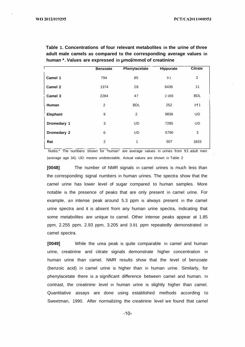
Table 1. Concentrations of four relevant metabolites in the urine of threeadult male camels as compared to the corresponding average values in
human * . Values are expressed in mo /mmo of creatinine
Benzoate Phenylacetate Hippurate Citrate
Camel 1 794 85 9 1 2
Camel 2 1374 28 6436 11
Camel 3 2284 47 2 166 BDL
Human 2 BDL 252 3 1
Elephant 9 2 9836 UD
Dromedary 1 3 UD 7285 UD
Dromedary 2 6 UD 5790 3
Rat 2 1 507 1833
Notes:* The numbers shown for "human" are average values in urines from 3 adult men
(average age 34). UD: means undetectable. Actual values are shown in Table 2
[0048] The number of NMR signals in camel urines is much less than
the corresponding signal numbers in human urines. The spectra show that the
camel urine has lower level of sugar compared to human samples. More
notable is the presence of peaks that are only present in camel urine. For
example, an intense peak around 5.3 ppm is always present in the camel
urine spectra and it is absent from any human urine spectra, indicating that
some metabolites are unique to camel. Other intense peaks appear at 1.85
ppm, 2.255 ppm, 2.93 ppm, 3.205 and 3.91 ppm repeatedly demonstrated in
camel spectra.
[0049] While the urea peak is quite comparable in camel and human
urine, creatinine and citrate signals demonstrate higher concentration in
human urine than camel. NMR results show that the level of benzoate
(benzoic acid) in camel urine is higher than in human urine. Similarly, for
phenylacetate there is a significant difference between camel and human. In
contrast, the creatinine level in human urine is slightly higher than camel.
Quantitative assays are done using established methods according to
Sweetman, 1990. After normalizing the creatinine level we found that camel
contains remarkably more benzoate than human (Table 1). Similarly,
phenylacetate is significantly higher in camel (Table 1). The results confirm
that the detected metabolites in camel urine have a unique profile with respect
to their type, number and concentrations. The high level of benzoate and
phenylacetate represent an exceptional difference of camel urine.
[0050] The clinical use of phenylacetate (PA) and benzoate in lowering
plasma ammonium levels in patients with hyperammonemia represent the
hallmark of their therapeutic action (Enns et a/., 2007, N. Engl. J. Med. 356,
2282-2292). In certain metabolic diseases resulting from defects in urea-cycle
enzymes, ammonium which cannot be converted to urea accumulates to a
toxic level that can be lethal. This drug combination of PA and benzoate,
marketed as Ammonul®, is useful to treat patients with inborn errors of
metabolism of the urea-cycle enzyme and prevents several complications
such as encephalopathy and death (Enns et a/., 2007, N. Engl. J. Med. 356,
2282-2292). In fact, phenylacetate, through mitochondrial conjugation with
glutamine, results in phenylacetyglutamine. Similarly, benzoate combines with
glycine thus forming benzoylglycine (hippurate) (see Fig. 3). These two non¬
toxic compounds are easily eliminated in the urine. Using this well established
drug combination consisting of phenylacetate and benzoate, Enns (Enns et
a/., 2007, N . Engl. J. Med. 356, 2282-2292) reported results of a 25 year
clinical study demonstrating an overall survival of 84 % following the
treatment.
[0051] In addition to their usefulness in treating urea cycle enzyme
defects, both phenylacetate and benzoate are found to be potentially useful
for the treatment of multiple sclerosis, as disease characterized by
demyelination presumably due to a T-cell auto-immune response. In a mouse
model of multiple sclerosis (Brahmachari et a/., 2009, J. Immunol. 183, 5917-
5927), both compounds exert immunomodulatory and anti-inflammatory
effects which are mediated by immune T-cells and involve among others,
suppression of NF-κΒ and nitric oxide synthetase; these actions ultimately
result in the alleviation of the disease.
[0052] Phenylacetate (PA) and phenyl butyrate (PB), the parent
compound from which it is metabolized in humans and animals, both display
other important bioactivities other than those cited above. Chemically, these
metabolites (PA and PB) belong to a group of aromatic fatty acids having a
stable phenyl ring. They are proven useful in many diseases, ranging from
cancer, sickle cell anemia, ALS and Huntington's disease.
[0053] Phenylacetate is originally discovered as a plant hormone that
regulates cell growth it has been studied in the past two decades as anti¬
cancer arid cellular differentiating compounds. PA inhibits the growth of
several cancer cell types of different lineages and, in some instances, it
promotes their differentiation to a non-cancerous phenotype. Of interest are
the effects of PA and PB on gliomas and neuroblastomas, originally thought to
be mediated by inhibition of protein prenylation cholesterol and fatty acid
biosynthesis. Subsequent studies have demonstrated that PA and PB inhibits
the growth of several neoplastic cell types including breast cancer (Liu et a/.,
2007, Cancer Chemother. Pharmacol. 59, 217-225), prostate cancer, colon
cancer and thyroid carcinoma. Furthermore, PA and PB can potentiate the
action of other hormones such as estrogens and retinoids in regulating cancer
cell growth. These anti-cancer actions of PA and PB have prompted clinical
trials especially that these compounds display little toxicity (Lin et a/., 2009,
Clin. Cancer Res. 15, 6241-6249; Cudkowicz et al., 2009, Amyotroph. Lateral.
Scler. 10, 99-106). The overall picture that emerges is that PA and PB act as
mild agents that synergize or cooperate with other bioactive agents, some of
which may be endogenous. In fact, physiological or pharmacologic actions of
PA and PB can act alone or in synergy with differentiating agents such as
retinoid to down-regulate key cell-cycle genes as well as angiogenic and
growth factors and that promote tumor cell growth. Studies regarding the
molecular mechanisms of action of PA has shed light on two important
pathways for its action: induction of histone acetylation which regulates
chromatin structure and regulation of steroid / nuclear receptor gene
expression. In fact, PA and PB can bind and activate the Peroxisome
Proliferator-activiated Receptor γ (PPAR) which belongs to the steroid

receptor superfamily of ligand-activated transcription factors (Samid et al.,
2000, Clin. Cancer Res. 6, 933-941). The latter finding would suggest that
these compounds act similar to steroidal hormonal drugs and would explain
the pleotropic action of PA and PB compounds in different diseases and
different physiological settings to regulate metabolism, inflammation, cancer
growth and cell differentiation.
[0054] Thus, although camel urine has been known for its healing
properties in the Arabic traditional medicine which is practiced even today in
Saudi Arabia, the identification of its active molecules has not yet been
reported.
[0055] There is also disclosed a method of preparing camel urine
extracts. The extracts may be prepared through the fractionation of camel
urine samples using techniques well known in the art. For example, the
sample may be fractionated using exclusion chromatography techniques or
any other method known in the art for the separation of molecules based on
their size, shape, hydrophilicity, hydrophobicity, charge, polarity, or any other
inherent physical characteristics which may be employed for the isolation (or
exclusion) of molecules from the urine sample of interest. The preferred
method for the preparation of extracts according to the present invention is by
using size exclusion filtering membranes excluding a fraction of molecules
having a molecular weight larger than a set weight (i.e. having a
predetermined molecular weight cut-off). The urine samples are filtered with
the filtering membrane to fractionate the sample in a filtrate comprising
smaller molecules and a retentate of larger molecules. Suitable molecular
weight for exclusion are from at least 100 kDa, or from at least 50, or from at
least 30 kDa, or from at least 10 kDa, or from at least 3 kDa. The preferred
molecular weight for exclusion (i.e. the preferred molecular weight cut-off) is
from at least 0 kDa.
[0056] In embodiments, there is described an extract from camel urine
which comprises an NMR spectrum as set forth in Fig. 1. The extract may be
used for the preparation of medicaments and/or for the treatment of various
diseases such as cancer, including but not limited to breast cancer, prostate
cancer, and thyroid cancer; metabolic diseases such as defects in the urea-
cycle enzymes; multiple sclerosis, sickle cell anaemia, amyotrophic lateral
sclerosis and Huntington's disease.
[0057] In embodiments, there is described an extract from camel urine
which comprises a HPLC fractionation spectrum as set forth in Fig. 8A, and
preferably comprising fraction C of said HPLC fractionation spectrum as set
forth in Fig. 8A. The extract may be used for the preparation of medicaments
and/or for the treatment of various diseases such as cancer, including but not
limited to breast cancer, prostate cancer, and thyroid cancer; metabolic
diseases such as defects in the urea-cycle enzymes; multiple sclerosis, sickle
cell anaemia, amyotrophic lateral sclerosis and Huntington's disease.
[0058] In embodiments, there is described an extract from camel milk
prepared according to the method described above. Desert Bedouins and
even ordinary people living in Saudi Arabia today are also known to mix camel
urine and milk to get therapeutic effects. We therefore examined whether
camel milk contains any remarquable molecules. Indeed, the milk from adult
camels contains significant amounts of benzoate (table 5) albeit in lower
amounts than in camel urine. However, camel milk is devoid of phenyl
acetate.
[0059] In use, the extracts of the present invention may be used alone,
or combined to each other for any therapeutic use for which they may be
useful. According to another embodiment, the extracts of the present
invention may also be used in food composition, such as for example
nutraceutical compositions. Nutraceuticals are food or food product that
provides health and medical benefits, including the prevention and treatment
of disease. Products according to the present invention may range from
isolated nutrients, dietary supplements, prebiotic, specific diets and herbal
product supplement, and processed foods such as cereals, soups, and
beverages. The compositions of the present invention may also be used as a
functional food, a food ingredient, a food additive, a natural food additive, a
non-food ingredient, a cosmeto-food, a pharmaceutical, and a food
supplement."
[0060] The present invention will be more readily understood by
referring to the following examples which are given to illustrate the invention
rather than to limit its scope.
EXAMPLE 1
Animals
[0061] Urine are collected from 15 male and 5 female adult camels
(Camelus dromedarius) raised in separate geographic areas in Saudi Arabia,
Jordan, Egypt, and Bahrain. In addition, urines are obtained from 2 adult
dromedaries, 2 alpaca, and one llama and two elephants from a zoo near
Montreal, Canada. Furthermore, urines are obtained from two adult male
Sprague-Dawley laboratory rats. The urine samples are transported to the
laboratory either on ice or after freezing.
EXAMPLE 2
NMR Spectroscopy
[0062] A urine sample (0.5 ml) is centrifuge-filtered at 2,000 g and 4°C
for 15 minutes by using 10 kDa centrifugal filter tubes (Millipore, Billerica, MA,
USA) to remove molecules and particles in the sample that have molecular
weights of at least 0 kDa. The centrifugal filters are washed several times
with 0.5 ml water, then centrifuged at 12000 x g at 4°C to remove glycerol
from the filter membrane until no NMR signal is observed in the filtrate. Four
hundred microliters of the filtrate are transferred to a 5 mm NMR tube and 100
µ Ι of D20 (Cambridge Isotope Laboratories, Inc.) are added to the NMR tube.
[0063] The NMR spectra are obtained using a Varian Inova 600 MHz
instrument (Palo Alto, California). All NMR experiments are obtained using a
one-dimensional presaturation sequence with gradients pulse sequence (zgpr
in the standard Bruker pulse sequence library). The 1H NMR spectra are
recorded by collecting 64 and 128 free induction decays (FIDs) and digitized
into 64 K complex data points over a spectral width of 12 ppm. The recycle
delay time is set to 5 sec and the receiver gain is kept at a constant value of
32. Before Fourier transform, the FID values are multiplied by an exponential
function equivalent to 1.0 Hz line broadening factor. All spectra are then
visually phased and adjusted manually where necessary.
EXAMPLE 3
Identification of urine compounds using NMR spectroscopy
[0064] Select spectra are examined using the Chenomx NMR Suite
(Chenomx Inc., Edmonton, Alberta, Canada). The metabolites' signals are
examined and the assignments are made by fitting and comparing of the
experimental spectra with reference spectra from the database using the
Chenomx NMR Profiler software.
EXAMPLE 4
Identification of phenylacetate and benzoate in camel urine using Mass
Spectrometry
[0065] Target chemicals are determined using an Agilent 7890 A GC
(or equivalent) with 5975C MSD equipped with an Agilent 7683B automatic
liquid sampler and an HP-5MS GC (or equivalent) column (30 m , 0.25mm i.d.,
0.25 m film thickness). Helium is used as the carrier gas, with a column flow
rate of 1.0 ml/min in constant flow mode. Injector temperature is 280°C. The
GC-MSD interface quadruple and the ion source temperatures are set at 280,
170 and 230°C, respectively. The GC oven temperature is kept at 50°C for
0.5 min, followed by the first ramp at 5°C / min to 225°C, second ramp at 3°C
/ min to 280°C, and holding for 1 min. Prior to quantification process, mass
spectra and GC retention times of each compound from mlz 50 to 550 are
obtained in full scan mode. The mass spectrometer used is operating in
electron impact with an ionization voltage of 70 EV. The sample is injected in
pulsed splitless mode.
[0066] The demonstration of BNZ and PAA in camel urine are
independently confirmed by three laboratories using Gas Chromatography-

Mass Spectrometry {GC-MS) (Fig. 2). The MS at the top of each peak is
searched using NIST (National Institute of Standards and Technology, an
agency of the U.S. Commerce Department) library with the best fit. Usually
the best-matched 25 compounds are displayed. The mass spectrum of
benzoic acid on El ionization shows molecular ions C He at m/z= 122.
Their intensity is 95% of the base peak, which is characterized by C6H5CO+
benzoyl ions at m/z=105. Other prominent fragments are C6H5+ ions are at
m/z=77 (90%) and C H3+ ions at m/z=51 (70%). The mass spectrum of
phenylacetic acid on El ionization shows molecular ions C8H80 2+ at m/z=136.
Their intensity is 30% of the base peak, which is characterized by C6H5CH2+
ions at m/z=91 .

EXAMPLE 5
Identification of other components of camel urine using Mass
Spectrometry
[0067] Urine samples from 2 camels are subjected to mass
spectrometry as described above in Example 4 .

EXAMPLE 6
Identification of other components of camel urine using Mass
Spectrometry compared to human urine
Table 3. Organic Acids and Metabolites in UrineMetabolite name (acid) Camel Human
Average SEM Average SEM
ACETOACETIC 0.00 0.00 1.55 0.1 9ADIPIC 1.33 0.22 1.00 0.08
BENZOIC 1484 00 124.82 2 45BUTYRYLGLYCINE 1.67 0.22 0.00 0.00
CITRIC 4.33 0.88 3 10.82 19.1 6ETHYLMALONIC 2.00 0 .17 2.82 0.1 9FUMARIC 0.33 0.08 0.55 0.06GLUTARIC 0.67 0 .17 0.45 0.05GLYCERIC 4.33 0.36 4.00 0.46GLYCOLIC 6.00 0.54 43.73 1.64
HEXANOYLGLYCINE 0.67 0.08 0.00 0.00HIPPURIC 2897.67 458.52 251 .73 24.35HOMOGENTISIC 0.67 0.08 0.00 0.00HOMOVANILLIC (HVA) 1.33 0 .11 2.00 0.07ISOBUTYRYLGLYCINE 5.00 0.38 0.27 0.03ISOVALERYLGLYCINE 3.00 0.23 0.73 0.08LACTIC 60.67 5.65 22.45 2 .12
MALONIC 0.33 0.08 0.09 0.02METHYLCITRIC 4.00 0.39 1.36 0.05METHYLMALONIC 3.00 0.38 0.91 0.02METHYLSUCCINIC 9.33 0.73 0.91 0.05MEVALONOLACTONE 0.00 0.00 0.18 0.04N-ACETYLASPARTIC 0.33 0.08 4.36 0 .17OXALIC 102.00 8.66 22.27 0.63OXOPROLINE(PYROGLUTAMIC) 3.67 0.33 27.36 1.08PHENYLPROPIONYLGLYCINE 0.67 0 .17 0.00 0.00PHENYLACETIC 53.33 4 64 0 00PHENYLLACTIC . 0.33 0.08 0.00 0.00PHENYLPYRUVIC 0.00 0.00 0.00 0.00PROPIONYLGLYCINE 0.67 0 .17 0.09 0.02PYRUVIC 1.00 0.09 11.55 1.28SEBACIC 2.00 0.1 7 0.00 0.00SUBERIC 2.67 0.24 1.09 0 .12SUCCINYLACETONE 0.00 0.00 0.00 0.00VANILLYMANDELIC(VMA) 0.00 0.00 1.36 0.072-HYDROXYADIPIC 1.00 0.09 0.00 0.002-HYDROXYGLUTARIC 1.33 0 .11 3 .18 0.1 82-HYDROXYISOCAPROIC 0.00 0.00 0.00 0.002-HYDROXYISOVALERIC 1.00 0 .14 0.09 0.022-HYDROXYPHENYLACETIC 1.67 0 .13 0.45 0.072-METHYL-3-HYDROXYBUTYRIC 8.67 0.66 2.55 0.092-METHYLACETOACETIC 2.33 0 .19 1.91 0.06
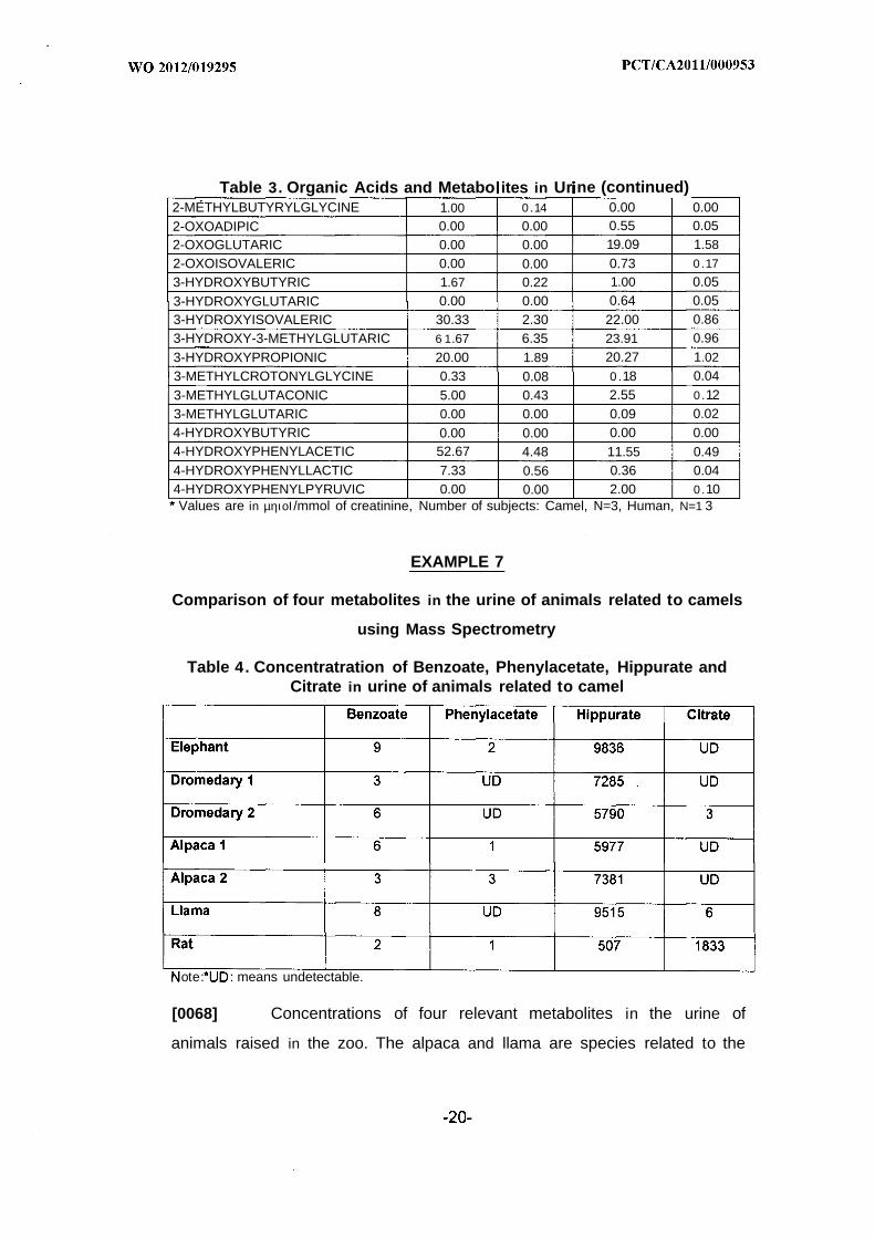
Table 3. Organic Acids and Metabo ites in Ur ne (continued)2-METHYLBUTYRYLGLYCINE 1.00 0 .14 0.00 0.002-OXOADIPIC 0.00 0.00 0.55 0.052-OXOGLUTARIC 0.00 0.00 19.09 1.58
2-OXOISOVALERIC 0.00 0.00 0.73 0 .17
3-HYDROXYBUTYRIC 1.67 0.22 1.00 0.05
3-HYDROXYGLUTARIC 0.00 0.00 0.64 0.053-HYDROXYISOVALERIC 30.33 2.30 22.00 0.863-HYDROXY-3-METHYLGLUTARIC 6 1.67 6.35 23.91 0.96
3-HYDROXYPROPIONIC 20.00 1.89 20.27 1.02
3-METHYLCROTONYLGLYCINE 0.33 0.08 0 .18 0.04
3-METHYLGLUTACONIC 5.00 0.43 2.55 0 .123-METHYLGLUTARIC 0.00 0.00 0.09 0.024-HYDROXYBUTYRIC 0.00 0.00 0.00 0.004-HYDROXYPHENYLACETIC 52.67 4.48 11.55 0.494-HYDROXYPHENYLLACTIC 7.33 0.56 0.36 0.044-HYDROXYPHENYLPYRUVIC 0.00 0.00 2.00 0 .10Values are in µηιοΙ/mmol of creatinine, Number of subjects: Camel, N=3, Human, N=1 3
EXAMPLE 7
Comparison of four metabolites in the urine of animals related to camels
using Mass Spectrometry
Table 4. Concentratration of Benzoate, Phenylacetate, Hippurate andCitrate in urine of animals related to camel
ote: : means undetectable.
[0068] Concentrations of four relevant metabolites in the urine of
animals raised in the zoo. The alpaca and llama are species related to the
camel. Rats were raised in a laboratory. A total of 57 metabolites are assayed
but this table shows only 4 . Values are expressed in pmol/mmol of creatinine.
EXAMPLE 8
Fractionation of camel urine using reversed phase-HPLC
[0069] Adult male camel urine previously lyophilized is reconstituted
with an equivalent amount of 0.1 % trifluoroacetic acid (TFA) in water, then
placed on ice and processed for HPLC within two hours. 10 to 100 microliters
of this reconstituted urine solution is injected into either a Waters™ HPLC
apparatus or an equivalent Beckman "Gold"™ HPLC apparatus. Both
apparatuses are fitted with a 300 mm C-18™ column. Elution from the column
is done using an increasing linear isocratic gradient of acetonitrile in water
containing 0.1 % TFA. The gradient is increased from 5% to 75% acetonitrile.
The absorbancy is monitored at 220 nm wavelength during the elution and
fractions are collected manually into plastic tubes. After collection, the
fractions are promptly placed in a Savant™ Speed-vac apparatus in order to
dry the samples. Now referring to Fig. 4A, the gradient is shown with the x-
axis indicating time, in minutes. The absorbency at 220 nm is recorded on the
y-axis.
EXAMPLE 9
Bioassay of the collected fractions following HPLC separation
[0070] The fractions (peaks) indicated by arrows on Fig. 4B. are
lyophilized and each is reconstituted in one (1) ml of RPMI culture medium
(without serum) and tested for biological activity using MCF7 cells. The
biological activity is tested in absence (Vehicle control, i.e. RPMI medium
alone) or presence of 10 microliters of the reconstituted fractions. It can be
seen from Fig. 4B that fraction C display an inhibitory activity.
EXAMPLE 10
Assessment of cell proliferation
[0071] The breast cancer cell line called MCF7 is obtained from the
American Type Tissue Culture Collection. The KS cells are passaged and the
culture medium is changed every other day in presence or in absence of any
of the samples mentioned above for the indicated periods ranging from 24-96
hrs. 3H-thymidine incorporation is measured as described (Guo WX et al.,
1996, Am J Pathol 148: 1999-2008). In most experiments, data are reported
as means ± SEM of sextuplet determinations. Statistical analysis is
determined by student t-test.
EXAMPLE 11
Comparison of four metabolites in the milk of camels using Mass
Spectrometry
Table 5. Concentrations of Benzoate, Phenylacetate, Hippurate andCitrate in the milk of 5 adult camels.
Benzoate Phenylacetate Lactate Citrate
Camel #EG2 20 UD 30 3326
Camel #EG3 23 UD 42 3091
Camel #EG4 11 UD 55 3087
Camel #EG5 38 UD 46 3461
Camel #EG6 17 UD 27 3455
Note: *UD: means undetectable.
[0072] Concentrations of four relevant metabolites in the milk of 5 adult
camels. A total of 57 metabolites are assayed but this table shows only 4 .
Values are expressed in pmol/mmol of creatinine*

EXAMPLE 12
Identification of other components of camel milk using MassSpectrometry
Table 6. Organic Acids and Metabolites in Milk
# ACIDES mo /L1 ACETOACETIQUE 12 ADIPIQUE 03 BENZOIQUE 384 BUTYRYLGLYCINE 05 CITRIQUE 34616 ETHYLMALONIQUE 07 FUMARIQUE 18 GLUTARIQUE 09 GLYCERIQUE
10 GLYCOLIQUE 0HEXANOYLGLYCINE 0
12 HIPPURIQUE13 HOMOGENTISIQUE 014 HOMOVANILLIQUE (HVA) 115 ISOBUTYRYLGLYCINE 016 ISOVALERYLGLYCINE 017 LACTIQUE 4618 MALONIQUE 019 METHYLCITRIQUE 020 METHYLMALONIQUE 02 1 METHYLSUCCINIQUE 022 MEVALONOLACTONE 023 N-ACETYLASPARTIQUE 024 OXAUQUE 925 OXOPROLINE(PYROGLUTAMIQUE) 426 PHENYLPROPIONYLGLYCINE 027 PHENYLACETIQUE 028 PHENYLLACTIQUE 029 PHENYLPYRUVIQUE 1830 PROPIONYLGLYCINE 13 1 PYRUVIQUE 1032 SEBACIQUE 033 SUBERIQUE 034 SUCCINYLACETONE 035 VANILLYMANDEUQUE (VMA) 036 2-HYDROXYADIPIQUE 037 2-HYDROXYGLUTARIQUE 138 2-HYDROXYISOCAPROIQUE 039 2-HYDROXYISOVALERIQUE 040 2-HYDROXYPHENYLACETIQUE 04 1 2-METHYL-3-HYDROXYBUTYRIQUE 042 2-METHYLACETOACETIQUE * 043 2-METHYLBUTYRYLGLYCINE 044 2-OXOADIPIQUE 745 2-OXOGLUTARIQUE 1546 2-OXOISOVALERIQUE 047 3-HYDROXYBUTYRIQUE 148 3-HYDROXYGLUTARIQUE 049 3-HYDROXYISOVALERIQUE 050 3-HYDROXY-3-METHYLGLUTARIQUE 05 1 3-HYDROXYPROPIONIQUE 3352 3-METHYLCROTONYLGLYCINE 053 3-METHYLGLUTACONIQUE 054 3-METHYLGLUTARIQUE 055 4-HYDROXYBUTYRIQUE 056 4-HYDROXYPHENYLACETIQUE 057 4-HYDROXYPHENYLLACTIQUE 158 4-HYDROXYPHENYLPYRUVIQUE 26

EXAMPLE 13
Identification of components of rat urine using Mass Spectrometry
[0073] Urine from two adult male Sprague-Dawley laboratory rats is
obtained and subjected to mass spectrometry as described above. The
organic acid content is determined:
Table 7. Organic Acids in rat urine

Table 7. Organic Acids in rat urine (continued)
[0074] While preferred embodiments have been described above and
illustrated in the accompanying drawings, it will be evident to those skilled in
the art that modifications may be made without departing from this disclosure.
Such modifications are considered as possible variants comprised in the
scope of the disclosure.

CLAIMS:
1 . An extract from camel urine comprising an NMR spectrum as set forth
in figure 1.
2 . An extract from camel urine comprising a HPLC fractionation spectrum
as set forth in figure 8A.
3 . The extract according to claim 2 , wherein said extract comprises a
fraction C of said HPLC fractionation spectrum as set forth in figure 8A.
4 . An extract from camel milk comprising at least one compound chosen
from benzoate, lactate and citrate.
5 . The extract according to claim 4 , further comprising at least one
compound chosen from acetoacetic acid, fumaric acid, glyceric acid,
homovanillic acid, oxalic acid, oxoprolic acid, phenylpyruvic acid,
propionylglycinic acid, pyruvic acid, 2-hydroxyglutaric acid, 2-oxoadipic acid,
2-oxoglutaric, 3-hydroxybutiric acid, 3 hydroxypropionic acid, 4-
hydroxyphenyllactic acid, and 4-hydroxyphenylpyruvic acid.
6 . A pharmaceutical composition comprising:
• an extract from camel according to any one of claims 1 to 5 , or
combinations thereof; and
• a pharmaceutically acceptable carrier.
7 . A pharmaceutical composition comprising:
• at least one compound as identified by a peak of an NMR
spectrum as defined in figure , a HPLC fractionation spectrum
as set forth in figure 8A or a fraction C thereof; and
• a pharmaceutically acceptable carrier.
8 . The use of an extract from camel according to any one of claims 1 to 5 ,
or combination thereof, for the preparation of a medicament for the treatment
of a disease.
9 . The use of an extract from camel according to any one of claims 1 to 5 ,
or combination thereof, for the treatment of a disease.
10. The use of at least one compound as identified by a peak of an NMR
spectrum as set forth in figure 1, a HPLC fractionation spectrum as set forth in
figure 8A or a fraction C thereof for the preparation of a medicament for the
treatment of a disease.
1. The use of at least one compound as identified by a peak of an NMR
spectrum as set forth in figure 1 , a HPLC fractionation spectrum as set forth in
figure 8A, or a fraction C thereof for the treatment of a disease.
12. The use according to any one of claims 10 - 11, wherein said at least
one compound is benzoate, phenylacetate or a combination thereof.
13. The use according to any one of claims 10 - 11, wherein said at least
one compound is chosen from Butyrylglycinic acid, Citric acid, Ethylmalonic
acid, Glyceric acid, Glycolic acid, Glutaric acid, Hexanoylglycenic acid,
Hippuric acid, Homovanillic acid Homogentisic acid, Isobutyrylglycinic acid,
Isovalerylglycinic acid, Lactic acid, Malonic acid, Methylcitric acid,
Methylmalonic acid, Methylsuccinic acid, N-acetylaspartic acid, Oxalic acid,
Oxoprolinic (pyroglutamic) acid, Phenylpropioniglycinic acid, Phenyllactic,
Propionylglycinic acid, Pyruvic acid, Sebacic acid, Suberic acid, 2-
hydroxyadipic acid, 2-hydroxyglutaric acid, 2-hydroxyisovaleric acid, 2-
hydroxyphenylacetic acid, 2-methyl-3-hydroxybutiric acid, 2-
methylacetoacetique acid, 2-methylbutyrylglycinic acid, 3-hydroxyisovaleric
acid, 3-hydroxy-3-methylglutaric acid, 3-hydroxybutyric acid, 3-
hydroxypropionic acid, 3-methylcrotonylglycinic acid, 3-methylglutaconic acid,

4-hydroxyphenylacetic acid, 4-hydroxyphenyl lactic acid, retinoic acid
derivatives.
14. The use according to any one of claims 8 - 13, wherein said disease is
chosen from a cancer, a neoplasm, a multidrug resistant tumor, and a
haematological malignancy.
15. The use according to claim 14, wherein said cancer is chosen from a
breast cancer.a colon cancer, a prostate cancer, a malignant glyoma, and a
thyroid cancer.
16. The use according to any one of claims 8 - 11 and 13, wherein said
disease is a metabolic defect in a urea-cycle enzyme.
17. The use according to any one of claims 8 - 13, wherein said disease is
chosen from multiple sclerosis, sickle cell anaemia, amyotrophic lateral
sclerosis, and Huntington's disease.
18. A method of treating a disease in a subject in need thereof, the method
comprising administering to said subject a therapeutically effective amount of
of an extract from camel according to any one of claims 1 to 5 or combination
thereof.
19. A method of treating a disease in a subject in need thereof, the method
comprising administering to said subject a therapeutically effective amount of
at least one compound as identified by a peak of an NMR spectrum as set
forth in figure 1, an HPLC fractionation spectrum as set forth in figure 8A or a
fraction C thereof.
20. The method according to claim 19, wherein said at least one
compound is benzoate, phenylacetate or a combination thereof.
2 1 . The method according to claim 19, wherein said at least one
compound is chosen from Butyrylglycinic acid, Citric acid, Ethylmalonic acid,
Glyceric acid, Glycolic acid, Glutaric acid, Hexanoylglycenic acid, Hippuric
acid, Homovanillic acid Homogentisic acid, Isobutyrylglycinic acid,
Isovalerylglycinic acid, Lactic acid, Malonic acid, Methylcitric acid,
Methylmalonic acid, Methylsuccinic acid, N-acetylaspartic acid, Oxalic acid,
Oxoprolinic (pyroglutamic) acid, Phenylpropioniglycinic acid, Phenyllactic,
Propionylglycinic acid, Pyruvic acid, Sebacic acid, Suberic acid, 2-
hydroxyadipic acid, 2-hydroxyglutaric acid, 2-hydroxyisovaleric acid, 2-
hydroxyphenylacetic acid, 2-methyl-3-hydroxybutiric acid, 2-
methylacetoacetique acid, 2-methylbutyrylglycin c acid, 3-hydroxyisovaleric
acid, 3-hydroxy-3-methylglutaric acid, 3-hydroxybutyric acid, 3-
hydroxypropionic acid, 3-methylcrotonylglycinic acid, 3-methylglutaconic acid,
4-hydroxyphenylacetic acid, 4-hydroxyphenyllactic acid.
22. The method according to any one of claims 18 - 21, wherein said
disease is chosen from a cancer, a neoplasm, a multidrug resistant tumor,
and a haematological malignancy.
23. The method according to claim 22, wherein said cancer is chosen from
a breast cancer, a colon cancer, a prostate cancer, a malignant glyoma and a
thyroid cancer,
24. The method according to any one of claims 18 - 19 and 2 1, wherein
said disease is a metabolic defect in a urea-cycle enzyme.
25. The method according to any one of claims 18 - 21, wherein said
disease is chosen from multiple sclerosis, sickle cell anemia, amyotrophic
lateral sclerosis, and Huntington's disease.
26. A method of producing an extract from camel urine having an NMR
spectrum as set forth in figure 1 comprising:
• removing a fraction of at least 0 kDa from a camel urine sample to
produce an extract having at least one molecule of molecular weight
lower than 10 kDa.
27. The method according to claim 26, wherein said removing is by
filtration of said camel urine sample.
28. The method according to claim 27, wherein said filtration is performed
with a filter membrane.
29. The method according to claim 28, wherein said filter membrane has
pores preventing passage of molecules having at least 10 kDa.
30. The method according to any one of claims 28 - 29, wherein said filter
membrane is treated with water prior to filtration of said camel urine sample.


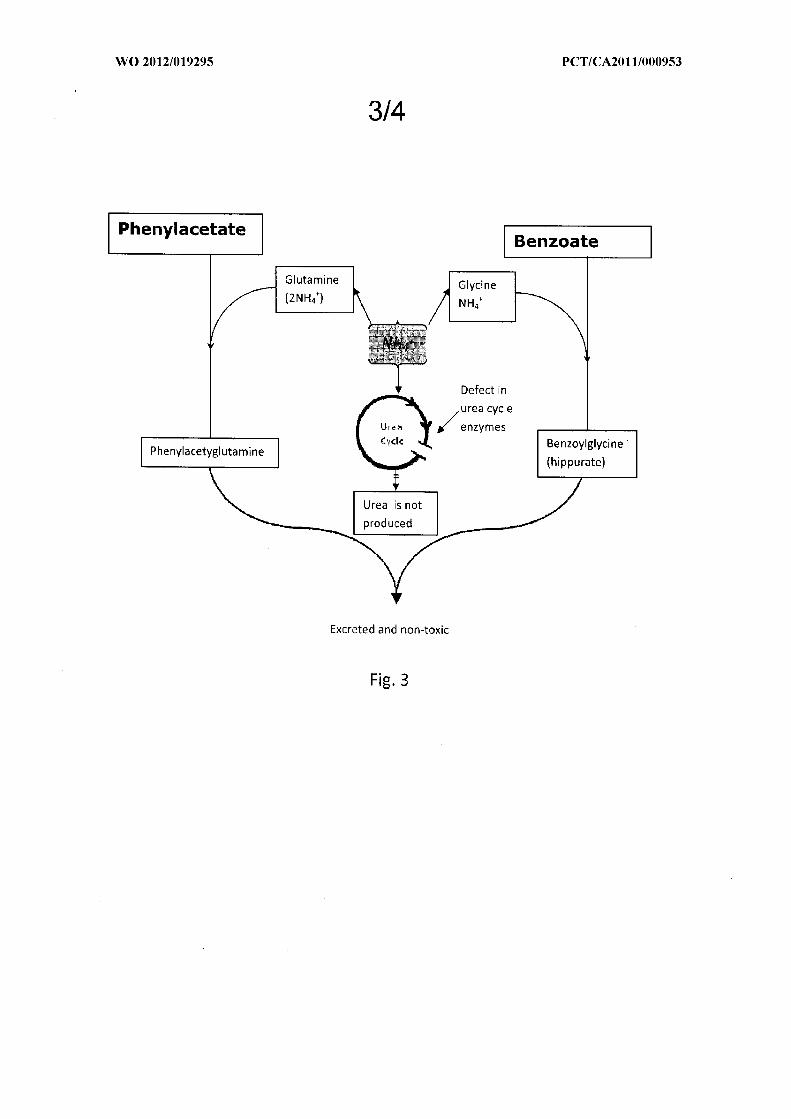
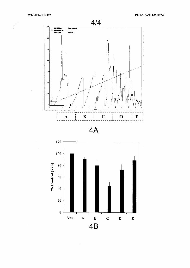
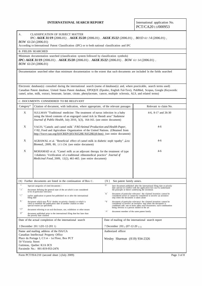
INTERNATIONAL SEARCH REPORT International application No.
PCT/CA20 11/000953
A . CLASSIFICATION OF SUBJECT MATTERIPC: A61K 31/19 (2006.01) . A61K 35/20 (2006.01) . A61K 35/22 (2006.01) . B01D 61 4 (2006.01) .
BOW 61/24 (2006.01)According to International Patent Classification (IPC) or to both national classification and IPC
B . FIELDS SEARCHED
Minimum documentation searched (classification system followed by classification symbols)
IPC: A61K 31/19 (2006.01) . A61K 35/20 (2006.01) . A61K 35/22 (2006.01) . BOW 61 14 (2006.01) .BOW 61/24 (2006.01)
Documentation searched other than minimum documentation to the extent that such documents are included in the fields searched
Electronic database(s) consulted during the international search (name of database(s) and, where practicable, search terms used)
Canadian Patent database. United States Patent database, EPOQUE (Epodoc, English Ful-Text), PubMed, Scopus, Google (Keywords:camel, urine, milk, extract, benzoate, lactate, citrate, phenylacetate, cancer, multiple sclerosis, ALS, and related terms)
C. DOCUMENTS CONSIDERED TO BE RELEVANT
Category' Citation of document, with indication, where appropriate, of the relevant passages Relevant to claim No.
X SULLMAN "Traditional medicine: The treatment of navus infection n a baby 4-6, 8-17 and 26-30using the blood contents of an engorged camel tick in Shendi area' " SudaneseJournal of Public Health, July 2010, 5(3), 164-165. (see entire document)
X YAGTL "Camels and camel milk " FAO Animal Production and Health Paper, 4-682, Food and Agriculture Organization of the United Nations. (Obtained from
http://¾wvv.tao.org/I)OCREP/()03/X6528E/X6528E()0.htm) (see entire document)
X AGRAWAL et al. "Beneficial effect of camel milk in diabetic neph opathy" 4-6Biomed., 2009, 80, 13 1-134. (see entire document)
X MOHAMAD et al. "Camel milk as an adjuvant therapy for the treatment of type 4-6
1 diabetes: Verification of a traditional ethnomedical practice" Journal ofMedicinal Food, 2009, 12(2), 461-465. (see entire document)
[X] Further documents are listed in the continuation of Box C . [X ] See patent family annex.
Special categories of cited documents : later document published after the international filing date or prioritydate and not m conflict with the application but citecTto understand
document defining the general state of the art which is not considered the principle or theorv underlying the inventionto be of particular relevance
X" document of particular relevance the claimed invention cannot beearlier application or patent but published on or after the international considered novel or cannot be considered to involve an inventivefiling date step when the document is taken alone
document which may t r n doubts on priority clami(s) or which is "Y document of particular relevance the claimed invention cannot becited to establish the publication date of another citation or other considered to involve an inventive step when the document isspecial reason (as specified) combined rath one or more other such documents such combination
being obvious to a person skilled m the artdocument referring to an oral disclosure, use, exhibition or other means
document member of the same patent familydocument published prior to the international filing date but later thanthe priority date claimed
Date of the actual completion of the international search Date of mailing of the international search report
1 December 201 1 (01-12-201 1) 7 December 20 1 (07-12-20 1 1)
Name and mailing address of the ISA CA Authorized officerCanadian Intellectual Property OfficePlace du Portage I, CI 14 - 1st Floor, Box PCT Wesley Sharman (819) 934-232650 Victoria StreetGatineau, Quebec K1A 0C9Facsimile No.: 001-819-953-2476
Form PCT/ISA/210 (second sheet ) (July 2009) Page 3 of 8

INTERNATIONAL SEARCH REPORT International application No.
PCT/CA20 11/000953
Box No. II Observations where certain claims were found unsearchable (Continuation of item 2 of the first sheet)
This international search report has not been established in respect of certain claims under Article 17(2)(a) for the followingreasons :
1 . [X] Claim os. : 18-25
because they relate to subject matter not required to be searched by this Authority, namely :
Although claims 18-25 are directed to methods of medical treatment of the human or animal body (Rule s39. 1(iv) of the PCT), asearch has been carried out on the alleged effects of the specific compounds defined in the present claims in treating disease.
2 . [X] Claim Nos. : 1-30
because they relate to parts of the international application that do not comply with the prescribed requirements to such an extentthat no meaningful international search can be carried out, specifically :
(see extra sheet)
3 . [ ] Claim Nos. :
because they are dependent claims and are not drafted n accordance with the second and th d sentences of Rule 6 .4( a).
Box No. Ill Observations where unity of invention is lacking (Continuation of item 3 of first sheet)
This International Searching Authority found multiple inventions in this international application, as follow s :
(see extra sheet)
1. [ ] As all required additional search fees were timely paid by the applicant, this international search report covers all
searchable claims.
2 . [X] As all searchable claims could be searched without effort justifying additional fees, this Authority did not invite
payment of additional fees.
3 . [ ] As only some of the required additional search fees were timely paid by the applicant, this international search report
covers only those claims for which fees were paid, specifically claim Nos. :
4 . [ ] No required additional search fees were timely paid by the applicant. Consequently, this international search report is
restricted to the invention first mentioned in the claims; it is covered bv claim Nos. :
Remark on Protest [ ] The additional search fees were accompanied by the applicant's protest and, where applicable,
the payment of a protest fee.
[ ] The additional search fees were accompanied by the applicant's protest but the applicable protest
fee was not paid within the time limit specified in the invitation.
[ ] No protest accompanied the payment of additional search fees.
Form PCT/ISA/210 (continuation of first sheet (2)) (July 2009) Page 2 of 8

INTERNATIONAL SEARCH REPORT International application No.
PCT/CA20 11/000953
C (Continuation). DOCUMENTS CONSIDERED TO BE RELEVANT
Category* Citation of document, with mdication, where appropriate, of the relevant passages Relevant to claim No.
X SHABO et a "Etiology of autism and camel milk as therapy" Int. J. Dis. Human 4-6Dev., 2005, 4(2), 67-70. (see entire document)
X WO2005023208 (KALE.TAMN) 17 March 2005 (17-03-2005) (see entire 4-6document)
X US20090297622 (KHORSHJD) 3 December 2009 (03-12-2009) (see entire 8-17 and 26-30document)
X K ORS et al. "h vitro anticancer agent: I - Tissue culture of human lung 8-17 and 26-30cancer cells A549. Π - Tissue culture study of mice leukemia cells L1210"International Journal of Cancer Research, 2006, 2(4), 330-344. (see entiredocument)
X 8-17 and 26-30
KHORSHLD "Potential anticancer natural product against human lung cancercells" Trends in Medical Research, 2009, 4( 1), 8-15. (see entire document)
X 8-17 and 26-30
KHORSrD et al. "Ototoxic activity of bioactive fractions from PM 701"Electronic Journal of Environmental, Agricultural and Food Chemistry:, 2009,8( 11), 1091-1098. (see entire document)
X 8-17 and 26-30
EL-SHAHAWY et al. "Spectral analysis, molecular orbital calculations andantimicrobial activity of PMF fraction extracted from PM-70 " InternationalJournal ofPharma and Bio Sciences, April-June 2010, 1(2), 1-20. (see entiredocument)
X 8-17 and 26-30
MOSHREF "PM701, a highly selective anticancerous agent against L1210leukemia cells: h vivo clinical and histopathological study" Journal of KingAbdiilaziz University - Medical Sciences, 2007, 14(4), 85-99. (see entire
X document) 8-17 and 26-30
KABARITY et al. "Camel urine as a possible anticarcinogenic agent" Arab GulfX J. Scient. Res. Agric. Biol. Sci., 1988, B6( 1), 55-63. (see entire document) 8-17
ENNS et al. "Survival after treatment with phenylacetate and benzoate for urea-cycle disorders" New England Journal of Medicine, 2007, 356(22), 2282-2292.
X (see entire document) 8-17
BL<ACHMACHAPJ et al. "Sodium benzoate, a metabolite of cinnamon and a foodadditive, reduces microglial and astroglial inflammatory responses" Journal of
X 8-17Immunology, 2009, 183, 5917-5927. (see entire document)
LIU et al. "Modulation by phenylacetate of early estrogen-mediated events nMCF-7 breast cancer cells" Cancer Chemother. Pharmacol., 2007, 59, 217-225.
X 8-17(see entire document)
WOODHOUSE "Chemotherapy investigations n cancer with reference to theinfluence of certain organic dibasic acids, diamino compounds and nitrocompounds on tumors n mice" Cancer Research, 1947, 7, 398-401 . (see entire
X 8-17document)
X 8-17GB1200928 (FERLUX) 5 August 1970 (05-08-1970) (see entire document)
LAVERMICOCCA et al. "Purification and characterization of novel antifungalcompounds form the sourdough Lactobacillus plantanim strain 2 Applied andEnvironmental Microbiology -, 2000, 66( 9), 4084-4090. (see entire document)
Form PCT/ISA/210 (continuation of second sheet) (July 2009) Page 4 of 8
INTERNATIONAL SEARCH REPORT International application No.
PCT/CA20 11/000953
C (Continuation). DOCUMENTS CONSIDERED TO BE RELEVANT
Category* Citation of document, with mdication, where appropriate, of the relevant passages Relevant to claim No.
X EVANS et al. "Retinoids: Present role and future potential' British Journal of 8-17Cancer, 1999, 80( 1/2), 1-8. (see entire document)
X LUPIEN et al. "Long-term treatment of homozygous familial 8-17hypercholesterolemia with 3-hydiOxy-3-methylglutanc acid" The Lancet, 1978,283-284. (see entire document)
X LUPIEN et al. "Effects of 3-hydroxy-3-methylglutaric acid on plasma and low- 8-17density lipoprotein cholesterol levels in familial hypercholesterolemia " Journal ofClinical Pharmacology, 1979, 120-126. (see entire document)
X PARK et al. "Pyruvate slows disease progression in a G93A SOD1 mutant 8-17
transgenic mouse model" Netiroscience Letters, 2007, 413, 265-269. (see entiredocument)
X DIEULEVEUX et al. "Antimicrobial spectrum and target site of D-3-phenyUactic 8-17
acid" International Journal of Food Microbiology, 1998, 40, 177-183. (see entiredocument)
X SHECHTER et al. "Hydrophenyl acetate derivatives inhibit protein tyrosine 8-17
kinase activity and proliferation n Nb2 rat lymphoma cells and insulin-inducedlipogenesis in rat adipocytes' "Molecular and Cellular Endocrinology;, 1991, 80,183-192. (see entire document)
X OHASHI et al. "The inhibitory effect of glycolic acid and lactic acid on melanin 8-17
synthesis in melanoma cells" Experimental Dermatology;, 2003, 12(S2) 43-50.(see entire document)
X YANG et al. "Effects of glycolic acid on the induction of apoptosis via caspase-2 8-17
activation in human leukemia cell line (HL-60)" Food and Chemical Toxicology;,2004, 42, 1777-1784. (see entire document)
L GARCIA RUIZ et la. "Cerebrospinal fluid homovanillic acid is reduced h 1-30
untreated Huntington's disease" Clinical Neuropharmacology, 18( 1), 58-63. (seeentire document)
L ROSA et al. "'Inhibition of energy metabolism by 2-methylacetoacetate and 2- 1-30
methyl-3-hydroxybutyrate n cerebral cortex of developing rats" J. Inherit. Metab.Dis., 2005, 28, 501-5 15. (see entire document)
L WATANABE et al. "Measurement of 3-hydroxyisovaleric acid in urine of biotin- 1-30
deficient infants and mice by P C Joumal of Nutrition, 2005, 135(3), 615-618.(see entire document)
Form CT ISA 2 10 (continuation of second sheet) (July 2009) Page 5 of 8
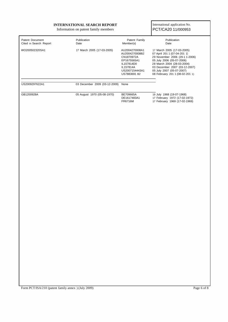
INTERNATIONAL SEARCH REPORT International application No.
Information on patent family members PCT/CA20 11/000953
Patent Document Publication Patent Family PublicationCited in Search Report Date Member(s) Date
WO200502320SA1 17 March 2005 (17-03-2005) AU2004270008A1 17 March 2005 (17-03-2005)AU2004270008B2 07 April 201 1 (07-04-201 1CN1870972A 29 November 2006 (29-1 1-2006)EP1675565A1 05 July 2006 (05-07-2006)IL157814D0 28 March 2004 (28-03-2004)IL157814A 03 December 2007 (03-12-2007)US2007154443A1 05 July 2007 (05-07-2007)US7883691 B2 08 February 201 1 (08-02-201 1)
US2009297622A1 03 December 2009 (03-12-2009) None
GB1200928A 05 August 1970 (05-08-1970) BE709665A 19 July 1968 (19-07-1968)DE1617483A1 17 February 1972 (17-02-1972)FR6716M 17 February 1969 (17-02-1969)
Form PCT/ISA/210 (patent family annex ) (July 2009) Page 6 of 8
INTERNATIONAL SEARCH REPORT International application No.
PCT/CA20 11/000953
Continuation of Box II:
Claims 1-30 of the current claims relate at least in part to extracts from camel urine compositions comprising said extractsuse of said extracts methods of using said extracts and methods of producing said extracts wherein the extracts arecharacterized by an NMR spectrum an HPLC spectrum or a specific fraction of an HPLC spectrum. However an NMR orHPLC spectrum cannot be viewed as fully and completely defining an extract the components of an extract or their relativeamounts. Along the same lines claims 7. 10-17 and 19-25 of the current claims relate at least in part to compositionscomprising compounds uses of compounds and methods of using compounds wherein the compound is identified by a peak inan NMR spectrum by a peak in HPLC spectrum or by a peak in a fraction of an HPLC spectrum. However a compoundcannot legitimately be identified by a single peak in an NMR spectrum or by a peak in an HPLC spectrum.
As a result these claims fail to fully and completely define the extract or the compounds identified by a peak in an NMR orHPLC spectrum in any true or meaningful manner causing the true scope of these claims to be indeterminable. Hence theseclaims so lack clarity in view of Article 6 of the PCT and so lack support and disclosure in view of Article 5 of the PCT that asearch over the entire scope of the present claims is not possible. Consequently a search has only been carried out for thoseparts of the claims that have been fully and completely defined by the present claims and supported by the present description.This would include claims encompassing extracts that can be viewed as fully defined (such as those defined in claims 4 and 5)and compounds that are fully identified (such as those defined in claims 12. 13. 20 and 2 1).
Therefore no search has been carried out for the subject matter of claims 1-3 and 7. Furthermore the search carried out forclaims 6. 8. 9 and 18 (and those claims dependent on these claims) only relates to their dependence on claims 4 and 5. Nosearch has been carried out for the subject matter of claims 6. 8. 9 and 18 (and those dependent on these claims) when claims6. 8. 9 and 18 are dependent on claims 1-3. Finally any search carried out for the subject matter of claims 10. 1 and 19 (andthose dependent on these claims) only relates to the use of compounds that are fully defined namely those defined in claims12. 13. 20 and 2 1.
Form PCT/ISA/210 (extra sheet) (July 2009) Page 7 of 8
INTERNATIONAL SEARCH REPORT International application No.
PCT/CA20 11/000953
Continuation of Box III:
The claims of the present application do not comply with the requirements of Rule 13 of the PCT. The present claims are notrelated to one invention or to a group of inventions that are so linked as to form a single general inventive concept.
The present claims are directed towards extracts from either camel urine or camel milk compositions comprising saidextracts use of said extracts to treat disease methods of using said extracts to treat disease and methods of producing saidurine extracts. In addition the present claims are directed towards compositions comprising compounds uses of compoundsand methods of using compounds wherein the compound is identified by a peak in an NMR spectrum by a peak in HPLCspectrum or by a peak in a fraction of an HPLC spectrum wherein said spectra are those obtained for a camel urine extract.
These claims cannot be viewed as being linked by a single general inventive concept. The use of a bodily fluid of a camel(either urine or milk) to treat disease does not link the present claims since the use of camel urine and camel milk to treatdisease is well-known (see for instance the teachings of the present description and D1-D13). Similarly camel urine andcamel milk extracts are known in the art (see for instance D6-D13) and such extracts cannot be viewed as i eiitive in light ofthe known use of camel urine and camel milk to treat disease. Therefore the use of such extract also cannot act as a linkinginventive feature. As a result claims encompassing extracts from camel urine and claims encompassing extracts from camelmilk cannot be viewed as linked by a single inventive concept.
In addition claims encompasses specific compounds or combinations of specific compounds are also not linked by a singleinventive concept. The compounds defined in present claims 4. 5. 12. 13, 20 and 21 are all known compounds. Theirpresence in camel milk or their identification in a camel urine does not link these compounds by a common inventive concept.This is particularly apparent since the compounds defined in claim 4 are known to be present in milk. In addition the mereidentification of a known compound in camel urine does not provide novelty to the compounds or to the known uses of theseknown compounds. As a result, since an extract as defined in claims 4 and 5 are viewed as a composition, each distinctcomposition whether directed to a single compound or a combination of compounds (as encompasses by present claims 4 and5 (i.e. at least one compound" includes a combination of two or more compounds) represents a separate and distinct allegedim eiition. Furthermore insofar as these compounds and combinations are known their use in the treatment of each potentialdisease also represents a separate and distinct alleged invention. Thus, for example, extracts comprising benzoate represent aseparate alleged invention compared to extracts comprising lactate or extracts comprising benzoate and pyruvic acid. Inaddition, the use of an extract comprising benzoate to treat breast cancer represents a separate alleged invention compared tothe use of an extract comprising benzoate to treat multiple sclerosis or the use of an extract comprising benzoate and pyruvicacid to treat breast cancer.
Similarly, claims 12, 13. 20 and 1 all encompass the use (or method of using) of a single known compound or a combinationof known compounds to treat disease. Since these compounds are all known the treatment of each separate and distinctdisease wit each separate and distinct compound or combination of compounds represents its own separate and distinctallegedly inventive concept since each are directed to an allegedly new use of a known compound or a combination of knowncompounds. Thus for example the use of citric acid to treat thyroid cancer represents a separate alleged invention comparedto the se of sebacic acid to treat thyroid cancer the se of the combination of citric acid and sebacic acid to treat thyroidcancer the use of citric acid to treat Huntington ' s disease or the use of citric acid and sebacic acid to treat Huntington ' sdisease.
Claims 26-30 are directed to a general method of producing an extract from camel urine. Since extracts of camel urine areknown (see D7-D13). this method also involves a separate and distinct allegedly inventive concept.
Form PCT/ISA/210 (extra sheet) (July 2009) Page 8 of 8

US 2014003 7722Al
(19) United States (12) Patent Application Publication (10) Pub. N0.: US 2014/0037722 A1
Mousa et al. (43) Pub. Date: Feb. 6, 2014
(54) METHODS AND COMPOSITIONS OF CAMEL A61K 31/12 (2006.01) DERIVED PRODUCTS A23C 9/152 (2006.01)
(7 1) Applicants: Shaker A. Mousa, Wynantskill, NY A 61K 31/33 7 (200601) (US); Abdulqader Al Haider, Riyadh (52) U-S- Cl (SA); AbdelgalilAbdelgader, Riyadh CPC ............. .. A61K 9/4866 (2013.01); A23C 9/152
(SA); Abdullah M- Aldahmash, Riyadh (2013.01); A61K 47/42 (2013.01); A61K Egg; Abdulkareem Alm°men> Rlyadh 31/337 (2013.01); A61K 31/704 (2013.01);
A61K 31/12 (2013.01); A61K31/727 (2013.01) (72) Inventors; Shaker A, Mousa, wynamskill, NY USPC .......... .. 424/452; 514/775; 424/729; 424/769
(US); Abdulqader Al Haider, Riyadh (SA); Abdelgalil Abdelgader, Riyadh (SA); Abdullah M. Aldahmash, Riyadh (57) ABSTRACT (SA); Abdulkareem Almomen, Riyadh (SA) The present invention provides a composition, a dairy prod
(21) APPL NO; 13/953,888 uct, and a method for treating a disorder in a subject. The composition includes (i) polymeric nanoparticles and (ii)
(22) Filed? Jul- 30: 2013 camel derived glycosaminoglycans (GAG)s ionic complex
Related U‘s‘ Application Data encapsulated into the nanoparticles, at least one active ingre _ _ _ _ d1ent encapsulated into the nanopartlcles, or combinatlons
(60) lfrogglsgonal apphcanon NO‘ 61/678’165’ ?led on Aug‘ thereof. The nanoparticles are lactoferrin nanoparticles ’ ' including camel derived lactoferrin, casein nanoparticles
Publication Classi?cation including camel derived casein, or combinations thereof. The dairy product includes ice cream or frozen yogurt, Wherein
(51) Int‘ C1‘ the ice cream or frozen yogurt includes the composition and A61K 9/48 (2006.01) . . . . .
A 61K 47/42 (200601) is derived from camel m1lk or other specles of m1lk. The A61K 31/727 (200601) method for treating a disorder in a subject includes adminis A61K 31/704 (2006.01) tering a therapeutic dose of the composition to the subject.

Patent Application Publication Feb. 6, 2014 Sheet 1 0f 21 US 2014/0037722 A1
gm w magi ..
.3 w
. - a Wm
+ m m
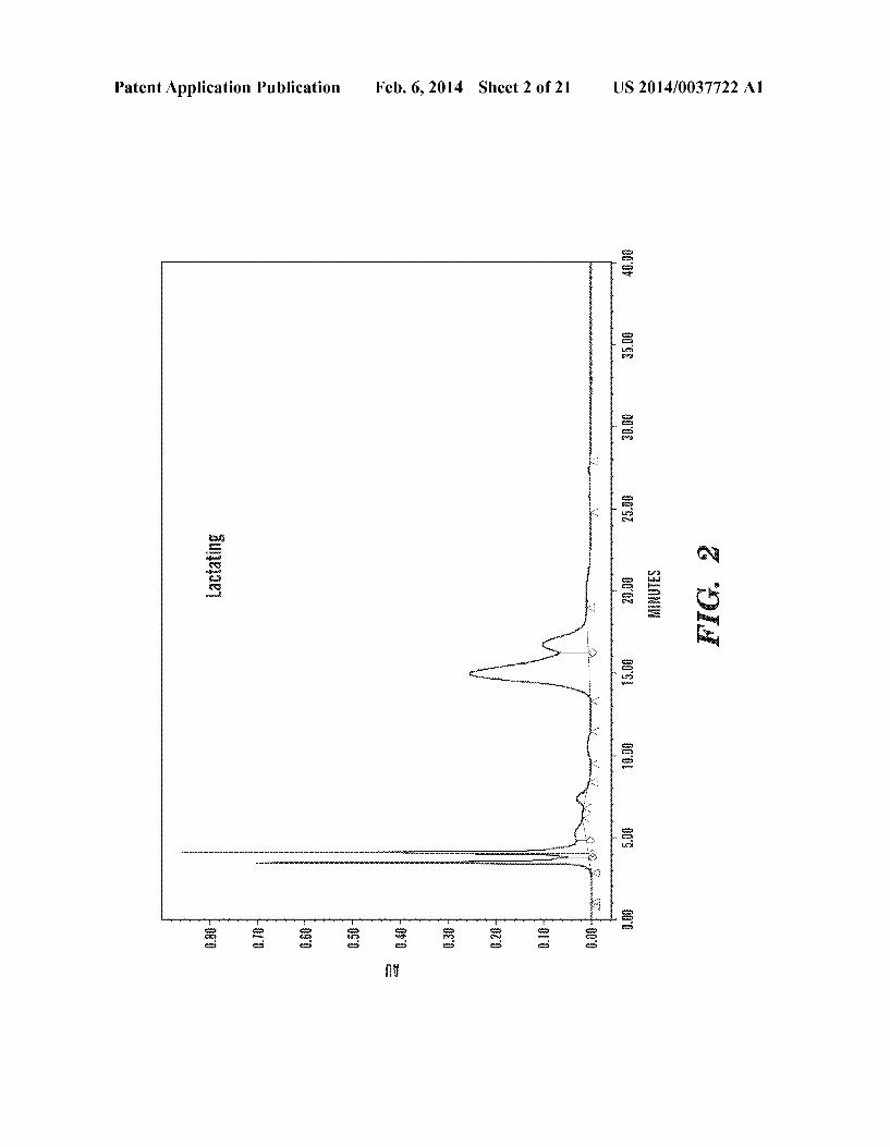
Patent Application Publication Feb. 6, 2014 Sheet 2 0f 21 US 2014/0037722 A1
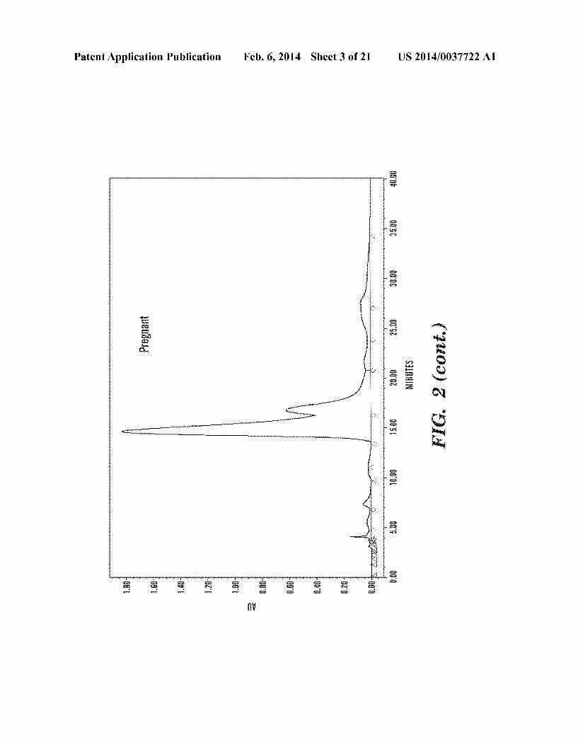
Patent Application Publication Feb. 6, 2014 Sheet 3 0f 21 US 2014/0037722 A1
33
“gamma
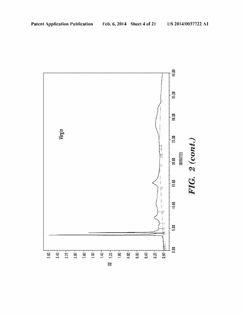
Patent Application Publication Feb. 6, 2014 Sheet 4 0f 21 US 2014/0037722 A1
I gm - a; ‘ $3 , gm ‘ Q: - E.“ g a: i mm." 1am." I am 1mm ( a: 1 3w

Patent Application Publication Feb. 6, 2014 Sheet 5 0f 21 US 2014/0037722 A1
33
E; H KEEN FEENH gig Ha?g?gs? EE‘MEWEWS mm?m??
3mm sham 3.x 3.2 Q , . ,, jifrflfialvsxxgiirirr/
-3
.~ _
, M a:
_
M a:
-2: is."
a $3
I ,2.”
.M
Q -92
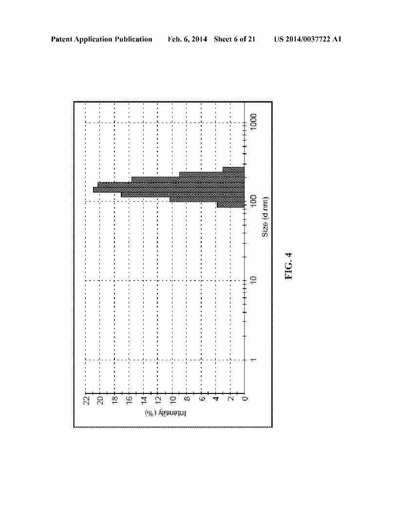

Feb. 6, 2014 Sheet 7 0f 21 US 2014/0037722 A1
Emma? E ZQEEEEE “Mum
W mwx?wumw-§mw-wmz,wwm&w§ up mmmuwg ............................................... ..
Patent Application Publication
:3 (‘76) MESNEiNE
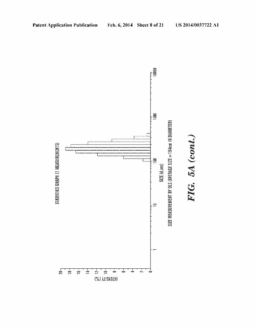
Patent Application Publication Feb. 6, 2014 Sheet 8 0f 21 US 2014/0037722 A1
‘ ‘ (2555a
SWiSiiCg GRQPH (1 EWEEESUREMENK)
IiiIFHg? YIHH! SiZHdmm) SiZE ME?SUREMENT 3‘! B13 (JKWRMBE SEZEZEQAnm 1N BUWEEFER) F1 G. 5A (QQYMJ
T
my".
(‘36) MESNEINE
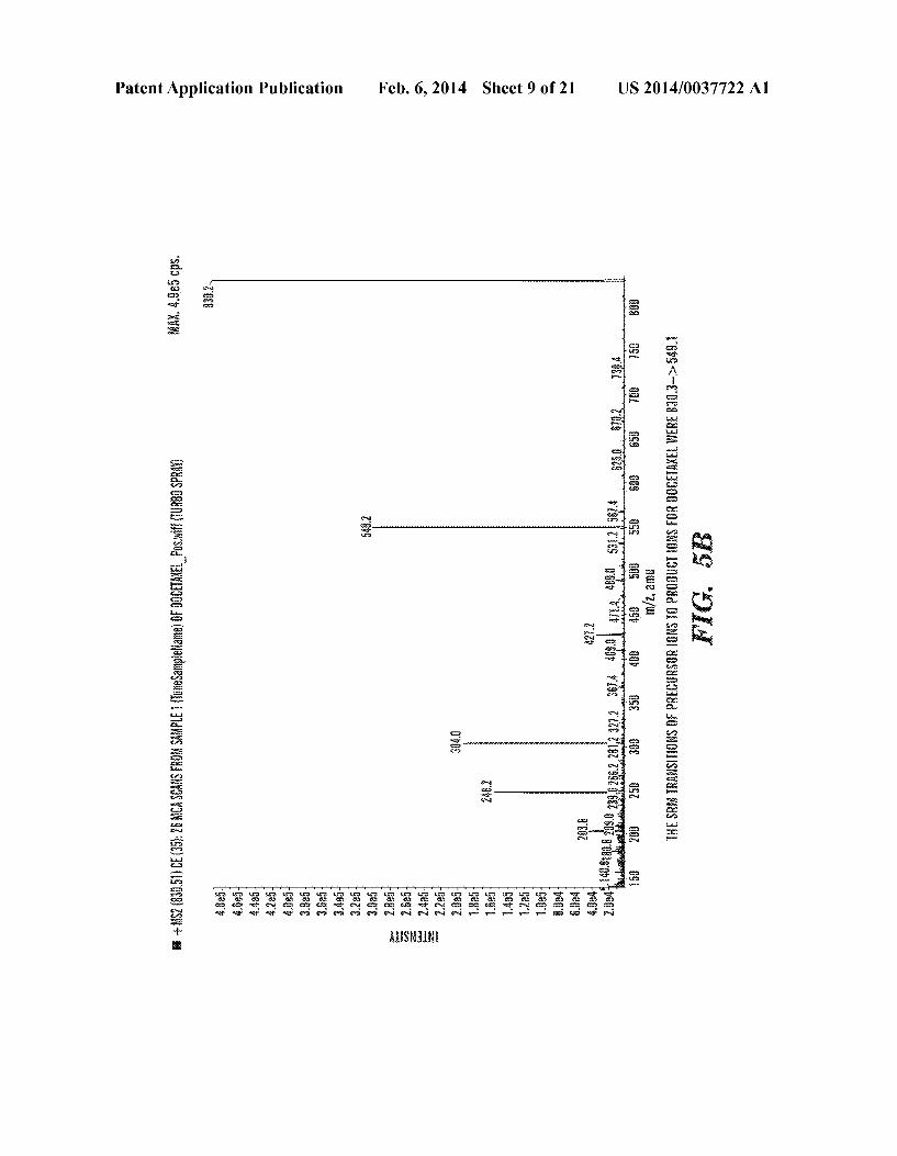
US 2014/0037722 A1 Feb. 6, 2014 Sheet 9 0f 21
AiESNEiNE
Patent Application Publication

Patent Application Publication Feb. 6, 2014 Sheet 10 0f 21 US 2014/0037722 A1
ENYENSEW. 8??»
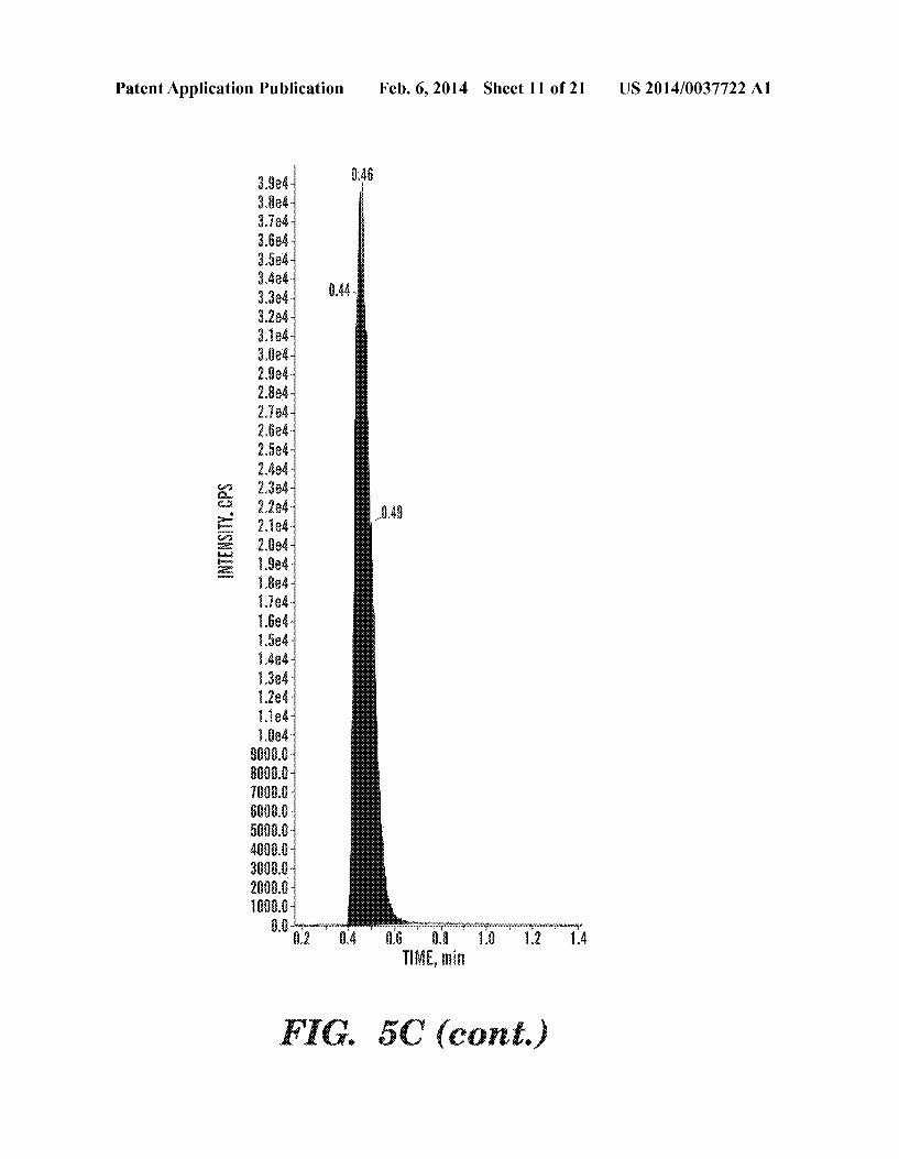
Patent Application Publication
ENYENSETY. CPS
3.984 3.3% 3Ja4~ 3.554 3.584 Mei 3.3% 312% 3.184‘ 3.5384 2.994 Meli 2.7%‘ mm 2.5% 2.464 2.3% 2.224“ 21%
Feb. 6, 2014 Sheet 11 0f 21 US 2014/0037722 A1
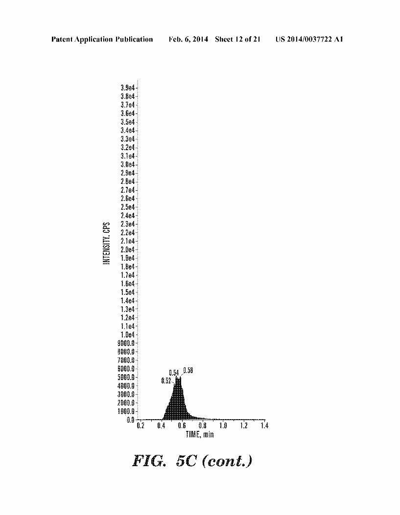
Patent Application Publication Feb. 6, 2014 Sheet 12 0f 21 US 2014/0037722 A1
BNIENSBW. EPS
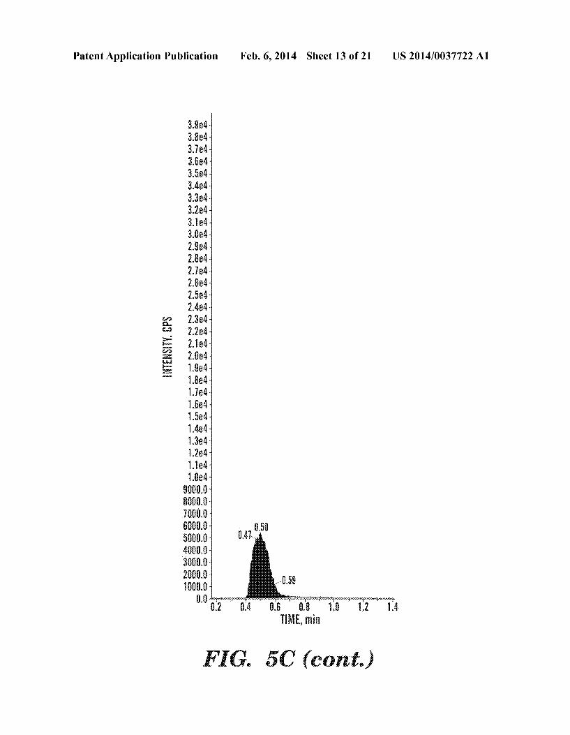
Patent Application Publication
ENYENSEW. 3%
3.884 3.324 Sled 3.834 3.534‘ 3.4e4~ 3.384 3.2% 3.? MA 3M4 2.9% 2.8% 2.?24 med *
2.5e4~ 2.424 2.3e4~ 2.284 2.1%4 -
Med '
L§e4~ 1.8%
Feb. 6, 2014 Sheet 13 0f 21 US 2014/0037722 A1

Patent Application Publication Feb. 6, 2014 Sheet 14 0f 21 US 2014/0037722 A1
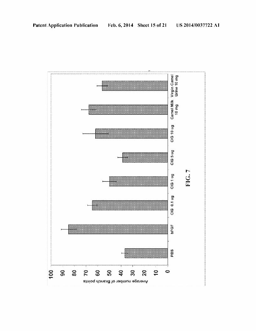
Feb. 6, 2014 Sheet 15 0f 21 US 2014/0037722 A1
Q: 2 main
m: 55
a: 3 m6
@wEmU Em»; 5:2 umEwU
singed qsumg ,io xaqwnu a?wanv
Patent Application Publication
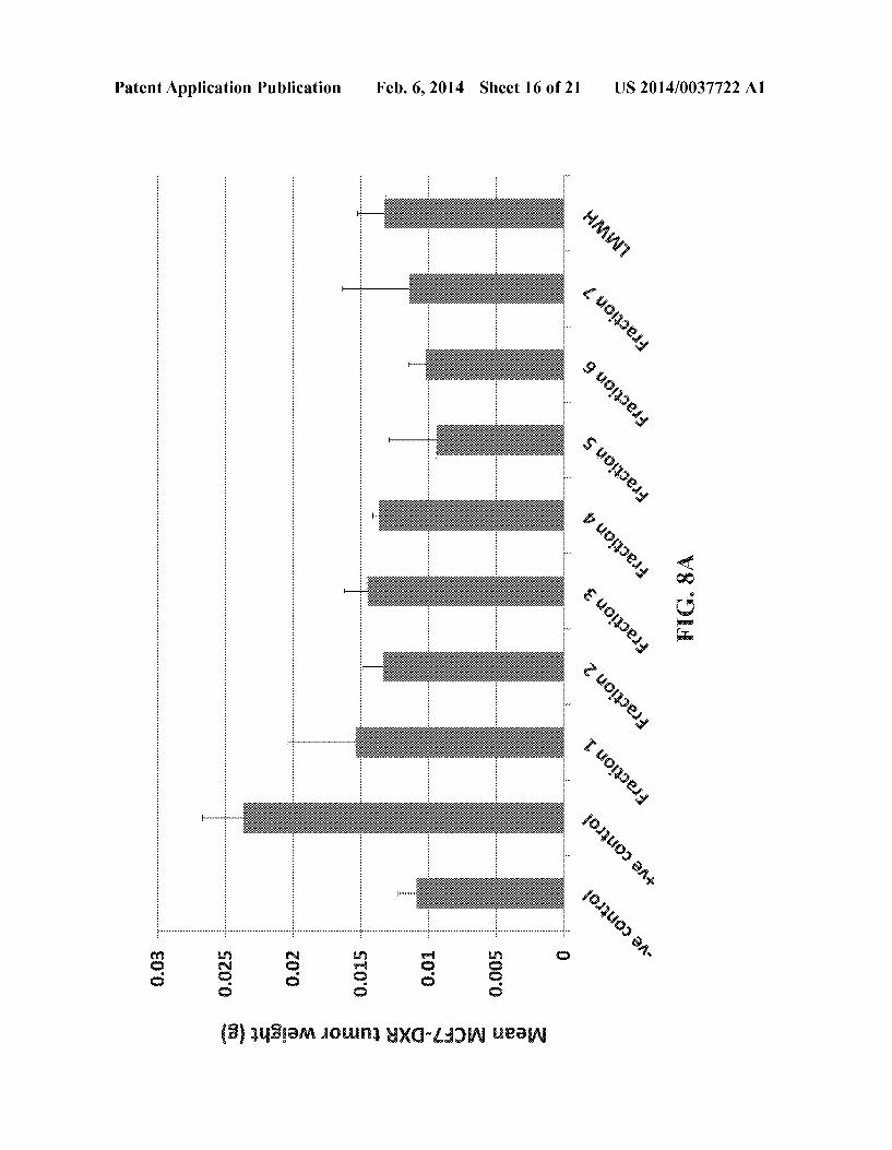
Patent Application Publication Feb. 6, 2014 Sheet 16 0f 21 US 2014/0037722 A1
6. iii-3 6.025 6 I 1 6.6%
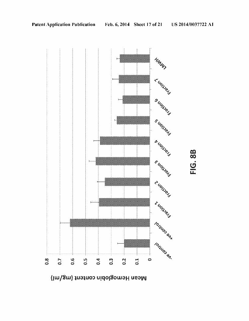
Patent Application Publication Feb. 6, 2014 Sheet 17 0f 21 US 2014/0037722 A1

N. cozumi m cnmum?
US 2014/0037722 A1 Feb. 6, 2014 Sheet 18 0f 21 Patent Application Publication
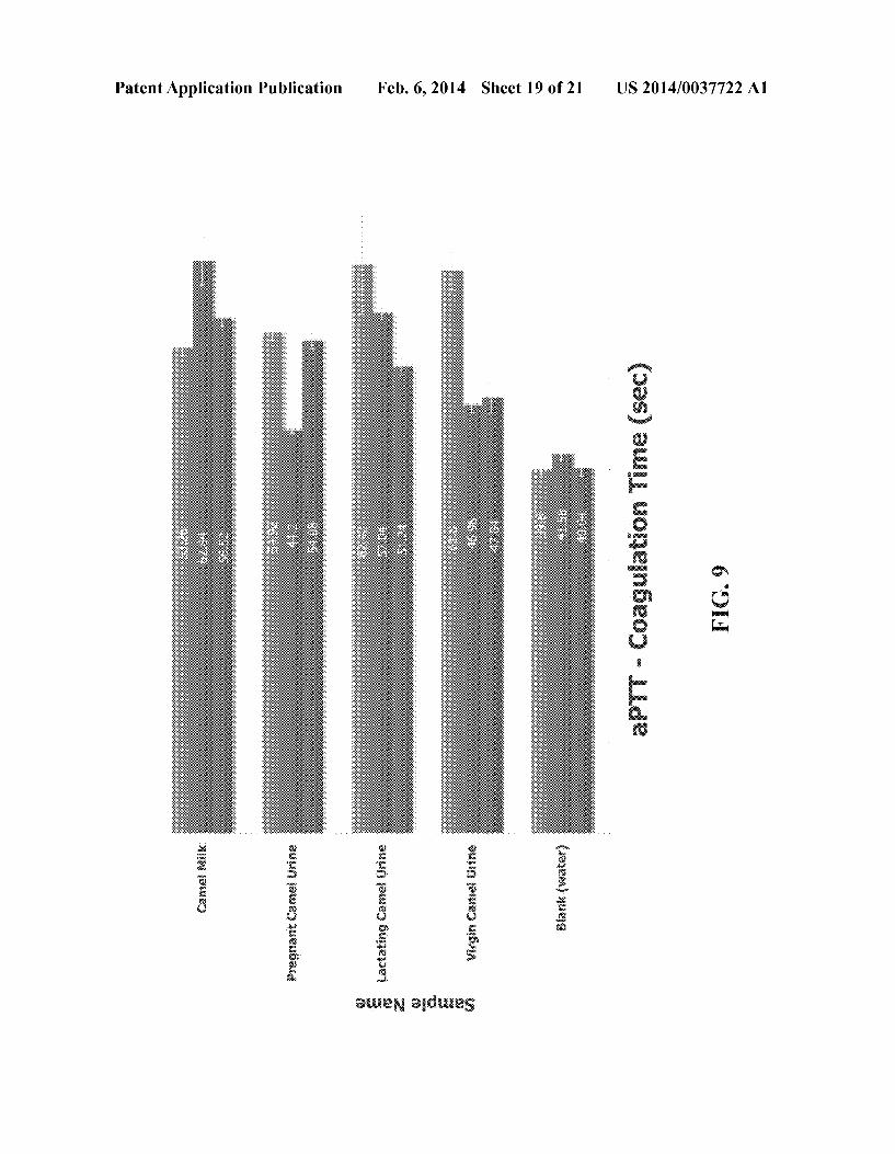
Patent Application Publication Feb. 6, 2014 Sheet 19 0f 21 US 2014/0037722 A1
.
amwwv. .
VEE “$55
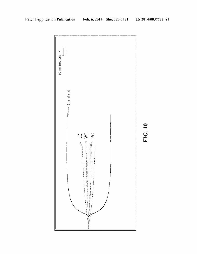

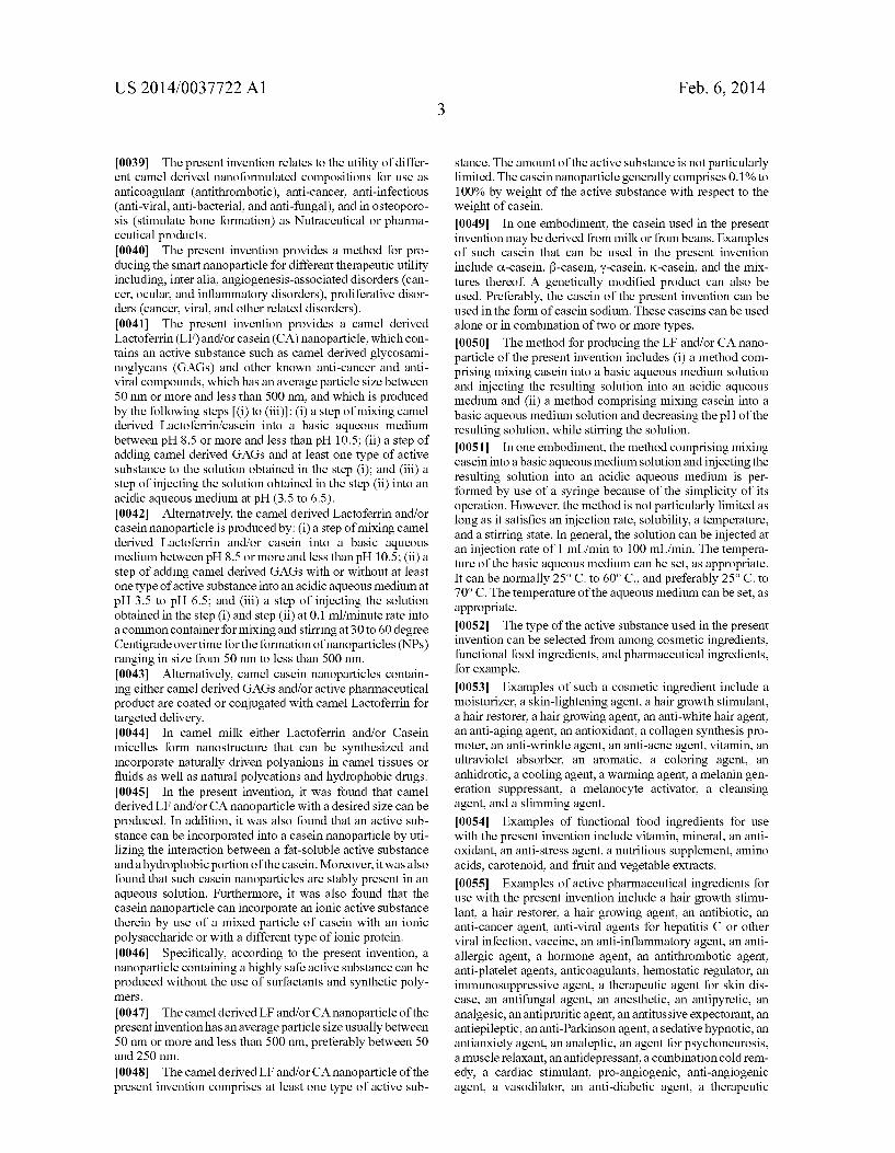

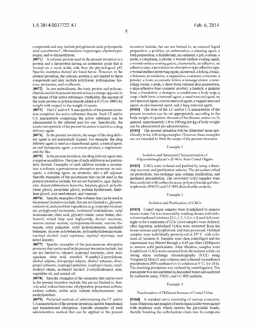
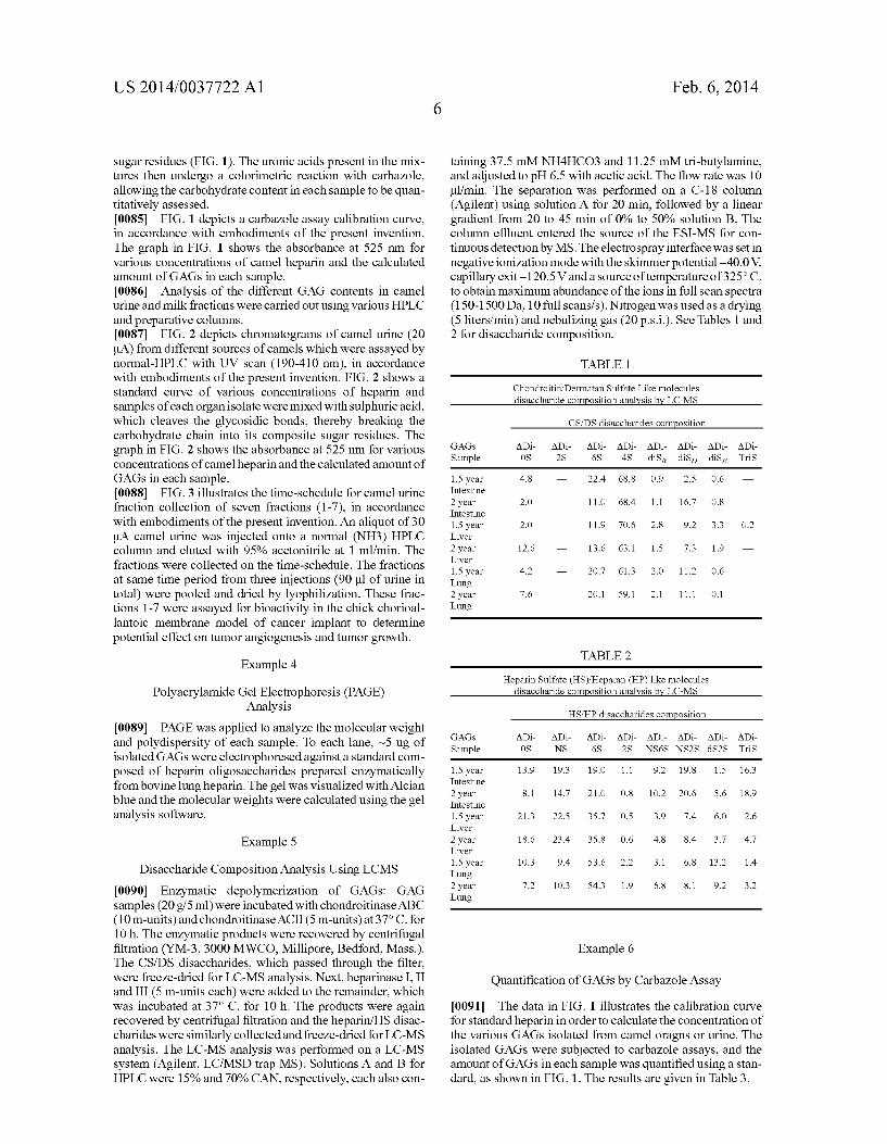
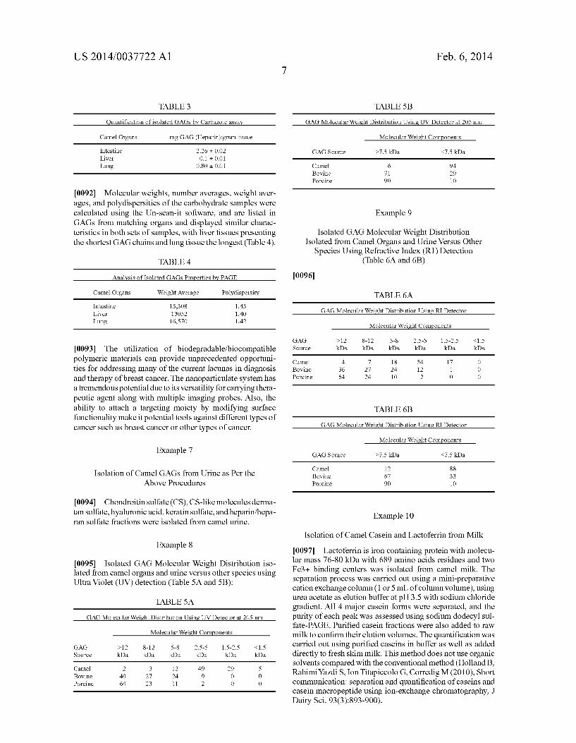


US 20090297622A1
(19) United States (12) Patent Application Publication (10) Pub. No.: US 2009/0297622 A1
Khorshid (43) Pub. Date: Dec. 3, 2009
(54) SEPARATION AND FORMULATION OF (30) Foreign Application Priority Data BIOACTIVE FRACTION AND SUBFRACTION FROM C AMEL URINE WORK As Jan. 1, 2008 (GC) ..................................... .. GC9962
ANTICANCER AGENT Publication Classi?cation
(51) Int. Cl. (76) Inventor: Faten A. Khorshid, Jeddah (SA) A61K 35/23 (2006.01)
A61P 35/00 (2006.01) Correspondence Address: (52) U.S. Cl. ...................................................... .. 424/558
ABELMAN, FRAYNE & SCHWAB (57) ABSTRACT 666 THIRD AVENUE’ 10TH FLOOR A pharmaceutical composition includes an effective amount NEW YORK’ NY 10017 (Us) of bio-active fraction PMF or subfraction PMFK obtained
from the lyophiliZed camel urine or coW urine Which coded in 21 A 1' N '2 12/178 152 publications With PM701, that is used as anti-cancer drug
( ) pp 0 ’ selected from an anticancer compound PM701, Which are targeted selectively the cancer cells Without affecting the
(22) Filed: Jul. 23, 2008 normal cells.
Main Cause of Death in Saudi Arabia Projected 2005
@ Cardiovascular diseases
Ei Other chronic diseases
IE Injuries
ElCummunieah-ie diseases
El Can eer
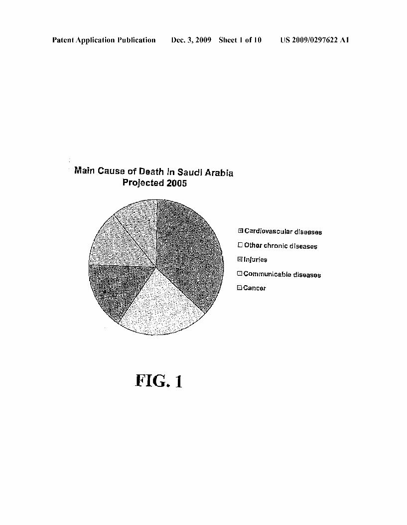
Patent Application Publication Dec. 3, 2009 Sheet 1 0f 10 US 2009/0297622 A1
3 Main Cause of Death in Saudi Arabia Projected 2005
@ Cerdievascuiar diseases
D‘ Other chmnic diseases
E5 Injuries
ElCommunisabie diseases
E‘ICaneer
Patent Application Publication Dec. 3, 2009 Sheet 2 0f 10 US 2009/0297622 Al
N .UHH , . .5 nm mu:
.7 -N m?m?g no m: Hi my. “Bump
u.
w .
-~ 25%: uu Hm E5 w a 3 ud. ,
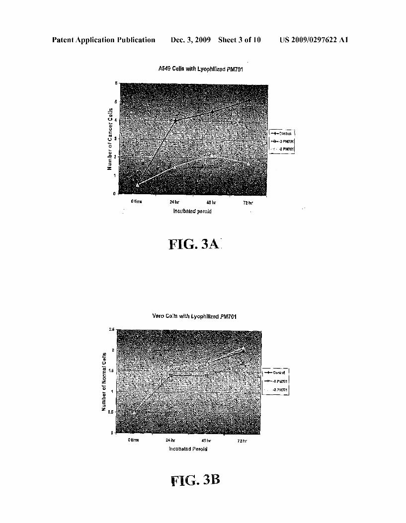
Patent Application Publication Dec. 3, 2009 Sheet 3 0f 10 US 2009/0297622 A1
A549 sens with Lyophilized PMm
Number ufCancer Cells
0 lime 24 hr 48 hr 72 hr
Incubated peruid -
FIG. 3A:
Vern Cells with Lyophilized PM701
Number of Nnrmai Cells
Oilma 24 hr 45 hr 72 hr
incubated Peroid
FIG. 3B
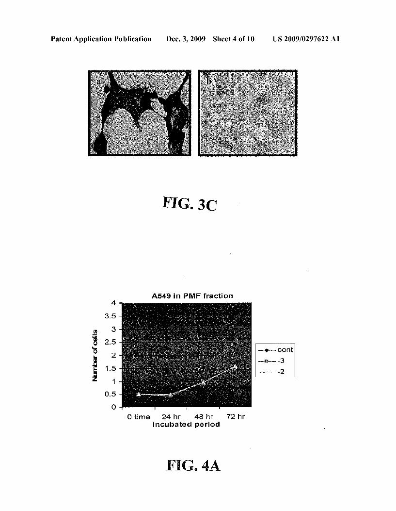
Patent Application Publication Dec. 3, 2009 Sheet 4 0f 10 US 2009/0297622 A1
A549 in PMF fraction
0 time 24 hr 48 hr 72 hr incubated period
F IG. 4A

Patent Application Publication Dec. 3, 2009 Sheet 5 0f 10 US 2009/0297622 A1
HFS Cells with PNEF Fraction
CI ‘.‘lrrls 24 hr 45 hr 7'2 hr
incubated Peroid
FIG. 4B

Patent Application Publication Dec. 3, 2009 Sheet 6 0f 10 US 2009/0297622 A1
A549 Cells with PMFK Subfraction
Number of Cancer cells
n time 24 hr 45 hr 72 hr
incu bated Percald ‘
FIG. 5A
HFS Cells with PM FK Subfraction
Number of Normal Cells
n time 24 hr 45 hr 72 hr
lncubated Perold
Fl. 5

Patent Application Publication Dec. 3, 2009 Sheet 7 0f 10 US 2009/0297622 A1
FIG. 5C
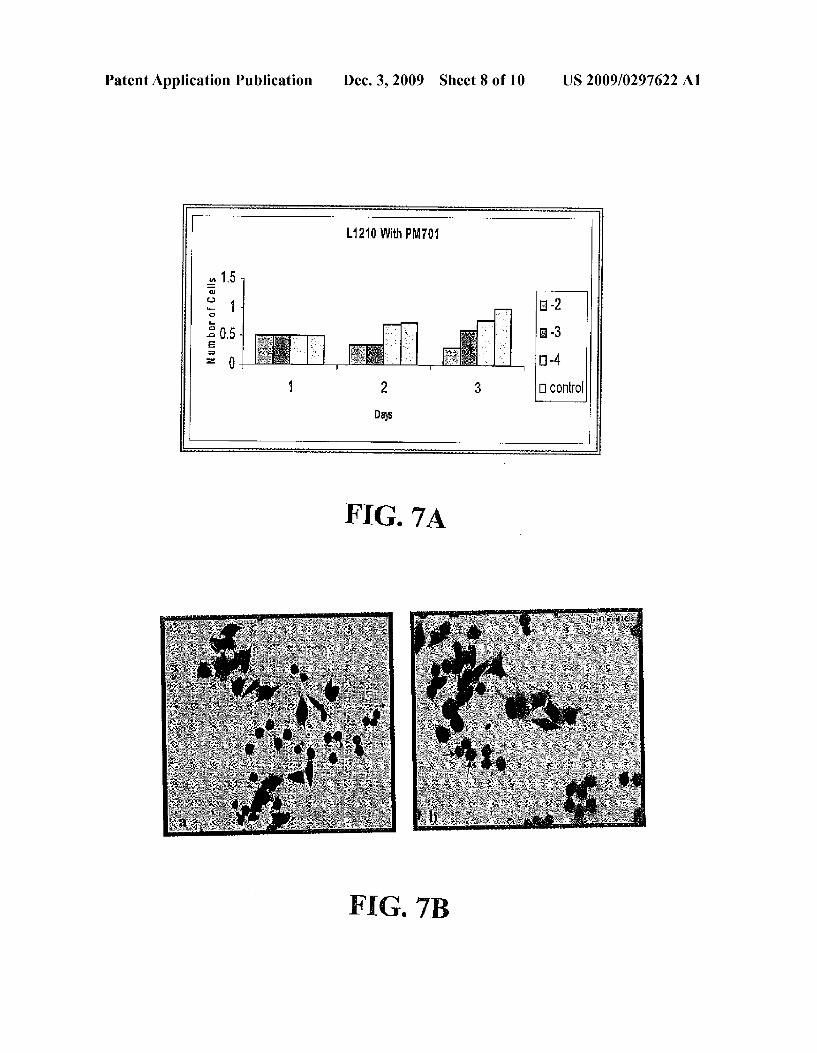
Patent Application Publication Dec. 3, 2009 Sheet 8 0f 10 US 2009/0297622 A1
L1210 With PMTM
-2
3 E -
I] control
5150 1 nu“ BEQEBQFSZ
FIG. 7A
FIG. 7B
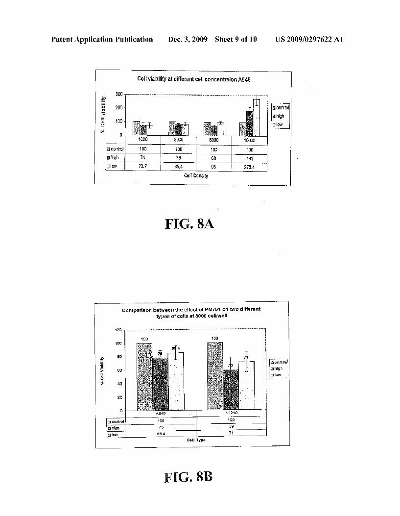
Patent Application Publication Dec. 3, 2009 Sheet 9 0f 10 US 2009/0297622 A1
Cell viability at dih‘ere nt cell can centraion A549
300 E‘ % a control 3 a high I u E! law gs:
Cell Density
FIG. 8A
Comparison between the effect of PMTM on two different types ofcelis at 3000 cell/well
no
10::
a, an
E E1 ciuntrui 5 EU a high
3 a 10w
3* 40
0
Cell Type
FIG. 8B

Patent Application Publication Dec. 3, 2009 Sheet 10 0f 10 US 2009/0297622 A1
FIG. 9

US 2009/0297622 A1
SEPARATION AND FORMULATION OF BIOACTIVE FRACTION AND SUBFRACTION
FROM CAMEL URINE WORK AS ANTICANCER AGENT
BACKGROUND OF THE INVENTION
[0001] 1. Field of the Invention [0002] The present invention relates to an absolutely novel use of camel urine for medicinal purposes and also relates to the preparation of it by lyophiliZation as a drug that called PM701 or fractionating it to active fractions that are coded as PMF and PMFK. More speci?cally, the present invention relates to isolating the bio-active fraction PMF and the most effective subfraction PMFK from the lyophiliZed PM701 of the adult single-humped Arabian camel, species camelus dromedarius and the absolutely novel use of these bio-active fractions as a selective anti-cancer agent. The fractionation Will be implicated in reducing the dosage of the Whole lyo philiZed urine and increasing its ef?cacy. The invention also relates to the preparation of a novel pharmaceutical compo sition (capsules) comprising an effective amount of the bio active fraction PMF or the bioactive subfraction in a compo sition of 435 mg/capsule With a range 7 or 5 capsules/day respectively, Which are targeted to the human cancer tissues through their anti-proliferative and apoptotic activities With out harming the other normal tissues. [0003] 2. Description of the Related Art [0004] Cancers are uncontrolled cell proliferations that result from the accumulation of genetic changes in cells endoWed With proliferative potential. After a variable latency period during Which they are clinically silent, the malignant cells progress to aggressive invasive and metastatic stages With tumor formation and Wide-spread dissemination throughout the body. [0005] Despite important advances in treatment, cancers still account for 28% of death in Western countries, and more than this ratio in other countries. Treatment of cancer has relied mainly on surgery, chemotherapy, radiotherapy and more recently immunotherapy. HoWever, for the most fre quent types of cancers (lung, breast, colo-rectal and the leu kemias) complete remission and cure has not been achieved. Therefore, the development of neW approaches for treating cancer patients is highly desirable and critically needed par ticularly for those patients Whose disease has progressed to a metastatic stage and are refractory to standard chemotherapy. [0006] The World health organiZation published that cancer is a leading cause of death WorldWide. From a total of 58 million deaths WorldWide in 2005, cancer accounts for 7.6 million (or 13%) of all deaths, in 2005 cancer killed approxi mately 12,000 people in Saudi Arabia, as shoWn in FIG. 1, With 8,000 of those people Were under the age of 70. [0007] In Prophet Mohamed Medicine (peace upon him), camel urine is suggested for drinking to improve some symp toms mainly associated With tumor formation in the body. Therefore, camel urine has been used in the Islamic and Arab World, sold and distributed in different siZe bottles. But it is never scienti?cally tested. The applicant considered it is WorthWhile to scienti?cally look at this and de?ne its value through in vitro and in vivo assays. The applicant has devel oped the curiosity about it and asked a number of questions: What possible importance can camel urine have? Whether the component camel urine is having any activity on tumor for mation by itself or some of its parts? Does the fresh urine have any effects on human cancer tissues? Could one formulate
Dec. 3, 2009
this urine in a convenience form for human use? Does the formulation of urine enhance its activity or reduce it? Dose the camel urine contain microorganism? Is it safe to use it as a drug or does it have toxicological effects on human diverse organs? [0008] In addition, could one fractionate the lyophiliZed camel urine in order to separate the bio-active fractions? To Which part of the cell does the camel urine react? What are the biochemical, biophysical, and biological evidences for its ef?cacy? [0009] Camel’s urine can be considered as an effective animal origin substance/secretion With the capacity of improvement of some symptoms mainly associated With tumors formation in the body but it does need substantiation through scienti?c experimentation. Thus, the applicant con sidered it WorthWhile to scienti?cally look at this and de?ne its values through in vitro and in vivo assays. The applicant in the ?rst instance probed Whether it contained anti-cancer agent since such a property Would make it a highly useful natural substance. In related art, use of ‘piperine’ as a bio availability enhancer has been described in US. Pat. Nos. 5,616,593 and 5,972,382. Also coW’s urine distillate or a dried fraction used for improving activity and bioavailability of antibiotics drugs as described in later US. Pat. No. 6,896, 907.
BRIEF SUMMARY OF THE INVENTION
[0010] Some of the above questions have been solved and addressed herein. To answer the ?rst set of questions the applicant collected the camel urine (PM701) from natural pastures in various areas Within Jeddah, Makah, Madinah and Riyadh governorate (Saudi Arabia) in different siZe bottles at any time of the day. The urine Was on CLED media and blood agar in sterile condition and did not notice any groWth of microorganism. Tissue cultures of human cancer tissues and normal tissues Were in studying the effect of (camel urine) PM701 on the behavior of cancer cells and normal cells. PM701 appears to target the cancer cells and have anti-pro liferative, apoptotic ef?cacy on them. [0011] Surprisingly, the same PM701 exhibited nourishing effects on normal healthy cells; this implies that PM701 have a selectively killing effect on cancer cells and reparative effect on normal dividing cells, these results leading to this inven tion. The novelty of the invention lies in the fact revealed through precise experimentation that the PM701 action and its effectiveness is achievable only in the range of concentra tion Which is literally in nano to micro-gram levels. That should be the reason for detection such a valuable potential of PM701 in targeted to the cancer cells. The utiliZed of fresh PM701 remains non-acceptable and non-convenience for human use therefore, the applicant also further lyophiliZed the liquid PM701 to obtain 0.2 g/ml of poWder. We re-exam ined the lyophiliZed PM701 on normal cells and diverse can cer cells in both cell culture and animal models, Which shoWed the same anti-cancer e?icacy and that Was mediated by apoptosis as determined by an MTT test and electron microscopy examination. [0012] The applicant thought of utiliZing PM701 as an alternative drug for cancer therapy since it shoWed a target effect on cancer cells and no side effects on the normal tis sues, but the amount of lyophiliZed PM701 dosage per day Was as a load in the body (46 capsules/day), ultimately lead ing to use one of the Ways, Which has been feasible for drastically reducing the daily dosage of this anti-cancer agent

US 2009/0297622 A1
PM701 and increasing the ef?ciency of the dosage activity too and has also high commercial importance. Therefore the bio-guided fractionation approach Was used With lyophiliZed PM701, Which leadus to the isolation and identi?cation of the bio-active fractions, Which is responsible for the anti-cancer e?icacy observed With the Whole urine. [0013] For the purpose of the present invention the folloW ing terms are de?ned beloW. [0014] The term “anti-cancer therapy” is intended to mean growth inhibition/eradication of primary tumors, stabiliZa tion of tumor groWth, inhibition of metastasis formation, or prevention of tumor formation. Furthermore, anticancer activity also covers any combination betWeen our substances and other knoWn or investigational anticancer agents, in order to improve the therapeutic e?icacy of drugs. [0015] The main purpose of this Work is to reach an opti mum alternative drug for cancer treatment other than radia tion or chemotherapy. [0016] A mainstream approach is treating cancer With neW methods other than chemotherapy and radiotherapy, Which have very bad side effects on normal tissues. [0017] Another objective of the invention is to provide neW use of the PM701 as a selective anti-cancer agent. [0018] In another objective of the invention is to provide a method for improving activity of PM701 via its bio-active fractions. [0019] Also another objective of the invention is to provide a process for the isolation of the active fractions form PM701 of camel urine or coW urine. [0020] Still another objective of the invention is to provide the bio-active fractions of lyophiliZed PM701 as in an effec tive amount as a novel pharmaceutical composition (cap sules). [0021] The important objective of the invention is to pre vent destruction of normal tissues during the process of can cer treatment.
[0022] To reach a protocol for treating cancer patients With available substances and at a loW cost. [0023] The invention relates to a neW use of knoWn abun dantly available camel urine and coW urine as anti-cancer agent and to provide the bio-active fractions of it that be useful in cancer treatment. In accordance With one aspect of the invention there is provided a novel anti-cancer pharma ceutical composition comprising an acceptable, effective anti-cancer amount of bio-active fractions of PM701. The invented bio-active fractions targeted the cancer tissues With out any side effects on the normal tissues.
BRIEF DESCRIPTION OF THE SEVERAL VIEWS OF THE DRAWINGS
[0024] FIG. 1 is a chart of projected causes of death in Saudi Arabia. [0025] FIG. 2 illustrates the chromatographic subfraction ation of the organic soluble fraction PMF led to the isolation of 7 subfractions referred to as G1 -G7.
[0026] FIG. 3A illustrates a curve of the in vitro cytotoxic effect of lyophiliZed PM701 using human lung carcinoma cell line, A549 in different incubated periods comparing With non treated cancer cells.
[0027] FIG. 3B illustrates a curve of the in vitro non-cyto toxic effect of lyophiliZed PM701 using human foreskin cell line, HFS in different incubated periods comparing With non treated normal cells.
Dec. 3, 2009
[0028] FIG. 3C illustrates the in vitro cytotoxic effect of lyophiliZed PM701 using human lung carcinoma cell line, A549. [0029] FIG. 4A Illustrates a curve of the in vitro cytotoxic effect of PMF fraction using human lung carcinoma cell line, A549 in different incubated periods comparing With non treated cancer cells.
[0030] FIG. 4B illustrates a curve of the in vitro non-cyto toxic effect of PMF fraction using human foreskin cell line, HFS in different incubated periods comparing With non treated normal cells. [0031] FIG. 4C illustrates the in vitro cytotoxic effect of PMF fraction using human lung carcinoma cell line, A549. [0032] FIG. 5A illustrates a curve of the in vitro cytotoxic effect of PMFK subfraction using human lung carcinoma cell line, A549 in different incubated periods comparing With non treated cancer cells.
[0033] FIG. 5B illustrates a curve of the in vitro non-cyto toxic effect of PMFK subfraction using human foreskin cell line, HFS in different incubated periods comparing With non treated normal cells. [0034] FIG. 5C illustrates the in vitro cytotoxic effect of PMFK subfraction using human lung carcinoma cell line, A549. [0035] FIG. 6A illustrates the in vitro cytotoxic effect of —2 and —3 concentration of PMF fraction using human lung carcinoma cell line, A549. [0036] FIG. 6B illustrates the in vitro cytotoxic effect of —2 and —3 concentration of PMFK subfraction using human lung carcinoma cell line, A549. [0037] FIG. 7 illustrates ultrastructural morphological hall marks features that characteriZe apoptosis, as shoWn by chro matin condensation and membrane blebbing in treated cells With FIG. 8A illustrates an MTT test shoW the effect of tWo different concentrations of PM701: —2 or high and —3 or loW on four different cell densities l, 3, 6 and l0><l03 cell/Well. [0038] FIG. 8B illustrates an MTT test shoWs the effect of PM701 on tWo types ofcarcinogenic cells A549 and L12 1 0 at 3 x l 03/Well.
[0039] FIG. 9 illustrates schematically the process of frac tionation and subfractionation.
DETAILED DESCRIPTION OF THE INVENTION
[0040] As shoWn in FIGS. 2-9, the present invention solves the problem of searching for and obtaining a plentifully, avail able, cheap, and natural material (PM701) as an anti-cancer agent With a higher and selective potency on cancer cells, obtained from camel urine or coW urine. Additionally, the bio-active fraction of PM701 Was isolated, Which is coded PMF due to its highly selectively and highly cytotoxic prop erties on cancer cells, Which is responsible on the Whole PM701 effect. Furthermore, PMF Was subfractionated and led to puri?cation of seven subfractions, the seventh Which is coded PMFKhas cytotoxic properties on cancer cells as much as PMF cytotoxic properties. [0041] PMF and PMFK have cytotoxic and killing activi ties in vitro on lung cancer cell line A549 and leukemic cancer cell line Ll2l0 Without affecting normal human foreskin cells HFS. More important Was the obvious effects on inhi bition the cancer cell division activity through the morpho logical hallmarks and biochemical features that characteriZe apoptosis, as shoWn by loss of cell viability, chromatin con densation, and reducing metabolic activity using an MTT test.
US 2009/0297622 A1
[0042] In another embodiment, PM701 Was used in vivo in treating animal models that Were inoculated With cancer cells; the result of in vivo testing is as satisfactory as in vitro effect at tissue culture level. [0043] In an embodiment of the present invention a phar maceutical composition comprising an effective amount of PMF or PMFK as bioactive anticancer compounds and phar maceutically acceptable additives selected from anticancer compounds. [0044] In another embodiment, PMF and PMFK are used as bioavailability anticancer therapy directly or in combination With other anticancer molecules. [0045] In yet another embodiment, PMF and PMFK are induced the apoptosis of cancer cells. [0046] In yet another embodiment, the PM701 fraction ation helps us to isolate the bio-active molecules, Which act better on the target cancer cells by decreasing its prolifera tion. [0047] In yet another embodiment, the cancer cells may be lung, leukemia or any cancer cells. [0048] In yet another embodiment, PMF used in vitro in the range betWeen 80 ug/ml to 800 ug/ml and PMFK used in the range betWeen 0.2 ug/ml to 2 ug/ml. [0049] In still another embodiment, the bioactive fractions (PMF and PMFK) enhance the antiproliferative and apoptotic activities of anti-cancer agents (PM701) for 2.8 folds. [0050] The methodology folloWed by us for this screening included speci?cally designed in vitro and in vivo bioassays as described beloW. The cell lines used in this invention Were obtained from cell bank of Tissue Culture Unit in King Fahd Medical Research Center (KFMRC).
Development of PoWder Form
[0051] For enhancing the utility and convenience of appli cation of PM701, liquid PM701 Was lyophiliZed to reach a solid form. The applicant further fractionated the solid form to obtain the bioactive fraction(s), Which is also free of the typical smell of camel urine that it is more readily acceptable to the humans. For this purpose the lyophiliZed PM701 Was fractionated as described by the folloWing procedure: [0052] Step 1: PM701 Was collected in the stainless steel container directly from the camel, Which is maintained in hygienic environment. [0053] Step 2: 90 gm ofliquid PM701 Was added to Micro crystalline cellulose (10 gm). This Will give 100 gm of a mixture, that froZen at —80° C. in Pyrex apparatus for 20-24 hrs. [0054] Steps 3: 100 gm of the mixture is lyophiliZed in the lyophiliZer at room temperature for 5 days to obtain 20 gm of solid form PM701 .
[0055] Step 4: Mixtures leave in a descator With calcium chloride, in present of vacuum pressure for one day at room temperature [0056] Step 5: The lyophiliZed PM701 is packed in fridge in a steriliZed glass container for further use.
Fractionation of a LyophiliZed PM701 Separation of Active Ingredients
[0057] The folloWing steps Were performed. [0058] i. Solvent Extraction Method for Fractionation [0059] Step 1: A sample of 5 mg of lyophiliZed PM701 Was sonicated With methanol three times each 30 ml to give about 750 mg of methanol fraction, Which is called (PMF).
Dec. 3, 2009
[0060] ii. Molecular Sieving Method for Subfractionation [0061] Step 1: Subfractionation of PMF using column of different type of gel With different sieving capacity (e.g. Sephadex LH-20, Sephadex-25, -50, . . . ).
[0062] Step 2: A sample of 1.5 mg of The methanol fraction (PMF) Was chromatographed on Silica gel column and Was eluted With folloWing solvents systems each 250 ml, chloro form, 10% methanol in chloroform, 20% methanol in chlo roform, 30% methanol in chloroform, 40% methanol in chlo roform, 60% methanol in chloroform folloWed With methanol. [0063] Step 3: The seven subfractions are puri?ed and indi vidual sub fractions are separated by high-performance liquid chromatography. [0064] Step 4: All subfractions, Which comes out of column Were tested for similar activity as that of PM701 at the tissue culture level. [0065] The organic solvent soluble fractions and subfrac tions Were found to have a strong antiproliferative activity in a panel of human cancer cell lines derived from lung and leukemia. In vitro, the fractions and subfractions demonstrate antiproliferative and antiapoptotic activities in tissue culture experiments, as shoWn in FIGS. 2 and 9.
Assay for In Vitro Dose Determination
[0066] a. The optimum inhibitory concentration of PM701 is evaluated and determined against different cancer cells, lung cancer cell line (A549), leukemic cell line (L1012) through the in vitro tissue culture experiments. [0067] b. The optimum inhibitory concentration of PM701 shoWing a cytotoxic effect on cancer cells While there is no any cytotoxic effect on normal cells, suggesting that PM701 can act selectively on cancer cells While nourishing normal cells. [0068] 1 ml dissolved PM701 or PMF or PMFK in 10 ml standard media, Which is called —1 (high). [0069] 1 ml dissolved PM701 or PMF or PMFK in 100 ml standard media, Which is called —2 [0070] 1 ml dissolved PM701 or PMF or PMFK in 1,000 ml standard media, Which is called —3 (mid). [0071] 1 ml dissolved PM701 or PMF or PMFK in 10,000 ml standard media, Which is called —4. [0072] 1 ml dissolved PM701 or PMF or PMFK in 100,000 ml standard media, Which is called —5 (loW). [0073] The in vitro experiments shoWed that the best effect observed When used —2 and —3 concentrations, so We ?xed the in vitro and in vivo doses using these medium concentration.
In Vitro Antiproliferative Activity Assay (Cell Culture)
[0074] 1. Cell Lines and Cell Culture [0075] a. A549 (Lung cancer commercial cell line obtained from King Fahd Medical Research Center (KFMRC) is inoculated at a density of about 0.5><105 cells in MEM medium in the Wells of 24 Well plate. L1210 cells Were prepared by the same Way in RPMI (1 640) supplemented With 10% heat-inactivated fetal calf serum.
[0076] b. This is replaced With fresh medium after 24 hours in each Well.
[0077] c. The test component(s) is added at desired concen trations in different Wells just after the medium replacement.
US 2009/0297622 A1
[0078] d. Observations are recorded on the cell count after 0, 24, 48, and 72 hours for which the following steps are required. [0079] i. The medium is removed from the wells. [0080] ii. The wells are rinsed with 1 ml PBS (Phosphate buffer saline). [0081] iii. To each well 500 pl of freshly prepared trypsin (0.1% in PBS) solution is added. [0082] iv. Typsin solution is removed after 30 seconds and the plate is gently tapped till the cells are released from the plate surface. [0083] v. Fresh 1 ml of growth medium is added and agi tated with a pipette to obtain a cell suspension.
[0084] vi. Cell suspension was prepared (1:1): 20 ml of cells with 20 ml of (0.4%) Trypan blue, 10 pl of cell suspen sion is taken on the haemocytometer and a cover glass is placed over the counting chamber. [0085] vii. The number of viable cells is counted in 5 big squares and the readings are taken from 5 microscopic ?elds to determine the average.
[0086] viii. The cell count (titer per ml) in the original sample is then calculated as average count><104.
Composition of Minimum Essential Medium (MEM)
[0087] MEM powder (lCN):9.95 g, NaHCO3 (Powder) :22 g, glutamine (Powder):0.3 g L, Non essential amino acid (l00><):l0 ml, Hepes (100x) solution:10 ml, antibiotic mix (Penicillin+Streptomycin):10 ml, deionZed-distilled water:1 liter.
[0088] Stirrer for 1 hrs at room temperature, PH (6.8-7.4). [0089] Sterile-?ltered through a 0.22 um ?lter and stored at +4 co.
[0090] FIG. 3A illustrates a curve of the in vitro cytotoxic effect of lyophiliZed PM701 using human lung carcinoma cell line, A549 in different incubated periods comparing with non treated cancer cells.
[0091] FIG. 3B illustrates a curve of the in vitro non-cyto toxic effect of lyophiliZed PM701 using vero cell line in different incubated periods comparing with non treated nor mal cells.
[0092] FIG. 3C illustrates the effect of lyophiliZed PM701 on the cell morphology of human lung carcinoma cell line, A549. Cancer cells A549 imaged (40><) after incubation for 24 h, ?xed and stained with Coomassie blue (a) in PM 701. Note the damage of cells as compared with the control cells that were incubated in MEM media (b). [0093] FIG. 4A Illustrates a curve of the in vitro cytotoxic effect of PMF fraction using human lung carcinoma cell line, A549 in different incubated periods comparing with non treated cancer cells.
[0094] FIG. 4B illustrates a curve of the in vitro non-cyto toxic effect of PMF fraction using human foreskin cell line, HFS in different incubated periods comparing with non treated normal cells. [0095] FIG. 4C Illustrates the effect of PMF fraction on the cell morphology of human lung carcinoma cell line, A549, a. treated cells; b. non treated cells (40><). [0096] FIG. 5A illustrates a curve of the in vitro cytotoxic effect of PMFK subfraction using human lung carcinoma cell line, A549 in different incubated periods comparing with non treated cancer cells.
Dec. 3, 2009
[0097] FIG. 5B illustrates a curve of the in vitro non-cyto toxic effect of PMFK subfraction using human foreskin cell line, HFS in different incubated periods comparing with non treated normal cells. [0098] FIG. 5C illustrates the effect of PMFK subfraction on the cell morphology of human lung carcinoma cell line, A549, a. treated cells; b. non treated cells (20><). [0099] FIG. 6 illustrates ultrastructural morphological hall marks features that characteriZe apoptosis, as shown by chro matin condensation and membrane blebbing in treated cells with PM701.
Cytotoxicity Assay
[0100] The MTT test is used for determination of the cyto toxicity or the anticarcinogenic effects of PM701 on two types of cancer cells, A549 and L1210. This test measures the cell viability as a percentage of control untreated cells. [0101] An MTT assay was performed to evaluate the growth effects of PM 701. The MTT assay is calorimetric assay based on the tetraZolium salt MTT that detects cell viability. Dissolved MTT is converted to an insoluble purple formaZan by cleavage of the tetraZolium ring by dehydroge nase enZymes in living but not dead cells. [0102] MTT was dissolved in phosphate buffered saline (PBS) at 5 mg/ml and ?ltered through a 0.22 pm ?lter to steriliZe and remove the small amount of insoluble residue then stored at 2-8 CO for frequent use. Stock solution of MTT is added to each culture being assayed to equal one tenth the original culture volume. [0103] Using isopropanol is measured by spectrophoto metrically yielding absorbance as a function of concentration of converted dye. [0104] Exponentially growing cells (3x10³ cells/ 100 [1.1) were seeded in 96-well plates and incubated for 24 h. Cells were then treated continuously with the various fractions and subtractions. At a selected time, 10 pl of stock MTT solution was added to all wells for the assay. After a further period of incubation (4 hours), the medium was aspirated from the wells as completely as possible without disturbing the forma Zan crystals. Then, 100 pl of isopropanol is added to each well for dissolving the resulting precipitate. The concentration of the dye is then measured at 570 nm on plate reader (Micro plate Reader Model 450; Bio-Rad). The optical density obtained is directly related to the viability of cells. [0105] The MTT assay distinguishes between viable and non-viable cells on the basis that physiologically active mito chondria metaboliZes the MTT only in viable cells. The lC50 was calculated as the concentration of drug causing a 50% inhibition in the absorbance compared to cells treated with solvent alone. [0106] FIG. 8A illustrates an MTT test show the effect of two different concentrations of PM701: —2 or high and —3 or low on four different cell densities 1, 3, 6 and 10><103 cell/ well. [0107] FIG. 8B illustrates an MTT test shows the effect of PM701 on two types ofcarcinogenic cells A549 and L1210 at 3 x 1 03/ well.
Results
[0108] Methanol soluble fraction (PMF) but not water soluble fractions, was found to have a potent antiproliferative activity in A549 and L1210 cell lines. Further chromato graphic subfractionation of these organic soluble extract led

US 2009/0297622 A1
to the isolation of 7 fractions referred to as G1 -G7, as shown in FIG. 2 Which illustrates the chromatographic subfraction ation of the organic soluble fraction PMF led to the isolation of 7 subfractions referred to as G1-G7. Solvent system: CHCl3-MeOH-Water 65:35:6. Spray Reagent: P-anisalde hyde Reagent. Heating at 110° C. for 5 min. [0109] The morphological changes of treated cells With lyophiliZed PM701 characterize apoptosis, as shoWn by loss of cell viability, membrane blebbing, and chromatin conden sation. and shoWn in FIGS. 7A and 7B. FIG. 7A illustrates the in vitro cytotoxic effect of —2 and —3 concentration of PM701 using mice leukemia cells, L1210. FIG. 7B illustrates the effect of PM 701 on the cell morphology of mice leukemia cells, L1210, a. treated cells; b. non treated cells. Note the normal cell siZe (arroWs) (40x). [0110] Methanol soluble fraction PMF Was found to have a good anticancer activity carcinoma cell lines.A dose relation ship Was also observed, as shoWn in FIG. 6.
Alternative Embodiments
[0111] Pharmaceutical compositions having anticancer properties can also be obtained from coW urine in the same manner described herein for obtaining such pharmaceutical compositions from camel urine. In the alternative embodi ment, the pharmaceutical composition includes a least one anticancer agent; and a coW urine distillate or a dried fraction (GM-IV) obtained from coW urine distillate. The coW urine distillate may be present in a concentration range of 0.001 pl/ml to 100 pl/ml. The pharmaceutical composition may include Taxol. The dried fraction (GM-IV) of the coW urine distillate may be obtained by lyophiliZation, and may have the folloWing physical characteristics: a White color, a solid crys talline form, Water solubility, a melting point above 4000 C., a speci?c gravity of 1.006 and an RF value in methanol: chloroform (50:50) phase 0.65. Preferably, the dried fraction (GM-IV) obtained from the coW urine distillate is devoid of a coW urine smell.
[0112] While the invention has been described in connec tion With speci?c embodiments thereof, it Will be understood that it is capable of further modi?cations and this application is intended to cover any variations, uses, or adaptations of the invention folloWing, in general, the principles of the invention and including such departures from the present disclosure as come Within knoWn or customary practice Within the art to Which the invention pertains and as may be applied to the essential features hereinbefore set forth, and as folloWs in the scope of the appended claims. [0113] Still more interesting observation is that the in vivo study on the animal models, Which indicated that (PM 701) has the ability to limit cancer progression in treated animals by at least 3 folds, Which means that it has a favorable anti mitotic effect. Further patent in this direction have been in progress.
What is claimed is: 1. A novel use of camel urine coded as PM701 as an
anti-cancer agent. 2. A pharmaceutical composition comprising: at least one anticancer agent, and a camel urine lyophiliZed or a dried fractions (PMF and PMFK) obtained from camel urine lyophiliZed.
3. The composition of claim 2, Wherein said camel urine is the adult single-humped Arabian camel Camelus drom edarius.
Dec. 3, 2009
4. The composition of claim 2, Wherein the composition is obtained in a poWder form by the lyophiliZation of PM701 in the range of 20 g for each 100 ml of fresh liquid PM701.
5. The composition of claim 4, comprising the folloWing physical characteristics: a yelloWish color, a solid crystalline form, a sharp (offensive) odor, a pH is 8.3, Water insoluble and a speci?c gravity of 1.045.
6. The composition of claim 4 Wherein the composition exhibits a target selective anti-cancer effect in causing anti proliferative and apoptotic activities in vitro on diverse cancer tissues representing tWo different types of cancers (human lung cells (A549) and mice leukemic cells (L1210)) and nourishing effects on human normal tissues of human fore skin and vero cells.
7. The composition of claim 4, Wherein the composition is fractionated by a number of trials in order to isolate the most effective bio-active fraction (PMF) through the bio-guided fractionation.
8. The composition of claim 7 obtained by fractionation in the range 150 mg/ g of lyophiliZed PM701 .
9. The composition of claim 7, Wherein the fraction iso lated from the lyophiliZed PM701 is subjected for further subfractionation in solvent systems each 250 ml consisting essentially of chloroform and methanol to obtain seven sub fractions, With a most effective subfraction being called PMFK.
10. The composition of claim 9 obtained by subfraction ation in the range 108.7 mg/ g of PMF.
11. The composition of claim 10 comprising about 108.7 mg/g of the composition including about 150 mg/g of lyo philiZed PM701.
12. The composition of claim 7, Wherein concentration effects on the cell viability and proliferation of the cancer cells Was at concentrations in the range of 80 pg and 800 pg.
13. The composition of claim 9, Wherein concentration effects on the cell viability and proliferation of the cancer cells Was at concentrations in the range of 0.2 pg and 2 pg.
14. The composition of claim 7, comprising an effective amount of the composition in the amount of 435 mg/capsule With 7 capsules/day.
15. The composition of claim 9, comprising an effective amount of the composition in the amount of 435 mg/capsule With 5 capsules/day.
16. The composition of claim 2, Wherein the dried PM 701 of the camel urine lyophiliZed can be obtained by distillation.
17. The composition of claim 7, Wherein the dried fractions (PMF and PMFK) obtained from the camel urine lyophiliZed are devoid of a camel urine smell.
18. The composition of claim 9, Wherein the dried fractions (PMF and PMFK) obtained from the camel urine lyophiliZed are devoid of a camel urine smell.
19. A pharmaceutical composition comprising: a least one anticancer agent; and
a coW urine distillate or a dried fraction (GM-IV) obtained from coW urine distillate.
20. The pharmaceutical composition of claim 19, Wherein the coW urine distillate is present in a concentration range of 0.001 pl/ml to 100 pl/ml.
21. The pharmaceutical composition of claim 19, further comprising Taxol.
US 2009/0297622 A1
22. The pharmaceutical composition of claim 19, wherein the dried fraction (GM-IV) of the coW urine distillate is obtained by lyophiliZation.
23. The composition of claim 22, Wherein the dried fraction (GM-IV) obtained from the coW urine distillate has the fol lowing physical characteristics: a White color, a solid crystal line form, Water solubility, a melting point above 4000 C., a
Dec. 3, 2009
speci?c gravity of 1.006 and an RF value in methanol: chlo roform (50:50) phase 0.65.
24. The composition of claim 22, Wherein the dried fraction (GM-IV) obtained from the coW urine distillate is devoid of a coW urine smell.