Science First - Novodent · In this issue of Science First, we present to you the scientific...
40
d m p F a Science First Volume 3, Issue 1 2015 Individualized CAD/CAM restorations
Transcript of Science First - Novodent · In this issue of Science First, we present to you the scientific...
d mp
Fa
Science FirstVolume 3, Issue 1
2015
Individualized CAD/CAM restorations

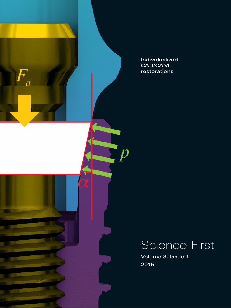
Cover picture: 3D rendering showing a precise fit between NobelProcera Abutment, NobelReplace Conical Connection implant and clinical screw. Selecting the matching abutment and using the dedicated clinical screw is crucial for system performance, since any small misfit can lead to extreme load and stress conditions and may result in system failure.
Introduction
Contents
Introduction Patients want their teeth restored 4The whole is greater than the sum of its parts 5History of NobelProcera® 7Advantages of CAD/CAM dentistry 8
Nobel Biocare CAD/CAM abutments
Scientific evidence 12On third-party implants 16Pivotal study 17Overview of studies 18
Nobel Biocare CAD/CAM implant bridges
Scientific evidence 20Pivotal study 21Overview of studies 22
Nobel Biocare CAD/CAM implant bars
Scientific evidence 27Overview of studies 29
Nobel Biocare CAD/CAM crowns and bridges
Scientific evidence 30Pivotal study 31
Cement vs. screw retention 32
References 35
Introduction
« Nobel Biocare is helping you to treat more patients
better than anyone else in the industry.”»
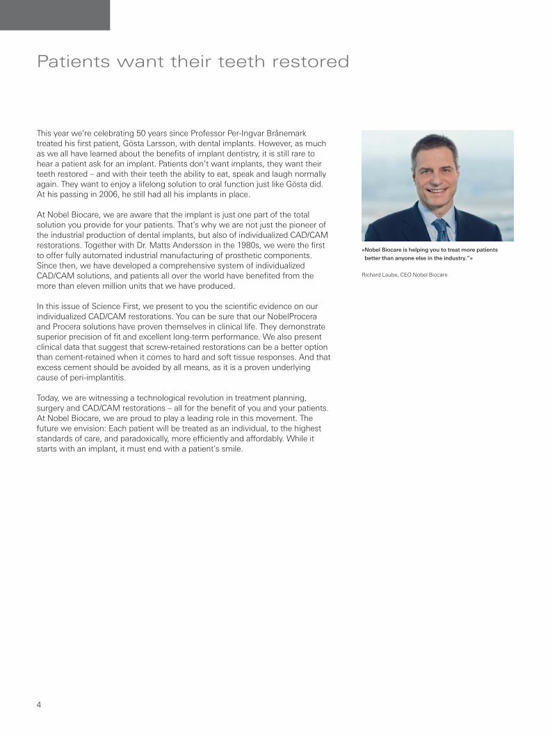
Richard Laube, CEO Nobel Biocare
Patients want their teeth restored
This year we’re celebrating 50 years since Professor Per-Ingvar Brånemark treated his first patient, Gösta Larsson, with dental implants. However, as much as we all have learned about the benefits of implant dentistry, it is still rare to hear a patient ask for an implant. Patients don’t want implants, they want their teeth restored – and with their teeth the ability to eat, speak and laugh normally again. They want to enjoy a lifelong solution to oral function just like Gösta did. At his passing in 2006, he still had all his implants in place.
At Nobel Biocare, we are aware that the implant is just one part of the total solution you provide for your patients. That’s why we are not just the pioneer of the industrial production of dental implants, but also of individualized CAD/CAM restorations. Together with Dr. Matts Andersson in the 1980s, we were the first to offer fully automated industrial manufacturing of prosthetic components. Since then, we have developed a comprehensive system of individualized CAD/CAM solutions, and patients all over the world have benefited from the more than eleven million units that we have produced.
In this issue of Science First, we present to you the scientific evidence on our individualized CAD/CAM restorations. You can be sure that our NobelProcera and Procera solutions have proven themselves in clinical life. They demonstrate superior precision of fit and excellent long-term performance. We also present clinical data that suggest that screw-retained restorations can be a better option than cement-retained when it comes to hard and soft tissue responses. And that excess cement should be avoided by all means, as it is a proven underlying cause of peri-implantitis.
Today, we are witnessing a technological revolution in treatment planning, surgery and CAD/CAM restorations – all for the benefit of you and your patients. At Nobel Biocare, we are proud to play a leading role in this movement. The future we envision: Each patient will be treated as an individual, to the highest standards of care, and paradoxically, more efficiently and affordably. While it starts with an implant, it must end with a patient’s smile.
Introduction
4

The whole is greater than the sum of its parts
Selecting the best implant-supported restorative solution for their patients is a key challenge for clinicians. For every restoration type there is a variety of manufacturers providing all types of components. Then there are the options offered by conventional casting, too. The resulting plethora of restorative solutions demands that every clinician navigates these options to meet the requirements of long-term performance, clinical safety, cost efficiency and patient satisfaction.
Designed and tested as part of a systemA key aspect of performance assessment is that a system is only as strong as its weakest link, and that the performance of any component depends not only on the component itself, but also on its interactions within the system. Conse-quently, the appropriate test of any component is as a part of that system. For this reason, Nobel Biocare conducts testing and research not only on separate components such as implants, abutments and screws, but always on the entire system, too. Nobel Biocare investigates systems from their design to the end user including assessment of: engineering and manufacturing processes, clinical research, quality assurance, and post-market surveillance. Only with this approach can the system function safely and reliably for many years.
Understanding the parameters that influence long-term performanceBoth theory (e.g. finite element analysis) and biomechanical testing indicate that several parameters can impact the performance of an implant system. These parameters include joint compression (the force that acts at the implant-abutment interface under loading conditions), preload (the tensile force keeping the pieces together) and friction coefficient (which depends on the surface materials that are in contact). In addition, there’s the force that the patient exerts on the system by chewing, as well as the length of the contact between the abutment and the implant. Plus, in a conical connection implant, the angle of the abutment within the implant cone. A small change in any of these pa-rameters, even one not visible to the eye, can lead to extreme load and stress conditions that result in system failure.
Precise fit maintains joint stabilityThe interface between implant and abutment is critical for joint stability. Manual adjustment of a cast or use of a substitute abutment can alter the contact angle and contact length. This can result in an undefined contact situation that could bring unknown risks to the patient. Conse quently, selecting the matching abutment is crucial for system performance, as it not only affects the fit of the restoration on the implant itself, but may also impact performance-relevant parameters.1
Precise fit ensures long-term performance
Joint compression (p) depends on a number of variables such as preload (tensile force Fa), friction angle (α) and contact length (l). Small changes in any of these parameters can lead to extreme load and stress conditions, which can cause implants to fracture.
Fa ∗ cos( ρ )∗ cos( )p=————————— dm ∗π ∗ l ∗ sin(ρ + )
α— 2 α— 2
➞➞➞➞
➞➞➞➞
➡ Fa
dmp
l
α
Introduction
5
Introduction
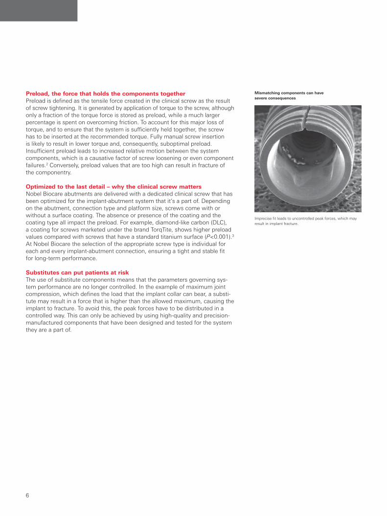
Mismatching components can have severe consequences
Imprecise fit leads to uncontrolled peak forces, which may result in implant fracture.
Preload, the force that holds the components togetherPreload is defined as the tensile force created in the clinical screw as the result of screw tightening. It is generated by application of torque to the screw, although only a fraction of the torque force is stored as preload, while a much larger percentage is spent on overcoming friction. To account for this major loss of torque, and to ensure that the system is sufficiently held together, the screw has to be inserted at the recommended torque. Fully manual screw insertion is likely to result in lower torque and, consequently, suboptimal preload. Insufficient preload leads to increased relative motion between the system components, which is a causative factor of screw loosening or even component failures.2 Conversely, preload values that are too high can result in fracture of the componentry.
Optimized to the last detail – why the clinical screw mattersNobel Biocare abutments are delivered with a dedicated clinical screw that has been optimized for the implant-abutment system that it’s a part of. Depending on the abutment, connection type and platform size, screws come with or without a surface coating. The absence or presence of the coating and the coating type all impact the preload. For example, diamond-like carbon (DLC), a coating for screws marketed under the brand TorqTite, shows higher preload values compared with screws that have a standard titanium surface (P<0.001).3 At Nobel Biocare the selection of the appropriate screw type is individual for each and every implant-abutment connection, ensuring a tight and stable fit for long-term performance.
Substitutes can put patients at riskThe use of substitute components means that the parameters governing sys-tem performance are no longer controlled. In the example of maximum joint compression, which defines the load that the implant collar can bear, a substi-tute may result in a force that is higher than the allowed maximum, causing the implant to fracture. To avoid this, the peak forces have to be distributed in a controlled way. This can only be achieved by using high-quality and precision-manufactured components that have been designed and tested for the system they are a part of.
Introduction
6
Introduction

In 1983, Dr. Matts Andersson first presented his groundbreaking innovation: fully automated industrial CAD/CAM* dental prosthetic production. Today, NobelProcera continues to lead the field as it delivers restorations of outstanding quality. Patients all over the world have benefited from the more than eleven million individualized units that have been delivered since the fabrication of the first coping over thirty years ago.
The roaring 80s of implant dentistryThe 1980s were a historic period for implant-based oral rehabilitation. The pub-lication of Professor Per-Ingvar Brånemark’s ten year follow-up clinical data in 1982 led to global acceptance of dental implants as a treatment method.4 In 1983, Professor Matts Andersson developed the Procera method of repeatable high-precision manufacturing for individualized dental restorations, beginning with titanium crowns. Nobelpharma, which would later become Nobel Biocare, saw the potential in Procera and acquired the technology in 1988. The break-through came with the production of all-ceramic crowns in 1989. Later, bridges, abutments and implant bridges in both titanium and ceramic followed.
From Procera to NobelProceraIn 2009, Procera was relaunched as NobelProcera. This saw the introduction of a new scanner offering unique optical scanning through conoscopic holo graphy, easy-to-use software and advanced centralized manufacturing. At the same time, fixed and fixed-removable overdenture bars were introduced. Today, NobelProcera offers the full range of screw- and cement-retained solutions – from single-unit to full-arch restorations, both for Nobel Biocare and other major implant systems.
Precision-manufacturing at its bestNobelProcera approaches the development of new products with advanced engineering, thorough verification, meticulous validation and specialized manufacturing techniques and tooling. The result: consistent precision of fit and exceptional product quality. All NobelProcera restorations are developed and produced according to the Medical Devices Quality Management System ISO 13485:2003. This means that all processes are regularly audited by the British Standards Institution (BSI), a notified body conducting a conformity assessment under the relevant EU Directives, and inspected by competent authorities such as the US Food and Drug Administration (FDA). This has established confidence that clinicians and patients always receive the best quality products.
In 1983, Professor Andersson developed the Procera method of repeatable high-precision manufacturing for dental restorations.
The first titanium coping was fabricated with the help of ordinary machines that are available in a toolmaker’s workshop.
Thorough quality control ensures that NobelProcera restora-tions are ready to use (production plant in Chiba, Japan).
History of NobelProcera®
* Computer-aided design / computer-aided manufacturing.
Introduction
7
Introduction
Nobel Biocare efficiently produces precise, durable and esthetic tooth- and implant-supported CAD/CAM prosthetics. Computer-aided design and manufacturing ensures precision of fit, while milling enables the use of high-strength, durable, and biocompatible materials. In addition, using CAD/CAM protocols reduces manual labor and removes the risks associated with the casting technique.
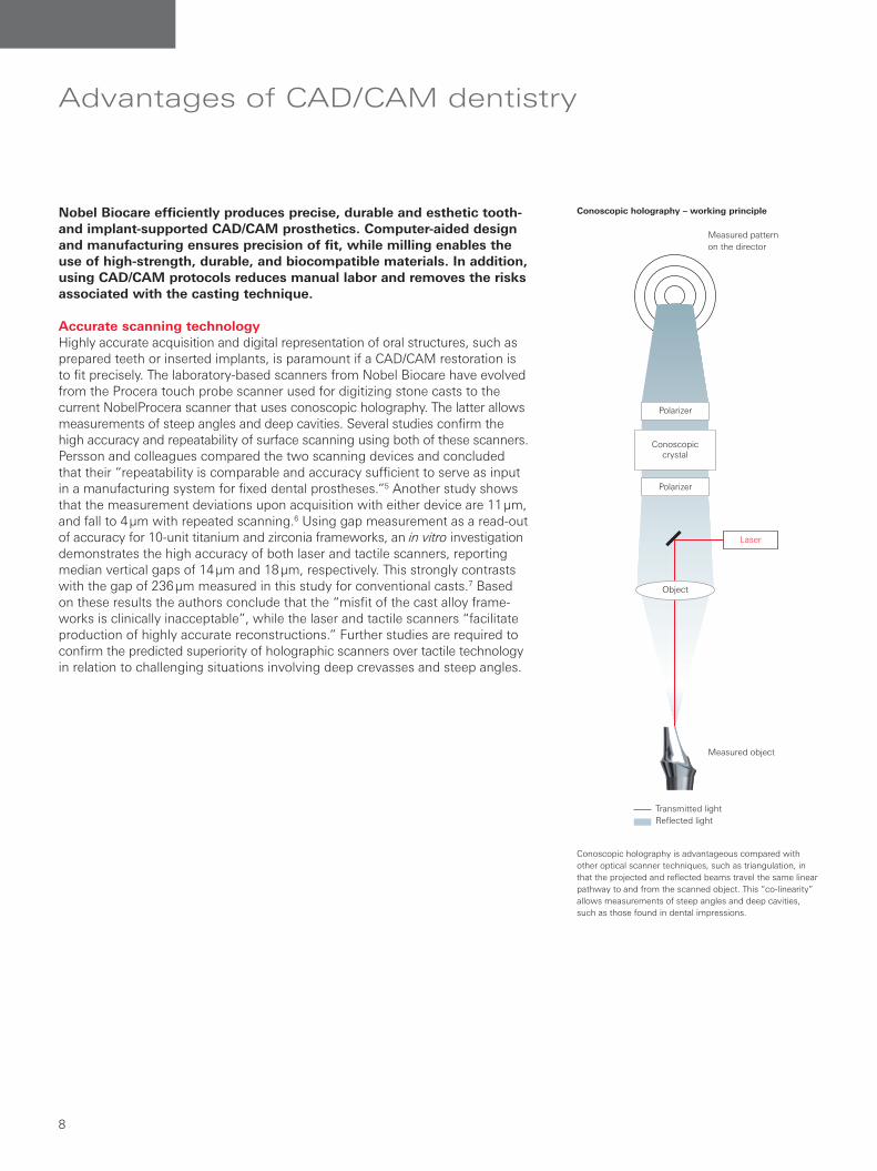
Accurate scanning technologyHighly accurate acquisition and digital representation of oral structures, such as prepared teeth or inserted implants, is paramount if a CAD/CAM restoration is to fit precisely. The laboratory-based scanners from Nobel Biocare have evolved from the Procera touch probe scanner used for digitizing stone casts to the current NobelProcera scanner that uses conoscopic holography. The latter allows measurements of steep angles and deep cavities. Several studies confirm the high accuracy and repeatability of surface scanning using both of these scanners. Persson and colleagues compared the two scanning devices and concluded that their “repeatability is comparable and accuracy sufficient to serve as input in a manufacturing system for fixed dental prostheses.”5 Another study shows that the measurement deviations upon acquisition with either device are 11 µm, and fall to 4 µm with repeated scanning.6 Using gap measurement as a read-out of accuracy for 10-unit titanium and zirconia frameworks, an in vitro investigation demonstrates the high accuracy of both laser and tactile scanners, reporting median vertical gaps of 14 μm and 18 µm, respectively. This strongly contrasts with the gap of 236 µm measured in this study for conventional casts.7 Based on these results the authors conclude that the “misfit of the cast alloy frame-works is clinically inacceptable”, while the laser and tactile scanners “facilitate production of highly accurate reconstructions.” Further studies are required to confirm the predicted superiority of holographic scanners over tactile technology in relation to challenging situations involving deep crevasses and steep angles.
Advantages of CAD/CAM dentistry
Conoscopic holography – working principle
Conoscopic holography is advantageous compared with other optical scanner techniques, such as triangulation, in that the projected and reflected beams travel the same linear pathway to and from the scanned object. This “co-linearity” allows measurements of steep angles and deep cavities, such as those found in dental impressions.
Measured pattern on the director
Measured object
Conoscopiccrystal
Laser
Polarizer
Object
Polarizer
Transmitted lightReflected light
Introduction
8
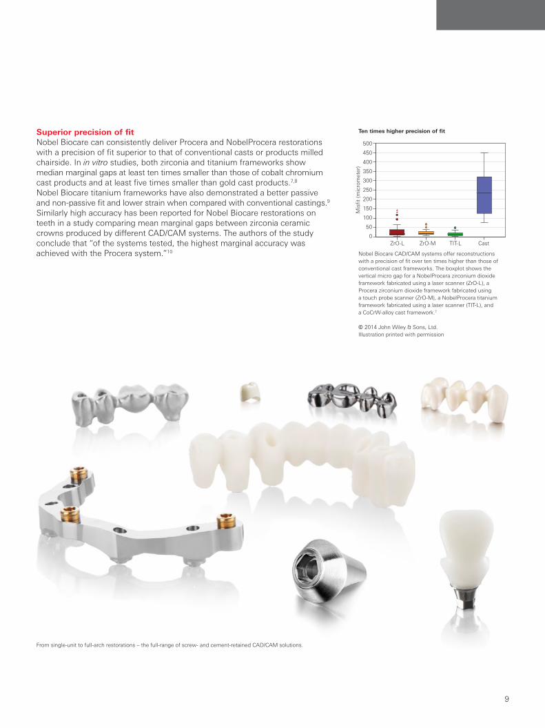
Nobel Biocare CAD/CAM systems offer reconstructions with a precision of fit over ten times higher than those of conventional cast frameworks. The boxplot shows the vertical micro gap for a NobelProcera zirconium dioxide framework fabricated using a laser scanner (ZrO-L), a Procera zirconium dioxide framework fabricated using a touch probe scanner (ZrO-M), a NobelProcera titanium framework fabricated using a laser scanner (TIT-L), and a CoCrW-alloy cast framework.7
© 2014 John Wiley & Sons, Ltd. Illustration printed with permission
Ten times higher precision of fitSuperior precision of fitNobel Biocare can consistently deliver Procera and NobelProcera restorations with a precision of fit superior to that of conventional casts or products milled chairside. In in vitro studies, both zirconia and titanium frameworks show median marginal gaps at least ten times smaller than those of cobalt chromium cast products and at least five times smaller than gold cast products.7,8 Nobel Biocare titanium frameworks have also demonstrated a better passive and non-passive fit and lower strain when compared with conventional castings.9 Similarly high accuracy has been reported for Nobel Biocare restorations on teeth in a study comparing mean marginal gaps between zirconia ceramic crowns produced by different CAD/CAM systems. The authors of the study conclude that “of the systems tested, the highest marginal accuracy was achieved with the Procera system.”10
From single-unit to full-arch restorations – the full-range of screw- and cement-retained CAD/CAM solutions.
(a)
(b)
Implant
Rel
ativ
e ef
fect
15 13 11 21 230.
00.
20.
40.
60.
81.
0
ZrO-LZrO-MTIT-LCast
500
450
400
350
300
250
200
150
100
50
0
Mis
fit (m
icro
met
er)
ZrO-L ZrO-M TIT-L Cast
Introduction
9
Introduction

Excellent strength in vitroCAD/CAM technology has introduced individualized prosthetics made from materials such as titanium or zirconia, the use of which is limited in traditional laboratory-based workflows. Titanium was the first raw material used in the Procera manufacturing process. Since its market entry in 1984, it has remained the gold standard due to high strength and biocompatibility.11 Over the last few years, an ever-increasing demand for esthetic properties has paved the way for ceramics such as zirconia, which offers both durability and tooth-like color.12 A number of independent investigations have demonstrated the excellent raw material strength of titanium and zirconia used by the Nobel Biocare CAD/CAM technology.13–25 Although considerable variations in fracture load can be observed between the different studies, comparative studies reveal that Nobel Biocare materials have an equivalent or superior strength to that of conventional cast materials.21,23
Nobel Biocare restorations maintain outstanding strength after exposure to fatigue stress in an artificial oral environment. Att and colleagues performed a series of in vitro tests aimed at evaluating fracture load after thermo-mechanical cycling set to mimic five years of function. The two studies demonstrate that all restorations, including titanium and zirconia, “exceeded the minimum limits of the fracture resistance for anterior restorations.”15,16
Exceptional durability in a clinical settingAs expected, use of stronger materials and individualized design has a marked positive influence on the strength and durability of CAD/CAM restorations in the clinical setting. Improved strength and durability have been reported for various Nobel Biocare frameworks, including implant bridges and implant bars. In a comparison of 10-unit titanium frameworks with gold alloy cast, the 5-year prosthesis survival rate was 100.0% vs. 97.1%, respectively.26 In addition, patients with Nobel Biocare restorations needed fewer appointments and experienced significantly fewer phonetic problems, fewer fistulas, fewer veneer fractures and no implant failures. Plus, a lower number of patients had their prostheses temporarily removed for adjustments.26 Similar results have been demonstrated in a study comparing conventional and Nobel Biocare CAD/CAM implant bar-retained overdentures, where CAD/CAM restorations experienced a significant reduction in technical complications.27,28 Fewer complications during the follow-up period have also been reported by Moberg and colleagues. They investigated Procera titanium frameworks supported by Nobel Biocare implants in comparison with conventional cast titanium frameworks supported by an alternative implant system.29
Complications recorded during a 3-year follow-up of a randomized prospective study with 40 edentulous patients treated with either Nobel Biocare implants and Procera frameworks or an alternative implant system with conventional titanium cast frameworks.29
Nobel Biocare CAD/CAM titanium frameworks are associated with fewer technical and biological complications
12
Technical complications
Acrylic fracture
Acrylic tooth loss
Filling loss
Biological complications
Hyperplasia
Adjustment of bridge/mucosa space
Peri-implant bone reduction
1
1
1
1
0
2
2
4
3
3
Brånemark System with Procera framework (n=20)
Alternative implant system with cast titanium framework (n=20)
Introduction
10

BiocompatibilityAll Nobel Biocare medical devices are made from biocompatible materials. Un-alloyed grade 2 titanium and the grade 5 alloyed titanium (Ti-6Al-4V) have both shown resistance to corrosion and a limited ion release in response to contact with a live environment. This results in low ion leakage and favorable tissue re-sponse including osseointegration.11,30 Similarly, zirconia has shown biocompati-bility in vitro and in vivo.12,30
Biocompatibility of restorative materials plays an important role not only in osseointegration, but also with respect to appropriate soft tissue attachment. It also influences the adhesion of bacteria. Mustafa and colleagues report that the adhesion and activity of human gingival fibroblasts is greater on industrially manufactured zirconia in comparison with polished and veneered structures.31
Bacterial adhesion is believed to be part of the first step both in biofilm formation and in initiation of an inflammatory response that could possibly lead to bone resorption and implant failure.32 In vitro tests demonstrated that the numbers of bacteria adhering to saliva- or saliva-plus-serum-covered surfaces of titanium, zirconia and hydroxyapatite (an enamel surrogate) are comparable. This led the authors to conclude that zirconia is “suitable material for manufacturing implant abutments with biological properties similar to titanium.”33
The results of clinical studies support the in vitro findings on the biocompatibility of CAD/CAM materials. A report of fifty clinical cases with a simplified tech-nique for reconstructing emergence profiles during implant restoration using Nobel Biocare Abutments in titanium and zirconia shows that, when these abutments are used at the provisional crown stage, the restorations exhibit excellent esthetics and healthy gingival tissues.34
Less chair time and fewer clinical visitsUse of CAD/CAM technology has led to a significant shortening of chair time during the prosthetic procedure and a significant reduction in the number of follow-up appointments. In a retrospective study comparing two patient cohorts, one with gold alloy cast frameworks and the other with Nobel Biocare CAD/CAM titanium restorations, the authors demonstrate that patients under-going conventional treatment had to attend more clinical appointments, and that the mean time for completion of their permanent prosthesis was almost 60% longer.26 The authors largely attribute these changes to the improved fit associated with the computer-aided design and production, as well as to the high durability of the materials.
Mean number of clinical visits per patient during the follow-up period. Nobel Biocare CAD/CAM titanium frameworks are associated with fewer follow-up visits due to higher precision of fit and stronger materials.26
Fewer follow-up visits
Gold alloy castNobel Biocare titanium framework
1 year
2 years
3 years
4 years
5 years
0 2 4 6 8 10Number of visits
Introduction
11
Introduction
Nobel Biocare CAD/CAM abutments are individualized solutions that combine long-term clinical stability with high esthetic results. This is due to their wide versatility, homogenous and biocompatible materials, and anatomic design.
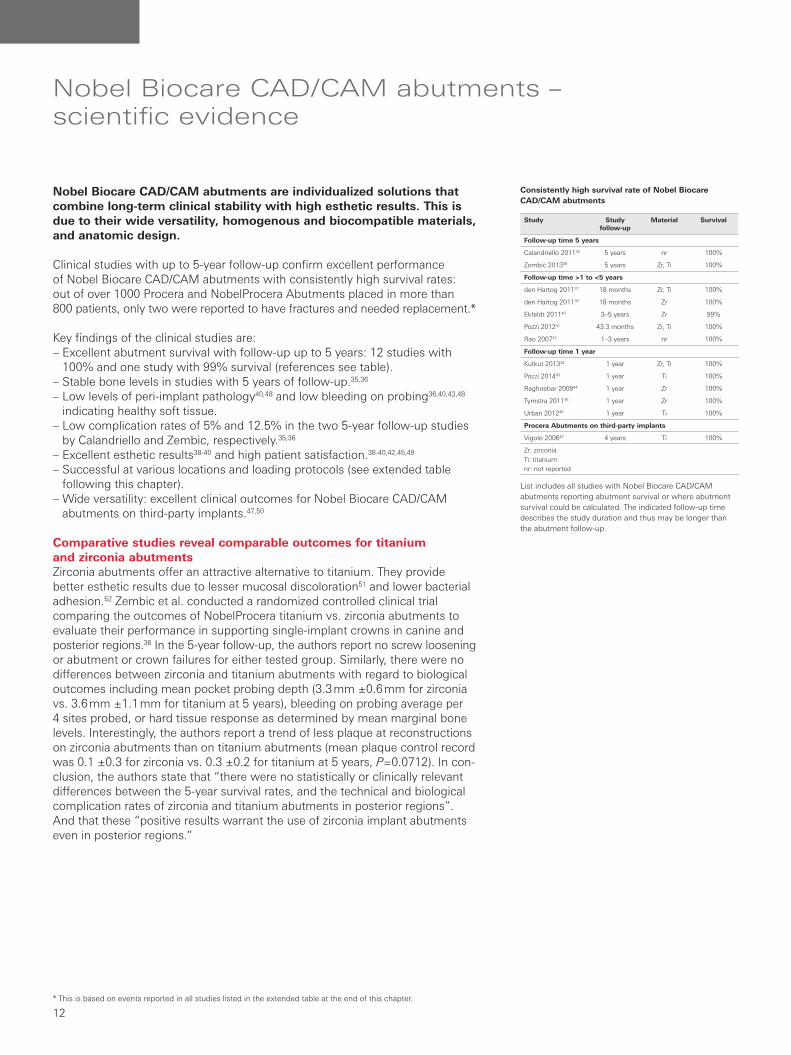
Clinical studies with up to 5-year follow-up confirm excellent performance of Nobel Biocare CAD/CAM abutments with consistently high survival rates: out of over 1000 Procera and NobelProcera Abutments placed in more than 800 patients, only two were reported to have fractures and needed replacement.*
Key findings of the clinical studies are:– Excellent abutment survival with follow-up up to 5 years: 12 studies with
100% and one study with 99% survival (references see table).– Stable bone levels in studies with 5 years of follow-up.35,36
– Low levels of peri-implant pathology40,48 and low bleeding on probing36,40,43,48 indicating healthy soft tissue.
– Low complication rates of 5% and 12.5% in the two 5-year follow-up studies by Calandriello and Zembic, respectively.35,36
– Excellent esthetic results38-40 and high patient satisfaction.38-40,42,45,49
– Successful at various locations and loading protocols (see extended table following this chapter).
– Wide versatility: excellent clinical outcomes for Nobel Biocare CAD/CAM abutments on third-party implants.47,50
Comparative studies reveal comparable outcomes for titanium and zirconia abutmentsZirconia abutments offer an attractive alternative to titanium. They provide better esthetic results due to lesser mucosal discoloration51 and lower bacterial adhesion.52 Zembic et al. conducted a randomized controlled clinical trial comparing the outcomes of NobelProcera titanium vs. zirconia abutments to evaluate their performance in supporting single-implant crowns in canine and posterior regions.36 In the 5-year follow-up, the authors report no screw loosening or abutment or crown failures for either tested group. Similarly, there were no differences between zirconia and titanium abutments with regard to biological outcomes including mean pocket probing depth (3.3 mm ±0.6 mm for zirconia vs. 3.6 mm ±1.1 mm for titanium at 5 years), bleeding on probing average per 4 sites probed, or hard tissue response as determined by mean marginal bone levels. Interestingly, the authors report a trend of less plaque at reconstructions on zirconia abutments than on titanium abutments (mean plaque control record was 0.1 ±0.3 for zirconia vs. 0.3 ±0.2 for titanium at 5 years, P=0.0712). In con-clusion, the authors state that “there were no statistically or clinically relevant differences between the 5-year survival rates, and the technical and biological complication rates of zirconia and titanium abutments in posterior regions”. And that these “positive results warrant the use of zirconia implant abutments even in posterior regions.”
Nobel Biocare CAD/CAM abutments – scientific evidence
* This is based on events reported in all studies listed in the extended table at the end of this chapter.
List includes all studies with Nobel Biocare CAD/CAM abutments reporting abutment survival or where abutment survival could be calculated. The indicated follow-up time describes the study duration and thus may be longer than the abutment follow-up.
Consistently high survival rate of Nobel Biocare CAD/CAM abutments
Study Study follow-up
Material Survival
Follow-up time 5 years
Calandriello 201135 5 years nr 100%
Zembic 201336 5 years Zr, Ti 100%
Follow-up time >1 to <5 years
den Hartog 201137 18 months Zr, Ti 100%
den Hartog 201139 18 months Zr 100%
Ekfeldt 201140 3–5 years Zr 99%
Pozzi 201241 43.3 months Zr, Ti 100%
Rao 200742 1–3 years nr 100%
Follow-up time 1 year
Kutkut 201334 1 year Zr, Ti 100%
Pozzi 201443 1 year Ti 100%
Raghoebar 200944 1 year Zr 100%
Tymstra 201145 1 year Zr 100%
Urban 201246 1 year Ti 100%
Procera Abutments on third-party implants
Vigolo 200647 4 years Ti 100%
Zr: zirconiaTi: titanium nr: not reported
Abutments
12

Excellent functional and esthetic outcomesAnother clinical study with Nobel Biocare CAD/CAM abutments in zirconia used for single-tooth restorations, mostly in the anterior maxilla, reports low rates of both technical and biological complications at 1-year follow-up.40 25 patients with 40 abutments underwent an evaluation with a longer follow-up of 3–5 years, which confirmed the good performance of zirconia abutments. The peri-implant bone level on all measurable implants was 0.16 mm ±0.72 mm (0.29 mm ±0.87 mm on randomly selected 25 implants). The mean bleeding on probing was slightly higher around the implant-supported restorations than at the mesial, but not the distal, neighboring teeth (0.18 ±0.2 vs. 0.07 ±0.11, P=0.0199; and vs. 0.14 ±0.27, P=0.5545). The esthetic outcomes were assessed as excellent (73%) or good (27%). The authors conclude that “zirconia abutments for single-implant crowns seem to demonstrate good short-term technical and biological results.”
Cement- and screw-retained solutionsClinical studies confirm excellent outcomes for zirconia and titanium abut-ments with both cement- and screw-retention systems. A recent report from a randomized clinical trial with single-tooth implants in the anterior jaw includes 38 screw-retained and 53 cement-retained restorations. It reports a 100% abutment and restoration survival rate as well as good performance in terms of function and esthetics. In addition, the study shows high patient satisfaction (score 9.0 ±1.0 out of maximum 10) after 18 months of follow-up.37,38
One-piece solution: screw-retained crowns for direct veneeringSeveral clinical studies used screw-retained crowns for direct veneering. They demonstrate promising clinical outcomes in short-term follow-up reports.37-40,45,49 Ekfeldt and colleagues conducted a retrospective evaluation of the records of 130 patients with 185 single-tooth implant restorations, 90 of which had the veneering porcelain baked directly to the zirconia abutment. At the 1-year follow-up, implant and abutment survival rates were both 99%, and the rates of complications were low. The authors conclude that “there were no significant differences in changes for any of the soft tissue registrations or the peri-implant marginal bone level” between the conventional two-piece abutment-crown restoration and the one-piece solution.40
A Agenesis of tooth 22 and small peg-shaped tooth 12. B Zirconia abutment with porcelain baked to the abutment;
palatal view. C Front view at 1-year examination: a single-implant
restoration region 22, a ceramic veneer on tooth 12. D Radiograph taken at the insertion of the restoration.
© 2011 John Wiley & Sons, Ltd.Illustrations printed with permission
Excellent esthetic results: screw-retained crowns for direct veneering40
A
C
B
D
Abutments
13
Abutments

Healthy soft tissueCustom abutments offer an individualized contour and emergence profile and are able to provide good soft tissue support. Clinical studies that evaluate soft tissue outcomes with NobelProcera and Procera Abutments confirm these proposed advantages of CAD/CAM abutments.– In three studies reporting plaque accumulation, 236 out of the summed
242 investigated sites had no visible plaque.37,39,43
– Esthetic analysis was conducted in three studies, with pink esthetic score (PES)53 mean values ranging from 6.3 ±1.7 to 7.1 ±1.5 (where 0 is the minimum, and 10 is the maximum and denotes healthy soft tissue). Satisfactory ICAI (implant crown aesthetic index54) mucosa was reported in three studies and ranged from 56.6% to 100%.38–40
– In all studies using Nobel Biocare CAD/CAM abutments (18 studies, 1146 implants, 1061 abutments), peri-implantitis and peri-implant mucositis, as defined by authors, is reported in 336,55 and in 11 patients,40,48 respectively.
– Bleeding on probing ranged from 0 to 1.4 ±0.7536,40,43,48 and pocket probing depth ranged from 2.2 mm ±0.84 mm to 5.3 mm ±1.5 mm.36,37,39,45–49
High patient satisfactionExcellent clinical outcomes combined with good esthetic results lead to high patient satisfaction, as evidenced by the studies with Nobel Biocare CAD/CAM abutments that assess patient responses.– Two randomized clinical trials comparing different implant designs using
titanium and zirconia abutments in the esthetic zone of 133 patients report high satisfaction of 84.5 out of 100 and 9.0 out of 10 on two visual analog scales.38,49
– Another randomized clinical trial comparing different loading protocols using zirconia abutments reports high patient satisfaction of 92.7% (immediate loading) and 89.0% (delayed loading) after 18 months of follow-up.39
– A clinical 3- to 5-year follow-up of 25 patients with 40 single-tooth restorations with zirconia abutments reports esthetic patient satisfaction of 90% (median 100%) and functional patient satisfaction of 94% (median 100%).40
– A pilot study with 10 patients who were missing two adjacent teeth in the maxillary esthetic zone reports very high patient satisfaction with an average score of 9.0 (out of 10) on a visual analog scale.45
– A prospective study reports that the 46 patients found the esthetic and functional results excellent (95.6%) or good (4.3%). The authors state that “a general impression of satisfaction of the patients was observed, as they expressed amazement over the absence of symptoms.”42
Definition of the esthetic assessment using the pink esthetic score (PES)53
21
4
3
5
PES evaluates five variables: mesial papilla (1), distal papilla (2), curvature of facial mucosa (3), level of facial mucosa (4), and root convexity/soft tissue color and texture (5). During an assessment each variable is assigned a score of 0, 1, or 2, with 0 being absent or having major discrepancy, 1 being incomplete or having minor discrepancy, and 2 being com-plete or having no discrepancy. Under optimum conditions all these scores add up to 10. The threshold of clinical acceptability is set at 6.53
Patients are highly satisfied with the protocols involving Nobel Biocare CAD/CAM abutments. Different questionnaires may have been used by different studies.
High patient satisfaction
Mea
n le
vel o
f sa
tisfa
ctio
n (%
) 100
80
60
40
20
0
Study den Hartog38
den Hartog37
Ekfeldt40 Rao42 Tymstra45 Tymstra49
Patients 93 62 25 46 10 40
Material Zi, Ti Zi Zi nr Zi Zi, Ti
* Immediate loading** Delayed loading# Esthetic satisfaction§ Functional satisfactionZi: zirconiaTi: titaniumnr: not reported
* ** #§
Abutments
14
Engineered to be effectiveKey findings from the in vitro experiments on Nobel Biocare CAD/CAM abutments assessing strength, durability and consistent precision of fit:– Nobel Biocare abutments show comparable or superior fracture load and
bending moments in nine in vitro studies with various protocols. These include after aging and comparisons with stock abutments.56–65
– Detorque values of zirconia abutments do not change with increasing loading cycles. This suggests high system stability and resistance to screw loosening.66
– Rotational freedom between implant and abutment ranges from 2.01° to 4.13°,59,67–69 with all values falling below the threshold of 5°, excess of which is associated with screw loosening.70
– Mean micro gaps between Nobel Biocare CAD/CAM abutments and supporting implants range from 0.06 μm to 10.5 μm.67,71–75
Points to consider when working with Nobel Biocare CAD/CAM abutments– Fracture load of zirconia abutments is not affected by manual grinding as
long as the appropriate guidelines are followed (stress-free preparation with water cooling and using fine-grained cutting diamonds).57 Manual adjustment of the abutment at the implant-abutment interface should be avoided, as this can lead to misfit. This problem was experienced by Gigandet and colleagues who had manually adjusted the Procera Abutment and consequently could not investigate its rotational play.59
– Metallic inserts in the zirconia abutments increase their strength.64
– As expected due to material strength characteristics, titanium abutments are stronger than zirconia abutments in in vitro testing (fracture load). However, both meet the strength requirements for clinical use.62,65
– To minimize potential bacterial leakage and ensure long-term stability of the prosthesis, abutments should be tightened to manufacturer-recommended torque levels.1,74
– For proper seating of the screw head, use the original screws provided with the Nobel Biocare abutments.76
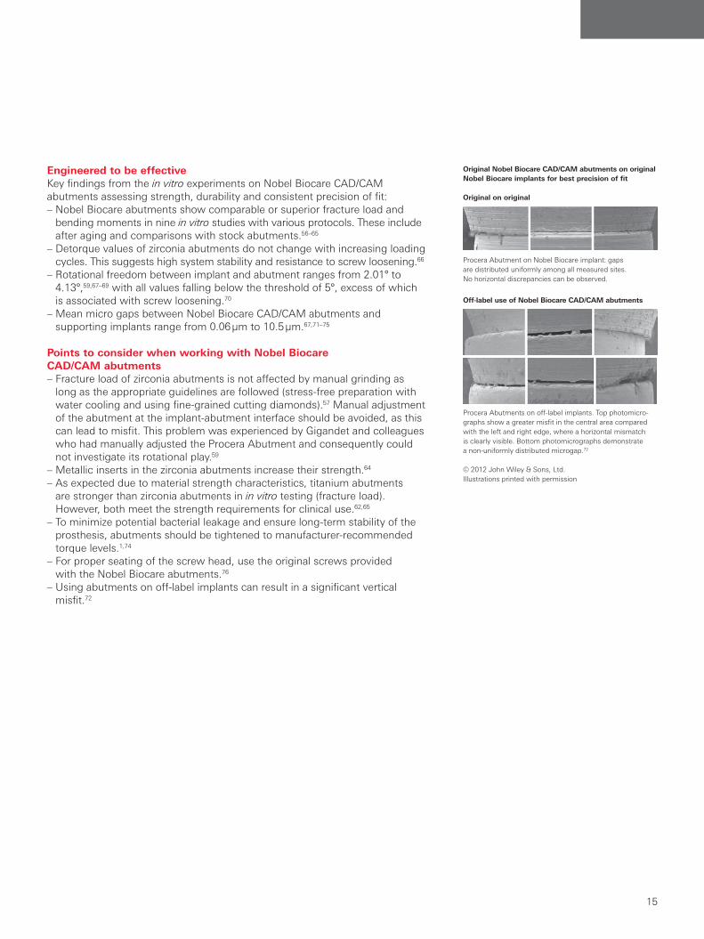
– Using abutments on off-label implants can result in a significant vertical misfit.72
Original Nobel Biocare CAD/CAM abutments on original Nobel Biocare implants for best precision of fit
Original on original
Off-label use of Nobel Biocare CAD/CAM abutments
Procera Abutment on Nobel Biocare implant: gaps are distributed uniformly among all measured sites. No horizontal discrepancies can be observed.
Procera Abutments on off-label implants. Top photomicro-graphs show a greater misfit in the central area compared with the left and right edge, where a horizontal mismatch is clearly visible. Bottom photomicrographs demonstrate a non-uniformly distributed microgap.72
© 2012 John Wiley & Sons, Ltd.Illustrations printed with permission
Abutments
15
Abutments
Nobel Biocare CAD/CAM abutments on third-party implants are engineered to provide a precise fit. The outstanding quality and excellent performance of these abutments is confirmed by both in vitro testing and clinical investigations.
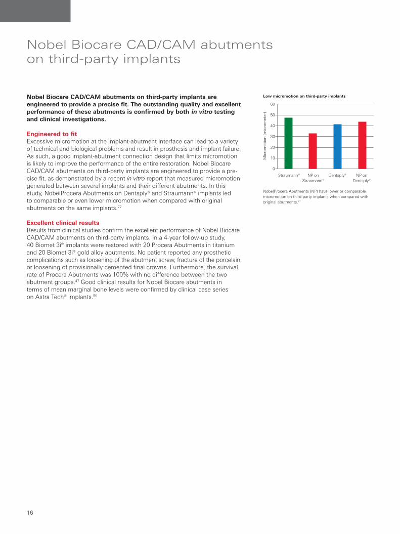
Engineered to fitExcessive micromotion at the implant-abutment interface can lead to a variety of technical and biological problems and result in prosthesis and implant failure. As such, a good implant-abutment connection design that limits micromotion is likely to improve the performance of the entire restoration. Nobel Biocare CAD/CAM abutments on third-party implants are engineered to provide a pre-cise fit, as demonstrated by a recent in vitro report that measured micromotion generated between several implants and their different abutments. In this study, NobelProcera Abutments on Dentsply® and Straumann® implants led to comparable or even lower micromotion when compared with original abutments on the same implants.77
Excellent clinical resultsResults from clinical studies confirm the excellent performance of Nobel Biocare CAD/CAM abutments on third-party implants. In a 4-year follow-up study, 40 Biomet 3i® implants were restored with 20 Procera Abutments in titanium and 20 Biomet 3i® gold alloy abutments. No patient reported any prosthetic complications such as loosening of the abutment screw, fracture of the porcelain, or loosening of provisionally cemented final crowns. Furthermore, the survival rate of Procera Abutments was 100% with no difference between the two abutment groups.47 Good clinical results for Nobel Biocare abutments in terms of mean marginal bone levels were confirmed by clinical case series on Astra Tech® implants.50
Nobel Biocare CAD/CAM abutments on third-party implants
NobelProcera Abutments (NP) have lower or comparable micromotion on third-party implants when compared with original abutments.77
Low micromotion on third-party implants
Straumann® NP on Straumann®
Dentsply® NP on Dentsply®
Mic
rom
otio
n (m
icro
met
er)
60
50
40
30
20
10
0
Abutments
16

Original abstract
Objectives: To test the survival rates, and the technical and biological complication rates of customized zirconia and titanium abutments 5 years after crown insertion.Materials and methods: Twenty-two patients with 40 single implants in maxillary and mandibular canine and posterior regions were included. The implant sites were randomly assigned to zirconia abutments supporting all-ceramic crowns or titanium abutments supporting metal-ceramic crowns. Clinical examinations were performed at baseline, and at 6, 12, 36 and 60 months of follow-up. The abutments and reconstructions were examined for technical and/or biological complications. Probing pocket depth (PPD), plaque control record (PCR) and Bleeding on Probing (BOP) were assessed at abutments (test) and analogous contralateral teeth (control). Radiographs of the implants revealed the bone level (BL) on mesial (mBL) and distal sides (dBL). Data were statistically analyzed with nonparametric mixed models provided by Brunner and Langer and STATA (P < 0.05).
Results: Eighteen patients with 18 zirconia and 10 titanium abutments were available at a mean follow-up of 5.6 years (range 4.5–6.3 years). No abutment fracture or loss of a recon-struction occurred. Hence, the survival rate was 100% for both. Survival of implants supporting zirconia abutments was 88.9% and 90% for implants supporting titanium abutments. Chipping of the veneering ceramic occurred at three metal- ceramic crowns supported by titanium abutments. No signifi-cant differences were found at the zirconia and titanium abutments for PPD (mean PPD ZrO2 3.3 ± 0.6 mm, mPPD Ti 3.6 ± 1.1 mm), PCR (mPCR ZrO2 0.1 ± 0.3, mPCR Ti 0.3 ± 0.2) and BOP (mBOP ZrO2 0.5 ± 0.3, mBOP Ti 0.6 ± 0.3). Moreover, the BL was similar at implants supporting zirconia and titanium abutments (mBL ZrO2 1.8 ± 0.5, dBL ZrO2 2.0 ± 0.8; mBL Ti 2.0 ± 0.8, dBL Ti 1.9 ± 0.8).Conclusions: There were no statistically or clinically relevant differences between the 5-year survival rates, and the techni-cal and biological complication rates of zirconia and titanium abutments in posterior regions.
Five-year results of a randomized controlled clinical trial comparing zirconia and titanium abutments supporting single-implant crowns in canine and posterior regions
Zembic A, Bosch A, Jung RE, Hammerle CH, Sailer IClin Oral Implants Res. 2013;24:384-390
Clinical and radiographical 5-year follow-up of an all-ceramic crown (ACC) on a zirconia abutment in region 14. Apparent buccal recessions on both neighboring teeth.
Clinical and radiographical 5-year follow-up of a metal-ceramic crown (MCC) on a titanium abutment in region 24.
© 2013 John Wiley & Sons, Ltd. Printed with permission
wileyonlinelibrary.com/journal/clr CLINICAL ORAL IMPLANTS RESEARCH
O F F I C I AL P U B L I CAT I O N O F T H E E U R O P EAN AS S O C I AT I O N F O R O S S EO I N T EG R AT I O N
Editor-in-Chief
Niklaus P. Lang, Switzerland
Associate Editors
T. Berglundh, Sweden
G. E. Salvi, Switzerland
H. P. Weber, USA
Clinical Research
Tissue Physiology
Wound Healing
Microbiology
Material Sciences
Prosthodontic Research
Occlusion of Oral Implants
Vol 24 • Issue No. 4 • April 2013
CL
INIC
AL
OR
AL
IMP
LA
NT
S RE
SEA
RC
HV
olume 24:4 • 2013 • (pp. 355–474)
CLINICALORAL IMPLANTSRESEARCH
Volume 24 · Number 4 · April 2013
Contents
Clinical Oral Implants Research is covered by Current Contents®/Clinical Medicine,the Science Citation Index®, and SciSearch®.
Printed in MalaysiaISSN:0905-7161
This journal is available online at Wiley Online Library. Visit wileyonlinelibrary.com/journal/clr to search the articles and register for table of contents e-mail alerts.
Review ArticleLong-term survival of calcium phosphate-coated dental implants: a meta-analytical approach to the clinical literature
Original ArticlesExperimental periodontitis and peri-implantitis in dogs
Bone tissue in different parts of the edentulous maxilla and mandible
In vitro assessment of artifacts induced by titanium dental implants in cone beam computed tomography
Five-year results of a randomized controlled clinical trial comparing zirconia and titanium abutments supporting single-implant crowns in canine and posterior regions
The impact of dis-/reconnection of laser microgrooved and machined implant abutments on soft- and hard-tissue healing
Histological evaluation at different times after augmentation of extraction sites grafted with a magnesium-enriched hydroxyapatite: double-blinded randomized controlled trial
Buccal bone remodeling after tooth extraction using the fl apless approach with and without synthetic bone grafting. A histomorphometric study in dogs
Histomorphometrical and molecular evaluation of endosseous dental implants sites in humans: correlation with clinical and radiographic aspects
Serum bone formation marker correlation with improved osseointegration in osteoporotic rats treated with simvastatin
A prospective study on implants installed with fl apless-guided surgery using the all-on-four concept in the mandible
Variation in arterial supply to the fl oor of the mouth and assessment of relative hemorrhage risk in implant surgery
Comparative evaluation of different calcium phosphate-based bone graft granules – an in vitro study with osteoblast-like cells
Bio-Oss® blocks combined with BMP-2 and VEGF for the regeneration of bony defects and vertical augmentation
A novel technique for tailored surface modifi cation of dental implants – a step wise approach based on plasma immersion ion implantation
Osteotome technique with injectable tissue-engineered bone and simultaneous implant placement by cell therapy
355 B. A. J. A. van Oirschot, E. M. Bronkhorst, J. J. J. P. van den Beucken, G. J. Meijer, J. A. Jansen & R. Junker
363 O. Carcuac, I. Abrahamsson, J.-P. Albouy, E. Linder, L. Larsson & T. Berglundh
372 J. Lindhe, E. Bressan, D. Cecchinato, E. Corrá, M. Toia & B. Liljenberg
378 G. I. Benic, M. Sancho-Puchades, R. E. Jung, H. Deyhle & C. H. F. Hämmerle
384 A. Zembic, A. Bösch, R. E. Jung, C. H. F. Hämmerle & I. Sailer
391 G. Iglhaut, K. Becker, V. Golubovic, H. Schliephake & I. Mihatovic
398 L. Canullo, F. Heinemann, T. Gedrange, R. Biffar & C. Kunert-Keil
407 F. Suaid, M. F. M. Grisi, S. L. S. Souza, D. B. Palioto, M. Taba Jr & A. B. Novaes Jr
414 A. C. Pereira, P. P. C. Souza, J. A. C. Souza, T. A. Silva, A. C. Batista & R. F. Ribeiro-Rotta
422 Z. Du, J. Chen, F. Yan, N. Doan, S. Ivanovski & Y. Xiao
428 R. A. Landázuri-Del Barrio, J. Cosyn, W. N. De Paula, H. De Bruyn & E. Marcantonio Jr
434 Y. Katsumi, R. Tanaka, T. Hayashi, T. Koga, R. Takagi & H. Ohshima
441 A. Bernhardt, A. Lode, F. Peters & M. Gelinsky
450 C. Schmitt, R. Lutz, H. Doering, M. Lell, J. Ratky & K. A. Schlegel
461 L. Meirelles, E. T. Uzumaki, J. H. C. Lima, C. A. Muller, T. Albrektsson, A. Wennerberg & C. S. Lambert
468 Y. Yamada, S. Nakamura, M. Ueda & K. Ito
available online at wileyonlinelibrary.com/journal/clr
Abutments
17
Abutments
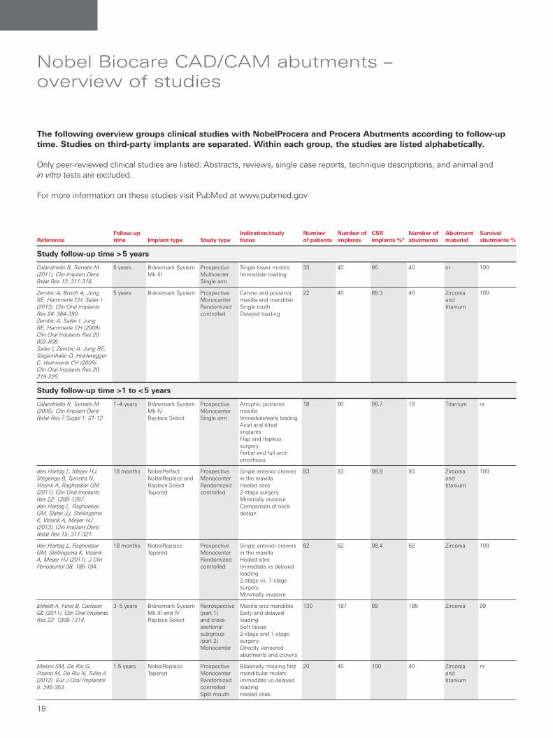
Nobel Biocare CAD/CAM abutments – overview of studies
The following overview groups clinical studies with NobelProcera and Procera Abutments according to follow-up time. Studies on third-party implants are separated. Within each group, the studies are listed alphabetically.
Only peer-reviewed clinical studies are listed. Abstracts, reviews, single case reports, technique descriptions, and animal and in vitro tests are excluded.
For more information on these studies visit PubMed at www.pubmed.gov
ReferenceFollow-up time Implant type Study type
Indication/study focus
Number of patients
Number of implants
CSR implants %*
Number of abutments
Abutment material
Survival abutments %
Study follow-up time > 5 years
Calandriello R, Tomatis M (2011). Clin Implant Dent Relat Res 13: 311-318.
5 years Brånemark System Mk III
ProspectiveMulticenterSingle arm
Single lower molarsImmediate loading
33 40 95 40 nr 100
Zembic A, Bosch A, Jung RE, Hammerle CH, Sailer I (2013). Clin Oral Implants Res 24: 384-390.Zembic A, Sailer I, Jung RE, Hammerle CH (2009). Clin Oral Implants Res 20: 802-808.Sailer I, Zembic A, Jung RE, Siegenthaler D, Holderegger C, Hammerle CH (2009). Clin Oral Implants Res 20: 219-225.
5 years Brånemark System ProspectiveMonocenterRandomized controlled
Canine and posterior maxilla and mandibleSingle toothDelayed loading
22 40 89.3 40 Zirconia and titanium
100
Study follow-up time >1 to < 5 years
Calandriello R, Tomatis M (2005). Clin Implant Dent Relat Res 7 Suppl 1: S1-12
1–4 years Brånemark System Mk IVReplace Select
ProspectiveMonocenterSingle arm
Atrophic posterior maxillaImmediate/early loadingAxial and tilted implantsFlap and flapless surgeryPartial and full-arch prosthesis
18 60 96.7 19 Titanium nr
den Hartog L, Meijer HJ, Stegenga B, Tymstra N, Vissink A, Raghoebar GM (2011). Clin Oral Implants Res 22: 1289-1297.den Hartog L, Raghoebar GM, Slater JJ, Stellingsma K, Vissink A, Meijer HJ (2013). Clin Implant Dent Relat Res 15: 311-321.
18 months NobelPerfect NobelReplace and Replace Select Tapered
ProspectiveMonocenterRandomizedcontrolled
Single anterior crowns in the maxillaHealed sites 2-stage surgeryMinimally invasive Comparison of neck design
93 93 98.9 93 Zirconia and titanium
100
den Hartog L, Raghoebar GM, Stellingsma K, Vissink A, Meijer HJ (2011). J Clin Periodontol 38: 186-194.
18 months NobelReplace Tapered
ProspectiveMonocenterRandomizedcontrolled
Single anterior crowns in the maxillaHealed sites Immediate vs delayed loading2-stage vs. 1-stage surgeryMinimally invasive
62 62 98.4 62 Zirconia 100
Ekfeldt A, Furst B, Carlsson GE (2011). Clin Oral Implants Res 22: 1308-1314.
3–5 years Brånemark System Mk III and IVReplace Select
Retrospective (part 1) and cross-sectional subgroup (part 2)Monocenter
Maxilla and mandible Early and delayed loadingSoft tissue2-stage and 1-stage surgeryDirectly veneered abutments and crowns
130 187 99 185 Zirconia 99
Meloni SM, De Riu G, Pisano M, De Riu N, Tullio A (2012). Eur J Oral Implantol 5: 345-353.
1.5 years NobelReplace Tapered
ProspectiveMonocenterRandomizedcontrolledSplit mouth
Bilaterally missing first mandibular molarsImmediate vs delayed loadingHealed sites
20 40 100 40 Zirconia and titanium
nr
Abutments
18

nr: not reported* If the CSR is not reported separately in the study, the percentage of surviving implants was calculated.
ReferenceFollow-up time Implant type Study type
Indication/study focus
Number of patients
Number of implants
CSR implants %*
Number of abutments
Abutment material
Survival abutments %
Pozzi A, Sannino G, Barlat-tani A (2012). J Prosthet Dent 108: 286-297.
43.3 months (mean, range 36–54 months)
NobelSpeedy ProspectiveMonocenterSingle arm
Atrophic posterior maxilla Partial prosthesisImmediate loadingAxial and tilted implantsExtraction and healed sites Minimally invasive NobelGuide
27 81 96.3 81 Zirconia and titanium
100
Rao W, Benzi R (2007). J Prosthet Dent 97: S3-S14.
1–3 years Replace Select Tapered
ProspectiveMonocenterSingle arm
Molar single crowns in the mandibleImmediate loadingMinimally invasiveFlapless surgery NobelGuide
46 51 100 51 nr 100
Study follow-up time 1 year
Kutkut A, Abu-Hammad O, Mitchell R (epub ahead 2013). Journal of Oral Implantology.
1 year nr MonocenterSingle armConsecutive case series
Single tooth Esthetics and soft tissueDelayed loading
50 50 100 50 Zirconia and titanium
100
Pozzi A, Agliardi E, Tallarico M, Barlattani A (epub ahead 2012). Clin Implant Dent Relat Res.
1 year NobelActiveNobelSpeedy Groovy
ProspectiveRandomizedcontrolledSplit mouth
Partially edentulous Soft tissue healthDelayed loading
34 88 100 88 Titanium 100
Raghoebar GM, Slater JJ, Hartog L, Meijer HJ, Vissink A (2009). Int J Oral Maxillofac Surg 38: 736-743.
1 year NobelReplace ProspectiveMonocenterSingle arm
Single toothHealed sitesConnective tissue grafting2-stage surgeryDelayed loading
45 45 100 45 Zirconia 100
Tallarico M, Vaccarella A, Marzi GC (2011). Eur J Oral Implantol 4: 13-20.
1 year Brånemark System Mk IIINobelSpeedy Groovy
ProspectiveMonocenterRandomizedcontrolled
Mandible and maxillaSingle crowns and fixed partial dentures 1-stage vs 2-stage surgeryDelayed loading
47 89 97.8 60 Titanium nr
Tymstra N, Raghoebar GM, Vissink A, Den Hartog L, Stellingsma K, Meijer HJ (2011). J Clin Periodontol 38: 74-85.
1 year NobelPerfectNobelReplace
ProspectiveMonocenterRandomizedcontrolled
Anterior maxilla Comparison of implant designs2-stage surgeryDelayed loading
40 80 100 80 Zirconia and titanium
nr
Tymstra N, Raghoebar GM, Vissink A, Meijer HJ (2011). Clin Oral Implants Res 22: 207-213.
1 year NobelReplace Tapered
ProspectiveMonocenterRandomizedcontrolled
Anterior maxilla Comparison of 2 implants vs. 1 implant and cantilever2-stage surgeryDelayed loading
10 15 100 15 Zirconia 100
Urban T, Kostopoulos L, Wenzel A (2012). Clin Oral Implants Res 23: 1389-1397.
1 year Brånemark System Mk III
ProspectiveRandomizedcontrolled
Single molar crowns Immediate placementBone grafting 2-stage surgery
92 92 83.7 77 Titanium 100
Nobel Biocare abutments on third-party implant systems
Khzam N, Mattheos N, Roberts D, Bruce WL, Ivanovski S (epub ahead 2014). Journal of Esthetic and Restorative Dentistry
12–37 months
Astra Tech Case series Extraction sitesImmediate loadingSingle toothSoft tissueFlapless surgery
13 15 100 15 Zirconia nr
Vigolo P, Givani A, Majzoub Z, Cordioli G (2006). J Prosthodont 15: 250-256.
4 years Biomet 3i Prospective Monocenter Randomized controlled Split mouth
Single crownBilateral edentulous sitesGold alloy vs titanium abutments2-stage surgery
20 20 100 20 Titanium 100
Abutments
19
Abutments

Nobel Biocare implant-retained bridges offer optimum flexibility with documented long-term clinical success. Use of titanium or zirconia instead of conventional casting alloys introduces materials of higher strength and biocompatibility, and leads to fewer biological and technical complications and a longer prosthetic survival. In addition, industrial manufacturing enables production of frameworks from single blocks. This avoids local weakening due to welding procedures.
Key findings of the clinical studies are: Nobel Biocare CAD/CAM implant bridges show excellent survival rates between 93% and 100% after up to 10 years, with most studies demonstrating 100% survival (see extended table following this chapter). In addition, technical and biological complications are low: – Only 1% to 3% of final restorations fractured as reported in six studies with
up to 10 years of follow-up.26,78,82–85 Fractures of provisional restorations occurred in 2% to 20% of restorations as reported in eleven studies with up to 5 years of follow-up.82,83,85–93
– Porcelain chipping, including minor events, is reported in eight studies (range 4% to 48% in up to 10 years of follow-up).41,78,79,81,82,90,94,95
– Peri-implantitis is reported in only two studies, occurring in 4 out of 81 patients (4.9%).82,83
Nobel Biocare CAD/CAM implant bridges also demonstrate high patient satisfaction with regard to function and esthetics:– Esthetics, phonetics and mastication are assessed by three studies.
According to the returned patient questionnaires, the respective outcomes were considered excellent or very good by 83%, 73%, and 91% of patients with edentulous mandibles, and 83.4%–87.5%, 87.5%–91.7%, and 75%–90.6% of patients with edentulous maxillae.96–98
– Two studies, with a total of 212 patients treated for maxillary or mandibular edentulism with the All-on-4® treatment concept and NobelProcera or Procera Implant Bridges, report no esthetic or functional (phonetic, masticatory, comfort, hygienic) complaints.85,99
– One study evaluating patient satisfaction on a visual analog scale (VAS) reports an esthetic VAS score of 98.1% and a functional VAS score of 95.5% after 3 years of function.81
Excellent precision of fitThree-dimensional evaluation of passive fit, made possible by using industrial non-contact scanners, reveals that NobelProcera Implant Bridges provide superior precision of fit when compared with conventional cast restorations (P < 0.001).100 A fit assessment of contacting surfaces indicates shrinkage towards the pontic site in conventional casts, whereas NobelProcera restora-tions show equal circumferential fit. Interestingly, these differences between manufacturing techniques were not found when the total surface areas were analyzed, which emphasizes the need for a detailed analysis of component congruence.
Nobel Biocare CAD/CAM implant bridges – scientific evidence
List includes all studies on NobelProcera and Procera Implant Bridges with a minimum of 5 years’ follow-up and reporting restoration survival rates.
High survival of Nobel Biocare CAD/CAM implant bridges in long-term clinical follow-up
Study Study follow-up
Material Survival
Ortorp 201278 10 years Ti 95.6%
Jemt 201126 5 years Ti 100%
Malo 201179 5 years Ti+ Zi (crowns only) 98.6%
Pettersson 201380 5 years Ti+ Zi (crowns only) 100%
Pozzi 201381 5 years Zi 100%
Zr: zirconiaTi: titanium
Implant bridges
20

Original abstract
Purpose: To evaluate 1-year implant survival and marginal bone loss around implants that support fixed partial dentures loaded immediately or after 3 months, and effects from abutment usage.Materials and methods: In this 2005 to 2009 randomized, parallel-group, clinical trial, 50 partially edentulous patients each received three Brånemark TiUnite implants (Nobel Biocare, Göteborg, Sweden), mostly in the posterior maxilla. Two implants were fitted with abutments: a TiUnite surface and a machine-milled surface; the suprastructure was attached directly at implant level for the third implant. After randomized allocation, implants were immediately loaded with a fixed temporary bridge (test group) or left unloaded for 3 months (control group). A permanent fixed suprastructure replaced the temporary bridge after 6 months (test). Hard and soft tissues were examined during pretreatment and surgery plus 2 days, 14 days, 4 weeks, 3 months, and 1 year after surgery.
Results: After 1 year, four implants were lost in the test and two in the control groups (1-year survival rates of 94.9% [test] and 97.2% [control], with no significant intergroup difference). Resonance frequency analysis values indicated a similar pattern in both groups, with implant stability quotient (ISQ) reduction between 2 and 4 weeks. The test group had a significantly lower ISQ than the control group at these appointments. After 1 year, marginal bone losses around the implants were, on average, 1.32 mm (test, standard error of the mean [SEM] 0.08) and 1.24 mm (control, SEM 0.08), with no significant intergroup difference. Significantly larger marginal bone loss was observed at implants without abutment compared with implants with abutment.Conclusions: For both groups, this study showed similar implant survival rates and marginal bone loss. Larger bone loss was found at implants loaded without attached abutments.
Immediately Loaded Implants with or without Abutments Supporting Fixed Partial Dentures: 1-Year Results from a Prospective, Randomized, Clinical Trial
Göthberg C, André U, Gröndahl K, Ljungquist B, Thomsen P, Slotte, CClin Implant Dent Relat Res. Epub ahead 2013
Clinical and radiographic images from a representative test patient. A. Preoperative view. B. Three implants placed in the left maxilla. C. Temporary fixed prosthesis placed 2 days after surgery. D. Permanent fixed prosthesis placed 6 months after surgery. E. Soft tissue appearance at 1-year follow-up. F. Intra-oral radiographs at 1-year follow-up (composite image)
© 2013 John Wiley & Sons, Ltd. Printed with permission
CLINICAL IMPLANT DENTISTRY
and Related Research
E DI T OR S: William Becker and Lars Sennerby
V O LU M E 1 5 | N U M B E R 1 | F E B R U A R Y 2 0 1 3 I S S N 1 523 - 0 8 9 9
This journal is available online at Wiley Online Library. Visit onlinelibrary.wiley.comto search the articles and register for table of contents e-mail alerts
B CA
D E F
Implant bridges
21
Implant bridges

ReferenceFollow-up time
Restoration and implant type Study type Indication/study focus
Number ofpatients/ implants
Number of restorations or abutments
Restoration material
Survival rate restoration / implants %*
Study follow-up time at least 5 years
Jemt T, Stenport V (2011). Int J Prosthodont 24: 356-362.
Jemt T, Stenport V, Friberg B (2011). Int J Prosthodont 24: 345-355.
5 years Procera 10-units
Brånemark System
Retrospective2 cohortsMonocenter
Gold alloy vs ProceraMaxillaHealed sites2-stage surgeryDelayed loading
109 / 670 109 Titanium
Veneering:resin teeth
100 / 97.3
Malo P, Nobre M, Lopes A (2011). Eur J Oral Implantol 4: 227-243.
5 years Procera full-arch
Multi-unit Abutments
Brånemark System Mk III and Mk IVNobelSpeedy Groovy
RetrospectiveMonocenterSingle arm
Fully edentulous maxillaAll-on-4®Immediate loadingMinimally invasive
221 / 995 221 Titanium
Veneering:acrylic or Procera Crown Zirconia with NobelRondo
98.6 / 95.8
Örtorp A, Jemt T (2012). Clin Implant Dent Relat Res 14: 88-99.
Örtorp A, Jemt T (2004). Clin Implant Dent Relat Res 6: 199-209.
Örtorp A, Jemt T (2002). Clin Implant Dent Relat Res 4: 104-109.
Örtorp A, Jemt T (2000). Clin Implant Dent Relat Res 2: 2-9.
10 years Procera full-arch
Abutments: standard, EsthetiCone, angulated
Brånemark System Mk II
ProspectiveMonocenterComparative
Edentulous maxilla and mandible Delayed loadingComparison of frameworks
65 / 367 67 Titanium
Veneering: resin teeth
10 years:95.6 / 95.0
5 years:98.3 / 95.0
3 years:98.3 / 95.3
1 year:100 / 97.8
Pettersson P, Sennerby L (epub ahead 2013). Clin Implant Dent Relat Res
5 years Procera partial and full-arch Procera copings
Esthetic and angulated abutments
Replace Select Tapered
RetrospectiveMonocenterSingle arm
Fully and partially edentulousHealed and extraction sitesImmediate and delayed loading
88 / 271 121 TitaniumZirconia (crowns only)
100 / 99.6
Pozzi A, Tallarico M, Barlattani A (epub ahead 2013). Journal of Oral Implantology.
5 years NobelProcera full-arch
Non-engaging abutments
NobelSpeedy GroovyNobelSpeedy Replace NobelActive
ProspectiveMonocenterSingle arm
EdentulousFlapless and mini-flapNobelGuideImmediate loading
16 / 132 18 Zirconia
IPS e.max crowns
100 / 100
Sanna AM, Molly L, van Steenberghe D (2007). J Prosthet Dent 97: 331-339.
5 years Procera full-arch
Guided abutments
Brånemark System TiUnite
RetrospectiveMonocenterSingle arm
EdentulousFlaplessNobelGuideImmediate loading
30 / 212 30 Titanium
Veneering: resin
nr / 91.5
Nobel Biocare CAD/CAM implant bridges – overview of studies
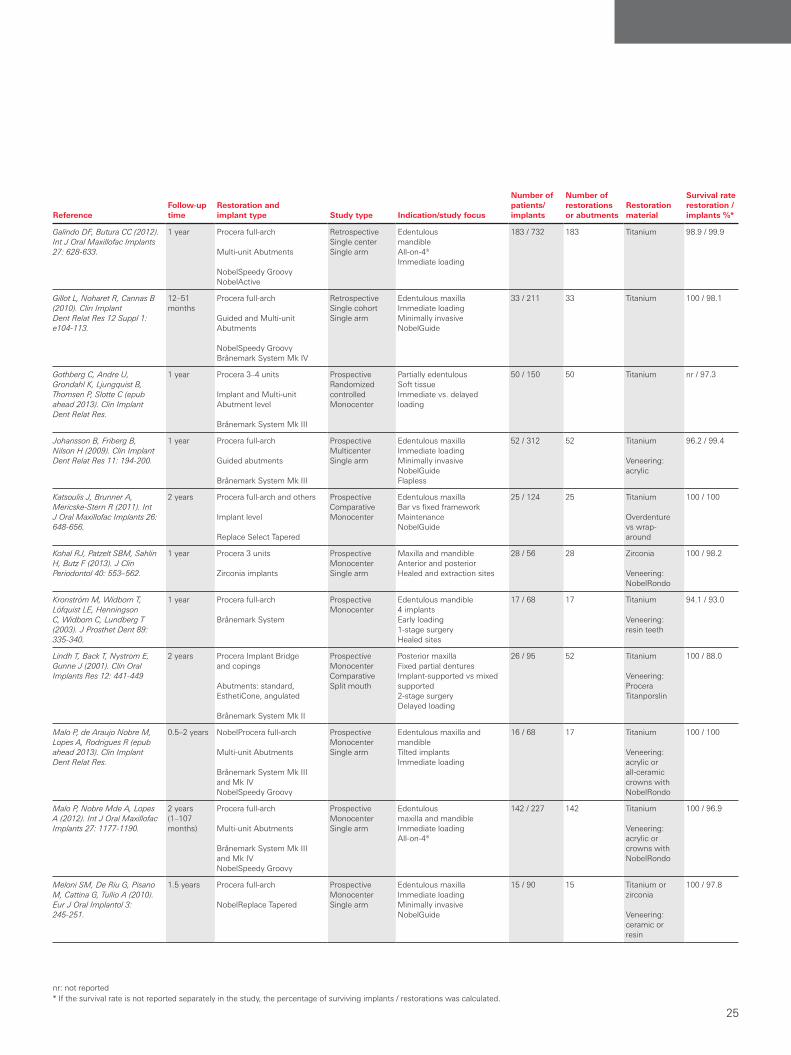
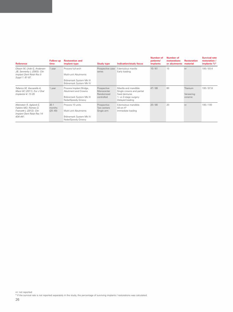
The following overview groups clinical studies with NobelProcera and Procera Implant Bridges according to follow-up time. Within each group, the studies are listed alphabetically.
Only peer-reviewed clinical studies are listed. Abstracts, reviews, single case reports, technique descriptions, and animal and in vitro tests are excluded.
For more information on these studies visit PubMed at www.pubmed.gov
Implant bridges
22

ReferenceFollow-up time
Restoration and implant type Study type Indication/study focus
Number ofpatients/ implants
Number of restorations or abutments
Restoration material
Survival rate restoration / implants %*
Study follow-up time at least 3 years and < 5 years
Agliardi EL, Pozzi A, Stappert CF, Benzi R, Romeo D, Gherlone E (epub ahead 2012). Clin Implant Dent Relat Res.
55.53 months (36–78)
Procera full-arch
Multi-unit Abutments
NobelSpeedy Groovy Brånemark System Mk IV
ProspectiveMonocenterSingle arm
Edentulous maxillaAxial and tilted implantsExtraction and healed sites Immediate FunctionMinimal invasive
32 / 192 48 Titanium 100 / 99.0
Calandriello R, Tomatis M (2005). Clin Implant Dent Relat Res 7 Suppl 1: S1-12.
1–4 years Procera partial and full-arch, and others
Angulated and Procera abutments
Brånemark System Mk IVReplace Select
ProspectiveMonocenterSingle arm
Atrophic posterior maxilla Immediate/early function Axial and tilted implantsFlap and flapless
18 / 60 19 Titanium 100 / 96.7
Cavalli N, Barbaro B, Spasari D, Azzola F, Ciatti A, Francetti L (epub ahead 2012). Int J Dent.
38.8 months (12–73)
Procera full-arch
Multi-unit Abutments
NobelSpeedy GroovyBrånemark System Mk IV
RetrospectiveMonocenterSingle arm
Edentulous maxilla All-on-4®Immediate loading
34 / 136 34 Titanium 100 / 100
Francetti L, Romeo D, Corbella S, Taschieri S, Del Fabbro M (2012). Clin Implant Dent Relat Res 14: 646-654.
52.8 (mandible), 33.8 (maxilla) months (22–66)
Procera full-arch
Multi-unit Abutments
NobelSpeedy GroovyBrånemark System Mk IV
ProspectiveTwo centersSingle arm
Edentulousmaxilla and mandible All-on-4®Immediate loadingMinimal invasiveSoft tissue healthHealed and extraction sites
47 / 196 49 nr 100 / 100
Malo P, de Araujo Nobre M, Lopes A, Ferro A, Moss S (epub ahead 2013). Clin Implant Dent Relat Res.
0.5–7 years NobelProcera full-arch
Brånemark System ZygomaNobelSpeedy Groovy
RetrospectiveMonocenterSingle arm
Edentulous atrophic maxillaExtra-maxillary techniqueAll-on-4®Immediate loading
352 / 1542 352 Titanium
Veneering:acrylic orall-ceramic crowns with NobelRondo
99.7 / 98.2
Malo P, de Araujo Nobre M, Lopes A, Francischone C, Rigolizzo M (2012). Clin Implant Dent Relat Res 14 Suppl 1: e139-150.
3–5 years Procera full-arch
Multi-unit Abutments
Brånemark System Mk III and Mk IVNobelSpeedy Groovy
RetrospectiveMonocenterSingle arm
Edentulous maxillaAll-on-4®Immediate loadingMinimally invasive
242 / 968 242 Titanium
Veneering:acrylic orall-ceramic crowns with NobelRondo
100 / 98.0
Malo P, Nobre M, Lopes A (2013). Eur J Oral Implantol 6: 273-283.
3 years NobelProcera full-arch
NobelSpeedy GroovyNobelSpeedy Replace
RetrospectiveMonocenterSingle arm
Edentulous atrophic maxillaAll-on-4®Immediate loading
70 / 280 70 Titanium
Veneering:acrylic or all-ceramic crowns with NobelRondo
100 / 98.2
Malo P, Nobre M, Lopes A, Francischone C, Rigolizzo M (2012). Eur J Oral Implantol 5: 37-46.
Malo P, Nobre Mde A, Lopes I (2008). J Prosthet Dent 100: 354-366.
3 years
1 year
NobelProcera full-arch
Multi-unit Abutments
Brånemark System Zygoma and others
RetrospectiveMonocenterSingle arm
Edentulous atrophic maxillaExtra-maxillary techniqueAll-on-4®Immediate loading
39 / 169 39 Titanium
Veneering:acrylic or all-ceramic crowns with NobelRondo
100 / 100
Moberg LE, Kondell PA, Sagulin GB, Bolin A, Heimdahl A, Gynther GW (2001). Clin Oral Implants Res 12: 450-461.
3 years Procera full-arch and others
Brånemark System and others
ProspectiveMonocenterRandomized controlled
Edentulous mandible2-stage vs 1-stageComparison of systemsDelayed loading
20 / 102 20 Titanium 100 / 97.9
nr: not reported* If the survival rate is not reported separately in the study, the percentage of surviving implants / restorations was calculated.
Implant bridges
23
Implant bridges

ReferenceFollow-up time
Restoration and implant type Study type Indication/study focus
Number ofpatients/ implants
Number of restorations or abutments
Restoration material
Survival rate restoration / implants %*
Papaspyridakos P, Lal K (2013). Clin Oral Implants Res 24: 659-665.
3 years (2–4)
Procera full-arch
Implant leve
ProspectiveMonocenter
Edentulous maxilla and mandible FlaplessNobelGuide
14 / 103 16 Zirconia
Veneering: porcelain
100 / 100
Pozzi A, Holst S, Fabbri G, Tallarico M (epub ahead 2013). Clin Implant Dent Relat Res.
42.3 months (3–5 years)
NobelProcera full-arch
Non-engaging abutments
NobelSpeedy GroovyNobelSpeedy ReplaceNobelActiveNobelReplace Tapered
RetrospectiveMonocenterSingle arm
Edentulous maxilla and mandible Soft tissue healthHealed and extraction sites
22 / 170 26 Zirconia
Veneering: Noritake Cerabien
100 / 100
Sjostrom M, Sennerby L, Nilson H, Lundgren S (2007). Clin Implant Dent Relat Res 9: 46-59.
3 years Procera full-arch
Standard and angulated abutments
Brånemark System
ProspectiveMonocenterSingle arm
Edentulous atrophic maxillaBone grafting2-stage surgeryDelayed loading
25 / 192 25 Titanium nr / 90.0
Study follow up time < 3 years
Agliardi E, Clerico M, Ciancio P, Massironi D (2010). Quintessence Int 41: 285-293.
30.1 months(19–47)
Procera full-arch
Multi-unit Abutments
NobelSpeedy Groovy Brånemark System Mk IV
ProspectiveSingle cohortSingle arm
Edentulous atrophic mandibleAll-on-4®Immediate loading
24 / 96 24 Titanium 100 / 100
Agliardi E, Panigatti S, Clerico M, Villa C, Malo P (2010). Clin Oral Implants Res 21: 459-465.
26.9 months (4–59)
Procera full-arch
Multi-unit Abutments
NobelSpeedy GroovyBrånemark System Mk IV
ProspectiveSingle cohortSingle arm
Edentulous maxilla and mandible All-on-4®Immediate loading Soft tissue
173 / 692 154 nr 100 / 99.2
Agliardi EL, Francetti L, Ro-meo D, Del Fabbro M (2009). Int J Oral Maxillofac Implants 24: 887-895.
27.2 months (18–42)
Procera full-arch
Multi-unit Abutments
NobelSpeedy GroovyBrånemark System Mk IV
ProspectiveSingle cohortSingle arm
Edentulous maxillaImmediate loadingSoft tissue Extraction and healed sitesStraight and angulated implants
20 / 120 20 nr 100 / 100
Engquist B, Astrand P, Anzen B, Dahlgren S, Engquist E, Feldmann H, Karlsson U, Nord PG, Sahlholm S, Svardstrom P (2002). Clin Implant Dent Relat Res 4: 93-103.
Engquist B, Astrand P, Anzen B, Dahlgren S, Engquist E, Feldmann H, Karlsson U, Nord PG, Sahlholm S, Svardstrom P (2004). Clin Implant Dent Relat Res 6: 90-100.
1 year Procera full-arch
Abutment and implant level
Brånemark System
ProspectiveBi-centerComparative
Edentulous mandible4 implants per jaw Delayed vs early loading1-stage vs 2-stage surgery
108 / 432 108 Titanium
Veneering: acrylic
93.0 / 94.4
Francetti L, Agliardi E, Testori T, Romeo D, Taschieri S, Fabbro MD (2008). Clin Implant Dent Relat Res 10: 255-263.
22.4 months (6–43)
Procera full-arch
Multi-unit Abutments
NobelSpeedy GroovyBrånemark System Mk IV
ProspectiveSingle cohortSingle arm
Edentulous mandibleAll-on-4®Immediate loadingMinimally invasiveSoft tissue Healed and extraction site
62 / 248 62 nr 100 / 100
Friberg B, Jemt T (2010). Clin Implant Dent Relat Res 12 Suppl 1: e56-62.
1 year Procera full-arch
Brånemark System Mk III and Mk IV
RetrospectiveMonocenterSingle arm
Edentulous mandible4 implants per jaw 1-stage surgeryEarly loading
75 / 300 75 Titanium
Veneering: resin
98.5 / 98.5
Fröberg KK, Lindh C, Ericsson I (2006). Clin Implant Dent Relat Res 8: 187-197.
18 months Procera full-arch
Brånemark System Mk III
ProspectiveComparativeSplit mouth
Edentulous mandibleMachined vs TiUniteImmediate loading
15 / 89 15 Titanium 100 / 100
Implant bridges
24
Implant bridges
nr: not reported* If the survival rate is not reported separately in the study, the percentage of surviving implants / restorations was calculated.
ReferenceFollow-up time
Restoration and implant type Study type Indication/study focus
Number ofpatients/ implants
Number of restorations or abutments
Restoration material
Survival rate restoration / implants %*
Galindo DF, Butura CC (2012). Int J Oral Maxillofac Implants 27: 628-633.
1 year Procera full-arch
Multi-unit Abutments
NobelSpeedy GroovyNobelActive
RetrospectiveSingle centerSingle arm
Edentulous mandibleAll-on-4® Immediate loading
183 / 732 183 Titanium 98.9 / 99.9
Gillot L, Noharet R, Cannas B (2010). Clin Implant Dent Relat Res 12 Suppl 1: e104-113.
12–51 months
Procera full-arch
Guided and Multi-unit Abutments
NobelSpeedy Groovy Brånemark System Mk IV
RetrospectiveSingle cohortSingle arm
Edentulous maxillaImmediate loadingMinimally invasiveNobelGuide
33 / 211 33 Titanium 100 / 98.1
Gothberg C, Andre U, Grondahl K, Ljungquist B, Thomsen P, Slotte C (epub ahead 2013). Clin Implant Dent Relat Res.
1 year Procera 3–4 units
Implant and Multi-unit Abutment level
Brånemark System Mk III
ProspectiveRandomized controlledMonocenter
Partially edentulous Soft tissue Immediate vs. delayed loading
50 / 150 50 Titanium nr / 97.3
Johansson B, Friberg B, Nilson H (2009). Clin Implant Dent Relat Res 11: 194-200.
1 year Procera full-arch
Guided abutments
Brånemark System Mk III
Prospective MulticenterSingle arm
Edentulous maxillaImmediate loadingMinimally invasiveNobelGuideFlapless
52 / 312 52 Titanium
Veneering:acrylic
96.2 / 99.4
Katsoulis J, Brunner A, Mericske-Stern R (2011). Int J Oral Maxillofac Implants 26: 648-656.
2 years Procera full-arch and others
Implant level
Replace Select Tapered
ProspectiveComparativeMonocenter
Edentulous maxillaBar vs fixed frameworkMaintenanceNobelGuide
25 / 124 25 Titanium
Overdenture vs wrap-around
100 / 100
Kohal RJ, Patzelt SBM, Sahlin H, Butz F (2013). J Clin Periodontol 40: 553–562.
1 year Procera 3 units
Zirconia implants
Prospective MonocenterSingle arm
Maxilla and mandible Anterior and posteriorHealed and extraction sites
28 / 56 28 Zirconia
Veneering: NobelRondo
100 / 98.2
Kronström M, Widbom T, Löfquist LE, Henningson C, Widbom C, Lundberg T (2003). J Prosthet Dent 89: 335-340.
1 year Procera full-arch
Brånemark System
ProspectiveMonocenter
Edentulous mandible4 implantsEarly loading1-stage surgeryHealed sites
17 / 68 17 Titanium
Veneering: resin teeth
94.1 / 93.0
Lindh T, Back T, Nystrom E, Gunne J (2001). Clin Oral Implants Res 12: 441-449
2 years Procera Implant Bridge and copings
Abutments: standard, EsthetiCone, angulated
Brånemark System Mk II
Prospective Monocenter ComparativeSplit mouth
Posterior maxillaFixed partial denturesImplant-supported vs mixed supported2-stage surgeryDelayed loading
26 / 95 52 Titanium
Veneering: Procera Titanporslin
100 / 88.0
Malo P, de Araujo Nobre M, Lopes A, Rodrigues R (epub ahead 2013). Clin Implant Dent Relat Res.
0.5–2 years NobelProcera full-arch
Multi-unit Abutments
Brånemark System Mk III and Mk IVNobelSpeedy Groovy
ProspectiveMonocenterSingle arm
Edentulous maxilla and mandibleTilted implantsImmediate loading
16 / 68 17 Titanium
Veneering:acrylic orall-ceramic crowns with NobelRondo
100 / 100
Malo P, Nobre Mde A, Lopes A (2012). Int J Oral Maxillofac Implants 27: 1177-1190.
2 years (1–107 months)
Procera full-arch
Multi-unit Abutments
Brånemark System Mk III and Mk IVNobelSpeedy Groovy
ProspectiveMonocenterSingle arm
Edentulousmaxilla and mandible Immediate loadingAll-on-4®
142 / 227 142 Titanium
Veneering:acrylic or crowns with NobelRondo
100 / 96.9
Meloni SM, De Riu G, Pisano M, Cattina G, Tullio A (2010). Eur J Oral Implantol 3: 245-251.
1.5 years Procera full-arch
NobelReplace Tapered
ProspectiveMonocenterSingle arm
Edentulous maxillaImmediate loadingMinimally invasiveNobelGuide
15 / 90 15 Titanium or zirconia
Veneering: ceramic or resin
100 / 97.8
Implant bridges
25
Implant bridges
ReferenceFollow-up time
Restoration and implant type Study type Indication/study focus
Number ofpatients/ implants
Number of restorations or abutments
Restoration material
Survival rate restoration / implants %*
Olsson M, Urde G, Andersen JB, Sennerby L (2003). Clin Implant Dent Relat Res 5 Suppl 1: 81-87.
1 year Procera full-arch
Multi-unit Abutments
Brånemark System Mk IIIBrånemark System Mk IV
Prospective case series
Edentulous maxillaEarly loading
10 / 61 10 nr 100 / 93.4
Tallarico M, Vaccarella A, Marzi GC (2011). Eur J Oral Implantol 4: 13-20.
1 year Procera Implant Bridge, Abutment and Crowns
Brånemark System Mk IIINobelSpeedy Groovy
ProspectiveMonocenterRandomized controlled
Maxilla and mandible Single crowns and partial fixed dentures 1- vs 2-stage surgeryDelayed loading
47 / 89 60 Titanium
Veneering: ceramic
100 / 97.8
Weinstein R, Agliardi E, Fabbro MD, Romeo D, Francetti L (2012). Clin Implant Dent Relat Res 14: 434-441.
30.1 months(20–40)
Procera 10 units
Multi-unit Abutments
Brånemark System Mk IVNobelSpeedy Groovy
ProspectiveTwo centersSingle arm
Edentulous mandibleAll-on-4®Immediate loading
20 / 80 20 nr 100 / 100
nr: not reported* If the survival rate is not reported separately in the study, the percentage of surviving implants / restorations was calculated.
Implant bridges
26

Nobel Biocare CAD/CAM implant bars – scientific evidence
Nobel Biocare CAD/CAM implant bars offer an important improvement in bar retention technology by allowing the use of high-quality materials together with the high accuracy of industrial manufacturing. Compared with gold bars, they provide a higher precision of fit and a striking improvement in performance for the patient.
Bar-retained overdenturesBar retention, the option closest to a fixed prosthesis, provides edentulous patients with improved function, facial esthetics and comfort, as well as improved nutrition, psychosocial status and quality of life. However, gold bars, though currently considered standard in the industry due to several decades of clinical follow-up, are plagued by technical complications and lead to suboptimal survival rates of both implants and prostheses.
Key findings of clinical studies with Nobel Biocare CAD/CAM implant bars are:– High survival rates of implants and restorations (100%).27,101
– Restorations are associated with stable peri-implant crestal bone levels.28
– Significantly fewer technical and biological complications in comparison with conventional cast frameworks.27,28
– High patient satisfaction.27
Better options for an individualized designCAD/CAM technology allows the use of higher quality materials and increases the accuracy of the industrially manufactured components. In an in vitro comparison, the precision of fit of NobelProcera Implant Bars Overdenture was significantly higher than that observed with soldered gold bars.102
Significant improvement in quality of lifeIn a multicenter study, preliminary results on 14 patients from two centers evaluating NobelProcera Implant Bars Overdenture loaded with the final prosthesis no later than three months after implant placement show significant improvements in quality of life.101 OHIP-21 scores improved both from pre-treatment assessment to prosthetic loading and at 6-month follow-up (P<0.001). The authors conclude that overdentures on milled titanium bars are a successful treatment.
Vertical micro gap measured in vitro for titanium bars produced with a photogrammetric and a laser scanner is lower than soldered gold bars or zirconium dioxide bars, indicating a high precision of fit of NobelProcera CAD/CAM design (titanium laser).102
© 2013 ElsevierIllustration printed with permission
Individualized NobelProcera Implant Bar Overdenture on four implants after 6 months. In this study, patient satisfaction at 6 months after implant insertion (scale 1–10) was high for retention (9), speaking (8.6), esthetics (9) and overall (8.5).101
Courtesy of Dr. M. Stocchero, University of Padova, Italy
High precision of fit with NobelProcera Implant Bars
High patient satisfaction
Titanium(Photo-
grammetry)
Titanium(Laser)
Zirconium dioxide(Photo-
grammetry)
Gold(Soldered)
500
125
100
75
50
25
0
Ver
tical
gap
(mic
rom
eter
)
P=0.023P=0.008
P=0.001
27
Implant bars Implant bridges
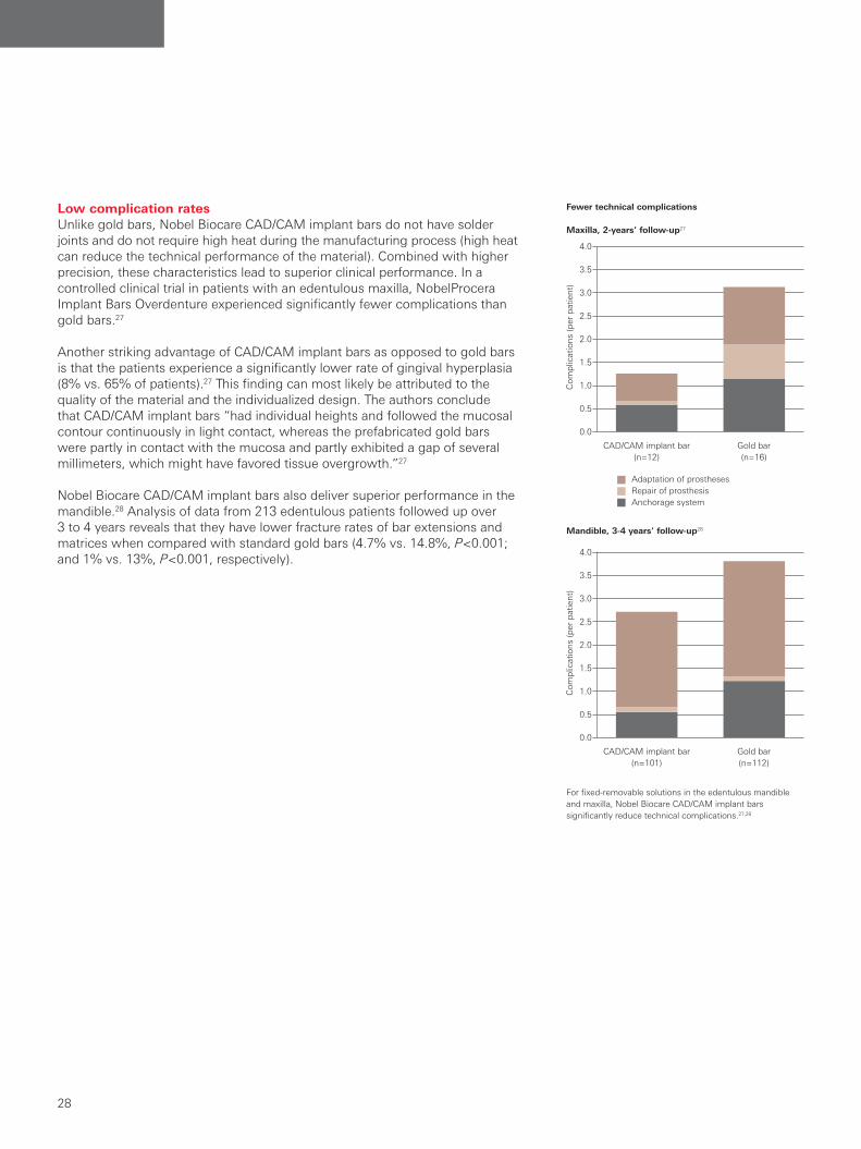
Low complication rates Unlike gold bars, Nobel Biocare CAD/CAM implant bars do not have solder joints and do not require high heat during the manufacturing process (high heat can reduce the technical performance of the material). Combined with higher precision, these characteristics lead to superior clinical performance. In a controlled clinical trial in patients with an edentulous maxilla, NobelProcera Implant Bars Overdenture experienced significantly fewer complications than gold bars.27
Another striking advantage of CAD/CAM implant bars as opposed to gold bars is that the patients experience a significantly lower rate of gingival hyperplasia (8% vs. 65% of patients).27 This finding can most likely be attributed to the quality of the material and the individualized design. The authors conclude that CAD/CAM implant bars “had individual heights and followed the mucosal contour continuously in light contact, whereas the prefabricated gold bars were partly in contact with the mucosa and partly exhibited a gap of several millimeters, which might have favored tissue overgrowth.”27
Nobel Biocare CAD/CAM implant bars also deliver superior performance in the mandible.28 Analysis of data from 213 edentulous patients followed up over 3 to 4 years reveals that they have lower fracture rates of bar extensions and matrices when compared with standard gold bars (4.7% vs. 14.8%, P<0.001; and 1% vs. 13%, P<0.001, respectively).
For fixed-removable solutions in the edentulous mandible and maxilla, Nobel Biocare CAD/CAM implant bars significantly reduce technical complications.27,28
Fewer technical complications
Maxilla, 2-years’ follow-up27
Adaptation of prosthesesRepair of prosthesisAnchorage system
CAD/CAM implant bar (n=12)
Gold bar (n=16)
Com
plic
atio
ns (p
er p
atie
nt)
4.0
3.5
3.0
2.5
2.0
1.5
1.0
0.5
0.0
Mandible, 3-4 years’ follow-up28
CAD/CAM implant bar (n=101)
Gold bar (n=112)
Com
plic
atio
ns (p
er p
atie
nt)
4.0
3.5
3.0
2.5
2.0
1.5
1.0
0.5
0.0
Implant bars
28
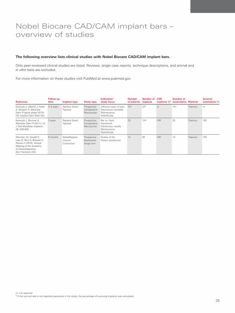
Nobel Biocare CAD/CAM implant bars –overview of studies
ReferenceFollow-up time Implant type Study type
Indication/ study focus
Number of patients
Number of implants
CSR implants %*
Number of restorations Material
Survival restorations %
Katsoulis J, Walchli J, Kobel S, Gholami H, Mericske-Stern R (epub ahead 2013). Clin Implant Dent Relat Res.
3–4 years Replace Select Tapered
ProspectiveComparativeMonocenter
Different types of barsEdentulous mandibleMaintenanceNobelGuide
101 231 nr 101 Titanium nr
Katsoulis J, Brunner A, Mericske-Stern R (2011). Int J Oral Maxillofac Implants 26: 648-656.
2 years Replace Select Tapered
ProspectiveComparativeMonocenter
Bar vs. fixed frameworkEdentulous maxillaMaintenanceNobelGuide
25 124 100 25 Titanium 100
Stoccher, M, Sivolell S, Lops D, Ricci S, Bressan E, Romeo E (2015). Annual Meeting of the Academy of Osseointegration, San Francisco USA.
6 months NobelReplace Conical Connection
ProspectiveMulticenterSingle arm
Quality of lifePatient satisfaction
14 56 100 14 Titanium 100
The following overview lists clinical studies with Nobel Biocare CAD/CAM implant bars.
Only peer-reviewed clinical studies are listed. Reviews, single case reports, technique descriptions, and animal and in vitro tests are excluded.
For more information on these studies visit PubMed at www.pubmed.gov
nr: not reported* If the survival rate is not reported separately in the study, the percentage of surviving implants was calculated.
Implant bars
29
Implant bars
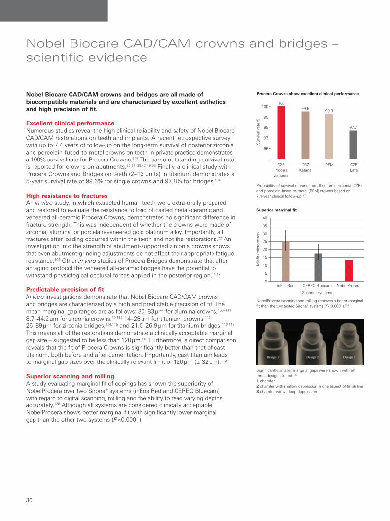
Nobel Biocare CAD/CAM crowns and bridges – scientific evidence
Nobel Biocare CAD/CAM crowns and bridges are all made of biocompatible materials and are characterized by excellent esthetics and high precision of fit.
Excellent clinical performanceNumerous studies reveal the high clinical reliability and safety of Nobel Biocare CAD/CAM restorations on teeth and implants. A recent retrospective survey with up to 7.4 years of follow-up on the long-term survival of posterior zirconia and porcelain-fused-to-metal crowns on teeth in private practice demonstrates a 100% survival rate for Procera Crowns.103 The same outstanding survival rate is reported for crowns on abutments.35,37–39,42,49,55 Finally, a clinical study with Procera Crowns and Bridges on teeth (2–13 units) in titanium demonstrates a 5-year survival rate of 99.6% for single crowns and 97.8% for bridges.104
High resistance to fracturesAn in vitro study, in which extracted human teeth were extra-orally prepared and restored to evaluate the resistance to load of casted metal-ceramic and veneered all-ceramic Procera Crowns, demonstrates no significant difference in fracture strength. This was independent of whether the crowns were made of zirconia, alumina, or porcelain-veneered gold platinum alloy. Importantly, all fractures after loading occurred within the teeth and not the restorations.22 An investigation into the strength of abutment-supported zirconia crowns shows that even abutment-grinding adjustments do not affect their appropriate fatigue resistance.105 Other in vitro studies of Procera Bridges demonstrate that after an aging protocol the veneered all-ceramic bridges have the potential to withstand physiological occlusal forces applied in the posterior region.14,17
Predictable precision of fitIn vitro investigations demonstrate that Nobel Biocare CAD/CAM crowns and bridges are characterized by a high and predictable precision of fit. The mean marginal gap ranges are as follows: 30–83 µm for alumina crowns,106–111 8.7–44.2 µm for zirconia crowns,10,112 14–28 µm for titanium crowns,113 26–89 µm for zirconia bridges,114,115 and 21.0–26.9 µm for titanium bridges.116,117 This means all of the restorations demonstrate a clinically acceptable marginal gap size – suggested to be less than 120 µm.118 Furthermore, a direct comparison reveals that the fit of Procera Crowns is significantly better than that of cast titanium, both before and after cementation. Importantly, cast titanium leads to marginal gap sizes over the clinically relevant limit of 120 µm (± 32 μm).113
Superior scanning and millingA study evaluating marginal fit of copings has shown the superiority of NobelProcera over two Sirona® systems (inEos Red and CEREC Bluecam) with regard to digital scanning, milling and the ability to read varying depths accurately.128 Although all systems are considered clinically acceptable, NobelProcera shows better marginal fit with significantly lower marginal gap than the other two systems (P<0.0001).
Probability of survival of veneered all-ceramic zirconia (CZR) and porcelain-fused-to-metal (PFM) crowns based on 7.4-year clinical follow-up.103
Procera Crowns show excellent clinical performance
CZR Procera Zirconia
CRZ Katana
PFM CZR Lava
10099.5
99.3
97.7
Sur
viva
l rat
e %
100
99
98
97
96
NobelProcera scanning and milling achieves a better marginal fit than the two tested Sirona® systems (P<0.0001).128
Significantly smaller marginal gaps were shown with all three designs tested:128 1 chamfer2 chamfer with shallow depression in one aspect of finish line 3 chamfer with a deep depression
Superior marginal fit
inEos Red CEREC Bluecam NobelProcera
Mis
fit (m
icro
met
er)
Scanner systems
40
35
30
25
20
15
10
5
0
Crowns and bridges
30
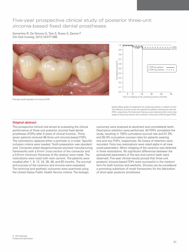
Original abstract
This prospective clinical trial aimed at evaluating the clinical performance of three-unit posterior zirconia fixed dental prostheses (FDPs) after 5 years of clinical function. Thirty- seven patients received 48 three-unit zirconia-based FDPs. The restorations replaced either a premolar or a molar. Specific inclusion criteria were needed. Tooth preparation was standard-ized. Computer-aided design/computer-assisted manufacturing frameworks with a 9 mm2 cross section of the connector and a 0.6 mm minimum thickness of the retainer were made. The restorations were luted with resin cement. The patients were recalled after 1, 6, 12, 24, 36, 48, and 60 months. The survival and success of the ceramics and zirconia were evaluated. The technical and aesthetic outcomes were examined using the United States Public Health Service criteria. The biologic
outcomes were analyzed at abutment and contralateral teeth. Descriptive statistics were performed. All FDPs completed the study, resulting in 100% cumulative survival rate and 91.9% and 95.4% cumulative success rates for patients wearing one and two FDPs, respectively. No losses of retention were recorded. Forty-two restorations were rated alpha in all mea-sured parameters. Minor chipping of the ceramics was detected in three restorations. No significant differences between the periodontal parameters of the test and control teeth were observed. Five-year clinical results proved that three-unit posterior zirconia-based FDPs were successful in the medium term for both function and aesthetic. Zirconia can be considered a promising substitute of metal frameworks for the fabrication of short-span posterior prostheses.
Five-year prospective clinical study of posterior three-unit zirconia-based fixed dental prostheses
Sorrentino R, De Simone G, Tete S, Russo S, Zarone FClin Oral Investig. 2012;16:977-985
Five-year recall evaluation of a zirconia FDP.
Kaplan-Meier graph of chipping of the veneering ceramic in relation to time. Two different survival curves are reported for patients wearing one and two FDPs, respectively. No framework fractures were detected while minor chip-pings of veneering ceramic were noticed in three [out of 48 included] FDPs.
© 2012 SpringerPrinted with permission
6 12 24 36 48 60
0.9540.9190.960
1 FDP per patient: 2 FDPs per patient:
Cum
ulat
ive
surv
ival
%
Time to chipping of the veneering ceramic (months)
1.0
0.9
0.8
0.7
0.6
0.5
0.4
0.3
0.2
0.1
0.0
Crowns and bridges
31
Crowns and bridges
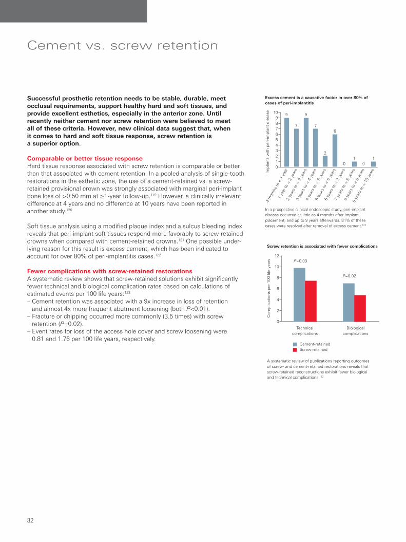
Cement vs. screw retention
Successful prosthetic retention needs to be stable, durable, meet occlusal requirements, support healthy hard and soft tissues, and provide excellent esthetics, especially in the anterior zone. Until recently neither cement nor screw retention were believed to meet all of these criteria. However, new clinical data suggest that, when it comes to hard and soft tissue response, screw retention is a superior option.
Comparable or better tissue responseHard tissue response associated with screw retention is comparable or better than that associated with cement retention. In a pooled analysis of single-tooth restorations in the esthetic zone, the use of a cement-retained vs. a screw- retained provisional crown was strongly associated with marginal peri-implant bone loss of >0.50 mm at ≥1-year follow-up.119 However, a clinically irrelevant difference at 4 years and no difference at 10 years have been reported in another study.120
Soft tissue analysis using a modified plaque index and a sulcus bleeding index reveals that peri-implant soft tissues respond more favorably to screw-retained crowns when compared with cement-retained crowns.121 One possible under-lying reason for this result is excess cement, which has been indicated to account for over 80% of peri-implantitis cases.122
Fewer complications with screw-retained restorations A systematic review shows that screw-retained solutions exhibit significantly fewer technical and biological complication rates based on calculations of estimated events per 100 life years:123
– Cement retention was associated with a 9x increase in loss of retention and almost 4x more frequent abutment loosening (both P<0.01).
– Fracture or chipping occurred more commonly (3.5 times) with screw retention (P=0.02).
– Event rates for loss of the access hole cover and screw loosening were 0.81 and 1.76 per 100 life years, respectively.
In a prospective clinical endoscopic study, peri-implant disease occurred as little as 4 months after implant placement, and up to 9 years afterwards. 81% of these cases were resolved after removal of excess cement.122
A systematic review of publications reporting outcomes of screw- and cement-retained restorations reveals that screw-retained reconstructions exhibit fewer biological and technical complications.123
Excess cement is a causative factor in over 80% of cases of peri-implantitis
Screw retention is associated with fewer complications
11
6
2
7
9
7
9
00
Impl
ants
with
per
i-im
plan
t di
seas
e4
mon
ths
to <
1 y
ear
1 ye
ar to
< 2
yea
rs
2 ye
ars
to <
3 y
ears
3 ye
ars
to <
4 y
ears
4 ye
ars
to <
5 y
ears
5 ye
ars
to <
6 y
ears
6 ye
ars
to <
7 y
ears
7 ye
ars
to <
8 y
ears
8 ye
ars
to <
9 y
ears
9 ye
ars
to <
10
year
s
109876543210
Technical complications
Biological complications
Com
plic
atio
ns p
er 1
00 li
fe y
ears
12
10
8
6
4
2
0
Cement-retainedScrew-retained
P=0.03
P=0.02
Cement vs. screw retention
32
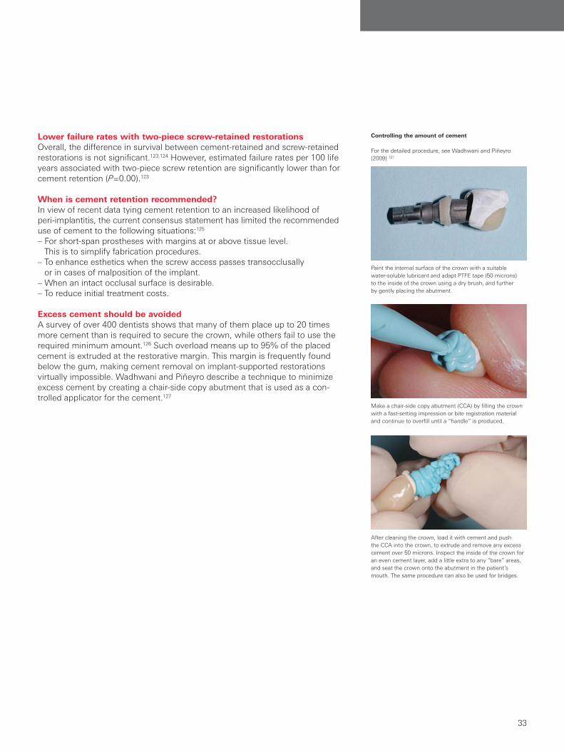
Lower failure rates with two-piece screw-retained restorationsOverall, the difference in survival between cement-retained and screw-retained restorations is not significant.123,124 However, estimated failure rates per 100 life years associated with two-piece screw retention are significantly lower than for cement retention (P=0.00).123
When is cement retention recommended?In view of recent data tying cement retention to an increased likelihood of peri-implantitis, the current consensus statement has limited the recommended use of cement to the following situations:125
– For short-span prostheses with margins at or above tissue level. This is to simplify fabrication procedures.
– To enhance esthetics when the screw access passes transocclusally or in cases of malposition of the implant.
– When an intact occlusal surface is desirable.– To reduce initial treatment costs.
Excess cement should be avoidedA survey of over 400 dentists shows that many of them place up to 20 times more cement than is required to secure the crown, while others fail to use the required minimum amount.126 Such overload means up to 95% of the placed cement is extruded at the restorative margin. This margin is frequently found below the gum, making cement removal on implant-supported restorations virtually impossible. Wadhwani and Piñeyro describe a technique to minimize excess cement by creating a chair-side copy abutment that is used as a con-trolled applicator for the cement.127
Paint the internal surface of the crown with a suitable water-soluble lubricant and adapt PTFE tape (50 microns) to the inside of the crown using a dry brush, and further by gently placing the abutment.
Make a chair-side copy abutment (CCA) by filling the crown with a fast-setting impression or bite registration material and continue to overfill until a “handle” is produced.
After cleaning the crown, load it with cement and push the CCA into the crown, to extrude and remove any excess cement over 50 microns. Inspect the inside of the crown for an even cement layer, add a little extra to any “bare” areas, and seat the crown onto the abutment in the patient’s mouth. The same procedure can also be used for bridges.
Controlling the amount of cement
For the detailed procedure, see Wadhwani and Piñeyro (2009) 127
Cement vs. screw retention
33
Cement vs. screw retention

NobelProcera ASC Abutment – versatility of screw retention combined with predictable abutment performanceThe NobelProcera Angulated Screw Channel (ASC) Abutment positions the screw access hole at an angle of up to 25°. In the esthetic zone this means that the screw access channel can be more palatal to allow for optimized esthetics. For posterior restorations the access channel can be positioned more mesially to allow for easier handling. The NobelProcera ASC Abutment has been tested in biomechanical and 3D numerical fatigue-strength assays, where it demon-strated strength and performance equal or better than that of the NobelProcera Abutment with a straight screw access channel (data on file).
29-year old female patient with agenesis of both lateral superior incisors. Porcelain Maryland Bridges are affected by continuous decementation.
Try-in of two NobelProcera ASC Abutments supported by two NobelActive NP implants.
The angulated screw channel enables a palatally placed screw access hole to improve esthetics.
Esthetic result with two directly veneered NobelProcera ASC Abutments.
Courtesy of Dr. Juan Zufia and Santiago Dalmau, Spain
Excellent esthetic outcomes with the NobelProcera ASC Abutment
Cement vs. screw retention
34

1 Sui X, Wei H, Wang D, Han Y, Deng J, Wang Y, Wang J, Yang J. Experimental research on the relationship between fit accuracy and fracture resistance of zirconia abutments. J Dent. 2014 Oct;42(10):1353-9.
2 Gratton DG, Aquilino SA, Stanford CM. Micromotion and dynamic fatigue properties of the dental implant-abutment interface. J Prosthet Dent 2001;85(1):47-52.
3 Vizer T, Maia C, Fuchs F, Liechti M, Heuberger P. Development of a test model to evaluate the pre-load of screw-retained dental implant systems. European Cells and Materials 2014;27( Suppl. 2):16.
4 Brånemark PI, Albrektsson T. Titanium implants permanently penetrating human skin. Scand J Plast Reconstr Surg. 1982;16(1):17-21.
5 Persson A, Andersson M, Oden A, Sandborgh-Englund G. A three-dimensional evaluation of a laser scanner and a touch-probe scanner. J Prosthet Dent 2006;95(3):194-200.
6 Holst S, Persson A, Wichmann M, Karl M. Digitizing implant position locators on master casts: comparison of a noncontact scanner and a contact-probe scanner. Int J Oral Maxillofac Implants 2012;27(1):29-35.
7 Katsoulis J, Mericske-Stern R, Rotkina L, et al. Precision of fit of implant-supported screw-retained 10-unit computer-aided-designed and computer-aided-manufactured frameworks made from zirconium dioxide and titanium: an in vitro study. Clin Oral Implants Res 2014;25(2):165–74.
8 Sierraalta M, Vivas JL, Razzoog ME, Wang RF. Precision of fit of titanium and cast implant frameworks using a new matching formula. Int J Dent epub ahead 2012:374315.
9 Karl M, Holst S. Strain development of screw-retained implant-supported fixed restorations: procera implant bridge versus conventionally cast restorations. Int J Prosthodont 2012;25(2):166-9.
10 Martinez-Rus F, Suarez MJ, Rivera B, Pradies G. Evaluation of the absolute marginal discrepancy of zirconia-based ceramic copings. J Prosthet Dent 2011;105(2):108-14.
11 Steinemann SG. Titanium--the material of choice? Periodontol 2000 1998;17:7-21.
12 Hisbergues M, Vendeville S, Vendeville P. Zirconia: Established facts and perspectives for a biomaterial in dental implantology. J Biomed Mater Res B Appl Biomater 2009;88(2):519-29.
13 Alhasanyah A, Vaidyanathan TK, Flinton RJ. Effect of Core Thickness Differences on Post-Fatigue Indentation Fracture Resistance of Veneered Zirconia Crowns. J Prosthodont 2013;22(5).
14 Att W, Grigoriadou M, Strub JR. ZrO2 three-unit fixed partial dentures: comparison of failure load before and after exposure to a mastication simulator. J Oral Rehabil 2007;34(4):282-90.
15 Att W, Kurun S, Gerds T, Strub JR. Fracture resistance of single-tooth implant-supported all-ceramic restorations: an in vitro study. J Prosthet Dent 2006;95(2):111-6.
16 Att W, Kurun S, Gerds T, Strub JR. Fracture resistance of single-tooth implant-supported all-ceramic restorations after exposure to the artificial mouth. J Oral Rehabil 2006;33(5):380-6.
17 Att W, Stamouli K, Gerds T, Strub JR. Fracture resistance of different zirconium dioxide three-unit all-ceramic fixed partial dentures. Acta Odontol Scand 2007;65(1):14-21.
18 Kokubo Y, Tsumita M, Kano T, Fukushima S. The influence of zirconia coping designs on the fracture load of all-ceramic molar crowns. Dent Mater J 2011;30(3):281-85.
19 Kokubo Y, Tsumita M, Sakurai S, et al. The effect of core framework designs on the fracture loads of all-ceramic fixed partial dentures on posterior implants. J Oral Rehabil 2007;34(7):503-7.
20 Larsson C, Holm L, Lovgren N, Kokubo Y, Vult von Steyern P. Fracture strength of four-unit Y-TZP FPD cores designed with varying connector diameter. An in vitro study. J Oral Rehabil 2007;34(9):702-9.
21 Persson M, Bergman M. Metal-ceramic bond strength. Acta Odontol Scand 1996;54(3):160-65.
22 Potiket N, Chiche G, Finger IM. In vitro fracture strength of teeth restored with different all-ceramic crown systems. J Prosthet Dent 2004;92(5):491-5.
23 Snyder MD, Hogg KD. Load-to-fracture value of different all-ceramic crown systems. J Contemp Dent Pract 2005;6(4):54-63.
24 Vult von Steyern P, Ebbesson S, Holmgren J, Haag P, Nilner K. Fracture strength of two oxide ceramic crown systems after cyclic pre-loading and thermocycling. J Oral Rehabil 2006;33(9):682-9.
25 Wood KC, Berzins DW, Luo Q, et al. Resistance to fracture of two all-ceramic crown materials following endodontic access. J Prosthet Dent 2006;95(1):33-41.
26 Jemt T, Stenport V. Implant treatment with fixed prostheses in the edentulous maxilla. Part 2: prosthetic technique and clinical maintenance in two patient cohorts restored between 1986 and 1987 and 15 years later. Int J Prosthodont 2011;24(4):356-62.
27 Katsoulis J, Brunner A, Mericske-Stern R. Maintenance of implant-supported maxillary prostheses: a 2-year controlled clinical trial. Int J Oral Maxillofac Implants 2011;26(3):648-56.
28 Katsoulis J, Walchli J, Kobel S, Gholami H, Mericske-Stern R. Complications with Computer-Aided Designed/Computer-Assisted Manufactured Titanium and Soldered Gold Bars for Mandibular Implant-Overdentures: Short-Term Observations. Clin Implant Dent Relat Res epub ahead 2013.
29 Moberg LE, Kondell PA, Sagulin GB, et al. Branemark System and ITI Dental Implant System for treatment of mandibular edentulism. A comparative randomized study: 3-year follow-up. Clin Oral Implants Res 2001;12(5):450-61.
30 Degidi M, Artese L, Scarano A, et al. Inflammatory infiltrate, microvessel density, nitric oxide synthase expression, vascular endothelial growth factor expression, and proliferative activity in peri-implant soft tissues around titanium and zirconium oxide healing caps. J Periodontol 2006;77(1):73-80.
31 Mustafa K, Wennerberg A, Arvidson K, et al. Influence of modifying and veneering the surface of ceramic abutments on cellular attachment and proliferation. Clin Oral Implants Res 2008;19(11):1178-87.
32 Madianos PN, Bobetsis YA, Kinane DF. Generation of inflammatory stimuli: how bacteria set up inflammatory responses in the gingiva. J Clin Periodontol 2005;32 Suppl 6:57-71.
33 Lima EM, Koo H, Vacca Smith AM, Rosalen PL, Del Bel Cury AA. Adsorption of salivary and serum proteins, and bacterial adherence on titanium and zirconia ceramic surfaces. Clin Oral Implants Res 2008;19(8):780-5.
34 Kutkut A, Abu-Hammad O, Mitchell R. Esthetic Considerations for Reconstructing Implant Emergence Profile Using Titanium and Zirconia Custom Implant Abutments: Fifty Case Series Report. Journal of Oral Implantology epub ahead 2013.
35 Calandriello R, Tomatis M. Immediate Occlusal Loading of Single Lower Molars Using Branemark System(R) Wide Platform TiUnite Implants: A 5-Year Follow-Up Report of a Prospective Clinical Multicenter Study. Clin Implant Dent Relat Res 2011;13(4):311-8.
36 Zembic A, Bosch A, Jung RE, Hammerle CH, Sailer I. Five-year results of a randomized controlled clinical trial comparing zirconia and titanium abutments supporting single-implant crowns in canine and posterior regions. Clin Oral Implants Res 2013;24(4):384-90.
37 den Hartog L, Meijer HJ, Stegenga B, et al. Single implants with different neck designs in the aesthetic zone: a randomized clinical trial. Clin Oral Implants Res 2011;22(11):1289-97.
38 den Hartog L, Raghoebar GM, Slater JJ, Stellingsma K, Vissink A, Meijer HJ. Single-Tooth Implants with Different Neck Designs: A Randomized Clinical Trial Evaluating the Aesthetic Outcome. Clin Implant Dent Relat Res 2013;15(3):311-21.
39 den Hartog L, Raghoebar GM, Stellingsma K, Vissink A, Meijer HJ. Immediate non-occlusal loading of single implants in the aesthetic zone: a randomized clinical trial. J Clin Periodontol 2011;38(2):186-94.
40 Ekfeldt A, Furst B, Carlsson GE. Zirconia abutments for single-tooth implant restorations: a retrospective and clinical follow-up study. Clin Oral Implants Res 2011;22(11):1308-14.
41 Pozzi A, Sannino G, Barlattani A. Minimally invasive treatment of the atrophic posterior maxilla: A proofof-concept prospective study with a follow-up of between 36 and 54 months. J Prosthet Dent 2012;108(5):286-97.
42 Rao W, Benzi R. Single mandibular first molar implants with flapless guided surgery and immediate function: preliminary clinical and radiographic results of a prospective study. J Prosthet Dent 2007;97(6 Suppl):S3-S14.
43 Pozzi A, Agliardi E, Tallarico M, Barlattani A. Clinical and Radiological Outcomes of Two Implants with Different Prosthetic Interfaces and Neck Configurations: Randomized, Controlled, Split-Mouth Clinical Trial. Clin Implant Dent Relat Res 2014;16(1):96–106.
44 Raghoebar GM, Slater JJ, Hartog L, Meijer HJ, Vissink A. Comparison of procedures for immediate reconstruction of large osseous defects resulting from removal of a single tooth to prepare for insertion of an endosseous implant after healing. Int J Oral Maxillofac Surg 2009;38(7):736-43.
45 Tymstra N, Raghoebar GM, Vissink A, Meijer HJ. Dental implant treatment for two adjacent missing teeth in the maxillary aesthetic zone: a comparative pilot study and test of principle. Clin Oral Implants Res 2011;22(2):207-13.
46 Urban T, Kostopoulos L, Wenzel A. Immediate implant placement in molar regions: a 12-month prospective, randomized follow-up study. Clin Oral Implants Res 2012;23(12):1389–97.
47 Vigolo P, Givani A, Majzoub Z, Cordioli G. A 4-year prospective study to assess peri-implant hard and soft tissues adjacent to titanium versus gold-alloy abutments in cemented single implant crowns. J Prosthodont 2006;15(4):250-6.
48 Meloni SM, De Riu G, Pisano M, De Riu N, Tullio A. Immediate versus delayed loading of single mandibular molars. One-year results from a randomised controlled trial. Eur J Oral Implantol 2012;5(4):345-53.
References
35
ReferencesCement vs. screw retention

49 Tymstra N, Raghoebar GM, Vissink A, et al. Treatment outcome of two adjacent implant crowns with different implant platform designs in the aesthetic zone: a 1-year randomized clinical trial. J Clin Periodontol 2011;38(01):74-85.
50 Khzam N, Mattheos N, Roberts D, Bruce WL, Ivanovski S. Immediate Placement and Restoration of Dental Implants in the Esthetic Region: Clinical Case Series. Journal of Esthetic and Restorative Dentistry epub ahead 2014:n/a-n/a.
51 Jung RE, Pjetursson BE, Glauser R, et al. A systematic review of the 5-year survival and complication rates of implant-supported single crowns. Clin Oral Implants Res 2008;19(2):119-30.
52 Scarano A, Piattelli M, Caputi S, Favero GA, Piattelli A. Bacterial adhesion on commercially pure titanium and zirconium oxide disks: an in vivo human study. J Periodontol 2004;75(2):292-6.
53 Belser UC, Grutter L, Vailati F, et al. Outcome evaluation of early placed maxillary anterior single-tooth implants using objective esthetic criteria: a cross-sectional, retrospective study in 45 patients with a 2- to 4-year follow-up using pink and white esthetic scores. J Periodontol 2009;80(1):140-51.
54 Meijer HJ, Stellingsma K, Meijndert L, Raghoebar GM. A new index for rating aesthetics of implant-supported single crowns and adjacent soft tissues--the Implant Crown Aesthetic Index. Clin Oral Implants Res 2005;16(6):645-9.
55 Tallarico M, Vaccarella A, Marzi GC. Clinical and radiological outcomes of 1- versus 2-stage implant placement: 1-year results of a randomised clinical trial. Eur J Oral Implantol 2011;4(1):13-20.
56 Alqahtani F, Flinton R. Postfatigue fracture resistance of modified prefabricated zirconia implant abutments. J Prosthet Dent 2014;112(2):299-305.
57 Att W, Yajima ND, Wolkewitz M, Witkowski S, Strub JR. Influence of Preparation and Wall Thickness on the Resistance to Fracture of Zirconia Implant Abutments. Clin Implant Dent Relat Res 2012;14(suppl 1):e196-e203.
58 Delben JA, Barao VAR, Dos Santos PH, Assuncao WG. Influence of Abutment Type and Esthetic Veneering on Preload Maintenance of Abutment Screw of Implant-Supported Crowns. Journal of Prosthodontics 2014;23(2):134-39.
59 Gigandet M, Bigolin G, Faoro F, Burgin W, Bragger U. Implants with Original and Non-Original Abutment Connections. Clin Implant Dent Relat Res 2014;16(2):303–11.
60 Kerstein RB, Radke J. A comparison of fabrication precision and mechanical reliability of 2 zirconia implant abutments. Int J Oral Maxillofac Implants 2008;23(6):1029-36.
61 Kim S, Kim HI, Brewer JD, Monaco EA, Jr. Comparison of fracture resistance of pressable metal ceramic custom implant abutments with CAD/CAM commercially fabricated zirconia implant abutments. J Prosthet Dent 2009;101(4):226-30.
62 Muhlemann S, Truninger TC, Stawarczyk B, Hammerle CH, Sailer I. Bending moments of zirconia and titanium implant abutments supporting all-ceramic crowns after aging. Clin Oral Implants Res 2014;25(1):74–81.
63 Protopapadaki M, Monaco EA, Jr., Kim HI, Davis EL. Comparison of fracture resistance of pressable metal ceramic custom implant abutment with a commercially fabricated CAD/CAM zirconia implant abutment. J Prosthet Dent 2013.
64 Sailer I, Sailer T, Stawarczyk B, Jung RE, Hammerle CH. In vitro study of the influence of the type of connection on the fracture load of zirconia abutments with internal and external implant-abutment connections. Int J Oral Maxillofac Implants 2009;24(5):850-8.
65 Truninger TC, Stawarczyk B, Leutert CR, et al. Bending moments of zirconia and titanium abutments with internal and external implant-abutment connections after aging and chewing simulation. Clin Oral Implants Res 2012;23(1):12-18.
66 Delben JA, Gomes EA, Barao VA, Kuboki Y, Assuncao WG. Evaluation of the effect of retightening and mechanical cycling on preload maintenance of retention screws. Int J Oral Maxillofac Implants 2011;26(2):251-6.
67 Garine WN, Funkenbusch PD, Ercoli C, Wodenscheck J, Murphy WC. Measurement of the rotational misfit and implant-abutment gap of all-ceramic abutments. Int J Oral Maxillofac Implants 2007;22(6):928-38.
68 Lang LA, Wang RF, May KB. The influence of abutment screw tightening on screw joint configuration. J Prosthet Dent 2002;87(1):74-9.
69 Vigolo P, Fonzi F, Majzoub Z, Cordioli G. An in vitro evaluation of titanium, zirconia, and alumina procera abutments with hexagonal connection. Int J Oral Maxillofac Implants 2006;21(4):575-80.
70 Binon PP. The effect of implant/abutment hexagonal misfit on screw joint stability. Int J Prosthodont 1996;9(2):149-60.
71 Baldassarri M, Hjerppe J, Romeo D, et al. Marginal accuracy of three implant-ceramic abutment configurations. Int J Oral Maxillofac Implants 2012;27(3):537-43.
72 de Morais Alves da Cunha T, de Araújo RPC, da Rocha PVB, Amoedo RMP. Comparison of Fit Accuracy between Procera® Custom Abutments and Three Implant Systems. Clin Implant Dent Relat Res 2012;14(6):890-95.
73 Hamilton A, Judge RB, Palamara JE, Evans C. Evaluation of the fit of CAD/CAM abutments. Int J Prosthodont 2013;26(4):370-80.
74 Smith NA, Turkyilmaz I. Evaluation of the sealing capability of implants to titanium and zirconia abutments against Porphyromonas gingivalis, Prevotella intermedia, and Fusobacterium nucleatum under different screw torque values. J Prosthet Dent epub ahead 2014.
75 Yuzugullu B, Avci M. The implant-abutment interface of alumina and zirconia abutments. Clin Implant Dent Relat Res 2008;10(2):113-21.
76 Lang LA, Sierraalta M, Hoffensperger M, Wang RF. Evaluation of the precision of fit between the Procera custom abutment and various implant systems. Int J Oral Maxillofac Implants 2003;18(5):652-8.
77 Karl M, Taylor TD. Parameters determining micromotion at the implant-abutment interface. Int J Oral Maxillofac Implants 2014;29(6):1338-47.
78 Ortorp A, Jemt T. CNC-milled titanium frameworks supported by implants in the edentulous jaw: a 10-year comparative clinical study. Clin Implant Dent Relat Res 2012;14(1):88-99.
79 Malo P, Nobre M, Lopes A. The rehabilitation of completely edentulous maxillae with different degrees of resorption with four or more immediately loaded implants: a 5-year retrospective study and a new classification. Eur J Oral Implantol 2011;4(3):227-43.
80 Pettersson P, Sennerby L. A 5-Year Retrospective Study on Replace Select Tapered Dental Implants. Clin Implant Dent Relat Res epub ahead 2013.
81 Pozzi A, Tallarico M, Barlattani A. Monolithic lithium disilicate full-contour crowns bonded on CAD/CAM zirconia complete-arch implant bridges with 3 to 5 years of follow-up. Journal of Oral Implantology epub ahead 2013.
82 Cavalli N, Barbaro B, Spasari D, et al. Tilted implants for full-arch rehabilitations in completely edentulous maxilla: a retrospective study. Int J Dent epub ahead 2012.
83 Francetti L, Romeo D, Corbella S, Taschieri S, Del Fabbro M. Bone Level Changes Around Axial and Tilted Implants in Full-Arch Fixed Immediate Restorations. Interim Results of a Prospective Study. Clin Implant Dent Relat Res 2012;14(5):646-54.
84 Galindo DF, Butura CC. Immediately loaded mandibular fixed implant prostheses using the all-on-four protocol: a report of 183 consecutively treated patients with 1 year of function in definitive prostheses. Int J Oral Maxillofac Implants 2012;27(3):628-33.
85 Malo P, Nobre M, Lopes A. Immediate loading of ‘All-on-4’ maxillary prostheses using trans-sinus tilted implants without sinus bone grafting: a retrospective study reporting the 3-year outcome. Eur J Oral Implantol 2013;6(3):273-83.
86 Agliardi E, Panigatti S, Clerico M, Villa C, Malo P. Immediate rehabilitation of the edentulous jaws with full fixed prostheses supported by four implants: interim results of a single cohort prospective study. Clin Oral Implants Res 2010;21(5):459-65.
87 Calandriello R, Tomatis M. Simplified treatment of the atrophic posterior maxilla via immediate/early function and tilted implants: A prospective 1-year clinical study. Clin Implant Dent Relat Res 2005;7 Suppl 1:S1-12.
88 Francetti L, Agliardi E, Testori T, et al. Immediate rehabilitation of the mandible with fixed full prosthesis supported by axial and tilted implants: interim results of a single cohort prospective study. Clin Implant Dent Relat Res 2008;10(4):255-63.
89 Gillot L, Cannas B, Buti J, Noharet R. A retrospective cohort study of 113 patients rehabilitated with immediately loaded maxillary cross-arch fixed dental prostheses in combination with immediate implant placement. Eur J Oral Implantol 2012;5(1):71-9.
90 Gothberg C, Andre U, Grondahl K, et al. Immediately Loaded Implants with or without Abutments Supporting Fixed Partial Dentures: 1-Year Results from a Prospective, Randomized, Clinical Trial. Clin Implant Dent Relat Res epub ahead 2013.
91 Malo P, de Araujo Nobre M, Lopes A, Francischone C, Rigolizzo M. “All-on-4” Immediate-Function Concept for Completely Edentulous Maxillae: A Clinical Report on the Medium (3 Years) and Long-Term (5 Years) Outcomes. Clin Implant Dent Relat Res 2012;14 Suppl 1:e139-50.
92 Meloni SM, De Riu G, Pisano M, Cattina G, Tullio A. Implant treatment software planning and guided flapless surgery with immediate provisional prosthesis delivery in the fully edentulous maxilla. A retrospective analysis of 15 consecutively treated patients. Eur J Oral Implantol 2010;3(3):245-51.
93 Olsson M, Urde G, Andersen JB, Sennerby L. Early loading of maxillary fixed cross-arch dental prostheses supported by six or eight oxidized titanium implants: results after 1 year of loading, case series. Clin Implant Dent Relat Res 2003;5 Suppl 1:81-87.
94 Papaspyridakos P, Lal K. Computer-assisted design/computer-assisted manufacturing zirconia implant fixed complete prostheses: clinical results and technical complications up to 4 years of function. Clin Oral Implants Res 2013;24(6):659-65.
References
36

95 Pozzi A, Holst S, Fabbri G, Tallarico M. Clinical Reliability of CAD/CAM Cross-Arch Zirconia Bridges on Immediately Loaded Implants Placed with Computer-Assisted/Template-Guided Surgery: A Retrospective Study with a Follow-Up between 3 and 5 Years. Clin Implant Dent Relat Res epub ahead 2013.
96 Agliardi E, Clerico M, Ciancio P, Massironi D. Immediate loading of full-arch fixed prostheses supported by axial and tilted implants for the treatment of edentulous atrophic mandibles. Quintessence Int 2010;41(4):285-93.
97 Agliardi EL, Francetti L, Romeo D, Del Fabbro M. Immediate rehabilitation of the edentulous maxilla: preliminary results of a single-cohort prospective study. Int J Oral Maxillofac Implants 2009;24(5):887-95.
98 Agliardi EL, Pozzi A, Stappert CF, et al. Immediate Fixed Rehabilitation of the Edentulous Maxilla: A Prospective Clinical and Radiological Study after 3 Years of Loading. Clin Implant Dent Relat Res 2014;16(2):292–302.
99 Malo P, Nobre Mde A, Lopes A. Immediate Rehabilitation of Completely Edentulous Arches with a Four-Implant Prosthesis Concept in Difficult Conditions: An Open Cohort Study with a Mean Follow-up of 2 Years. Int J Oral Maxillofac Implants 2012;27(5):1177-90.
100 Holst S, Karl M, Wichmann M, Matta RE. A technique for in vitro fit assessment of multi-unit screw-retained implant restorations: Application of a triple-scan protocol. J Dent Biomech epub 2012;3:1758736012452181.
101 Stocchero M, Sivolella S, Lops D, et al. A clinical evaluation of individualized titanium milled bar overdentures on 4 tapered implants in the mandible or maxilla. Preliminary 6-months data. Annaul Meeting of the Academy of Osseointegration. San Francisco, USA; 2015.
102 Katsoulis J, Mericske-Stern R, Yates DM, et al. In vitro precision of fit of computer-aided design and computer-aided manufacturing titanium and zirconium dioxide bars. Dent Mater 2013;29(9):945-53.
103 Ozer F, Mante FK, Chiche G, et al. A retrospective survey on long-term survival of posterior zirconia and porcelain-fused-to-metal crowns in private practice. Quintessence Int 2014;45(1):31-8.
104 Lovgren R, Andersson B, Carlsson GE, Odman P. Prospective clinical 5-year study of ceramic-veneered titanium restorations with the Procera system. J Prosthet Dent 2000;84(5):514-21.
105 Cabrera T, Rekow, Stappert. Fatigue analysis of individualized zirconia implant-abutments and crowns. J Dent Res 2007;86(Spec Iss A).
106 Kokubo Y, Ohkubo C, Tsumita M, et al. Clinical marginal and internal gaps of Procera AllCeram crowns. J Oral Rehabil 2005;32(7):526-30.
107 Limkangwalmongkol P, Kee E, Chiche GJ, Blatz MB. Comparison of marginal fit between all-porcelain margin versus alumina-supported margin on Procera Alumina crowns. J Prosthodont 2009;18(2):162-6.
108 May KB, Russell MM, Razzoog ME, Lang BR. Precision of fit: the Procera AllCeram crown. J Prosthet Dent 1998;80(4):394-404.
109 Naert I, Van der Donck A, Beckers L. Precision of fit and clinical evaluation of all-ceramic full restorations followed between 0.5 and 5 years. J Oral Rehabil 2005;32(1):51-7.
110 Rahme HY, Tehini GE, Adib SM, Ardo AS, Rifai KT. In vitro evaluation of the “replica technique” in the measurement of the fit of Procera crowns. J Contemp Dent Pract 2008;9(2):25-32.
111 Sulaiman F, Chai J, Jameson LM, Wozniak WT. A comparison of the marginal fit of In-Ceram, IPS Empress, and Procera crowns. Int J Prosthodont 1997;10(5):478-84.
112 Kokubo Y, Tsumita M, Kano T, Sakurai S, Fukushima S. Clinical marginal and internal gaps of zirconia all-ceramic crowns. J Prosthodont Res 2011;55(1):40-43.
113 Jesus Suarez M, Lozano JF, Paz Salido M, Martinez F. Marginal fit of titanium metal-ceramic crowns. Int J Prosthodont 2005;18(5):390-1.
114 Att W, Komine F, Gerds T, Strub JR. Marginal adaptation of three different zirconium dioxide three-unit fixed dental prostheses. J Prosthet Dent 2009;101(4):239-47.
115 Gonzalo E, Suarez MJ, Serrano B, Lozano JF. Marginal fit of Zirconia posterior fixed partial dentures. Int J Prosthodont 2008;21(5):398-9.
116 Takahashi T, Gunne J. Fit of implant frameworks: an in vitro comparison between two fabrication techniques. J Prosthet Dent 2003;89(3):256-60.
117 Torsello F, di Torresanto VM, Ercoli C, Cordaro L. Evaluation of the marginal precision of one-piece complete arch titanium frameworks fabricated using five different methods for implant-supported restorations. Clin Oral Implants Res 2008;19(8):772-79.
118 McLean JW, von Fraunhofer JA. The estimation of cement film thickness by an in vivo technique. Br Dent J 1971;131(3):107-11.
119 Slagter KW, den Hartog L, Bakker NA, et al. Immediate placement of dental implants in the esthetic zone: a systematic review and pooled analysis. J Periodontol 2014;85(7):e241-50.
120 Vigolo P, Mutinelli S, Givani A, Stellini E. Cemented versus screw-retained implant-supported single-tooth crowns: a 10-year randomised controlled trial. Eur J Oral Implantol 2012;5(4):355-64.
121 Weber HP, Kim DM, Ng MW, Hwang JW, Fiorellini JP. Peri-implant soft-tissue health surrounding cement- and screw-retained implant restorations: a multi-center, 3-year prospective study. Clin Oral Implants Res 2006;17(4):375-9.
122 Wilson TG, Jr. The positive relationship between excess cement and peri-implant disease: a prospective clinical endoscopic study. J Periodontol 2009;80(9):1388-92.
123 Wittneben JG, Millen C, Bragger U. Clinical performance of screw- versus cement-retained fixed implant-supported reconstructions-a systematic review. Int J Oral Maxillofac Implants 2014;29 Suppl:84-98.
124 Cicciu M, Beretta M, Risitano G, Maiorana C. Cemented-retained vs screw-retained implant restorations: an investigation on 1939 dental implants. Minerva Stomatol 2008;57(4):167-79.
125 Wismeijer D, Bragger U, Evans C, et al. Consensus Statements and Recommended Clinical Procedures Regarding Restorative Materials and Techniques for Implant Dentistry. Int J Oral Maxillofac Implants 2014;29(Supplement):137-40.
126 Wadhwani C, Hess T, Pineyro A, Opler R, Chung KH. Cement application techniques in luting implant-supported crowns: a quantitative and qualitative survey. Int J Oral Maxillofac Implants 2012;27(4):859-64.
127 Wadhwani C, Pineyro A. Technique for controlling the cement for an implant crown. J Prosthet Dent 2009;102(1):57-8.
128 Al-Aali K. Marginal Fit of Milled Copings Utilizing Digital Scanners [#48]. 43rd Annual Meeting & Exhibition of the AADR. Charlotte, USA; 2014.
References
37
References

Notes


nobelbiocare.com
78
32
2 G
B 1
50
2 P
rin
ted
in G
erm
any
© N
ob
el B
ioca
re S
ervi
ces
AG
, 2
01
5.
All
rig
hts
res
erve
d.
No
bel
Bio
care
, th
e N
ob
el B
ioca
re lo
go
typ
e an
d a
ll o
ther
tra
dem
arks
are
, if
no
thin
g e
lse
is s
tate
d o
r is
evi
den
t fr
om
th
e co
nte
xt in
a c
erta
in c
ase,
tra
dem
arks
of
No
bel
Bio
care
. P
leas
e re
fer
to n
ob
elb
ioca
re.c
om
/tra
dem
arks
fo
r m
ore
info
rmat
ion
. P
rod
uct
imag
es a
re n
ot
nec
essa
rily
to
sca
le.
Dis
clai
mer
: S
om
e p
rod
uct
s m
ay n
ot
be
reg
ula
tory
cle
ared
/re
leas
ed f
or
sale
in a
ll m
arke
ts.
Ple
ase
con
tact
th
e lo
cal N
ob
el B
ioca
re s
ales
off
ice
for
curr
ent
pro
du
ct a
sso
rtm
ent
and
ava
ilab
ility
.