School of Pharmacy, University of Lincoln, Joseph Banks ...
36
1 Synthetic receptors for the recognition and discrimination of post-translationally methylated lysines Dr. Tobias Gruber* School of Pharmacy, University of Lincoln, Joseph Banks Laboratories, Green Lane, Lincoln LN6 7DL, United Kingdom. *E-mail: [email protected]
Transcript of School of Pharmacy, University of Lincoln, Joseph Banks ...
1
Synthetic receptors for the recognition and discrimination of
post-translationally methylated lysines
Dr. Tobias Gruber*
School of Pharmacy, University of Lincoln, Joseph Banks
Laboratories, Green Lane, Lincoln LN6 7DL, United Kingdom.
*E-mail: [email protected]

2
Synthetic receptors for the recognition and discrimination of post-translationally
methylated lysines
Dr. Tobias Gruber, University of Lincoln
Photo: Tobias Gruber; 50.495 N, 11.467 O

3
Abstract
Post-translational modifications (PTMs) describe the chemical alteration of proteins after their
biosynthesis in ribosomes. PTMs play important roles in cell biology including the regulation
of gene expression, cell-cell interactions and the development of different diseases. A
prominent class of PTMs is the side chain methylation of lysine. For the analysis and
discrimination of differently methylated lysines antibodies are widely used, though, methylated
peptide and protein targets are known to be particularly difficult to be differentiated by
antibody-based affinity reagents; an additional challenge can be batch-to-batch reproducibility.
The application of mass spectrometry techniques for methyllysine discrimination requires a
complex sample preparation and is not suited for working in cells. The desire to overcome
above-named challenges promoted the development of synthetic receptor molecules that
recognize and bind methyllysines. Such ‘artificial antibodies’ are of interest for a number of
applications, e.g. as reagents in biochemical assays, for the isolation and purification of post-
translationally methylated proteins and for the tracking of signalling pathways. Moreover, they
offer new approaches in diagnostics and therapy. This review delivers an overview of the broad
field of methyllysine binding and covers a wide range of synthetic receptors used for the
recognition of methylated lysines including calixarenes, resorcinarenes, pillararenes, disulfide
cyclophanes, cucurbituriles and acyclic receptors.
Keywords:
Post-translational modification
Lysine methylation
Artificial antibodies
Synthetic receptors
Epigenetics
Graphical Abstract
Very distinguished: The side chain methylation of lysine is a
prominent post-translational modification. The desire to
understand the function of this modification promoted the
development of synthetic molecules that recognize and bind
methylated lysines. This review delivers an overview of
established methyllysine hosts and provides a summary of their
affinities and applications.

4
Tobias Gruber studied chemistry at the University of Freiberg/Saxony
and earned his “Diplom” (= M.Sc.) in 2004. Later he carried out
research on calixarenes and related receptors in the group of Edwin
Weber and received his PhD in 2008. After one year with Manfred
Jung in Freiburg/Breisgau were he has been working on histone
demethylase inhibitors, Tobias moved to Oxford joining the group of
Christopher Schofield where he studied the (bio)syntheses of β- and
γ-lactams. In 2011, he started his independent research career in
Braunschweig and moved later back to Freiberg. Since 2015, Tobias
is a Senior Lecturer in the School of Pharmacy at the University of
Lincoln. His research interests include: synthetic receptors as tools for chemical biology and
drug delivery, crystal engineering and pre-formulation of APIs, new antimicrobial agents.

5
1. Introduction
Post-translational modifications (PTMs) describe the chemical alteration of proteins after their
biosynthesis in ribosomes. They occur in almost all known proteins and only after post-
translational modification is the protein able to fulfil its biological duty in or out of the cell.
One can differentiate four different types of PTMs: 1. addition of other proteins or peptides
(e.g. ubiquitination), 2. change of the chemical nature of an amino acid (e.g. arginine
citrulline), 3. structural alterations (e.g. formation of disulfide bridges) and 4. addition of other
functionalities (e.g. methylation, phosphorylation, glycosylation). Together, PTMs play
important roles in cell-biology including the regulation of gene expression, cell-cell
interactions and development of diseases.[1]
A well-studied class of proteins subjected to PTMs are histones. They are basic, i.e. cationic,
proteins found in the eukaryotic cell nuclei and form complexes with the anionic DNA to form
chromatin. To date five core histone proteins have been described, viz. H1, H2A, H2B, H3 and
H4. Histones are rich in lysine and the accessibility of the DNA for transcription is regulated
by PTMs in the lysine side-chains, generally by an interplay of acetylation and methylation.[2]
Thereby, methylation and demethylation is catalyzed by respective enzymes.
Methyltransferases introduce one, two or three methyl groups to the Nε positions of different
lysine residues resulting in mono-, di- or trimethylation; demethylases promote the loss of
methyl groups (Scheme 1).[3] Lysine methyltransferases and lysine demethylases as well as
methyllysine binding proteins, e.g. chromodomain- and plant homeodomain-containing
proteins, are essential drug targets.[4] One of the largest families of histone binding domains
(histone readers) are plant homeodomain (PHD) fingers. They stabilize the chromatin by
binding to post-translationally modified and unmodified H3 proteins. PHD fingers control the
activity of nuclear enzymes maintaining the physiological PTM equilibrium and, therefore, are
crucial for cell homeostasis.
A)
B)
C)
D)
Scheme 1. A) Lysine (de-)methylation is catalyzed by methyltransferases and
demethylases leading to B) monomethyl-, C) dimethyl- or D) trimethyllysine. Under
physiological conditions all three species appear as ammonium ions.
So far, the best characterized histone methylations are H3K4mex (histone H3 x times
methylated at Lys in position 4), H3K9mex, H3K27mex and H4K20mex. Methylation at H3K4
6
is connected with active euchromatin, i.e. actively transcripted genes,[5] and methylation at
H3K9 is typically found in gene silencing.[6] H3K27me3 is associated with inactive gene
promoters,[7] whereas H4K20 methylation is a key player in genomic integrity.[8] Histone
methylation patterns can predict the clinical outcome of a variety of cancers and especially the
loss of H4K20 trimethylation is regarded as a hallmark of human cancer.[9] Lysine methylation
is also playing a fundamental role in neuronal disorders such as depression. In the absence of
stress the brain-derived neutrotropic factor (Bdnf) shows modest levels of histone H3
acetylation and no H3K27 dimethylation. Chronic defeat stress – an animal model of human
depression – induces demethylation of histone H3K27. Moreover, related patterns of histone
acetylation and methylation play a crucial role in addiction.[10]
The desire to analyze, understand and quantify lysine methylation promoted the development
of synthetic molecules that recognize and bind methyllysines.[11] Antibodies against differently
methylated lysines are widely used,[12] although methylated peptide and protein targets are
known to be particularly difficult to differentiate by antibody-based affinity reagents.[13]
Furthermore, most antibodies cannot penetrate cells[14] and they are expensive to produce
requiring, in many cases, the use of animals. An additional challenge can be batch-to-batch
reproducibility.[15] This leads to massive extra-costs for validating an antibody and to replace
ineffective batches.[16] The application of other techniques for methyllysine discrimination
such as mass spectrometry generated important tools for PTM analysis,[17] though, often require
a complex sample preparation.[18] Mass spectroscopic analyses can be time-consuming and
sometimes complicated due to complex mixtures containing low PTM concentrations and large
ionized proteins.[19] Additionally, instrument-based techniques are not suited for working in
cells.
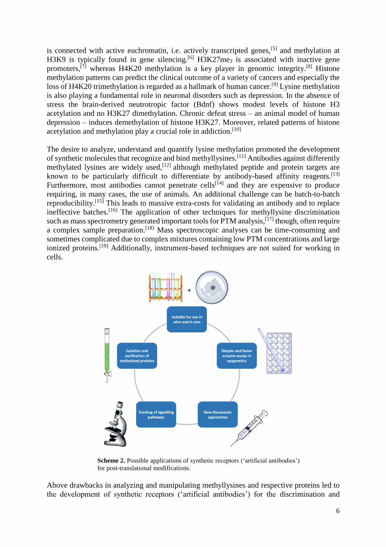
Scheme 2. Possible applications of synthetic receptors (‘artificial antibodies’)
for post-translational modifications.
Above drawbacks in analyzing and manipulating methyllysines and respective proteins led to
the development of synthetic receptors (‘artificial antibodies’) for the discrimination and

7
detection of methyllysines in water and biological media as well as in cells.[20] They are well-
defined small molecules, stable under ambient conditions and can be produced – more or less
cost-efficient – without the need for animals. Actual and potential applications of synthetic
receptors for methyllysines are shown in Scheme 2.
In the last years a broad range of synthetic ammonium ion receptors have been reported such
as cyclic peptides, crown ethers, calixarenes and cyclodextrins.[21] Thereby, for the recognition
of ammonium ions four types of interactions between host and guest are characteristic:
1. Steric and molecular complementarity
2. Ion pairs and salt bridges
3. Hydrogen bonds
4. Cation∙∙∙π interactions
Organic solvents allow strong hydrogen bonding between host and guest resulting in higher
associations constants than observed for polar solvents, such as methanol and water.[22] In these
more challenging media, hydrogen bonds between host and guest will provide less impetus for
binding.[23] As a consequence, other non-covalent interactions are amplified. In case of
methyllysines their recognition is based on the complexation of methylammonium ions.
Additional methyl groups at the NH2 moiety in ε-position of lysine does not change the overall
charge, however, result in less hydrophilic side chains. Hence, C-H∙∙∙π interactions and the
hydrophobic effect have a larger influence on the complexation.
Obviously, the ideal synthetic receptor for methyllysines will be able to discriminate them from
other amino acids and methylamines. However, it will also be able to distinguish between four
lysines featuring the most subtle differences in size, shape, basicity and lipophilicity resulting
from the presence of 0-3 methyl groups at the Nɛ position. Crystallographic studies of
methylated lysines complexed by peptides and proteins revealed that di- and trimethylated
lysine residues prefer so-called “aromatic cages”.[24, 25] The latter were the inspiration for quite
a number of synthetic methyllysine receptors. However, some successful candidates not even
contain phenyl moieties as discussed later.
The dissociation constant (Kd) is a measure for the thermodynamic stability of a host/guest
complex. It can be used to describe the affinity between receptor (host) and ligand (guest); a
high affinity of both binding partners is expressed by a low dissociation constant of the
complex. The most commonly used techniques for determining Kd values are: NMR
spectroscopy and isothermal titration calorimetry (ITC),[26] fluorescence displacement (FD),[27]
fluorescence anisotropy (FA),[28] and surface plasmon resonance SPR.[29] For comparison
reasons this review uses only Kd values in µM. For some methyllysine receptors these were
calculated from the respective association constant Ka (Kd=1/Ka). A detailed compilation of the
dissociation constants for all compounds discussed here can be found in Tables 1-11 and the
ESI (Tables S1-S8). As a matter of lucidity the lowest Kd values and highest selectivity are
shaded in each table.
Due their rather easy experimental set-up complexation studies in solution only deliver
restricted information on the geometry of host/guest complexes. Complementary X-ray
structures allow to study interactions between hosts and guests in more detail and give insight
into respective conformational parameter such as distances and torsions angles. So far, five
complexes of synthetic receptors with methylated lysines have been described in the
Cambridge Structural Database (CSD) and the Protein Data Bank (PDB), respectively. They
will be discussed in the corresponding sections of this review.

8
2. Calixarenes
2.1. Sulfonatocalixarenes
One way to mimic “aromatic cages” found in proteins are aromatic macrocycles with
cyclophanes[30] as typical examples. Cyclophanes (=cyclic phenylalkanes) were one of the first
synthetic receptors used for the recognition and complexation of alkylammonium ions. A
prominent subfamily are so-called calix[n]arenes, i.e. m-cyclophanes featuring phenolic groups
and a defined cavity; the digit in brackets defines the number of aromatic units.[31] The phenolic
region of a calixarene is called lower rim, the opposite section upper rim. The best researched
calixarenes are the cyclic tetramers, i.e. calix[4]arenes. They can exist in four different main
conformations (cone, partial cone, 1,3-alternate and 1,2-alternate), which are determined by
the respective lower rim substituents, temperature, solvent and possible complexing ions (e.g.
Na+ will force a flexible calix[4]arene into the cone form).[32]
Calixarenes and their derivatives are rather easy to synthesize and some show remarkable
bioactivity.[33] This is also true for sulfonatocalixarenes,[34] which have already been studied
extensively with respect to their inclusion properties in aqueous solution (ammonium ions,[35]
amino acids,[36] biological buffer components,[37] neutral species[38]) as well as in solid state.[39]
Studies carried out by Hof and co-workers[40] found that p-tetrasulfonatocalix[4]arene (1)
(Scheme 3) is able to discriminate between tri-, di-, mono- and unmethylated lysine with
dissociation constants of 27, 62, 250 and 1,923 μM, respectively, and a Kme0/Kme3 selectivity
of 71 (NMR, sodium phosphate buffer) (Table 1). Using fluorescence displacement assays and
glycine buffer even lower dissociation constants (7.7 μM for trimethyllysine) and higher
selectivity (>130) have been observed.[41] Primary drivers for the recognition events are
favorable enthalpies of binding (electrostatic interactions, non-classical hydrophobic
effects).[42]
Scheme 3. Structures of calixarenes 1-5 featuring varying
lower and upper rim substituents as well as ring sizes.
Further research revealed that biologically more relevant concentrations of sodium and
potassium ions in the buffer medium decrease the affinity of methyllysines to 1 by a factor of

9
2-3; lowering the temperature decreases dissociations constants (ca. 2 μM/K) (Table S1).[43]
Complexation studies with methylated and unmethylated peptides showed that both have
higher affinities to 1, though lower selectivity, than observed for the simple amino acids. As
one reason repulsive forces between the carboxylate anions of the amino acid guests and the
sulfonato group of the host have been discussed. Another reason could be attractive secondary
interactions between 1 and the amide backbone and neighboring side chains in the histone
peptides.[44] By way of interest, 1 binds H3K4me3, H3K9me3 and H3K27me3 equally well (Kd
=5.0-9.1 μM) as determined by ITC and, hence, possess a rather low specificity towards
different trimethylated lysines sites.
The interactions of tetrasulfonatocalix[4]arene (1) with trimethyllysines are in some cases
strong enough to compete with those of natural protein receptors. It has been shown that 1 is
able to interrupt the binding of histone H3K4me3 with its native protein binder ING2 PHD. By
way of interest the calixarene host disrupts the interaction of ING2 PHD and the trimethylated
histone (IC50 = 108 μM), though not between ING2 PHD and the dimethylated histone.[45]
Table 1. Dissociation constants for the binding of
p-tetrasulfonatocalix[4]arene (1) to differently
methylated lysines and respective histone peptides
(40 mM sodium phosphate buffer, pH 7.4).
Ref. Guest Kd
[μM] S[a] T [K] method
[40] Kme0 1923 - 298 NMR
[40] Kme1 250 8 298 NMR
[40] Kme1 333 - 303 ITC
[40] Kme2 62 31 298 NMR
[40] Kme2 95 - 303 ITC
[40] Kme3 27 71 298 NMR
[40] Kme3 28 - 303 ITC
[42] H3K4[b] 46 303 ITC
[42] H3K4me3 5.0 9 303 ITC [42] H3K9 101 303 ITC [42] H3K9me3 7.2 14 303 ITC [42] H3K27 220 303 ITC [42] H3K27me3 5.4 41 303 ITC [42] H3K36 128 303 ITC [42] H3K36me3 9.1 14 303 ITC
[a] S: Kme0/Kmex selectivity
[b] H3K4 = +H3N-ARTKQTAY-CONH2; H3K9 =
Ac-TARKSTGY-CONH2; H3K27 = Ac-
AARKSAPY-CONH2; H3K36 = Ac-
GGVKKPHY-CONH2
The complexes of tetrasulfonatocalixarene (1) with dimethyllysine hen egg-white lysozyme[46]
and cytochrome c containing nonmethylated lysines[47] give interesting insights into the driving
forces for methyllysine recognition. In the asymmetric unit of the dimethyllysine hen egg-white
lysozyme calixarene complex two host molecules bind to dimethyllysine residues and two bind
to arginine residues. The calixarenes that complex the dimethyllysine are in a symmetrical cone
conformation with interplanary angles of 61.7/62.7 ° and 65.1/66.6 °, respectively (Scheme
4a). One of the Nɛ methyl groups of each guest points directly into the calixarene cavity. The
distance between the carbon atom and the centroids of the calixarene phenyl rings range from

10
3.51 to 3.91 Å (four C-H∙∙∙π interactions). The second methyl group points towards two of the
sulfonato residues [d(C∙∙∙O)=3.57, 3.46 Å] and develops only one C-H∙∙∙π interaction (3.89 Å)
with the aromatic cavity.
The asymmetric unit of the complex of 1 with cytochrome c contains three guest molecules,
which all cap the protein at lysine residues. The calixarene host is again in a cone conformation,
though its geometry is somewhat distorted (interplanary angles 51.4/86.7 °, 58.4/80.4 °,
59.0/88.9 °). The lysine guests are interacting with the receptor cavity via their alkyl backbone.
The found C-H∙∙∙π interactions involve carbon atoms Cɛ (2-3 contacts per host/guest unit, 3.27-
3.99 Å) and Cδ (1-2 contacts per host/guest unit, 3.73-3.97 Å) (Scheme 4b). The ε-ammonium
unit is bend towards two the sulfonate groups preventing cation∙∙∙π interactions; a similar
feature is observed in the complex of lysine with 1 (CSD code: WIXSOL)[48]. Taking under
consideration the rather unspecific inclusion of lysine, dimethyllysine and arginine residues,
the binding behaviour of 1 seems to be rather ‘promiscuous’ – at least in the solid state.
A)
B)
Scheme 4. Details of the X-ray structures of tetrasulfonatocalixarene 1 with
A) dimethyllysine hen egg-white lysozyme (PDB code: 4N0J/4PRU)[47]
and B) cytochrome c (PDB codes: 3TYI;[46] unpublished alternative
binding mode: 4YE1). (For both complexes only one host/guest unit is
shown. Hydrogen atoms are omitted for clarity.)
The conformation of the calixarene host is of crucial importance for its inclusion behaviour.
The etherified calixarene 2 (Scheme 3) is in a so-called pinched cone conformation. Its closed
cavity prevents efficient trimethyllysine uptake. For calixarenes 3 and 4 the ethylene glycol
handles facilitate an open cavity leading to H3K27me3 affinities of 85 and 20 μM,
respectively.[42] Also the ring size of the calixarene has been varied. The resulting
hexasulfonatocalix[6]arene (5) still possesses a moderate selectivity for trimethylated lysine
over lysine (Kme0/Kme3=5), though presents only low affinities towards both (5,000 μM for
Kme0 and 1,074 μM for Kme3) (Table S2).[43]
In order to further increase the affinity of p-tetrasulfonatocalixarene 1 towards higher
methylated lysines, Hof and co-workers suggested to exchange one SO3- group against a phenyl
moiety or a bromine atom.[49] The resulting trisulfonato receptors (6-12) (Scheme 5) have the
potential to develop additional C-H∙∙∙π interactions towards the backbone of the lysine guests.
Within the series of receptors 6-12 only the calixarene featuring the underivatized phenyl
substituent (6) showed higher affinity (16 μM) and selectivity (150) towards trimethylysine in
comparison to 1 (27 μM, 71); no information has been given for the affinities towards the
recognition mono- and dimethyllysines. In later studies hosts 6-8, 11 and 12 have been
employed in fluorescence displacement assays to study their affinity to histone peptide H3K27
in its trimethylated and unmethylated form.[50] As observed for mother compound 1, the
affinities for the peptides are much higher than for the isolated amino acids (0.34-1.86 μM),

11
though the Kme0/Kmex selectivity decreases in most cases (e.g. from 150 to 25 for 6) (Table
3).
Scheme 5. For an improved binding of higher methylated
lysines one SO3- group in 1 has been exchanged against a
phenyl moiety or a bromine atom leading to trisulfonato
calixarenes 6-12
Table 3. Dissociation constants for the complexes of
trisulfonatocalixarenes 6-12 with lysine and
trimethyllysine and the respective histone peptide
H3K27mex (pH 7.4).
Host Guest Kd
[μM] S[a] solv. method
6[49] Kme0 2380 - A[b] NMR
6[49] Kme3 16 150 A NMR
6[49] Kme3 13 - A ITC
6[50] H3K27 19 - B[c] FD
6[50] H3K27me3 0.75 25 B FD
7[49] Kme0 4762 - A NMR
7[49] Kme3 476 10 A NMR
7[50] H3K27 11.3 - B FD
7[50] H3K27me3 0.88 13 B FD
8[49] Kme0 7143 - A NMR
8[49] Kme3 169 42 A NMR
8[50] H3K27 23 - B FD
8[50] H3K27me3 1.86 12 B FD
9[49] Kme0 9091 - A NMR
9[49] Kme3 588 16 A NMR
10[49] Kme0 5000 - A NMR
10[49] Kme3 192 26 A NMR
11[50] H3K27 2.7 - B FD
11[50] H3K27me3 0.34 8 B FD
12[49] Kme0 2273 - A NMR
12[49] Kme3 256 9 A NMR
12[50] H3K27 11.3 - B FD
12[50] H3K27me3 0.88 13 B FD [a] S: Kme0/Kmex selectivity
[b] A: 40 mM sodium phosphate buffer
[c] B: 10 mM sodium phosphate buffer

12
Only recently, the interactions of calixarenes 6 and 12 with cytochrome c have been studied in
solution and the crystalline state (PDB codes: 5KPF for 6 and 5LFT for 12).51 Though only
unmethylated lysines are available in cytochrome c, the dissociation constant for the complex
with 12 is 20 µM. This is slightly lower than for the complex of cytochrome c with parent
calixarene 1 (Kd=28 µM). In the crystalline complexes of 6 and 12 the NH3+ moieties of the
lysine residues avoid the cavity of the calixarenes similar to the respective X-ray structure of
1.
Kimura et al. employed trisulfonated calixarenes for the design of multivalent ligands.[52] The
conjugation of 6 via short amide linkers delivered mono-, di- and trivalent (13) receptors
(Scheme 6). The di- and trivalent ligands have higher binding affinities for methylated and
nonmethylated histones (0.39-0.86 μM) than the monovalent receptor (8.9-20.4 μM) (Table 4).
Noteworthy, for the trivalent system the Kd differences of respective complexes with histones
H3 and H4, acetylated histone H4KAc and H3K27me3 are only rather small. H3 and H4 are
rich in arginine and lysine, which suggests that nonspecific electrostatic interactions between
the charged guest and the calixarene host overrule the more discriminating cation∙∙∙π and C-
H∙∙∙π interactions between the methylated lysines the host cavity.[53] It should also be
considered that in this case the peptides were immobilized on a chip and the binding has been
studied using surface plasmon resonance (SPR).
Scheme 6. For trivalent receptor 13 three molecules of calixarene 6
have been conjugated via amide linkers. The affinity of 13 to
trimethyllysine is almost 23 times higher than observed for the
respective monovalent host.
Further research focussed on discriminating lysine trimethylation at specific lysine residues,
viz. H3K9me3 and H3K4me3. For that purpose a series of calixarene amides and sulfonamides
(14-19) has been synthesized (Scheme 7).[44],[45] They feature a somewhat higher flexibility in
comparison to the biphenyl calixarenes discussed above. Most (sulfon)amide receptors bind to
H3K9me3 and H3K4me3 with dissociation constants below 1 μM as determined by
fluorescence displacement assays (1: Kd=0.12 μM [H3K9me3], 0.02 μM [H3K4me3]; 14, 16-

13
18: Kd=0.14-0.49 μM [H3K9me3], 0.02-0.09 μM [H3K4me3]). Only for hosts 15 and 19 lower
affinities have been found (15: Kd=4.8 μM [H3K9me3], 7.8 μM [H3K4me3]; 19: Kd=0.51 μM
[H3K9me3], 1.6 μM [H3K4me3]). The H3K9me3/H3K4me3 selectivity is rather low for 15, 18
and 19 (0.3-1.6) and somewhat higher for 14 (9.5) and 17 (9.8), which is similar to the one of
calixarene 1 (7.8) (Table S3). (N.B.: Due to the different analytical methods applied a direct
comparison with data from Tables 1 and 2 is difficult.)
Table 4. Dissociation constants Kd [μM] for receptor 6 as
mono-, di - and trivalent amide conjugate to H4 and H3
tails (SPR, chip-immobilized peptides).
Peptide
Host H4[a] H4KAc[b] H3[c] H3K27me3[d]
Monovalent 17.8 10.0 20.4 8.9
Divalent 1.8 3.3 1.8 0.39
Trivalent (13) 0.73 0.86 0.51 0.39
[a] H-SGRGKGGKGLGKGGAKRHRKGGK(biotin)-NH2
[b] H-SGRG-K(Ac)-GG-K(Ac)-GLG-K(Ac)-GGA-K(Ac)-
RHR-K(Ac)-GGK(biotin)-NH2
[c] Ac-RKSTGGKAPRKQLATKAAR-Kme0-GGK(biotin)-NH2
[d] Ac-RKSTGGKAPRKQLATKAAR-Kme3-GGK(biotin)-NH2
Scheme 7. Structures of sulfonatocalixarenes 14-20 featuring one or two aromatic substituents at the upper
rim.
By way of interest, calixarenes 1 and 14-16 are able to disrupt the binding between the natural
histone binder CHD4 PHD2[54] and histone H3K9me3 (Kd=0.9 μM) in vitro without disturbing
the interaction between CHD4 and the unmethylated histone H3K9me0 (Kd=19 μM);

14
compound 14 showed the highest activity. Furthermore, all four compounds are active in
disrupting heterochromatin markers in cells.[45]
Due to its high affinities for trimethylated peptides, calixarene 14 has been employed in
supramolecular affinity chromatography for methylation-targeted proteomics. Linking 14 to
agarose beads allowed the resolution of histone peptides (H3K4mex, H3K27mex and
H3K27mex) on the basis of their methylation.[55, 56]
The recognition and purification of methyllysines with calixarenes is a crucial achievement,
though also their quantification has been in the focus. In 2013, a macrocyclic sensor array and
its method of use have been patented.[57] Thereby, a macrocycle is connected to a dye for
identifying histone-code-related analytes. Several macrocycles (e.g. calix[n]arenes, cyclo-
dextrines, cucurbit[n]urils), analytes (e.g. methyllysines, methylarginines, phosphotyrosines)
and fluorophores (e.g. fluorescein, dansyl, pyrene) have been disclosed. Only recently, Beatty
et al. presented trisulfonatocalixarene 20 (Scheme 7) as a tool for photochemical sensing of
trimethyllysines in biological media.[58] Macrocycle 20 features intrinsic fluorescence[59] and
the recognition and sensing processes tolerate various salts, metal ions and enzymatic
cofactors.
A)
B)
Scheme 8. A) Structure of NBD-labelled host 21. B) After the selective
binding of 21 to a trimethylated site of the peptide, a free lysine residue
nearby reacts with the NBD moiety in an SNAr reaction.
Gober and Waters used trisulfonatocalixarene 21 for the affinity labelling of Kme3-containing
histone peptides (Scheme 8). Thereby, the receptor unit of the molecule complexes
trimethyllysine in the peptide guest and the nitrobenzoxadiazole (NBD) group acts as the
reagent covalently labelling a nonmethylated lysine in the same peptide (SNAr mechanism).
The selectivity and rate of the labelling reaction proved to be significantly dependent on salt

15
and reagent concentration as well as pH. The utility of this new tool has been demonstrated in
a turn-on fluorescence HDAC assay.[60] NBD is a particularly attractive fluorophore as its
photophysical properties allow the use of a fluorescein isothiocyanate (FITC) filter.[61]
2.2. Carboxycalixarenes
Leung, Gruber and co-workers described a simple, readily synthesized monocarboxycalixarene
(22) that selectively binds to di/trimethylammonium groups (Table 6).[62] In comparison to
respective tri- and tetrasulfonatocalixarenes a monocarboxycalixarene is bearing only a single
charge with possible advantages with respect to cell permeability. Furthermore, the complex
of a monocarboxycalixarene and lysines will be neutral, which could be of interest for the
extraction or the delivery of (methylated) lysines. Carboxycalixarene 22 binds di- and
trimethyllysine with dissociations constants of 70 μM and 60 μM, respectively, though the
affinities towards non- and monomethylated lysines are very low (Kd>500 μM) (Table 5).
Receptor 22 is able to recognize methyllysines even in complex mixtures such as E. coli cell
lysate as demonstrated by NMR spectroscopy.
Table 5. Dissociation constants of
carboxycalixarene 22 with differently
methylated lysine guests (300 K,
NMR, 50 mM sodium phosphate
buffer, pH 7.5).
Guest Kd [μM]
Kme0 >500
Kme1 >500
Kme2 70
Kme3 60
PATGGV-Kme1-KPHRY >500
PATGGV-Kme2-KPHRY 60
PATGGV-Kme3-KPHRY 65
AR-Kme1-STGGK >500
AR-Kme2-STGGK 50
AR-Kme3-STGGK 50
choline 65
carnitine 60
meldonium 50
4-dimethylaminopyridine
(DMAP) 95

16
The energy-minimized complexes of 22 with methyllysines reveal, that only for higher
methylated lysines cation···π-interactions contribute to the recognition event (Scheme 9).
Furthermore, C-H···π-interactions seem to play a vital role for the formation and the stability
of the host/guest complexes. Di- und trimethyllysine develop four and five of these contacts,
respectively. Thereby, all of the N-methyl groups are involved in the interaction. In the case of
monomethyllysine two N-H∙∙∙O-hydrogen bond prevents the occurrence of C-H···π- and
cation···π-contacts.
In the energy-minimized structure of 22 with lysine three C-H···π-interactions and an
intramolecular N-H∙∙∙O-hydrogen bond involving the guest carboxylate and the ε-ammonium
unit has been observed. The N-H∙∙∙O prevent a close contact of the ammonium cation with the
π-electron rich cavity. Such a behavior has already been found in the X-ray structure of lysine
in the complex with tetrasulfonatocalixarene (1),[48] cytochrome c[47] and
hexaphosphonatocalix[6]arene (PDB code: 5LYC),[63] respectively.
A)
B)
C)
D)
Scheme 9. The energy-minimized host/guest complexes of 22 with trimethyllysine (A),
dimethyllysine (B), monomethyllysine (C) and lysine (D) demonstrate the preference
for higher methylated lysines. Only in A) and B) stabilizing C-H∙∙∙π and cation∙∙∙π
interactions are observed. (MacroModel V.9.8; OPLS_2001 force field; MCMM;
solvent: water; 20,000 steps.)
In contrast to tetra- and trisulfonatocalixarenes the binding of 22 to the di- and trimethyllysine
motif does not improve significantly when changing from free amino acids to peptides (Kd=50-
65 μM) (Table 5). The peptide PATGGV-Kme3-KPHRY is actually bound even worse than
the free amino acid – despite the fact that the trimethyllysine has a direct lysine neighbor in the
peptide. Host 22 only contains one anionic charge at the upper rim, though recognizes di- and
trimethyllysines in the same order of magnitude as tetrasulfonatocalixarene 1. Hence, the
repulsive forces between host and the amino acid carboxylate are much lower in 22 compared
to 1. As a consequence, the more or less unchanged affinities between 22 and the methyllysine
peptides must be primarily attributed to the lack secondary interactions between host and guest.

17
2.3. Structure-activity relationship of calixarenes
Due to the diverse analytical methods employed for the determination of the dissociation
constants, structure-activity relationships of the different calixarene hosts needs to be discussed
cautiously. Here, additional work seems necessary in order to gain more reliable data. In
general, it can be stated that higher charged receptors lead to higher affinities and selectivity (1
vs. 22). Calix[4]arenes are favoured over calix[6]arenes (1 vs. 5) and alternative anionic groups
at the upper rim lead to similar affinities for trimethyllysines.
In tetrasulfonatocalixarenes, the substitution of one SO3- group against a non-functionalized
phenyl substituent improves the binding of trimethyllysine and the Kme0/Kmex selectivity (1
vs. 6). However, functionalized phenyl substituents as in 7-11 result in drastically decreased
binding affinities and selectivity. In contrast, for histone peptide H3K27me3 the electronically-
activated p-methylphenyl group at the upper rim (11) delivered the highest affinity. The phenyl
moiety can also be attached to the calixarene via amide and sulphonamide bonds (14-19). The
additional flexibility resulted in highly active receptors for histone peptides H3K4me3 and
H3K9me3. It is somewhat surprising that also the plain tetrasulfonatocalixarene 1 is able to
complex both peptides with nanomolar affinity.
3. Resorcinarenes
Similar to calixarenes, resorcinarenes are m-cyclophanes, though bear two phenol groups at
each aromatic moiety instead of one. Resulting from their synthesis most resorcinarenes feature
additional alkyl groups at the methylene bridges. Their cavity size is comparable to those of
calixarenes. Four of the eight phenolic protons of resorcinarenes can be dissociated at pH >12.
The introduction of electron-withdrawing groups (such as –CN) increases the OH acidity of
the resorcinarene resulting in a pKa in the physiological pH region.[64] Chen and co-workers
deployed this phenomenon in tetracyano receptor 23 (Scheme 10), which proved to be a
powerful receptor for tetraalkylammonium ions with dissociation constants of 0.9-1.7 μM.[65]
Hamilton and co-workers further studied this receptor as methyllysine host revealing a
dissociations constant of 21 μM for Kme3, 68 μM for Kme2, 476 μM for Kme1 and over 1,000
μM for nonmethylated lysine.[66] These are comparable to the values found for
tetrasulfonatocalixarene (1). Both receptors, 1 and 23, have been successfully screened for their
potential to inhibit the KDM4A(=JMJD2A)-catalysed demethylation of a histone peptide
(H3K9me3) in vitro.
Scheme 10. Structure of tetracyanoresorcinarene 23.
The electron-withdrawing cyano groups result in a
lower pKa compared to the mother resorcinarene and,
hence, improve receptor solubility and the binding of
alkylammonium ions.

18
Despite their strong O-H∙∙∙O hydrogen bonds between adjacent OH functions resorcinarenes
are quite flexible molecules; bridging the phenol moieties leads to more rigid cavitands. An
example are tetraphosphonate hosts in which two OH groups of neighboring arene units are
each connected via a P=O bridge. This family of cavitands proved successful as synthetic
receptors for N-methylammonium salts as reported for receptor 24.[67] (Scheme 11)
Interestingly, the authors could also show that for tetraphosphonate cavitands the depth of
insertion of an N-Me group into a host cavity – determined by X-ray crystallography – can be
correlated to the binding constant. The affinity for the N+-Me group originates from three types
of non-covalent interactions between host and guest: a) cation∙∙∙dipole interactions (N+···O═P),
b) cation∙∙∙π interactions of the methyl group with the aromatic cavity and c) two hydrogen
bonds between the two nitrogen protons and two adjacent P═O bridges. By way of interest,
these features are also found in the X-ray structures of respective tetraphosphonates with
monomethyllysines as reported by Geremia, Dalcanale and co-workers,[68] viz. 25 in its
complex with methyllysine ∙ 2 HCl ∙ 6.4 CF3CH2OH ∙ H2O (CSD code: OJISEH) and 26 in its
complex with methyllysine ∙ HCl ∙ H2O (CSD code: IKOZUF) (Scheme 12).
Scheme 11. Tetraphosphonate cavitands 24-27
feature different alkyl residues at the methylene
bridge (R1) and the phosphonate ester (R2).
Scheme 12. X-ray structure of 25 in its complex
with methyllysine ∙ 2 HCl, CF3CH2OH and water
(1:6.4:1), CSD code: OJISEH. (Only the cavitand
and the methyllysine guest are shown.)

19
For cavitands 26 and 27 the dissociations constants of their complexes with monomethyllysine
have been determined. Unsurprisingly, the complex in MeOH (27: Kd=0.9 μM) has a higher
stability than those in aqueous media (26: Kd=671 μM in H2O; 885 μM in NaCl-containing
sodium phosphate buffer) (Table S5). Data for the complexation of non-methylated or di- and
trimethylated is not given in the reference.
In the following tetraphosphonate cavitands have been applied for the development of an
analytical platform based on plasmon-free surface enhanced Raman scattering (SERS). Using
this approach Alessandri et al. have been able to distinguish N-methyllysine hydrochloride
from lysine hydrochloride in water.[69] Later a tetraphosphonate cavitand for the recognition of
monomethyllysine in histone peptides has been introduced. It allows the discrimination of
single monomethylated peptides from multi monomethylated ones.[70]
Hooley, Zhong and co-workers introduced a fluorescence-based supramolecular tandem assay
for the in situ monitoring of a lysine demethylase (JMJD2E) or methyltransferase (PRDM9).[71]
This site-selective displacement assay system contains only three resorcinarene-based
cavitands (28-30) (Scheme 13). As all three have different charges their combination allows
the simultaneous investigation of different methylation sites, e.g. peptide sequence AR-Kme3-
ST (H3K9me3) over T-Kme3-QTA (H3K4me3) and AAR-Kme3-S (H3K27me3). (N.B.:
Receptor 30 featuring four positive charges is able to recognize an alkylammonium ion!)
Scheme 13. Structures of cavitands 28-30.
Receptor 28 and related hosts[72] are well known
for their high affinities towards alkylammonium
ions.[73]
4. Pillararenes
Not all electron-rich macrocycles have high affinities to alkylammonium ions and are suited to
discriminate methyllysines as demonstrated by pillararene 31 (Scheme 14).[74] It features ten
carboxyl groups as well as a stable cavity and is able to bind basic amino acids such as Lys,
Arg and His with dissociation constants of 555 μM, 169 μM and 667 μM. Driving forces are
electrostatic interactions between COO- and the cationic side chains and hydrophobic
interactions. The complex of 31 with trimethyllysine was found to be less stable as KMe3 is
not able to act as a hydrogen bond donor in contrast to Lys.

20
Scheme 14. Pillararene 31 has a stable, electron-rich
cavity though binds lysine (555 μM) with a higher
affinity than trimethyllysine (769 μM).
5. Disulfide cyclophanes
Already back in 1990 Dougherty et al. developed a cyclophane consisting of two xylene and
two ethenoanthracene units connected via ether bridges. It binds acetylcholine with a
dissociation constant of 50 μM, a value comparable to those of biological recognition sites.[75]
In the following the xylene units have been replaced by a series of (hetero)aromatic units
(Scheme 15a).[76] The resulting receptors favor peralkylated ammonium ions over lower
alkylated ammonium ions due to the higher desolvation barriers for the latter. Later, the group
demonstrated the importance of cation∙∙∙π interactions for the complexation of alkylammonium
ions.[77] In 2008, Otto and co-workers[78] employed the – appropriately functionalized –
building blocks of the first Dougherty receptor for the composition of a dynamic combinatorial
library (DCL).[79] The components of this library proved to be disulfide macrocycles with 32
as one example (Scheme 15b). All its members have been studied towards its ability to
recognize peralkylated ammonium ions, which were also used as templates in the reaction.
A)
B)
Scheme 15. A) General structure of Dougherty’s ethenoanthracene receptors. B) Structure of disulfide
cyclophane 32 found in a dynamic combinatorial library (DCL) with A and B as only building blocks.
Waters and co-workers used A, B and related aromatic dithiols to create a dynamic
combinatorial library similar to the one of Otto, though, in this case dipeptide Ac-Kme3-G-NH2

21
has been used as molecular target. By way of interest, again receptor 32 (Scheme 15c) was the
most amplified and was found – after separation and purification – to bind methylated lysines.
The binding affinity for histone H3K9me3 is about 25 μM, which is similar to the binding of
native HP1 chromodomain, a biological methyllysine receptor (Table 9).[80] Moreover, 32 can
discriminate differently methylated lysines.[81] Interestingly, the binding strength and
selectivity varies quite drastically depending on the method used for its determination, e.g. 25
μM vs. 2.6 μM and >48 vs. 8.5 for histone H3K9me3 (fluorescence anisotropy vs. ITC).[82] The
authors explain this with incomplete desalting of the samples measured by fluorescence
anisotropy.
Table 9. Dissociation constants for the binding of disulfide
macrocycle 32 and HP1 chromodomain to histone peptide
H3K9 in its different methylation states
Host Guest Kd [μM] S[a] solv. method
rac-32[81] H3K9me0 >1200 - A[b] FA
rac-32[81] H3K9me1 166 >7 A FA
rac-32[81] H3K9me2 58 >20 A FA
rac-32[81] H3K9me3 25 >48 A FA
rac-32[82] H3K9me0 22 - B[c] ITC
rac-32[82] H3K9me1 13.9 1.6 B ITC
rac-32[82] H3K9me2 6.3 3.5 B ITC
rac-32[82] H3K9me3 2.6 8.5 B ITC
HP1[80] H3K9me0 >1000 - C[d] FA
HP1[80] H3K9me1 96 >10 C FA
HP1[80] H3K9me2 15 >66 C FA
HP1[80] H3K9me3 10 >100 C FA
[a] S: Kme0/Kmex selectivity
[b] A: 10 mM phosphate buffer; 300 K; pH 8.5
[c] B: 10 mM borate buffer; 299 K; pH 8.5
[d] C: phosphate buffer, 25 mM NaCl, 1 mM DTT; 288 K;
pH 7.5
In order to increase and vary the binding affinities and selectivity of receptor 32, the Waters
group used the DCL approach to generate macrocycles 33-38 (Scheme 16). They have
impressively high affinities to histone peptide H3K9 in its different methylation forms ranging
from 0.13-2.6 μM (trimethylation), 0.18-13.2 μM (dimethylation), 1.0-40 μM (mono-
methylation) and 1.8-58 μM (unmethylated peptide) (Table S6). [82],[83],[84] For a broad
application of disulfide receptors as trimethyllysine sensors a late stage modification has been
suggested.[85] As other synthetic receptors present here the hosts from dynamic combinatorial
libraries have been employed in fluorogenic sensor platforms using an indicator displacement
system.[86]

22
Scheme 16. The variation of the aryl unit in the Waters receptor (32) produce disulfide
macrocycles 33-38. Two COOH functions at the exchangeable aromatic moiety as in 33, 35 and
37 result in lower dissociation constants (0.13-0.30 μM) and – in most cases – better selectivity
(8.1-35) compared to 32 (Kd=2.6 μM; 8.5).
Disulfide cyclophanes 32-38 show interesting structure-activity relationships primarily based
on the location and number of COO- functions at the individual aryl units. When the position
of the carboxylate in 32 is changed from meta to ortho (34) or a phenylene residue is inserted
(38) the affinity towards methyllysines is scarcely affected (change from 2.6 to 2.3 and 2.6/2.2,
respectively). In case of 34 the Kme0/Kme3 selectivity stays more or less the same (change
from 8.5 to 9.6), for 38 it decreases significantly (2.7/4.7). The exchange of the benzene moiety
in 32 against a naphthalene ring – leading to host 36 – results in a much higher Kme0/Kme3
selectivity (>58) than observed for 32 (8.5), though a more or less unchanged affinity for
trimethyllysine (1.4 μM). An explanation deliver improved C-H∙∙∙π-interactions facilitated by
the naphthalene ring, which benefit the higher methylated lysines. Cyclophanes 33, 35 and 37
are featuring an additional COO- group each, which support secondary interactions with the
peptide chain. These obviously lead to much lower dissociation constants for the complex with
trimethyllysine (0.13-0.30 μM) and very good (33, 35) or good (37) Kme0/Kme3 selectivity
(35, 34 and 16/8.1, respectively).
Most receptors discussed here have been designed to achieve high affinities and selectivity
towards trimethylation. Though, one isomer of macrocycle 37 shows a slightly higher affinity
for dimethylated histone protein H3K9 (Kd=0.20 μM) than for the trimethylated one (Kd=0.22
μM). This has also been demonstrated for peptide Ac-Kmex GGY-NH2 with Kd=3.32 μM
(dimethylation) and 4.30 μM (trimethylation), respectively.[84]
Macrocycle 33 is a good example for a synthetic receptor with comparably high affinities for
trimethyllysines in different peptides, though different Kme0/Kmex selectivity. For histone
peptides H3K9 (Ac-WGGG-QTAR-Kmex-STG-NH2) and H3K36 (Ac-WGGG-TGGV-Kmex-
KPH-NH2) the trimethylated lysine is complexed with the same affinity (Kd=0.3 μM).

23
However, the affinities of 33 for the unmethylated histone peptides are 10.5 μM (H3K9) and
≈70 μM (H3K36).[82]
For receptor 33 the Waters group also studied the binding affinity towards lysine and
trimethyllysines under the influence of neighboring arginine and lysine residues, i.e. possible
secondary interactions between host and guest. Their research revealed that, in general, the
binding improves when additional cationic side chains (Arg or Lys) are present in the peptide.
However, the position of the additional cationic amino acid residue has only a little influence
on the binding affinities (Kd=0.31-0.62 μM) and the Kme0/Kme3 selectivity decreases in most
cases (20-58), except for a lysine in direct neighborhood (100) (Table 10).[87]
Table 10. Dissociation constants for the binding
of 33 to non- and trimethylated model peptides
featuring varying distances of a neighboring Arg
and Lys. (299 K, ITC, 10 mM borate buffer, pH
8.5)[87]
Ac-WGGGG-Zi-3-Zi-2-Zi-1-Kmex-GGG-NH2
Zi-3 Zi-2 Zi-1 x Kd
[μM]
Kme0/Kme3
selectivity
G G G 0 140 67
G G G 3 2.1
G G R 0 12 20
G G R 3 0.62
G R G 0 13 29
G R G 3 0.46
R G G 0 17 34
R G G 3 0.50
G G K 0 31 100
G G K 3 0.31
G K G 0 23 58
G K G 3 0.40
6. Cucurbiturils
Cucurbit[n]urils are synthetic macrocycles consisting of glycoluril monomers linked by
methylene bridges. In contrast to all other synthetic hosts discussed here, cucurbiturils do not
contain aromatic moieties. Nevertheless, they have proven as versatile receptors in
supramolecular chemistry.[88] Unlike underivatized calixarenes and other cyclophanes
cucurbiturils are water-soluble. In the last years mainly cucurbit[7]uril (Scheme 17) has been
studied with respect to its ability to form complexes with lysine and other amino acids[89] as
well as tetraalkylammonium ions (Kd=1.0-8.3 μM).[90],[91] In 2013, Macartney and co-worker
studied the selective molecular recognition of methylated lysines by cucurbiturils with
dissociations constants of 0.5 μM, 17 μM, 556 μM, 1,887 μM for tri-, di- and monomethylation
as well as lysine, respectively.[92] Interestingly, 39 binds to trimethylated lysine over 3,500
times better than to lysine. This is the highest selectivity observed so far for the recognition of
methylated lysines. Thus, cucurbit[7]uril has even a higher selectivity than natural protein
receptors (ING2 = 1,500[93], ADDATRX = 7.4[94] / 28[95], HP1 > 100[80]) (Table S7). In
cucurbituril complexes the high affinities can clearly not be explained by cation∙∙∙π and C-H∙∙∙π
interactions, though by the release of high energy water from the hydrophobic cavity (non-
classical hydrophobic effect)[96] and ion-dipole interactions.

24
Scheme 17. Cucurbit[7]uril (39) is the
synthetic receptor with the highest
selectivity for trimethylated lysine
observed so far.
Recently, Crowley and co-workers described the complex of dimethylated Ralstonia
solanacearum lectin with cucurbit[7]uril (39) (PDB codes: 6F7W/6F7X).[97] In the X-ray
structure they found three different modes of binding, suggesting an incomplete filled host
cavity (Scheme 18), which could also explain the rather low affinity in solution (Kd≈1,000 μM).
Like calixarene 1, cucurbit[7]uril (39) has also been reported to recognize proteins not
containing methyllysines with rather high affinity. An example is the complex with human
insulin (PDB code: 3Q6E), in which the N-terminal phenylalanine residue is preferentially
recognized by the cucurbituril over other amino acid sidechains (Kd=0.7 μM).[98]
Scheme 18. Detail of the complex of cucurbit[7]uril
(39) and dimethylated Ralstonia solanacearum
lectin (PDB code: 6F7W).[97] (Only one host/guest
entity is shown. Hydrogen atoms are omitted for
clarity.)
Zong and co-workers employed sulfonatocalixarenes 1 and 5 as well as cucurbituril 39 in the
separation of methylated histone proteins by host-mediated capillary electrophoresis.[99] The
molecular recognition event changes the electrophoretic mobility of the differently methylated
peptides. The addition of calixarenes 1 and 5 to the background electrolyte led to their
successful separation; cucurbituril 39 has been less effective.

25
7. Acyclic receptors
The recognition of amino acids by synthetic receptors is not restricted to cyclic host systems.
Early examples for acyclic receptors comprise guanidium receptors[100] introduced by Schmuck
and the molecular tweezers[101] of Klärner and Schrader. Other examples of acyclic hosts are
tripodal receptors on the base of substituted trimethyl- and triethylbenzenes. These have proven
as very successful hosts especially for the recognition of sugars.[102] So far only one tripodal
receptor, viz. trisindol 40 (Scheme 19), has been studied towards its capacity to complex
methylated lysines. However, as 40 shows a low degree of preorganization only unsatisfying
affinities towards trimethyllysine have been observed (15,873 μM[103]/4,000 μM[104]). Due to
their higher hydrophobicity tetraalkylammonium ions with long alkyl chains are complexed
with much higher affinities, e.g. tetrabutylammonium chloride with a dissociations constant of
37 μM[103]/142 μM (Table S8).[104]
Scheme 19. Trisindol receptor 40
complexes long chain tetraalkyl-
ammonium ions with high affinitiy,
though, is less suited for the binding of
trimethyllysine.
8. Summary and outlook
The article on methyllysine recognition by tetrasulfonatocalixarene 1 by Hof and his group in
2010 demonstrated for the first time the feasibility of small molecule receptors as potential
hosts for post-translationally modified lysines as free amino acids, in peptides and in proteins.
Since then quite a number of receptors for differently methylated lysines has been described.
Some of them were already studied before with respect to their ability to bind alkylammonium
ions, some were newly introduced. Interestingly, only one family of methyllysine hosts, viz.
the ethenoanthraceno receptors, is chiral despite the methyllysine guests occur only as single
enantiomer – at least in natural peptides and proteins.
In general, it can be stated that methyllysine hosts have higher affinities to methyllysine-
containing peptides and proteins than to methyllysines as amino acids. An explanation deliver
attractive secondary interactions between the synthetic hosts and the peptide guests. However,
in some cases it is problematic to directly compare the binding constants of the various
receptors due to varying experimental techniques and conditions in the different references.
Hence, it seems not constructive to choose the methyllysine host with the lowest dissociation
constant and nominate a ‘winner’. Different applications will require different receptor
26
properties. Depending on the envisaged use overall charge or pH dependency[105] may be more
important than selectivity. In some cases competing guests such as Nα-methylamino acids[67]
or arginine[106] need to be considered; some receptors may not be stable under the assessed
conditions, e.g. due to S-S bonds.[107] In some cases the low solubility in water may restricted
the applications of the ‘synthetic antibodies’. Another possible challenge is the provision of the
artificial methyllysine host: tetrasulfonatocalix[4]arene (1) and cucurbit[7]uril (39) are both
commercially available, many others require elaborate synthetic procedures making it more
difficult for users beyond chemistry to work with them.
Interestingly, only a rather low number of X-ray structures of methyllysine complexes have
been described. Thereby, the binding preferences of the synthetic receptor in solution is not
always directly comparable with the situation in solid state – as shown for the rather
promiscuous binding of some title compounds. However, X-ray structures of methyllysine
inclusion compounds and complexes give valuable information on non-covalent interactions
between host and guest and can help to further improve the selectivity and specificity (generic
vs. specific receptors).
On the long run, research on hosts for methyllysines will also benefit the development of
‘artificial antibodies’ for similar post-translational modifications such as methylarginine,[108]
methylhistidine,[109] and methyladenin.[110] The general concepts found for methyllysine
binding will further stimulate the recognition of related guest species such as methylamines
like ecstasy,[111] amphetamines[112] or other illegal substances.[113] Already, synthetic
methyllysine receptors have prompted the development of macrocycles for the detoxification
of organophosphonates, which can be – and are – misused as chemical warfare agents. By way
of example, Kubik and co-workers reported on a series of trisulfonatocalixarenes for the
catalytic hydrolysis of V-type nerve agents and soman demonstrating once again the variability
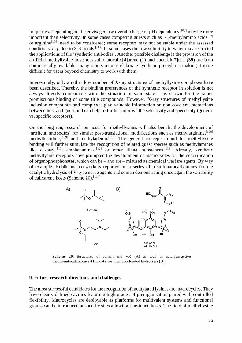
of calixarene hosts (Scheme 20).[114]
A)
B)
Scheme 20. Structures of soman and VX (A) as well as catalytic-active
trisulfonatocalixarenes 41 and 42 for their accelerated hydrolysis (B).
9. Future research directions and challenges
The most successful candidates for the recognition of methylated lysines are macrocycles. They
have clearly defined cavities featuring high grades of preorganization paired with controlled
flexibility. Macrocycles are deployable as platforms for multivalent systems and functional
groups can be introduced at specific sites allowing fine-tuned hosts. The field of methyllysine

27
recognition is still in its infancies and the author of this review can only speculate about future
developments. It can be assumed that prospective receptors will be designed either to be
specific or generic in their recognition behavior – in both cases with high Kme0/Kmex
selectivity. As proposed by the author and co-workers macrocycles with intrinsic fluorescence
could be one way forward.[59] Furthermore, bioinformatics combined with molecular modelling
and crystallographic approaches will help to design new generations of ‘artificial antibodies’
for the recognition of the methyllysine motif. A challenge for the future is clearly the better
comparability between the different synthetic hosts. The reviewed literature uses six different
analytical techniques to determine the dissociations constant of respective complexes, which
makes authentic structure-activity relationships rather difficult. It would also be helpful to
screen not only lysine and trimethyllysine, but also mono- and dimethyllysines.
As shown in Scheme 2 synthetic receptors for trimethyllysines offer a broad range of
applications. Significant progress has been made for the analysis and purification of
methyllysines. In several cases the target compounds have been employed in assays using
demethylases and/or methyltransferases. Moreover, the biological activity of some
methyllysine receptors is a promising start for new therapeutic agents. In no way synthetic
receptors will be able to fully replace antibodies, though the reviewed literature delivers
promising points of contacts for amendatory applications.
The use of synthetic methyllysine receptor as therapeutic agents is only possible with an
acceptable toxicity. Single injected doses of tetrasulfonatocalixarene 1 (equivalent to 2-5 g in
humans) showed no acute toxicity[115] and no toxicity towards various tumor cell lines has been
observed.[116] It develops no haemolytic toxicity observed for concentrations up to 5 mM[117]
and is not activating neutrophils, hence, does not induce an immune response[118]. Also
cucurbiturils have demonstrated a rather low toxicity.[119] Despite they are able to cross the cell
membrane of mouse embryo cells,[120] cucurbiturils lack of cytotoxicity in mammalian cells
(up to 1 mM).[121] Intravenously administered cucuribt[7]uril (39) has demonstrated no toxicity
at doses up to 200 mg/kg,[122] though has a measureable cardiotoxicity at concentrations > 500
μM.[123] Other synthetic receptors for methyllysine may have much higher toxicity levels,
whose determination should be a point of contact for further studies.
Most of the synthetic receptors for methyllysines discussed here also recognize a broad range
of other alkylammonium ions. In some cases the latter are complexed with even higher
affinities than methyllysines. As most applications use the recognition of methylated lysines in
biological media naturally occurring alkylammonium ions may disturb the desired recognition
event. An example are cucurbiturils, that – when transferred into the cell – would not only
recognize methyllysines, but may also interact with spermine and spermidine with the resulting
complexes affecting DNA-modifying enzymes.[124] More studies are necessary to rule out
cross-interactions, especially as hosts 1 and 14-16 have been described to disrupt the interaction
of histone peptides with their natural protein binder, hence, already demonstrated their
influence on DNA activity.
Another broad topic for future research are off-target effects of synthetic receptors for
methylated lysines. A possible example is tetrasulfonatocalixarene 1 and its ability to hydrolyze
ATP (Scheme 21).[125] After application of 1 on cells or on organisms – aiming for the
discrimination of methyllysines – the artificially decreased ATP level could lead to artefacts as
(ATP)-dependent chromatin remodeling enzymes[126] may be influenced. Would other
sulfonatocalixarenes be active as well? Other untried interactions are conceivable as 1 is also
a high affinity blocker of chloride channels[127] and volume-regulated anion channels.[128]
Furthermore, it is known for its antithrombotic and anticoagulant properties.[129] By way of
interest, both calixarene 1 and cucurbituril 39 inhibit amyloid fibrillation by multipoint

28
hydrophobic interactions,[130] which may lead to competing host/guest interactions in
respective systems.
Scheme 21. Proposed complex of 1 with ATP
during the catalytic hydrolysis of the guest.[125]
The blood-brain barrier prevents the uptake of large hydrophilic and highly anionic artificial
hosts as shown for tetrasulfonatocalixarene 1.[115] Nevertheless, in the long term the use of
synthetic receptors for methyllysines in the brain may be feasible. Several methyllysine
receptors (e.g. 23, 39, etc.) have also a high affinities to acetylcholine and the complexation of
the neurotransmitter could lead to artefacts when screening for methyllysines. Possible are also
off-target effects: receptor 43 (Scheme 22) – closely related to tetracyano receptor 23 – was
found to inhibit the hydrolysis of acetylcholine,[131] hence, may influence synapse activities.
Future research could help to establish if guest hydrolysis is a general characteristic of synthetic
trimethylammonium receptors.
Scheme 22. Resorcinarene 43 is an inhibitor of
acetylcholine hydrolysis.
10. Acknowledgments
Financial support from the School of Pharmacy, University of Lincoln, is gratefully
acknowledged. TG wants to thank Manfred Jung (Institute of Pharmaceutical Sciences,
University of Freiburg/Breisgau) for introducing him to the fascinating field of epigenetics.
11. References
1 T. Jenuwein, C. D. Allis, Science 2001, 293, 1074-1080.
2 M. Luo, Chem. Rev. 2018, Ahead of Print. DOI: 10.1021/acs.chemrev.8b00008

29
3 a) W. Sippl, M. Jung (Eds.), Epigenetic Targets in Drug Discovery, Wiley-VCH, Weinheim,
2009; b) S. Krishnan, S. Horowitz, R. C. Trievel, ChemBioChem 2011, 12, 254-263; c) R. L.
Hancock, M. I. Abboud, T. J. Smart, E. Flashman, A. Kawamura, C. J. Schofield, R. J.
Hopkinson, ChemBioChem 2018, 19, 917-921.
4 a) C. B. Yoo, P. A. Jones, Nat. Rev. Drug Discovery 2006, 5, 37-50; b) P. Chi, C. D. Allis, G.
G. Wang, Nat. Rev. Cancer 2010, 10, 457-469; c) M. A. Dawson, T. Kouzarides, Cell 2012,
150, 12-27.
5 L. M. Soares, P. C. He, Y. Chun, H. Suh, T. Kim, S. Buratowski, Mol Cell. 2017, 68, 773-
785.
6 J.-A. Park, A.-J. Kim, Y. Kang, Y.-J. Jung, H. K. Kim, K.-C. Kim, Mol Cell. 2011, 31, 343-
349.
7 E. Martinez-Garcia, J. D. Licht, Nat. Gen. 2010, 42, 100-101.
8 S. Jørgensen, G. Schotta, C. S. Sørensen, Nucleic Acids Res. 2013, 41, 2797-2806.
9 Y. Yokoyama, A. Matsumoto, M. Hieda, Y. Shinchi, E. Ogihara, M. Hamada, Y. Nishioka,
H. Kimura, K. Yoshidome, M. Tsujimoto, N. Matsuura, Breast Cancer Research 2014, 16,
R66
10 N. Tsankova, W. Renthal, A. Kumar, E. J. Nestler, Nat. Rev. 2007, 8, 355-367.
11 B. D. Smith (Ed.), Synthetic Receptors for Biomolecules, Royal Society of Chemistry, 2015.
12 S. M. Fuchs, B. D. Strahl, Epigenomics 2011, 3, 247-249.
13 a) I. Bock, A. Dhayalan, S. Kudithipudi, O. Brandt, P. Rathert, A. Jeltsch, Epigenetics 2011,
6, 256-263; b) S. B. Rothbart, B. M. Dickson, J. R. Raab, A. T. Grzybowski, K. Krajewski, A.
H. Guo, E. K. Shanle, S. Z. Josefowicz, S. M. Fuchs, C. D. Allis, T. R. Magnuson, A. J.
Ruthenburg, B. D. Strahl, Mol. Cell 2015, 59, 502-511.
14 Y. Zhao, D. Lou, J. Burkett, H. Kohler, J. Immunol. Methods 2001, 254, 137-145.
15 J. R. Couchman, J. Histochem. Cytochem. 2009, 57, 7-8.
16 A. Schonbrunn, Mol. Endocrinol. 2014, 28, 1403-1407.
17 a) M. Bremang, A. Cuomo, A. M. Agresta, M. Stugiewicz, V. Spadotto, T. Bonaldi, Mol.
BioSyst. 2013, 9, 2231-2247; b) A. Guo, H. Gu, J. Zhou, D. Mulhern, Y. Wang, K. A. Lee, V.
Yang, M. Aguiar, J. Kornhauser, X. Jia, J. Ren, S. A. Beausoleil, J. C. Silva, V. Vemulapalli,
M. T. Bedford, M. J. Comb, Mol. Cell. Proteomics 2014, 13, 372-387.
18 a) B. A. Garcia, S. Mollah, B. M. Ueberheide, S. A. Busby, T. L. Muratore, J. Shabanowitz,
D. F. Hunt, Nature Protocols 2007, 2, 933-938; b) Z. Wu, Z. Cheng, M. Sun, X. Wan, P. Liu,
T. He, M. Tan, Y. Zhao, Mol. Cell. Proteomics 2015, 14, 329-339.
19 E. S. Witze, W. M. Old, K. A. Resing, N. G. Ahn, Nat. Methods 2007, 4, 798-806.

30
20 a) K. D. Daze, F. Hof, Acc. Chem. Res. 2013, 46, 937-945; b) A. Shaurya, K. I. Dubicki, F.
Hof, Supramol. Chem. 2014, 26, 583-590; c) F. Hof, Chem. Commun. 2016, 52, 10093-10108.
21 A. Späth, B. König, Beilstein J. Org. Chem. 2010, 6, doi: 10.3762/bjoc.3.32.
22 E. A. Kataev, C. Müller, Tetrahedron 2014, 70, 137-167.
23 a) T. H. Rehm, C. Schmuck, Chem. Soc. Rev. 2010, 39, 3597-3611; b) E. Persch, O. Dumele,
F. Diederich, Angew. Chem. Int. Ed. 2015, 54, 3290-3327.
24 S. D. Taverna, H. Li, A. J. Ruthenburg, C. D. Allis, D. J. Patel, Nat. Struct. Biol. 2007, 14,
1025-1040.
25 C. Rapp, E. Goldberger, N. Tishbi, R. Kirshenbaum, Proteins 2014, 82, 1494-1502.
26 P. Thordarson, Chem. Soc. Rev. 2011, 40, 1305-1323.
27 P. Kuzmič, M. L. Moss, J. L. Kofron, D. H. Rich, Anal. Biochem. 1992, 65-69.
28 T. D. Pollard, Mol. Biol. Cell 2010, 21, 4061-4067.
29 Y. Tang, X. Zeng, J. Liang, J. Chem. Educ. 2010, 87, 742-746.
30 R. Gleiter, H. Hopf (Eds.), Modern Cyclophane Chemistry, Wiley: Weinheim, 2004.
31 C. D. Gutsche, Calixarenes, The Royal Society of Chemistry, 2008.
32 a) M. Gruner, C. Fischer, T. Gruber, E. Weber, Supramol. Chem. 2010, 22, 256-266; b) T.
Gruber, M. Gruner, C. Fischer, W. Seichter, E. Weber, P. Bombicz, New J. Chem. 2010, 34,
250-259; c) C. Fischer, T. Gruber, D. Eißmann, W. Seichter, E. Weber, Cryst. Growth Des.
2011, 11, 1989-1994; d) T. Gruber, F. Eißmann, M. Gruner, L. G. Reuter, W. Seichter, E.
Weber, CrystEngComm. 2014, 16, 3730-3736.
33 a) R. V. Rodik, V. I. Boyko, V. I. Kalchenko, Curr. Med. Chem. 2009, 16, 1630-1655; b) M.
Giuliani, I. Morbioli, F. Sansone, A. Casnati, Chem Commun. 2015, 51, 14140-141159; c) M.
M. Naseer, M. Ahmed, S. Hameed, Chem. Biol. Drug Des. 2017, 89, 243-256.
34 a) D.-S. Guo, Y. Liu, Acc. Chem. Res. 2014, 47, 1925-1934; b) C. Hoskins, A. D. M. Curtis,
Nanomed. Res. 2015, 2, 00028.
35 a) J. L. Atwood, L. J. Barbour, P. C. Junk, G. W. Orr, Supramol. Chem. 1995, 5, 105-108;
b) L. Memmi, A. N. Lazar, A. Brioude, V. Ball, A. W. Coleman, Chem. Commun. 2001, 2474-
2475; c) H.-J. Buschmann, L. Mutihac, E. Schollmeyer, J. Incl. Phenom. Macrocyclic Chem.
2003, 46, 133-137; d) F. Perret, A. N. Lazar, A. W. Coleman, Chem. Commun. 2006, 2425-
2438; e) G. Arena, A. Casnati, A. Contino, A. Magrì, F. Sansone, D. Sciotto, R. Ungaro, Org.
Biomol. Chem. 2006, 4, 243-249.
36 C. Bonaccorso, S. Gentile, F. G. Gulino, D. Sciotto, Lett. Org. Chem. 2009, 6, 598-603.

31
37 E. da Silva, F. Nouar, A. W. Coleman, B. Rather, M. J. Zaworotko, J. Incl. Phenom.
Macrocyclic Chem. 2003, 46, 161-166.
38 N. Kon, N. Iki, S. Miyano, Org. Biomol. Chem. 2003, 1, 751-755.
39 O. Danylyuk, K. Suwinska, Chem. Commun. 2009, 5799-5813.
40 C. S. Beshara, C. E. Jones, K. D. Daze, B. J. Lilgert, F. Hof, ChemBioChem 2010, 11, 63-
66.
41 M. Florea, S. Kudithipudi, A. Rei, M. J. Gonzalez-Alvarez, A. Jeltsch, W. M. Nau, Chem.
Eur. J. 2012, 18, 3521-3528.
42 K. D. Daze, T. Pinter, C. S. Beshara, A. Ibraheem, S. A. Minaker, M. C. Ma, R. J. M.
Courtemanche, R. E. Campbell, F. Hof, Chem. Sci. 2012, 3, 2695-2699.
43 K. D. Daze, C. E. Jones, B. J. Lilgert, C. S. Beshara, F. Hof, Can. J. Chem. 2013, 1072-1076.
44 H. F. Allen, K. D. Daze, T. Shimbo, A. Lai, C. A. Musselman, J. K. Sims, P. A. Wade, F.
Hof, T. G. Kutateladze, Biochem. J. 2014, 459, 505-512.
45 M. Ali, K. D. Daze, D. E. Strongin, S. B. Rothbart, H. Rincon-Arano, H. F. Allen, J. Li, B.
D. Strahl, F. Hof, T. G. Kutateladze, J. Biol. Chem. 2015, 290, 22919-22930.
46 a) R. E. McGovern, A. A. McCarthy, P. B. Crowley, Chem. Commun. 2014, 50, 10412-
10415; b) R. E. McGovern, B. D. Snarr, J. A. Lyons, J. McFarlane, A. L. Whiting, I. Paci, F.
Hof, P. B. Crowley, Chem. Sci. 2015, 6, 442-449.
47 R. E. McGovern, H. Fernandes, A. R. Khan, N. P. Power, P. B. Crowley, Nat. Chem. 2012,
4, 527-533.
48 M. Selkti, A. W. Coleman, I. Nicolis, N. Douteau-Guevel, F. Villain, A. Tomas, C. De
Rango, Chem. Commun. 2000, 161-162.
49 K. D. Daze, M. C. Ma, F. Pineux, F. Hof, Org. Lett. 2012, 14, 1512-1515.
50 S. Tabet, S.F. Douglas, K. D. Daze, G. A. E. Garnett, K. J. H. Allen, E. M. M. Abrioux, T.
T. H. Quon, J. E. Wulff, F. Hof, Bioorg. Med. Chem. 2013, 21, 7004-7010.
51 A. M. Doolan, M. L. Rennie, P. B. Crowley, Chem. Eur. J. 2018, 24, 984-991.
52 Y. Kimura, N. Saito, K. Hanada, J. Liu, T. Okabe, S. A. Kawashima, K. Yamatsugu, M.
Kanai, ChemBioChem 2015, 16, 2599-2604.
53 J. E. Beaver, M. L. Waters, ACS Chem Biol. 2016, 11, 643-653.
54 C. A. Musselman, J. Ramírez, J. K. Sims, R. E. Mansfield, S. S. Oliver, J. M. Denu, J. P.
Mackay, P. A. Wade, J. Hagman, T. G. Kutateladze, PNAS 2012, 109, 787-792.
55 G. A. E. Garnett, M. J. Starke, A. Shaurya, J. Li, F. Hof, Anal. Chem. 2016, 88, 3697-3703.

32
56 F. Hof, G. A. E. Garnett, U.S. Pat. 20170052154 A1, 2017.
57 F. Hof, S. Minaker, K. Daze, S. Tabet, M. Ma, PCT Int. Appl. WO 2013091074 A1
20130627, 2013.
58 M. A. Beatty, J. Borges-González, N. J. Sinclair, A. T. Pye, F. Hof, J. Am. Chem. Soc. 2018,
140, 3500-3504.
59 M. Bürger, F. Katzsch, E. Brendler, T. Gruber, J. Org. Chem. 2015, 80, 4882-4892.
60 I. N. Gober, M. L. Waters, J. Am. Chem. Soc. 2016, 138, 9452-9459.
61 R. Lalor, H. Baillie-Johnson, C. Redshaw, S. E. Matthews, A. Mueller, J. Am. Chem. Soc.,
2008, 130, 2892-2893.
62 T. Hanauer, R. J. Hopkinson, K. Patel, Y. Li, D. Correddu, A. Kawamura, V. Sarojini, I. K.
H. Leung, T. Gruber, Org. Biomol. Chem. 2017, 15, 1100-1105.
63 M. L. Rennie, A. M. Doolan, C. L. Raston, P. B. Crowley, Angew. Chem. Int. Ed. 2017, 56,
5517-5521.
64 W.-H. Chen, M. Nishikawa, S.-D. Tan, M. Yamamura, A. Satake, Y. Kobuke, Chem.
Commun. 2004, 872-873.
65 W.-H. Chen, Y. Wei, S.-D. Tan, B. Wang, Z.-L. Xu, Supramol. Chem. 2005, 17, 469-473.
66 H. Peacock, C. C. Thinnes, A. Kawamura, A. D. Hamilton, Supramol. Chem. 2016, 28, 575-
581.
67 M. Dionisio, G. Oliviero, D. Menozzi, S. Federici, R. M. Yebeutchou, F. P. Schmidtchen, E.
Dalcanale, P. Bergese, J. Am. Chem. Soc. 2012, 134, 2392-2398.
68 R. Pinalli, G. Brancatelli, A. Pedrini, D. Menozzi, D. Hernández, P. Ballester, S. Geremia,
E. Dalcanale, J. Am. Chem. Soc. 2016, 138, 8569-8580.
69 I. Alessandri, E. Biavardi, A. Gianoncelli, P. Bergese, E. Dalcanale, ACS Appl. Mater.
Interfaces 2016, 8, 14944-14951.
70 N. Bontempi, E. Biavardi, D. Bordiga, G. Candiani, I. Alessandri, P. Bergese, E. Dalcanale,
Nanoscale 2017, 9, 8639-8646.
71 Y. Liu, L. Perez, A. D. Gill, M. Mettry, L. Li, Y. Wang, R. J. Hooley, W. Zhong, J. Am.
Chem. Soc. 2017, 139, 10964-10967.
72 a) E. Menozzi, H. Onagi, A. L. Rheingold, J. Rebek Jr., Eur. J. Org. Chem. 2005, 17, 3633-
3636; b) Y. Kubo, K. Tsuruzoe, S. Okuyama, R. Nishiyabu, T. Fujihara, Chem. Commun. 2010,
46, 3604-3606.

33
73 a) M. P. Schramm, R. J. Hooley, J. Rebek Jr., J. Am. Chem. Soc. 2007, 129, 9773-9779; b)
B. Soberats, E. Sanna, G. Martorell, C. Rotger, A. Costa, Org. Lett. 2014, 16, 840-843.
74 C. Li, J. Ma, L. Zhao, Y. Zhang, Y. Yu, X. Shu, J. Li, X. Jia, Chem. Commun. 2013, 49,
1924-1926.
75 D. A. Dougherty, D. A. Stauffer, Science 1990, 250, 1558-1560.
76 P. C. Kearney, L. S. Mizoue, R. A. Kumpf, J. E. Forman, A. McCurdy, D. A. Dougherty, J.
Am. Chem. Soc. 1993, 115, 9907-9919.
77 a) S. M. Ngola, D. A. Dougherty, J. Org. Chem. 1998, 63, 4566-4567; b) S. M. Ngola, P. C.
Kearney, S. Mecozzi, K. Russel, D. A. Dougherty, J. Am. Chem. Soc. 1999, 121, 1192-1201.
78 P. T. Corbett, J. K. Sanders, S. Otto, Chem. Eur. J. 2008, 14, 2153-2166.
79 W. Zhang, Y. Jin (Eds.), Dynamic Covalent Chemistry: Principles, Reactions, and
Applications, John Wiley & Sons: Chichester, 2018.
80 R. M. Hughes, K. R. Wiggins, S. Khorasanizadeh, M. L. Waters, PNAS 2007, 104, 11184-
11188.
81 L. A. Ingerman, M. E. Cuellar, M. L. Waters, Chem. Commun. 2010, 46, 1839-1841.
82 N. K. Pinkin, M. L. Waters, Org. Biomol. Chem. 2014, 12, 7059-7067.
83 J. E. Beaver, B. C. Peacor, J. V. Bain, L. I. James, M. L. Waters, Org. Biomol. Chem. 2015,
13, 3220-3226.
84 I. N. Gober, M. L. Waters, Org. Biomol. Chem. 2017, 15, 7789-7795.
85 N. K. Pinkin, A. N. Power, M. L. Waters, Org. Biomol. Chem. 2015, 13, 10939-10945.
86 B. C. Peacor, C. M. Ramsay, M. L. Waters, Chem. Sci. 2017, 8, 1422-1428.
87 N. K. Pinkin, I. Liu, J. D. Abron, M. L. Waters, Chem. Eur. J. 2015, 21, 17981-17986.
88 S. Dinesh, K. Jayshree, K. Khedkar, P. Min, K. Kimoon, Chem. Soc. Rev. 2015, 44, 8747-
8761.
89 a) H. Zhang, M. Grabenauer, M. T. Bowers, D. V. Dearden, J. Phys. Chem. A 2009, 113,
1508-1517; b) J. W. Lee, H. H. L. Lee, Y. H. Ko, K. Kim, H. I. Kim, J. Phys. Chem. B 2015,
119, 4628-4636; c) E. Kovalenko, M. Vilaseca, M. Díaz-Lobo, A. N. Masliy, C. Vicent, V. P.
Fedin, J. Am. Soc. Mass Spectrom. 2016, 27, 265-276.
90 A. D. St-Jacques, I. W. Wyman, D. H. Macartney, Chem. Common. 2008, 4936-4938.
91 I. W. Wyman, D. H. Macartney, Org. Biomol. Chem. 2010, 8, 253-260.
92 M. A. Gamal-Eldin, D. H. Macartney, Org. Biomol. Chem. 2013, 11, 488-495.

34
93 P. V. Peña, F. Davrazou, X. Shi, W. L. Walter, V. V. Verkhusha, O. Gozani, R. Zhao, T. G.
Kutateladze, Nature 2006, 442, 100-103.
94 S. Iwase, B. Xiang, S. Ghosh, T. Ren, P. W. Lewis, J. C. Cochrane, C. D. Allis, D. J. Picketts,
D. J. Patel, H. Li, Y. Shi, Nat. Struct. Mol. Biol. 2011, 18, 769-776.
95 S. Eustermann, J.-C. Yang, M. J. Law, R. Amos, L. M. Chapman, C. Jelinska, D. Garrick,
D. Clynes, R. J. Gibbons, D. Rhodes, D. R. Higgs, D. Neuhaus, Nat. Struct. Mol. Biol. 2011,
18, 777-782.
96 F. Biedermann, W. M. Nau, H.-J. Schneider, Angew. Chem. Int. Ed. 2014, 53, 11158-11171.
97 F. Guagnini, P. M. Antonik, M. L. Rennie, P. O’Byrne, A. R. Khan, R. Pinalli, E. Dalcanale,
P. B. Crowley, Angew. Chem. Int. Ed. 2018, 57, 7126-7130.
98 J. M. Chinai, A. B. Taylor, L. M. Ryno, N. D. Hargreaves, C. A. Morris, P. J. Hart, A. R.
Urbach, J. Am. Chem. Soc. 2011, 133, 8810-8813.
99 J. Lee, L. Perez, Y. Liu, H. Wang, R. J. Hooley, W. Zhong, Anal. Chem. 2018, 90, 1881-
1888.
100 a) C. Schmuck, Chem. Commun. 1999, 843-844; b) C. Schmuck, V. Bickert, Org. Lett.
2003, 5, 4579-4581; c) C. Schmuck, S. Graupner, Tetrahedron Lett. 2005, 46, 1295-1298; d)
C. Schmuck, L. Geiger, J. Am. Chem. Soc. 2005, 127, 10486-10487; e) C. Schmuck, U.
Machon, Chem. Eur. J. 2005, 11, 1109-1118.
101 a) S. Dutt, C. Wilch, T. Gersthagen, P. Talbiersky, K. Bravo-Rodriguez, M. Hanni, E.
Sánchez-García, C. Ochsenfeld, F.-G. Klärner, T. Schrader, J. Org. Chem. 2013, 78, 6721-
6734; b) F.-G. Klärner, T. Schrader, Acc. Chem. Res. 2013, 46, 967-978; c) S. Acharya, B. M.
Safaie, P. Wongkongkathep, M. I. Ivanova, A. Attar, F.-G. Klärner, T. Schrader, J. A. Loo, G.
Bitan, L. J. Lapidus, J. Biol. Chem. 2014, 289, 10727-10737; d) M. Mallon, S. Dutt, T.
Schrader, P. B. Crowley, ChemBioChem 2016, 17, 774-783; e) T. Schrader, G. Bitan, F.-G.
Klärner, Chem. Commun. 2016, 52, 11318-11334.
102 a) A. Vacca, C. Nativi, M. Cacciarini, R. Pergoli, S. Roelens, J. Am. Chem. Soc. 2004, 126,
16456-16465; b) M. Cacciarini, E. Cordiano, C. Nativi, S. Roelens, J. Org. Chem. 2007, 72,
3933-3936; c) M. Mazik, M. Kuschel, Chem. Eur. J. 2008, 14, 2405-2419; d) J.-R. Rosien, W.
Seichter, M. Mazik, Org. Biomol. Chem. 2013, 11, 6569-6579.
103 A. L. Whiting, N. M. Neufeld, F. Hof, Tetrahedron Lett. 2009, 50, 7035-7037.
104 A. L. Whiting, F. Hof, Org. Biomol. Chem. 2012, 10, 6885-6892
105 S.-D. Tan, W.-H. Chen, A. Satake, B. Wang, Z.-L. Xu, Y. Kobuke, Org. Biomol. Chem
2004, 2, 2719-2721.
106 L. I. James, J. E. Beaver, N. W. Rice, M. L. Waters, J. Am. Chem. Soc. 2013, 135, 6450-
6455.

35
107 M. Karimi, M. T. Ignasiak, B. Chan, A. K. Croft, L. Radom, C. H. Schiesser, D. I. Pattison,
M. J. Davies, Scientific Reports 6:38572 | DOI: 10.1038/srep38572.
108 J. M. Leiper, P. Vallance, Eur. J. Clin. Pharmacol. 2006, 62, 33-38.
109 M. G. Zeece, Q. Chu, S. J. Jones, T. L. Woods, W. J. Reville, J. Cap. Electrophor. 1996, 3,
55-59.
110 W.-S. Aik, J. S. Scotti, H. Choi, L. Gong, M. Demetriades, C. J. Schofield, M. A.
McDonough, Nucleic Acids Res. 2014, 42, 4741-4754.
111 D. Masseroni, E. Biavardi, D. Genovese, E. Rampazzo, L. Prodi, E. Dalcanale, Chem.
Commun. 2015, 51, 12799-12802.
112 a) B. Botta, A. Tafi, F. Caporuscio, M. Botta, L. Nevola, I. D’Acquarica, C. Frachetti, M.
Speranza, Chem. Eur. J. 2008, 14, 3585-3595; b) E. Biavardi, S. Federici, C. Tudisco, D.
Menozzi, C. Massera, A. Sottini, G. G. Condorelli, P. Bergese, E. Dalcanale, Angew. Chem.
Int. Ed. 2014, 53, 9183-9188; c) Y. Jang, M. Jang, H. Kim, S. J. Lee, E. Jin, J. Y. Koo, I.-C.
Hwang, Y. Kim, Y. H. Ko, I. Hwang, J. H. Oh, K. Kim, Chem 2017, 3, 641-651.
113 S. Ganapati, S. D. Grabitz, S. Murkli, F. Scheffenbichler, M. I. Rudolph, P. Y. Zavalij, M.
Eikermann, L. Isaacs, ChemBioChem 2017, 18, 1583-1588.
114 C. Schneider, A. Bierwisch, M. Koller, F. Worek, S. Kubik, Angew. Chem., Int. Ed. 2016,
55, 12668-12672.
115 A. W. Coleman, S. Jebors, S. Cecillon, P. Perret, D. Garin, D. Marti-Battle, M. Moulin, New
J. Chem. 2008, 32, 780-782.
116 A. W. Coleman, A. N. Lazar, L. G. Bagetto, S. Magnard, M. H. Michaud, Fr. Pat., FR
2899900, 2007.
117 E. Da Silva, P. Shahgaldian, A. W. Coleman, Int. J. Pharm. 2004, 273, 57-62.
118 M. H. Paclet, C. F. Rousseau, C. Yannick, F. Morel, A. W. Coleman, J. Incl. Phenom.
Macrocycl. Chem. 2006, 55, 353-357.
119 S. Walker, R. Oun, F. J. McInnes, N. J. Wheate, Israel J. Chem. 2011, 51, 616-624.
120 P. Montes-Navajas, M. Gonzalez-Bejar, J. C. Scaiano, H. Garcia, Photochem. Photobiol.
Sci. 2009, 8, 1743-1747.
121 N. J. Wheate, C. Limantoro, Supramol. Chem. 2016, 28, 849-856.
122 V. D. Uzunova, C. Cullinane, K. Brix, W. M. Nau, A. I. Day, Org. Biomol. Chem. 2010, 8,
2037-2042.
123 H. Chen, J. Y. W. Chan, X. Yang, I. W. Wyman, D. Bardelang, D. H. Macartney, S. M. Y.
Lee, R. Wang, RSC Adv. 2015, 5, 30067-30074.
36
124 C. Parente Carvalho, A. Norouzy, V. Ribeiro, W. M. Nau, U. Pischel, Org. Biomol. Chem.
2015, 13, 2866-2869.
125 Y. Tian-Ming, Y. Zhi-Feng, W. Li, G. Jin-Ying, Y. Si-De, S. Xian-Fa, Spectrochim. Acta
A. 2002, 58, 3033-3038.
126 M. Vignali, A. H. Hassan, K. E. Neely, J. L. Workman, Mol. Cell. Biol. 2000, 20, 1899-
1910 127 A. K. Singh, C. J. Venglarik, R. J. Bridges, Kidney Int., 1995, 48, 985-993.
128 G. Droogmans, J. Prenen, J. Eggermont, T. Voets, V. Nilius, Am. J. Physiol. 1998, 275,
c646-c652.
129 K. M. Hwang, Y. M. Qi, S.-Y. Liu, B. Thomas, C. Lee, S. Bruno, W. Choy, J. Chen, Patent
US5409959, 1995.
130 a) H. H. Lee, T. S. Choi, S. J. C Lee, J. W. Lee, J. Park, Y. H. Ko, W. J. Kim, K. Kim, H. I.
Kim, Angew. Chem. Int. Ed. 2014, 53, 7461-7465; b) Z. Wang, S. Tao, X. Dong, Y. Sun, Chem.
Asian J. 2017, 12, 341-346.
131 H.-J. Schneider, U. Schneider, J. Org. Chem. 1987, 52, 1613-1615.