Scanning Electron Microscopy of Selected Members - Applied and
3
APPLIED MICROBIOLOGY, Oct. 1969, p. 694696 Copyright © 1969 American Society for Microbiology Vol. 18, No. 4 Printed in U.S.A. Scanning Electron Microscopy of Selected Members of the Streptomyces hygroscopicus Group ALMA DIETZ A-nm JOHN MATHEWS Research Laboratories, The Upjohn Company, Kalamazoo, Michigan 49001 Received for publication 11 July 1969 Distinctive variations in whole spore morphology and spore surface morphology of Streptomyces hygroscopicus strains, which had been observed previously by the authors by the preshadowed carbon replica technique, were confirmed by obser- vations with the scanning electron microscope. Previous publications by the authors (1, 2) de- scribed the similarities in spore morphology among certain members of the Streptomyces hygroscopicus group and other Streptomyces species which are not classified as S. hygro- scopicus. We also showed that five cultures designated as varieties of Streptomyces hygro- scopicus had spores which differed from the holotype. The present study, describing the in situ scan- ning electron microscopy of six members of the S. hygroscopicus group, was done to evaluate this new technique and to determine the validity of previous observations of spore morphology. Six cultures selected for this study were S. hygroscopicus CBS; S. halstedii CBS; S. hygro- scopicus var. decoyicus NRRL 2666; S. hygro- scopicus var. glebosus ATCC 14607; S. hygroscopi- cus var. odoratus IFO 1545; S. hygroscopicus var. ossamyceticus ATCC 15420. These organisms were seeded on Czapek's sucrose agar in petri plates containing inclined coverslips by the method of Williams and Davies (3). After 10 days of incubation, the coverslips were carefully withdrawn, leaving a narrow band of agar and attendant growth attached to the coverslip. These coverslips were cemented to aluminum stubs and coated with a thin film of gold in a vacuum evaporator provided with a variable tilt, rotary specimen support. The metal-plated coverslips were examined in a Stereoscan scanning electron microscope (Cambridge Scientific Instruments, Ltd.); instrument time was generously made available by Engis Equipment Co., Morton Grove, Ill. The holotype of S. hygroscopicus has the same spiral spore chain as shown by carbon repligraphy with the transmission electron microscope (Fig. 1 and 2). The rugose surface, consisting of numerous pits bordered by ridges of spore wall material, is identical to the surface found by carbon repligraphy. S. halstedii is again shown to be identical in spore morphology to the holotype, S. hygroscopi- cus (Fig. 3 and 4). This is further evidence that this organism should be redescribed and reclassi- fied as a member of the S. hygroscopicus group. S. hygroscopicus var. decoyicus (Fig. 5 and 6), S. hygroscopicus var. glebosus (Fig. 7 and 8), S. hygroscopicus var. odoratus (Fig. 9 and 10), and S. hygroscopicus var. ossamyceticus (Fig. 11 and 12) are all shown to have spores that differ from the spores of the holotype (Fig. 1 and 2). This confirms our previous observation (1, 2) by carbon repligraphy that these cultures do not conform in spore morphology to S. hygroscopicus, and thus warrant redescription and reclassifica- tion. S. hygroscopicus var. decoyicus (Fig. 5 and 6) presents a chain of smooth, unsymmetrical, individual spores arranged in a spiral fashion. S. hygroscopicus var. glebosus (Fig. 7 and 8) has a similar chain of smooth, individual spores in spiral configuration. However, these spores are more symmetrical in appearance. S. hygroscopicus var. odoratus (Fig. 9 and 10) has smooth, indi- vidual spores in short spiral groupings. S. hy- groscopicus var. ossamyceticus (Fig. 11 and 12) has rough surfaced spores arranged in straight or slightly curved chains. These chains appear to have been twisted. We present these pictures as evidence that scanning electron microscopy is a valuable, new technique in studying spore morphology of the streptomycetes and in determining the in situ relationship of spores to the aerial mycelium. Such depth of field is not available with any other microscope. We believe that the scanning electron microsope will prove to be invaluable for definitive streptomycete characterization. 694 on December 19, 2018 by guest http://aem.asm.org/ Downloaded from
Transcript of Scanning Electron Microscopy of Selected Members - Applied and
APPLIED MICROBIOLOGY, Oct. 1969, p. 694696Copyright © 1969 American Society for Microbiology
Vol. 18, No. 4Printed in U.S.A.
Scanning Electron Microscopy of Selected Membersof the Streptomyces hygroscopicus Group
ALMA DIETZ A-nm JOHN MATHEWS
Research Laboratories, The Upjohn Company, Kalamazoo, Michigan 49001
Received for publication 11 July 1969
Distinctive variations in whole spore morphology and spore surface morphologyof Streptomyces hygroscopicus strains, which had been observed previously by theauthors by the preshadowed carbon replica technique, were confirmed by obser-vations with the scanning electron microscope.
Previous publications by the authors (1, 2) de-scribed the similarities in spore morphologyamong certain members of the Streptomyceshygroscopicus group and other Streptomycesspecies which are not classified as S. hygro-scopicus. We also showed that five culturesdesignated as varieties of Streptomyces hygro-scopicus had spores which differed from theholotype.The present study, describing the in situ scan-
ning electron microscopy of six members of theS. hygroscopicus group, was done to evaluate thisnew technique and to determine the validity ofprevious observations of spore morphology.
Six cultures selected for this study were S.hygroscopicus CBS; S. halstedii CBS; S. hygro-scopicus var. decoyicus NRRL 2666; S. hygro-scopicus var. glebosus ATCC 14607; S. hygroscopi-cus var. odoratus IFO 1545; S. hygroscopicus var.ossamyceticus ATCC 15420. These organismswere seeded on Czapek's sucrose agar in petriplates containing inclined coverslips by themethod of Williams and Davies (3). After 10days of incubation, the coverslips were carefullywithdrawn, leaving a narrow band of agar andattendant growth attached to the coverslip. Thesecoverslips were cemented to aluminum stubs andcoated with a thin film of gold in a vacuumevaporator provided with a variable tilt, rotaryspecimen support. The metal-plated coverslipswere examined in a Stereoscan scanning electronmicroscope (Cambridge Scientific Instruments,Ltd.); instrument time was generously madeavailable by Engis Equipment Co., MortonGrove, Ill.The holotype of S. hygroscopicus has the same
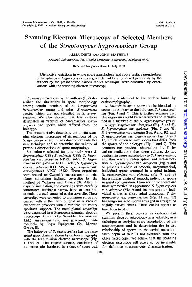
spiral spore chain as shown by carbon repligraphywith the transmission electron microscope (Fig.1 and 2). The rugose surface, consisting ofnumerous pits bordered by ridges of spore wall
material, is identical to the surface found bycarbon repligraphy.
S. halstedii is again shown to be identical inspore morphology to the holotype, S. hygroscopi-cus (Fig. 3 and 4). This is further evidence thatthis organism should be redescribed and reclassi-fied as a member of the S. hygroscopicus group.
S. hygroscopicus var. decoyicus (Fig. 5 and 6),S. hygroscopicus var. glebosus (Fig. 7 and 8),S. hygroscopicus var. odoratus (Fig. 9 and 10), andS. hygroscopicus var. ossamyceticus (Fig. 11 and12) are all shown to have spores that differ fromthe spores of the holotype (Fig. 1 and 2). Thisconfirms our previous observation (1, 2) bycarbon repligraphy that these cultures do notconform in spore morphology to S. hygroscopicus,and thus warrant redescription and reclassifica-tion. S. hygroscopicus var. decoyicus (Fig. 5 and6) presents a chain of smooth, unsymmetrical,individual spores arranged in a spiral fashion.S. hygroscopicus var. glebosus (Fig. 7 and 8)has a similar chain of smooth, individual sporesin spiral configuration. However, these spores aremore symmetrical in appearance. S. hygroscopicusvar. odoratus (Fig. 9 and 10) has smooth, indi-vidual spores in short spiral groupings. S. hy-groscopicus var. ossamyceticus (Fig. 11 and 12)has rough surfaced spores arranged in straight orslightly curved chains. These chains appear tohave been twisted.We present these pictures as evidence that
scanning electron microscopy is a valuable, newtechnique in studying spore morphology of thestreptomycetes and in determining the in siturelationship of spores to the aerial mycelium.Such depth of field is not available with anyother microscope. We believe that the scanningelectron microsope will prove to be invaluablefor definitive streptomycete characterization.
694
on Decem
ber 19, 2018 by guesthttp://aem
.asm.org/
Dow
nloaded from
VOL. 18, 1969
Fio. 1-6. Fig. 1, Streptomyces hygroscopicus CBS; Fig. 2, Streptomyces hygroscopicus CBS; Fig. 3, Strepto-myces halstedfi CBS; Fig. 4, Streptomyces halstedii CBS; Fig. 5, Streptomyces hygroscopicus var. decoyicusNRRL 2666; Fig. 6, Streptomyces hygroscopicus var. decoyicus NRRL 2666. Each index mark represents 2 pm.
695NOTES
on Decem
ber 19, 2018 by guesthttp://aem
.asm.org/
Dow
nloaded from

NOTES APPL. MICROBIOL.
FIG. 7-12. Fig. 7, Streptomyces hygroscopicus var. glebosus ATCC 14607; Fig. 8, Streptomyces hygroscopicusvar. glebosus ATCC 14607; Fig. 9, Streptomyces hygroscopicus var. odoratus IFO 1545; Fig. 10, Streptomyceshygroscopicus var. odoratus IFO 1545; Fig. 11, Streptomyces hygroscopicus var. ossamyceticus ATCC 15420;Fig. 12, Streptomyces hygroscopicus var. ossamyceticus ATCC 15420. Each index mark represents 2 ,sm.
LITERATURE CITED
1. Dietz, A., and J. Mathews. 1962. Taxonomy by carbon repli-cation. I. An examination of Streptomyces hygroscopicus.Appl. Microbiol. 10:258-263.
2. Dietz, A., and J. Mathews. 1968. Taxonomy by carbon rep-
lication. II. Examination of eight additional culturesof Streptomyces hygroscopicus. Appl. Microbiol. 16:935-941.
3. Williams, S. T., and F. L. Davies. 1967. Use of a ScanningElectron Microscope for the examination of actinomycetes.J. Gen. Microbiol. 48:171-177.
696
on Decem
ber 19, 2018 by guesthttp://aem
.asm.org/
Dow
nloaded from