SCANNING ELECTRON MICROSCOPY OF LEAF OF …ijrpc.com/files/32-272.pdf · SCANNING ELECTRON...
9
IJRPC 2012, 2(2) Bhatt et al ISSN: 22312781 470 INTERNATIONAL JOURNAL OF RESEARCH IN PHARMACY AND CHEMISTRY Available online at www.ijrpc.com SCANNING ELECTRON MICROSCOPY OF LEAF OF LITSEA CHINENSIS LAM Bhatt SP 1* and Pandya SS 2 1 Jodhpur National University, Jodhpur, Rajasthan, India. 2 B. Pharmacy College, Kakanpura, Godhra, Panchmahal, Gujarat, India. INTRODUCTION The scanning electron microscope (S.E.M.) with its relatively high resolution and great depth of focus, far surpassing that of the light microscope, is an important addition to the range of techniques available for structural studies in biology. Thus far this instrument has produced its most impressive results on the fine architecture of the surfaces of hard tissues which do not require complex histological techniques. 1 Although some results merely confirm previous light microscope studies, the SEM has made it possible to elucidate the real nature of many features lying at or beyond the limit of resolution of the light microscope and at the same time revealed many, completely unsuspected, new structures. To date, in the botany, the SEM has been utilized in fields such as wood, seed, fruit, and spore anatomy, palynology, palaeobotany, and the study of the distribution and physical nature of the cuticular layers of leaves. 1,2 Detailed studies have been made on diverse groups ranging from the angiosperms to the algae and fungi, bryophytes by comparison have been rather neglected. The surface topography of a few spores has been examined. 3,4 The scanning electron microscope (S.E.M.) gives considerable depth of focus at high magnification, an advantage over the light microscope. 5 It was therefore thought useful to examine the histology of leaf of Litsea chinensis Lam with S.E.M. Litsea chinensis Lam (syn: Litsea glutinosa) is a Lauraceae member commonly known as Meda lakadi in Hindi, Medsakhah in Sanskrit, Neluka in Assami and Meda or Va Lakdi in local Gujarati language. Plant is believed to be effective for its wound healing, antidiarrhoeal, and anti-inflammatory property in traditional and folk lore system of medicine. 6-10 Yet so far scanty of anatomical studies of the leaf for the Litsea chinensis Lam reported. This study reports the anatomical characterization of leaf of Litsea chinensis Lam with Scanning Electron Microscope. Leaf histology was studied to see useful features for distinguishing between plant species, standardisation in plants known to have similar structure. MATERIALS AND METHODS Study and collection of plant Leaf samples were collected from the vicinity of Jorhat, Assam. Preliminary data provided by Medicinal and aromatic plant Department of Research Article ABSTRACT Microscopic structure of Litsea chinensis Lam was studied with the help of scanning electron microscope for pharmacognostical and taxonomic utility. Despite its wide distribution and medicinal use, it has little attention on microscopy reported in literature. It has homogenous and characteristic leaf anatomy. Presence of abundant mucilage, developed vascular bundle, hypodermis, presence of ground material and tissues might eventually allow recognition and standardization of plant. Keywords: Leaf anatomy, Litsea chinensis Lam, Lauraceae, SEM
Transcript of SCANNING ELECTRON MICROSCOPY OF LEAF OF …ijrpc.com/files/32-272.pdf · SCANNING ELECTRON...
IJRPC 2012, 2(2) Bhatt et al ISSN: 22312781
470
INTERNATIONAL JOURNAL OF RESEARCH IN PHARMACY AND CHEMISTRY Available online at www.ijrpc.com
SCANNING ELECTRON MICROSCOPY OF LEAF OF
LITSEA CHINENSIS LAM
Bhatt SP1* and Pandya SS2 1Jodhpur National University, Jodhpur, Rajasthan, India.
2 B. Pharmacy College, Kakanpura, Godhra, Panchmahal, Gujarat, India.
INTRODUCTION The scanning electron microscope (S.E.M.) with its relatively high resolution and great depth of focus, far surpassing that of the light microscope, is an important addition to the range of techniques available for structural studies in biology. Thus far this instrument has produced its most impressive results on the fine architecture of the surfaces of hard tissues which do not require complex histological techniques.1 Although some results merely confirm previous light microscope studies, the SEM has made it possible to elucidate the real nature of many features lying at or beyond the limit of resolution of the light microscope and at the same time revealed many, completely unsuspected, new structures. To date, in the botany, the SEM has been utilized in fields such as wood, seed, fruit, and spore anatomy, palynology, palaeobotany, and the study of the distribution and physical nature of the cuticular layers of leaves. 1,2
Detailed studies have been made on diverse groups ranging from the angiosperms to the algae and fungi, bryophytes by comparison have been rather neglected. The surface topography of a few spores has been examined.3,4
The scanning electron microscope (S.E.M.) gives considerable depth of focus at high magnification, an advantage over the light microscope.5 It was therefore thought useful to examine the histology of leaf of Litsea chinensis Lam with S.E.M. Litsea chinensis Lam (syn: Litsea glutinosa) is a Lauraceae member commonly known as Meda lakadi in Hindi, Medsakhah in Sanskrit, Neluka in Assami and Meda or Va Lakdi in local Gujarati language. Plant is believed to be effective for its wound healing, antidiarrhoeal, and anti-inflammatory property in traditional and folk lore system of medicine.6-10 Yet so far scanty of anatomical studies of the leaf for the Litsea chinensis Lam reported. This study reports the anatomical characterization of leaf of Litsea chinensis Lam with Scanning Electron Microscope. Leaf histology was studied to see useful features for distinguishing between plant species, standardisation in plants known to have similar structure. MATERIALS AND METHODS Study and collection of plant Leaf samples were collected from the vicinity of Jorhat, Assam. Preliminary data provided by Medicinal and aromatic plant Department of
Research Article
ABSTRACT Microscopic structure of Litsea chinensis Lam was studied with the help of scanning electron microscope for pharmacognostical and taxonomic utility. Despite its wide distribution and medicinal use, it has little attention on microscopy reported in literature. It has homogenous and characteristic leaf anatomy. Presence of abundant mucilage, developed vascular bundle, hypodermis, presence of ground material and tissues might eventually allow recognition and standardization of plant. Keywords: Leaf anatomy, Litsea chinensis Lam, Lauraceae, SEM
IJRPC 2012, 2(2) Bhatt et al ISSN: 22312781
471
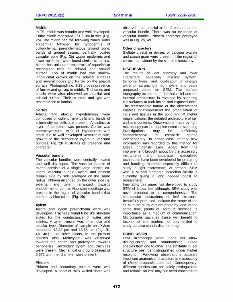
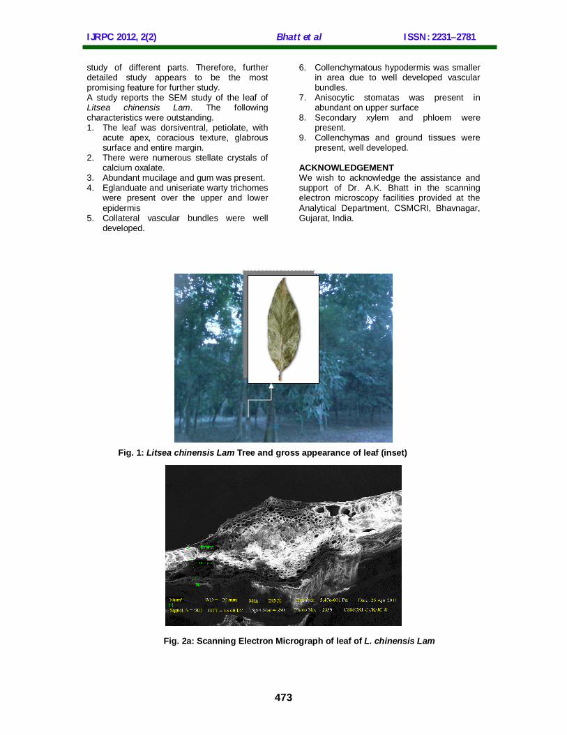
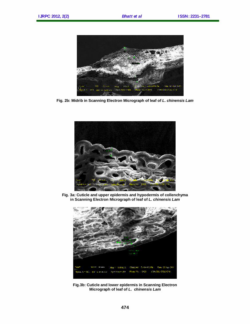
north east institute of science and technology of Jorhat indicated that the place, environment and habitat were meteorological near the Jorhat were climate and environments were suitable. Taxonomic authentification Taxonomic determination — Litsea chinensis Lam has been identified and collected from the vicinity of Jorhat. Of the so many variety of Litsea present in Assam, to determine the species found at the study sites, blooming trees were chosen at the area. Vouchers were prepared and submitted in the Herbarium at the Botanical Survey of India, North East Region, Shillong, Meghalaya. Herbarium was examined for cross-reference using the taxonomic identification key and the plant was identified as Litsea chinensis Lam. Sampling — Samples were obtained from the well developed trees site, at 3 m from the ground by direct plucking method. The anatomical description was based on well known botanical literature. Preparation for SEM For observations by scanning electron microscopy, fragments of about 5 mm square of the leaves and was washed, at room temperature and fixed on aluminum probe unit. Observations were carried in crayo (constant pressure) mode with the help of German made SEM Leo1 1430VP, version 1. Samples were examined in constant temperature -10 0C at variable distance. RESULT AND DISCUSSION General anatomy of leaf Externally, the leaf was green with well developed epipodium, mesopodium and hypopodium. It was foliage leaf arranged alternatively. Hopopodium exhibited base and stipules. Petiole was symmetrical. Leaf was lanceolate, glabrous, coriaceous, pinnate venation with entire margin and acute apex; of variable in size (L:16-26 cm x B: 4-8cm) It smells characteristic and taste mucilaginous. Depending from stem maturity, leaf exhibits variable size and maturity fig. 1. Microscopy of the leaf The leaf was dorsiventral. It consists of lamina and midrib. Both are distinctive and well developed. Several ridges and furrows were present in upper and lower part of the midrib (Fig. 2a).
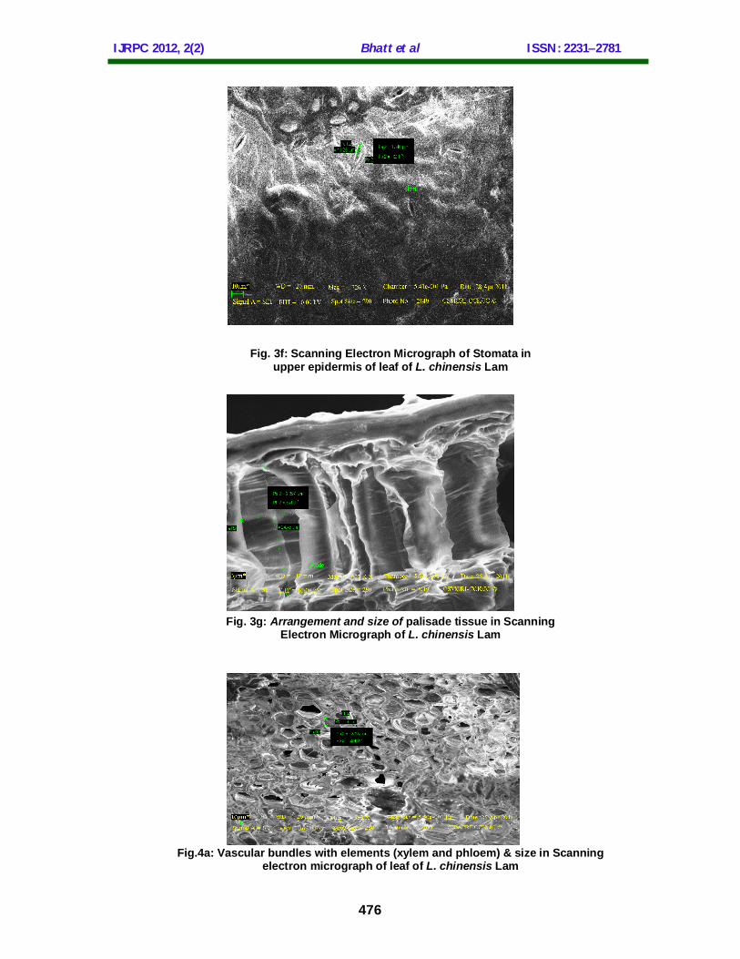
Lamina Entire lamina measured 108.3 µm in size (Fig. 2b). Cuticle, upper and lower epidermis, palisade tissue layer, parenchymetous cells and vascular bundles were present and well developed. Epidermis There were two epidermial layers on abaxial and adaxial surfaces of the leaf. Each was uniseriate, composed of a row of compactly set rectangular or squarish cells and have thin and sinuous cell walls on adaxial and abaxial surfaces. The cells measured 11.36 µm x 4.90 µm in size. The outer walls were cutinised and possess thin cuticle. The thickness was being more pronounced in the cells of the upper epidemis 7.401 µm than those on the lower side 3.3 µm (Fig. 3a & 3b.). Trichomes Eglanduate of 53.47 µm and glanduate of 128.8 µm size, unicellular warty sometimes filliform trichomes were present on both the surface of lamina and midrib (Fig. 3c & 3d.). Eglanduate trichomes have thick cell wall and pointed apex. Stomata Stomata of anisocytic type was present abundantly on upper surface while less in lower surface. An arrangement of guard cells and subsidiary cells or accessory cells was readily distinguished from adjacent epidermal cells. The stomata measured 13.28 µm X3.34 µm (Fig. 3e & 3f). Mesophyll Lamina was dorsiventral. The ground tissues forming the mesophyll, was differentiated into palisade and spongy cells. The palisade cells occur towards upper epidermis. Average size of palisade cells measured 9.892 µm x 24.45 µm (Fig. 3g). They were columnar cells with scanty intercellular spaces and remains arranged more or less at right angles to the upper epidermis. Abundant chloroplasts were present. There were two layers of palisade cells. The spongy cells occur towards the lower epidermis. These cells were oval, spherical or irregular in shape. They were quite loosely arranged with conspicuous intercellular spaces. The number of chloroplast was much less here which explains the pale green colour of the lower surface of the leaf. Vascular bundles were present in lamina. The bundles were surrounded by the parenchyma cells.
IJRPC 2012, 2(2) Bhatt et al ISSN: 22312781
472
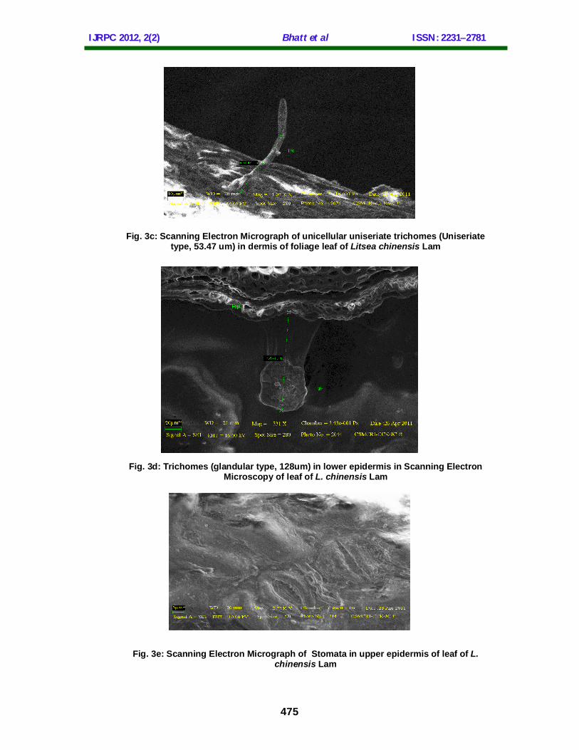
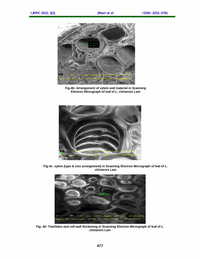
Midrib In TS, midrib was broader and well developed. Entire midrib measured 261.2 um in size (Fig. 2b). The midrib had the following zones, outer epidermis, followed by hypodermis of collenchyma, parenchymetous ground zone, bands of ground tissues, centrally located vascular zone (Fig. 2b) Upper epidermis and lower epidermis were found similar to lamina. Midrib has uniseriate epidermis of squarish or rectangular cells on adaxial and abaxial surface. Top of midrib had two shallow longitudinal groves on the adaxial surfaces and several ridges and furrow on the abaxial surface. Photograph no. 3.18 proves presence of furrow and groves in midrib. Trichomes and cuticle were also observed on abaxial and adaxial surface. Their structure and type was resemblance to lamina. Cortex Adaxial and abaxial hypodermises were composed of collenchyma cells and bands of sclerenchyma cells are present. A distinctive layer of cambium was present. Cortex was parenchymetous. Area of Hypodermis was small due to well developed vascular bundle, growth of the secondary layers in vascular bundles. Fig. 2b illustrates its presence and character. Vascular bundle The vascular bundles were centrally located and well developed. The vascular bundle of midrib consists of a single large central co-lateral vascular bundle. Xylem and phloem remain side by side arranged on the same radius. Phloem arranged on the outer side i.e. external and xylem arranged towards endodermis or centre. Abundant mucilage was present in the region of vascular bundle that confirm by blue colour (Fig. 2b) Xylem Xylem and xylem parenchyma were well developed. Tracheae found tube like structure suited for the conductance of water and solutes. A xylem vessel was of annular and circular type. Diameter of sample unit Xylem measured 12.22 µm and 13.68 µm (Fig. 2b, 4b, 4c.). Like other dicots, in the present species also Metaxylem was observed towards the centre and protoxylem towards peripherals. Secondary xylem and trachides were present. Mechanical or ground tissues of 6.672 µm inner diameter were present. Phloem Phloem and secondary phloem were well developed. A band of thick walled fibers was
observed the abaxial side of phloem of the vascular bundle. There was an evidence of vascular bundle. Phloem character portrayed well in Fig. 2b, 4d. Other characters Stellate crystal or druses of calcium oxalate and starch grain were present in the region of cortex that evident by the simple microscopy. DISCUSSION The results of leaf anatomy and foliar characters, especially vascular system, trichome types, and localization of crystals were of surprisingly high systematic value proposed based on SEM. The surface topography examined in detailed relief and the internal architecture is revealed by scanning cut surfaces to look inside and exposed cells. The stereoscopic nature of the observations enables to comprehend the organization of cells and tissues in the solid and at higher magnifications, the detailed architecture of cell wall and contents. Observations made by light microscopy can be supplemented or scanning investigations may be sufficiently comprehensive to establish criteria independently. In either case entirely new information was recorded by this method for Litsea chinensis Lam. Apart from the improvement brought about by the accessory instruments and apparatus, specialised techniques have been developed for preparing and handling materials especially difficult to study in light microscope. At present, SEM with TEM and elemental detection facility is currently giving a long needed boost to researchers. Inevitably, this paper has developed to study SEM of Litsea leaf although; SEM study was never intended to be comprehensive. The spectacular illustrations of leaf structure, beautifully produced, indicate the scope of the SEM in the study of plant anatomy, and, at the same time, plenty of literature stresses its importance as a medium of communication. Micrographs such as these will benefit to taxonomist and readers not only limited to study but also standardise the drug. CONCLUSION Leaf microscopy alone does not allow distinguishing and standardising Litsea species from one to other. The similarity in leaf structure best be distinguished under higher resolution. Following observations appears important anatomical characters in microscopy of Litsea chinensis Lam leaf. Consequently, different species can not solely distinguished and reliable on leaf only but need concomitant
IJRPC 2012, 2(2) Bhatt et al ISSN: 22312781
473
study of different parts. Therefore, further detailed study appears to be the most promising feature for further study. A study reports the SEM study of the leaf of Litsea chinensis Lam. The following characteristics were outstanding. 1. The leaf was dorsiventral, petiolate, with
acute apex, coracious texture, glabrous surface and entire margin.
2. There were numerous stellate crystals of calcium oxalate.
3. Abundant mucilage and gum was present. 4. Eglanduate and uniseriate warty trichomes
were present over the upper and lower epidermis
5. Collateral vascular bundles were well developed.
6. Collenchymatous hypodermis was smaller in area due to well developed vascular bundles.
7. Anisocytic stomatas was present in abundant on upper surface
8. Secondary xylem and phloem were present.
9. Collenchymas and ground tissues were present, well developed.
ACKNOWLEDGEMENT We wish to acknowledge the assistance and support of Dr. A.K. Bhatt in the scanning electron microscopy facilities provided at the Analytical Department, CSMCRI, Bhavnagar, Gujarat, India.
Fig. 1: Litsea chinensis Lam Tree and gross appearance of leaf (inset)
Fig. 2a: Scanning Electron Micrograph of leaf of L. chinensis Lam
IJRPC 2012, 2(2) Bhatt et al ISSN: 22312781
474
Fig. 2b: Midrib in Scanning Electron Micrograph of leaf of L. chinensis Lam
Fig. 3a: Cuticle and upper epidermis and hypodermis of collenchyma
in Scanning Electron Micrograph of leaf of L. chinensis Lam
Fig.3b: Cuticle and lower epidermis in Scanning Electron
Micrograph of leaf of L. chinensis Lam
IJRPC 2012, 2(2) Bhatt et al ISSN: 22312781
475
Fig. 3c: Scanning Electron Micrograph of unicellular uniseriate trichomes (Uniseriate type, 53.47 um) in dermis of foliage leaf of Litsea chinensis Lam
Fig. 3d: Trichomes (glandular type, 128um) in lower epidermis in Scanning Electron Microscopy of leaf of L. chinensis Lam
Fig. 3e: Scanning Electron Micrograph of Stomata in upper epidermis of leaf of L. chinensis Lam
IJRPC 2012, 2(2) Bhatt et al ISSN: 22312781
476
Fig. 3f: Scanning Electron Micrograph of Stomata in
upper epidermis of leaf of L. chinensis Lam
Fig. 3g: Arrangement and size of palisade tissue in Scanning Electron Micrograph of L. chinensis Lam
Fig.4a: Vascular bundles with elements (xylem and phloem) & size in Scanning electron micrograph of leaf of L. chinensis Lam
IJRPC 2012, 2(2) Bhatt et al ISSN: 22312781
477
Fig.4b: Arrangement of xylem and material in Scanning Electron Micrograph of leaf of L. chinensis Lam
Fig.4c: xylem (type & size arrangement) in Scanning Electron Micrograph of leaf of L.
chinensis Lam
Fig. 4d: Trachides and cell wall thickening in Scanning Electron Micrograph of leaf of L. chinensis Lam
IJRPC 2012, 2(2) Bhatt et al ISSN: 22312781
478
REFERENCES 1. Carr KE. Applications of scanning
electron microscopy in biology. Internat. Rev. Cytol. 1971;30: 183-255.
2. Stant MY. The role of scanning electron microscope in plant anatomy. Kew bulletin 1973;28(1):105-115.
3. Mozingo H, Klein P, Zeevi Y, Lewis ER. Scanning electron microscope studies on Sphagnum imbricatum. The Bryologist. 1969;72:484-488.
4. Taylor J, Kaufman PB and Bigelow WC. Scanning electron microscope examination of spore and elaters of Targionia hypophylla. The Bryologist. 1972;74:497-498.
5. McNeil KE and Skerman VBD. Examination of Myxobacteria by scanning electron microscopy. International Journal of Systemic Bacteriology. 1972;22(4):243-250.
6. Bennet SSR. “Name changing in flowering plants of India and adjacent regions”, Treaser Publisher, Dehra Dun. 1986; 339-40.
7. Bora PJ. Yogendra Kumar., “Floristic diversity of Assam; study of pabitora wildlife santury”, Daya publishing house, Assam. 2003Page no. 291.
8. Kirtikar KR and Basu BD. Indian medicinal plants Vol:III. International Book Distributors, Dehradun. 2005.;2157-2161.
9. Dept. of Ayurveda, Yoga, Unani, Sidhha and Homeopathy, New Delhi, Ministry of Health and Family Welfare. Govt. of India. The Ayurvedic Pharmacopoeia of India 2006; 1st Edn, Part –I, Vol; V.108-9.
10. Cooke T. “Flora of the presidency of Bombay”, Vol III, 2nd Edn, Botanical Survey of India, Govt. of Indian. Page no 31-33