Scanning Electron Micrographs of two species of Sturnophagoides
8
217 Tropical Biomedicine 25(3): 217–224 (2008) Scanning Electron Micrographs of two species of Sturnophagoides (Acari: Astigmata: Pyroglyphidae) mites in Malaysia Mariana, A. 1 , Santana Raj, A.S. 2 , Ho, T.M. 1 , Tan, S.N. 1 and Zuhaizam, H. 1 1 Acarology Unit, Institute for Medical Research, 50588 Kuala Lumpur, 2 Electron Microscopy Unit, Institute for Medical Research, 50588 Kuala Lumpur. Email: [email protected] Received 14 April 2008; received in revised form 20 September 2008; accepted 23 September 2008 Abstract. Scanning electron microscope (SEM) images of two dust mites, Sturnophagoides brasiliensis and Sturnophagoides halterophilus, are presented to provide an improved visualization of the taxonomic characters of these mites. Sturnophagoides halterophilus can be differentiated from S. brasiliensis by their expanded genu and femur of leg I. The differences in morphology of male and female S. brasiliensis are also discussed. INTRODUCTION Worldwide, mites have been found inhabiting house dust. In Malaysia, one of the less common species found is Sturnophagoides brasiliensis Fain, 1967 (Ho & Nadchatram, 1984; Ho & Nadchatram, 1985; Mariana et al., 2000). Usually S. brasiliensis is not the most abundant mite found, however in a survey of a students’ hostel, it was the most common and abundant mite recovered (Ho & Mariana, 1994). Another species, Sturnophagoides halterophilus Fain & Feinberg 1970, was also collected in the same survey but in a much lower density. As many as 67-69 S. brasiliensis mites per gram of dust had been recovered from mattresses (Ho & Mariana, 1994; Mariana, 2002). Sturnophagoides brasiliensis has been reported to produce allergens responsible for asthma and rhinitis (Arlian, 1991; Chew et al., 1999; Mariana, 2002). The genus Sturnophagoides was first described (as a subgenus of Derma- tophagoides) by Fain (1967a). Amongst the species described under this genus are S. brasiliensis (Fain, 1967b) and S. halterophilus (Fain & Feinberg, 1970). The genus was then further described by Fain (1971) and Wharton (1976). Illustration of S. brasiliensis and S. halterophilus by scanning electron micrographs (SEM) has not been widely reported. SEMs of these species are presented here to provide an improved visualization of the morphological characters of the mites. General morphology Sturnophagoides brasiliensis is a minute mite, with idiosoma measuring about 0.18 to 0.25 mm in length and 0.07 to 0.14 mm in width (Figure 1). Gnathosoma of S. brasiliensis is composed of a pair of palps and chelicerae which are supported by a bulb (subcapitulum) (Figure 2). The palps are a simple two-segmented structure with sensory hairs. The chelicerae are three- segmented; the third segment is dentate. Like other Pyroglyphids, the mite has a pair of long scapular external setae (sce) extending from the outer margin of the idiosoma and a pair of shorter scapular internal setae (sci) on the inner part of the idiosoma (Figure 3). The anterior of the 217 - 224 Mariana A.pmd 11/17/2008, 10:55 AM 217
Transcript of Scanning Electron Micrographs of two species of Sturnophagoides
217
Tropical Biomedicine 25(3): 217–224 (2008)
Scanning Electron Micrographs of two species of
Sturnophagoides (Acari: Astigmata: Pyroglyphidae) mites
in Malaysia
Mariana, A.1, Santana Raj, A.S.2, Ho, T.M.1, Tan, S.N.1 and Zuhaizam, H.11Acarology Unit, Institute for Medical Research, 50588 Kuala Lumpur,2Electron Microscopy Unit, Institute for Medical Research, 50588 Kuala Lumpur.Email: [email protected] 14 April 2008; received in revised form 20 September 2008; accepted 23 September 2008
Abstract. Scanning electron microscope (SEM) images of two dust mites, Sturnophagoides
brasiliensis and Sturnophagoides halterophilus, are presented to provide an improvedvisualization of the taxonomic characters of these mites. Sturnophagoides halterophilus can bedifferentiated from S. brasiliensis by their expanded genu and femur of leg I. The differences inmorphology of male and female S. brasiliensis are also discussed.
INTRODUCTION
Worldwide, mites have been found inhabitinghouse dust. In Malaysia, one of the lesscommon species found is Sturnophagoides
brasiliensis Fain, 1967 (Ho & Nadchatram,1984; Ho & Nadchatram, 1985; Mariana et al.,2000). Usually S. brasiliensis is not the mostabundant mite found, however in a surveyof a students’ hostel, it was the most commonand abundant mite recovered (Ho & Mariana,1994). Another species, Sturnophagoides
halterophilus Fain & Feinberg 1970, was alsocollected in the same survey but in a muchlower density. As many as 67-69 S.
brasiliensis mites per gram of dust had beenrecovered from mattresses (Ho & Mariana,1994; Mariana, 2002). Sturnophagoides
brasiliensis has been reported to produceallergens responsible for asthma and rhinitis(Arlian, 1991; Chew et al., 1999; Mariana,2002).
The genus Sturnophagoides was firstdescribed (as a subgenus of Derma-
tophagoides) by Fain (1967a). Amongst thespecies described under this genus areS. brasiliensis (Fain, 1967b) and S.
halterophilus (Fain & Feinberg, 1970). Thegenus was then further described by Fain(1971) and Wharton (1976). Illustration ofS. brasiliensis and S. halterophilus byscanning electron micrographs (SEM) hasnot been widely reported. SEMs of thesespecies are presented here to provide animproved visualization of the morphologicalcharacters of the mites.
General morphology
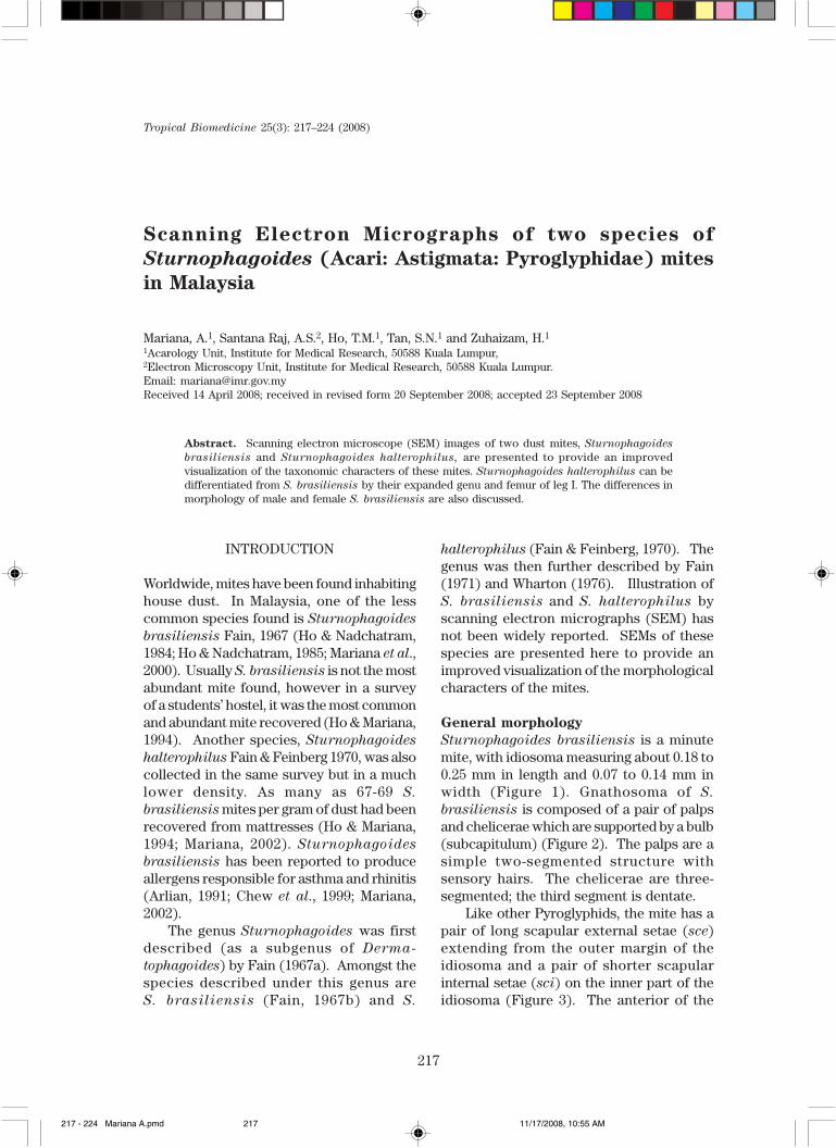
Sturnophagoides brasiliensis is a minutemite, with idiosoma measuring about 0.18 to0.25 mm in length and 0.07 to 0.14 mm inwidth (Figure 1). Gnathosoma of S.
brasiliensis is composed of a pair of palpsand chelicerae which are supported by a bulb(subcapitulum) (Figure 2). The palps are asimple two-segmented structure withsensory hairs. The chelicerae are three-segmented; the third segment is dentate.
Like other Pyroglyphids, the mite has apair of long scapular external setae (sce)extending from the outer margin of theidiosoma and a pair of shorter scapularinternal setae (sci) on the inner part of theidiosoma (Figure 3). The anterior of the
217 - 224 Mariana A.pmd 11/17/2008, 10:55 AM217
218
Figure 1. Ventral view of male S. brasiliensis (left) and S. halterophilus (right).
Figure 2. Anterior region of gnathosoma showing chelicera and palp.
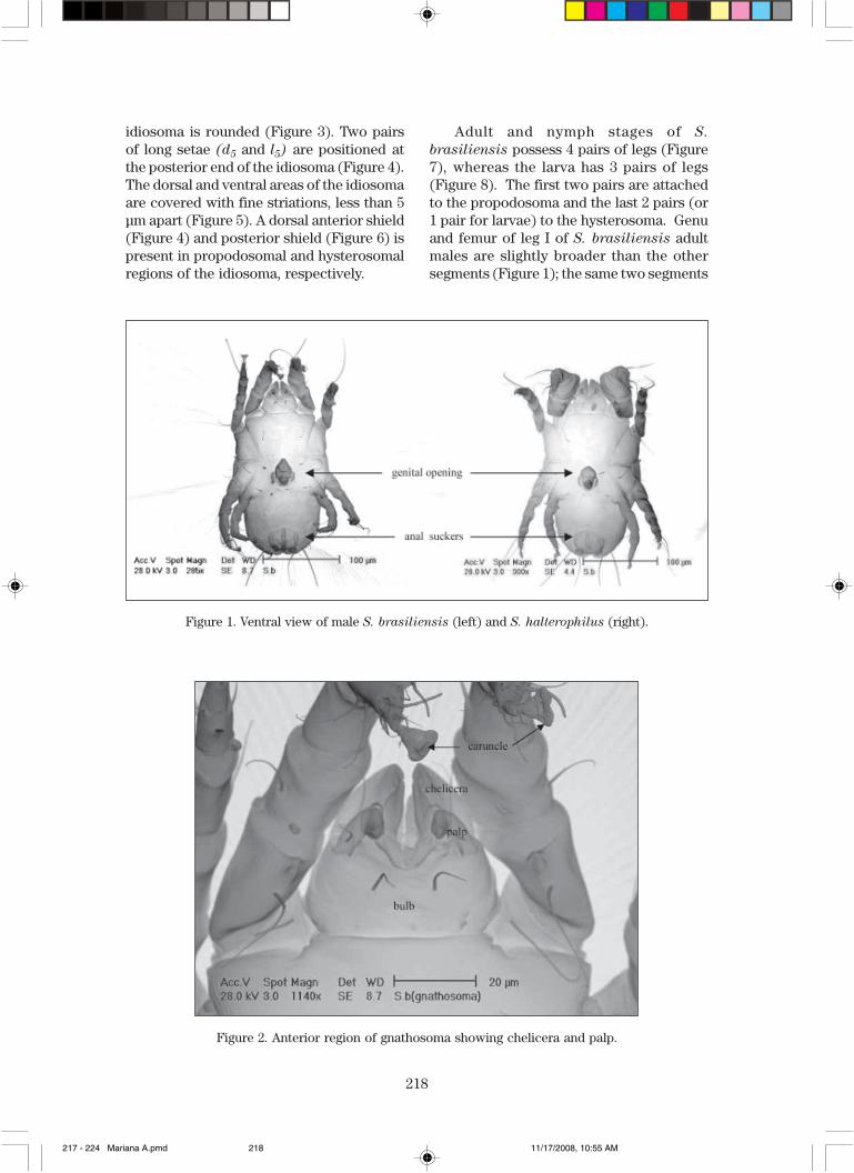
idiosoma is rounded (Figure 3). Two pairsof long setae (d5 and l5) are positioned atthe posterior end of the idiosoma (Figure 4).The dorsal and ventral areas of the idiosomaare covered with fine striations, less than 5µm apart (Figure 5). A dorsal anterior shield(Figure 4) and posterior shield (Figure 6) ispresent in propodosomal and hysterosomalregions of the idiosoma, respectively.
Adult and nymph stages of S.
brasiliensis possess 4 pairs of legs (Figure7), whereas the larva has 3 pairs of legs(Figure 8). The first two pairs are attachedto the propodosoma and the last 2 pairs (or1 pair for larvae) to the hysterosoma. Genuand femur of leg I of S. brasiliensis adultmales are slightly broader than the othersegments (Figure 1); the same two segments
217 - 224 Mariana A.pmd 11/17/2008, 10:55 AM218

219
Figure 3. Rounded anterior of idiosoma.
Figure 4. Long d5 and l5 setae of S. brasiliensis (left) and S. halterophilus (right).
Figure 5. Idiosoma with fine body striations.
217 - 224 Mariana A.pmd 11/17/2008, 10:55 AM219
220
Figure 6. Posterior end of idiosoma showing hysterosomal shield.
Figure 7. Nymph of S. brasiliensis.
Figure 8. Larvae of S. brasiliensis.
217 - 224 Mariana A.pmd 11/17/2008, 10:55 AM220

221
of S. halterophilus are 3 to 4 times broader(Figure 9). All tarsi of S. brasiliensis and S.
halterophilus end in caruncles.
Sexual dimorphism
Male S. brasiliensis has a pair of ventralterminal anal suckers surrounded by an oval-
shaped chitinous arc (Figure 10). The shapeof the arc is not unique to this species but isshared with S. halterophilus (Ho, 1986). Allfemales possess a vulva lip and an externalterminal opening (Figure 11).
Other than different internal sex organs,male and female S. brasiliensis mites also
Figure 9. Anterior view of male S. brasiliensis (left) and S. halterophilus (right).
Figure 10 . Ventral of male showing genital opening and the oval-shaped arc around anal suckers of S.
brasiliensis (above) and S. halterophilus (bottom).
217 - 224 Mariana A.pmd 11/17/2008, 10:55 AM221

222
Figure 11. Ventral view of female S.brasiliensis mite showing genital opening.
differ in the size and shape of their dorsalhysterosomal shield (Figure 12). Femaleshave a smaller shield compared to the males.It is cone-shaped and not reaching coxae IV.
The shield in males is much longer than wideand extending anteriorly above coxae IV. Ashield of the same shape can also be seen inmale S. halterophilus (Figure 13). The
Figure 12. Posterior end of idiosoma of a male (left) and a female S. brasiliensis.
Figure 13. Dorsal hysterosomal shield of a male S. halterophilus.
217 - 224 Mariana A.pmd 11/17/2008, 10:55 AM222

223
striations in dorsal hysterosomal area aredifferent for male and female S. brasiliensis
(Figure 12). No comparison of the dorsalstriations of different sexes of S.
halterophilus mites is possible because untilto date, no female S. halterophilus has beendescribed. There are suggestions thatperhaps S. halterophilus is a heteromorphicmale of S. brasiliensis (Fain et al., 1988,1990). Dorsal striations are also seen inimmature stages (Figure 14).
Acknowledgements. The authors wish tothank the Director, Institute for MedicalResearch, Kuala Lumpur, Malaysia forpermission to publish this paper. We alsowish to thank Ms Teh Hamidah Zamzuri andMs Aida Suhana Rosli for their assistance inprocessing the mites for scanning electronmicrography.
Figure 14. Striations in the dorsal hysterosomalarea of a nymph S. brasiliensis.
REFERENCES
Arlian, L.G. (1991). House dust miteallergens: A review. Experimental and
Applied Acarology 10: 167-186.Chew, F.T., Lim, S.H., Goh, D.Y.T. & Lee, B.W.
(1999). Sensitization to local dust mitefauna in Singapore. Allergy 54(11):
1150-59.Fain, A. (1967a). Deux nouvelles especes de
Dermatophagoidinae. Rattachement decettesous-famille aux Pyroglyphidae(Sarcoptiformes). Acarologia IX(4):
870-881.Fain, A. (1967b). Le genre Dermato-
phagoides Bogdanov, 1864 sonimportance dans les allergiesrepiratoires et cutanees chez l’homme(Psoroptidae: Sarcoptiformes).Acarologia IX(1): 179-225.
Fain, A. & Feinberg, J.G. 1970. Un nouvelacarien provenant des poussieres d’unemaison a Singapour (Sarcoptiformes:Pyroglyphidae). Acarologia XII(1): 164-167.
Fain, A. (1971). Genre Sturnophagoides
Fain, 1967. Buletin Insitut royal des
Sciences naturelles de Belgique 47(8):
2.Fain, A., Guerin, B. & Hart, B.J. (1988).
Acariens et allergies. Varennes-en-
Argonne: Allerbio.
Fain, A., Guerin, B. & Hart, B.J. (1990). Mitesand allergic disease. Varennes-en-
Argonne: Allerbio.
Ho, T.M. & Nadchatram, M. (1984).Distribution of house dust mites in a newsettlement in Jengka, Pahang, Malaysia.Tropical Biomedicine 1: 49-54.
Ho, T.M. & Nadchatram, M. (1985).Distribution of Dermatophagoides
pteronyssinus (Astigmata: Pyrogly-phidae) in Cameron Highlands, Malaysia.Tropical Biomedicine 2: 54-58.
Ho, T.M. (1986). Pyroglyphid mites found inhouse dust in Peninsular Malaysia.Tropical Biomedicine 3: 89-93.
217 - 224 Mariana A.pmd 11/17/2008, 10:55 AM223

224
Ho, T.M. & Mariana, A. (1994). The efficacyof a vacuum cleaner for the control ofdust mites in mattresses. Tropical
Biomedicine 11(2): 135-8.Mariana, A., Ho, T.M., Sofian-Azirun, M. &
Wong, A.L. (2000). House dust mite faunain the Klang Valley, Malaysia. Southeast
Asian Journal of Tropical Medicine and
Public Health 31(4): 712-721.
Mariana, A. (2002). The biology &distribution of allergen producing miteswith particular reference to Blomia
tropicalis (Acarina: Astigmata:Echimyopodidae) in the Klang Valley,Malaysia. PhD Thesis, University ofMalaya, Kuala Lumpur, Malaysia.
Wharton, G.W. (1976). Sturnophagoides.Journal of Medical Entomology 12(6):
601.
217 - 224 Mariana A.pmd 11/17/2008, 10:55 AM224