Sarah Richardson, 2020 Introduction
24
Sarah Richardson, 2020 1 Pregnancy-Related Lumbopelvic Pain Introduction: Pregnancy-related lumbopelvic pain involves either low back pain, pelvic girdle pain, or a combination of the two. Pregnancy-related lumbopelvic pain is the most common musculoskeletal complaint experienced during female pregnancy. 1 There, unfortunately, is a huge variance in the reported rate of lumbopelvic pain experienced during pregnancy due to differences in the population studied and study design. 2 Due to the inconsistencies in research related to pregnancy-related lumbopelvic pain, the prevalence of this condition is estimated to range between 24 to 90%. 3 More specifically, it is estimated that more than 50% of pregnant females experience low back pain and between 10 and 65% experience pelvic girdle pain. 1 More often than not, individuals who experience pain during pregnancy will continue to experience symptoms for a prolonged period following delivery. For instance, a study found that about half of females with initial pregnancy-related low back pain will continue to experience pain one year postpartum, and 20% are symptomatic three years following delivery. 4 Another study reported that postpartum pain at three months and three years is estimated to occur at a rate of over 40% and 23%, respectively. 5 Pregnancy-related low back pain typically begins within the second trimester or around 22 weeks of pregnancy. 4 Pregnancy-related pelvic girdle pain typically begins by the first trimester and peaks between the 24 th and 36 th week of pregnancy. 4 Pregnancy- related pelvic girdle pain usually resolves spontaneously within six-months postpartum but eight to 10% will experience persistent pain for up to two years postpartum. 4 Pregnancy-related lumbopelvic pain is considered to be chronic when the presence of pain within the lumbar or pelvic region is experienced for more than six months. 6 Since there is little difference in the function and long-term prognosis in those reporting either pregnancy-related pelvic or low back pain, for this paper we will be discussing pregnancy-related lumbopelvic pain with an
Transcript of Sarah Richardson, 2020 Introduction
Sarah Richardson, 2020
1
Pregnancy-Related Lumbopelvic Pain
Introduction:
Pregnancy-related lumbopelvic pain involves either low back pain, pelvic girdle pain, or a
combination of the two. Pregnancy-related lumbopelvic pain is the most common
musculoskeletal complaint experienced during female pregnancy.1 There, unfortunately, is a
huge variance in the reported rate of lumbopelvic pain experienced during pregnancy due to
differences in the population studied and study design.2 Due to the inconsistencies in research
related to pregnancy-related lumbopelvic pain, the prevalence of this condition is estimated to
range between 24 to 90%.3 More specifically, it is estimated that more than 50% of pregnant
females experience low back pain and between 10 and 65% experience pelvic girdle pain.1 More
often than not, individuals who experience pain during pregnancy will continue to experience
symptoms for a prolonged period following delivery. For instance, a study found that about half
of females with initial pregnancy-related low back pain will continue to experience pain one year
postpartum, and 20% are symptomatic three years following delivery.4 Another study reported
that postpartum pain at three months and three years is estimated to occur at a rate of over 40%
and 23%, respectively.5 Pregnancy-related low back pain typically begins within the second
trimester or around 22 weeks of pregnancy.4 Pregnancy-related pelvic girdle pain typically
begins by the first trimester and peaks between the 24th and 36th week of pregnancy.4 Pregnancy-
related pelvic girdle pain usually resolves spontaneously within six-months postpartum but eight
to 10% will experience persistent pain for up to two years postpartum.4 Pregnancy-related
lumbopelvic pain is considered to be chronic when the presence of pain within the lumbar or
pelvic region is experienced for more than six months.6 Since there is little difference in the
function and long-term prognosis in those reporting either pregnancy-related pelvic or low back
pain, for this paper we will be discussing pregnancy-related lumbopelvic pain with an
Sarah Richardson, 2020
2
assumption that this term encompasses those with low back pain, pelvic pain, and a combination
of the two.5
Due to inconsistencies within research related to the prevalence and definition of pregnancy-
related lumbopelvic pain, many of these individuals do not receive adequate care from health
care providers. For instance, many females have reported feeling disappointed in the care and
attention they received from health care professions when consulting about their pregnancy-
related lumbopelvic pain.5 More specifically, they shared that a lot of professionals were
dismissive of their symptoms and seemed uninterested in their concerns.5 Instead, it appeared
that providers were more interested in other medical needs such as hypertension and less
interested in their pain experience.5 Unsurprisingly, inadequate education is often cited as a
reason why health professionals are dismissive of pain symptoms.5 With staggeringly high
estimates for the rate of pregnancy-related lumbopelvic pain, and current reports of inadequate
care, physical therapists have a responsibility to better serve this patient population through
informed, evidence-based, and empathetic care. Therefore, this paper seeks to inform physical
therapists about the anatomical contributions, speculated etiology, typical patient presentation,
suggested examination approach, and evidence-based treatment options for pregnancy-related
lumbopelvic pain.
Anatomy:
The pelvis is formed by the paired innominate bones, sacrum, and coccyx.7 The pelvis is
supported by a mesh-network of strong ligaments that help make up the anterior and lateral walls
of the pelvis visualized in Figures 1 and 2 of Appendix A.7 The iliolumbar ligament stabilizes,
strengthens, and restricts rotational movement at the lumbosacral joint.8 This ligament starts at
the top of the transverse process of the fifth lumbar vertebrae and attaches to the posterior part of

Sarah Richardson, 2020
3
the iliac crest.8 The anterior sacroiliac ligament connects the lateral sacrum to the ilium.8 The
sacrotuberous ligament runs from the sacrum and coccyx to the ischial tuberosity.8 The
sacrospinous ligament runs from the ischial spine to the lateral sacrum and coccyx.8 Other
ligaments include the sacrococcygeal ligaments, pubic symphysis ligaments, and endopelvic
fascia ligament.8 In the female pelvis there are also ligaments within the reproductive system that
support the internal genitalia which are divided into three categories: broad ligament, ligaments
related to the ovary, and ligaments related to the uterus.8 The ligaments specific to the female
reproductive system can be seen in Figure 3 of Appendix A. The broad ligament extends from
the lateral sidewalls of the pelvis to the uterus at midline.8 This ligament covers the fallopian
tubes and ovaries anteriorly and posteriorly.8 Two ligaments attach to the ovary: the ovarian
ligament and the suspensory ligament.8 Four ligaments attach to the uterus which includes the
round ligament, cardinal ligament, pubocervical ligament, and uterosacral ligament.8
The pelvic floor muscles are divided into three layers and are illustrated with a lateral
view in Figure 4 of Appendix A. The first layer is considered the superficial layer which includes
the ischiocavernosus, bulbocavernosus, superficial transverse perineal muscles, and external anal
sphincter muscle.6,7 The second layer is deep to the superficial layer and contains a thick, fibrous
sheet of dense fascia called the perineal membrane.7 The second layer is often referred to as the
urogenital diaphragm and includes the deep transverse perineal, sphincter urethrovaginalis,
compressor urethra, and external urethral sphincter muscles.6,7 The third layer is considered to be
the pelvic diaphragm and includes the levator ani (pubococcygeus, puborectalis, iliococcygeus)
and coccygeus muscles.6,7 The levator ani is innervated by the anterior ramus of S4 and branches
of the pudendal nerve (S2 – S4).7 The coccygeus is innervated by the anterior rami of S4 and
S5.7 The pubococcygeus acts to support the pelvic viscera and pulls the coccyx towards the pubic
Sarah Richardson, 2020
4
bone.7 The puborectalis acts to elevate and constrict the anal canal.7 The iliococcygeus supports
the viscera and pulls the rectum and vagina forward. The coccygeus flexes the coccyx, supports
the viscera, and stabilizes the sacroiliac joint.7 The pelvic wall, or the lateral borders of the pelvic
musculature, consists of the piriformis and obturator internus muscles.7
Lumbopelvic pain can occur as a result of a primary dysfunction of the pelvic floor
muscles or abdominal muscles, ligaments, tendons, nerves, or as a functional adaptation to other
disorders within the pelvis or spine.6 Any of the previously discussed anatomical structures can
influence the severity and intensity of lumbopelvic pain during pregnancy. For example, pain
within the pelvic floor muscles can refer to the pelvis, back, abdomen, bladder, buttocks, and
distal leg.6 Various skeletal pathologies can also contribute to lumbopelvic pain such as
sacroiliac dysfunction; symphysis pubis inflammation or injury; and coccyx injury or
malposition.6 Muscular etiologies contributing to lumbopelvic pain include levator ani and
piriformis syndromes. Muscular injury can be a result of trauma, repetitive use, or underuse. Pain
in this region can also develop secondary to other pain syndromes such as irritable bowel
syndrome, pudendal nerve neuropathy, and painful bladder syndrome.6
Etiology and Risk Factors:
Unfortunately, pregnancy-related lumbopelvic pain has a poorly understood etiology.5
Many hypothesize that there are hormonal, mechanical, traumatic, degenerative, and metabolic
factors that contribute to this condition.5 In pregnant females, hormonal changes often
accompany an increase in body mass. During pregnancy the hormones relaxin, progesterone, and
estrogen all increase. Relaxin is a hormone produced by the placenta that increases in early
pregnancy, peaks at the end of the first trimester, but remains high until late pregnancy.9
Estrogen enhances relaxin receptor sensitivity, thus heightening its influence on joint laxity. Joint

Sarah Richardson, 2020
5
laxity is considered one of the etiologies influencing low back pain and pelvic pain in pregnant
females. For example, pregnant females with moderate to severe posterior pelvic pain often
exhibit asymmetric sacroiliac joint laxity.9 Additionally, individuals with pregnancy-related low
back pain tend to have greater pubic symphysis mobility compared to asymptomatic pregnant
females.9
It is recommended that females gain between 25 to 35 pounds during pregnancy, with
over half of this weight gain occurring in the abdomen.9 This abdominal growth causes an
anterior shift in the center of gravity, which exerts additional static and dynamic loads on the
axial skeleton.9 This also causes postural compensations to occur which can influence the
development of pregnancy-related lumbopelvic pain as depicted in Figure 1 of Appendix B. With
a growing abdomen, the abdominal muscles are stretched and become weak which increases the
strain placed on the lumbar muscles.9 The pelvis during pregnancy rotates within the sagittal
plane which causes compensatory hyperlordosis since the center of gravity has shifted anteriorly
as seen in Figure 2 of Appendix B. This anterior shift in the center of gravity causes a flexion
moment of the lumbar spine with further increases the load experienced by the lumbar
musculature.9 Additionally, this anterior shift in the center of gravity causes a greater anterior
pelvic tilt, increasing the load through the sacroiliac ligaments attempting to resist this rotation.9
With hormones increasing the laxity of these ligaments combined with exaggerated
compensatory postures, axial loading of the spine will significantly increase. A study found that
spinal compression from physical activity is higher and post-activity recovery takes longer to
occur in pregnant females with low back pain compared to asymptomatic pregnant or non-
pregnant females.9

Sarah Richardson, 2020
6
Some researchers propose that peripheral and central neuropathic mechanisms influence
central sensitization in females before, during, and after pregnancy thus heightening their pain
experience.5 Others believe that vascular changes contribute to pregnancy-related lumbopelvic
pain.9 The growing uterus can put compression on the aorta and vena cava when the pregnant
female is in a supine position. This compression can increase the risk for thromboembolism and
subsequent stasis with decreased oxygen saturation.9 If either were to occur, hypoxemia could
compromise the metabolic activity of the surrounding neural structures.9
The risk factors associated with pregnancy-related lumbopelvic pain are multifactorial.
The previously discussed pregnancy-related structural changes such as increased joint laxity,
anterior displacement of the center of gravity, increased axial loading, and vascular changes all
increase one's risk for developing pain.4 Research suggests, however, that physical activity may
lessen the degree of biomechanical change that occurs as pregnancy advances by decreasing the
axial load on the spine, increasing joint stabilization through surrounding musculature, and
enhancing spinal alignment and segmental motion.4
Once pregnancy-related lumbopelvic pain has been experienced, there is an 85% greater
likelihood of developing recurrent pain.4 Risk factors can be categorized based on when they are
experienced during pregnancy but it should be noted that some risk factors occur in multiple
categories. Risk factors for pregnancy-related lumbopelvic pain during pregnancy include back
pain before pregnancy that required intervention; abdominal pain before pregnancy; and history
of pregnancy-related lumbopelvic pain in previous pregnancies.5 Risk factors during postpartum
include a history of back pain pre-pregnancy; pain during pregnancy; increased pain intensity
during pregnancy; increased pain intensity during delivery; and history of pregnancy-related
lumbopelvic pain in previous pregnancies. Risk factors for chronic or persistent pregnancy-

Sarah Richardson, 2020
7
related lumbopelvic pain include a body mass index > 25; a younger age; and hypermobility, but
their influence is still debated in the literature.5
Patient Presentation:
Due to the unknown etiology of pregnancy-related lumbopelvic pain and the various
structures that can impact pain experienced in this population, the patient presentation will vary.
Typically, patients with pregnancy-related lumbopelvic pain will have a difficult time localizing
and describing their pain.5,6 Pain could be localized to the pubis symphysis, posterior pelvis at
the sacroiliac joint, low back, gluteal region, groin, radiate down the distal leg or be a
combination of any of the aforementioned areas.5,6 The intensity and frequency of pain will also
vary.5 In some cases pain is constant whereas for others it is intermittent.5 Given the previously
discussed influence of hormones on joint laxity, many patients will report variations of pain
related to the timing of their menstrual cycle.6 The pain may be described by the patient as
aching, cutting, dull, or stabbing.5 However, pain is more often unilateral and patients commonly
describe “heaviness” or “pelvic pressure” when discussing their pain experience.6 Additionally,
pain in the low back that radiates to the levator ani insertion is common.6 Pain cycle and
aggravators may help a therapist determine what structures are contributing most significantly to
the patient’s pain experience. For instance, pain that radiates to the hip and down the back of the
thigh is characteristic of neuropathy or piriformis muscle irritation.6 Pain in the afternoon that
worsens throughout the day is common in individuals with levator ani syndrome.6 Other
musculoskeletal signs include worsened pain with prolonged sitting or standing, anxiety, bowel
movements, physical activity, or during sexual intercourse.6 Other commonly reported activities
that may evoke pain in this patient population include lying, parting or lifting legs, turning in
Sarah Richardson, 2020
8
bed, getting up from a chair or bed, walking, climbing stairs, lifting light loads, housework,
childcare, and shopping.5
If pain is experienced only in the low back, patients will often report the first experience of
pain before pregnancy.9 Additionally, they will present with decreased lumbar range of motion
and may experience tenderness to palpation of the lumbar paraspinals.9 Their pain may also be
exaggerated with prolonged postures of over 30 minutes, and forward spinal flexion.9
Contrastingly, if a patient reports pain only in the pelvic region, then their pain typically is not
experienced until their first pregnancy.9 Their lumbar range of motion will appear relatively
normal but they may experience tenderness to palpation of the sacroiliac joint and gluteal
musculature.9 These patients will also likely experience pain with walking and standing.9 It is not
uncommon for patients with pregnancy-related lumbopelvic pain to also report symptoms like
dyspareunia, voiding dysfunction, and constipation.6 Depending on your setting, patients may
not be forthright with symptoms related to bowel, bladder, and sexual dysfunction so additional
questioning may be required during the examination. It is also important to ask about the history
of trauma to the pelvis from falls, automobile accidents, sports, exercise, or childbirth
instrumentation.6 Additionally, it is not uncommon for patients with pregnancy-related
lumbopelvic pain to have a history of physical or psychological abuse.6 History of trauma is
often difficult to determine or detect but signs or reported symptoms may include difficulties
with focusing and memory; difficulty sleeping and relaxing; fear and anxiety around taking risks;
self-doubt and perfectionism; chronic fatigue and exhaustion; difficulty maintaining motivation
and a sense of purpose; chronic pain; digestive problems; and muscle tension during physical
contact.10
Suggested Examination:
Sarah Richardson, 2020
9
A thorough history should be taken at the start of an examination with consideration
made to the typical patient presentation previously discussed. This should be performed through
active listening of the patient’s pain story regarding the site of pain, activity limitations, history
of pain before current pregnancy or in previous pregnancies.5 The therapist should also explore
the patient’s concerns regarding the nature of the pain and assure the health and wellbeing of the
unborn child, as this is a common concern expressed by pregnant females experiencing pain mid-
pregnancy.5
Following a thorough patient history, the following items should be assessed: gait and
seated posture; integrity of the lumbar spine and pelvic joints; skin integrity; muscle strength,
sensation, and reflex testing; and pelvic provocation testing.6,9 A pelvic floor therapist should
also perform an internal pelvic and rectal examination.6 If an individual is not a pelvic floor
physical therapist, this should be a referral when pregnancy-related lumbopelvic pain is
suspected since there is an association between lumbopelvic pain and bowel, bladder, and sexual
function. If pregnancy-related lumbopelvic pain is suspected, therapists may observe a slowed
gait with a limp and frequent position changes when the patient is seated due to discomfort.6
Therapists during the lumbar spine and pelvic joint assessment are looking at the curvature,
symmetry, and tone of the paraspinals and surrounding musculature.6 In patients with pregnancy-
related lumbopelvic pain, therapists may observe limitations in the range of motion of the hip or
lumbar spine and may find that the sacroiliac joint and pubic symphysis are tender to palpation.6
During the skin assessment, therapists are looking for scarring, lesions, skin changes, atrophic or
dermatologic changes.6 During an internal pelvic and rectal exam, therapists are visualizing and
palpating for perineal lift during a pelvic floor muscle contraction followed by full relaxation to
assess tone, voluntary muscle control, and rule out pelvic organ prolapse.6 If the pelvic floor

Sarah Richardson, 2020
10
musculature is healthy, neither contraction nor relaxation should be painful.6 A therapist,
however, may find trigger points in the pubococcygeus, iliococcygeus, obturator internus, and
coccygeus during internal palpation which indicates increased tone.6 Lastly, a therapist can
perform pelvic provocation tests, outlined in Table 2 of Appendix C, to determine if pelvic girdle
pain is present.9 Given the multifactorial etiology and heterogeneous patient presentation,
therapists should keep also keep in mind other musculoskeletal, neurologic, and systemic
diagnoses. An extensive but not comprehensive list of differential diagnoses for pregnancy-
related low back and pelvic girdle pain can be found in Table 1 of Appendix C. If symptoms are
severe enough or believed to be associated with neurologic compromise, then therapists should
consider referring for imaging. MRI is the preferred imaging for females during pregnancy since
it is safe for the mother and child.9 However, the timing of the MRI may influence its
appropriateness. For instance, the International Commission on Non-Ionizing Radiation
Protection recommends delaying elective MRI imaging until after the first trimester of
pregnancy.9 The American Congress of Obstetricians and Gynecologists state that during
pregnancy, imaging procedures with non-ionizing radiation should be considered first over
ionizing- radiography when appropriate.9
Treatment Options:
Overall, there are mixed findings and limited evidence-based treatment approaches for
pregnancy-related lumbopelvic pain. A systematic review found moderate evidence for the use
of exercise therapy to reduce pain intensity, disability, and sick leave for patients with
pregnancy-related lumbopelvic pain.3 All studies reviewed included extensive education that
discussed anatomy, pathology, changes during pregnancy, posture, self-management, activity
modifications, activities of daily living, and relaxation.3 Reviewers concluded that extensive

Sarah Richardson, 2020
11
education has a positive effect on pain, disability, and sick leave.3 For example, the therapist may
make postural and activity modification recommendations to decrease the impact of lumbopelvic
pain during pregnancy. It is suggested that to reduce pressure on visceral structures, females
should either position themselves on all fours and alternate between spinal flexion and extension
or bend forward in a chair and practice breathing control.5 To reduce hyperlordosis, females
should use a well-supported chair that keeps their low back in a neutral position. It is also
encouraged for females to alter their position often, in seated or standing.5 When standing up
from a chair, females can place their feet further apart than normal and lean forward with their
shoulders over their thighs before pushing into standing.5 Lastly, when lying, it is recommended
that females lay on their side with their hips and knees flexed, with a pillow between their
knees.5 Therapists can also make recommendations for the use of external supports. A systematic
review found strong evidence for the use of pelvic belts in treating lumbopelvic pain during
pregnancy.11 Different types of pelvic belts reduced pain intensity, but a rigid belt was found to
be most effective for those with symphyseal pain.11 For females with pelvic girdle pain only, a
non-rigid belt was most effective and authors concluded that pelvic belts should be the first
choice to stabilize the pelvis before exercise.11 Additionally, therapists may recommend wearing
supportive shoes and sleeping on a firm mattress during pregnancy since these are general
guidelines supported by the American College of Obstetricians and Gynecologists.12
In terms of exercise prescription, a meta-analysis found that exercise during pregnancy
prevented new episodes of sick leave by over 20%.1 Unfortunately, the authors also found
exercise to have no statistically significant protective effect on the development of lumbopelvic
pain during pregnancy.1 Authors did note, however, that exercises that improve muscle strength
and endurance are more effective in the prevention of new episodes of pain when they are

Sarah Richardson, 2020
12
habitual.1 A Cochrane Review found that when exercise interventions were compared to usual
prenatal care, females experienced significant improvements in back pain and levels of
disability.13 Reviewers found moderate-quality evidence that eight to twelve-week exercise
programs can reduce the number of females who report low back and pelvic pain during
pregnancy.13 Reviewers also found moderate-quality evidence that manual therapy significantly
reduced low back pain, pelvic pain, and functional disability more so than standard prenatal
care.13 Manual therapy included stretching, gentle pressure, resistance, acupuncture, and
craniosacral therapy. Another systematic review found evidence that pelvic floor muscle training
was an effective treatment for pregnancy-related lumbopelvic pain since it significantly reduced
pain and disability short-term.14 However, pelvic floor muscle training was not found to be
superior to the usual physical therapy care of electrical stimulation, manual therapy, ultrasound,
and back strengthening exercises.14 There is also promising evidence that yoga-based
interventions are helpful for pain reduction and prevention of pregnancy-related lumbopelvic
pain.12 Unfortunately, it is near impossible to recommend a particular type of exercise for this
diagnosis because no reviews have conclusively shared a recommendation for the type of
exercise to use. Although there are no recommended exercises to use, there are guidelines for
exercises to avoid during pregnancy.
To ensure safety with exercise prescription in pregnant patients, the American College of
Obstetricians and Gynecologists committee created a guideline for safe-exercise during
pregnancy.15 They stated that physical activity during pregnancy is associated with minimal
risks, however, given the normal anatomic, physiological changes, and fetal requirements,
females may need to modify their exercise routines.15 Additionally, a thorough evaluation is
required before starting exercise during pregnancy. Females with uncomplicated pregnancies are

Sarah Richardson, 2020
13
encouraged to participate in aerobic and strength conditioning before, during, and after
pregnancy as it promotes physical fitness, prevents comorbidities, manages weight, and improves
psychological well-being.15 There are, however, absolute and relative contraindications to
exercise during pregnancy. Additionally, there are warning signs providers should recognize to
stop exercise as well as safe and unsafe activities. These contraindications, warning signs, and
safe activities are listed in Tables 1 – 3 in Appendix D.
In terms of treatment options outside of the scope of physical therapy, pharmacologic
management, acupuncture, massage, behavioral therapy, and relaxation may be helpful in this
patient population.9,12 Oral acetaminophen is the first choice analgesic for back pain during
pregnancy due to its low levels of teratogenic properties.9 FDA classifies this drug as level B
meaning there is currently no evidence that use during pregnancy causes harm to the fetus
according to animal studies.9 If NSAIDs are used, they should only be used on a short-term basis
and restricted to the first and second trimester because it can prematurely close the ductus
arteriosus which is essential for fetal circulation.9 Full dose aspirin, methocarbamol, and opioids
should be avoided as they have been found to cause birth defects.9
Surgical intervention is not often recommended in this patient population. It is also
important to note that pain should never be the sole indication for surgery in pregnant patients.9
If pain is unbearable and unresponsive to conservative measures, or if neurological compromise
is suspected, surgery may be used sparingly in this patient population.9 If surgery is required,
then careful consideration is made to the patient's position during surgery depending on the
timeline of their pregnancy. For instance, if surgery occurs in the first trimester the patient is
positioned in prone.9 If surgery occurs in the second trimester, then the patient is positioned in
side-lying.9 If surgery occurs in the third trimester, then the patient is placed in left side-lying
Sarah Richardson, 2020
14
because right side-lying increases the risk for compression of the vena cava.9 Additionally, if
surgery is scheduled to occur after 34 weeks’ gestation, then surgeons determine if delivery
needs to occur before surgery for the overall safety of the mother and child.9
Conclusion:
Overall, pregnancy-related lumbopelvic pain can significantly impact the quality of life,
psychological well-being, and function of females well-beyond their delivery date. Despite
estimates of high prevalence rates, many females continue to report inadequate care related to
lumbopelvic pain during pregnancy. Health care providers, especially physical therapists, have a
unique opportunity and responsibility to provide informed and empathetic care to these patients.
Physical therapists can better serve this patient population if they have a deep understanding of
anatomical and biomechanical changes that occur during pregnancy and their influence on
lumbopelvic pain. The information outlined in this paper is intended to enhance the physical
therapist’s knowledge regarding the recognition and management of pregnancy-related
lumbopelvic pain. Topics addressed within this paper will allow physical therapists to strengthen
their subjective and objective examination procedures, improve their patient education, and
refine their plan of care to incorporate evidence-supported interventions.
Sarah Richardson, 2020
15
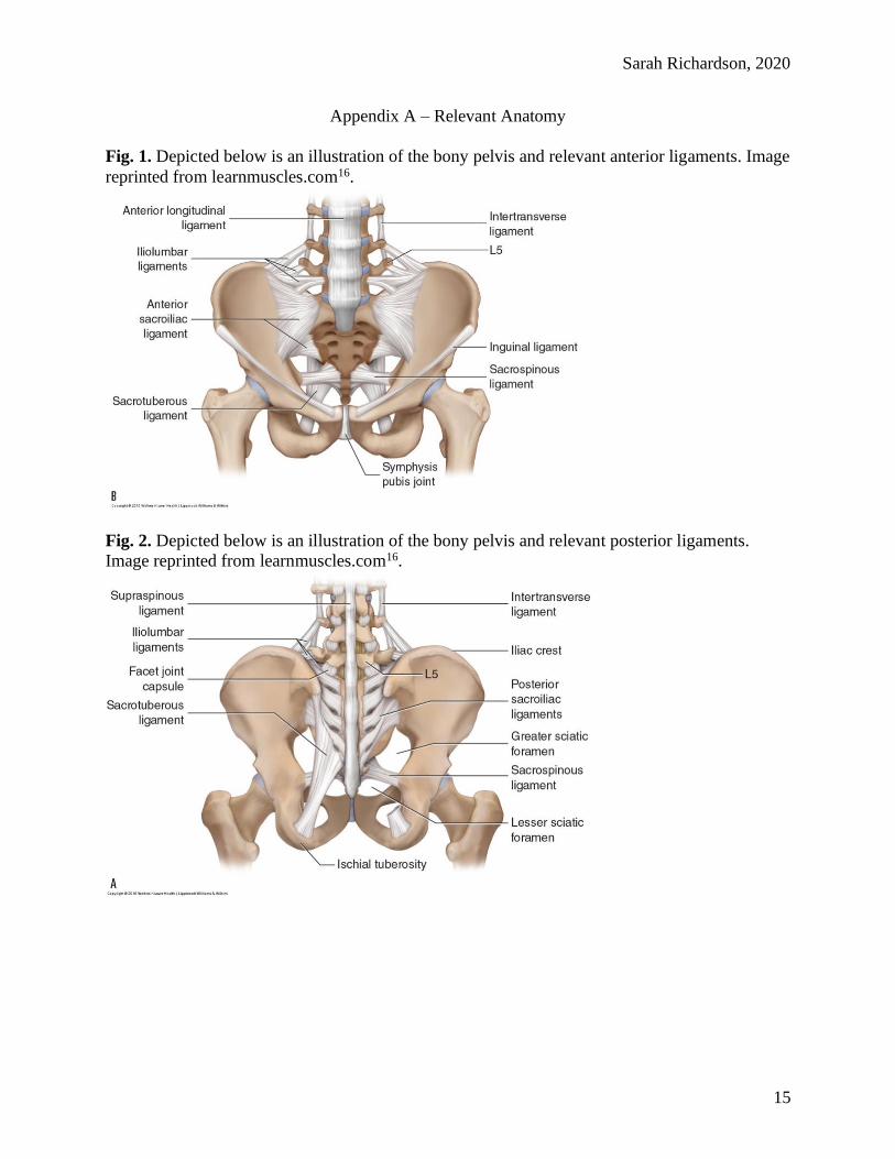
Appendix A – Relevant Anatomy
Fig. 1. Depicted below is an illustration of the bony pelvis and relevant anterior ligaments. Image
reprinted from learnmuscles.com16.
Fig. 2. Depicted below is an illustration of the bony pelvis and relevant posterior ligaments.
Image reprinted from learnmuscles.com16.

Sarah Richardson, 2020
16
Fig. 3. Depicted below is an illustration of the female reproductive system and relevant female-
specific ligaments. Image reprinted from ovarian anatomy.17
Fig. 4. Depicted below is an illustration of the pelvic floor and wall muscles of the female pelvis.
Image reprinted from Netter, 2002.18

Sarah Richardson, 2020
17
Appendix B – Biomechanical Changes During Pregnancy
Fig. 1. Depicted below is an illustration demonstrating and describing the musculoskeletal
compensations seen during pregnancy. Image reprinted from Casagrande et al., 2015.9

Sarah Richardson, 2020
18
Fig. 2. Depicted below is an illustration demonstrating anterior pelvic tilt and compensatory
hyperlordosis experienced during pregnancy. Image reprinted from Casagrande et al., 2015.9

Sarah Richardson, 2020
19
Appendix C – Physical Therapy Examination
Table 1: Differential Diagnosis for Pregnancy-Related Lumbopelvic Pain. Table adapted from
Casagrande et al., 2015.9
Differential Diagnosis for Low Back Pain (LBP) and Pelvic Girdle Pain (PGP) in
pregnancy
LBP PGP
Osteoarthritis – lumbar spine, SI joint,
hip
Painful visceral pathologies of the pelvis – urogenital
or Gastrointestinal
Stress fracture – sacrum, ilium, femur Syphilitic lesion of pubis
Lumbar disk herniation Lumbar disk herniation
Lumbar radiculopathy Lumbar radiculopathy
Spondylolisthesis/spondylolysis Rheumatism
Rheumatism Sciatica
Sciatica Spinal stenosis
Spinal stenosis Lumbar spine arthritis
Lumbar spine arthritis Tuberculosis
Tuberculosis Urinary Tract Infection
Urinary Tract Infection Rupture of symphysis pubis
Rupture of symphysis pubis Sacroiliac Joint sprain
Sacroiliac Joint sprain Osteitis pubis
Osteitis pubis Chorioamnionitis
Chorioamnionitis Femoral vein thrombosis
Femoral vein thrombosis Preterm labor
Preterm labor Placental abruption
Placental abruption Round ligament pain
Round ligament pain Bone or soft tissue tumor
Bone or soft tissue tumor
Ankylosing spondylitis
fibromyalgia
Red degeneration of leiomyoma
Pregnancy-related osteoporosis
Cauda equina
Osteomyelitis
Sarah Richardson, 2020
20
Lumbar facet arthropathy
Osteonecrosis of hip
Appendicitis
Pyelonephritis
Hydronephrosis
Renal Calculi
Aortic aneurysm
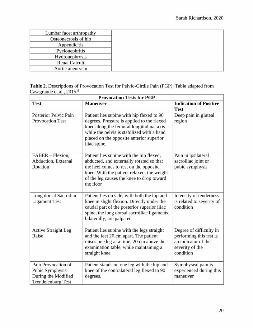
Table 2. Descriptions of Provocation Test for Pelvic-Girdle Pain (PGP). Table adapted from
Casagrande et al., 2015.9
Provocation Tests for PGP
Test Maneuver Indication of Positive
Test
Posterior Pelvic Pain
Provocation Test
Patient lies supine with hip flexed to 90
degrees. Pressure is applied to the flexed
knee along the femoral longitudinal axis
while the pelvis is stabilized with a hand
placed on the opposite anterior superior
iliac spine.
Deep pain in gluteal
region
FABER – Flexion,
Abduction, External
Rotation
Patient lies supine with the hip flexed,
abducted, and externally rotated so that
the heel comes to rest on the opposite
knee. With the patient relaxed, the weight
of the leg causes the knee to drop toward
the floor
Pain in ipsilateral
sacroiliac joint or
pubic symphysis
Long dorsal Sacroiliac
Ligament Test
Patient lies on side, with both the hip and
knee in slight flexion. Directly under the
caudal part of the posterior superior iliac
spine, the long dorsal sacroiliac ligaments,
bilaterally, are palpated
Intensity of tenderness
is related to severity of
condition
Active Straight Leg
Raise
Patient lies supine with the legs straight
and the feet 20 cm apart. The patient
raises one leg at a time, 20 cm above the
examination table, while maintaining a
straight knee
Degree of difficulty in
performing this test is
an indicator of the
severity of the
condition
Pain Provocation of
Pubic Symphysis
During the Modified
Trendelenburg Test
Patient stands on one leg with the hip and
knee of the contralateral leg flexed to 90
degrees.
Symphyseal pain is
experienced during this
maneuver
Sarah Richardson, 2020
21
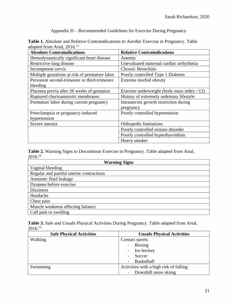
Appendix D – Recommended Guidelines for Exercise During Pregnancy
Table 1. Absolute and Relative Contraindications to Aerobic Exercise in Pregnancy. Table
adapted from Artal, 2016.15
Absolute Contraindications Relative Contraindications
Hemodynamically significant heart disease Anemia
Restrictive lung disease Unevaluated maternal cardiac arrhythmia
Incompetent cervix Chronic Bronchitis
Multiple gestations at risk of premature labor Poorly controlled Type 1 Diabetes
Persistent second-trimester or third-trimester
bleeding
Extreme morbid obesity
Placenta previa after 26 weeks of gestation Extreme underweight (body mass index <12)
Ruptured chorioamniotic membranes History of extremely sedentary lifestyle
Premature labor during current pregnancy Intrauterine growth restriction during
pregnancy
Preeclampsia or pregnancy-induced
hypertension
Poorly controlled hypertension
Severe anemia Orthopedic limitations
Poorly controlled seizure disorder
Poorly controlled hyperthyroidism
Heavy smoker
Table 2. Warning Signs to Discontinue Exercise in Pregnancy. Table adapted from Artal,
2016.15
Warning Signs
Vaginal bleeding
Regular and painful uterine contractions
Amniotic fluid leakage
Dyspnea before exercise
Dizziness
Headache
Chest pain
Muscle weakness affecting balance
Calf pain or swelling
Table 3. Safe and Unsafe Physical Activities During Pregnancy. Table adapted from Artal,
2016.15
Safe Physical Activities Unsafe Physical Activities
Walking Contact sports:
- Boxing
- Ice hockey
- Soccer
- Basketball
Swimming Activities with a high risk of falling:
- Downhill snow skiing

Sarah Richardson, 2020
22
- Water skiing
- Off-road cycling
- Gymnastics
- Horseback riding
Stationary Cycling Scuba Diving
Low-impact aerobics Sky Diving
Certain types of yoga and modified Pilates
For previously active females:
- Running or jogging
- Racquet sports
- Strength training
Sarah Richardson, 2020
23
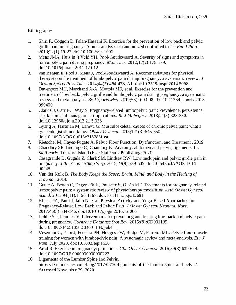
Bibliography
1. Shiri R, Coggon D, Falah-Hassani K. Exercise for the prevention of low back and pelvic
girdle pain in pregnancy: A meta-analysis of randomized controlled trials. Eur J Pain.
2018;22(1):19-27. doi:10.1002/ejp.1096
2. Mens JMA, Huis in ’t Veld YH, Pool-Goudzwaard A. Severity of signs and symptoms in
lumbopelvic pain during pregnancy. Man Ther. 2012;17(2):175-179.
doi:10.1016/j.math.2011.12.012
3. van Benten E, Pool J, Mens J, Pool-Goudzwaard A. Recommendations for physical
therapists on the treatment of lumbopelvic pain during pregnancy: a systematic review. J
Orthop Sports Phys Ther. 2014;44(7):464-473, A1. doi:10.2519/jospt.2014.5098
4. Davenport MH, Marchand A-A, Mottola MF, et al. Exercise for the prevention and
treatment of low back, pelvic girdle and lumbopelvic pain during pregnancy: a systematic
review and meta-analysis. Br J Sports Med. 2019;53(2):90-98. doi:10.1136/bjsports-2018-
099400
5. Clark CJ, Carr EC, Way S. Pregnancy-related lumbopelvic pain: Prevalence, persistence,
risk factors and management implications. Br J Midwifery. 2013;21(5):323-330.
doi:10.12968/bjom.2013.21.5.323
6. Gyang A, Hartman M, Lamvu G. Musculoskeletal causes of chronic pelvic pain: what a
gynecologist should know. Obstet Gynecol. 2013;121(3):645-650.
doi:10.1097/AOG.0b013e318283ffea
7. Rietschel M, Hayes-Fugate A. Pelvic Floor Function, Dysfunction, and Treatment . 2019.
8. Chaudhry SR, Imonugo O, Chaudhry K. Anatomy, abdomen and pelvis, ligaments. In:
StatPearls. Treasure Island (FL): StatPearls Publishing; 2020.
9. Casagrande D, Gugala Z, Clark SM, Lindsey RW. Low back pain and pelvic girdle pain in
pregnancy. J Am Acad Orthop Surg. 2015;23(9):539-549. doi:10.5435/JAAOS-D-14-
00248
10. Van der Kolk B. The Body Keeps the Score: Brain, Mind, and Body in the Healing of
Trauma.; 2014.
11. Gutke A, Betten C, Degerskär K, Pousette S, Olsén MF. Treatments for pregnancy-related
lumbopelvic pain: a systematic review of physiotherapy modalities. Acta Obstet Gynecol
Scand. 2015;94(11):1156-1167. doi:10.1111/aogs.12681
12. Kinser PA, Pauli J, Jallo N, et al. Physical Activity and Yoga-Based Approaches for
Pregnancy-Related Low Back and Pelvic Pain. J Obstet Gynecol Neonatal Nurs.
2017;46(3):334-346. doi:10.1016/j.jogn.2016.12.006
13. Liddle SD, Pennick V. Interventions for preventing and treating low-back and pelvic pain
during pregnancy. Cochrane Database Syst Rev. 2015;(9):CD001139.
doi:10.1002/14651858.CD001139.pub4
14. Vesentini G, Prior J, Ferreira PH, Hodges PW, Rudge M, Ferreira ML. Pelvic floor muscle
training for women with lumbopelvic pain: A systematic review and meta-analysis. Eur J
Pain. July 2020. doi:10.1002/ejp.1636
15. Artal R. Exercise in pregnancy: guidelines. Clin Obstet Gynecol. 2016;59(3):639-644.
doi:10.1097/GRF.0000000000000223
16. Ligaments of the Lumbar Spine and Pelvis.
https://learnmuscles.com/blog/2017/08/30/ligaments-of-the-lumbar-spine-and-pelvis/.
Accessed November 29, 2020.

Sarah Richardson, 2020
24
17. CH27 Ovarian Anatomy.
https://www.apsubiology.org/anatomy/2020/2020_Exam_Reviews/Exam_5/CH27_Ovaria
n_Anatomy.htm. Accessed November 29, 2020.
18. 4: Pelvic floor diaphragm, medial view. Adapted from [Netter, 2002] | Download Scientific
Diagram. https://www.researchgate.net/figure/Pelvic-floor-diaphragm-medial-view-
Adapted-from-Netter-2002_fig4_216453509. Accessed November 29, 2020.