Sandhya H and Srinath Rao - scipress.com · Sandhya H and Srinath Rao Plant Tissue Culture and...
7
Role of growth regulators on in vitro callus induction and direct regeneration in Physalis minima Linn. Sandhya H and Srinath Rao Plant Tissue Culture and Genetic Engineering Laboratory, Department of Post Graduate Studies and Research in Botany, Gulbarga University, Kalaburagi- 585106 Karnataka, INDIA. *E-mail address: [email protected] Keywords: Physalis minima, Node, Stem, Leaf, callus and growth regulators. ABSTRACT. Suitable protocol for induction of callus and regeneration was developed from different explants viz., node, stem and leaves in Physalis minima. MS basal medium supplemented with various concentrations (1.0-4.0mg/l) of auxins like 2,4-Dichlorophenoxy acetic acid (2,4-D), α-naphthalene acetic acid (NAA) and Indole- 3-acetic acid (IAA) and cytokinins (0.5-1.5mg/l) like BAP or Kn were used. All the three explants responded for induction of callus, however stem explants were found superior, followed by node and leaf. Callus induction was observed in all the auxins and combination of growth regulators used with varied mass (2010±1.10) and highest percentage of callus induction was observed from stem at 2.0mg/l 2,4-D (90%) followed by NAA (70%) and IAA (50%). Organogenesis was induced when nodal explants were transferred on MS medium supplemented with 2,4-D and Kn at various concentrations, maximum being on 2.0mg/l 2,4-D + 1.0mg/l Kn (90%). Regenerated shoots were elongated on 0.5mg/l GA 3 . The shoots were subsequently rooted on MS + 1.0mg/l IBA (95%) medium. Rooted shoots were hardened and acclimatized, later they were transferred to polycups containing soil, cocopeat and sand in the ratio 1:2:1. 1. INTRODUCTION Physalis minima L. belongs to the family Solanaceae. It is a small herbaceous plant grows as weed in crop fields, which is commonly known by several names, like native goose berry, Wild cape goose berry and Pygmy ground cherry. It is an annual herb, 20-50cm high at its maturity. Typical Physalis growth pattern and shape with some branching and pretty yellow flowers borne at nodes, fruits ripen in a couple of months and are enclosed in a green-yellow papery calyx which protects them from pests (tradewindsfruit.com). Geographically they are distributed in East Asia, China, Himalayas and in some places of Australia (Prasad et al., 2009). The technique of plant tissue culture at present is attracting worldwide attention, because plant cells are able to synthesize specific compounds, especially various secondary metabolites usefull as medicines and food additives (Sheeba et al.,2013). Solanaceous plants are able to synthesize a wide range of alkaloids with interesting pharmaceutical activities (Yamada and Tabata, 1997; Alireza et al., 2006). This species is used medicinally as tonic, diuretic, laxative, anti-inflammatry, enlargement of the spleen and abdominal troubles and as a helpful remedy in ulceration of the bladder, fruit is of this plant are used to cure spleen disorders, the leaves are crushed and applied over snakebite site (Mungole et al.,2011). 2. MATERIALS AND METHODS Plant Material Physalis minima L. were collected from different areas around Gulbarga. In the present investigations explants like node, stem and leaves were used. They were soaked in solution containing Bavistin (1.0% w/v, a fungicide) and tween 20 for 15- 20mn and then the explants were washed under running tap water and rinsed thrice with distilled water. The explants were then International Letters of Natural Sciences Online: 2015-07-31 ISSN: 2300-9675, Vol. 44, pp 38-44 doi:10.18052/www.scipress.com/ILNS.44.38 © 2015 SciPress Ltd., Switzerland SciPress applies the CC-BY 4.0 license to works we publish: https://creativecommons.org/licenses/by/4.0/
-
Upload
nguyenliem -
Category
Documents
-
view
220 -
download
0
Transcript of Sandhya H and Srinath Rao - scipress.com · Sandhya H and Srinath Rao Plant Tissue Culture and...

Role of growth regulators on in vitro callus induction and direct regeneration in Physalis minima Linn.
Sandhya H and Srinath Rao
Plant Tissue Culture and Genetic Engineering Laboratory,
Department of Post Graduate Studies and Research in Botany, Gulbarga University,
Kalaburagi- 585106 Karnataka, INDIA.
*E-mail address: [email protected]
Keywords: Physalis minima, Node, Stem, Leaf, callus and growth regulators.
ABSTRACT. Suitable protocol for induction of callus and regeneration was developed from
different explants viz., node, stem and leaves in Physalis minima. MS basal medium supplemented
with various concentrations (1.0-4.0mg/l) of auxins like 2,4-Dichlorophenoxy acetic acid (2,4-D),
α-naphthalene acetic acid (NAA) and Indole- 3-acetic acid (IAA) and cytokinins (0.5-1.5mg/l) like
BAP or Kn were used. All the three explants responded for induction of callus, however stem
explants were found superior, followed by node and leaf. Callus induction was observed in all the
auxins and combination of growth regulators used with varied mass (2010±1.10) and highest
percentage of callus induction was observed from stem at 2.0mg/l 2,4-D (90%) followed by NAA
(70%) and IAA (50%). Organogenesis was induced when nodal explants were transferred on MS
medium supplemented with 2,4-D and Kn at various concentrations, maximum being on 2.0mg/l
2,4-D + 1.0mg/l Kn (90%). Regenerated shoots were elongated on 0.5mg/l GA3. The shoots were
subsequently rooted on MS + 1.0mg/l IBA (95%) medium. Rooted shoots were hardened and
acclimatized, later they were transferred to polycups containing soil, cocopeat and sand in the ratio
1:2:1.
1. INTRODUCTION
Physalis minima L. belongs to the family Solanaceae. It is a small herbaceous plant grows
as weed in crop fields, which is commonly known by several names, like native goose berry, Wild
cape goose berry and Pygmy ground cherry. It is an annual herb, 20-50cm high at its maturity.
Typical Physalis growth pattern and shape with some branching and pretty yellow flowers borne at
nodes, fruits ripen in a couple of months and are enclosed in a green-yellow papery calyx which
protects them from pests (tradewindsfruit.com). Geographically they are distributed in East Asia,
China, Himalayas and in some places of Australia (Prasad et al., 2009). The technique of plant
tissue culture at present is attracting worldwide attention, because plant cells are able to synthesize
specific compounds, especially various secondary metabolites usefull as medicines and food
additives (Sheeba et al.,2013). Solanaceous plants are able to synthesize a wide range of alkaloids
with interesting pharmaceutical activities (Yamada and Tabata, 1997; Alireza et al., 2006). This
species is used medicinally as tonic, diuretic, laxative, anti-inflammatry, enlargement of the spleen
and abdominal troubles and as a helpful remedy in ulceration of the bladder, fruit is of this plant are
used to cure spleen disorders, the leaves are crushed and applied over snakebite site (Mungole et
al.,2011).
2. MATERIALS AND METHODS
Plant Material
Physalis minima L. were collected from different areas around Gulbarga. In the present
investigations explants like node, stem and leaves were used. They were soaked in solution
containing Bavistin (1.0% w/v, a fungicide) and tween 20 for 15- 20mn and then the explants were
washed under running tap water and rinsed thrice with distilled water. The explants were then
International Letters of Natural Sciences Online: 2015-07-31ISSN: 2300-9675, Vol. 44, pp 38-44doi:10.18052/www.scipress.com/ILNS.44.38© 2015 SciPress Ltd., Switzerland
SciPress applies the CC-BY 4.0 license to works we publish: https://creativecommons.org/licenses/by/4.0/

sterilized with 70% Ethyl alcohol for 3-4mn followed by washing with double distilled water and
then surface sterilized with 0.1% (w/v) mercuric chloride for 3-4mn and subsequently washed
thoroughly in sterile distilled water to remove the traces of mercuric chloride. They were then
inoculated on MS medium (Murashige and Skoog, 1962) supplemented with various auxins viz.,
2,4-Dichloro phenoxyaceticacid (2,4-D) (1.0-4.0mg/l), α-naphthalene acetic acid (NAA) (1.0-
4.0mg/l) and Indole- 3-acetic acid (IAA) (1.0-4.0mg/l) alone or supplemented with cytokinins like
6-Benzyl amino purine (BAP) (0.5-1.5mg/l) and Kinetin (Kn) (0.5-1.5mg/l). The pH of the medium
was adjusted to 5.6-5.8 prior to autoclaving at 1210C for 20mn. The cultures were maintained at
25±10C with 16/8 hr photoperiod at 40µm m
-2 s
-1 provided by cool white fluorescent tubes. The
callus was maintained by regular sub-culture at 4 weeks of interval on fresh medium with the same
composition or on regeneration media. The data was subjected to statistical analysis.
Table: 1. Effect of auxins on induction of callus in Physalis minima L.
Data represents average of three replicates; each replicate consists of 25 cultures. Mean ±Standard
error. Mean followed by the different superscript in column are not significantly different from each
other. a=P<0.05, b=P<0.01 and c=P<0.001 levels according to ANOVA.
Table: 2. Effect of 2,4-D (2.0mg/l) in combination with cytokinins on induction and growth of
callus in Physalis minima L.
Growth
Hormone
Conc.
mg/l
Stem
Fresh Wt. (mg) Dry Wt. (mg) Frequency (%)
2,4-D+BAP
2.0+0.5 1923±0.10b
252±1.00b
70
2.0+1.0 1852±1.15b
235±1.25b
80
2.0+1.5 1620±1.00a
201±0.00a
60
2,4-D+Kn
2.0+0.5 3580±0.00a
595±1.12b
90
2.0+1.0 2310±1.00c
445±0.00c
80
3.0+1.5 2110±1.20c
430±0.10c
70
Growth
Hormone
Conc.
mg/l
Stem
Node Leaf
Fresh Wt.
(mg)
Dry Wt.
(mg)
Frequency
(%)
Fresh Wt.
(mg)
Dry Wt.
(mg)
Frequency
(%)
Fresh Wt.
(mg)
Dry Wt.
(mg)
Frequency
(%)
2,4-D
1.0
1125±1.15c 138±0.11c 40 1011±0.01c 120±1.30c 45 1002±0.00a 115±0.00a 40
2.0
2010±1.10a 428±0.11a 90 1301±1.20a 229±1.30a 70 1100±1.20b 199±1.00a 50
3.0
1850±1.00b 231±1.10b 60 1120±0.05b 180±0.00b 50 985±1.00a 96±1.17c 45
4.0
1714±1.20b 198±1.20b 50 1105±1.45c 196±0.00b 50 890±0.12b 81±1.21c 40
NAA
1.0
1021±0.05b 130±0.20b 45 1010±1.10c 120±1.11c 50 1095±1.20b 187±0.01c 60
2.0
1195±1.23a 228±0.01a 70 1098±020b 190±1.23b 55 1011±0.20b 119±1.37c 50
3.0
1101±1.10a 201±0.00a 50 1001±1.15b 117±1.20c 40 986±1.25c 96±0.10b 45
4.0
998±0.20c 102±1.10c 40 898±1.25c 84±0.00c 40 875±1.22c 81±1.73c 40
IAA
1.0
992±0.00b 98±1.30b 45 980±0.20c 95±1.20b 45 870±1.23c 79±1.45c 35
2.0
1102±0.10b 195±1.21b 50 992±1.20b 97±1.11b 40 902±1.11c 88±1.00a 45
3.0 1011±1.20a 121±1.10c 40 1060±0.22a 170±0.10a 30 1048±0.00a 155±0.00a 40
4.0 976±0.11b 94±1.20c 35 901±1.11c 88±1.76c 35 917±1.10c 89±1.76c 30
International Letters of Natural Sciences Vol. 44 39

Data represents average of three replicates; each replicate consists of 25 cultures. Mean ±Standard
error. Mean followed by the different superscript in column are not significantly different from each
other. a=P<0.05, b=P<0.01 and c=P<0.001 levels according to ANOVA.
Table: 3. Effect of 2,4-D (2.0mg/l) in combination with Kn on direct regeneration of multiple
shoots in Physalis minima L.
Growth
Hormone
Conc.
(mg/l)
Node
No. of Multiple Shoots Frequency (%)
2,4-D+Kn
2.0+0.5 3.10±0.17b
70
2.0+1.0 7.26±0.60a
95
2.0+1.5 3.20±0.25b
70
Data represents average of three replicates; each replicate consists of 25 cultures. Mean ±Standard
error. Mean followed by the different superscript in column are not significantly different from each
other. a=P<0.05, b=P<0.01 and c=P<0.001 levels according to ANOVA.
Table: 4. Effect of IBA on root induction from in vitro raised shoots of Physalis minima L. after 30
days of culture.
Growth
Hormone
Conc.
(mg/l) No. of roots Frequency (%)
IBA
1.0 15.20±0.30a
95
2.0 8.35±0.11c
70
3.0 4.64±0.28c
70
Data represents average of three replicates; each replicate consists of 25 cultures. Mean ±Standard
error. Mean followed by the different superscript in column are not significantly different from each
other. a=P<0.05, b=P<0.01 and c=P<0.001 levels according to ANOVA.
3. RESULTS AND DISCUSSION
The induction of callus and regeneration of plant depends on explants and influenced by the
concentrations and combinations of growth regulators in the medium. Different explants were
cultured on MS medium supplemented with various auxins and cytokinins in varied combinations
to assess the morphogenetic potential of the explants.
4. CALLUS INDUCTION
In the present investigations node, stem and leaf explants were inoculated on MS medium
fortified with different concentrations of 2,4-D, NAA and IAA alone or in combination with BAP
or Kn for callus induction. All the three explants responded for callus induction with varying
degree, stem explants proved best followed by node and leaf. Highest frequency (90% ; 18 days)
was noticed in stem explants followed by node (70% ; 20 days ) and leaf (50% ; 25 days).
Among the three auxins tested the frequency of callus induction was maximum on MS
medium supplemented with 2.0mg/l 2,4-D, followed by NAA and IAA. From the data presented in
Table-1, it can be noticed that with an increase in the concentration of auxins there was an increase
in the frequency and growth of the callus upto 2.0mg/l and at 4.0mg/l the frequency and growth of
callus decreased. Maximum frequency (90%) of callus induction and highest biomass
(2010±1.10mg) was observed on MS medium supplemented with 2.0mg/l 2,4-D when stem were
used as explants followed by nodal explants (1301±1.20mg) with 70% frequency and leaf explants
(1100±1.20mg) with 50% frequency (Table-1; Plate-I a). However other auxins viz., NAA and IAA
showed poor response with respect to induction and further growth of callus at all the
40 Volume 44
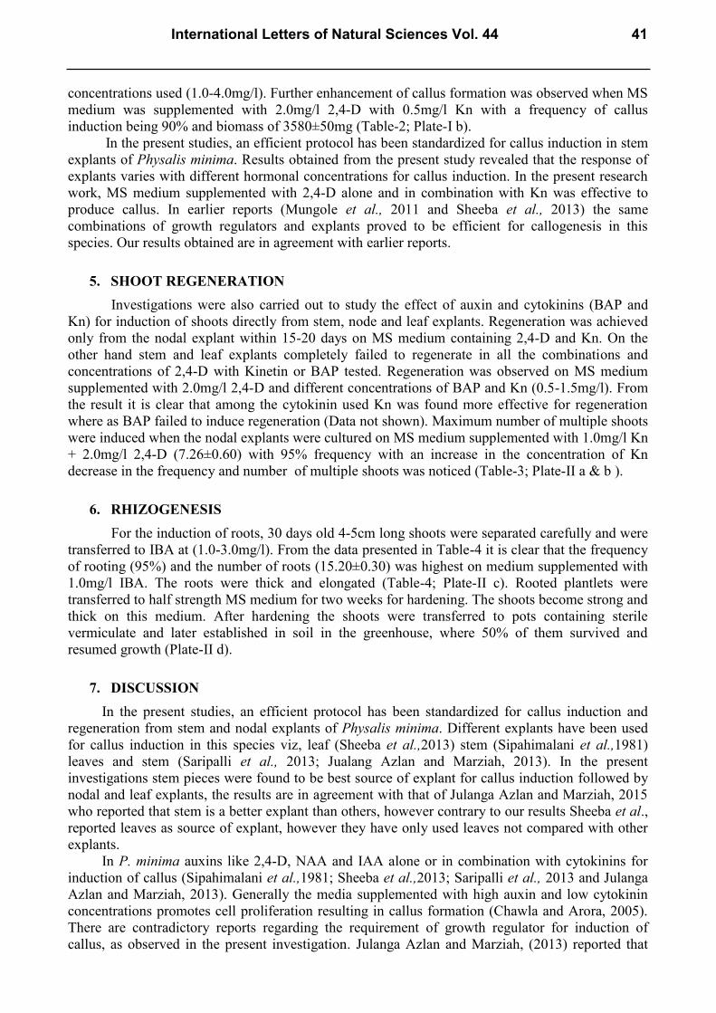
concentrations used (1.0-4.0mg/l). Further enhancement of callus formation was observed when MS
medium was supplemented with 2.0mg/l 2,4-D with 0.5mg/l Kn with a frequency of callus
induction being 90% and biomass of 3580±50mg (Table-2; Plate-I b).
In the present studies, an efficient protocol has been standardized for callus induction in stem
explants of Physalis minima. Results obtained from the present study revealed that the response of
explants varies with different hormonal concentrations for callus induction. In the present research
work, MS medium supplemented with 2,4-D alone and in combination with Kn was effective to
produce callus. In earlier reports (Mungole et al., 2011 and Sheeba et al., 2013) the same
combinations of growth regulators and explants proved to be efficient for callogenesis in this
species. Our results obtained are in agreement with earlier reports.
5. SHOOT REGENERATION
Investigations were also carried out to study the effect of auxin and cytokinins (BAP and
Kn) for induction of shoots directly from stem, node and leaf explants. Regeneration was achieved
only from the nodal explant within 15-20 days on MS medium containing 2,4-D and Kn. On the
other hand stem and leaf explants completely failed to regenerate in all the combinations and
concentrations of 2,4-D with Kinetin or BAP tested. Regeneration was observed on MS medium
supplemented with 2.0mg/l 2,4-D and different concentrations of BAP and Kn (0.5-1.5mg/l). From
the result it is clear that among the cytokinin used Kn was found more effective for regeneration
where as BAP failed to induce regeneration (Data not shown). Maximum number of multiple shoots
were induced when the nodal explants were cultured on MS medium supplemented with 1.0mg/l Kn
+ 2.0mg/l 2,4-D (7.26±0.60) with 95% frequency with an increase in the concentration of Kn
decrease in the frequency and number of multiple shoots was noticed (Table-3; Plate-II a & b ).
6. RHIZOGENESIS
For the induction of roots, 30 days old 4-5cm long shoots were separated carefully and were
transferred to IBA at (1.0-3.0mg/l). From the data presented in Table-4 it is clear that the frequency
of rooting (95%) and the number of roots (15.20±0.30) was highest on medium supplemented with
1.0mg/l IBA. The roots were thick and elongated (Table-4; Plate-II c). Rooted plantlets were
transferred to half strength MS medium for two weeks for hardening. The shoots become strong and
thick on this medium. After hardening the shoots were transferred to pots containing sterile
vermiculate and later established in soil in the greenhouse, where 50% of them survived and
resumed growth (Plate-II d).
7. DISCUSSION
In the present studies, an efficient protocol has been standardized for callus induction and
regeneration from stem and nodal explants of Physalis minima. Different explants have been used
for callus induction in this species viz, leaf (Sheeba et al.,2013) stem (Sipahimalani et al.,1981)
leaves and stem (Saripalli et al., 2013; Jualang Azlan and Marziah, 2013). In the present
investigations stem pieces were found to be best source of explant for callus induction followed by
nodal and leaf explants, the results are in agreement with that of Julanga Azlan and Marziah, 2015
who reported that stem is a better explant than others, however contrary to our results Sheeba et al.,
reported leaves as source of explant, however they have only used leaves not compared with other
explants.
In P. minima auxins like 2,4-D, NAA and IAA alone or in combination with cytokinins for
induction of callus (Sipahimalani et al.,1981; Sheeba et al.,2013; Saripalli et al., 2013 and Julanga
Azlan and Marziah, 2013). Generally the media supplemented with high auxin and low cytokinin
concentrations promotes cell proliferation resulting in callus formation (Chawla and Arora, 2005).
There are contradictory reports regarding the requirement of growth regulator for induction of
callus, as observed in the present investigation. Julanga Azlan and Marziah, (2013) reported that
International Letters of Natural Sciences Vol. 44 41

2,4-D and kinetin was best for callus induction in this species, Sipahimalani et al., (1981) reported
that combination of 2,4-D + BAP favoured callus formation, however they have not used kinetin in
combination with 2.4-D, on the contrary Prasad et al., (2009) and Saripalli et al., (2013) reported
combination of BAP + NAA was better for callus induction with stem explants. It is very difficult
to explain the contradictory reports with respect to requirement of growth regulators using the same
(stem) explants. Sheeba et al., (2013) reported combination of IAA and BAP for callus induction in
this species, this varied results may be due to the different explants (Leaves) by them and stem in
our investigation, however again there is a contradictory report of callus induction from leaf explant
(Saripalli et al., 2013) who reported 1.0mg/l BAP + 0.5mg/l α-NAA suitable for callus induction in
this species.
The results obtained from present research work, MS medium supplemented with 2,4-D and
in combination with Kn was effective to produce multiples from nodal explant.
Different explants have been used for induction of multiple shoots in this species viz, shoot
tip (Intzaar et al., 2013; Ramar and Ayyadurai 2014) leaf, shoot tip and nodes (Sheeba et al., 2013).
A single explant has been used and an attempt to compare the response of other explants is not
made, however, Sheeba et al., (2013) have reported that nodal explants were superior to shoot tips
with respect to induction of multiple shoots, the results are in conformity with our investigations.
There are reports that the shoot regeneration using BAP and coconut water on MS medium
from nodal explants in P. minima (Afroz et al., 2009) and from leaf and nodal explants of P.
minima were responsive to organogenesis with BAP, IAA and IBA at different hormones (Sheeba
et al.,2010; Ashwinin Solanki and Dipali Gupta, 2013). In the present investigation multiple shoots
were induced on MS medium supplemented with 2, 4-D and kn. Highest frequency (95%) and
maximum (7.26±0.60) number of multiple shoots were recorded on medium supplemented with
1.0mg/l kn + 2.0mg/l 2,4-D.
Plate - I
Plate I. In vitro callus induction of Physalis minima L. a) Induction of callus from stem explants on
MS + 2.0mg/l 2,4-D b) Induction of callus from stem explants on MS + 2.0mg/l 2,4-D +0.5mg/l
Kn.
a b
42 Volume 44

Plate - II
Plate II. In vitro shoot induction, proliferation and acclimatization from nodal explants of Physalis
minima L. a) Initiation of direct multiple shoots from nodal explants on MS + 2.0mg/l 2,4-D +
1.0mg/l Kn. b) Multiple shoot formation from in vitro derived nodal explants on MS + 2.0mg/l 2,4-
D + 1.0mg/l Kn after 15 days. c) Root induction on MS + 1.0mg/l IBA. d) Acclimatized potted
plant.
There are contradictory reports
There are also reports in different species like P. pubescens, stating that MS basal medium
supplemented with BAP and NAA is optimum for regeneration and callus induced from leaf and
nodal segments (Rao et al.,2004), shoots were initiated from callus obtained from the apical leaf
explants only not from the root and node explants calli (Mungole et al.,2011) and the maximum
number of multiple shoots were achieved from nodal and stem explants on BAP, GA3 and 2,4-D in
P. peruviana by (Ramar et al.,2014). This shows that organogenesis as callogenesis is also highly
dependent on genotype. In our investigation organogenesis was not obtained on BAP, IAA and
NAA supplemented media, however earlier reports mentioned above have BAP as suitable
cytokinin for induction of shoots. Among these results nodal explants is common in all the reports
only differs in hormonal concentrations. Our results are in against with these reports.
8. CONCLUSION
The method standardized in the present study for the production of callus and plants from
intermodal and nodal explants is useful for the callogenesis and multiplication as well as for the
genetic manipulation. The plant is known to have important secondary metabolites Physalin, so
callus can be used for secondary metabolite production and this protocol is expected to be very
useful for the mass production of this species.
a
b
d c
d
International Letters of Natural Sciences Vol. 44 43

References
[1] Prasad S. H. K. R., Swapna N. L., Rajasekhar D., Anthonamma K. and Prasad M. (2009).
Preliminary phytochemical and antimicrobial spectrum of cultured tissues of Physalis
minima (L). Int. J. Chem. Sci.:7(4): 2719-2725.
[2] Sheeba E., Parvathy S. and Palanivel S. (2010). Direct Regeneration from leaves and nodes
explants of Physalis minima LINN. European Journal of Applied Sciences. 2(2): 58-61.
[3] Alireza I., Oshaghi M. A. and Majd A. (2006). Distribution of atropine and scopolamine in
different organs and stages of development in Datura stramonium L. (Solanaceae). Structure
and ultrstructure of biosynthesizing cells. Acta Biologica Cracoviensia Series Botanica. Vol.
48 (1), pp. 13-18.
[4] Yamada Y. and Tabata M. (1997). Plant biotechnology of tropane alkaloids. Plant
Biotechnology. Vol. 14, pp. 1-10.
[5] Mungole A. J., Doifode V. D., Kamble R. B., Chaturvedi A. and Zanwar P. (2011). In- vitro
callus induction and shoot regeneration in Physalis minima L. Annals of Biological
Research. 2(2): 79-85.
[6] Murashige T. and Skoog F. (1962). A revised medium for rapid growth and bioassays with
tobacco cultures. Physiol. Plants. 15: 473-497.
[7] Chawla H. S. and Arora A. (2005). Organogenic plant regeneration via callus induction in
Chick pea (Cicer arietinum L.)- Role of genotypes, growth regulators and explants. Indian
Journal of Biotechnology. Vol. 4, pp. 251-256.
[8] Sheeba E., Palanivel S. and Parvathi S. (2013). Effect of plant growth regulators on callus
induction in Physalis minima Linn. International Journal of Innovative Research in Science,
Engineering and Technology. Vol. 2 (9): 4847- 4851.
[9] Afroz F., Hassan A. K. M. S., Bari L. S., Sultana R., Begum N., Jahan M. A. A. and Khatun
R. (2009). In vitro shoot proliferation and plant regeneration of Physalis minima L. a
Perennial Medicinal Herb. Bangladesh J. Sci. Ind. Res. 44 : (4) 453-456.
[10] Ashwani Solanki and Dipali Gupta (2013). In vitro shoot multiplication in Physalis minima
var. Indica L. Biosciences Boitechnology Research Asia. Vol. 10 (1), 371-374.
[11] Rao Y. V., Ravishankar A., Lakshmi T. V. R. and Rao R. K. G. (2004).Plant regeneration in
Physalis pubescens L. and its induced mutant. Plant Tissue Cult. 14 (1) : 9-15.
[12] Ramar K., Ayyadurai V. and Arulprakash T. (2014). In vitro shoot multiplication and plant
regeneration of Physalis peruviana L. An important medicinal plant. International Journal of
Current Microbiology and Applied Sciences. Vol. 3 (3), pp. 456-464.
[13] Sipahimalani AT, Bapat VA, Rao PS and Chadha MS. (1981). Biosynthetic potential of
cultured tissues and regenerated plants of Physalis minima L. Nat. Procd. (Lloyadia) 44(1):
114-118.
[14] Intzaar S., Akram M. and Afrasiab H. (2013). High frequency multiple shoot formation of
pygmy groundcherry (Physalis minima): An endangered medicinal plant. International
Journal of Agriculture and Biology. 15(4): 755-760.
[15] Jualang Azlan Gansau and Marziah mahmood. (2013). Growth characterstics and production
of physalins from Physalis minima hairy roots in shake flasks. Kasetsart J. (Nat. Sci.) 47:
748-759.
[16] Saripalli H. R., Nandam L. S., Teka Z. and Madanprasad. (2013). Preliminary
phytochemical studies and efficacy of chloroform extracts of cultured tissues of Physalis
minima (L.) against pathogens. Global Journal of Biology, Agriculrure and Health sciences.
Vol. 2 (4): 187-190.
44 Volume 44