Sample Human Biology March - Pearson Education · Figure 9.1 The brain and spinal cord form ......
13
EDEXCEL INTERNATIONAL GCSE (9 –1) HUMAN BIOLOGY Student Book Philip Bradfield, Steve Potter eBook included Uncorrected proof, all content subject to change at publisher discretion. Not for resale, circulation or distribution in whole or in part. ©Pearson 2017
Transcript of Sample Human Biology March - Pearson Education · Figure 9.1 The brain and spinal cord form ......

EDEXCEL INTERNATIONAL GCSE (9 –1)
HUMAN BIOLOGYStudent Book Philip Bradfield, Steve Potter
eBookincluded
Unc
orre
cted
pro
of, a
ll co
nten
t sub
ject
to c
hang
e at
pub
lishe
r dis
cret
ion.
Not
for r
esal
e, c
ircul
atio
n or
dis
tribu
tion
in w
hole
or i
n pa
rt. ©
Pea
rson
201
7
EDEXCEL INTERNATIONAL GCSE (9 –1)
HUMAN BIOLOGYStudent Book
Philip BradfieldSteve Potter
Unc
orre
cted
pro
of, a
ll co
nten
t sub
ject
to c
hang
e at
pub
lishe
r dis
cret
ion.
Not
for r
esal
e, c
ircul
atio
n or
dis
tribu
tion
in w
hole
or i
n pa
rt. ©
Pea
rson
201
7

CONTENTS iii
COURSE STRUCTURE
ABOUT THIS BOOK
ASSESSMENT OVERVIEW
UNIT 1
UNIT 2
UNIT 3
UNIT 4
APPENDIX
GLOSSARY
INDEX
IV
VI
VIII
2
52
124
192
257
262
289
Unc
orre
cted
pro
of, a
ll co
nten
t sub
ject
to c
hang
e at
pub
lishe
r dis
cret
ion.
Not
for r
esal
e, c
ircul
atio
n or
dis
tribu
tion
in w
hole
or i
n pa
rt. ©
Pea
rson
201
7
UNIT 3 COORDINATION 149
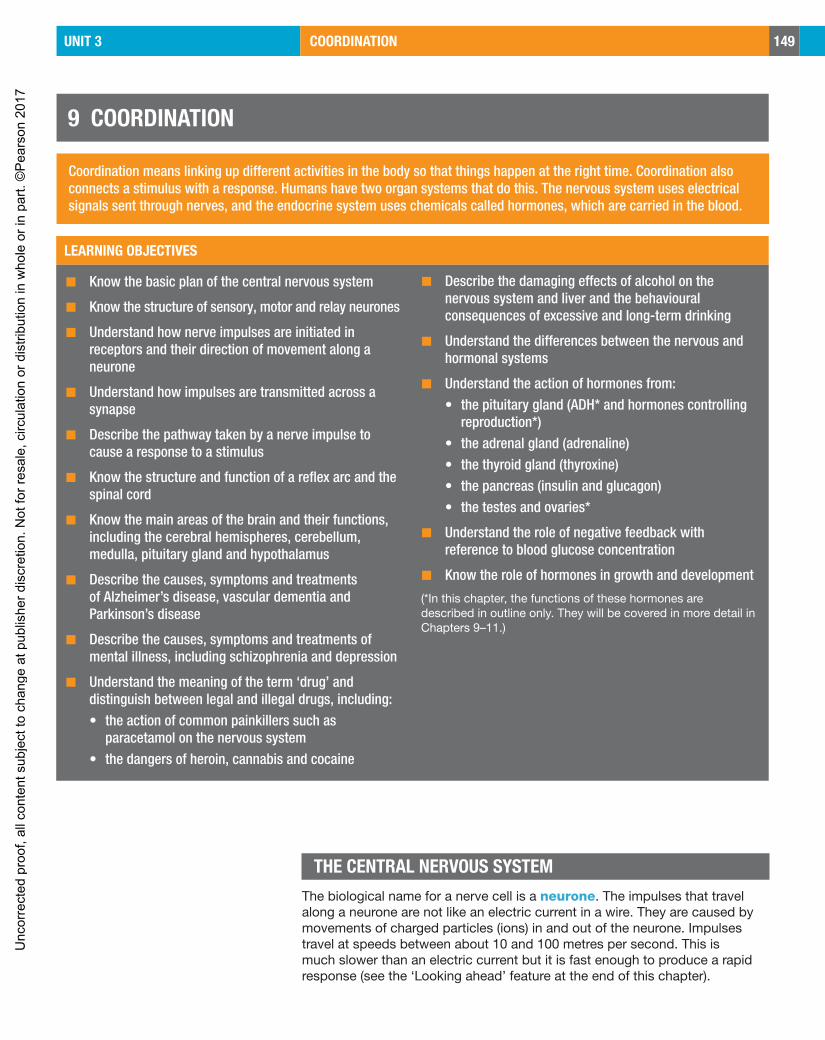
9 COORDINATION
LEARNING OBJECTIVES
◼ Know the basic plan of the central nervous system
◼ Know the structure of sensory, motor and relay neurones
◼ Understand how nerve impulses are initiated in receptors and their direction of movement along a neurone
◼ Understand how impulses are transmitted across a synapse
◼ Describe the pathway taken by a nerve impulse to cause a response to a stimulus
◼ Know the structure and function of a reflex arc and the spinal cord
◼ Know the main areas of the brain and their functions, including the cerebral hemispheres, cerebellum, medulla, pituitary gland and hypothalamus
◼ Describe the causes, symptoms and treatments of Alzheimer’s disease, vascular dementia and Parkinson’s disease
◼ Describe the causes, symptoms and treatments of mental illness, including schizophrenia and depression
◼ Understand the meaning of the term ‘drug’ and distinguish between legal and illegal drugs, including:• the action of common painkillers such as
paracetamol on the nervous system• the dangers of heroin, cannabis and cocaine
◼ Describe the damaging effects of alcohol on the nervous system and liver and the behavioural consequences of excessive and long-term drinking
◼ Understand the differences between the nervous and hormonal systems
◼ Understand the action of hormones from:• the pituitary gland (ADH* and hormones controlling
reproduction*)• the adrenal gland (adrenaline)• the thyroid gland (thyroxine)• the pancreas (insulin and glucagon)• the testes and ovaries*
◼ Understand the role of negative feedback with reference to blood glucose concentration
◼ Know the role of hormones in growth and development
(*In this chapter, the functions of these hormones are described in outline only. They will be covered in more detail in Chapters 9–11.)
Coordination means linking up different activities in the body so that things happen at the right time. Coordination also connects a stimulus with a response. Humans have two organ systems that do this. The nervous system uses electrical signals sent through nerves, and the endocrine system uses chemicals called hormones, which are carried in the blood.
THE CENTRAL NERVOUS SYSTEMThe biological name for a nerve cell is a neurone. The impulses that travel along a neurone are not like an electric current in a wire. They are caused by movements of charged particles (ions) in and out of the neurone. Impulses travel at speeds between about 10 and 100 metres per second. This is much slower than an electric current but it is fast enough to produce a rapid response (see the ‘Looking ahead’ feature at the end of this chapter).
Unc
orre
cted
pro
of, a
ll co
nten
t sub
ject
to c
hang
e at
pub
lishe
r dis
cret
ion.
Not
for r
esal
e, c
ircul
atio
n or
dis
tribu
tion
in w
hole
or i
n pa
rt. ©
Pea
rson
201
7
150 UNIT 3 COORDINATION
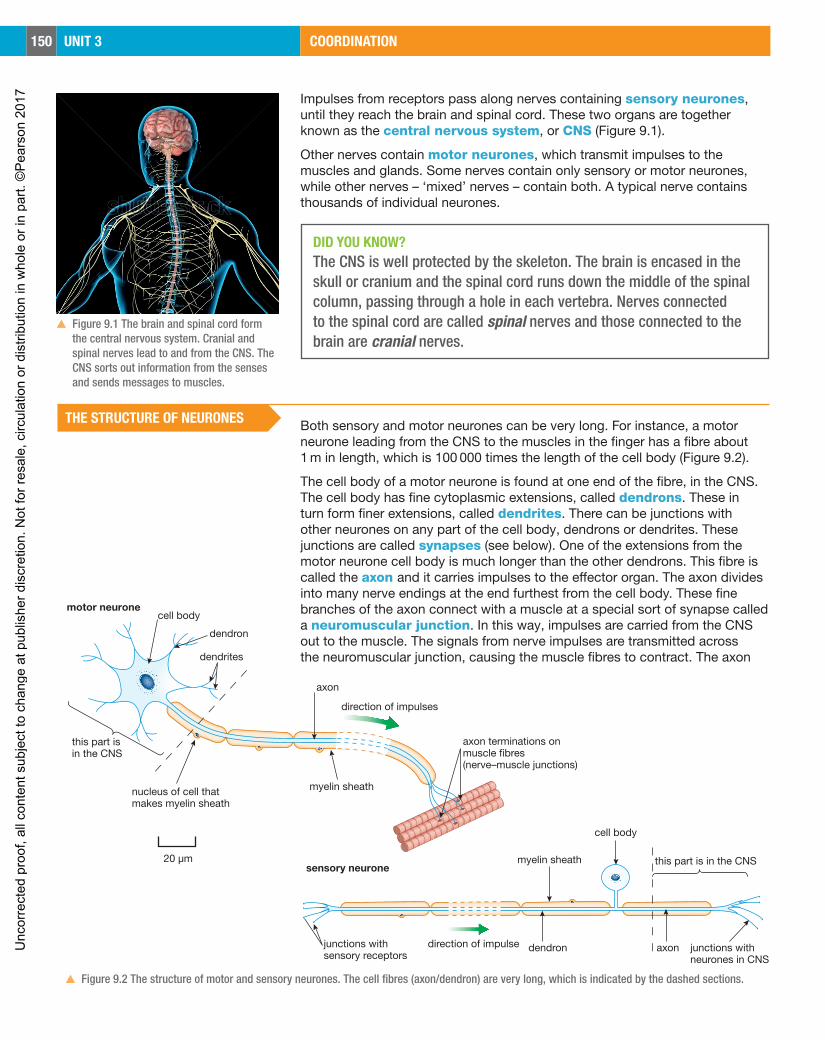
▲▲ Figure 9.1 The brain and spinal cord form the central nervous system. Cranial and spinal nerves lead to and from the CNS. The CNS sorts out information from the senses and sends messages to muscles.
Impulses from receptors pass along nerves containing sensory neurones, until they reach the brain and spinal cord. These two organs are together known as the central nervous system, or CNS (Figure 9.1).
Other nerves contain motor neurones, which transmit impulses to the muscles and glands. Some nerves contain only sensory or motor neurones, while other nerves – ‘mixed’ nerves – contain both. A typical nerve contains thousands of individual neurones.
DID YOU KNOW?The CNS is well protected by the skeleton. The brain is encased in the skull or cranium and the spinal cord runs down the middle of the spinal column, passing through a hole in each vertebra. Nerves connected to the spinal cord are called spinal nerves and those connected to the brain are cranial nerves.
THE STRUCTURE OF NEURONES Both sensory and motor neurones can be very long. For instance, a motor neurone leading from the CNS to the muscles in the finger has a fibre about 1 m in length, which is 100 000 times the length of the cell body (Figure 9.2).
The cell body of a motor neurone is found at one end of the fibre, in the CNS. The cell body has fine cytoplasmic extensions, called dendrons. These in turn form finer extensions, called dendrites. There can be junctions with other neurones on any part of the cell body, dendrons or dendrites. These junctions are called synapses (see below). One of the extensions from the motor neurone cell body is much longer than the other dendrons. This fibre is called the axon and it carries impulses to the effector organ. The axon divides into many nerve endings at the end furthest from the cell body. These fine branches of the axon connect with a muscle at a special sort of synapse called a neuromuscular junction. In this way, impulses are carried from the CNS out to the muscle. The signals from nerve impulses are transmitted across the neuromuscular junction, causing the muscle fibres to contract. The axon
dendron
motor neurone
sensory neurone
cell body
dendron
dendrites
axon
direction of impulses
axon terminations onmuscle fibres(nerve–muscle junctions)
myelin sheathnucleus of cell thatmakes myelin sheath
this part isin the CNS
myelin sheath
cell body
this part is in the CNS
junctions withneurones in CNS
axondirection of impulsejunctions withsensory receptors
20 µm
▲▲ Figure 9.2 The structure of motor and sensory neurones. The cell fibres (axon/dendron) are very long, which is indicated by the dashed sections.
Unc
orre
cted
pro
of, a
ll co
nten
t sub
ject
to c
hang
e at
pub
lishe
r dis
cret
ion.
Not
for r
esal
e, c
ircul
atio
n or
dis
tribu
tion
in w
hole
or i
n pa
rt. ©
Pea
rson
201
7

UNIT 3 COORDINATION 151
has an outer covering or ‘sheath’ made of a fatty material called myelin. This myelin sheath insulates the axon, preventing ‘short circuits’ with other axons, and also speeds up the conduction of the impulses. The sheath is formed by the membranes of special cells that wrap themselves around the axon as it develops. Cells with a myelin sheath are described as myelinated.
A sensory neurone has a similar structure to a motor neurone, but the cell body is located on a side branch of the fibre, just outside the CNS. The fibre from the sensory receptor to the cell body is actually a dendron, while the fibre from the cell body to the CNS is a short axon. As with motor neurones, fibres of sensory neurones are often myelinated.
SYNAPSES Synapses are essential to the working of the nervous system. The CNS is made of many billions of nerve cells, and these have links with many others, through synapses. In the brain, each neurone may form synapses with thousands of other neurones, so the number of possible pathways through the system is almost unlimited.
A synapse is actually a gap between two nerve cells. The electrical impulses passing through the neurones do not cross this gap. Instead, impulses arriving at a synapse cause the ends of the fine branches of the axon to secrete a chemical called a neurotransmitter. This chemical diffuses across the gap and attaches to the membrane of the second neurone. It then starts off new impulses in the second cell (Figure 9.3). After the neurotransmitter has ‘passed on the message’, it is broken down by an enzyme.
Remember that many nerve cells, particularly those in the brain, have thousands of synapses with other neurones. The output of one cell may depend on the inputs from many cells adding together. In this way, synapses are important for integrating information in the CNS (Figure 9.4).
synaptic connections withother neurones
cell body of neurone
axon
impulses arrive down axonof first neurone neurotransmitter
di�uses acrosssynapse
neurotransmitterattaches tomembrane ofsecond neurone
impulse started insecond neurone
neurotransmitter brokendown by enzyme fromsecond neurone
12
3
4
5
▲▲ Figure 9.3 The sequence of events happening at a synapse.
impulses in incoming neuronesimpulses in outgoing neurone
▲▲ Figure 9.4 Synapses allow the output of one nerve cell to be a result of integration of information from many other cells.
Unc
orre
cted
pro
of, a
ll co
nten
t sub
ject
to c
hang
e at
pub
lishe
r dis
cret
ion.
Not
for r
esal
e, c
ircul
atio
n or
dis
tribu
tion
in w
hole
or i
n pa
rt. ©
Pea
rson
201
7
152 UNIT 3 COORDINATION
Because synapses rely on the movement of chemicals, it is easy for other chemicals to interfere with the way they work. These chemicals may imitate the neurotransmitter, or prevent it from acting. This is how many well-known drugs, both useful and harmful, work.
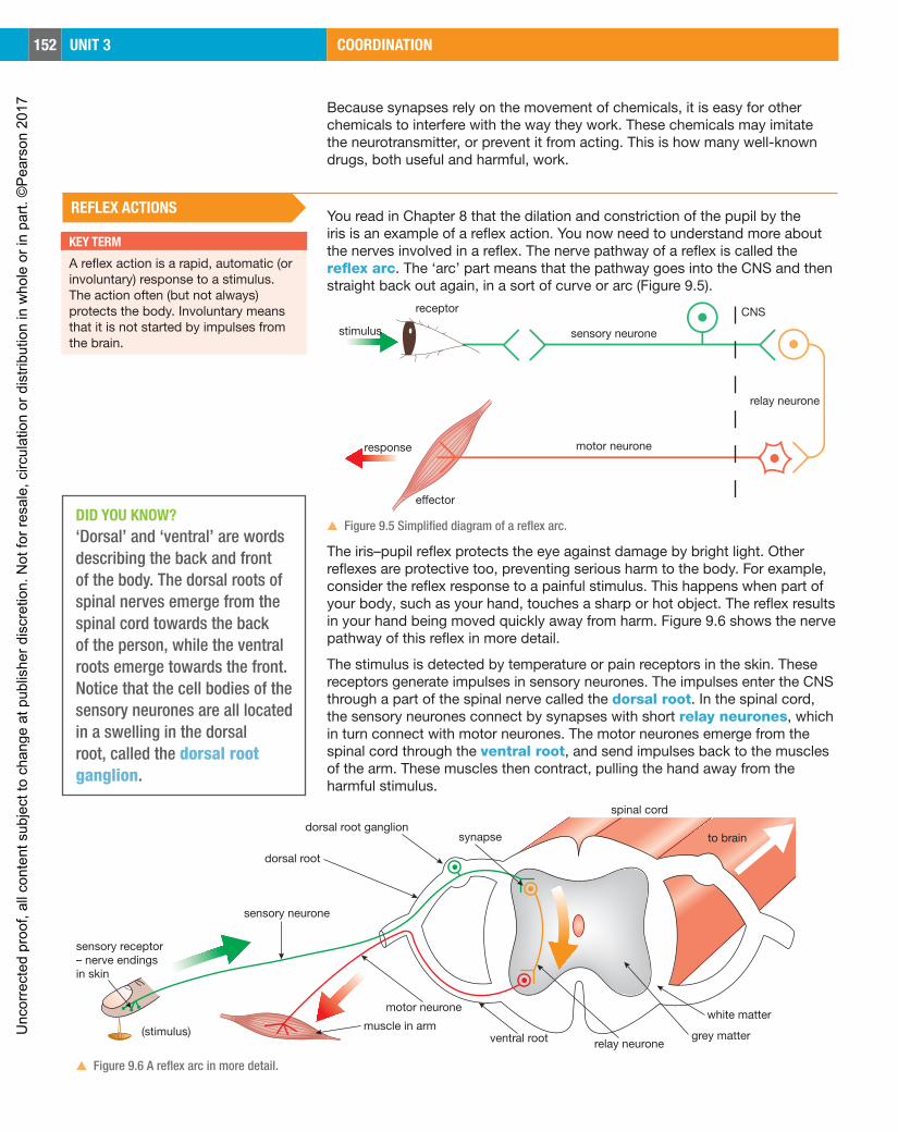
REFLEX ACTIONS You read in Chapter 8 that the dilation and constriction of the pupil by the iris is an example of a reflex action. You now need to understand more about the nerves involved in a reflex. The nerve pathway of a reflex is called the reflex arc. The ‘arc’ part means that the pathway goes into the CNS and then straight back out again, in a sort of curve or arc (Figure 9.5).
stimulus
response
receptor
e�ector
CNS
sensory neurone
motor neurone
relay neurone
▲▲ Figure 9.5 Simplified diagram of a reflex arc.
The iris–pupil reflex protects the eye against damage by bright light. Other reflexes are protective too, preventing serious harm to the body. For example, consider the reflex response to a painful stimulus. This happens when part of your body, such as your hand, touches a sharp or hot object. The reflex results in your hand being moved quickly away from harm. Figure 9.6 shows the nerve pathway of this reflex in more detail.
The stimulus is detected by temperature or pain receptors in the skin. These receptors generate impulses in sensory neurones. The impulses enter the CNS through a part of the spinal nerve called the dorsal root. In the spinal cord, the sensory neurones connect by synapses with short relay neurones, which in turn connect with motor neurones. The motor neurones emerge from the spinal cord through the ventral root, and send impulses back to the muscles of the arm. These muscles then contract, pulling the hand away from the harmful stimulus.
sensory neurone
dorsal root
dorsal root ganglionsynapse
spinal cord
to brain
white matter
grey matterrelay neuroneventral root
motor neurone
muscle in arm(stimulus)
sensory receptor– nerve endingsin skin
▲▲ Figure 9.6 A reflex arc in more detail.
KEY TERM
A reflex action is a rapid, automatic (or involuntary) response to a stimulus. The action often (but not always) protects the body. Involuntary means that it is not started by impulses from the brain.
DID YOU KNOW?‘Dorsal’ and ‘ventral’ are words describing the back and front of the body. The dorsal roots of spinal nerves emerge from the spinal cord towards the back of the person, while the ventral roots emerge towards the front. Notice that the cell bodies of the sensory neurones are all located in a swelling in the dorsal root, called the dorsal root ganglion.
Unc
orre
cted
pro
of, a
ll co
nten
t sub
ject
to c
hang
e at
pub
lishe
r dis
cret
ion.
Not
for r
esal
e, c
ircul
atio
n or
dis
tribu
tion
in w
hole
or i
n pa
rt. ©
Pea
rson
201
7
UNIT 3 COORDINATION 153
The middle part of the spinal cord consists mainly of nerve cell bodies, which give it a grey colour. This is why it is known as grey matter. The outer part of the spinal cord is called white matter, and has a whiter appearance because it contains many axons with fatty myelin sheaths.
Impulses travel through the reflex arc in a fraction of a second, so the reflex action is very fast and does not need to be started by impulses from the brain. This does not mean, however, that the brain is unaware of what is going on: in the spinal cord, the reflex arc neurones also form synapses with nerve cells leading to and from the brain. The brain therefore receives information about the stimulus. This is how we feel the pain.
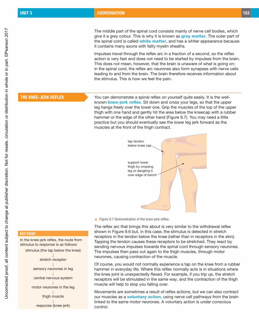
THE KNEE-JERK REFLEX You can demonstrate a spinal reflex on yourself quite easily. It is the well-known knee-jerk reflex. Sit down and cross your legs, so that the upper leg hangs freely over the lower one. Grip the muscles of the top of the upper thigh with one hand and gently hit the area below the kneecap with a rubber hammer or the edge of the other hand (Figure 9.7). You may need a little practice but you should eventually see the lower leg jerk forward as the muscles at the front of the thigh contract.
support lowerthigh by crossingleg or dangling itover edge of bench
tap tendonbelow knee cap
▲▲ Figure 9.7 Demonstration of the knee-jerk reflex.
The reflex arc that brings this about is very similar to the withdrawal reflex shown in Figure 9.6 but, in this case, the stimulus is detected in stretch receptors in the tendon below the knee (rather than in receptors in the skin). Tapping the tendon causes these receptors to be stretched. They react by sending nervous impulses towards the spinal cord through sensory neurones. The impulses then pass out again to the thigh muscles, through motor neurones, causing contraction of the muscle.
Of course, you would not normally experience a tap on the knee from a rubber hammer in everyday life. Where this reflex normally acts is in situations where the knee joint is unexpectedly flexed. For example, if you trip up, the stretch receptors will be stimulated in the same way, and the contraction of the thigh muscle will help to stop you falling over.
Movements are sometimes a result of reflex actions, but we can also contract our muscles as a voluntary action, using nerve cell pathways from the brain linked to the same motor neurones. A voluntary action is under conscious control.
KEY POINT
In the knee-jerk reflex, the route from stimulus to response is as follows:
stimulus (the tap below the knee)↓
stretch receptor↓
sensory neurones in leg↓
central nervous system↓
motor neurones in the leg↓
thigh muscle↓
response (knee jerk)
Unc
orre
cted
pro
of, a
ll co
nten
t sub
ject
to c
hang
e at
pub
lishe
r dis
cret
ion.
Not
for r
esal
e, c
ircul
atio
n or
dis
tribu
tion
in w
hole
or i
n pa
rt. ©
Pea
rson
201
7

154 UNIT 3 COORDINATION
THE BRAINWe began to understand the functions of different parts of the brain by studying people who had suffered brain damage through accident or disease. Nowadays, we have very advanced electronic equipment that can record the activity in a normal living brain. However, we still do not fully understand the workings of this most complex organ of the body.
Your brain is sometimes called your ‘grey matter’. This is because the positions of the grey and white matter are reversed in the brain compared with the spinal cord. The grey matter, mainly made of nerve cell bodies, is on the outside of the brain, while the axons that form the white matter are in the middle of the brain. The brain is made up of different parts, each with a specific function (Figure 9.8).
cranium (skull)
cerebrum
cavity in brainfilled with fluid
cerebellum
spinal cord
vertebral column(spine)
medulla
pituitary gland(makeshormones)
hypothalamus
space around brainfilled with fluid
▲▲ Figure 9.8 Section through the human brain, showing its main parts.
The largest part of the brain is the cerebrum, made of two cerebral hemispheres. The cerebrum is the source of all our conscious thoughts. It has an outer layer called the cerebral cortex, with many folds all over its surface (Figure 9.9).
The cerebrum has three main functions.
▲◾ It contains sensory areas that receive and process information from all our sense organs.
▲◾ It has motor areas, from which all our voluntary actions originate.
▲◾ It is the origin of ‘higher’ activities, such as memory, reasoning, emotions and personality.
Different parts of the cerebrum carry out particular functions. For example, the sensory and motor areas are always situated in the same place in the cortex (Figure 9.10). Some parts of these areas deal with more information than others. Large parts of the sensory area deal with impulses from the fingers and lips, for example. This is illustrated in Figure 9.11.
▲▲ Figure 9.9 A side view of a human brain. Notice the folded surface of the cerebral cortex.
Unc
orre
cted
pro
of, a
ll co
nten
t sub
ject
to c
hang
e at
pub
lishe
r dis
cret
ion.
Not
for r
esal
e, c
ircul
atio
n or
dis
tribu
tion
in w
hole
or i
n pa
rt. ©
Pea
rson
201
7

UNIT 3 COORDINATION 155
feet abdomen chest handsfingersmouthlipstongue
smell andtaste
skilledmovements
legsarms
main se
nsor
y area
hearing
vision
main motor areas
main sensory areas
‘higher’ and ‘association’ areas
▲▲ Figure 9.10 Different parts of the cerebrum carry out specific functions.
Behind the cerebrum is the cerebellum. This region is concerned with coordinating the contraction of sets of muscles, as well as maintaining balance. This is important when you are carrying out complicated muscular activities, such as running or riding a bike. Underneath the cerebrum, connecting the spinal cord with the rest of the brain, is the brain stem or medulla. This controls basic body activities such as heartbeat and breathing rate.
The pituitary gland is located at the base of the brain, just below a part of the brain called the hypothalamus. The pituitary gland secretes a number of chemical ‘messengers’, called hormones, into the blood. Hormones are described later in this chapter.
DISEASES OF THE BRAINThere are many different brain diseases. Some only develop in people as they grow old. We will look at three of these age-related brain diseases: Alzheimer’s disease, vascular dementia and Parkinson’s disease. You should know about the cause, symptoms and treatments for each disease.
ALZHEIMER’S DISEASE Alzheimer’s disease (usually just called Alzheimer’s) is a disease that causes dementia. People with dementia show a group of symptoms that affect normal activities such as eating or getting dressed. Their personalities may change, so that they cannot control their emotions, become agitated or see things that are not there.
Cause: The cause is not well understood, but it seems to be due to a build-up of two proteins in brain cells, called amyloid and tau. As these proteins build up, they damage and kill brain cells. As the disease progresses, more and more brain cells are damaged, leading to the symptoms of Alzheimer’s.
Symptoms: Early symptoms include:
▲◾ forgetting recent events, names and faces
▲◾ becoming increasingly repetitive, e.g. asking the same question after a short period of time
▲▲ Figure 9.11 A model of a human with its parts drawn in proportion to the amount of sensory information they send to the cortex of the brain (note that this does not apply to the eyes, which use more cortex than the rest of the body put together).
Unc
orre
cted
pro
of, a
ll co
nten
t sub
ject
to c
hang
e at
pub
lishe
r dis
cret
ion.
Not
for r
esal
e, c
ircul
atio
n or
dis
tribu
tion
in w
hole
or i
n pa
rt. ©
Pea
rson
201
7

UNIT 3 COORDINATION 169
CHAPTER QUESTIONS
1 The diagram shows some parts of the nervous system involved in a simple reflex action that happens when a finger touches a hot object.
Z
A
C
B
X
Y
HEATtemperature/ pain receptors
to muscles
a What type of neurone is:i neurone Aii neurone Biii neurone C?
b Describe the function of each of these types of neurone.c Which parts of the nervous system are shown by the labels X, Y and Z?d In what form is information passed along neurones?
e Explain how information passes from one neurone to another.
2 a Which part of the human brain is responsible for controlling each of the following actionsi keeping your balance when you walkii maintaining your breathing when you are asleepiii making your leg muscles contract when you kick a ball
b A ‘stroke’ is caused by a blood clot blocking the blood supply to part of the brain.i One patient, after suffering a stroke, was unable to move his left arm.
Which part of his brain was affected?ii Another patient lost her sense of smell following a stroke. Which part
of her brain was affected?c Strokes can also cause vascular dementia.
i What are the symptoms of vascular dementia?ii Describe two treatments that slow down the development of vascular
dementia.
3 Hormones are secreted by endocrine glands. a Explain the meaning of the three terms underlined in this sentence.
b Identify the hormones A to D in the table.
Hormone One function of this hormone
A stimulates the liver to convert glucose to glycogen
B controls the ‘fight or flight’ responses
C brings about a sudden increase in growth in teenage boys
D regulates the menstrual cycle
SKILLS ANALYSIS 7
SKILLS INTERPRETATION
SKILLS CRITICAL THINKING 6
SKILLS REASONING 7
SKILLS CRITICAL THINKING
6
7
6
7
7
SKILLS INTERPRETATION, ANALYSIS 6
Unc
orre
cted
pro
of, a
ll co
nten
t sub
ject
to c
hang
e at
pub
lishe
r dis
cret
ion.
Not
for r
esal
e, c
ircul
atio
n or
dis
tribu
tion
in w
hole
or i
n pa
rt. ©
Pea
rson
201
7

170 UNIT 3 COORDINATION
4 Explain these observations:a Mice that had their thyroid gland removed were less able to survive in
freezing conditions than mice with intact thyroid glands.b People living in areas of the world where there is no iodine in the drinking
water can develop a condition called a goitre.
5 The graph shows the changes in blood glucose in a healthy woman over a 12-hour period.
Blo
od g
luco
se/m
g pe
r 10
0 cm
3
of b
lood
Time of day
70
80
90
100
110
120
130
0800 1200 1600 2000
X Y
a Explain why there was a rise in blood glucose at X.b How does the body bring about a decrease in blood glucose at Y? Your
answer should include the words insulin, liver and pancreas.c Diabetes is a disease where the body cannot control the concentration of
glucose in the blood.i Why is this dangerous?ii Describe two ways in which a person with diabetes can monitor their
blood glucose level.iii Explain two ways in which a person with diabetes can help to control
their blood glucose level.
6 a Explain the meaning of the term ‘negative feedback’.b Describe the negative feedback loop involving the hormone glucagon
that takes place in response to a fall in blood glucose.
7 The graph shows the effect of different amounts of alcohol in the blood on the average reaction time of a group of adult men.
0 2Amount of alcohol in the blood/arbitrary units
Rea
ctio
n tim
e/se
cond
s
5 80
0.1
0.2
0.3
0.4
0.5
0.6
i Describe the results shown by the graph.
ii Calculate the change in reaction time when the concentration of alcohol in the blood increases from 0 to 8 units.
iii Use these results to explain why it is dangerous to drink alcohol and drive.
iv What are the effects on the liver of regularly drinking large amounts of alcohol?
SKILLS REASONING 8
SKILLS ANALYSIS, REASONING
SKILLS REASONING
SKILLS CRITICAL THINKING 7
8
8
SKILLS ANALYSIS
5
SKILLS PROBLEM SOLVING 6
SKILLS REASONING 7
SKILLS CRITICAL THINKING 6
Unc
orre
cted
pro
of, a
ll co
nten
t sub
ject
to c
hang
e at
pub
lishe
r dis
cret
ion.
Not
for r
esal
e, c
ircul
atio
n or
dis
tribu
tion
in w
hole
or i
n pa
rt. ©
Pea
rson
201
7

EDEXCEL INTERNATIONAL GCSE (9 –1)
HUMAN BIOLOGYStudent Book Philip Bradfield, Steve Potter
www.pearsonglobalschools.com
Edexcel International GCSE (9–1) Human Biology prepares students for the new2017 International GCSE (9–1) Human Biology specification. This resource provides comprehensive coverage of the new specification. This book, which includes access to the eBook, is designed to provide students with the best preparation possible for the examination:
• Written by highly experienced International GCSE Human Biology teachers and authors Philip Bradfield and Steve Potter
• Content is mapped closely to the specification to provide comprehensive coverage
• Exam practice throughout, with differentiated revision exercises and exam-style practice
• Signposted transferable skills• Integrated Pearson Progression Scale• Reviewed by a language specialist to ensure the book is written in a clear
and accessible style for students whose first language may not be English• Glossary of key Human Biology terminology, along with full answers
included on the eBook• eBook included.
Unc
orre
cted
pro
of, a
ll co
nten
t sub
ject
to c
hang
e at
pub
lishe
r dis
cret
ion.
Not
for r
esal
e, c
ircul
atio
n or
dis
tribu
tion
in w
hole
or i
n pa
rt. ©
Pea
rson
201
7