Modularly assembled designer TAL effector nucleases for targeted
Upload
phunghuongCategory
view
218download
2
Sample Collection
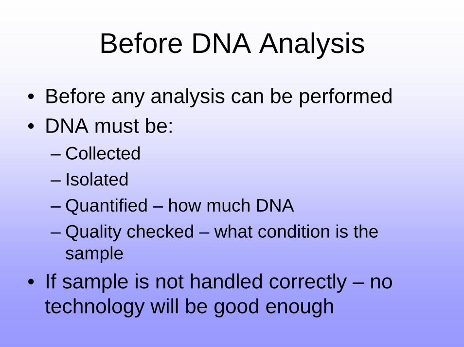
Before DNA Analysis
• Before any analysis can be performed• DNA must be:
– Collected– Isolated– Quantified – how much DNA– Quality checked – what condition is the
sample• If sample is not handled correctly – no
technology will be good enough
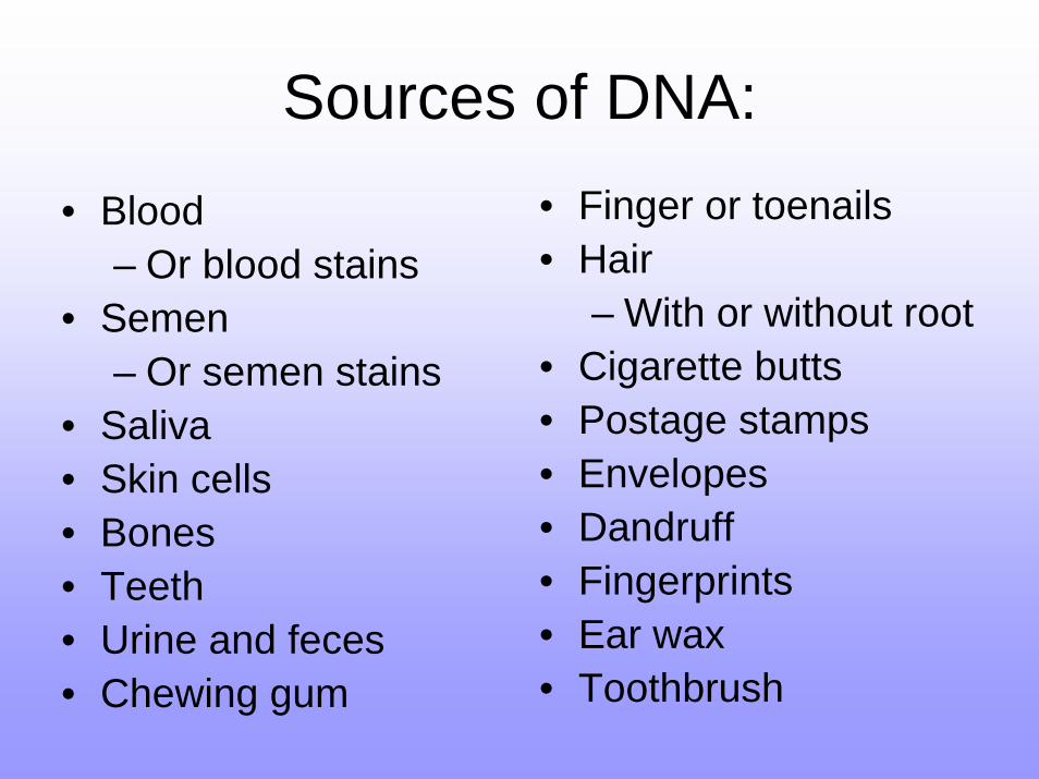
Sources of DNA:• Blood
– Or blood stains• Semen
– Or semen stains• Saliva• Skin cells• Bones• Teeth• Urine and feces• Chewing gum
• Finger or toenails• Hair
– With or without root• Cigarette butts• Postage stamps• Envelopes• Dandruff• Fingerprints• Ear wax• Toothbrush
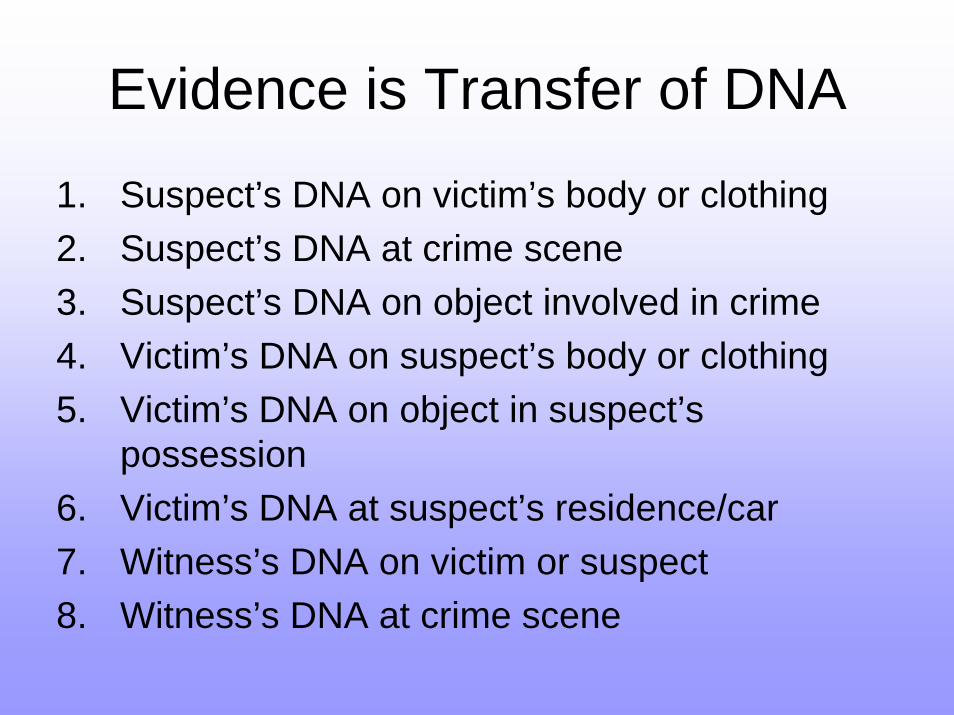
Evidence is Transfer of DNA
1. Suspect’s DNA on victim’s body or clothing2. Suspect’s DNA at crime scene3. Suspect’s DNA on object involved in crime4. Victim’s DNA on suspect’s body or clothing5. Victim’s DNA on object in suspect’s
possession6. Victim’s DNA at suspect’s residence/car7. Witness’s DNA on victim or suspect8. Witness’s DNA at crime scene
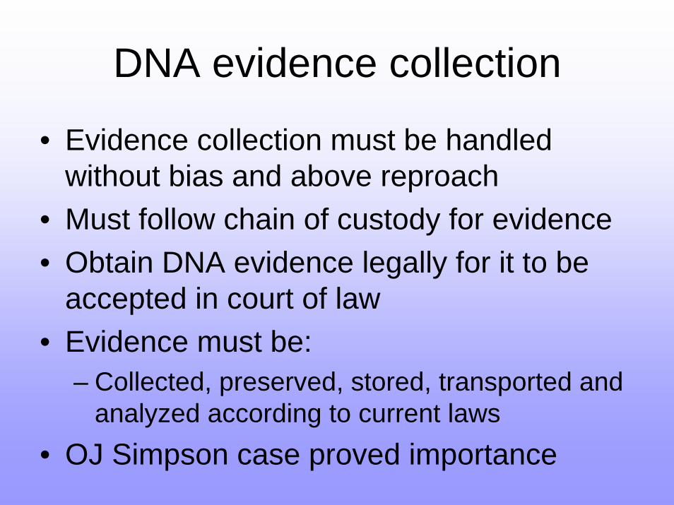
DNA evidence collection
• Evidence collection must be handled without bias and above reproach
• Must follow chain of custody for evidence • Obtain DNA evidence legally for it to be
accepted in court of law• Evidence must be:
– Collected, preserved, stored, transported and analyzed according to current laws
• OJ Simpson case proved importance
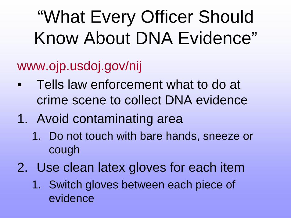
“What Every Officer Should Know About DNA Evidence”
www.ojp.usdoj.gov/nij• Tells law enforcement what to do at
crime scene to collect DNA evidence1. Avoid contaminating area
1. Do not touch with bare hands, sneeze or cough
2. Use clean latex gloves for each item1. Switch gloves between each piece of
evidence
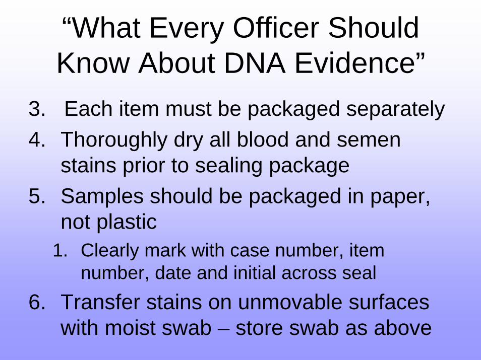
“What Every Officer Should Know About DNA Evidence”
3. Each item must be packaged separately4. Thoroughly dry all blood and semen
stains prior to sealing package5. Samples should be packaged in paper,
not plastic1. Clearly mark with case number, item
number, date and initial across seal6. Transfer stains on unmovable surfaces
with moist swab – store swab as above
Reference Samples
• Reference samples are DNA taken from people in order to compare to DNA collected
• Purposes of reference sample:– Suspect’s DNA for control of own items/home– Victim’s DNA as control– Family or roommates of victim as controls– Family DNA for paternity testing– Family DNA for identifying victim’s body
Reference Samples
• DNA is usually taken from either:– Blood sample– Cheek cells
• Buccal swabs:– Rubbing q-tip like swab against cheek cells– Swab either dried or pressed against treated
collection card and card is dried• Alternatives:
– Disposable toothbrush, mouth wash, etc
Transport and Storage of DNA
• Transport:– DNA evidence needs to be labeled and
handled according to laws in order to be admissible in court
• Storage:– Stains/swabs need to be dried to prevent
mold and bacteria growth– DNA is best preserved dry and cold– 4°C to -20°C is ideal for un-extracted DNA
sample
Identification of DNA Evidence
• Before the DNA can be extracted the sample needs to be identified at the crime scene
• Presumptive testing• Identifying any DNA that is present• Three primary stains:
– Blood– Semen– Saliva
Presumptive Testing• Identify the presence of DNA:
– Detection of blood/blood stains– Detection of semen/semen stains– Direct observation of semen– Detection of saliva/saliva stains
• The following may be important and informative but are used less often:– Vaginal secretions– Hair– Urine or Feces
Detection of Blood
• Blood is made of:– Plasma– Red blood cells– White blood cells– Platelets
• Most presumptive testing – checks for presence of hemoglobin
• Hemoglobin is in red blood cells– Carries oxygen and contains iron atoms
Detection of Blood• Immunochromatographic:
– Identifies higher primate or human blood only– Down to 0.07ug of blood
• Luminol:– Can be sprayed onto surfaces rapidly– Blood will react and glow in the dark -
luminescence– Detects a stain even if it’s been diluted
(washed) 10 million times!– Does not inhibit later DNA testing of sample
Serology• Utilize antibody – antigen reactions to
identify specific chemicals• Done in or on blood• Usually used:
– Identify what antibodies a person has– Example – if you have been immunized
• Can also be used:– Identify a blood stain– Identify a person’s blood type
Detection of Semen
• Presumptive testing identifies:– Sperm cells directly– Acid phosphatase (AP)– Prostate Specific Antigen (PSA)
• Some males do not make sperm:– After vasectomy– Aspermic or oligospermic men
• Therefore need to observe enzymes that are present in semen as well
Detection of SemenAcid phosphatase:
– Secreted by prostate gland– Purple color with Fast Blue B solution – Fluorescence with Fast Blue B under UV light
Prostate specific antigen:– Antigen on cells in seminal fluid– Also present in breast milk for some reason,
but in much lower concentrations– PSA kit similar to home pregnancy test– Measures presence and concentration
Direct observation of semen
• Recover sample or stain of semen• Reconstitute in deionized water• Fix to a slide• Examine under microscope• “Christmas Tree” stain will mark different
parts of sperm different colors:– Head is red and tail is green
• Makes easier to identify sperm cells from female epithelial cells
Detection of Saliva
• Saliva contains amylase• Identify and estimate levels of amylase• Phadebas test:
– Uses an antibody of amylase• Starch Iodine radial diffusion test:
– Iodine causes starch to turn dark purple– Amylase is an enzyme that digests starch– Presence and concentration of amylase can
be identified by darkness of purple
DNA Extraction
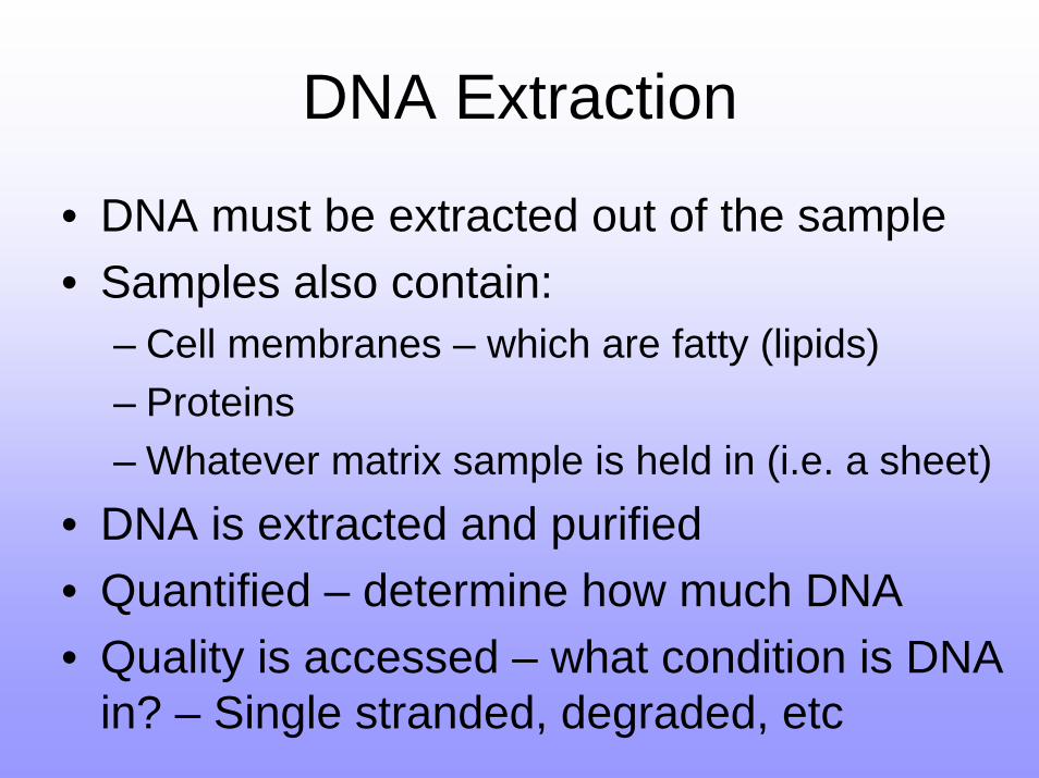
• DNA must be extracted out of the sample• Samples also contain:
– Cell membranes – which are fatty (lipids)– Proteins– Whatever matrix sample is held in (i.e. a sheet)
• DNA is extracted and purified• Quantified – determine how much DNA• Quality is accessed – what condition is DNA
in? – Single stranded, degraded, etc
DNA Extraction
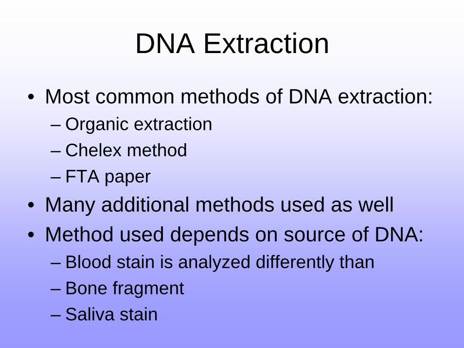
• Most common methods of DNA extraction:– Organic extraction– Chelex method– FTA paper
• Many additional methods used as well• Method used depends on source of DNA:
– Blood stain is analyzed differently than– Bone fragment– Saliva stain
DNA Extraction
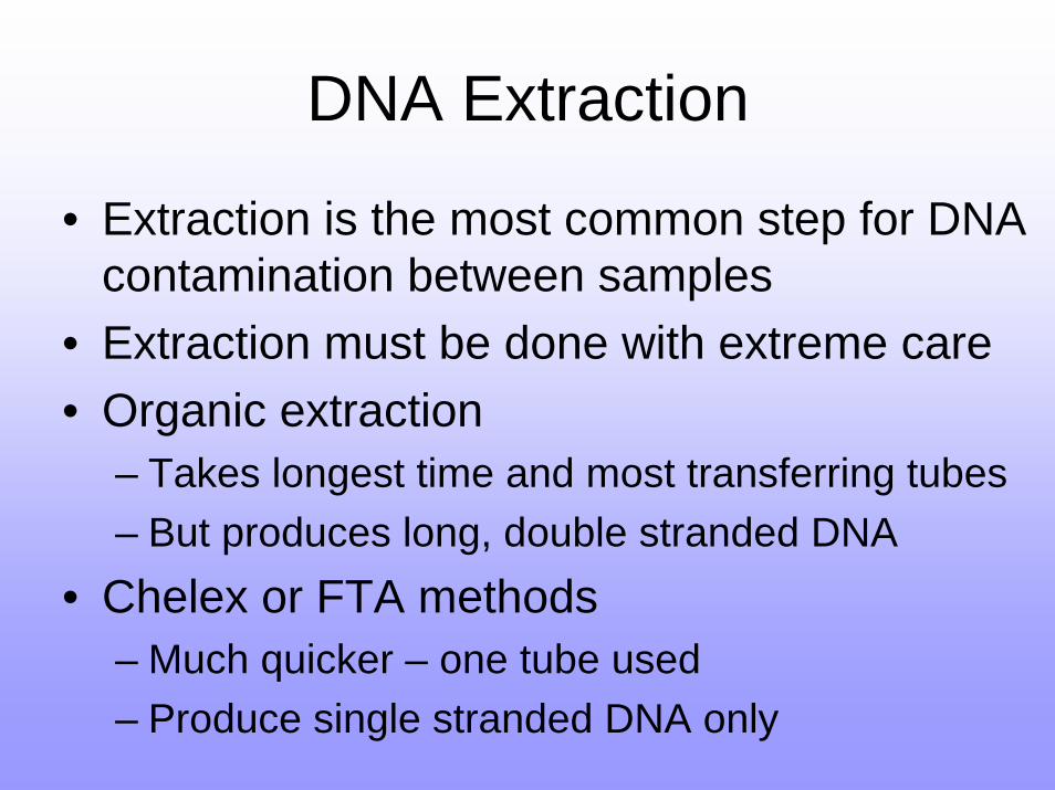
• Extraction is the most common step for DNA contamination between samples
• Extraction must be done with extreme care• Organic extraction
– Takes longest time and most transferring tubes– But produces long, double stranded DNA
• Chelex or FTA methods– Much quicker – one tube used– Produce single stranded DNA only
Organic Extraction
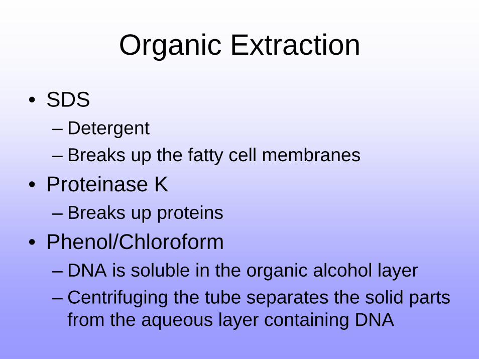
• SDS– Detergent– Breaks up the fatty cell membranes
• Proteinase K– Breaks up proteins
• Phenol/Chloroform– DNA is soluble in the organic alcohol layer– Centrifuging the tube separates the solid parts
from the aqueous layer containing DNA
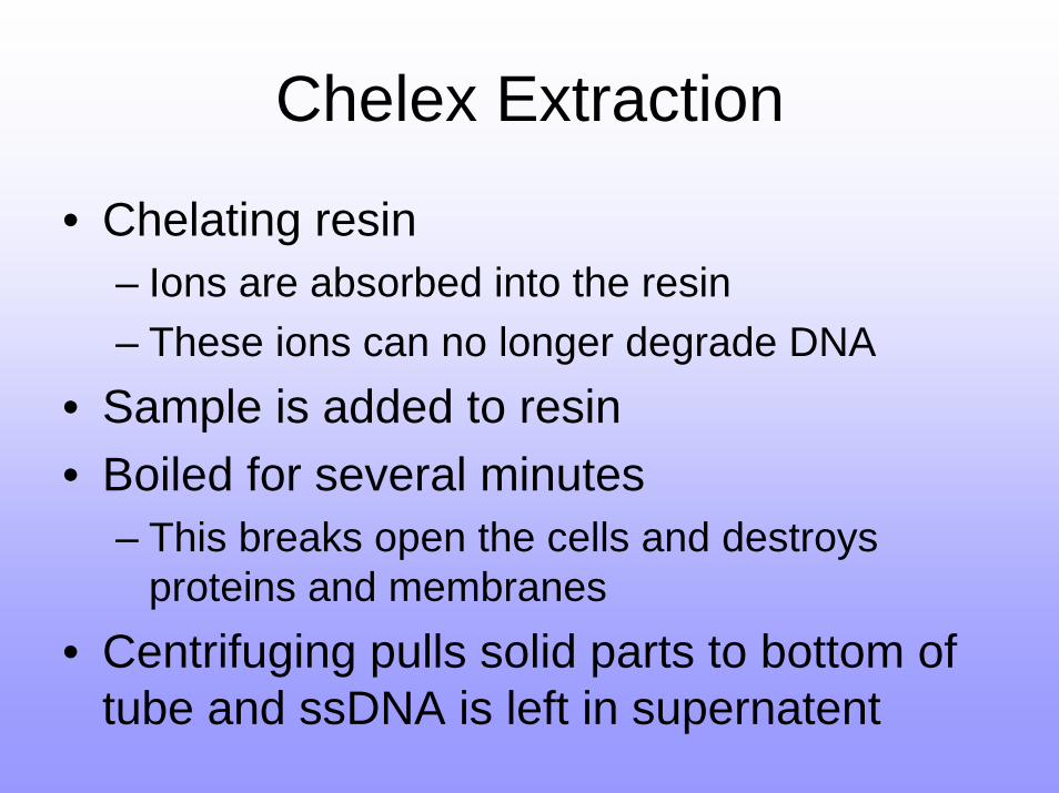
Chelex Extraction
• Chelating resin– Ions are absorbed into the resin– These ions can no longer degrade DNA
• Sample is added to resin• Boiled for several minutes
– This breaks open the cells and destroys proteins and membranes
• Centrifuging pulls solid parts to bottom of tube and ssDNA is left in supernatent
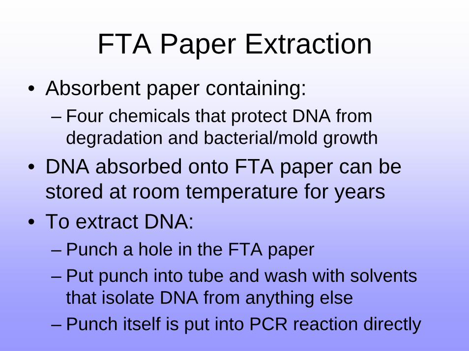
FTA Paper Extraction• Absorbent paper containing:
– Four chemicals that protect DNA from degradation and bacterial/mold growth
• DNA absorbed onto FTA paper can be stored at room temperature for years
• To extract DNA:– Punch a hole in the FTA paper– Put punch into tube and wash with solvents
that isolate DNA from anything else– Punch itself is put into PCR reaction directly
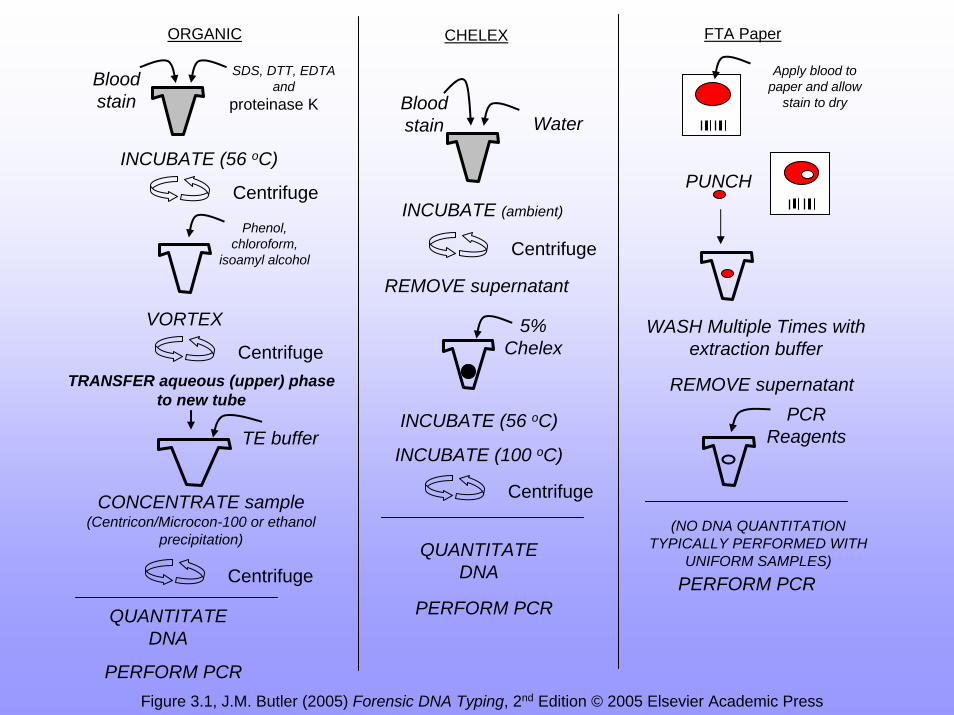
ORGANIC FTA PaperCHELEX
Blood stain
PUNCH
WASH Multiple Times with extraction buffer
PERFORM PCR
PCR Reagents
SDS, DTT, EDTA and
proteinase K
INCUBATE (56 oC)
Phenol,chloroform,
isoamyl alcohol
QUANTITATE DNA
Apply blood to paper and allow
stain to dryBlood stain
VORTEX
(NO DNA QUANTITATION TYPICALLY PERFORMED WITH
UNIFORM SAMPLES)
Water
INCUBATE (ambient)
5% Chelex
INCUBATE (100 oC)
REMOVE supernatant
INCUBATE (56 oC)
QUANTITATE DNA
PERFORM PCR
PERFORM PCR
Centrifuge
Centrifuge
Centrifuge
Centrifuge
REMOVE supernatantTRANSFER aqueous (upper) phase to new tube
CONCENTRATE sample (Centricon/Microcon-100 or ethanol
precipitation)
Centrifuge
TE buffer
Figure 3.1, J.M. Butler (2005) Forensic DNA Typing, 2nd Edition © 2005 Elsevier Academic Press
Other Methods of Extraction
• Three most commonly used methods:– Organic extraction– Chelex– FTA paper
• Other methods also used:– Differential extraction– Direct capture of sperm cells– Solid phase extraction

Differential Extraction
• Separate sperm cells from epithelial cells• Separating the male’s fraction from the
female’s fraction allows more reliable assignment of suspect’s DNA
• How it works:– Sperm cells are more resistant to being
broken apart than epithelial cells– Break up the epithelial cells first – extract
DNA from female subject separately
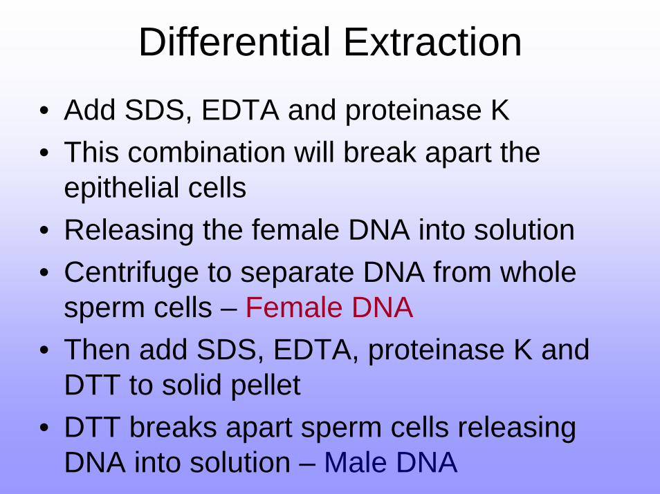
Differential Extraction• Add SDS, EDTA and proteinase K• This combination will break apart the
epithelial cells • Releasing the female DNA into solution• Centrifuge to separate DNA from whole
sperm cells – Female DNA• Then add SDS, EDTA, proteinase K and
DTT to solid pellet• DTT breaks apart sperm cells releasing
DNA into solution – Male DNA
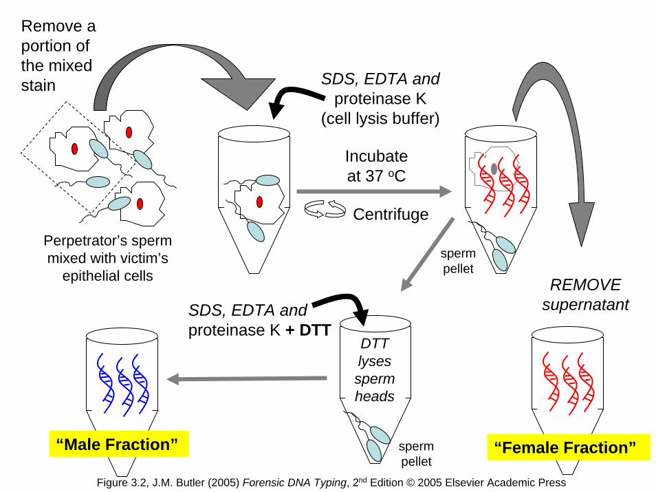
Perpetrator’s sperm mixed with victim’s
epithelial cells
Centrifuge
REMOVE supernatant
SDS, EDTA and proteinase K
(cell lysis buffer)
Remove a portion of the mixed stain
SDS, EDTA and proteinase K + DTT
Incubate at 37 oC
sperm pellet
DTT lyses sperm heads
“Male Fraction” “Female Fraction”sperm pellet
Figure 3.2, J.M. Butler (2005) Forensic DNA Typing, 2nd Edition © 2005 Elsevier Academic Press
Capture of Sperm Cells
• Physically capturing sperm cells and then extracting DNA directly from cell
• Antibody beads:– Beads that are coating in antibodies against
sperm proteins– Sperm cells will stick to beads, all other cells
will be washed away• Laser capture microdissection:
– Use a laser to trap a sperm cell under thin film

Solid Phase Extraction
• QIAamp spin columns• Nucleic acids are absorbed into silica
beads in high salt solution• pH < 7.5 – 95% of DNA will be bound to
beads• pH > 8 and low salt – DNA that was bound
will be eluted into solution• Effective and fast way to isolate DNA
More Methods of Extraction
• In addition, there are still other methods of extracting DNA in the works:– Adding high molar NaCl and proteinase K and
vigorous shaking and centrifugation– Closed tube extraction with heat resistant
protease– Microwave heating used with organic
extraction to speed up the time needed– Others…
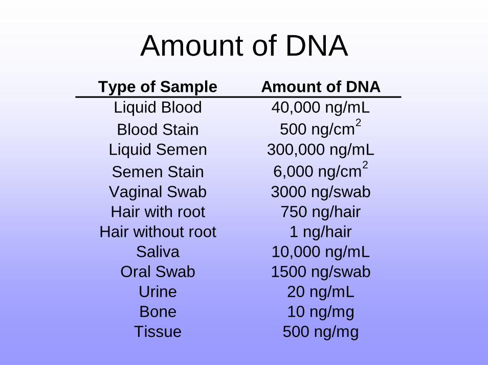
Amount of DNAType of Sample Amount of DNA
Liquid Blood 40,000 ng/mLBlood Stain 500 ng/cm2
Liquid Semen 300,000 ng/mLSemen Stain 6,000 ng/cm2
Vaginal Swab 3000 ng/swabHair with root 750 ng/hair
Hair without root 1 ng/hairSaliva 10,000 ng/mL
Oral Swab 1500 ng/swabUrine 20 ng/mLBone 10 ng/mg
Tissue 500 ng/mg
DNA extraction
Extracting DNA has four goals:• Collecting the DNA
– Want to get as much DNA as possible– High quality DNA if possible
• Removing everything else– Proteins, cellular debris, sample matrix
• Removing PCR inhibitors• Avoid degradation of DNA
PCR Inhibitors
• PCR inhibitors will:– Lower the quality of PCR results– Lead to complete failure of PCR reaction
Two most common inhibitors for Forensic samples:
• Hemoglobin• Indigo dyes from denim• Bind the active site of the Taq polymerase
DNA Degradation
• If DNA is damaged it will not be able to be used for PCR
• Damaging agents:– Nucleases – digest nucleic acids– Bacterial or fungal growth – will degrade DNA– Oxidation – will damage the bases– Hydrolytic cleavage – water will break DNA’s
sugar phosphate backbone– UV damage – will produced thymine dimers
DNA Quantification
Final step – quality and quantify DNA• Must be human DNA only• Must be in good enough shape for PCR
results to be acceptable• Must know concentration of DNA so that
roughly the same amount of DNA is used for all reactions
• PCR reaction conditions depend on DNA concentration
DNA Quantification
Too much DNA:• Split peaks• Leaking into other colors/lanes• Peaks that are off the scaleToo little DNA:• Allele “drop-out”• Can be allele specific – so you will get the
wrong genotype without knowing it
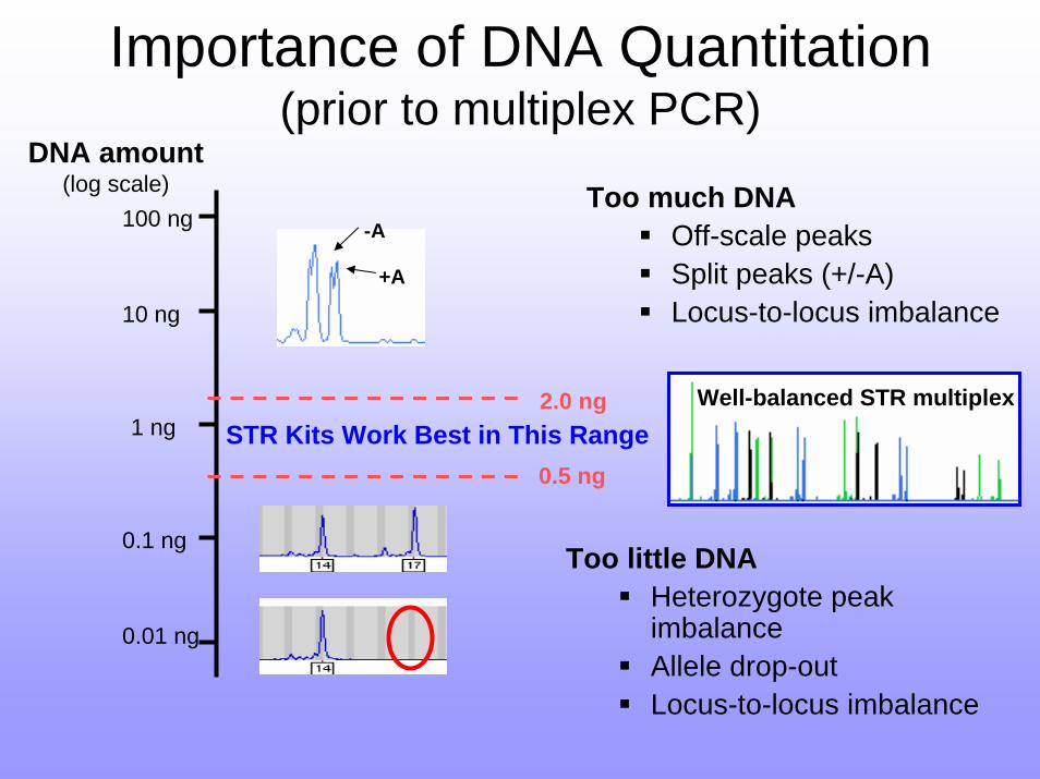
Importance of DNA Quantitation (prior to multiplex PCR)
DNA amount(log scale)
0.5 ng
-A
+A
Too much DNAOff-scale peaksSplit peaks (+/-A)Locus-to-locus imbalance
100 ng
10 ng
1 ng
0.1 ng
0.01 ng
2.0 ng
Too little DNAHeterozygote peak imbalanceAllele drop-outLocus-to-locus imbalance
STR Kits Work Best in This RangeWell-balanced STR multiplex
Quantification Methods
• Absorbance at 260 nm wavelength– Not as reliable or consistent– Uses too much of the sample
Commonly used for Forensics:• Slot blot• Florescence based approaches/kits• Real-Time PCR
– Quantitative PCR reaction
Slot Blot Method
• 40 base pair probe is primate specific• Probe is either radioactive or fluorescence• Probe is washed over DNA sample that is
captured on nylon membrane– Like southern blot
• Intensity of sample blot is compared to ladder of standards of known concentrations – read by instrumentation
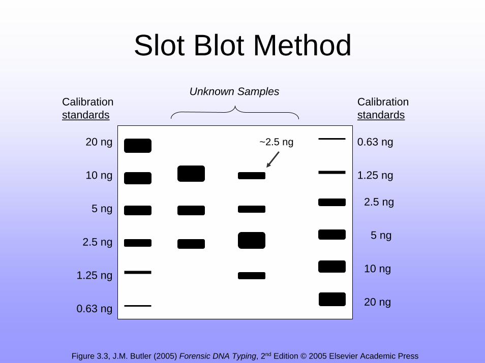
20 ng
10 ng
5 ng
2.5 ng
1.25 ng
0.63 ng20 ng
10 ng
5 ng
2.5 ng
1.25 ng
0.63 ng
Calibration standards
Calibration standards
Unknown Samples
~2.5 ng
Figure 3.3, J.M. Butler (2005) Forensic DNA Typing, 2nd Edition © 2005 Elsevier Academic Press
Slot Blot Method
PicoGreen assay
• PicoGreen is a fluorescent interchelating dye– Will be absorbed into DNA’s structure
• Florescence is changed by being bound to DNA
• Sample is then examined by fluorometer• Quantifies DNA quickly and easily• But is NOT human specific
AluQuant assay
• Probe binds to Alu repeats• Probe is washed over DNA sample• Probe lights up based on enzymatic
reaction• Light intensity is read – DNA is quantified • Can be automated• Is specific to human DNA only
Real-Time PCR
• Real-Time PCR is a PCR reaction so it effectively measures quantity and quality in same reaction
• Also produces a PCR product that can be genotyped directly
• Primers used are human specific so this reaction is human specific
• Will discuss details in next chapter/lecture
End Point PCR Method
• Just genotype with first STR marker• If it works then all the rest of the STR
markers should work as well• Can compare to standard curve with
samples of known concentrations too• Saves time, doesn’t waste any sample,
and ultimately this is what you are trying to figure out in the first place
Quantity of DNA Needed
• Most STR genotyping works best with around 1 ng of purified DNA
• More important that all DNA is at a constant concentration
• 1 genome copy of DNA = 3 picograms• If cell is diploid = 6 pg/genome• Therefore 1 ng (1000 pg) of DNA = ~ 333
copies of each locus
Any Questions?
Read Chapter Four
Guest Lecturer on Wednesday:Chief Medical Examiner