SALS, a WH2-Domain-Containing Protein, Promotes ...capping proteins in nonmuscle cells, which...
15
Developmental Cell Article SALS, a WH2-Domain-Containing Protein, Promotes Sarcomeric Actin Filament Elongation from Pointed Ends during Drosophila Muscle Growth Jianwu Bai, 1, * John H. Hartwig, 3 and Norbert Perrimon 1,2, * 1 Department of Genetics 2 Howard Hughes Medical Institute Harvard Medical School, 77 Avenue Louis Pasteur, Boston, MA 02115, USA 3 Hematology Division, Brigham and Women’s Hospital, Boston, MA 02115, USA *Correspondence: [email protected] (J.B.), [email protected] (N.P.) DOI 10.1016/j.devcel.2007.10.003 SUMMARY Organization of actin filaments into a well-orga- nized sarcomere structure is critical for muscle development and function. However, it is not completely understood how sarcomeric actin/ thin filaments attain their stereotyped lengths. In an RNAi screen in Drosophila primary muscle cells, we identified a gene, sarcomere length short (sals), which encodes an actin-binding, WH2 domain-containing protein, required for proper sarcomere size. When sals is knocked down by RNAi, primary muscles display thin myofibrils with shortened sarcomeres and increased sarcomere number. Both loss- and gain-of-function analyses indicate that SALS may influence sarcomere lengths by promoting thin-filament lengthening from pointed ends. Furthermore, the complex localization of SALS and other sarcomeric proteins in myofibrils reveals that the full length of thin filaments is achieved in a two-step process, and that SALS is required for the second elongation phase, most likely because it antagonizes the pointed-end capping protein Tropomodulin. INTRODUCTION Striated muscles in both insects and vertebrates have remarkably regular and precise organizations that are intimately linked to their function. Sarcomeres, the basic contractile units of myofibrils in muscles, are composed of actin (thin) and myosin (thick) filaments and their asso- ciated proteins (Figure 1A). Interestingly, although the length of thin filaments is strikingly uniform within individ- ual sarcomeres, there are significant differences in the ab- solute length of thin filaments in different muscle types of the same animal (Granzier et al., 1991) as well as in mus- cles of different species (Page and Huxley, 1963). Further- more, variations in thin-filament length are believed to be responsible for the distinct mechanical properties charac- teristic of each muscle (Burkholder et al., 1994), because the thick-filament length is fairly constant (Page and Hux- ley, 1963). According to the crossbridge theory of muscle contraction (Huxley and Simmons, 1971), the extent of overlap between thin and thick filaments determines the amount of force that a muscle can exert at different sarco- mere lengths. Finally, abnormal thin-filament lengths are thought to be the molecular basis for several debilitating muscle diseases, such as nemaline myopathy and dilated cardiomyopathy (Littlefield and Fowler, 1998; Sussman et al., 1999). Thus, the precise regulation of thin-filament length is critical for muscle function and muscle integrity. Sarcomeric actin filaments are organized with their two ends lined up in regular arrays (Figure 1A). The barbed (plus) ends are capped by CapZ and are crosslinked by a-actinin at the Z line. Tropomodulin (Tmod), another cap- ping protein, binds to the free pointed (minus) ends lo- cated in the middle of the sarcomere toward the H zone, a region free of thin filaments. In nonmuscle cells, it is gen- erally believed that most, if not all, actin assembly is regu- lated at the barbed ends, and that pointed ends are the sites of disassembly (i.e., treadmilling process) (Pollard et al., 2000). Thus, barbed-end and pointed-end capping proteins are thought to influence filament lengths in non- muscle cells by terminating growth at the barbed ends and by preventing shortening at the pointed ends, respec- tively (Fischer and Fowler, 2003). In contrast, the length of sarcomeric actin filaments in striated muscles is primarily regulated by modulation of the pointed-end capping activ- ity of Tmod (Fischer and Fowler, 2003). Excess Tmod in striated muscles causes shortening of thin filaments (Lit- tlefield et al., 2001; Mardahl-Dumesnil and Fowler, 2001; Sussman et al., 1998). Conversely, inhibition of Tmod ex- pression or function results in lengthening of thin filaments (Gregorio et al., 1995; Sussman et al., 1998). Thus, unlike capping proteins in nonmuscle cells, which prevent disas- sembly, Tmod limits actin assembly at the pointed ends of thin filaments. This may be due to the high actin concen- tration and highly stable actin arrays in muscle tissues. These observations strongly suggest that thin filaments can elongate from pointed ends (Littlefield et al., 2001; Mardahl-Dumesnil and Fowler, 2001; Sussman et al., 828 Developmental Cell 13, 828–842, December 2007 ª2007 Elsevier Inc.
Transcript of SALS, a WH2-Domain-Containing Protein, Promotes ...capping proteins in nonmuscle cells, which...
Developmental Cell
Article
SALS, a WH2-Domain-Containing Protein,Promotes Sarcomeric Actin Filament ElongationfromPointedEndsduringDrosophilaMuscleGrowthJianwu Bai,1,* John H. Hartwig,3 and Norbert Perrimon1,2,*1Department of Genetics2Howard Hughes Medical Institute
Harvard Medical School, 77 Avenue Louis Pasteur, Boston, MA 02115, USA3Hematology Division, Brigham and Women’s Hospital, Boston, MA 02115, USA
*Correspondence: [email protected] (J.B.), [email protected] (N.P.)
DOI 10.1016/j.devcel.2007.10.003
SUMMARY
Organization of actin filaments into a well-orga-nized sarcomere structure is critical for muscledevelopment and function. However, it is notcompletely understood how sarcomeric actin/thin filaments attain their stereotyped lengths.In an RNAi screen in Drosophila primary musclecells, we identified a gene, sarcomere lengthshort (sals), which encodes an actin-binding,WH2 domain-containing protein, required forproper sarcomere size. When sals is knockeddown by RNAi, primary muscles display thinmyofibrils with shortened sarcomeres andincreased sarcomere number. Both loss- andgain-of-function analyses indicate that SALSmay influence sarcomere lengths by promotingthin-filament lengthening from pointed ends.Furthermore, the complex localization of SALSand other sarcomeric proteins in myofibrilsreveals that the full length of thin filaments isachieved in a two-step process, and thatSALS is required for the second elongationphase, most likely because it antagonizes thepointed-end capping protein Tropomodulin.
INTRODUCTION
Striated muscles in both insects and vertebrates have
remarkably regular and precise organizations that are
intimately linked to their function. Sarcomeres, the basic
contractile units of myofibrils in muscles, are composed
of actin (thin) and myosin (thick) filaments and their asso-
ciated proteins (Figure 1A). Interestingly, although the
length of thin filaments is strikingly uniform within individ-
ual sarcomeres, there are significant differences in the ab-
solute length of thin filaments in different muscle types of
the same animal (Granzier et al., 1991) as well as in mus-
cles of different species (Page and Huxley, 1963). Further-
more, variations in thin-filament length are believed to be
828 Developmental Cell 13, 828–842, December 2007 ª2007 E
responsible for the distinct mechanical properties charac-
teristic of each muscle (Burkholder et al., 1994), because
the thick-filament length is fairly constant (Page and Hux-
ley, 1963). According to the crossbridge theory of muscle
contraction (Huxley and Simmons, 1971), the extent of
overlap between thin and thick filaments determines the
amount of force that a muscle can exert at different sarco-
mere lengths. Finally, abnormal thin-filament lengths are
thought to be the molecular basis for several debilitating
muscle diseases, such as nemaline myopathy and dilated
cardiomyopathy (Littlefield and Fowler, 1998; Sussman
et al., 1999). Thus, the precise regulation of thin-filament
length is critical for muscle function and muscle integrity.
Sarcomeric actin filaments are organized with their two
ends lined up in regular arrays (Figure 1A). The barbed
(plus) ends are capped by CapZ and are crosslinked by
a-actinin at the Z line. Tropomodulin (Tmod), another cap-
ping protein, binds to the free pointed (minus) ends lo-
cated in the middle of the sarcomere toward the H zone,
a region free of thin filaments. In nonmuscle cells, it is gen-
erally believed that most, if not all, actin assembly is regu-
lated at the barbed ends, and that pointed ends are the
sites of disassembly (i.e., treadmilling process) (Pollard
et al., 2000). Thus, barbed-end and pointed-end capping
proteins are thought to influence filament lengths in non-
muscle cells by terminating growth at the barbed ends
and by preventing shortening at the pointed ends, respec-
tively (Fischer and Fowler, 2003). In contrast, the length of
sarcomeric actin filaments in striated muscles is primarily
regulated by modulation of the pointed-end capping activ-
ity of Tmod (Fischer and Fowler, 2003). Excess Tmod in
striated muscles causes shortening of thin filaments (Lit-
tlefield et al., 2001; Mardahl-Dumesnil and Fowler, 2001;
Sussman et al., 1998). Conversely, inhibition of Tmod ex-
pression or function results in lengthening of thin filaments
(Gregorio et al., 1995; Sussman et al., 1998). Thus, unlike
capping proteins in nonmuscle cells, which prevent disas-
sembly, Tmod limits actin assembly at the pointed ends of
thin filaments. This may be due to the high actin concen-
tration and highly stable actin arrays in muscle tissues.
These observations strongly suggest that thin filaments
can elongate from pointed ends (Littlefield et al., 2001;
Mardahl-Dumesnil and Fowler, 2001; Sussman et al.,
lsevier Inc.
Developmental Cell
SALS Promotes Thin-Filament Lengthening
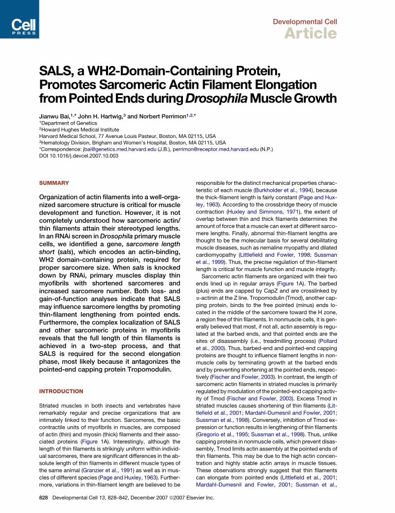
Figure 1. Muscle Defects Caused by dsRNAs Targeting sals in Primary Culture
(A) Schematic drawing of a myofibril with three sarcomeres.
(B) Representative fluorescence micrographs of primary muscles derived from myoblasts dissociated from WT embryos and treated with dsRNAs
targeting either lacZ as WT controls (left panels) or sals (middle panels) or actin (right panels). Muscles are stained with phalloidin for F-actin (red
in merge), an antibody against muscle myosin (MHC) (green in merge), and DAPI for nuclei (blue in merge). The bottom panels are phase images
of primary muscles shown in the above panels. Arrowheads point to the nuclei in muscles. The insets are confocal fluorescence images showing
the organization of sarcomeres at a higher magnification. The scale bar is 10 mm.
(C) A histogram showing the comparison of primary myofibril features among control lacZ RNAi, sals RNAi, actin RNAi muscles (12 days at 18�C), and
those derived from cells isolated from either sals homozygous mutant embryos (sals mutant) or embryos of Dmef2-Gal4, salsf07849/UAS-sals, salsf07849
(Rescue) (5 days at 25�C). These features include sarcomere length, myofibril length, and myofibril width, all of which are normalized with control lacZ
RNAi myofibrils. The length of the sarcomeres was determined by measuring the distance between Z lines. The lengths of sarcomeres in myofibrils of
sals RNAi and actin RNAi and the sals mutant are 1.77 ± 0.042 mm (average ± SEM) (range from 1.36 to 2.27 mm), 1.74 ± 0.08 mm (range from 1.30 to
2.38 mm), and 1.70 ± 0.075 mm (range from 1.30 to 2.15 mm), respectively, which are much shorter than the control sarcomere length (6 ± 0.73 mm
[range from 4.56 to 7.9 mm]) and that of Rescue myofibrils (5.5 ± 0.06 mm; range from 2.4 to 6.7 mm). The width of myofibrils from both sals and actin
RNAi and the sals mutant was �20% of controls (p < 0.01). The width of Rescue myofibrils is comparable to that of lacZ RNAi controls. Both the
lengths of myofibrils and the number of sarcomeres in myofibrils were calculated and normalized by the number of muscle nuclei. The average myo-
fibril length (per nucleus) is 60 ± 5 mm for sals RNAi myofibrils, versus 54 ± 4 mm for controls. n R 15 primary muscles with visual phenotypes scored in
each of three independent experiments.
1998). In addition to Tmod, several other proteins are also
involved in the regulation of thin-filament length. Tropomy-
osin (TM) stabilizes thin filaments by copolymerizing with
them and by modulating Tmod binding affinity to the
Developm
pointed ends (Littlefield and Fowler, 1998). The actin-
depolymerizing factor (ADF)/cofilin regulates thin-filament
depolymerization from their pointed ends (Mohri et al.,
2006), and nebulin may act as a molecular ruler that
ental Cell 13, 828–842, December 2007 ª2007 Elsevier Inc. 829

Developmental Cell
SALS Promotes Thin-Filament Lengthening
dictates thin-filament lengths in vertebrates (Bang et al.,
2006; McElhinny et al., 2005; Witt et al., 2006). Despite
the fact that the above-mentioned proteins are linked to
either capping, stabilization, or depolymerization of thin fil-
aments, the protein(s) responsible for promoting filament
elongation remains unknown.
To identify additional regulators of sarcomere assem-
bly, we developed a screening strategy based on RNA in-
terference (RNAi) in Drosophila primary muscle cells (J.B.
et al., unpublished data). Here, we report the characteriza-
tion of Sarcomere Length Short (SALS), a novel actin-
binding, WH2-containing protein identified by using this
approach. Loss of SALS causes shortening of thin fila-
ments and lack of actin incorporation at their pointed
ends. Our analyses reveal that SALS promotes thin-fila-
ment lengthening from the pointed ends, most likely by
antagonizing Tmod capping activity.
RESULTS
Primary Muscle Phenotypes Causedby the Disruption of sals by RNAiOur protocol for RNAi screening in primary muscle cells
(see Experimental Procedures) is based on the ability of
cultured myoblasts isolated from Drosophila gastrulating
embryos (4–6 hr after egg laying [AEL]) to fuse and differ-
entiate into striated muscles in the same sequence of de-
velopmental events as observed in vivo (Bernstein et al.,
1978). Simply bathing primary cells in medium containing
dsRNAs knocks down gene expression in primary mus-
cles (J.B. et al., unpublished data) (Figure 2I).
Using this method, we identified CG31374
(FBgn0051374), which we named ‘‘sarcomere length short
(sals)’’ based on the striking myofibril phenotypes caused
by RNAi. In primary cultures treated with dsRNAs against
sals (referred to as ‘‘sals RNAi’’ in the text), we observed
three distinct phenotypes (Figures 1B and 1C). First, sals
RNAi myofibrils contained much shortened sarcomeres
(the lengths of sarcomeres were only �30% of lacZ
RNAi controls). Second, these myofibrils were much thin-
ner. These phenotypes were also observed when primary
muscles were treated with dsRNAs simultaneously target-
ing all actin isoforms (referred to as ‘‘actin RNAi’’) (Figures
1B and 1C), which may reflect an arrest in myofibril as-
sembly due to a lack of actin building blocks. Third, sals
RNAi myofibrils possessed more sarcomeres than con-
trols, with an average of 35 and 9 per nucleus, respectively
(p < 0.001). Interestingly, the lengths of control versus sals
RNAi myofibrils were comparable (Figures 1B and 1C).
This is in contrast to actin RNAi myofibrils, which do not
exhibit an increase in sarcomere numbers (�7.5 versus
�9/per nucleus in controls), thus resulting in a shorter
myofibril length (Figures 1B and 1C). Finally, sals RNAi
muscles contained 3.46 ± 0.062 (average ± SEM) nuclei
per fiber, which was comparable to the numbers in con-
trols (�3.48 ± 0.5), suggesting that the phenotype that
we observed in sals RNAi myofibrils does not result from
fewer fusion events.
830 Developmental Cell 13, 828–842, December 2007 ª2007 E
Molecular Characterization of SALSsals maps to the cytological position 86E5-86E6
(Figure S1; see the Supplemental Data available with this
article online). It encodes three transcripts, CG31374-
RA, CG31374-RB, and CG31374-RC, derived from alter-
natively spliced RNAs (Figure 2A). CG31374-RA encodes
a 497 amino acid (aa) protein (SALS-PA, the short form of
SALS), whereas CG31374-RB and CG31374-RC encode
the same 899 aa protein (the long form of SALS) (Fig-
ure 2B). Both SMART and Pfam searches indicate that
the long form of SALS contains two related 10 aa domains
(KLRHVACNDR in D1 and KLKPTKTNDR in D2) (Figures
2B and 2C), corresponding to an actin-binding motif
known as Wiskott-Aldrich syndrome protein (WASP)-
homology domain 2 (WH2). WH2 is a phylogenetically
conserved domain found in a variety of proteins from yeast
to human, including verprolin, N-WASP, and WIP (Paunola
et al., 2002). The SALS long form also contains a profilin-
binding proline (Pro)-rich sequence located proximal to
the WH2 motifs (Figure 2B). This arrangement is similar
to that found in verprolin, N-WASP, and WIP, and it has
been proposed to be involved in actin-filament elongation
(Chereau et al., 2005). The shorter form, SALS-PA, lacks
most of the N terminus of the long form, including the
Pro-rich domain and the first WH2 domain (D1).
Whole-mount in situ hybridizations with antisense
probes, individually targeting CG31374-RA or CG31374-
RB specifically, showed that only the long splice form is
expressed during embryogenesis and is restricted to mus-
cles (Figures 2D–2H). Its expression initiates at late stage
13 (Figure 2E) and strengthens from stage 15 onward (Fig-
ures 2F and 2G). In addition, only dsRNAs targeting the
long sals isoform induce muscle phenotypes in primary
cultures (Figures 2A and 2I and data not shown). Immu-
nostaining of larval tissues with two different antibodies
targeting different regions of SALS (anti-SALS-Bai targets
both isoforms, and anti-SALS-Ma [Ma et al., 2006] only
targets the long form) revealed that the short form can
be found in nonmuscle tissues such as fat body, whereas
the long form is only expressed in muscle tissues during
larval stages (data not shown).
PBac{WH}f07849 Insertion Producesa Protein Null Allele of sals
PBac{WH}f07849, is a piggyBac transposon inserted at
the sals locus (Thibault et al., 2004). Based on the follow-
ing evidence, we established that the PBac{WH}f07849
insertion produces a protein null allele of sals. First,
PBac{WH}f07849 failed to complement Df(3R)M-Kx1
(86C1; 87B5), a deficiency uncovering sals, and both ho-
mozygous salsf07849 mutant embryos and trans-heterozy-
gous mutants over a deficiency (salsf07849/Df(3R)M-Kx1)
failed to hatch from the egg case (100% penetrant) and
showed similar muscle defects (see below and data not
shown). Second, SALS protein (�150 kDa) was not de-
tectable in homozygous sals07849 mutants (referred to as
‘‘sals mutant’’ below), as measured by immunostaining
and by western blot with both anti-SALS antibodies (Fig-
ures 2I–2K). Third, remobilization of the piggyBac element
lsevier Inc.

Developmental Cell
SALS Promotes Thin-Filament Lengthening
Figure 2. Genomic and Domain Organization, Homology, and Expression of SALS
(A) The sals locus spans �14 kb of genomic DNA. The transcribed regions (gray boxes) and introns (dotted lines) are indicated. The locations of the
salsf07849 piggyBac insertion PBac{WH}f07849 and of dsRNAs used to target different SALS isoforms are shown.
(B) Protein structures of the two protein isoforms encoded by the sals splice variants.
(C) Alignment of the SALS WH2 domains (bold and underlined) and their flanking sequences with those found in several actin-binding proteins. Amino
acids in red indicate conserved ones in the WH2 domain. The D2 domain in the two SALS isoforms displays 53% and 62% sequence identities with
the verprolin WH2 domain and the human WIP D1 domain, respectively.
(D–H) Whole-mount embryonic in situs with Dig-labeled antisense probes specifically targeting the long sals isoform (anterior is oriented toward the
left). (D, E, G, and H) Lateral views. (F) A dorsal view. pm, pharyngeal muscles; vm, visceral muscles that surround the gut; sm, somatic body wall
muscles. (D–G) WT embryos. (H) A twist (twi) mutant embryo showing no sals expression. Because twi mutant embryos lack mesoderm derivatives,
it is confirmed that the long splice form is restricted to muscle tissues.
(I) Western blots probed with rabbit anti-SALS-Ma and mouse anti-tubulin. (Left) Lysates prepared from WT and homozygous (salsf07849/salsf07849)
larvae (22–24 hr AEL), which were directly ground in SDS sampling buffer. (Right) Lysates of primary cells treated with dsRNAs targeting either
the lacZ or sals long form, after 12 days at 18�C. Anti-SALS-Ma antibody recognizes the SALS protein at �150 kDa (black arrows) and also cross-
reacts with unrelated bands (asterisks) that are detectable in all samples. The gray arrows indicate the location of the tubulin loading control.
(J and K) Fluorescence micrographs of (J) late-stage-16 WT and (K) sals mutant embryos stained with the anti-SALS-Bai antibody.
Scale bars are 50 mm.
Developmental Cell 13, 828–842, December 2007 ª2007 Elsevier Inc. 831

Developmental Cell
SALS Promotes Thin-Filament Lengthening
Figure 3. Muscle Defects in sals Mutants
(A–C) Confocal fluorescence micrographs of (A) somatic body wall muscles VL3 and VL4, (B) pharyngeal muscle, and (C) visceral muscle from em-
bryos of late stage 17 (19–21 hr AEL), which were visualized for F-actin, for a-actinin, and for nuclei by DAPI staining. The image in the top right panel of
(A) shows the same muscles as those in the three top panels on its left, but on a focal plane closer to the muscle periphery. White arrows in (A) point to
the termini of muscle VL3. Control sarcomeres in somatic body wall muscle VL3 at A3 averaged 7.7 ± 0.34 mm in length (range from 6.17 to 9.6 mm,
n = 12 embryos), whereas sals-deficient sarcomeres averaged 5.59 ± 0.45 mm in length (range from 3.4 to 6.9 mm, n = 12) in vivo.
(D) Confocal fluorescence micrographs of somatic body wall muscles VL3 and VL4 from WT larvae (top panel) and posthatching sals mutant embryos
(bottom two panels) at 28–30 hr AEL, visualized for F-actin.
The scale bars are 10 mm.
from PBac{WH}f07849 by using the piggyBac trans-
posase fully rescued the lethality phenotype. Fourth, pri-
mary muscles, derived from sals mutant embryos, showed
similar phenotypes to those associated with sals RNAi
(Figure 1C). Finally, expression of the long form of SALS
in primary muscles by using the muscle Gal4 driver
Dmef2-Gal4 was able to rescue sals mutant myofibril phe-
notypes in primary cultures (Figure 1C).
Muscle Defects in sals Mutant AnimalsTo confirm that the myofibril phenotypes associated with
sals RNAi in primary muscle cells reflect the function of
the gene in vivo, we examined the muscle defects in ho-
mozygous salsf07849 mutants at different developmental
stages by using a number of muscle markers. At stage
16, sals mutant embryos have normal muscle morphology
overall (Figure S2A), and the number of nuclei in mutant
muscles is comparable to that in their WT counterparts
(Figure S2B), showing that sals function is not involved in
early critical events of muscle development, such as myo-
blast specification or fusion. WT muscles do not become
mature with well-assembled myofibrils until late stage 17
(19–21 hr AEL) (Newman and Wright, 1981). We therefore
832 Developmental Cell 13, 828–842, December 2007 ª2007 E
examined the staining patterns of actin and the sarcomere
Z line protein a-actinin in somatic body wall, pharyngeal,
and visceral gut muscles of late-stage-17 sals mutants
(Figures 3A–3C). Although myofibrils in both WT and mu-
tant muscles showed striated sarcomeric patterns at this
stage, muscles in mutant embryos contained myofibrils
with numerous short sarcomeres, a finding analogous to
that observed in myofibrils in primary cultures from either
sals RNAi or sals mutant embryos. The primary difference
was the degree of reduction in sarcomere length, which
was less severe in mutant animals compared to sals
RNAi muscles grown in culture (�27% reduction versus
�70%) (see Discussion). In addition, whereas WT muscle
myofibrils were bundled transversely, mutant myofibrils
were thin and split and were distributed along the lateral
sides of muscles (Figure 3A). Furthermore, the nuclei in
mutant muscles were localized more toward the muscle
ends rather than evenly distributed, as observed for nuclei
in WT muscles (Figure 3A; see Figure S3 and its legend).
We further examined the muscle structure of ‘‘post-
hatched’’ sals mutants. WT larval somatic muscles
at 28–30 hr AEL displayed a well-organized myofibril
structure, and they were still growing longitudinally, as
lsevier Inc.
Developmental Cell
SALS Promotes Thin-Filament Lengthening
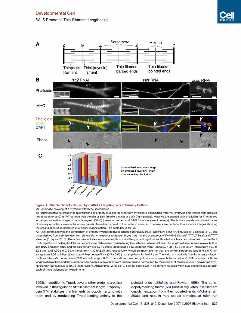
Figure 4. Examination of Myofibril Mor-
phology and Sarcomeric Organization
in sals Mutant Muscles by Confocal Anal-
ysis and Electron Microscopy
(A–D) Confocal micrographs of myofibrils from
WT (upper panels) and sals mutant embryos
(bottom panels) at 20–22 hr AEL. The left
panels show F-actin. The middle panels show
the stainings for different sarcomeric compo-
nents: (A) MHC for thick filaments; (B) KZ for
SLS in Z lines; (C) TM; and (D) Tmod. The right
panels show merged images from the left and
middle panels. Note the strong phalloidin stain-
ings at Z lines, and shorter thin-filament and
sarcomere lengths in sals mutant myofibrils
than those in WT, as revealed by phalloidin,
TM, and Tmod stainings.
(E–J) (E–H) Electron micrographs of larval mus-
cles from (E, G, and I) WT (w1118) and (F, H,
and J) the sals mutant (salsf07849/salsf07849)
at stages of (E–H) 20–22 hr AEL or (I and J)
28–30 hr AEL. (E and F) Transverse sections
at a higher magnification show that the basic
contractile unit of a thick filament (large black
dots) surrounded by an array of thin filaments
(small dots) can form correctly in both (E) WT
and (F) sals mutant myofibrils. Longitudinal
sections of (G) WT muscles and (H) muscles
of some mutant embryos (n = 10) show regular,
parallel, highly ordered arrays of myofilaments
with apparent interdigitating thin and thick fila-
ments (arrows) and well-organized Z lines (ar-
rowheads) that are characteristic of larval mus-
cles. Sarcomeres are in a contracted state in
both (G) and (H), and they are �4.5 mm and
�3.2 mm long, respectively. Glycogen granules
(g), mitochondria (M), and nuclei (N) are also
labeled in (G) and (H). (I and J) Sarcomeric
regions in muscles of an (I) older WT and (J)
mutant embryos (28–30 hr AEL). (J) Note that
mutants contain electron-dense structures
(black arrowheads), and their myofibrillar archi-
tecture was severely disrupted.
The scale bar is 10 mm in (A) for (A)–(D), 100 nm
in (E) for (E) and (F), 500 nm in (G) for (G) and (H),
and 500 nm in (I) for (I) and (J).
evidenced by the increased number of sarcomeres in
myofibrils (12 compared to 8 at 19–21 hr AEL) (upper panel
in Figure 3D). In contrast, the muscles of age-matched sals
mutant larvae had many more sarcomeres (�18 com-
pared to 12 in controls at 28–30 hr AEL), but they were
much shorter in length longitudinally (only �70% of their
WT counterparts) (middle panel of Figure 3D), indicating
that the growth rate of sals mutant muscles is much slower
than that of WT. In addition, some muscles showed a com-
plete disruption of myofibril morphology (bottom panel in
Figure 3D) (see Discussion). This extreme phenotype
was also evident under an electron microscopy (EM) anal-
ysis (Figures 4I and 4J). Interestingly, the numerous elec-
tron-dense patches seen under EM (arrowheads in
Figure 4J) resemble the condensed thin filaments in con-
junction with Z line materials seen in nemaline myopathy
(Yamaguchi et al., 1978).
Developm
Because sals mutants failed to hatch from the egg case,
we tested whether this was a result of their inability to
move. Peristaltic movement, a characteristic behavior
observed before hatching, was detectable in mutant
embryos at late stage 17 (20–22 hr AEL at 25�C) (Movie
S2). However, compared to control embryos, which
exhibit well-coordinated waves of strong forward and
backward peristalsis (Movie S1), sals mutants had less
frequent backward peristalsis, and the vigor of their con-
traction was much reduced (Figure S4; Movie S2). At
28–30 hr AEL, which was beyond the expected hatch
time, mutants displayed weak movements (Movie S3).
They eventually died in the egg case within a day. When
mutant animals were released from their egg case by
manual dissection, they were flaccid and immobile. We
conclude that although sals mutant embryos can move,
they cannot generate sufficient contractile force to escape
ental Cell 13, 828–842, December 2007 ª2007 Elsevier Inc. 833
Developmental Cell
SALS Promotes Thin-Filament Lengthening
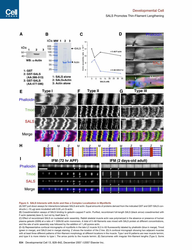
Figure 5. SALS Interacts with Actin and Has a Complex Localization in Myofibrils
(A) GST pull-down assays for interactions between SALS and actin. Equal amounts of proteins derived from the indicated GST and GST-SALS con-
structs (�10 mg) were incubated with 0.05 mm G-actin.
(B) Cosedimentation assays of SALS binding to gelsolin-capped F-actin. Purified, recombinant full-length SALS (black arrow) cosedimented with
F-actin (asterisk) (lane 2), but not by itself (lane 1).
(C) Effect of recombinant SALS on nucleated actin assembly. Rabbit skeletal muscle actin was polymerized in the absence or presence of human
plasma gelsolin (GSN) at a ratio of 1 GSN:50 actin monomers. A total of 5 nM filaments was mixed with SALS protein at different concentrations,
and the rate of actin assembly was followed by the addition of 1 mM pyrene actin.
(D–G) Representative confocal micrographs of myofibrils in the late-L2 muscle VL3 in A3 fluorescently labeled by phalloidin (blue in merge), Tmod
(green in merge), and SALS (red in merge) staining. Z shows the location of the Z line. (D) A confocal micrograph showing two adjacent muscles
with at least three different patterns of thin-filament morphology at different locations in the muscle. Type I and III patterns are near muscle termini,
and type II is more interior to type I. The arrow points to the muscle termini. (E) Sarcomeres with irregular thin-filament lengths (Type I). Some
834 Developmental Cell 13, 828–842, December 2007 ª2007 Elsevier Inc.
Developmental Cell
SALS Promotes Thin-Filament Lengthening
from the egg case because of their underlying muscle
defects.
sals Mutant Sarcomeres Have Short ThinFilamentsTo investigate the organization of sarcomeres in sals
mutant muscle myofibrils, we dissected WT and sals mu-
tants at 19–21 hr AEL, and stained muscles with phalloidin
as well as antibodies targeting a number of sarcomeric
markers (Figures 4A–4D). These include Drosophila mus-
cle Myosin Heavy Chain (MHC) (Figure 4A); KZ, an isoform
of the Drosophila Titin homolog Sallimus (SLS) localized at
the Z line (Machado et al., 1998) (Figure 4B); Tropomyosin
(TM) (Figure 4C); and Tmod (Figure 4D).
The overall distribution of these sarcomeric compo-
nents, except Tmod, appeared to be unaltered in the
sals-deficient myofibrils (Figures 4A–4D). Whereas Tmod
staining was found in or close to the M line of the sarco-
mere in WT myofibrils, its distribution was very close to
the Z line in mutants (Figure 4D). Furthermore, the length
of thin filaments was much shorter in mutant sarcomeres
(�0.85 ± 0.021 mm versus �2.75 ± 0.086 mm [range from
1.8 to 3.4 mm] in WT; myofibrils in body wall muscle VL3
in A3 or A4 were analyzed; n R 12). The reduced size of
thin-filament lengths found in mutant myofibrils was very
similar to that of sals-deficient myofibrils in primary culture
(data not shown). Short thin-filament lengths were inde-
pendently confirmed by phalloidin staining for F-actin (Fig-
ure 3) and by staining for TM (Figure 4C). Notably, the
width of MHC staining in mutant myofibrils, although
slightly shorter, was comparable to that in WT (Figure 4A),
suggesting that the thick-filament length was not dramat-
ically reduced. Altogether, these results indicate that
SALS is not critical for the normal organization of several
sarcomeric proteins, but that it is essential for achieving
the final thin-filament length. This conclusion was further
supported by an EM analysis of sals mutant muscles,
which had well-organized myofibril structures at 20–22
hr AEL, as revealed in both transverse and longitudinal
EM sections (Figures 4E–4H).
SALS Binds Both G-Actin and F-Actinand Associates with Filament Pointed EndsAs SALS (referred to as ‘‘the long form of SALS’’ in the text
below) has a predicted actin-binding domain, we deter-
mined whether it could bind actin. We first examined
whether SALS could directly bind to G-actin in vitro. Be-
cause the full-length GST-SALS protein is not expressed
well, we tested two complementary fusion proteins,
SALS(386–513) and SALS(477–596), in a pull-down assay.
Developm
SALS(386–513), which contains the two WH2 domains,
bound G-actin, whereas SALS(477–596), which lacks
the WH2 domains, did not (Figure 5A). This suggests
that the WH2 domains of SALS are sufficient for G-actin
binding.
We further evaluated the capacity of SALS to bind to ac-
tin filaments (F-actin) by using a cosedimentation assay.
The purified recombinant SALS protein by itself did not
sediment at 200,000 3 g (30 min). However, SALS cosedi-
mented with gelsolin-capped F-actin (Figure 5B), indicat-
ing that SALS binds F-actin through the pointed ends and/
or sides, but not through the barbed ends, which were
capped by gelsolin in the assay (Yin et al., 1981). The as-
sociation of SALS with F-actin is specific, because the
control protein GST failed to sediment with F-actin (data
not shown).
To investigate SALS pointed-end effects, we deter-
mined whether it affected actin elongation from gelsolin-
capped actin filaments. As shown in Figure 5C, we found
that SALS markedly slowed the rate of addition of actin
subunits at the pointed end of these capped, but not un-
capped, filaments. The effect was observed at a stoichi-
ometry that also eliminates monomer sequestration as
a mechanism of diminished pointed-end assembly. Alto-
gether, our data suggest that SALS is associated with
the filament pointed ends.
SALS Protein Is Enriched at Pointed Ends duringThin-Filament ElongationTo further characterize the role of SALS in sarcomere elon-
gation, we examined its localization pattern in muscles by
using two antibodies targeting two different regions of
SALS. Both antibodies showed an identical staining pat-
tern in muscles, and this pattern revealed that SALS is
specifically associated with thin filaments, but not with
thick filaments (data not shown). Because new sarco-
meres are constantly added to myofibrils during muscle
growth through three larval stages (L1, L2, L3) (Haas,
1950), we examined SALS localization in late-L2 or
early-L3 muscles because they are more easily prepared
for visualization.
We investigated the SALS localization pattern with re-
spect to F-actin (by phalloidin staining) and to Tmod (Fig-
ures 5D–5G), as well as to a-actinin (Figure S5) in somatic
body wall muscles. We detected a large variation in thin-fil-
ament morphology among different sarcomeres within the
same myofibril, although the lengths of these sarcomeres
were relatively uniform (Figures 5D–5G). Phalloidin has
been reported to have less accessibility to more organized
thin filaments, due to their association with actin-binding
thin-filament pointed ends protrude out, forming ragged ends toward the middle of the sarcomere (arrows). (F) Sarcomeres with a more uniform or-
ganization of actin filaments (Type II). (G) Sarcomeres with Tmod strongly localized in the M line and SALS preferentially found near the Z line (Type III).
(H) Schematic drawing showing the complex distributions of SALS (in red or light red) and Tmod (in green) relative to thin filaments (in blue), based on
the confocal micrographs shown in (E)–(G), during thin-filament assembly. Yellow indicates the merge of SALS and Tmod. The black, dotted, thin line:
M line; blue thin line: Z line. To simplify, thick filaments are not shown in sarcomeres.
(I) Confocal micrographs of isolated IFM myofibrils from pupae 72 hr APF (left panels) and 2-day-old adults (right panels) fluorescently labeled by
phalloidin (blue in merge) and stained for Tmod (green in merge) and SALS (red in merge). P, pointed ends.
The scale bar is 50 mm in (D), 10 mm in (E) for (E)–(G), and 5 mm in (I).
ental Cell 13, 828–842, December 2007 ª2007 Elsevier Inc. 835

Developmental Cell
SALS Promotes Thin-Filament Lengthening
proteins such as nebulin, which may block phalloidin from
binding to F-actin (Zhukarev et al., 1997). Thus, we deter-
mined the level of organization of thin filaments in sarco-
meres based on the phalloidin staining pattern: those
with broad, fuzzy staining were the least organized (top
panels in Figures 5E and 5F), and those with strong, Z
line staining were the most organized and usually had the
longest length, with their pointed ends meeting in the mid-
dle of the sarcomere as a discrete band (Figure 5G). Inter-
estingly, sarcomeres with less organized thin-filament
morphology were often detected at the muscle termini
(Figures 5D and 5E), where growth is known to take place
in vertebrate muscles (Dix and Eisenberg, 1990; Williams
and Goldspink, 1971). By analogy, we presume that such
sarcomeres represent newly formed sarcomeres, whereas
those with a more organized thin-filament morphology are
older and full-grown sarcomeres.
The distribution of a-actinin, a marker for thin-filament
barbed ends at the Z line, was always located in the mid-
line of the phalloidin staining (Figure S5), confirming that
thin filaments are relatively stable at the barbed ends (Lit-
tlefield et al., 2001). In contrast, several patterns of Tmod
and SALS localization could be distinguished and corre-
lated with the morphology of thin filaments in sarcomeres
(middle panels in Figures 5E–5G). Importantly, SALS was
localized at the pointed ends of thin filaments while they
were elongating (Figures 5E, 5F, and 5H), but it disap-
peared from the pointed ends of full-grown thin filaments
and relocated at the region flanking the Z line, where the
fuzzy Tmod staining pattern was also observed [Figures
5G and 5H]. This doublet pattern was more obvious in
myofibrils of visceral muscles of third-instar larvae
(Figure S5B). A similar distribution pattern has also been
observed previously for MLP (Muscle LIM protein), a pro-
tein implicated in myofibril growth (Arber et al., 1997). Be-
cause sals-deficient muscles also have thin myofibrils, we
speculate that this doublet pattern flanking the Z lines may
represent the sites at which newly assembled thin fila-
ments are located, which may also contribute to the trans-
verse growth of myofibrils. Notably, Tmod distribution was
not found at the tip of thin-filament ends in, presumably,
newly added sarcomeres, but these were the loci at which
the strong SALS staining was observed (arrows in
Figure 5E). Thus, we speculate that these ends may not
be capped strongly by Tmod while they are still actively
elongating, most likely through SALS activity.
Importantly, SALS is also expressed in the adult fly indi-
rect flight muscles (IFM) (Figure 5I). The IFM increase their
size by the synchronous lengthening of sarcomeres (from
�1.7 mm to 3.2 mm), whereas the sarcomere number re-
mains unchanged in myofibrils (Reedy and Beall, 1993).
This is unlike larval muscles, which grow by increasing
sarcomere number through the addition of newly assem-
bled sarcomeres to the existing myofibrils. Thus, IFM pro-
vide a better system by which to correlate the positions of
SALS to the growth of thin filaments. Indeed, we observed
strong localization of SALS at the pointed ends when the
thin filaments were still elongating along with sarcomere
lengthening in the IFM 72 hr after pupal formation (APF)
836 Developmental Cell 13, 828–842, December 2007 ª2007 E
(Figure 5I). Furthermore, SALS is less enriched at the
pointed ends when thin filaments reach their maximum
length, because sarcomere lengthening stops in the IFM
of 2-day-old adults (Figure 5I). Altogether, the distribution
of SALS in both larval muscles and adult IFM suggests that
SALS may be involved in thin-filament elongation from the
pointed ends.
SALS Is Required for GFPactin Incorporationat Pointed EndsTo provide further support for the pointed-end activity
of SALS, we established primary cell cultures from myo-
blasts isolated from embryos carrying the Dmef2-Gal4,
UAS-GFPactin transgenes. In these cells, GFPactin was
expressed at the stage at which myofibril assembly just
starts to occur. This unique timing of expression causes
the initial incorporation of GFPactin into thin filaments to
be found at both the Z line and the filament pointed
ends, either in primary muscles cultured within 5 days at
18�C or in early-L1 muscles (Figure 6A and data not
shown). This is consistent with the report from Littlefield
et al. (2001) that states that G-actin can be incorporated
into both barbed and pointed thin-filament ends. How-
ever, after an extended period of culture, either in vivo
(such as in third-instar larval muscles) or in primary culture
(such as those cultured for 12 days at 18�C), GFPactin was
found throughout the entire thin filaments, presumably
due to turnover of non-GFP-labeled, endogenous actins
and further incorporation of GFPactin (Roper et al.,
2005; data not shown). We thus only examined the effects
of dsRNAs targeting lacZ or sals on the initial sites of incor-
poration of GFPactin in the thin filaments of primary mus-
cles cultured within 5 days at 18�C.
Muscles in primary cultures treated with control lacZ
dsRNA were similar to muscles in untreated cultures,
and, in both cases, the majority displayed two incorpora-
tion sites for GFPactin (Figures 6A and 6C): one site colo-
calized with a-actinin at the Z line, where thin-filament
barbed ends are located; the other site mapped to the
middle of the sarcomere, at the thin-filament pointed
ends. Unlike control primary muscles, the majority of
sals RNAi primary muscles showed GFPactin incorpora-
tion only at the Z lines, with no incorporation at the pointed
ends (Figures 6B and 6C). These data strongly indicate
that SALS is required for GFPactin incorporation into
pointed ends, but not barbed ends.
Converse Effect of SALS and Tmodon Thin-Filament AssemblyGiven the decreased length of thin filaments observed
in the absence of a functional SALS protein, we tested
whether its overexpression could lead to a corresponding
increase in thin-filament length. Constitutive expression of
SALS in muscles disrupted their function, and 65% of em-
bryos carrying Dmef2-Gal4, UAS-sals failed to hatch at
29�C (n = 200). The remaining, although able to hatch,
crawled very slowly and later died. To obviate any compli-
cations resulting from the mechanical stress placed by
lsevier Inc.

Developmental Cell
SALS Promotes Thin-Filament Lengthening
Figure 6. SALS Is Required for GFPactin
Incorporation at Pointed Ends
(A and B) Confocal micrographs of primary
muscles cultured at 18�C for 5 days, derived
from embryos carrying Dmef2-Gal4/UAS-
GFPactin57B, treated with dsRNAs targeting
either (A) lacZ or (B) sals, visualized by GFP
for GFPactin (green in merge), and stained for
Tmod (red in merge) and for a-actinin (blue in
merge). Arrows, Z lines; arrowheads, pointed
ends.
(C) GFPactin incorporation sites. Visual scoring
of GFPactin incorporation sites in primary mus-
cles treated with dsRNAs targeting lacZ or sals.
Z>>P, cells in which GFPactin fluorescence
was much brighter at Z lines than at pointed
ends; Z = P, cells in which GFPactin fluores-
cence at Z lines and at pointed ends was of ap-
proximately equivalent brightness; Z<<P, cells
in which GFPactin fluorescence was much
dimmer at Z lines than at pointed ends; n, num-
ber of primary muscle cells counted.
The scale bar is 10 mm in (A) for (A) and (B).
other tissues on muscles in vivo, we examined muscle
phenotypes in primary culture.
Primary muscle cells isolated from Dmef2-Gal4, UAS-
sals embryos were cultured for 5 days at 29�C, fixed,
and stained with phalloidin and the sarcomeric marker
KZ. Strikingly, �90% of the primary muscles (n = 57)
had completely lost the stereotypical striated pattern of
thin and thick filaments (Figure 7B). This phenotype
was reminiscent of that observed in primary muscles
treated with a dsRNA against Tmod (Figure 7A; �80%
penetrant, n = 40), suggesting that excess SALS or loss
of Tmod may disrupt initial myofibril assembly (as indi-
cated in the model shown in Figure 7J). However, a small
percentage of muscles with excess SALS (�7%, n = 56)
contained myofibrils with an aberrant striated pattern
with longer than usual sarcomeres (Figure 7E), a pheno-
type that also could be found in Tmod dsRNA-treated
primary myofibrils (�5% penetrant) (Figure 7D), but never
found in controls (Figure 7C). These mild phenotypes
may reflect a relatively lower level of SALS expression
or a partial reduction of Tmod level caused by RNAi in
muscle cells, respectively. In the myofibrils containing
elongated sarcomeres, staining for KZ or phalloidin re-
vealed a relatively normal, striated Z line pattern, al-
though localization of KZ was slightly altered (middle
panels in Figures 7C–7E). However, the staining for
Tmod in the primary muscles with either Tmod RNAi or
overexpression of SALS was diffuse and did not exhibit
any regular striated pattern, as seen in controls (data
not shown). Controls had a clear and discrete phalloidin
staining band in the middle of the sarcomere, whereas
phalloidin staining in the middle of these lengthened sar-
comeres was fuzzy and irregular (Figures 7C–7E), show-
ing that their thin filaments are misaligned and have vari-
able lengths, and that some extend across the M line,
resulting in no obvious H zone. This observation indicates
that excess SALS can cause thin filaments to elongate
from the pointed ends.
Developmen
The antagonistic activities of SALS and Tmod suggest
that they might genetically interact in the thin-filament as-
sembly process. To document this interaction, we first
confirmed that excess Tmod would result in short thin-
filament lengths in primary cultured muscles (1.8 ± 0.042
mm versus 2.5 ± 0.034 mm in controls; average ± SD,
n R 15 primary muscles scored for each genotype), as pre-
viously reported to be the case in vertebrate muscles and
in fly IFM (Littlefield et al., 2001; Mardahl-Dumesnil and
Fowler, 2001) (Figures 7G and 7H). In addition, animals
overexpressing Tmod had much higher mortality rates
at both embryonic and larval stages compared to the
control animals carrying Dmef2-Gal4 alone (Figure 7F),
due to muscle defects resulting from short thin-filament
lengths (data not shown). Importantly, reduction of the
SALS level enhanced the effect caused by Tmod overex-
pression, as demonstrated by an increase in embryonic
mortality rate (Figure 7F) and a greater reduction in
thin-filament lengths in cultured muscles (Figures 7G–
7I). Thus, we conclude that Tmod and SALS functionally
antagonize each other in thin-filament assembly at the fil-
ament pointed ends.
DISCUSSION
SALS Promotes Thin-Filament Elongationfrom Pointed Ends In VivoIn this study, we report the characterization of SALS, an
actin-binding, WH2-domain-containing protein that is es-
sential for the development and function of Drosophila lar-
val muscles. Importantly, we show that SALS is required
for the elongation of thin filaments from pointed ends to at-
tain their final lengths in sarcomeres. These conclusions
are based on the findings that (1) inactivation of SALS by
either RNAi or genetic disruption leads to myofibrils con-
taining shortened thin filaments, (2) the localization of
SALS at the filament pointed ends coincides with the
growth of thin filaments, (3) GFPactin fails to incorporate
tal Cell 13, 828–842, December 2007 ª2007 Elsevier Inc. 837

Developmental Cell
SALS Promotes Thin-Filament Lengthening
Figure 7. Antagonistic Relationship between SALS and Tmod
(A and B) Fluorescence micrographs of primary muscles visualized by phalloidin. (A) Primary muscles were treated with a dsRNA targeting Tmod.
(B) Primary muscles were derived from fly embryos carrying Dmef2-Gal4, UAS-sals.
(C–E) Confocal micrographs of primary myofibrils stained by phalloidin (red in merge) and by anti-KZ antibodies for Z lines (green in merge). (C) Control
myofibrils treated with lacZ dsRNAs. Filament pointed ends joined at the midline of the sarcomere (arrowheads). (D) Myofibrils treated with Tmod
dsRNA. (E) Myofibrils derived from embryonic cells carrying Dmef2-Gal4, UAS-sals. Note the irregular sarcomere lengths in (D) and (E) compared
to those in (C) (white brackets). The white arrowheads in (D) and (E) point to nonuniform pointed ends of thin filaments, compared to the arrowheads
in (C).
(F) Mortality rate of animals of different genotypes at embryonic and larval stages.
(G–I) Confocal fluorescence micrographs of primary myofibrils derived from cells isolated from embryos of (G) Dmef2-Gal4, (H) UAS-Tmod/+;Dmef2-
Gal4/+, and (I) UAS-Tmod/+;Dmef2-Gal4/salsf07849, visualized by phalloidin (red in merge) and Tmod (green in merge). White lines, Z lines; white ar-
rowheads, pointed ends. Note that the average length of thin filaments in UAS-Tmod/+;Dmef2-Gal4/salsf07849 primary muscle myofibrils was �10%
shorter than that found in UAS-Tmod/+;Dmef2-Gal4/+ primary muscles (1.6 ± 0.079 mm versus 1.8 ± 0.042 mm; average ± SD, n R 15).
838 Developmental Cell 13, 828–842, December 2007 ª2007 Elsevier Inc.

Developmental Cell
SALS Promotes Thin-Filament Lengthening
into thin filaments through pointed ends in the absence of
SALS, and (4) SALS overexpression is sufficient to cause
overgrowth of thin filaments.
The discovery of SALS is significant because it demon-
strates the existence of a protein that appears to promote
thin-filament elongation from pointed ends in vivo. Our ge-
netic analyses suggest that SALS may act by antagonizing
Tmod pointed-end capping activity (Figure 7J). The antag-
onism may resemble that observed between Ena/VASP
proteins and the capping protein (Bear et al., 2002) or be-
tween formins and the capping protein (Zigmond et al.,
2003) at the barbed end. Loss of SALS would allow
Tmod to cap the pointed ends permanently. This is equiv-
alent to the effect of an increase in Tmod levels during
myofibril assembly, which converts Tmod from a dynamic
to a permanent cap by outcompeting SALS at the pointed
end and by preventing SALS from elongating actin fila-
ments. Conversely, overexpression of SALS leads to
a phenotype similar to loss of Tmod, because excess
SALS might act as an ‘‘anticapping’’ protein to compete
with and antagonize Tmod access to the filament pointed
ends. Finally, the complex localization patterns of Tmod
and SALS, together with their antagonistic relationships,
is suggestive of the mechanism deployed during normal
thin-filament assembly. Indeed, rapid actin assembly at
the uncapped filament pointed ends is consistent with
the localization of Tmod that lags behind SALS (Figure 5).
As a protein that binds both G-actin and F-actin, SALS
may function by stabilizing F-actin and/or through the di-
rect involvement of actin polymerization. To our surprise,
although it is strongly associated with the pointed ends,
SALS alone appears to inhibit filament elongation in vitro
(Figure 5), instead of promoting elongation, as supported
by all of the in vivo data. This discrepancy may imply that
other factors such as Tmod, TM, and profilin-actin are
needed in the biochemistry assay to observe the effect of
SALS in vitro. Indeed, the long form of SALS contains
Pro-rich profilin-binding sequences positioned immediately
N-terminal to the two tandem actin-binding WH2 domains.
Moreover, crystal structure analysis suggests that the Pro-
rich regions preceding WH2 domains may facilitate actin-
filament elongation by increasing the local concentration
of profilin-actin, or that they may mediate actin monomer
transfer directly to WH2 via profilin (Chereau et al., 2005).
How Does Lack of SALS Lead to ShortenedSarcomere Length and Increased SarcomereNumber in Myofibrils?Our analysis of sals mutant muscles suggests that the
‘‘short sarcomere’’ phenotype is a consequence of short-
ened thin filaments. This model is consistent with previous
findings that reduced sarcomere lengths are found in
muscles with altered expressions of proteins known to
be involved in the regulation of actin-filament lengths, for
instance when Tmod is present in excess (Sussman
et al., 1999). Furthermore, we propose that sarcomere
length may be specified during assembly in order to
achieve maximum actomyosin interaction. Based on the
crossbridge contraction theory (Huxley and Simmons,
1971), force generation and motion in striated muscle re-
sult from interactions between thin and thick filaments
through the cyclical formation and rupture of the actomy-
osin bonds that act as crossbridges in the overlap region
of the two filaments in sarcomeres. Previous studies
have shown that there exists a relationship between acto-
myosin bonds and sarcomere length whereby sarcomere
length decreases with an increase in actomyosin bonds
and vice versa (Telley and Denoth, 2007). Thus, we spec-
ulate that if the sarcomere length remained the same in
sals mutant myofibrils as in WT, the overlap between the
myosin and shortened actin filaments would be severely
reduced, completely impairing muscle function. Based
on these considerations, we speculate that sals mutant
myofibrils may compensate for this problem by adopting
a shorter sarcomere length, thereby restoring a more op-
timal level of actomyosin interaction.
Moreover, we propose that the mechanical constraints
placed on a muscle in vivo can explain the intriguing differ-
ence in sarcomere length that we observe in mutant sals
muscles in vivo compared to those grown in culture. As
we report, absence of SALS leads to 70% shortening of
the sarcomeres in the myotubes in culture (Figure 1), but
to only a 27% reduction in sarcomere length in muscles
in vivo (Figures 3 and 4). Because the sarcomeres are pre-
sumably contracting under a much heavier load in vivo
compared to myofibrils grown in culture, we reason that
sals mutant sarcomeres are not able to shorten as much
in vivo as they do in culture. We also note that such dra-
matic difference cannot be attributed to the age difference
between muscles of 22 hr-old sals mutant embryos and
those of R5 day-old sals mutant muscles in culture, since
there is no difference in sarcomere length between WT
embryonic and larval muscles.
In addition, sals mutant myofibrils grown in primary cul-
tures showed an increase in sarcomere number, resulting
in mutant myofibrils with a length similar to their WT coun-
terparts. Here again, the increase in sarcomere number
may reflect a compensatory mechanism intended to
achieve a strength of contraction comparable to that of
WT myofibrils. Indeed, shortened sarcomeres, which
have reduced actomyosin compared to WT sarcomeres,
may not be as efficient as a single contractile unit. Thus,
the addition of more such sarcomeres to a myofibril might
restore better functionality. This compensatory addition of
(J) Model of thin-filament assembly steps under conditions of either no SALS, normal SALS, or excess SALS. The final length of thin filaments is at-
tained in a two-step process. Short thin filaments can form under no SALS conditions (step 1), whereas longer thin filaments are achieved only in the
presence of SALS (step 2). However, excess SALS, depending on the timing and its dosage, can either disrupt sarcomere organization or increase
thin-filament length within sarcomeres.
The scale bar is 30 mm in (A) for (A) and (B), 10 mm in (C) for (C)–(E), and 10 mm in (G) for (G)–(I).
Developmental Cell 13, 828–842, December 2007 ª2007 Elsevier Inc. 839

Developmental Cell
SALS Promotes Thin-Filament Lengthening
sarcomeres probably also occurs in sals mutant myofibrils
in vivo. Indeed, muscles of sals mutant larvae at 28–30 hr
AEL had many more sarcomeres (�18 compared to 12 in
age-matched controls). However, the muscle length was
much shorter (only �70% of their WT counterparts) at
this stage, indicating that mutant muscles with shortened
sarcomeres may grow much more slowly than in WT at the
same stage. Moreover, from 28–30 hr AEL onward, sals
mutant muscles were completely disrupted, and mutant
animals eventually died. This is probably due to mechan-
ical stresses placed on the muscle by other tissues during
development. We speculate that the force generated by
sals mutant muscles of a shorter size may not be strong
enough to sustain these mechanical stresses in vivo,
and the mutant animals died long before their muscles
could achieve the same length as in WT. This is in contrast
to the sals mutant muscle in culture, which is able to attain
a final muscle length similar to WT controls while still main-
taining normal striated morphology after being cultured for
a long period of time, most likely due to the lack of the me-
chanical stresses present in the whole animal.
A Two-Step Model of the Thin-FilamentAssembly ProcessThin filaments are still assembled in the absence of SALS,
although their length is significantly shortened, indicating
that SALS is not required for initial thin-filament assembly
(Figures 1B and 4). However, SALS is absolutely required
for thin filaments to attain a longer length, and we show
that SALS promotes thin-filament lengthening from
pointed ends (Figures 5 and 6). These data reveal a two-
step process of thin-filament assembly in Drosophila larval
muscles (Figure 7J). During step 1, short filaments are as-
sembled, whereas during step 2, which depends on SALS
activity, the filaments expand to their final lengths from
their pointed ends (Figure 7J). actin RNAi presumably dis-
rupts both thin-filament assembly steps. Thus, actin RNAi
primary muscles contain both shortened sarcomeres that
have thin filaments that fail to elongate and shortened
myofibrils that fail to grow longitudinally through the addi-
tion of more sarcomeres, whose assembly presumably
requires assembly of short thin filaments (step 1). In con-
trast, sals RNAi only disrupts the second elongation
step, but it still allows the addition of short sarcomeres
containing short thin filaments to preexisting myofibrils
to attain normal myofibril length. A similar two-step pro-
cess has also been implicated in myofibril assembly of
the fly adult IFM (Mardahl-Dumesnil and Fowler, 2001).
Although we are not yet able to efficiently disrupt SALS
function to examine its role in IFM thin-filament assembly,
SALS is most likely involved, because it is localized at the
pointed ends of elongating thin filaments during IFM de-
velopment (Figure 5).
Possible SALS Activity in Vertebrate StriatedMuscle and Nonmuscle CellsStudies on myofibrillogenesis in vertebrate striated mus-
cles also suggest a similar two-step process in thin-fila-
ment assembly. For example, during early myofibrillogen-
840 Developmental Cell 13, 828–842, December 2007 ª2007 El
esis in the chick cardiac myoctyes, sarcomeres grow from
�1.5 mm to 1.8–2.5 mm, and their thin filaments elongate
accordingly (Gregorio and Antin, 2000). Delayed assembly
of Tmod at pointed end was thought to contribute to this
elongation process (Gregorio et al., 1995). In addition, in-
hibition of Tmod activity in these cells causes excessive
thin-filament elongation into H zones (Gregorio et al.,
1995). These newly assembled filaments from uncapped
pointed ends are unlikely to result from the sole modula-
tion of the function of currently known proteins such as
TM or cofilin, which maintain thin-filament length by stabi-
lizing filaments and/or inhibiting their depolymerization
from pointed ends (Littlefield and Fowler, 1998). Because
these proteins only limit existing filament size, but do not
contribute to actin assembly (Iwasa and Mullins, 2007), ef-
ficient pointed-end assembly is likely to involve a SALS-
like activity.
SALS-like activity is unlikely to play a role in actin as-
sembly in cellular processes that require actin filaments
to turn over rapidly, such as protrusion of the leading
edge of lamellipodia, where the pointed ends are the sites
for rapid filament disassembly. However, it may be impor-
tant for length specification of stable actin-filament struc-
ture that requires regulation of actin assembly from
pointed ends, such as in hair cell rootlets of chick cochlea
(Tilney and DeRosier, 1986). Thus, even though BLAST
searches failed to identify any close SALS orthologs in ver-
tebrates, functionally related proteins are likely to exist.
There are several uncharacterized WH2-containing pro-
teins present in human and mouse genomes (Paunola
et al., 2002), and one or more of these may regulate
actin-filament lengthening from pointed ends.
EXPERIMENTAL PROCEDURES
Drosophila Genetics
Drosophila strains used in this study were Dmef2-Gal4 (Ranganava-
kulu et al., 1996); UAS-GFPactin57B (Roper et al., 2005); TTG (TM3,
Twi-Gal4, UAS-2EGFP, Ser) and twiID96 (from Dr. A Michelson);
P{PZ}tho[1], PBac{WH}f07849 (Thibault et al., 2004); and Df(3R)M-
Kx1 (from the Bloomington Drosophila Stock Center). w1118 was
used as a control strain. Excision of the piggyBac element from
PBac{WH}f07849 was done as described (Thibault et al., 2004).
Embryonic Primary Cell Cultures and Primary Cell RNAi
Embryonic primary cell cultures were established mainly as described
(Bernstein et al., 1978). Cells were seeded and grown either in 384-well
optically clear plates (Costar) (in Figure 1B) or in LabTek 8-well cham-
ber coverglass slides (VWR) coated with human vitronectin (Chemicon)
(Volk et al., 1990) (used for culturing of primary muscles shown in
the insets of Figure 1B and in Figures 6 and 7), at 1.7–2.5 3 105
cells/cm2. Mature primary muscles used for analyses were usually
from 12-day cultures at 18�C or from 5-day cultures at 25�C or 29�C.
A detailed description of the establishment of the primary cell RNAi
protocol will be described elsewhere. Briefly, the primary cells were
isolated from postgastrula embryos (4–6 hr AEL) and were seeded in
plates containing dsRNAs at around 4 3 104 cells per well. After 22 hr
in serum-free M3 medium, additional serum-containing culture me-
dium was added to bring the solution to a final concentration of 10%
FCS. After an additional 11 days of culturing at 18�C, primary cells
were directly fixed with 4% formaldehyde (Polyscience, Niles, USA).
dsRNAs were prepared as described at http://flyrnai.org/. The oligos
sevier Inc.

Developmental Cell
SALS Promotes Thin-Filament Lengthening
for making dsRNAs used in this study can be found in the Supplemen-
tal Data.
Molecular Biology, Germline Transformation,
In Situ Hybridization, and Antibody Production
See the Supplemental Data.
Actin-Binding Assays and Actin-Polymerization Assay
The GST pull-down assays, cosedimentation assays, and pyrene
actin-polymerization assay were carried out as described (Martinez-
Quiles et al., 2001). The actin assembly rate was measured as
described (Pollard and Mooseker, 1981). Details can be found in the
Supplemental Data.
Immunofluorescence Microscopy and Image Analysis
Late embryos or larvae were dissected in Ca2+-free saline buffer (relax-
ing buffer) as described (Budnik et al., 2006). Myofibrils from dorsal
longitudinal muscles of pupal and adult IFM were isolated as described
(Mardahl-Dumesnil and Fowler, 2001). In general, dissected tissues
and primary cells were fixed in 4% formaldehyde in relaxing buffer
for 20–30 min, and the standard immunohistochemistry protocol was
then followed (Budnik et al., 2006). The stained samples were analyzed
on a Leica LSM NT confocal microscope. Primary antibodies used in
this study can be found in the Supplemental Data.
Analyses of myofibril features such as thin-filament length and sar-
comere length were performed from deconvoluted images by using
Leica confocal software TCS SP2. Thin-filament length was defined
as half the distance between Tmod peaks or half the width of the phal-
loidin bands or the distance between Tmod and neighboring a-actinin
peaks. Sarcomere length was defined as the distance between
a-actinin bands or between midlines of the phalloidin bands.
Electron Microscopy
See the Supplemental Data.
Supplemental Data
Supplemental Data include Supplemental Experimental Procedures,
five figures, and movies and are available at http://www.
developmentalcell.com/cgi/content/full/13/6/828/DC1/.
ACKNOWLEDGMENTS
We thank Drs. P. Beachy, J. Saide, W. Chia, A. Michelson, V. Fowler,
H. Bellen, and D. Andrew, and the Drosophila Bloomington Stock Cen-
ter for fly stocks and antibodies. We are also very grateful to R. Binari
and C. Villalta for technical help, J. Ni for help in the production of SALS
protein, A. Lebensohn for help in actin-binding assays, and A. Samso-
nova for help in data analysis of peristalsis movies. We also thank
V. Fowler, B. Mathey-Prevot, C. Bakal, C. Pitsouli, and J. Zirin for crit-
ical comments on the manuscript. J.B. was supported by the Damon
Runyon Cancer Research Foundation Fellowship DRG-1716-02. N.P.
is an Investigator of the Howard Hughes Medical Institute.
Received: August 22, 2006
Revised: July 20, 2007
Accepted: October 4, 2007
Published: December 3, 2007
REFERENCES
Arber, S., Hunter, J., Ross, J., Hongo, M., Sansig, G., Borg, J., Perriard,
J., Chien, K., and Caroni, P. (1997). MLP-deficient mice exhibit a
disruption of cardiac cytoarchitectural organization, dilated cardiomy-
opathy, and heart failure. Cell 88, 393–403.
Bang, M., Li, X., Littlefield, R., Bremner, S., Thor, A., Knowlton, K.,
Lieber, R., and Chen, J. (2006). Nebulin-deficient mice exhibit shorter
thin filament lengths and reduced contractile function in skeletal mus-
cle. J. Cell Biol. 173, 905–916.
Development
Bear, J., Svitkina, T., Krause, M., Schafer, D., Loureiro, J., Strasser, G.,
Maly, I., Chaga, O., Cooper, J., Borisy, G., et al. (2002). Antagonism
between Ena/VASP proteins and actin filament capping regulates fi-
broblast motility. Cell 109, 509–521.
Bernstein, S., Fyrberg, E., and Donady, J. (1978). Isolation and partial
characterization of Drosophila myoblasts from primary cultures of
embryonic cells. J. Cell Biol. 78, 856–865.
Budnik, V., Gorczyca, M., and Prokop, A. (2006). Selected methods for
the anatomical study of Drosophila embryonic and larval neuromuscu-
lar junctions. Int. Rev. Neurobiol. 75, 323–365.
Burkholder, T., Fingado, B., Baron, S., and Lieber, R. (1994). Relation-
ship between muscle fiber types and sizes and muscle architectural
properties in the mouse hindlimb. J. Morphol. 221, 177–190.
Chereau, D., Kerff, F., Graceffa, P., Grabarek, Z., Langsetmo, K., and
Dominguez, R. (2005). Actin-bound structures of Wiskott-Aldrich
syndrome protein (WASP)-homology domain 2 and the implications
for filament assembly. Proc. Natl. Acad. Sci. USA 102, 16644–16649.
Dix, D., and Eisenberg, B. (1990). Myosin mRNA accumulation and
myofibrillogenesis at the myotendinous junction of stretched muscle
fibers. J. Cell Biol. 111, 1885–1894.
Fischer, R.S., and Fowler, V.M. (2003). Tropomodulins: life at the slow
end. Trends in Cell Biology 13, 593–601.
Granzier, H., Akster, H., and Ter Keurs, H. (1991). Effect of thin filament
length on the force-sarcomere length relation to skeletal muscle. Am.
J. Physiol. Cell Physiol. 260, C1060–C1070.
Gregorio, C., and Antin, P. (2000). To the heart of myofibril assembly.
Trends Cell Biol. 10, 355–362.
Gregorio, C., Weber, A., Bondad, M., Pennise, C., and Fowler, V.
(1995). Requirement of pointed-end capping by tropomodulin to main-
tain actin filament length in embryonic chick cardiac myocytes. Nature
377, 83–86.
Haas, J. (1950). Cytoplasmic growth in the muscle fibers of larvae of
Drosophila melanogaster. Growth 14, 277–294.
Huxley, A., and Simmons, R. (1971). Proposed mechanism of force
generation in striated muscle. Nature 233, 533–538.
Iwasa, J., and Mullins, R. (2007). Spatial and temporal relationships be-
tween actin-filament nucleation, capping, and disassembly. Curr. Biol.
17, 395–406.
Littlefield, R., and Fowler, V. (1998). Defining actin filament length in
striated muscle: rulers and caps or dynamic stability? Annu. Rev.
Cell Dev. Biol. 14, 487–525.
Littlefield, R., Almenar-Queralt, A., and Fowler, V. (2001). Actin dynam-
ics at pointed ends regulates thin filament length in striated muscle.
Nat. Cell Biol. 3, 544–551.
Ma, Y., Creanga, A., Lum, L., and Beachy, P. (2006). Prevalence of off-
target effects in Drosophila RNA interference screens. Nature 443,
359–363.
Machado, C., Sunkel, C., and Andrew, D. (1998). Human autoanti-
bodies reveal titin as a chromosomal protein. J. Cell Biol. 141, 321–
333.
Mardahl-Dumesnil, M., and Fowler, V. (2001). Thin filaments elongate
from their pointed ends during myofibril assembly in Drosophila indi-
rect flight muscle. J. Cell Biol. 155, 1043–1053.
Martinez-Quiles, N., Rohatgi, R., Anton, M., Medina, M., Saville, S.,
Miki, H., Yamaguchi, H., Takenawa, T., Hartwig, J., Geha, R., et al.
(2001). WIP regulates N-WASP-mediated actin polymerization and
filopodium formation. Nat. Cell Biol. 3, 484–491.
McElhinny, A., Schwach, C., Valichnac, M., Mount-Patrick, S., and
Gregorio, C. (2005). Nebulin regulates the assembly and lengths of
the thin filaments in striated muscle. J. Cell Biol. 170, 947–957.
Mohri, K., Ono, K., Yu, R., Yamashiro, S., and Ono, S. (2006). Enhance-
ment of actin-depolymerizing factor/cofilin-dependent actin disas-
sembly by actin-interacting protein 1 is required for organized actin
al Cell 13, 828–842, December 2007 ª2007 Elsevier Inc. 841

Developmental Cell
SALS Promotes Thin-Filament Lengthening
filament assembly in the Caenorhabditis elegans body wall muscle.
Mol. Biol. Cell 17, 2190–2199.
Newman, S., and Wright, T. (1981). A histological and ultrastructural
analysis of developmental defects produced by the mutation, lethal(1)-
myospheroid, in Drosophila melanogaster. Dev. Biol. 86, 393–402.
Page, S., and Huxley, H. (1963). Filament lengths in striated muscle.
J. Cell Biol. 19, 369–390.
Paunola, E., Mattila, P., and Lappalainen, P. (2002). WH2 domain:
a small, versatile adaptor for actin monomers. FEBS Lett. 513, 92–97.
Pollard, T., and Mooseker, M. (1981). Direct measurement of actin po-
lymerization rate constants by electron microscopy of actin filaments
nucleated by isolated microvillus cores. J. Cell Biol. 88, 654–659.
Pollard, T., Blanchoin, L., and Mullins, R. (2000). Molecular mecha-
nisms controlling actin filament dynamics in nonmuscle cells. Annu.
Rev. Biophys. Biomol. Struct. 29, 545–576.
Ranganavakulu, G., Schulz, R., and Olson, E. (1996). Wingless induces
nautilus expression in the ventral mesoderm of the Drosopohila
embryo. Dev. Biol. 176, 143–148.
Reedy, M.C., and Beall, C. (1993). Ultrastructure of developing flight
muscle in Drosophila. I. Assembly of myofibrils. Dev. Biol. 160, 443–
465.
Roper, K., Mau, Y., and Brown, N. (2005). Contribution of sequence
variation in Drosophila actins to their incorporation into actin-based
structures in vivo. J. Cell Sci. 118, 3937–3948.
Sussman, M., Baque, S., Uhm, C., Daniels, M., Price, R., Simpson, D.,
Terracio, L., and Kedes, L. (1998). Altered expression of tropomodulin
in cardiomyocytes disrupts the sarcomeric structure of myofibrils.
Circ. Res. 82, 94–105.
Sussman, M., Welch, S., Gude, N., Khoury, P., Daniels, S., Kirkpatrick,
D., Walsh, R., Price, R., Lim, H., and Molkentin, J. (1999). Pathogenesis
of dilated cardiomyopathy: molecular, structural, and population anal-
yses in tropomodulin-overexpressing transgenic mice. Am. J. Pathol.
155, 2101–2113.
842 Developmental Cell 13, 828–842, December 2007 ª2007 E
Telley, I., and Denoth, J. (2007). Sarcomere dynamics during muscular
contraction and their implications to muscle function. J. Muscle Res.
Cell Motil. 28, 89–104.
Thibault, S., Singer, M., Wiyazaki, W., Milash, B., Dompe, N., Singh, C.,
Buchholz, R., Demsky, M., Fawcett, R., Francis-Lang, H., et al. (2004).
A complementary transposon tool kit for Drosophila melanogaster
using P and piggyBac. Nat. Genet. 36, 283–287.
Tilney, L., and DeRosier, D. (1986). Actin filaments, stereocilia, and hair
cells of the bird cochlea. IV. How the actin filaments become organized
in developing stereocilia and in the cuticular plate. Dev. Biol. 116, 119–
129.
Volk, T., Fessler, L., and Fessler, J. (1990). A role for integrin in the for-
mation of sarcomeric cytoarchitecture. Cell 63, 525–536.
Williams, P., and Goldspink, G. (1971). Longitudinal growth of striated
muscle fibers. J. Cell Sci. 9, 751–767.
Witt, C., Burkart, C., Labeit, D., MaNabb, M., Wu, Y., Granzier, H., and
Labeit, S. (2006). Nebulin regulates thin filament length, contractility,
and Z-disk structure in vivo. EMBO J. 25, 3843–3855.
Yamaguchi, M., Robson, R., Stromer, M., Dahl, D., and Oda, T. (1978).
Actin filaments form the backbone of nemaline myopathy rods. Nature
271, 265–267.
Yin, H., Hartwig, J., Maruyama, K., and Stossel, T. (1981). Ca2+ control
of actin filament length. Effects of macrophage gelsolin on actin poly-
merization. J. Biol. Chem. 256, 9693–9697.
Zhukarev, V., Sanger, J., Sanger, J., Goldman, Y., and Shuman, H.
(1997). Distribution and orientation of rhodamine-phalloidin bound to
thin filaments in skeletal and cardiac myofibrils. Cell Motil. Cytoskele-
ton 37, 363–377.
Zigmond, S., Evangelista, M., Boone, C., Yang, C., Dar, A., Sicheri, F.,
Forkey, J., and Pring, M. (2003). Formin leaky cap allows elongation in
the presence of tight capping proteins. Curr. Biol. 13, 1820–1823.
lsevier Inc.