Salivary Gland Neoplasms for Dummies! - lasop.comlasop.com/pgs/hdouts/2009-04_Thompson_Salivary...
27
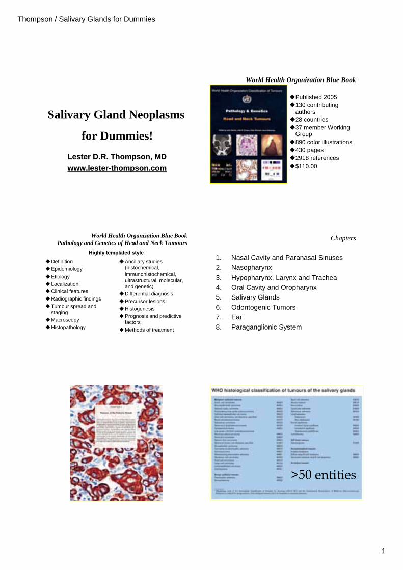
Thompson / Salivary Glands for Dummies 1 Salivary Gland Neoplasms Lester D.R. Thompson, Lester D.R. Thompson, MD MD www.lester www.lester- thompson.com thompson.com for Dummies! World Health Organization Blue Book Published 2005 130 contributing authors 28 countries 37 member Working Group 890 color illustrations 430 pages 2918 references $110.00 World Health Organization Blue Book Pathology and Genetics of Head and Neck Tumours Definition Epidemiology Etiology Localization Clinical features Radiographic findings Tumour spread and staging Macroscopy Histopathology Ancillary studies (histochemical, immunohistochemical, ultrastructural, molecular, and genetic) Differential diagnosis Precursor lesions Histogenesis Prognosis and predictive factors Methods of treatment Highly templated Highly templated style style Chapters 1. Nasal Cavity and Paranasal Sinuses 2. Nasopharynx 3. Hypopharynx, Larynx and Trachea 4. Oral Cavity and Oropharynx 5. Salivary Glands 6. Odontogenic Tumors 7. Ear 8. Paraganglionic System >50 entities
-
Upload
hoangthuan -
Category
Documents
-
view
227 -
download
0
Transcript of Salivary Gland Neoplasms for Dummies! - lasop.comlasop.com/pgs/hdouts/2009-04_Thompson_Salivary...
Thompson / Salivary Glands for Dummies
1
Salivary Gland Neoplasms
Lester D.R. Thompson, Lester D.R. Thompson, MDMDwww.lesterwww.lester--thompson.comthompson.com
for Dummies!
World Health Organization Blue Book
�Published 2005�130 contributing
authors�28 countries�37 member Working
Group�890 color illustrations�430 pages�2918 references�$110.00
World Health Organization Blue BookPathology and Genetics of Head and Neck Tumours
�Definition�Epidemiology�Etiology�Localization�Clinical features�Radiographic findings�Tumour spread and
staging�Macroscopy�Histopathology
�Ancillary studies (histochemical, immunohistochemical, ultrastructural, molecular, and genetic)
�Differential diagnosis�Precursor lesions�Histogenesis�Prognosis and predictive
factors�Methods of treatment
Highly templatedHighly templated stylestyle
Chapters
1. Nasal Cavity and Paranasal Sinuses2. Nasopharynx3. Hypopharynx, Larynx and Trachea4. Oral Cavity and Oropharynx5. Salivary Glands6. Odontogenic Tumors7. Ear8. Paraganglionic System
>50 entities
Thompson / Salivary Glands for Dummies
2
General Considerations
�1% of all tumors (considered under-reported)
�Most common in adults
�Increased frequency in females with Warthin tumor
�Fine needle aspiration first line screening test
�Little known about etiology
�Site helps separate benign and malignant
�Clinic stage is important
�Molecular techniques slow to catch on
Genetics
� Pleomorphic adenoma�8q12 (PLAG1) (40%) (increased PLAG1)
�12q14-15 (HMGA2) (8%)
�Mucoepidermoid carcinoma�t(11;19)(q21;p13): MECT1-MAML2
�About 70% of low grade tumors
Immunohistochemistry
�Ductal cell differentiation�Keratin (AE1/AE3), CAM5.2, EMA, CEA
�Myoepithelial cell differentiation�Smooth muscle actin, p63, S-100 protein,
calponin, GFAP, caldesmon, myosin, MSA
�Keratin (AE1/AE3), CAM5.2, CK14
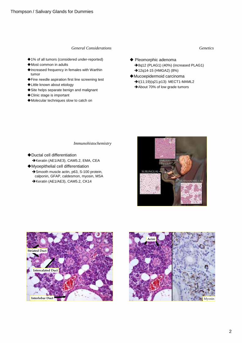
Normal Histology of Parotid
Interlobar Duct
Striated Duct
Intercalated Duct
Myosin
Acini
Thompson / Salivary Glands for Dummies
3
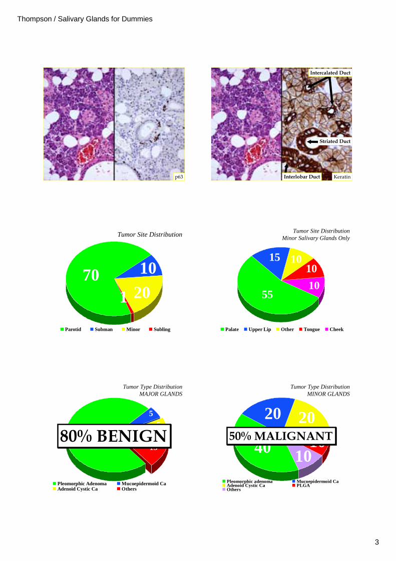
p63 Interlobar Duct
Intercalated Duct
Striated Duct
Keratin
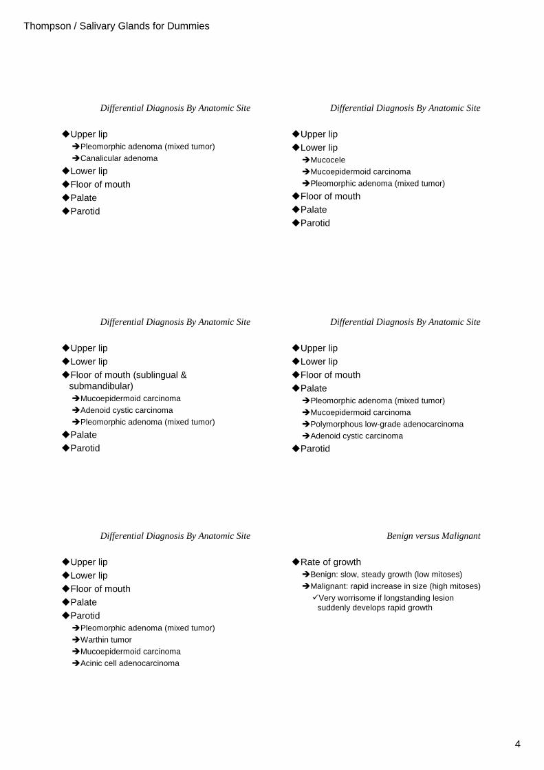
Tumor Site Distribution
701 20
10
Parotid Subman Minor Subling
Tumor Site DistributionMinor Salivary Glands Only
55
15 1010
10
Palate Upper Lip Other Tongue Cheek
Tumor Type DistributionMAJOR GLANDS
15
55
70
Pleomorphic Adenoma Mucoepidermoid CaAdenoid Cystic Ca Others
80% BENIGN80% BENIGN
Tumor Type DistributionMINOR GLANDS
40 10
2010
20
Pleomorphic adenoma Mucoepidermoid CaAdenoid Cystic Ca PLGAOthers
50% MALIGNANT50% MALIGNANT
Thompson / Salivary Glands for Dummies
4
Differential Diagnosis By Anatomic Site
�Upper lip�Pleomorphic adenoma (mixed tumor)
�Canalicular adenoma
�Lower lip�Floor of mouth�Palate�Parotid
Differential Diagnosis By Anatomic Site
�Upper lip�Lower lip
�Mucocele
�Mucoepidermoid carcinoma
�Pleomorphic adenoma (mixed tumor)
�Floor of mouth�Palate�Parotid
Differential Diagnosis By Anatomic Site
�Upper lip�Lower lip�Floor of mouth (sublingual &
submandibular)�Mucoepidermoid carcinoma
�Adenoid cystic carcinoma
�Pleomorphic adenoma (mixed tumor)
�Palate�Parotid
Differential Diagnosis By Anatomic Site
�Upper lip�Lower lip�Floor of mouth�Palate
�Pleomorphic adenoma (mixed tumor)
�Mucoepidermoid carcinoma
�Polymorphous low-grade adenocarcinoma
�Adenoid cystic carcinoma
�Parotid
Differential Diagnosis By Anatomic Site
�Upper lip�Lower lip�Floor of mouth�Palate�Parotid
�Pleomorphic adenoma (mixed tumor)
�Warthin tumor
�Mucoepidermoid carcinoma
�Acinic cell adenocarcinoma
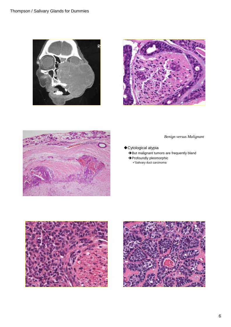
Benign versus Malignant
�Rate of growth�Benign: slow, steady growth (low mitoses)
�Malignant: rapid increase in size (high mitoses)
�Very worrisome if longstanding lesion suddenly develops rapid growth
Thompson / Salivary Glands for Dummies
5
Benign versus Malignant
�Relationship with surrounding structures�Fixation
�Benign: Freely movable (palate excluded) �Malignant: Adherent to surrounding tissue
�Ulceration�Benign: Overlying epithelium intact�Malignant: Ulceration of overlying epithelium
�Paresthesia (due to nerve invasion by tumor)�Benign: No change in sensation�Malignant: Paresthesia common
Benign versus Malignant
�Circumscription�Benign: Encapsulated; well circumscribed
�Malignant: Poorly circumscribed; infiltrative
. . . BUT — Be aware of multifocality and minor salivary gland location
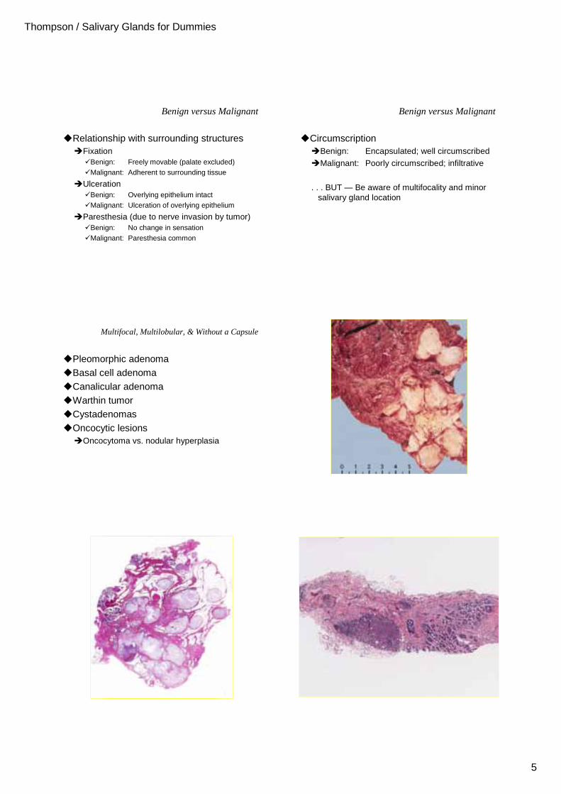
Multifocal, Multilobular, & Without a Capsule
�Pleomorphic adenoma�Basal cell adenoma�Canalicular adenoma�Warthin tumor�Cystadenomas�Oncocytic lesions
�Oncocytoma vs. nodular hyperplasia
Thompson / Salivary Glands for Dummies
6
Benign versus Malignant
�Cytological atypia�But malignant tumors are frequently bland
�Profoundly pleomorphic�Salivary duct carcinoma
Thompson / Salivary Glands for Dummies
7
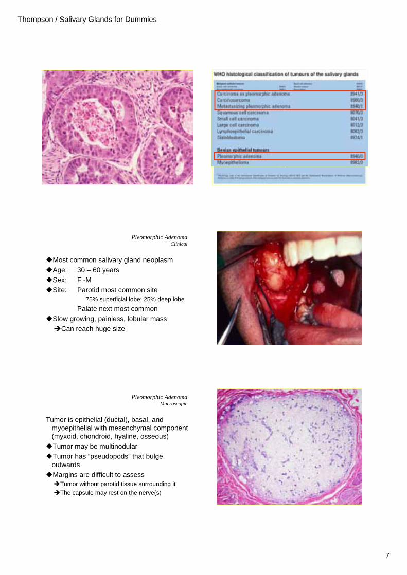
Pleomorphic AdenomaClinical
�Most common salivary gland neoplasm�Age: 30 – 60 years�Sex: F~M�Site: Parotid most common site
75% superficial lobe; 25% deep lobe
Palate next most common�Slow growing, painless, lobular mass
�Can reach huge size
Pleomorphic AdenomaMacroscopic
Tumor is epithelial (ductal), basal, and myoepithelial with mesenchymal component (myxoid, chondroid, hyaline, osseous)
�Tumor may be multinodular�Tumor has “pseudopods” that bulge
outwards�Margins are difficult to assess
�Tumor without parotid tissue surrounding it
�The capsule may rest on the nerve(s)

Thompson / Salivary Glands for Dummies
8
“Resection margins”
Multifocal recurrence
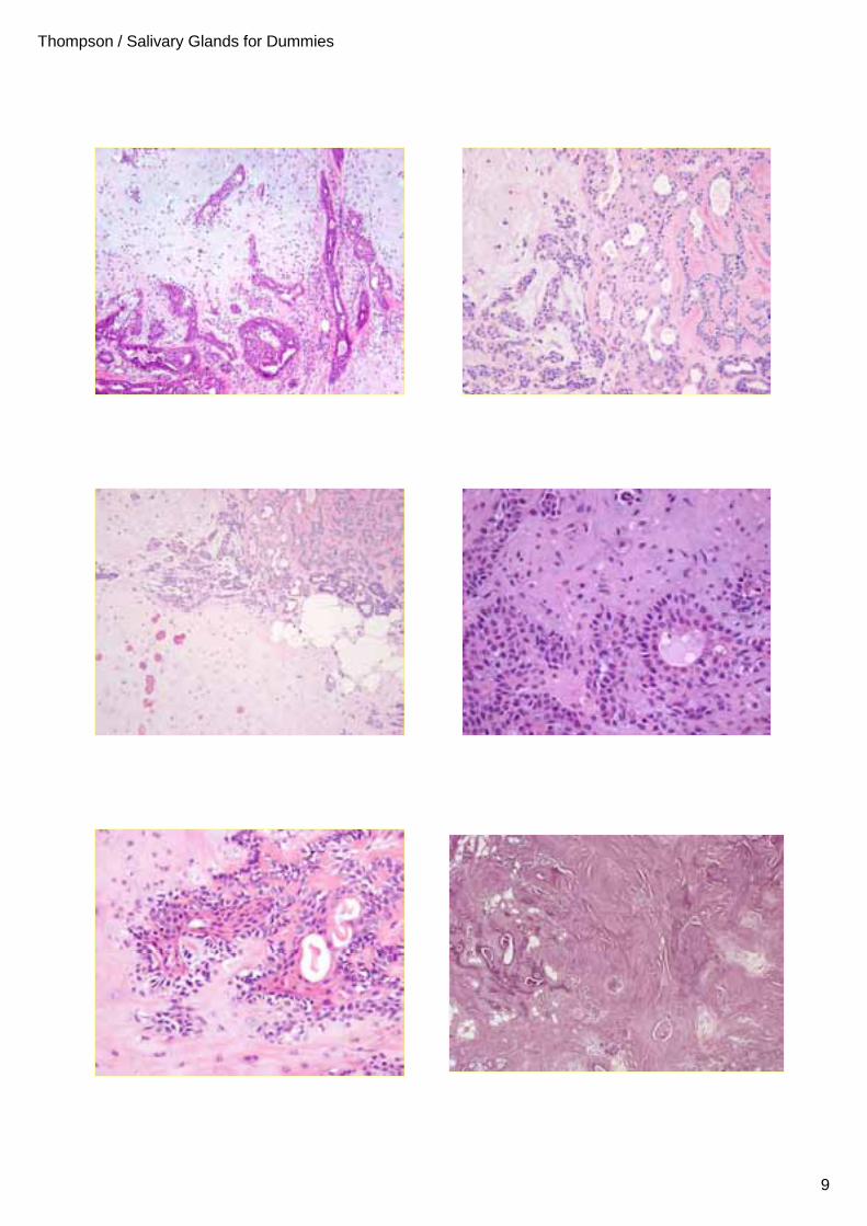
Pleomorphic Adenoma
�Remarkably variable histology�Solid, tubular, trabecular, cystic
�Cells literally “melt” into the chondroid or myxoid background stroma
�Stroma may be heavily fibrotic/hyalinized
�Spindled, epithelioid, glandular, & plasmacytoid cells
�Squamous metaplasia is common
�Increased mitotic figures s/p FNA
Thompson / Salivary Glands for Dummies
9
Thompson / Salivary Glands for Dummies
10
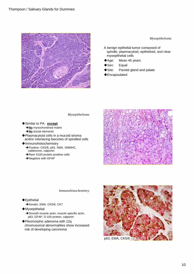
Myoepithelioma
A benign epithelial tumor composed of spindle, plasmacytoid, epithelioid, and clear myoepithelial cells
�Age: Mean 45 years�Sex: Equal�Site: Parotid gland and palate�Encapsulated
Myoepithelioma
�Similar to PA: except�No myxochondroid matrix�No ductal elements
�Plasmacytoid cells in a mucoid stroma and/or interlacing fascicles of spindled cells
�Immunohistochemistry�Positive: CK5/6, p63, SMA, SMMHC,
caldesmon, calponin�Rare S100 protein positive cells �Negative with GFAP
Immunohistochemistry
�Epithelial�Keratin, EMA, CK5/6, CK7
�Myoepithelial�Smooth muscle actin, muscle specific actin,
p63, GFAP, S-100 protein, calponin
�Pleomorphic adenoma with 12q chromosomal abnormalities show increased risk of developing carcinoma
p63, EMA, CK5/6
Thompson / Salivary Glands for Dummies
11
Pleomorphic Adenoma
�Recurrence vs. Recrudescence vs. Residual
�“Benign” metastasis�Malignant transformation can be seen in
long standing tumors
Pleomorphic Adenoma
Past Management�Local anaesthetic
�Direct incision over lump
�Remove some/any parotid tissue
�High recurrence rate �70% Lanier 1972
Present management�General anaesthesia
�Remove ALL parotid tissue (superficial and/or deep lobes)
�<2% recurrence 10 yr
Carcinoma Ex-Pleomorphic AdenomaDemographics
�About 6-10% of PA develop carcinoma�Represents about 12% of all salivary
malignancies�About 4% of all salivary gland tumors
�Must have pre-existing PA�Only clinical history is some cases�Long history of PA or frequent
recurrences�Risk of 1.5% at 5 years; 10% at 15 years
Carcinoma Ex-Pleomorphic AdenomaClinical
�Age: Elderly (usually >60 yrs)�Sex: M = F�Site: Majority in major glands
2/3 in parotid
�Sudden enlargement, with/without nerve symptoms
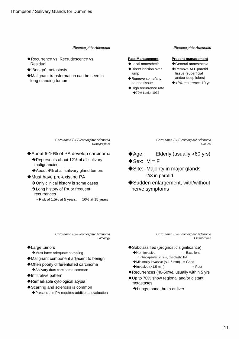
Carcinoma Ex-Pleomorphic AdenomaPathology
�Large tumors�Must have adequate sampling
�Malignant component adjacent to benign�Often poorly differentiated carcinoma
�Salivary duct carcinoma common
�Infiltrative pattern�Remarkable cytological atypia�Scarring and sclerosis is common
�Presence in PA requires additional evaluation
Carcinoma Ex-Pleomorphic AdenomaClassification
�Subclassified (prognostic significance)�Non-invasive = Excellent
�Intracapsular, in situ, dysplastic PA
�Minimally invasive (< 1.5 mm) = Good
�Invasive (>1.5 mm) = Poor
�Recurrences (40-50%), usually within 5 yrs�Up to 70% show regional and/or distant
metastases�Lungs, bone, brain or liver
Thompson / Salivary Glands for Dummies
12
Thompson / Salivary Glands for Dummies
13
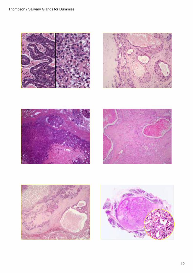
Ki67
Benign metastasizing pleomorphic adenoma
Thompson / Salivary Glands for Dummies
14
Carcinoma Ex-Pleomorphic AdenomaPrognostic Factors
�Pathologic stage�Size�Histologic grade and type�Proportion of carcinoma�Extent of invasion�Ki-67 labeling index
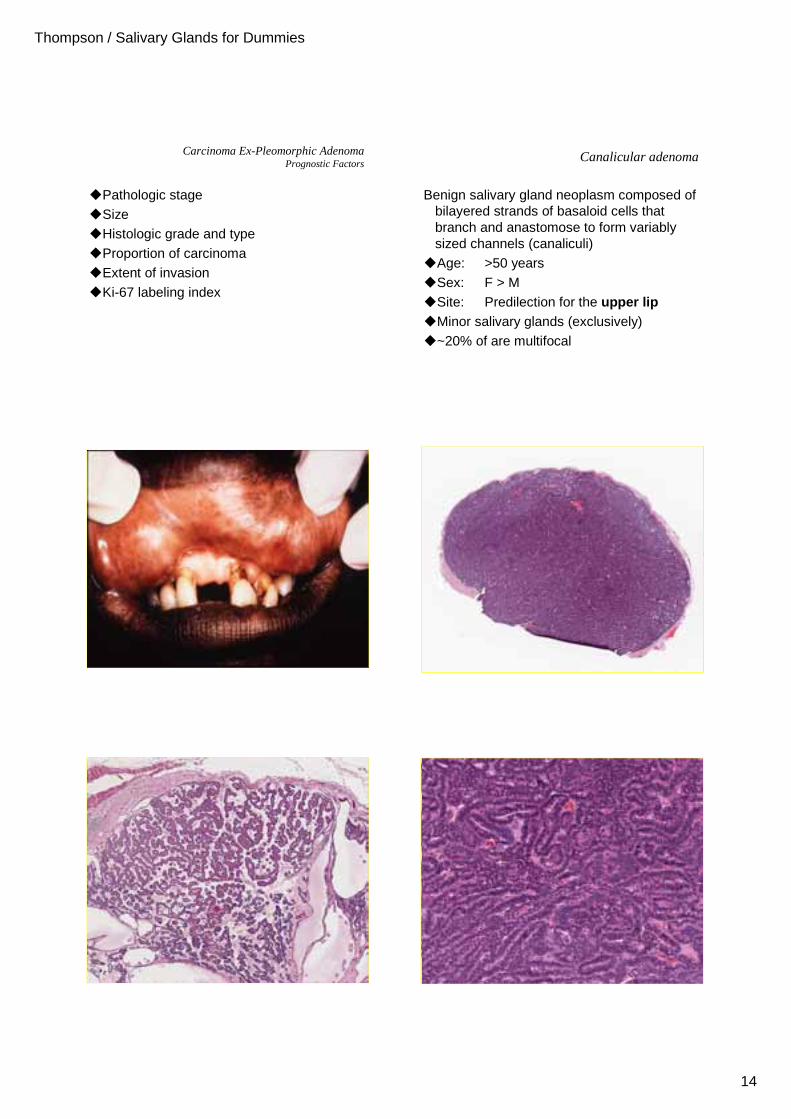
Canalicular adenoma
Benign salivary gland neoplasm composed of bilayered strands of basaloid cells that branch and anastomose to form variably sized channels (canaliculi)
�Age: >50 years�Sex: F > M�Site: Predilection for the upper lip�Minor salivary glands (exclusively)�~20% of are multifocal
Thompson / Salivary Glands for Dummies
15
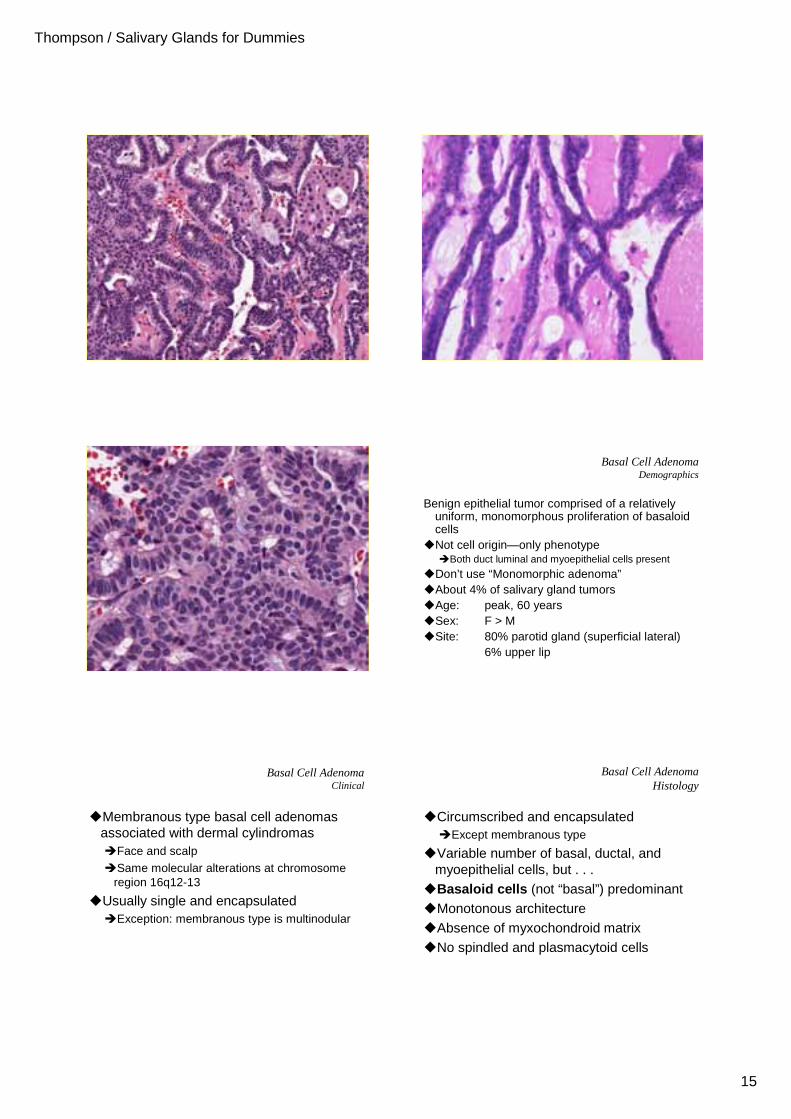
Benign epithelial tumor comprised of a relatively uniform, monomorphous proliferation of basaloid cells
�Not cell origin—only phenotype�Both duct luminal and myoepithelial cells present
�Don’t use “Monomorphic adenoma”�About 4% of salivary gland tumors�Age: peak, 60 years�Sex: F > M�Site: 80% parotid gland (superficial lateral)
6% upper lip
Basal Cell AdenomaDemographics
Basal Cell AdenomaClinical
�Membranous type basal cell adenomas associated with dermal cylindromas�Face and scalp
�Same molecular alterations at chromosome region 16q12-13
�Usually single and encapsulated�Exception: membranous type is multinodular
�Circumscribed and encapsulated�Except membranous type
�Variable number of basal, ductal, and myoepithelial cells, but . . .
�Basaloid cells (not “basal”) predominant�Monotonous architecture�Absence of myxochondroid matrix�No spindled and plasmacytoid cells
Basal Cell AdenomaHistology
Thompson / Salivary Glands for Dummies
16
Basal Cell AdenomaHistology
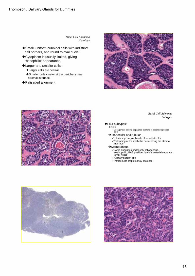
�Small, uniform cuboidal cells with indistinct cell borders, and round to oval nuclei
�Cytoplasm is usually limited, giving “basophilic” appearance
�Larger and smaller cells: �Larger cells are central
�Smaller cells cluster at the periphery near stromal interface
�Palisaded alignment
�Four subtypes:�Solid
�Collagenous stroma separates clusters of basaloid epithelial cells
�Trabecular and tubular�Interlacing, narrow bands of basaloid cells�Palisading of the epithelial nuclei along the stromal
interface�Membranous
�Large quantities of densely collagenous, eosinophilic, PAS positive, hyaline material separate tumor nests
�"Jigsaw puzzle"-like �Intracellular droplets may coalesce
Basal Cell AdenomaSubtypes
Thompson / Salivary Glands for Dummies
17
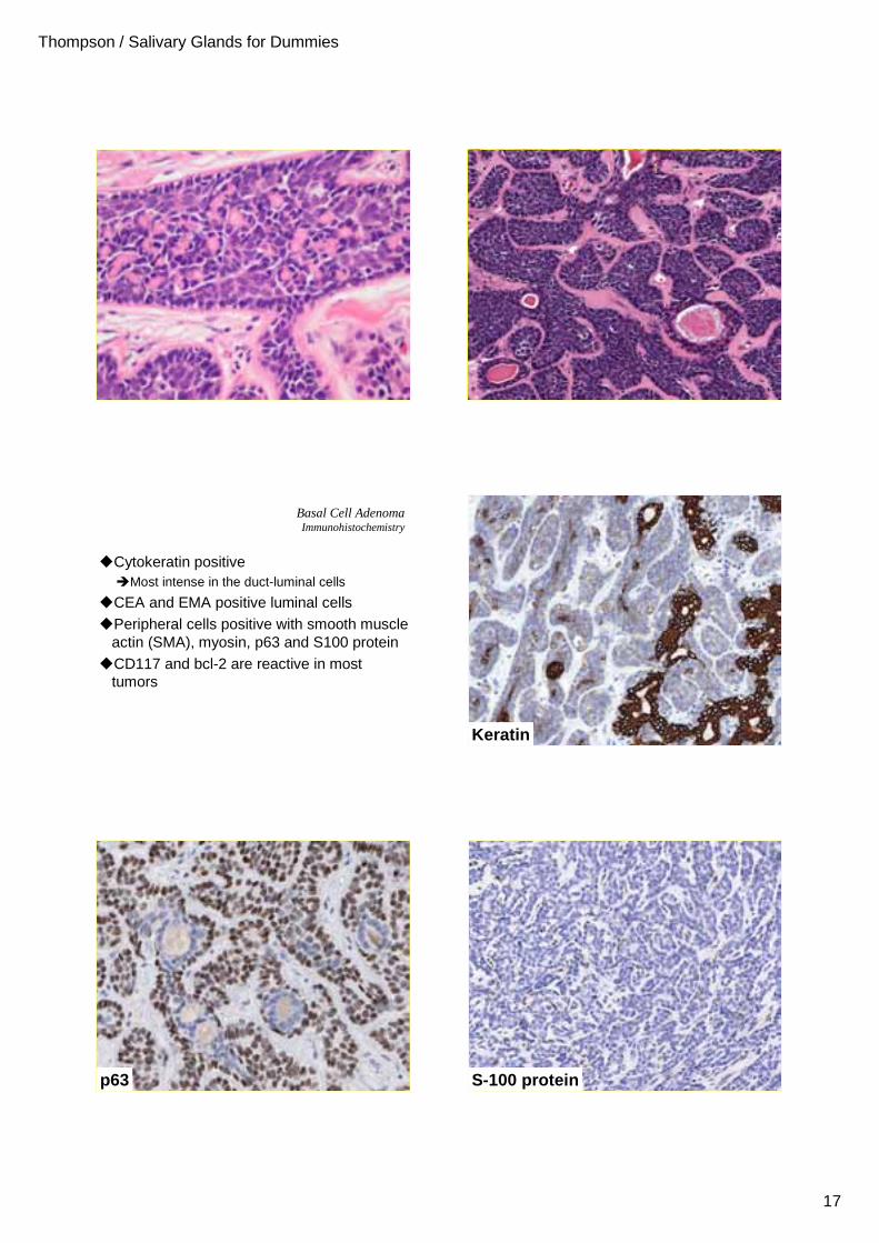
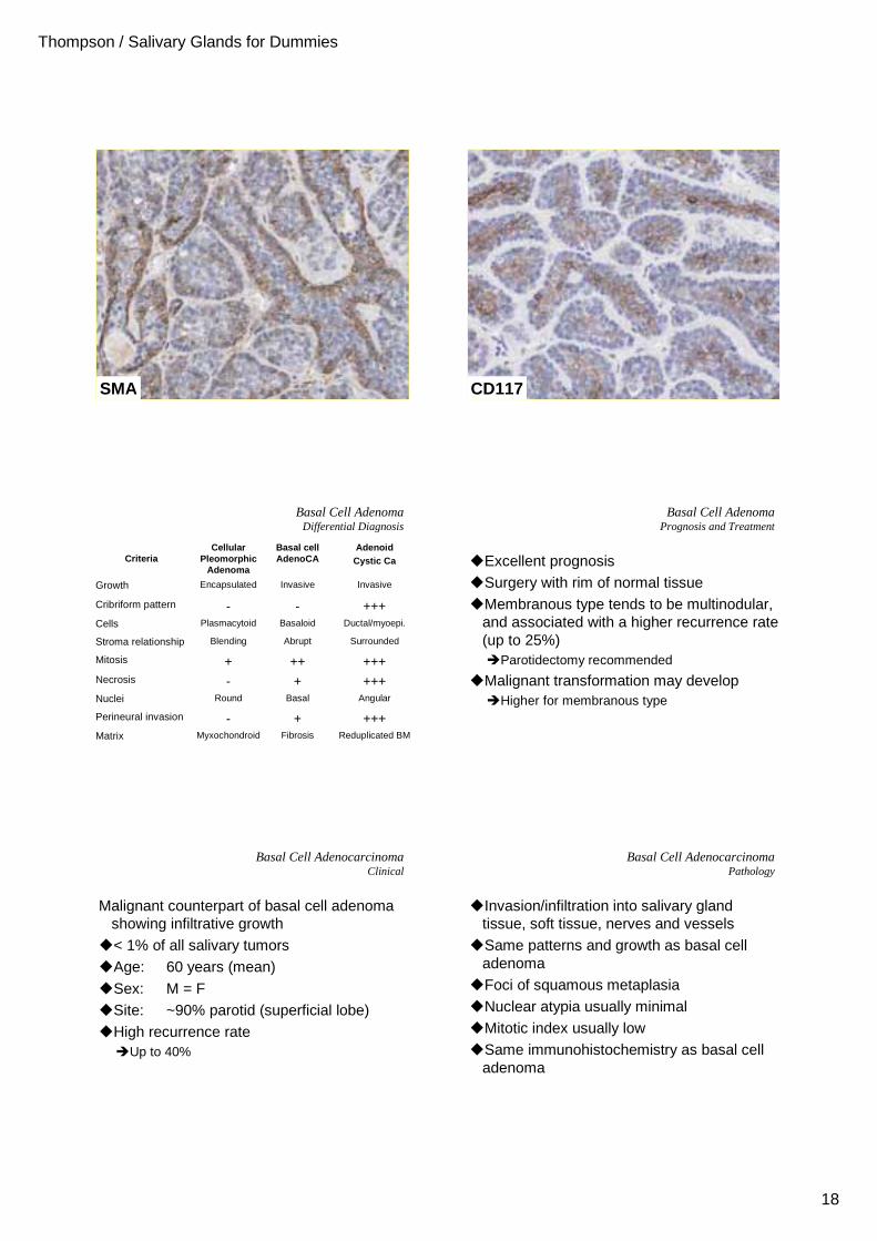
Basal Cell AdenomaImmunohistochemistry
�Cytokeratin positive�Most intense in the duct-luminal cells
�CEA and EMA positive luminal cells�Peripheral cells positive with smooth muscle
actin (SMA), myosin, p63 and S100 protein�CD117 and bcl-2 are reactive in most
tumors
Keratin
p63 S-100 protein
Thompson / Salivary Glands for Dummies
18
SMA CD117
Basal Cell AdenomaDifferential Diagnosis
Ductal/myoepi.BasaloidPlasmacytoidCells
Reduplicated BMFibrosisMyxochondroidMatrix
++++-Perineural invasion
AngularBasalRoundNuclei
++++-Necrosis
++++++Mitosis
SurroundedAbruptBlendingStroma relationship
+++--Cribriform pattern
InvasiveInvasiveEncapsulatedGrowth
Adenoid Cystic Ca
Basal cell AdenoCA
Cellular Pleomorphic
AdenomaCriteria
Basal Cell AdenomaPrognosis and Treatment
�Excellent prognosis�Surgery with rim of normal tissue�Membranous type tends to be multinodular,
and associated with a higher recurrence rate (up to 25%)�Parotidectomy recommended
�Malignant transformation may develop�Higher for membranous type
Basal Cell AdenocarcinomaClinical
Malignant counterpart of basal cell adenoma showing infiltrative growth
�< 1% of all salivary tumors�Age: 60 years (mean)
�Sex: M = F �Site: ~90% parotid (superficial lobe)�High recurrence rate
�Up to 40%
Basal Cell AdenocarcinomaPathology
�Invasion/infiltration into salivary gland tissue, soft tissue, nerves and vessels
�Same patterns and growth as basal cell adenoma
�Foci of squamous metaplasia�Nuclear atypia usually minimal�Mitotic index usually low�Same immunohistochemistry as basal cell
adenoma
Thompson / Salivary Glands for Dummies
19
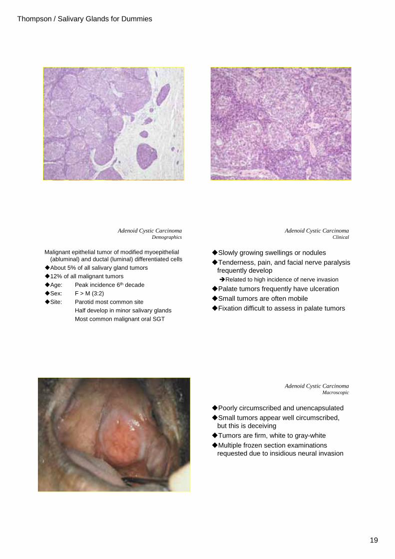
Adenoid Cystic CarcinomaDemographics
Malignant epithelial tumor of modified myoepithelial (abluminal) and ductal (luminal) differentiated cells
�About 5% of all salivary gland tumors
�12% of all malignant tumors
�Age: Peak incidence 6th decade
�Sex: F > M (3:2)
�Site: Parotid most common site
Half develop in minor salivary glands
Most common malignant oral SGT
Adenoid Cystic CarcinomaClinical
�Slowly growing swellings or nodules�Tenderness, pain, and facial nerve paralysis
frequently develop�Related to high incidence of nerve invasion
�Palate tumors frequently have ulceration �Small tumors are often mobile�Fixation difficult to assess in palate tumors
Adenoid Cystic CarcinomaMacroscopic
�Poorly circumscribed and unencapsulated�Small tumors appear well circumscribed,
but this is deceiving�Tumors are firm, white to gray-white
�Multiple frozen section examinations requested due to insidious neural invasion
Thompson / Salivary Glands for Dummies
20
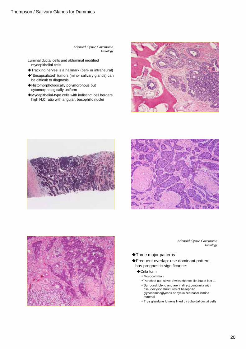
Adenoid Cystic CarcinomaHistology
Luminal ductal cells and abluminal modified myoepithelial cells
�Tracking nerves is a hallmark (peri- or intraneural)
�“Encapsulated” tumors (minor salivary glands) can be difficult to diagnosis
�Histomorphologically polymorphous but cytomorphologically uniform
�Myoepithelial-type cells with indistinct cell borders, high N:C ratio with angular, basophilic nuclei
Adenoid Cystic CarcinomaHistology
�Three major patterns�Frequent overlap: use dominant pattern,
has prognostic significance:�Cribriform
�Most common�Punched out, sieve, Swiss cheese-like but in fact …�Surround, blend and are in direct continuity with
pseudocystic structures of basophilic glycosaminoglycans or hyalinized basal lamina material
�True glandular lumens lined by cuboidal ductal cells
Thompson / Salivary Glands for Dummies
21
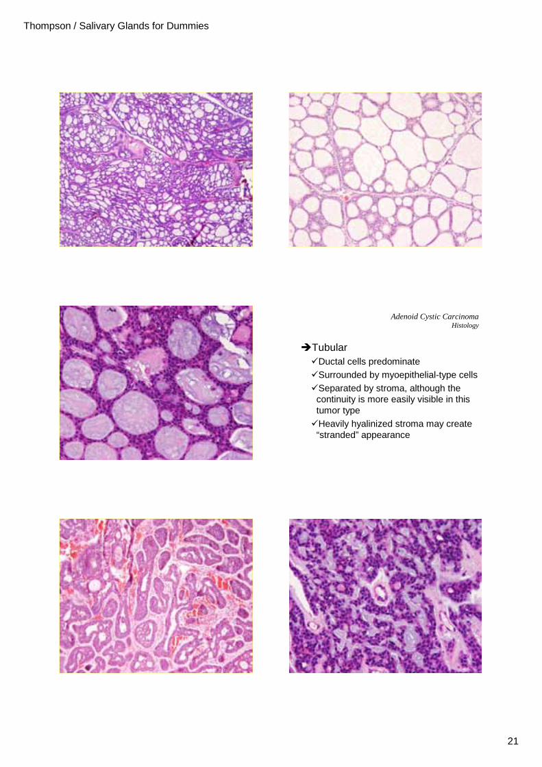
Adenoid Cystic CarcinomaHistology
�Tubular�Ductal cells predominate�Surrounded by myoepithelial-type cells�Separated by stroma, although the
continuity is more easily visible in this tumor type
�Heavily hyalinized stroma may create “stranded” appearance
Thompson / Salivary Glands for Dummies
22
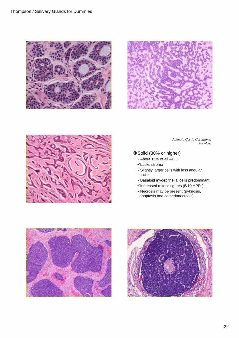
Adenoid Cystic CarcinomaHistology
�Solid (30% or higher)�About 15% of all ACC�Lacks stroma�Slightly larger cells with less angular
nuclei�Basaloid myoepithelial cells predominant�Increased mitotic figures (5/10 HPFs)�Necrosis may be present (pyknosis,
apoptosis and comedonecrosis)
Thompson / Salivary Glands for Dummies
23
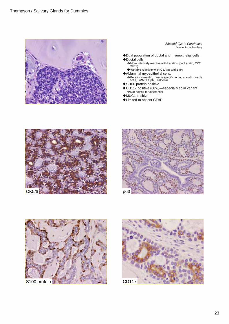
Adenoid Cystic CarcinomaImmunohistochemistry
�Dual population of ductal and myoepithelial cells�Ductal cells:
�More intensely reactive with keratins (pankeratin, CK7, CK19)
�Variable reactivity with CEA(p) and EMA�Abluminal myoepithelial cells:
�Keratin, vimentin, muscle specific actin, smooth muscle actin, SMMHC, p63, calponin
�S-100 protein positive�CD117 positive (80%)—especially solid variant
�Not helpful for differential�MUC1 positive�Limited to absent GFAP
CK5/6 p63
S100 protein CD117
Thompson / Salivary Glands for Dummies
24
Adenoid Cystic CarcinomaGrading
5%26%39%15-year survival
100%80%50%Recurrence
ManyFewRareMitoses
++++/-Pleomorphism
SolidCribriformTubularDominant pattern
+++++/-Perineural invasion*
++/-NoBone Invasion
++/-NoNecrosis
NeverDeceptiveGoodCircumscription
Grade 3 (20%)Grade 2 (35%)Grade 1 (45%)
*Nerves beyond the gland raise the grade
Adenoid Cystic CarcinomaMolecular Alterations
�About 50% have loss of chromosome 12q12
�About 30% have translocations between 9p13-23 and 6q
�LOH at 6q23-25: associated with a poorer prognosis
�Alteration p53: associated with tumor recurrence and progression to solid type
�Cribriform/tubular growth pattern�Polymorphous low grade adenocarcinoma:
�Exclusively minor salivary gland, “onion-skin”, lacks reduplicated basement membrane, cytologicallybland with vesicular chromatin
�Ductal and myoepithelial type cells�Pleomorphic adenoma
�Lacks invasion, blends with myxochondroid matrix, plasmacytoid cells
�Epithelial-myoepithelial carcinoma�Biphasic pattern
�Basaloid pattern�Basal cell adenoma, adenocarcinoma, solid variant of
adenoid cystic carcinoma
Adenoid Cystic CarcinomaDifferential Diagnosis
Adenoid Cystic CarcinomaDifferential Diagnosis – Basaloid Pattern
High-gradeLow-gradeBenignBiological behavior
++++-Vascular involvement
++++-Perineural invasion
-+++Squamous areas
++++-Necrosis
++++++Mitosis
++++++Atypia
+/-+++++Peripheral palisading
InvasiveInvasiveEncapsulatedGrowth
Solid Adenoid Cystic Ca
Basal cell AdenoCA
Basal cell AdenomaCriteria
Adenoid Cystic CarcinomaPrognosis and Management
�Indolent, but relentless, progressive growth�Worse prognosis with increasing clinical
stage�Tumor size, lymph node metastasis,
distant metastasis�10-year survival
�Stage I 75%
�Stage II 43%
�Stage III & IV 15%
Adenoid Cystic CarcinomaPrognosis and Management
�Up to 40% occult lymph node metastasis at presentation
�Late onset of metastases (lungs, bone)�Overall survival:
�5-year ~80%�10-year ~45%�15-year ~30%
Thompson / Salivary Glands for Dummies
25
Adenoid Cystic CarcinomaPrognosis and Management
�Worse outcome (recurrence or prognosis):�Solid histologic pattern (15-year: 5%)�Higher grade tumors �Perineural invasion associated with higher recurrence
rate (conflicting results) �Sinonasal primaries (worst prognosis)
�Palate have the best prognosis�Increased Ki-67 index (>5-10%)
�Radical surgery is treatment of choice�Surgical margin status affects recurrence not overall
survival
�Postoperative radiation therapy is commonly used
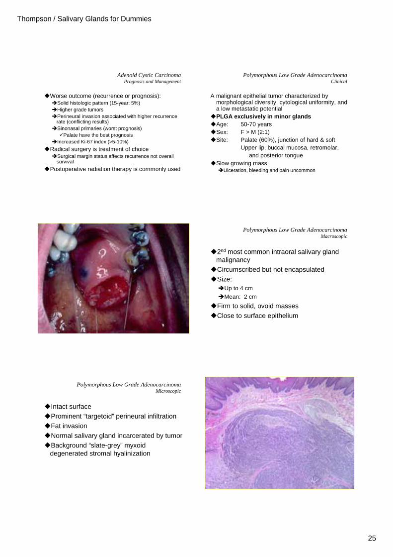
Polymorphous Low Grade AdenocarcinomaClinical
A malignant epithelial tumor characterized by morphological diversity, cytological uniformity, and a low metastatic potential
�PLGA exclusively in minor glands�Age: 50-70 years�Sex: F > M (2:1)�Site: Palate (60%), junction of hard & soft
Upper lip, buccal mucosa, retromolar,and posterior tongue
�Slow growing mass �Ulceration, bleeding and pain uncommon
Polymorphous Low Grade AdenocarcinomaMacroscopic
�2nd most common intraoral salivary gland malignancy
�Circumscribed but not encapsulated�Size:
�Up to 4 cm
�Mean: 2 cm
�Firm to solid, ovoid masses�Close to surface epithelium
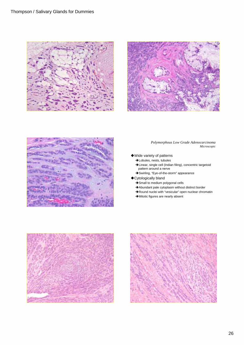
Polymorphous Low Grade AdenocarcinomaMicroscopic
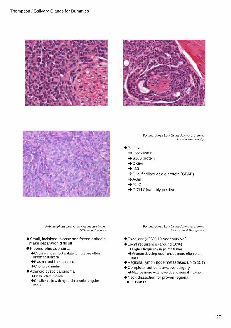
�Intact surface�Prominent “targetoid” perineural infiltration�Fat invasion�Normal salivary gland incarcerated by tumor�Background “slate-grey” myxoid
degenerated stromal hyalinization
Thompson / Salivary Glands for Dummies
26
Polymorphous Low Grade AdenocarcinomaMicroscopic
�Wide variety of patterns�Lobules, nests, tubules�Linear, single cell (Indian filing), concentric targetoid
pattern around a nerve�Swirling, “Eye-of-the-storm” appearance
�Cytologically bland�Small to medium polygonal cells�Abundant pale cytoplasm without distinct border�Round nuclei with “vesicular” open nuclear chromatin�Mitotic figures are nearly absent
Thompson / Salivary Glands for Dummies
27
Polymorphous Low Grade AdenocarcinomaImmunohistochemistry
�Positive:�Cytokeratin�S100 protein�CK5/6�p63�Glial fibrillary acidic protein (GFAP)�Actin�bcl-2�CD117 (variably positive)
Polymorphous Low Grade AdenocarcinomaDifferential Diagnosis
�Small, incisional biopsy and frozen artifacts make separation difficult
�Pleomorphic adenoma�Circumscribed (but palate tumors are often
unencapsulated)�Plasmacytoid appearance�Chondroid matrix
�Adenoid cystic carcinoma�Destructive growth�Smaller cells with hyperchromatic, angular
nuclei
Polymorphous Low Grade AdenocarcinomaPrognosis and Management
�Excellent (>95% 10-year survival)�Local recurrence (around 10%)
�Higher frequency in palate tumor�Women develop recurrences more often than
men
�Regional lymph node metastases up to 15%�Complete, but conservative surgery
�May be more extensive due to neural invasion
�Neck dissection for proven regional metastases