Saliva and oral health - Semantic Scholar...Saliva and oral health Saliva and oral health has...
50
Saliva and oral health an essential overview for the health professional fourth edition Michael Edgar, Colin Dawes & Denis O’Mullane
Transcript of Saliva and oral health - Semantic Scholar...Saliva and oral health Saliva and oral health has...
Saliva and oral health
Saliva and oral health has continued to be such a popular text on our evolvingknowledge of saliva and its functions that this 4th edition has been promptedin order to bring the reader fully up to date. The editors and authors havetaken the opportunity to fully revise the text to make it an essential guide to therole of this vital body fluid and the secrets it is still revealing.
This edition is still aimed primarily at the ‘progressive and inquisitivepractitioner’ but will doubtless find favour with undergraduate andpostgraduate dental students as well as other members of the dental team andhealth professionals.
Chapters include: The anatomy and physiology of the salivary glands,xerostomia, salivary clearance and its effects on oral health and The role ofsaliva in caries, erosion and calculus formation.
Saliva and oral health
an essential overview for the health professional
fourth edition
Michael Edgar, Colin Dawes & Denis O’Mullane
Saliva and oral health
fourth editionM
ichael Edgar, Colin D
awes &
Denis O
’Mullane
SHL_S+OH_A5_COV 13/11/2012 14:25 Page 1

SHL_S+OH_A5 13/11/2012 14:16 Page B
Saliva and oral healthFourth edition
Edited by
Michael EdgarEmeritus Professor of Dental Science,
The University of Liverpool, U K.
Colin DawesEmeritus Professor of Oral Biology, Faculty of Dentistry,
University of Manitoba, Winnipeg, MB, Canada
Denis O’MullaneEmeritus Professor/Consultant
Oral Health Services Research CentreUniversity Dental School and Hospital
WiltonCork, Ireland
2012Published by Stephen Hancocks Limited
SHL_S+OH_A5 13/11/2012 14:16 Page C
Colin DawesEmeritus Professor of Oral Biology
Faculty of Dentistry
University of Manitoba
Winnipeg, MB, Canada
Michael EdgarEmeritus Professor of Dental Science
The University of Liverpool, UK
Eva J HelmerhorstAssociate Professor
Department of Periodontology
and Oral Biology
Boston University Henry M Goldman
School of Dental Medicine, Boston,
MA 02118
Susan M HighamProfessor of Oral Biology
Department of Clinical Dental Sciences
School of Dentistry, Liverpool, UK
Mahvash NavazeshProfessor of Diagnostic Sciences
Division of Periodontology, Diagnostic
Sciences & Dental Hygiene
Associate Dean of Academic Affairs
& Student Life, Herman Ostrow School
of Dentistry of USC, Los Angeles,
California 90089-0641
Jacob M (‘Bob’) ten CateProfessor of Experimental Preventive
Dentistry and Director of Research,
Academic Centre for Dentistry
Amsterdam (ACTA), The Netherlands
Director, Netherlands Institute of
Dental Sciences
Peter M SmithSenior Lecturer in Oral Biology
Department of Clinical Dental Sciences
School of Dentistry, Liverpool, UK
Helen WheltonDirector, Oral Health Services Research
Centre, Professor in Dental Public Health
and Preventive Dentistry, University Dental
School and Hospital Wilton, Cork, Ireland
Contributors
SALIVA AND ORAL HEALTH
SHL_S+OH_A5 13/11/2012 14:16 Page D
Preface i
Saliva: functionality beyond oral health iii
1 Introduction: the anatomy and physiology of salivary glands 1
2 Mechanisms of salivary secretion 17
3 Factors influencing salivary flow rate and composition 37
4 Xerostomia and salivary gland hypofunction; aetiology, diagnosis, clinical implications and management 57
5 Salivary clearance and its effects on oral health 81
6 Saliva and the control of plaque pH 97
7 Protective functions of saliva 115
8 The role of saliva in mineral equilibria -
caries, erosion and calculus formation 135
Index 151
Contents
SHL_S+OH_A5 13/11/2012 14:16 Page E

Published by Stephen Hancocks Limited,Little Steine, Hill Farm Lane, Duns Tew, OX25 6JH© Stephen Hancocks Limited 2012.
ISBN 978-0-9565668-3-6All rights reserved. No part of this publication may be reproduced, stored in a retrievalsystem, or transmitted in any form or by any means: electronic, mechanical,photocopying, recording or otherwise, without either the permission of the publisher ora licence permitting restricted copying in the United Kingdom issued by the CopyrightLicensing Agency Limited, 90 Tottenham Court Road, London WC1 9HE.
Layout by Philip Giorgi and Kavita Graphics, [email protected] and bound by Dennis Barber Limited, Lowestoft, Suffolk
First edition 1990Second edition 1996Third edition 2004Fourth edition 2012
WOHP and all affiliated designs owned by and used courtesy of the Wm. Wrigley Jr. Company or its affiliates
SHL_S+OH_A5 13/11/2012 14:16 Page F

i
A fourth edition of Saliva and Oral Health seemed overdue, as the third edition waspublished in 2004 and considerable advances in several aspects of salivary researchhave occurred since that time.
Six of the eight chapter authors in the previous edition remain the same in the fourthedition. The untimely demise of Professor Jonathon Ship has necessitated a replacementand we are delighted that Professor Mahvash Navazesh has agreed to contribute thechapter on xerostomia. Professor Jorma Tenovuo indicated that, because of hisimpending retirement, he wished to be replaced and we are fortunate that Dr. EvaHelmerhorst agreed to write a chapter on salivary proteins, an area in which she hasmade several important contributions.
Although the eight chapters of the book concentrate on the effects of saliva on oralhealth, Drs. Michael Dodds and Taichi Inui, from our Sponsor, The Wrigley Company,have contributed a commentary on other functions of saliva of particular interest tothe food industry. These include the role of saliva in ‘mouth feel’ and the sensoryperception of foods and beverages and also the potential use of assays of salivarycomponents in the diagnosis of a number of systemic as well as oral diseases.
As in previous editions, the book is aimed not only at undergraduate andpostgraduate students but also at dental practitioners and other health professionalswho deal with oral diseases.
We are very grateful for the cooperation of all involved in the production of the newedition, including the new publisher. As ever, we are indebted to the Wm. Wrigley Jr.Company, in the person of Dr. M.W.J. Dodds, for underwriting the costs of producingthis fourth edition.
Michael EdgarColin DawesDenis O'Mullane
Preface
SHL_S+OH_A5 13/11/2012 14:16 Page i

ii
SALIVA AND ORAL HEALTH
SHL_S+OH_A5 13/11/2012 14:16 Page ii
iii
We are delighted to have had the opportunity to support the publication of Saliva andOral Health through all four editions, since the first one appeared in 1990. Over thesubsequent editions we have observed the addition of new research findings as theypertain to fundamental understanding of physiological secretory mechanisms, alongwith greater understanding of the roles of the organic components of saliva inprotecting the oral cavity against insult, to cite just two examples. It is particularlyencouraging to note that this book serves as a resource not just for researchers in thefield of saliva physiology and biochemistry, but also dental students, dental practitionersand other professionals with an interest in the health of the oral cavity, and we hopethis trend continues. In this context, the importance of saliva as a fluid that is crucialfor the maintenance of both oral and systemic health is a central dogma, and this aspectis comprehensively covered by the various chapter authors, who are all well respectedand highly published experts in their fields. However, beyond the function of saliva incontributing to the maintenance of both oral and systemic health, at the local level italso plays fundamental roles in delivering mouth comfort and oral sensations, as wellas in food processing in the oral cavity, that also contribute to the overall health andwellbeing of the body. As scientists with a keen interest in the functionality of saliva,but approaching this also from a food industry perspective, we would like to providesome additional thoughts on the importance of saliva beyond its local effects on themouth, teeth, and oral tissues, and briefly consider some areas of saliva function thatmay not be covered by the main chapter topics.
Saliva is an easily accessible medium which can be collected without the need forneedles, aseptic or sterile conditions, or other specialised apparatus, and commercialkits are currently available for measuring hormone levels, such as cortisol ortestosterone, illegal drugs and other substances in saliva. Recently, there has beenincreasing interest in, and support for, the use of saliva as a diagnostic fluid for screeningnot only oral conditions, such as caries, periodontal diseases or oral cancer, but also
Saliva: functionality beyond oral health.Comments from the SponsorMichael Dodds and Taichi Inui
SHL_S+OH_A5 13/11/2012 14:16 Page iii
iv
SALIVA AND ORAL HEALTH
for other systemic conditions, such as breast and pancreatic cancers.1 It is also possiblethat in the future the specific organic composition of saliva could function as anindicator of disease risk elsewhere in the body; for example, higher levels of the salivarymucins, MUC5B and MUC7 have been found in individuals who are suffering fromgastric diseases and are infected with Helicobacter pylori, the organism associated withgastric ulcers.2 In addition, there has been recent interest directed at salivary exosomesas a means to gain better understanding of cell-cell communications mediated by bodyfluids.3 Many studies in the psychology and exercise literature have used salivary cortisolor α-amylase as indicators of psychosocial or physical stress, as sympathetic nerveactivity stimulates salivary acinar cell release of macromolecules, including α-amylase.4,5
However, this approach has been criticised due to methodological concerns, such aslack of control of salivary flow rate, as well as physiological complications, such as thefact that amylase secretion is partially controlled by parasympathetic stimulation(described in Chapter 2), as well as the dissociation between serum and salivary cortisollevels.6,7 Thus, an understanding of saliva composition, secretory physiology and theimpact of functional stimulation is an important aspect of undertaking any type ofwork that involves objective measurement of saliva or any of its components.
Although not specifically a health-related issue, the treatment and prevention ofextrinsic dental staining is almost an industry in itself, both for the profession, and alsofrom the consumer goods stand point (i.e., toothpastes and over-the-counter whiteningtreatments). The mechanism of tooth staining is in part mediated by saliva composition.Specifically, certain salivary proteins, including the proline-rich proteins (PRPs) havebeen shown to bind salivary polyphenols but not lower-molecular-weight salivaryproteins, such as the histatins.8
The perception of astringency that is typically associated with consumption ofpolyphenol- and tannin-rich foods has a specific mechanism, involving binding andprecipitation of PRPs, as well as another salivary protein, statherin.9,10 Although astringencymay be perceived to be a negative sensation in response to consuming foods or beverages,a balanced level of astringency may also be seen to be an inherent aspect of the enjoymentof others, such as red wine or tea. It has been proposed that salivary PRPs, by bindingdietary tannins, constitute a protective mechanism against their health-detrimental effects.The parotid salivary glands of rats and mice increased in size and PRP content in responseto being fed a high tannin diet, while these rodents maintained normal weight gains.Conversely, hamsters fed a similar diet did not show this pattern of protein up-regulationand failed to demonstrate normal growth, suggesting that, at least in rodents, the PRPsconstitute an inducible protective mechanism against excessive consumption of dietarycomponents with a potential negative health impact. It has been argued that, from an
SHL_S+OH_A5 13/11/2012 14:16 Page iv
v
COMMENTS FROM THE SPONSOR
evolutionary standpoint, the constitutive presence in human saliva of high levels of PRPsand other tannin-binding proteins is a remnant of an earlier stage of human developmentwhen hominid populations relied on a diet rich in fruits, vegetables and nuts.9
The effect of saliva composition on mouth feel also seems to have been an historicallyunder-researched topic. This could relate both to health, through the maintenance of athin film of saliva to keep the oral tissues hydrated and lubricated, as well as to otheraspects of oral function. For example, saliva plays an important role in facilitating theswallowing of food, by influencing food particle size and lubrication of the food bolus.Salivary mucins and other glycoproteins help to form food into a coherent and slipperybolus which can be transported smoothly down the oesophagus. The cohesiveness of thefood bolus, rather than its water content, is considered to play a more important role indetermining whether the bolus can be swallowed, indicating the relative importance ofthe organic content.11 While it is generally accepted that the various glycosylated proteinsand mucins in saliva form a well hydrated film on the mouth tissues to provide orallubrication, facilitate food bolus formation and swallowing, and help to preventdesiccation of the mucosal tissues, much less is known about how minor alterations orpolymorphisms in the macromolecular composition of saliva could alter oral perception.The rheology, or viscoelasticity, of saliva has been shown to vary under differentconditions of stimulation, and it has been suggested that the inability of artificial salivasto be well tolerated by patients suffering from xerostomia is due to their failure to mimiceffectively the unique and complex rheological properties of human saliva.12 There is alsoincreased interest from scientists working in the food industry in understanding howsaliva can influence mouth feel, food processing and sensory perception of foods andbeverages. For example, salivary protein concentration was found to be correlated withmouth feel with semi-solid foods.13 Beyond this non-specific effect, the high molecularweight mucin, MUC5B, and α-amylase specifically adsorb to lysozyme-stabilisedemulsions that have the sensory perception of dryness (astringency), in contrast to thecreamy, β-lactoglobulin-stabilised emulsions, that attract the lower molecular weightmucin, MUC7, but not α-amylase.14 This effect is probably mediated by charge differences,but provides an interesting insight into how salivary composition may help modify howpleasant (creamy or rich) versus unpleasant (dry, or astringent) food textures areperceived. While an increasing number of studies have been conducted around salivaand oral perception, other attributes of mouth feel and food texture still remain to beexplained by physical and chemical properties of saliva, such as melting and stickiness.
Saliva is the medium for dissolving food taste molecules and carrying them to thetaste buds. However, rather than acting as a simple aqueous solvent, components of salivamay modify how tastants are perceived. Saliva stimulated by different tastants (pungent,
SHL_S+OH_A5 13/11/2012 14:16 Page v

vi
SALIVA AND ORAL HEALTH
tingling, and sour) has different protein profiles as assessed by electrophoresis and massspectrometry,15 and different tastants have been shown to affect the protein compositionof the saliva, which could affect subsequent perception of bitter or astringent stimuli.16,17
The salivary enzyme carbonic anhydrase VI, formerly known as gustin, is a zinc-bindingenzyme that has been shown to be associated with taste sensitivity to bitter stimuli, aswell as demonstrating an intriguing association with body mass index.18
In addition to the well known action of salivary α-amylase in initiating the processof starch digestion, saliva may also help stabilise, protect, and transport critical nutrientsso that they can be absorbed from the gastro-intestinal system. An example of this ishaptocorrin. This glycosylated salivary protein binds to vitamin B12 in the mouth,protecting it from digestion in the stomach, but is then cleaved off in the duodenumand replaced by intrinsic factor, finally facilitating absorption of the vitaminB12/intrinsic factor complex in the proximal ileum.19
Summary
As described throughout this book, saliva plays a pivotal role in the overall maintenanceof a healthy homeostatic condition in the oral cavity, which from the dental perspectiveis usually considered to be related to protection of the teeth and mucosal surfaces. Theoral cavity is also the starting point of the digestive system, so saliva has advantages inbeing easily accessible for sampling as well as providing an impact on systemic health.With the increasing volume of biological data becoming available due to newer analyticalmethodologies offered by proteomics and other so-called ‘omics’ approaches, thecontribution of saliva to overall human health and wellbeing is being explored from anholistic, or systems biology approach, emphasising interactions among constituents, thatis opening up novel aspects of the importance of this body fluid to the human condition.
References1 Sugimoto M, Wong DT, Hirayama A, Soga T, Tomita M. Capillary electrophoresis mass
spectrometry-based saliva metabolomics identified oral, breast and pancreatic cancer-specific profiles. Metabolomics 2010; 6:78-95.
2 Silva DG, Stevens RH, Macedo JM, Hirata R, Pinto AC, Alves LM, Veerman EC, Tinoco EM.Higher levels of salivary MUC5B and MUC7 in individuals with gastric diseases who harborHelicobacter pylori. Arch Oral Biol 2009; 54:86-90.
3 Palanisamy V, Sharma S, Deshpande A, Zhou H, Gimzewski J, Wong DT. Nanostructural andtranscriptomic analyses of human saliva derived exosomes. PLoS One 2010; 5:e8577.
SHL_S+OH_A5 13/11/2012 14:16 Page vi
vii
COMMENTS FROM THE SPONSOR
4 Rohleder N, Nater UM. Determinants of salivary alpha-amylase in humans andmethodological considerations. Psychoneuroendocrinology 2009; 34:469-485.
5 de Oliveira VN, Bessa A, Lamounier RP, de Santana MG, de Mello MT, Espindola FS.Changes in the salivary biomarkers induced by an effort test. Int J Sports Med 2010; 31:377-381.
6 Bosch JA, Veerman EC, de Geus EJ, Proctor GB. α-amylase as a reliable and convenientmeasure of sympathetic activity: don't start salivating just yet! Psychoneuroendocrinology2011; 36:449-453.
7 Hellhammer DH, Wüst S, Kudielka BM. Salivary cortisol as a biomarker in stress research.Psychoneuroendocrinology 2009; 34:163-171.
8 Proctor GB, Pramanik R, Carpenter GH, Rees GD. Salivary proteins interact with dietaryconstituents to modulate tooth staining. J Dent Res 2005; 84:73-78.
9 Bennick A. Interaction of plant polyphenols with salivary proteins. Crit Rev Oral Biol Med2002; 13:184-196.
10 Soares S, Vitorino R, Osorio H, Fernandes A, Venancio A, Mateus N, Amado F, de Freitas V.Reactivity of human salivary proteins families toward food polyphenols. J Agric Food Chem2011; 59:5535-5547.
11 de Loubens C, Magnin A, Doyennette M, Tréléa IC, Souchon I. A biomechanical model ofswallowing for understanding the influence of saliva and food bolus viscosity on flavor release.J Theoret Biol 2011; 280:180-188.
12 Stokes JR, Davies GA. Viscoelasticity of human whole saliva collected after acid andmechanical stimulation. Biorheology 2007; 44:141-160.
13 Engelen L, van den Keybus PA, de Wijk RA, Veerman EC, Amerongen A , Bosman F, Prinz JF,van der Bilt A. The effect of saliva composition on texture perception of semi-solids. Arch OralBiol 2007; 52:518-525.
14 Silletti E, Vitorino RM, Schipper R, Amado FM, Vingerhoeds MH. Identification of salivaryproteins at oil-water interfaces stabilized by lysozyme and beta-lactoglobulin. Arch Oral Biol2010; 55:268-278.
15 Lorenz K, Bader M, Klaus A, Weiss W, Görg A, Hofmann T. Orosensory stimulation effects onhuman saliva proteome. J Agric Food Chem 2011; 59:10219-10231.
16 Dinnella C, Recchia A, Vincenzi S, Tuorila H, Monteleone E. Temporary modification of salivaryprotein profile and individual responses to repeated phenolic astringent stimuli. Chem Senses2010; 35:75-85.
17 Quintana M, Palicki O, Lucchi G, Ducoroy P, Chambon C, Salles C, Morzel M. Short-termmodification of human salivary proteome induced by two bitter tastants, urea and quinine.Chem Percept 2009; 2:133-142.
18 Padiglia A, Zonza A, Atzori E, Chillotti C, Calo C, Tepper B J, Barbarossa IT. Sensitivity to 6-n-propylthiouracil is associated with gustin (carbonic anhydrase VI) gene polymorphism, salivaryzinc, and body mass index in humans. Am J Clin Nutr 2010; 92:539-545.
19 Wuerges J, Garau G, Geremia S, Fedosov SN, Petersen TE, Randaccio L. Structural basis formammalian vitamin B12 transport by transcobalamin. Proc Natl Acad Sci 2006; 103:4386-4391.
SHL_S+OH_A5 13/11/2012 14:16 Page vii

viii
SALIVA AND ORAL HEALTH
SHL_S+OH_A5 13/11/2012 14:16 Page viii

1
Saliva is the mixed glandular secretion which constantly bathes the teeth and the oralmucosa. It is constituted by the secretions of the three paired major salivary glands; theparotid, submandibular and sublingual. It also contains the secretions of the minorsalivary glands, of which there are hundreds contained within the submucosa of theoral mucosa and some gingival crevicular fluid.
The presence of saliva is vital to the maintenance of healthy hard (teeth) and soft(mucosa) oral tissues. Severe reduction of salivary output not only results in a rapiddeterioration in oral health but also has a detrimental impact on the quality of life forthe sufferer. Patients suffering from dry mouth can experience difficulty with eating,swallowing, speech, the wearing of dentures, trauma to and ulceration of the oralmucosa, taste alteration, poor oral hygiene, a burning sensation of the mucosa, oralinfections including Candida and rapidly progressing dental caries. The sensation of drymouth or xerostomia is becoming increasingly common in developed countries whereadults are living longer. Polypharmacy is very common among the older adult populationand many commonly prescribed drugs cause a reduction in salivary flow. Xerostomiaalso occurs in Sjögren’s syndrome, which is not an uncommon condition. In addition tospecific diseases of the salivary glands, salivary flow is usually severely impaired followingradiotherapy in the head and neck area for cancer treatment in both children and adultsof all ages. Clearly oral dryness is a problem which faces an increasingly large proportionof the population. An understanding of saliva and its role in oral health will help topromote awareness among health care workers of the problems arising when the quantityor quality of saliva is decreased; this awareness and understanding is important to theprevention, early diagnosis and treatment of the condition.
There is an extensive body of research on saliva as a diagnostic fluid. It has been usedto indicate an individual’s caries susceptibility; it has also been used to reflect systemicphysiological and pathological changes which are mirrored in saliva. One of the major
Introduction: the anatomy and physiology of salivary glands
Helen Whelton
1
SHL_S+OH_A5 13/11/2012 14:16 Page 1
2
SALIVA AND ORAL HEALTH
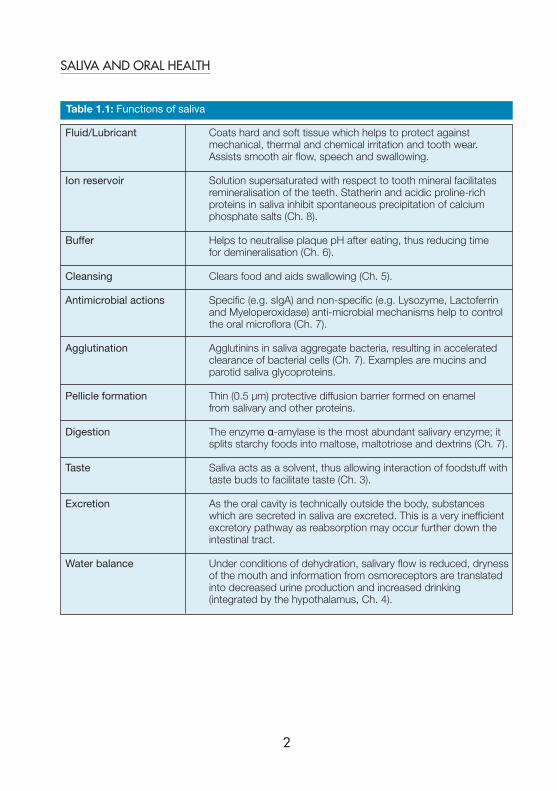
Table 1.1: Functions of saliva
Fluid/Lubricant Coats hard and soft tissue which helps to protect against mechanical, thermal and chemical irritation and tooth wear. Assists smooth air flow, speech and swallowing.
Ion reservoir Solution supersaturated with respect to tooth mineral facilitates remineralisation of the teeth. Statherin and acidic proline-rich proteins in saliva inhibit spontaneous precipitation of calcium phosphate salts (Ch. 8).
Buffer Helps to neutralise plaque pH after eating, thus reducing time for demineralisation (Ch. 6).
Cleansing Clears food and aids swallowing (Ch. 5).
Antimicrobial actions Specific (e.g. sIgA) and non-specific (e.g. Lysozyme, Lactoferrin and Myeloperoxidase) anti-microbial mechanisms help to control the oral microflora (Ch. 7).
Agglutination Agglutinins in saliva aggregate bacteria, resulting in accelerated clearance of bacterial cells (Ch. 7). Examples are mucins and parotid saliva glycoproteins.
Pellicle formation Thin (0.5 μm) protective diffusion barrier formed on enamel from salivary and other proteins.
Digestion The enzyme α-amylase is the most abundant salivary enzyme; it splits starchy foods into maltose, maltotriose and dextrins (Ch. 7).
Taste Saliva acts as a solvent, thus allowing interaction of foodstuff with taste buds to facilitate taste (Ch. 3).
Excretion As the oral cavity is technically outside the body, substances which are secreted in saliva are excreted. This is a very inefficient excretory pathway as reabsorption may occur further down the intestinal tract.
Water balance Under conditions of dehydration, salivary flow is reduced, dryness of the mouth and information from osmoreceptors are translated into decreased urine production and increased drinking (integrated by the hypothalamus, Ch. 4).
SHL_S+OH_A5 13/11/2012 14:16 Page 2
3
INTRODUCTION: THE ANATOMY AND PHYSIOLOGY OF SALIVARY GLANDS
benefits of saliva is that it is easily available for non-invasive collection and analysis. Itcan be used to monitor the presence and levels of hormones, drugs, antibodies, micro-organisms and ions.
This chapter will provide an overview of the functions of saliva, the anatomy andhistology of salivary glands, the physiology of saliva formation, the constituents of salivaand the use of saliva as a diagnostic fluid, including its role in caries risk assessment.Much of the material in this chapter will be covered in more detail in later chapters.
Functions of saliva
The complexity of this oral fluid is perhaps best appreciated by the consideration of itsmany and varied functions. The functions of saliva are largely protective; however, italso has other functions. Table 1.1 provides an overview of many of these functions.More detail is provided in subsequent chapters as indicated.
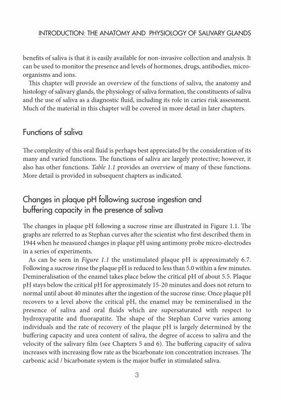
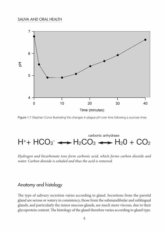
Changes in plaque pH following sucrose ingestion and buffering capacity in the presence of saliva
The changes in plaque pH following a sucrose rinse are illustrated in Figure 1.1. Thegraphs are referred to as Stephan curves after the scientist who first described them in1944 when he measured changes in plaque pH using antimony probe micro-electrodesin a series of experiments.
As can be seen in Figure 1.1 the unstimulated plaque pH is approximately 6.7.Following a sucrose rinse the plaque pH is reduced to less than 5.0 within a few minutes.Demineralisation of the enamel takes place below the critical pH of about 5.5. PlaquepH stays below the critical pH for approximately 15-20 minutes and does not return tonormal until about 40 minutes after the ingestion of the sucrose rinse. Once plaque pHrecovers to a level above the critical pH, the enamel may be remineralised in thepresence of saliva and oral fluids which are supersaturated with respect tohydroxyapatite and fluorapatite. The shape of the Stephan Curve varies amongindividuals and the rate of recovery of the plaque pH is largely determined by thebuffering capacity and urea content of saliva, the degree of access to saliva and thevelocity of the salivary film (see Chapters 5 and 6). The buffering capacity of salivaincreases with increasing flow rate as the bicarbonate ion concentration increases. Thecarbonic acid / bicarbonate system is the major buffer in stimulated saliva.
SHL_S+OH_A5 13/11/2012 14:16 Page 3
4
SALIVA AND ORAL HEALTH
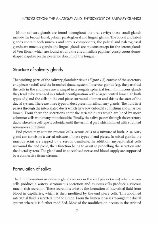
Anatomy and histology
The type of salivary secretion varies according to gland. Secretions from the parotidgland are serous or watery in consistency, those from the submandibular and sublingualglands, and particularly the minor mucous glands, are much more viscous, due to theirglycoprotein content. The histology of the gland therefore varies according to gland type.
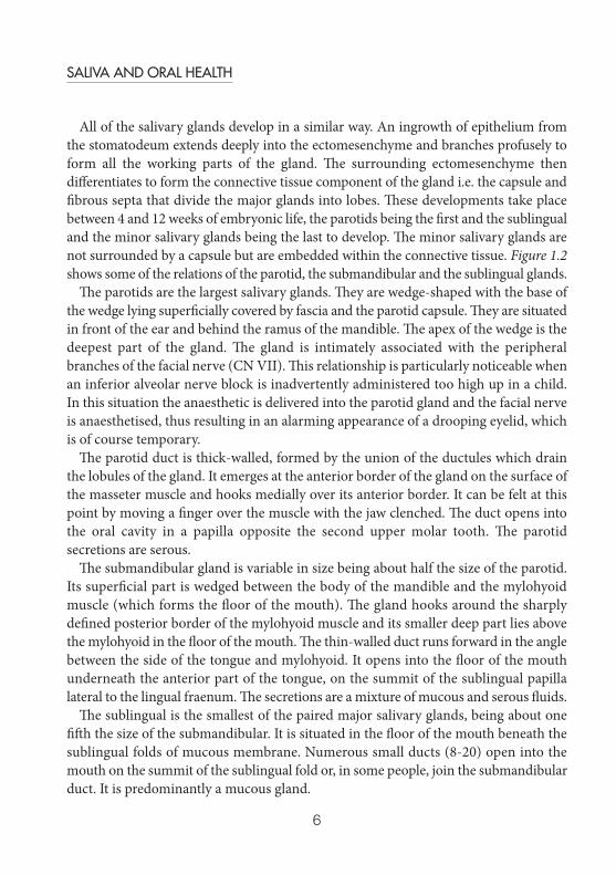
Hydrogen and bicarbonate ions form carbonic acid, which forms carbon dioxide andwater. Carbon dioxide is exhaled and thus the acid is removed.
Figure 1.1 Stephan Curve illustrating the changes in plague pH over time following a sucrose rinse
carbonic anhydrase
H++ HCO3- H2CO3 H20 + CO2
SHL_S+OH_A5 13/11/2012 14:16 Page 4

5
INTRODUCTION: THE ANATOMY AND PHYSIOLOGY OF SALIVARY GLANDS
Figure 1.2a Anatomy of Parotid Gland
Figure 1.2b Anatomy of Sublingual and Submandibular Glands
SHL_S+OH_A5 13/11/2012 14:16 Page 5
6
SALIVA AND ORAL HEALTH
All of the salivary glands develop in a similar way. An ingrowth of epithelium fromthe stomatodeum extends deeply into the ectomesenchyme and branches profusely toform all the working parts of the gland. The surrounding ectomesenchyme thendifferentiates to form the connective tissue component of the gland i.e. the capsule andfibrous septa that divide the major glands into lobes. These developments take placebetween 4 and 12 weeks of embryonic life, the parotids being the first and the sublingualand the minor salivary glands being the last to develop. The minor salivary glands arenot surrounded by a capsule but are embedded within the connective tissue. Figure 1.2shows some of the relations of the parotid, the submandibular and the sublingual glands.
The parotids are the largest salivary glands. They are wedge-shaped with the base ofthe wedge lying superficially covered by fascia and the parotid capsule. They are situatedin front of the ear and behind the ramus of the mandible. The apex of the wedge is thedeepest part of the gland. The gland is intimately associated with the peripheralbranches of the facial nerve (CN VII). This relationship is particularly noticeable whenan inferior alveolar nerve block is inadvertently administered too high up in a child.In this situation the anaesthetic is delivered into the parotid gland and the facial nerveis anaesthetised, thus resulting in an alarming appearance of a drooping eyelid, whichis of course temporary.
The parotid duct is thick-walled, formed by the union of the ductules which drainthe lobules of the gland. It emerges at the anterior border of the gland on the surface ofthe masseter muscle and hooks medially over its anterior border. It can be felt at thispoint by moving a finger over the muscle with the jaw clenched. The duct opens intothe oral cavity in a papilla opposite the second upper molar tooth. The parotidsecretions are serous.
The submandibular gland is variable in size being about half the size of the parotid.Its superficial part is wedged between the body of the mandible and the mylohyoidmuscle (which forms the floor of the mouth). The gland hooks around the sharplydefined posterior border of the mylohyoid muscle and its smaller deep part lies abovethe mylohyoid in the floor of the mouth. The thin-walled duct runs forward in the anglebetween the side of the tongue and mylohyoid. It opens into the floor of the mouthunderneath the anterior part of the tongue, on the summit of the sublingual papillalateral to the lingual fraenum. The secretions are a mixture of mucous and serous fluids.
The sublingual is the smallest of the paired major salivary glands, being about onefifth the size of the submandibular. It is situated in the floor of the mouth beneath thesublingual folds of mucous membrane. Numerous small ducts (8-20) open into themouth on the summit of the sublingual fold or, in some people, join the submandibularduct. It is predominantly a mucous gland.
SHL_S+OH_A5 13/11/2012 14:16 Page 6
7
INTRODUCTION: THE ANATOMY AND PHYSIOLOGY OF SALIVARY GLANDS
Minor salivary glands are found throughout the oral cavity; these small glandsinclude the buccal, labial, palatal, palatoglossal and lingual glands. The buccal and labialglands contain both mucous and serous components, the palatal and palatoglossalglands are mucous glands, the lingual glands are mucous except for the serous glandsof Von Ebner, which are found around the circumvallate papillae (conspicuous dome-shaped papillae on the posterior dorsum of the tongue).
Structure of salivary glands
The working parts of the salivary glandular tissue (Figure 1.3) consist of the secretoryend pieces (acini) and the branched ductal system. In serous glands (e.g. the parotids)the cells in the end piece are arranged in a roughly spherical form. In mucous glandsthey tend to be arranged in a tubular configuration with a larger central lumen. In bothtypes of gland the cells in the end piece surround a lumen and this is the start of theductal system. There are three types of duct present in all salivary glands. The fluid firstpasses through the intercalated ducts which have low cuboidal epithelium and a narrowlumen. From there the secretions enter the striated ducts which are lined by morecolumnar cells with many mitochondria. Finally, the saliva passes through the excretoryducts where the cell type is cuboidal until the terminal part which is lined with stratifiedsquamous epithelium.
End pieces may contain mucous cells, serous cells or a mixture of both. A salivarygland can consist of a varied mixture of these types of end pieces. In mixed glands, themucous acini are capped by a serous demilune. In addition, myoepithelial cellssurround the end piece, their function being to assist in propelling the secretion intothe ductal system. The gland and its specialised nerve and blood supply are supportedby a connective tissue stroma.
Formulation of saliva
The fluid formation in salivary glands occurs in the end pieces (acini) where serouscells produce a watery seromucous secretion and mucous cells produce a viscousmucin-rich secretion. These secretions arise by the formation of interstitial fluid fromblood in capillaries, which is then modified by the end piece cells. This modifiedinterstitial fluid is secreted into the lumen. From the lumen it passes through the ductalsystem where it is further modified. Most of the modification occurs in the striated
SHL_S+OH_A5 13/11/2012 14:16 Page 7

8
SALIVA AND ORAL HEALTH
ducts where ion exchange takes place and the secretion is changed from an isotonicsolution to a hypotonic one. The composition of saliva is further modified in theexcretory ducts before it is finally secreted into the mouth (see Chapter 2 for a detailedaccount of saliva secretory mechanisms).
Figure 1.3 Salivary Glandular Tissue
SHL_S+OH_A5 13/11/2012 14:16 Page 8

9
INTRODUCTION: THE ANATOMY AND PHYSIOLOGY OF SALIVARY GLANDS
Nerve supply
Secretion of saliva is a nerve-mediated reflex. The volume and type of saliva secreted iscontrolled by the autonomic nervous system.
The glands receive both parasympatheic and sympathetic nerve supplies. The reflexinvolves afferent receptors and nerves carrying impulses induced by stimulation, acentral hub (the salivary nuclei), and an efferent part consisting of parasympatheticand sympathetic autonomic nerve bundles that separately innervate the glands.
Taste and mastication are the principal stimuli (unconditioned reflex) but others suchas sight, thought and smell of food (conditioned reflex) also play a role. Taste andmechanical stimuli from the tongue and other areas of the mouth exciteparasympathetic nerve impulses in the afferent limbs of the salivary reflex which travelvia the glossopharyngeal (CN IX), facial (CN VII), vagal (CN X) (taste) and thetrigeminal (CN V) (chewing) cranial nerves.. These afferent impulses are carried to thesalivary nuclei located approximately at the juncture of the pons and the medulla. Inturn impulses from the salivary centres can be modulated i.e. stimulated or inhibitedby impulses from the higher centres in the central nervous system; for example, thetaste and smell centres in the cortex and the lateral hypothalamus where the regulationof feeding, drinking and body temperature occurs. Also, in stressful situations drymouth sometimes occurs, as a result of the inhibitory effect of higher centres on thesalivary nuclei. The secretory response of the gland is then controlled via theglossopharyngeal nerve synapsing in the otic ganglion, the postganglionicparasympathetic fibres carrying on to the parotid gland and via the facial nervesynapsing in the submandibular ganglion and carrying on to the sublingual andsubmandibular glands. Parasympathetic stimulation also increases the blood flow tothe salivary glands, increasing the supply of nutrition.
Other reflexes originating in the stomach and upper intestines also stimulatesalivation. For example, nausea or swallowing very irritating foods initiates reflexsalivation which serves to dilute or neutralise the irritating substances.
Sympathetic stimulation can also increase salivary flow to a moderate extent butmuch less so than parasympathetic stimulation. Sympathetic impulses are more likelyto influence salivary composition by increasing exocytosis from certain cells andinducing changes in the reabsorption of electrolytes. The relevant efferent sympatheticnerves originate in the spinal cord, synapse in the superior cervical ganglia and thentravel along blood vessels to the salivary glands.
Hormones such as androgens, oestrogens, glucocorticoids and peptides also influencesalivary composition.
SHL_S+OH_A5 13/11/2012 14:16 Page 9

10
SALIVA AND ORAL HEALTH
Blood supply
The blood supply to the glands also influences secretion. An extensive blood supply isrequired for the rapid secretion of saliva. There is a concentration of capillaries aroundthe striated ducts where ionic exchange takes place whilst a lesser density supplies theterminal secretory acini. The process of salivation indirectly dilates the blood vessels,thus providing increased nutrition as needed. Salivary secretion is usually accompaniedby a large increase in blood flow to the glands.
The main arterial supply to the parotid gland is by the superficial temporal andexternal carotid arteries. Venous drainage is provided by numerous veins which draininto the retromandibular and external jugular veins. Lymph drainage goes mainly viathe superficial and deep parotid nodes to the deep cervical nodes. The submandibulargland takes its arterial blood supply from branches of the facial artery and a fewbranches of the lingual artery. Venous drainage is via the common facial and lingualveins and lymph drainage goes via the submandibular lymph nodes and the deepcervical and jugular chains. The sublingual gland is served by the sublingual branch ofthe lingual artery as well as the submental branch of the facial artery and drainage isby the submental branch of the facial vein. Lymph drainage goes to the submandibularlymph nodes.
Physiology
CompositionThe composition of saliva varies according to many factors including the gland typefrom which it is secreted. The average compositions of both unstimulated and chewing-stimulated whole saliva are shown in Table 1.2.
Flow rateSalivary flow rate exhibits circadian variation and peaks in the late afternoon; theacrophase. Normal salivary flow rates are in the region of 0.3-0.4 ml/min whenunstimulated and 1.5-2.0 ml/min when stimulated, although both rates have widenormal ranges (see Chapter 3). Approximately 0.5 – 0.6 litres of saliva is secreted perday. The contribution of the different glands to whole saliva varies according to thelevel of stimulation. For unstimulated saliva, about 25% comes from the parotid glands,60% from the submandibular glands, 7-8% from the sublingual gland and 7-8% fromthe minor mucous glands. During sleep, flow rate is negligible. For highly stimulated
SHL_S+OH_A5 13/11/2012 14:16 Page 10

11
INTRODUCTION: THE ANATOMY AND PHYSIOLOGY OF SALIVARY GLANDS
Table 1.2: The composition of unstimulated and chewing-stimulated whole saliva (Courtesy of C Dawes). Cells are blank where quantitative data are not available
Unstimulated Stimulated
Water 99.55 % 99.53%1
Solids 0.45% 0.47%1
Mean ± S.D. Mean ± S.D.
Flow Rate 0.32 ± 0.232 2.08 ± 0.843
pH 7.04 ± 0.28 7.61 ± 0.174
Inorganic Constituents
Sodium (mmol/L) 5.76 ± 3.43 20.67 ± 11.744
Potassium (mmol/L) 19.47 ± 2.18 13.62 ± 2.704
Calcium (mmol/L) 1.32 ± 0.24 1.47 ± 0.354
Magnesium (mmol/L) 0.20 ± 0.08 0.15 ± 0.055
Chloride (mmol/L) 16.40 ± 2.08 18.09 ± 7.384
Bicarbonate (mmol/L) 5.47 ± 2.46 16.03 ± 5.064
Phosphate (mmol/L) 5.69 ± 1.91 2.70 ± 0.554
Thiocyanate (mmol/L) 0.70 ± 0.42 0.34 ± 0.206
Iodide (μmol/L) 13.8 ± 8.57
Fluoride (μmol/L) 1.37 ± 0.768 1.16 ± 0.649
Organic Constituents
Total protein (mg/L) 1630 ± 720 1350 ± 29010
Secretory IgA (mg/L) 76.1 ± 40.2 37.8 ± 22.56
MUC5B (mg/L) 830 ± 480 460 ± 20010
MUC7 (mg/L) 440 ± 520 320 ± 33010
Amylase (U = mg maltose/mL/min) 317 ± 290 453 ± 39011
Lysozyme (mg/L) 28.9 ± 12.6 23.2 ± 10.76
Lactoferrin (mg/L) 8.4 ± 10.3 5.5 ± 4.76
Statherin (μmol/L) 4.93 ± 0.6112
Albumin (mg/L) 51.2 ± 49.0 60.9 ± 53.011
Glucose (μmol/L) 79.4 ± 33.3 32.4 ± 27.113
Lactate (mmol/L) 0.20 ± 0.24 0.22 ± 0.174
Total Lipids (mg/L) 12.1 ± 6.314 13.615
Amino Acids (μmol/L) 78016 56717
Urea (mmol/L) 3.57 ± 1.26 2.65 ± 0.9218
Ammonia (mmol/L) 6.8619 2.57 ± 1.6420
Table 1.2: References1. Calculated from the concentrations of the components listed in Table 1.2 2. Becks H, Wainwright WW. XIII. Rate of flow of resting saliva of healthy individuals. J Dent
Res 1943; 22: 391-396.3. Crossner CG. Salivary flow in children and adolescents. Swed Dent J 1984; 8: 271-276.
SHL_S+OH_A5 13/11/2012 14:16 Page 11
12
SALIVA AND ORAL HEALTH
4. Dawes C, Dong C. The flow rate and electrolyte composition of whole saliva elicited by theuse of sucrose-containing and sugar-free chewing-gums. Arch Oral Biol 1995; 40: 699-705.
5. Gow BS. Analysis of metals in saliva by atomic absorption spectroscopy. II. Magnesium. J Dent Res 1965; 44: 890-894.
6. Jalil RA, Ashley FP, Wilson RF, Wagaiyu EG. Concentrations of thiocyanate,hypothiocyanite, ‘free’ and ‘total’ lysozyme, lactoferrin and secretory IgA in resting andstimulated whole saliva of children aged 12-14 years and the relationship with plaque andgingivitis. J Periodont Res 1993; 28: 130-136.
7. Tenovuo J, Makinen KK. Concentration of thiocyanate and ionisable iodine in saliva ofsmokers and non-smokers. J Dent Res 1976; 55: 661-663.
8. Bruun C, Thylstrup A. Fluoride in whole saliva and dental caries experience in areas withhigh or low concentrations of fluoride in the drinking water. Caries Res 1984; 18: 450-456.
9. Eakle WS, Featherstone JDB, Weintraub JA, Shain SG, Gansky SA. Salivary fluoride levelsfollowing application of fluoride varnish or fluoride rinse. Comm Dent Oral Epidemiol 2004; 32: 462-469.
10. Rayment SA, Liu B, Soares RV, Offner GD, Oppenheim FG, Troxler RF. The effects ofduration and intensity of stimulation on total protein and mucin concentrations in restingand stimulated whole saliva. J Dent Res 2001; 80: 1584-1587.
11. Gandara BK, Izutsu KT, Truelove EL, Mandel ID, Sommers EE, Ensign WY. Sialochemistryof whole, parotid, and labial minor gland saliva in patients with lichen planus. J Dent Res1987; 66: 1619-1622.
12. Contucci AM, Inzitari R, Agostino S, Vitali A, Fiorita A, Cabras T, Scarano E, Messana I.Statherin levels in saliva of patients with precancerous and cancerous lesions of the oralcavity: a preliminary report. Oral Diseases 2005; 11: 95-99.
13. Jurysta C, Bulur N, Oguzhan B, Satman I, Yilmaz TM, Malaisse WJ, Sener A. Salivaryglucose concentration and excretion in normal and diabetic subjects. J Biomed BiotechnolArticle ID 430426.Epub May 26 2009.
14. Brasser AJ, Barwacz CA, Dawson DV, Brogden KA, Drake DR, Wertz PW. Presence ofwax esters and squalene in human saliva. Arch Oral Biol 2011; 56: 588-591.
15. Larsson B, Olivecrona G, Ericson T. Lipids in human saliva. Arch Oral Biol 1996; 41: 105-110.16. Liappis N, Hildenbrand G. Freie Aminosäuren im Gesamptspeichel gesunder Kinder
Einfluss von Karies. Zahn Mund Kieferheilkd 1982 70: 829-835.17. Syrjänen SM, Alakuijala L, Alakuijala P, Markkanen SO, Markkanen H. Free amino acid
levels in oral fluids of normal subjects and patients with periodontal disease. Arch Oral Biol1990; 35: 189-193.
18. Macpherson LMD, Dawes C. Urea concentration in minor mucous gland secretions andthe effect of salivary film velocity on urea metabolism by Streptococcus vestibularis in anartificial plaque. J Periodont Res 1991; 26: 395-401.
19. Evans MW. The ammonia and inorganic phosphorus content of the saliva in relation to dietand dental caries. Aust J Dent 1951 55: 264-270.
20. Huizenga JR, Vissink A, Kuipers EJ, Gips CH. Helicobacter pylori & ammonia concentrationsof whole, parotid and submandibular/sublingual saliva. Clin Oral Invest 1999; 3: 84-87.
SHL_S+OH_A5 13/11/2012 14:16 Page 12
13
INTRODUCTION: THE ANATOMY AND PHYSIOLOGY OF SALIVARY GLANDS
saliva the contribution from the parotids increases to an estimated 50%, thesubmandibulars contribute 35%, the sublinguals 7-8% and 7-8% comes from the minormucous glands.
Many drugs used for the treatment of common conditions such as hypertension,depression and allergies (to mention but a few), also influence salivary flow rate andcomposition. Factors influencing salivary flow rate and composition are considered inmore detail in Chapter 3.
The determination of a patient’s salivary flow rate is a simple procedure. Bothunstimulated and stimulated flow rates can be measured and changes in flow can bemonitored over time to establish a norm for that patient. Measurement of salivary flowis considered further in Chapters 3 and 4. Other clinical investigations of salivaryfunction such as sialography and scintiscanning require referral for specialistevaluation.
Effects of ageingAlthough dry mouth is a reasonably common complaint of older adults, the totalsalivary flow rate is independent of age; reduced salivary flow rate does not occurprimarily as a result of the ageing process but is secondary to various diseases andmedications, the reduction in salivary flow being related to the number of medicationstaken simultaneously. Acinar cells, however, do degenerate with age. The submandibulargland is more sensitive to metabolic/physiological change, thus the unstimulatedsalivary flow, the majority of which is contributed by the submandibular gland, isaffected more by physiological changes.
Saliva as a diagnostic fluid
Caries risk assessmentA number of caries risk assessment tests based on measurements in saliva have beendeveloped. Examples are tests which measure salivary mutans streptococci andlactobacilli and salivary buffering capacity. High levels of mutans streptococci, i.e. >105
colony forming units (CFUs) per ml of saliva, are associated with an increased risk ofdeveloping caries. High levels of Lactobacilli (>105 CFUs per ml saliva) are foundamongst individuals with frequent carbohydrate consumption and are also associatedwith an increased risk of caries. Buffering capacity is a measure of the host’s ability toneutralise the reduction in plaque pH produced by acidogenic organisms. Salivary testsare useful indicators of caries susceptibility at the individual level where they can be
SHL_S+OH_A5 13/11/2012 14:16 Page 13

14
SALIVA AND ORAL HEALTH
used for prospective monitoring of caries preventive interventions and for profiling ofpatient disease susceptibility. Although many efforts have been made to identify a testor combination of tests to predict caries development, no one test has been found topredict this multifactorial disease accurately. In fact, past caries history in the primaryand permanent dentitions is presently the best indicator of caries susceptibility.
A number of salivary variables measured for caries risk assessment in dentistry arelisted in Table 1.3. Some of these variables are more accessible to the practitioner formeasurement than others. Whole salivary flow rates are easily measured although dueattention must be paid to the conditions under which saliva is collected. Eitherunstimulated or stimulated flow rate can be measured. Unstimulated flow is of interestbecause the usual state of the glands is at rest. For stimulated flow, various stimuli suchas gustatory (citric acid) and mechanical (chewing) stimulation will produce differentresults. Because of the circadian rhythm of salivary flow rate, repeated measurementsshould be made at the same time of day (for details of method of measurement ofunstimulated and stimulated flow rates see Ch. 3). Buffering capacity is easily measuredat the chairside using a commercially-available kit and may be measured onunstimulated or stimulated saliva; the buffering capacity of the former is usually lower.Paraffin-wax-stimulated saliva samples are used for bacteriological tests as chewingdislodges the flora into the saliva. Mutans streptococci and Lactobacilli may both becultured from stimulated saliva samples. Commercially-available chairside tests also
Table 1.3: Salivary variables measured for caries risk assessment
Variable Caries Risk Assessment
Flow rate At extremes of flow, flow rate is related to caries activity. Low flow rate is associated with increased caries and high flow rate is related to reduced caries risk.
Buffering capacity Higher buffering capacity indicates better ability to neutralise acid and therefore more resistance to demineralisation.
Salivary mutans streptococci >105 CFU/ml saliva indicates increased risk.
Salivary Lactobacilli >105 CFU/ml saliva indicates frequent carbohydrate consumption and therefore increased risk.
Fluoride ions Higher ambient levels of fluoride ions in saliva are associated with use of fluoride products or with water fluoridation.
Ca and P ions Higher levels associated with less caries.
SHL_S+OH_A5 13/11/2012 14:16 Page 14
15
INTRODUCTION: THE ANATOMY AND PHYSIOLOGY OF SALIVARY GLANDS
facilitate their measurement. The biochemical measurement of fluoride, calcium andphosphate requires special laboratory facilities which are not readily available to thepractitioner.
General diagnosticsAs increasingly sophisticated techniques are available for the study of genes, proteinsand bacteria, their application to saliva promises to extend the scope of oral diagnosticsto the study of systemic disease as well as oral disease and metabolism. Saliva is easilyavailable for non-invasive sampling and analysis and with careful collection andhandling presents possible opportunities for the identification of biomarkers for thetwo major oral diseases, periodontal disease and dental caries. As the concept ofpersonalised medicine has grown, the use of saliva for pharmacogenomics has alsoreceived attention. Pharmacogenomics studies the impact of genetic variation on drugresponse in patients. It correlates gene expression with a drug’s toxicity or efficacy. Apharmacogenomic test result can be used by physicians to select the most effective drugand dose with the least side effects in many different situations; it has the potential toreduce adverse reactions or even death through accidental overdose. The use of oralmucosal swabs instead of blood to collect DNA from cell samples forpharmacogenomics would be far less invasive for physicians and patients and is anatural development in oral diagnostics research. Other future areas for developmentinclude the use of saliva or oral swabs to study cancer biomarkers, not only for localbut also systemic disease.
Saliva can be used to monitor the presence and level of hormones, drugs, antibodies,and micro-organisms. It can be particularly useful where there are problems withvenipuncture; for example where study logistics require repeated sampling, whichmakes venipuncture uncomfortable or unacceptable. In many instances collections ofwhole saliva could be easier and more acceptable.
Developments in this important area of diagnostics are still at an early stage and manyof the uses listed below are in the early stages of development. In addition to showingpromise for the prediction of periodontal disease progression, caries levels, systemiccancer biomarkers and pharmacogenomics, analysis of saliva has been employed in:
• Pharmacokinetics, therapeutic drug monitoring of some drugs and metabolicstudies
• Monitoring of a number of drugs: Theophylline, Lithium, Phenytoin andCarbamazepine, Cortisol, Digoxin and Ethanol
• Testing for drugs of abuse
SHL_S+OH_A5 13/11/2012 14:16 Page 15

16
SALIVA AND ORAL HEALTH
• Evaluation and assessment of endocrine studies • Testosterone in the male• Progesterone in the female• Diagnostic Immunology - virus diagnosis and surveillance (e.g. antibodies
against the measles, rubella and mumps viruses)• Diagnosis of graft versus host disease • Screening tests.
AcknowledgementsMy thanks to Colin Dawes for compiling Table 1.2 and to Mairead Harding for her comments onthe draft.
Further reading• Baum BJ, Yates JR 3rd, Srivastava S, Wong DT, Melvin JE. Scientific frontiers: emerging
technologies for salivary diagnostics. Adv Dent Res 2011; 23: 360-368.• Ferguson DB. ed. Oral Bioscience. Edinburgh: Churchill Livingstone, 1999.• Halim A. Human Anatomy: Volume 3: Head, Neck and Brain. New Delhi: I.K. International
Publishing House Pvt. Ltd, 2009. • Kinney J, Morelli T, Braun T, Ramseier CA, Herr AE, Sugai JV, et al. Saliva pathogen
biomarker signatures and periodontal disease progression. J Dent Res 2011; 90: 752-758.• Malamud D, Tabak L, Eds. Saliva as a Diagnostic Fluid. Ann NY Acad Sci 694: 1993.• Nauntofte B, Tenovuo JO, Lagerlöf F. Secretion and composition of saliva. In: Dental Caries
The Disease and its Clinical Management. pp. 7-27. Fejerskov O, Kidd EAM, Eds. Oxford:Blackwell, Munksgaard, 2003.
• Nordlund A, Johansson I, Källestål C, Ericson T, Sjöström M, Strömberg N. Improved ability ofbiological and previous caries multimarkers to predict caries disease as revealed bymultivariate PLS modelling. BMC Oral Health 2009; 9: 28.
• deBurgh Norman JE, McGurk M, eds. Color atlas and text of salivary gland diseases,disorders and surgery. London: Mosby-Wolfe; 1995.
• Proctor GB, Carpenter GH. Regulation of salivary gland function by autonomic nerves. AutonNeurosci 2007; 133: 3-18.
• Yeh CK, Johnson DA, Dodds MW. Impact of aging on human salivary gland function: acommunity-based study. Aging (Milano) 1998; 10: 421-428.
SHL_S+OH_A5 13/11/2012 14:16 Page 16
17
Introduction
Salivary secretion may be defined as “A unidirectional movement of fluid, electrolytesand macromolecules into saliva in response to appropriate stimulation”. This simplestatement encapsulates most aspects of the secretory process. The critical words in thestatement are stimulation, fluid and electrolytes, macromolecules and finallyunidirectional.
‘Stimulation’ encompasses the neural mechanisms that integrate the response tosalivary stimuli, such as taste and mastication, and the processes within each salivaryacinar cell that communicate between the nervous system and the secretory machinery.All important aspects of salivation are regulated by nerves and this regulation ismediated through G-protein coupled receptors.
‘Fluid’, ‘electrolytes’ and ‘macromolecules’ describe defining components of saliva.The unique viscoelastic and antibacterial properties of saliva stem largely from itsprotein component. The electrolyte content adds acid buffering and remineralisationcapabilities and the fluid vehicle dilutes and clears the oral environment (see Chapters5, 7, and 8). Fluid and electrolyte secretion are functionally entwined, one is not possiblewithout the other and both are largely separate from the processes by which proteinsare synthesized and secreted.
The only way to achieve a ‘Unidirectional’ movement of fluid, electrolytes andmacromolecules across a cell is if one end of the cell behaves differently from the other.It has always been obvious that one end of a secretory acinar cell looks different fromthe other; what is equally true, but much less obvious is that this polarity extends toevery aspect of cell function, including the control of secretion.
Mechanisms of salivary secretion
Peter M Smith
2
SHL_S+OH_A5 13/11/2012 14:16 Page 17

18
SALIVA AND ORAL HEALTH
Stimulation
Neural control of salivationThe neural control of secretion is outlined in Figure 2.1. The primary stimulus forsalivation is taste1 and afferent input is carried to the solitary nucleus in the medullavia the facial (VII) and glossopharyngeal (IX) nerves. Input from mastication and fromother senses, such as smell, sight and thought are also integrated in the solitary nucleus.In man, taste and mastication are by far the most important stimuli of salivary secretion.Parasympathetic efferent pathways for the sublingual and submandibular glands arefrom the facial nerve via the submandibular ganglion and for the parotid gland fromthe glossopharyngeal nerve via the otic ganglion. These pathways regulate fluidsecretion by releasing acetylcholine (ACh) at the surface of the salivary gland acinarcells. Macromolecule secretion is regulated by noradrenaline (NorAd ornorepinephrine, US) release from sympathetic nerves. Sympathetic post-ganglionicpathways are from the cervical ganglion of the sympathetic chain. The division betweenparasympathetic and sympathetic control of different aspects of the secretory processis blurred slightly because parasympathetic nerves may also release peptides, such assubstance P and Vasoactive Intestinal Polypeptide (VIP) and NorAd will also bind toCa2+-mobilising α-adrenergic receptors.2
Figure 2.1 The first step in stimulus-secretion coupling is release of a neurotransmitter
Afferent pathways: taste; facial (VII) and glossopharyngeal (IX) nerves to solitary nucleus in the medulla. Alsoinput from higher centres in response to smell etc. Efferent pathways: Parasympathetic; sublingual andsubmandibular from facial nerve via submandibular ganglion. Parotid from glossopharyngeal via oticganglion. Sympathetic post-ganglionic from cervical ganglion of sympathetic chain.
SHL_S+OH_A5 13/11/2012 14:16 Page 18
19
MECHANISMS OF SALIVARY SECRETION
Second messengersSecond messengers carry the secretory stimulus from the nerves into the secretory cellsand provide a flexible coupling between the intracellular and extracellular environmentswith built in amplification. Amplification is one of the most significant aspects of 2ndmessenger signalling because it transduces a very small extracellular stimulus into alarge intracellular event.3
As shown in Figure 2.2, fluid secretion is activated by binding of ACh to muscarinicM3 receptors and macromolecule secretion by binding of NorAd to β-adrenergicreceptors. Both of these receptors belong to the very large and diverse G-protein-coupled receptor (GPCR) superfamily now known to mediate most responses tohormones and neurotransmitters.4 The wide diversity of responses controlled by GPCRsstems from the unique combinations of G-proteins coupled to the receptors. Ligandbinding to a GPCR leads to activation of the associated heterotrimeric G-protein byreplacement of bound GDP with GTP. The activated α-subunit of the G-proteindissociates from the βγ subunits and in turn activates a target enzyme4. The targetenzyme in fluid secretion is phospholipase C (PLC, activated by G-αq) and in proteinsecretion adenylate cyclase (activated by G-αs). The G-protein α subunit is selfinactivating because it has an intrinsic GTPase activity. Once GTP is hydrolysed toGDP, the α subunit and the enzyme it has activated switch off again. Nevertheless, therelatively slow rate of GTP hydrolysis means that a single activated target enzyme canprocess many molecules of substrate before it inactivates.
Figure 2.2 The second step in stimulus-secretion coupling is binding of neurotransmitter to receptorand activation of an intracellular enzyme
Members of the 7-membranespanning domain superfamily ofreceptors are linked toheterotrimeric G-proteins. Onactivation by neurotransmitter(1), the G-protein binds GTPinstead of GDP and is thusactivated. The α subunit of theactivated G-protein dissociatesfrom the βγ subunits (2) andbinds to and activates a targetenzyme (3).
SHL_S+OH_A5 13/11/2012 14:16 Page 19
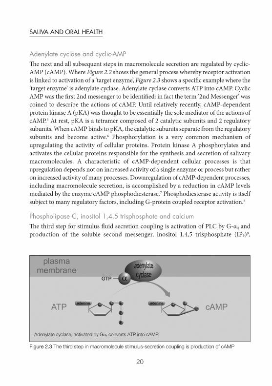
20
SALIVA AND ORAL HEALTH
Adenylate cyclase and cyclic-AMPThe next and all subsequent steps in macromolecule secretion are regulated by cyclic-AMP (cAMP). Where Figure 2.2 shows the general process whereby receptor activationis linked to activation of a ‘target enzyme’, Figure 2.3 shows a specific example where the‘target enzyme’ is adenylate cyclase. Adenylate cyclase converts ATP into cAMP. CyclicAMP was the first 2nd messenger to be identified: in fact the term ‘2nd Messenger’ wascoined to describe the actions of cAMP. Until relatively recently, cAMP-dependentprotein kinase A (pKA) was thought to be essentially the sole mediator of the actions ofcAMP.5 At rest, pKA is a tetramer composed of 2 catalytic subunits and 2 regulatorysubunits. When cAMP binds to pKA, the catalytic subunits separate from the regulatorysubunits and become active.6 Phosphorylation is a very common mechanism ofupregulating the activity of cellular proteins. Protein kinase A phosphorylates andactivates the cellular proteins responsible for the synthesis and secretion of salivarymacromolecules. A characteristic of cAMP-dependent cellular processes is thatupregulation depends not on increased activity of a single enzyme or process but ratheron increased activity of many processes. Downregulation of cAMP-dependent processes,including macromolecule secretion, is accomplished by a reduction in cAMP levelsmediated by the enzyme cAMP phosphodiesterase.7 Phosphodiesterase activity is itselfsubject to many regulatory factors, including G-protein coupled receptor activation.8
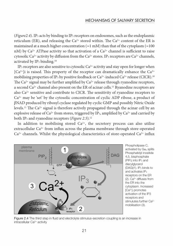
Phospholipase C, inositol 1,4,5 trisphosphate and calciumThe third step for stimulus fluid secretion coupling is activation of PLC by G-αq andproduction of the soluble second messenger, inositol 1,4,5 trisphosphate (IP3)9,
Figure 2.3 The third step in macromolecule stimulus-secretion coupling is production of cAMP
Adenylate cyclase, activated by Gαs converts ATP into cAMP.
SHL_S+OH_A5 13/11/2012 14:16 Page 20
21
MECHANISMS OF SALIVARY SECRETION
(Figure2.4). IP3 acts by binding to IP3 receptors on endosomes, such as the endoplasmicreticulum (ER), and releasing the Ca2+ stored within. The Ca2+ content of the ER ismaintained at a much higher concentration (≈1 mM) than that of the cytoplasm (≈100nM) by Ca2+ ATPase activity so that activation of a Ca2+ channel is sufficient to raisecytosolic Ca2+ activity by diffusion from the Ca2+ stores. IP3 receptors are Ca2+ channels,activated by IP3 binding.10
IP3 receptors are also sensitive to cytosolic Ca2+ activity and stay open for longer when[Ca2+]i is raised. This property of the receptor can dramatically enhance the Ca2+
mobilising properties of IP3 by positive feedback or Ca2+-induced Ca2+ release (CICR).10
The Ca2+ signal may be further amplified by Ca2+ release through ryanodine receptors,a second Ca2+ channel also present on the ER of acinar cells.11 Ryanodine receptors arealso Ca2+ sensitive and contribute to CICR. The sensitivity of ryanodine receptors toCa2+ may be ‘set’ by the cytosolic concentration of cyclic ADP ribose, a product ofβNAD produced by ribosyl cyclase regulated by cyclic GMP and possibly Nitric Oxidelevels.11 The Ca2+ signal is therefore actively propagated through the acinar cell by anexplosive release of Ca2+ from stores, triggered by IP3, amplified by Ca2+ and carried byboth IP3 and ryanodine receptors (Figure 2.5).12
In addition to mobilising stored Ca2+, the secretory process can also utiliseextracellular Ca2+ from influx across the plasma membrane through store-operatedCa2+ channels. Whilst the physiological characteristics of store-operated Ca2+ influx
Figure 2.4 The third step in fluid and electrolyte stimulus-secretion coupling is an increase inintracellular Ca2+ activity
Phospholipase C,activated by Gαq splitsPhosphatidyl inositide4,5, bisphosphate(PIP2) into IP3 anddiacylglycerol(DAG)(1). IP3 binds toand activates IP3
receptors on the ER(2). Ca2+ diffuses fromthe ER into thecytoplasm. Increased[Ca2+]i promotesactivation of the IP3receptors andstimulutes further Ca2+
mobilisation (3).
SHL_S+OH_A5 13/11/2012 14:16 Page 21

22
SALIVA AND ORAL HEALTH
have been extensively studied over the last 20 years,13 it is only recently that themolecular identity of both the Ca2+ sensor14 and the Ca2+ channel itself15 have beendiscovered. The former is now known to be the Ca2+ binding protein STromalInteraction Molecule (STIM)1, situated in the membrane of the endoplasmicreticulum.16 Dissociation of Ca2+ from the EF-hand (a finger or hand-shaped Ca2+
binding domain) Ca2+ binding region of STIM1 following depletion of the Ca2+ storesis the trigger for oligomerisation of STIM1 into a complex capable of interaction withthe plasma membrane Ca2+ channel protein Orai1 (named for the gatekeepers of heavenin Greek mythology and not for the city in Uttar Pradesh) which opens to allow Ca2+
influx.13,17 Down-regulation of the Ca2+ signal, following closure of both intracellularand extracellular channels, depends mainly on Ca2+ ATPase activity to pump the Ca2+
back into the stores or out of the cell.
MacromoleculesMacromolecules cannot cross the plasma membrane. At first sight, this might seem to bean insurmountable problem for a protein-secreting cell but the secret to protein secretionis to synthesise proteins for export within endosomes (Figure 2.6). Topologically at least,these proteins are never inside the cell and so do not have to cross the cell membrane toget out. Proteins are secreted when the endosome or vesicle into which they weresynthesised fuses with the plasma membrane in the process of exocytosis.
Figure 2.5 Actively propagated Ca2+ signal
IP3 stimulates Ca2+ release from IP3 receptors (IP3R) Ca2+ stimulates further Ca2+ release from IP3R andfrom ryanodine receptors (RyR). Release of Ca2+ by one receptor triggers activation of the next and thusactively propagates the signal. Thus a Ca2+ signal may start at the apical pole of the cell and then rapidlybecome cell-wide
SHL_S+OH_A5 13/11/2012 14:16 Page 22

23
MECHANISMS OF SALIVARY SECRETION
Synthesis of secretory proteins begins with gene transcription and manufacture ofmessenger RNA to carry the sequence information from the nucleus to ribosomes inthe cytoplasm. Secretory proteins start with a ‘signal sequence’ which targets thedeveloping polypeptide to the ER where it is N-glycosylated and folded into the correctthree-dimensional structure. Small membrane vesicles carry proteins from the ERthrough several layers of the Golgi apparatus for additional processing and ‘packaging’for export. Proteins move by default onwards from the ER; those destined to remain inthe cell contain specific ‘retention sequences’ to segregate them from secretory proteins.Secretory proteins are concentrated within Golgi condensing-vacuoles and stored insecretory vesicles. As these mature they are transported close to the apical membrane.In response to a secretory stimulus, secretory vesicles fuse with the plasma membraneand discharge their contents outside the cell.1,18
The secretory process may be divided into four stages. Synthesis, segregation andpackaging, storage and release. Each of these stages is regulated by phosphorylation oftarget proteins by cAMP-dependent pKA. Therefore an increase in cAMP stimulates:
• Transcription of genes for salivary proteins (e.g. proline-rich proteins).• Post-translational modification (e.g. glycosylation)• Maturation and translocation of secretory vesicles to the apical membrane• Exocytosis.
Figure 2.6 Secretory proteins are synthesised in endosomes
Proteins are synthesized inside secretory vesicles by ribosomes (R). Secretory vesicles mature and are stored until a secretory stimulus is received.
SHL_S+OH_A5 13/11/2012 14:16 Page 23

24
SALIVA AND ORAL HEALTH
Thus, an increase in the level of cAMP within the cell will stimulate every stepinvolved in the secretion of protein (Figure 2.7).19,20
The molecular components of exocytosis have been extensively studied and the keyplayers, soluble N-ethylmaleimide-sensitive fusion protein attachment protein receptors(SNAREs) are present in salivary gland acinar cells. In most secretory cells, unlikesalivary acinar cells, an increase in [Ca2+]i is the proximal trigger for exocytosis.Secretory vesicles have v-SNARES which recognise plasma membrane t-SNARES andthe two form tight complexes that link the two membranes and mediate the three stepsin regulated exocytosis; docking, priming and fusion20 (Figure 2.8). In neuronal cells,for example, secretory vesicles are docked and primed and await a Ca2+ signal to triggerexocytosis. Perhaps in salivary gland acinar cells, secretory vesicles wait for a secretory
Figure 2.7 cAMP and PKA
cAMP binding to the regulatory subunit of pKA releases and activates the catalytic subunit (1)The catalytic subunit phosphorylates and upregulates many components of the secretory pathway includingexocytosis (2). Recent work has also identified an Epac-mediated pKA-independent mechanism that mayalso be implicated in the regulation of exocytosis (3)
SHL_S+OH_A5 13/11/2012 14:16 Page 24
25
MECHANISMS OF SALIVARY SECRETION
stimulus at an earlier cAMP-dependent ‘brake’ point.20 The role of other intracellularsignals in the control of exocytosis is becoming better understood and both pKA-dependent and pKA-independent mechanisms have been identified that may beresponsible for the control of exocytosis in salivary acinar cells.7 Most recently, a rolefor an exchange protein directly activated by cAMP (Epac) has been identified in insulinsecretion in pancreatic β cells. In this relatively novel pKA-independent cAMP-mediated signaling pathway,5 Epac functions as guanine nucleotide exchange factor formembers of the ras family of small G-proteins and also binds directly to Rim2, a keycomponent of the exocytotic machinery. Whilst details of the mechanism remainunclear, preliminary studies allow the possibility of a similar mechanism in salivarygland cells.21
Not all secreted proteins originate in salivary gland cells. Saliva also contains plasmaproteins, for example the immunoglobulin, IgA. IgA is no more able to cross the plasmamembrane than any other protein and so crosses acinar cells in a membrane vesicle.Receptors for IgA on the basolateral membrane of the acinar cells bind IgA which istaken ‘within’ the cell by endocytosis. Following transcytosis of the vesicles containingIgA, the immunoglobulin is released into the saliva by exocytosis (Figure 2.9).22
Figure 2.8 A cAMP-dependent ‘brake’ point
Exocytosis occurs in three stages; docking, priming and fusion. The fusion process itself is Ca2+-dependent.However, earlier stages of the process, e.g. ‘docking’ could be cAMP-dependent. In salivary gland cells, thisstep is the rate- limiting ‘brake’ point in exocytosis.
SHL_S+OH_A5 13/11/2012 14:16 Page 25
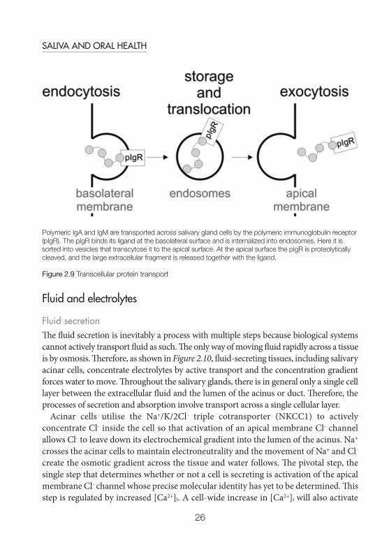
26
SALIVA AND ORAL HEALTH
Fluid and electrolytes
Fluid secretionThe fluid secretion is inevitably a process with multiple steps because biological systemscannot actively transport fluid as such. The only way of moving fluid rapidly across a tissueis by osmosis. Therefore, as shown in Figure 2.10, fluid-secreting tissues, including salivaryacinar cells, concentrate electrolytes by active transport and the concentration gradientforces water to move. Throughout the salivary glands, there is in general only a single celllayer between the extracellular fluid and the lumen of the acinus or duct. Therefore, theprocesses of secretion and absorption involve transport across a single cellular layer.
Acinar cells utilise the Na+/K/2Cl- triple cotransporter (NKCC1) to activelyconcentrate Cl- inside the cell so that activation of an apical membrane Cl- channelallows Cl- to leave down its electrochemical gradient into the lumen of the acinus. Na+
crosses the acinar cells to maintain electroneutrality and the movement of Na+ and Cl-
create the osmotic gradient across the tissue and water follows. The pivotal step, thesingle step that determines whether or not a cell is secreting is activation of the apicalmembrane Cl- channel whose precise molecular identity has yet to be determined. Thisstep is regulated by increased [Ca2+]i. A cell-wide increase in [Ca2+]i will also activate
Polymeric IgA and IgM are transported across salivary gland cells by the polymeric immunoglobulin receptor(pIgR). The pIgR binds its ligand at the basolateral surface and is internalized into endosomes. Here it issorted into vesicles that transcytose it to the apical surface. At the apical surface the pIgR is proteolyticallycleaved, and the large extracellular fragment is released together with the ligand.
Figure 2.9 Transcellular protein transport
SHL_S+OH_A5 13/11/2012 14:16 Page 26
27
MECHANISMS OF SALIVARY SECRETION
the basolateral K+ channel (SLO), which keeps the membrane potential at a highnegative value and thus preserves the driving force for Cl- efflux.
Electrolyte-led fluid transport movement is always isotonic. Once isotonicity isreached, there is no additional driving force for water movement. The ability of salivaryglands to generate an hypotonic saliva lies with the striated ducts. Striated duct cellspump electrolytes from the primary saliva by active transport. At first sight, it mightseem that this will simply reverse the secretory process, but the striated ducts areimpermeable to water, so there can be no osmotically driven water reabsorption. Thebasic outline of this secretory process was identified as the ‘two-stage hypothesis’ byThaysen et al22a in 1954. The fluid secretory process in the acinar cells has a muchgreater capacity than the electrolyte reabsorptive process in the ducts. This is why thecomposition of saliva changes with flow rate. At low, unstimulated, flow rates, saliva
Figure 2.10 Fluid secretion follows electrolyte secretion
The Na+/K+ ATPase makes direct use of ATP to pump Na+ out of the cell and create an inwardly directedNa+ gradient. This energises the Na+/K/2Cl- (NKCC1) cotransport system (1) which in turn concentrates Cl-
above its electrochemical potential (2). Increased [Ca2+]i opens the Ca2+-dependent K+ and Cl- channels andCl- crosses the apical membrane into the lumen of the acinus (2). Na+ follows Cl- across the cell to maintainelectroneutrality and the resultant osmotic gradient moves water (3)
SHL_S+OH_A5 13/11/2012 14:16 Page 27

28
SALIVA AND ORAL HEALTH
moves slowly through the ducts and the striated ducts are able to substantially modifythe composition of the saliva. At high, stimulated, flow rates, the saliva passes rapidlythrough the ducts with little alteration. The composition of saliva at high flow ratesmore closely resembles that of the primary saliva produced by the acinar cells.
Bicarbonate secretionThe secretory process for bicarbonate is similar to that for Cl- inasmuch as bicarbonateis concentrated within acinar cells and released following receipt of a secretory stimulus.Details of the process for bicarbonate are much less well understood than for Cl-. Inmost salivary glands, uptake is probably via a carbonic anhydrase mediated processthat depends ultimately on Na+/H+ exchange and the Na+ gradient. Efflux is probably
Figure 2.11 Fluid secretion follows electrolyte secretion
Carbon dioxide inside cells is converted to HCO3- and H+ by carbonic anhydrase. HCO3- is secreted across
the apical membrane of the cell through an anion channel (2). H+ are actively extruded across the basolateralmembrane by Na+/H+ exchange energised by the Na+ gradient which is created by the action of the Na+/K+
ATPase (1). If protons were not lost from the cell, carbonic anhydrase would be unable to generate HCO3-.
SHL_S+OH_A5 13/11/2012 14:16 Page 28
29
MECHANISMS OF SALIVARY SECRETION
via a bicarbonate-permeable channel (Figure 2.11). The Ca2+-dependent Cl- channel isbicarbonate permeable and bicarbonate efflux via this channel would be the mostsimple mechanism for bicarbonate secretion. Qualitatively at least, bicarbonatesecretion will be as effective as Cl- secretion as a mechanism for driving fluidmovement.19
Bicarbonate is one of the electrolytes reabsorbed by the striated ducts andbicarbonate concentration in unstimulated saliva is consequently low. A combinationof increased secretion and failure to reabsorb bicarbonate at high flow rates is thesimplest explanation of the much higher bicarbonate concentration in stimulated saliva.
Acinar cells secrete macromolecules and fluid and electrolytes. Striated duct cellsreabsorb electrolytes. Intercalated ducts lie between the acini and the striated ducts andseem to function more like acinar cells than striated duct cells. They probably makelittle contribution to protein secretion but may have an important role in bicarbonateand fluid secretion.
Calcium and phosphate secretionCalcium not only has a pivotal role in the control of secretion, it also has, along withphosphate (Pi), an important role in oral homeostasis, in particular with respect to theteeth. The mineral content of teeth is water soluble and teeth would demineralise in asimple bicarbonate-rich NaCl solution. Saliva contains, in addition, sufficient Ca2+ andPi to prevent demineralisation (Figure 2.12). There is therefore a unidirectional transportof Ca2+ and Pi across the acinar epithelial cells and into the saliva. All the componentsfor Ca2+ translocation are present in salivary acinar cells.23,24 Ca2+ pumped out of cellsacross the apical membrane of the cells would be replaced by Ca2+ that had ‘tunnelled’across the cells within the endoplasmic reticulum (see below and 25). Ca2+ influx acrossthe basolateral membrane via store-operated Ca2+ influx will replenish the Ca2+ withinthe stores. Thus Ca2+ may be translocated across cells without deranging the cellularprocesses which depend on low [Ca2+]i. Despite the probable involvement of primaryactive transport in the Ca2+ translocation process, Ca2+ activity in saliva is similar to thatof the blood. This is not the case for Pi, which may be concentrated several fold in saliva.Translocation of Pi therefore involves transepithelial active transport. The uphill, energy-dependent, step of Pi transport is across the basolateral membrane by the wellcharacterised Na+-dependent Pi transporter, NPT2b.26 Phosphate exits the acinar cellsdown its concentration gradient through an as yet unidentified pathway. The phosphateconcentration, like that of HCO3-, is modified by the passage of saliva through the ducts.NPT2b is also expressed on the apical membrane of ductal cells and it has been proposedthat here it functions to reabsorb Pi from the primary saliva.26
SHL_S+OH_A5 13/11/2012 14:16 Page 29
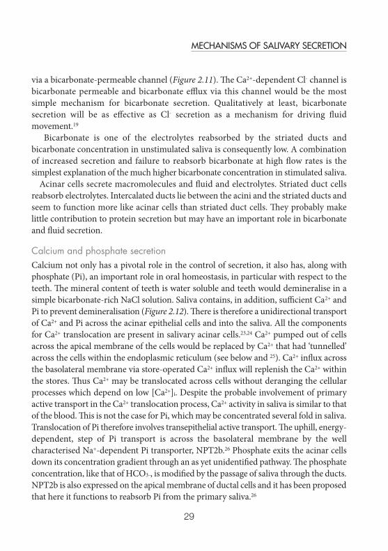
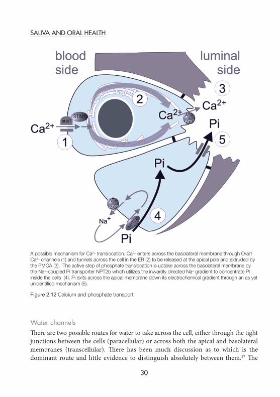
30
SALIVA AND ORAL HEALTH
Water channelsThere are two possible routes for water to take across the cell, either through the tightjunctions between the cells (paracellular) or across both the apical and basolateralmembranes (transcellular). There has been much discussion as to which is thedominant route and little evidence to distinguish absolutely between them.27 The
A possible mechanism for Ca2+ translocation. Ca2+ enters across the basolateral membrane through Orai1Ca2+ channels (1) and tunnels across the cell in the ER (2) to be released at the apical pole and extruded bythe PMCA (3). The active step of phosphate translocation is uptake across the basolateral membrane bythe Na+-coupled Pi transporter NPT2b which utilizes the inwardly directed Na+ gradient to concentrate Piinside the cells (4). Pi exits across the apical membrane down its electrochemical gradient through an as yetunidentified mechanism (5).
Figure 2.12 Calcium and phosphate transport
SHL_S+OH_A5 13/11/2012 14:16 Page 30
31
MECHANISMS OF SALIVARY SECRETION
intrinsic water permeability of the plasma membrane is very low and both apical andbasolateral membranes must therefore contain water channels to facilitate transcellularwater transport. Water channels in salivary acinar cells are members of the aquaporin(AQP) family. Aquaporins are membrane proteins composed of 4 subunits, each ofwhich has 6 membrane-spanning domains that form a water-permeable pore.Aquaporins come in two types, one of which transports only water and another whichis also permeable to glycerol. Neither type conducts ions.28 There are at least 10mammalian aquaporin isoforms and AQP5 has been localised to the apical membraneof salivary gland acinar cells. AQP5 knockout mice (Genetically modified mice thatcannot produce AQP5) show a 60% reduction in stimulated flow in airway mucosalglands which would suggest that at least this proportion of water flow is transcellular.29
UndirectionalIn normal circumstances the secretory process works only one way. Theunidirectionality of secretion is achieved by the barrier function of the acinar and ductcells in separating blood from saliva and, at a cellular level, by polarisation of structure
Figure 2.13 Histological polarity
Acinar and striated ducts are very obviously polarised. Acinar cells (A) have a high density of secretoryvesicles at the apical pole (1) and striated duct cells (B) have basal infoldings and a high density ofmitochondria (2).
SHL_S+OH_A5 13/11/2012 14:16 Page 31
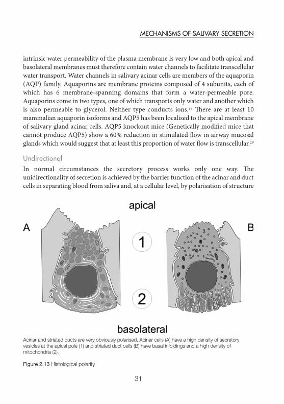

32
SALIVA AND ORAL HEALTH
(Figure 2.13) and function. Every cell type involved in salivary secretion is polarised inone way or another. Acinar and duct cells are connected together by tight junctions,which also form the division between the apical membrane which faces into the lumenof the gland and the basolateral membrane which faces the extracellular fluid. Thedifferent properties of these two membranes are fundamental to the polarisation of cellfunction necessary for unidirectional secretion.
Striated ducts are so called because in longitudinal section, their basolateral side hasa striped appearance. The stripes are caused by many infoldings of the basal membrane,crammed full of mitochondria (Figure 2.13). A high density of mitochondria, close tothe plasma membrane is usually indicative of primary active transport, in this case theNa+/K+ ATPase. The most obviously defining feature of acinar cells is the apical pole ofthe cell, densely packed with secretory vesicles.
From a functional perspective, the apical pole of the acinar cells is where all the mostcritical events occur. Secretory vesicles are directed by the actin cytoskeleton towardsthe apical pole of the cell and exocytosis occurs almost exclusively at the apical pole.
The key event in fluid secretion, activation of the Ca2+-dependent anion channel alsooccurs at the apical pole. There is growing evidence to indicate that the controlling Ca2+
signal originates at the apical pole of the cell and in certain circumstances, may berestricted to this pole of the cell (Figure 2.14).30
Calcium signals are very ‘expensive’ in terms of the metabolic cost of holding [Ca2+]i
at nanomolar levels and, because sustained elevated Ca2+ levels are cytotoxic, potentiallydangerous to the cell. Spatially restricted Ca2+ signals may be an elegant resolution toboth of these problems. It has proved very challenging to elucidate the mechanisms
Figure 2.14 Local Ca2+ signals
Sequential Ca2+ image maps taken over 20 s of a single mouse submandibular acinar cell loaded with theCa2+-sensitive dye fura-2 and stimulated with 20 nM ACh. The Ca2+ signal manifests only at the apical pole,at the bottom of the image. Each Ca2+ response lasted < 500 ms
SHL_S+OH_A5 13/11/2012 14:16 Page 32

33
MECHANISMS OF SALIVARY SECRETION
underlying local Ca2+ signals, not least of all, how the cell stops the signal frompropagating across the cell by Ca2+-induced Ca2+ release. A partial answer to thisquestion may simply be that spatially restricted Ca2+ signals are very brief. The apicalorigin of the Ca2+ signal is slightly odd, given that the ER, which is thought to be theprimary source of stored Ca2+, is almost exclusively distributed through the basolateralregion of the cell. Secretory vesicles, which have a very obvious apical location, havebeen proposed as a possible Ca2+ store.31 An alternative, and more widely accepted,mechanism depends on the reticulate nature of the ER. In this model, Ca2+ is stored atthe basolateral pole of the cell and ‘tunnels’ through the ER to the apical pole where itis released (Figure 2.14).25 This last mechanism also offers the intriguing possibility thatthe actin cytoskeleton that shapes the dynamic structure of the ER and regulatestransport of secretory vesicles to the secretory pole of the cell, might also have a role inthe control of fluid secretion.24 Ca2+ tunnelling also offers a mechanism of transepithelialtranslocation of Ca2+ that is intrinsically coupled to the process of fluid secretion,because the apical Ca2+ activity is greatest when secretion is triggered.
Pharmacological control of fluid and electrolyte secretionEvery step of stimulus-secretion coupling is potentially vulnerable to dysfunction underpathological conditions. The challenge for secretory physiologists studying autoimmunehyposalivation conditions, such as Sjögren’s syndrome, is to find the points at whichthe immune response could damage the secretory process.32,33 There is a growing bodyof evidence to suggest that severe glandular atrophy is the end-stage of Sjögren’ssyndrome and that glandular hypofunction occurs much earlier in the pathology of thecondition. Contrariwise, every step of stimulus-secretion coupling is a potential pointfor therapeutic intervention. Stimulation of fluid secretion by activation of muscarinicreceptors is one of the more obvious and accessible entry points. Pilocarpine, a naturallyoccurring alkaloid, is probably the best known therapeutic cholinomimetic agent andis distributed under the trade name ‘Salagen’. Cevimeline (evoxac) is anothercholinomimetic agent used therapeutically in the US which may have a higherspecificity for muscarinic M3 receptors than pilocarpine and potentially, therefore fewerside effects. The side effects of therapeutic application of salagen (15-30 mg/day) orevoxac (90 mg/day), sweating etc. are usually tolerated in preference to dry mouth (seeChapter 4). ACh itself is of little use therapeutically because it is so rapidly metabolised.Saliva production may be blocked by cholinergic receptor antagonists, such as atropine,which compete with ACh for muscarinic receptors and prevent the effects ofparasympathetic stimulation on fluid and electrolyte secretion. The most commoncause of dry mouth is as a side effect of xerogenic drugs used to treat other conditions.
SHL_S+OH_A5 13/11/2012 14:16 Page 33
34
SALIVA AND ORAL HEALTH
Microfluorimetry, electrophysiology and molecular biology are proving to be apowerful combination with which to study secretory mechanisms. For example:fluorescent probes for subcellular components, such as the ER, mitochondria or thenucleus may be used to visualise these organelles in living cells and determine theirrole in signal transduction. Caged agonists or second messengers may be used toprovide precise spatial mapping of intracellular responses and so further refine ourunderstanding of cellular polarisation. Genes for key elements of the secretorymachinery can be linked to fluorescent markers and expressed and visualised in isolatedacinar cells by transient transfection. Gene knockouts can help pinpoint the functionof specific proteins, such as AQP5 or ACh M3 receptors. There is now great scope andgreat potential in turning these powerful techniques towards understanding glandularpathologies
References1. Hector MP, Linden RWA. Reflexes of salivary secretion. In: Neural Mechanisms of Salivary
Gland Secretion, Garrett JR, Ekström J, Anderson LC, Eds. 1999, Karger: Basel. p. 196–217.2. Melvin JE, Yule D, Shuttleworth T, Begenisich T. Regulation of fluid and electrolyte secretion in
salivary gland acinar cells. Ann Rev Physiol 2005; 67: 445-469.3. Rodbell M. Nobel Lecture. Signal transduction: evolution of an idea. Biosci Rep 1995;
15: 117-133.4. Oldham WM, Hamm HE. Heterotrimeric G protein activation by G-protein-coupled receptors.
Nat Rev Mol Cell Biol 2008; 9: 60-71.5. Gloerich M, Bos JL. Epac: defining a new mechanism for cAMP action. Ann Rev Pharmacol
Toxicol 2010; 50: 355-375.6. Bossis I, Stratakis CA. Minireview: PRKAR1A: normal and abnormal functions. Endocrinology
2004; 145: 5452-5458.7. Seino S, Shibasaki T. PKA-dependent and PKA-independent pathways for cAMP-regulated
exocytosis. Physiol Rev 2005; 85: 1303-1342.8. Bender AT, Beavo JA, Cyclic nucleotide phosphodiesterases: molecular regulation to clinical
use. Pharmacol Rev 2006; 58: 488-520.9. Petersen OH, Tepikin AV. Polarized calcium signaling in exocrine gland cells. Ann Rev Physiol
2008; 70: 273-299.10. Dawson A. IP(3) receptors. Curr Biol 2003; 13: R424.11. Galione A, Churchill GC. Interactions between calcium release pathways: multiple
messengers and multiple stores. Cell Calcium 2002; 32: 343-354.12. Harmer AR, Gallacher DV, Smith PM. Role of Ins(1,4,5)P3, cADP-ribose and nicotinic acid -
adenine dinucleotide phosphate in Ca2+ signalling in mouse submandibular acinar cells.Biochem J 2001; 353: 555-560.
13. Putney JW, Bird GS. Regulation of calcium entry in exocrine gland cells and other epithelialcells. J Med Invest 2009; 56: 362-367.
SHL_S+OH_A5 13/11/2012 14:16 Page 34
35
MECHANISMS OF SALIVARY SECRETION
14. Liou J, Kim ML, Heo WD, Jones JT, Myers JW, Ferrell Jr JE, Meyer T. STIM is a Ca2+ sensoressential for Ca2+-store-depletion-triggered Ca2+ influx. Curr Biol 2005; 15: 1235-1541.
15. Feske S, Gwack Y, Prakriya M, Srikanth S, Puppel SH, Tanasa B, Hogan PG, Lewis RS, DalyM, Rao A. A mutation in Orai1 causes immune deficiency by abrogating CRAC channelfunction. Nature 2006; 441: 179-185.
16. Lewis RS. The molecular choreography of a store-operated calcium channel. Nature 2007;446: 284-287.
17. Soboloff J, Spassova MA, Tang XD, Hewavitharana T, Xu W, Gill DL. Orai1 and STIMreconstitute store-operated calcium channel function. J Biol Chem 2006; 281: 20661-20665.
18. Garrett JR. Effects of Autonomic Nerve Stimulations on Salivary Parenchyma and ProteinSecretion. In: Neural Mechanisms of Salivary Gland Secretion. Garrett JR, Ekström J,Anderson LC, Eds. 1999, Karger: Basel. pp. 59-79.
19. Turner RJ, Sugiya H. Understanding salivary fluid and protein secretion. Oral Dis 2002; 8: 3-11.20. Fujita-Yoshigaki J. Divergence and convergence in regulated exocytosis: the characteristics of
cAMP-dependent enzyme secretion of parotid salivary acinar cells. Cell Signal 1998. 10: 371-375.
21. Shimomura H, Imai A, Nashida T. Evidence for the involvement of cAMP-GEF (Epac) pathwayin amylase release from the rat parotid gland. Arch Biochem Biophys 2004; 431: 124-128.
22. Proctor GB, Garrett JR, Carpenter GH, Ebersole LE. Salivary secretion of immunoglobulin A by submandibular glands in response to autonomimetic infusions in anaesthetised rats. J Neuroimmunol 2003; 136: 17-24.
22a. Thaysen JH, Thorn NA, Schwartz IL. Excretion of sodium, potassium, chloride and carbondioxide in human parotid saliva. Am J Physiol 1954; 178: 155-159.
23. Peng JB, Brown EM, Hediger MA. Epithelial Ca2+ entry channels: transcellular Ca2+ transportand beyond. J Physiol 2003; 551: 729-740.
24. Harmer AR, Gallacher DV, Smith PM. Correlations between the functional integrity of theendoplasmic reticulum and polarized Ca2+ signalling in mouse lacrimal acinar cells: a role forinositol 1, 3, 4, 5-tetrakisphosphate. Biochem J 2002; 367: 137-143.
25. Petersen OH. Localization and regulation of Ca2+ entry and exit pathways in exocrine glandcells. Cell Calcium 2003; 33: 337-344.
26. Homann V, Rosin-Steiner S, Stratmann T, Arnold WH, Gaengler P, Kinne RKH. Sodium-phosphate cotransporter in human salivary glands: molecular evidence for the involvement ofNPT2b in acinar phosphate secretion and ductal phosphate reabsorption. Arch Oral Biol2005; 50: 759-768.
27. Loo DD, Wright EM, Zeuthen T. Water pumps. J Physiol 2002; 542: 53-60.28. Agre P, King LS, Yasui M, Guggino WB, Ottersen OP, Fujiyoshi Y, Engel A, Nielsen S. Aquaporin
water channels--from atomic structure to clinical medicine. J Physiol 2002; 542: 3-16.29. Song Y, Verkman AS. Aquaporin-5 dependent fluid secretion in airway submucosal glands.
J Biol Chem 2001; 276: 41288-41292.30. Thorn P, Lawrie AM, Smith PM, Gallacher DV, Petersen OH. Ca2+ oscillations in pancreatic
acinar cells: spatiotemporal relationships and functional implications. Cell Calcium 1993; 14: 746-757.
SHL_S+OH_A5 13/11/2012 14:16 Page 35

36
SALIVA AND ORAL HEALTH
31. Marty A. Calcium release and internal calcium regulation in acinar cells of exocrine glands. J Membr Biol 1991; 124: 189-197.
32. Dawson LJ, Christmas SE, Smith PM. An investigation of interactions between the immunesystem and stimulus-secretion coupling in mouse submandibular acinar cells. A possiblemechanism to account for reduced salivary flow rates associated with the onset of Sjögren’ssyndrome. Rheumatology 2000; 39: 1226-1233.
33. Caulfield VL, Balmer C, Dawson LJ, Smith PM. A role for nitric oxide-mediated glandularhypofunction in a non-apoptotic model for Sjögren’s syndrome. Rheumatology 2009; 48: 727-733.
SHL_S+OH_A5 13/11/2012 14:16 Page 36
37
IntroductionThis chapter covers the differences in flow rate and composition between unstimulatedsaliva (secreted continuously in the absence of exogenous stimulation) and stimulatedsaliva (secreted usually in response to masticatory or gustatory stimulation), the factorsinfluencing salivary flow rate and composition, and their physiological importance.
Unstimulated salivaUnstimulated whole saliva is the mixture of secretions found in the mouth in theabsence of exogenous stimuli such as tastants or chewing. It is composed of secretionsfrom the parotid, submandibular, sublingual, and minor mucous glands but it alsocontains gingival crevicular fluid, desquamated epithelial cells, bacteria, leucocytes(mainly from the gingival crevice), and possibly food residues, blood, and viruses.Unstimulated whole saliva is usually collected with the patient sitting quietly, with thehead down and mouth slightly open to allow the saliva to drip from the lower lip intoa beaker or similar receptacle over a given time, or the patient can spit out the saliva atregular intervals, while not swallowing. However, when saliva is spat out rather thandrooled, the number of bacteria and desquamated epithelial cells is increased. Themeasured flow rate is actually the difference between the volume secreted by thedifferent salivary glands and the volumes which may be lost by evaporation, if there ismouth-breathing, or mucosal absorption over the collection period.1
Several large studies of unstimulated salivary flow rates in healthy individuals (Table3.1)2 have found the average value for whole saliva to be about 0.3-0.4 ml/minute, but thenormal range is very large and includes individuals with very low flow rates who do notcomplain of a dry mouth. Such a broad normal range makes it difficult to say whether or
Factors Influencing Salivary Flow Rate and CompositionC. Dawes
3
SHL_S+OH_A5 13/11/2012 14:16 Page 37