
salim.pdf
5
A comparative study of three-dimensional saline infusion sonohysterography and diagnostic hysteroscopy for the classification of submucous fibroids R.Salim, C.Lee, A.Davies, B.Jolaoso, E.Ofuasia and D.Jurkovic 1 Department of Obstetrics and Gynaecology, King’s College Hospital, London, UK 1 To whom correspondence should be addressed at: Early Pregnancy and Gynaecology Assessment Unit, Suite 8, Golden Jubilee Wing, King’s College Hospital, London SE5 8RX, UK. E-mail: [email protected] BACKGROUND: The purpose of this study was to compare three-dimensional saline infusion sonohysterography (3D SI S) and di agnost ic hysteroscopy for the di agnosi s and cl assi fic at ion of submucous ut er ine fibroids. METHODS: This was a prospective double-blind study of 49 women who presented with a history of menorrhagia, diagnosed on non-enhanced two-dimensional ultrasonography with submucous fibroids. Fibroids were classified on 3D SIS according to the proportion of fibroid contained within the endometrial cavity, using the European Society of Hyst eros copy Classifi cati on of Submucou s Fibr oids. These results were then compare d with the findi ngs at diagn ostic hyster oscopy. RESULTS: A tota l of 61 submucou s fibro ids was identifie d in 49 symp toma tic women. Diagnostic hysteroscopy confirmed these findings in all cases. There was agreement between the two methods in 11/ 12 cas es of Typ e 0 fibr oid s (92 %), 34/37 (92 %) of Typ e I fibr oid s and 9/12 (75%) of Typ e II fibr oid s. The overall level of agreement was good with a kappa value of 0.80. CONCLUSIONS: There is a good overall agree- ment between 3D SIS and diagnostic hysteroscopy in classification of submucous fibroids. Agreement is better in cases where a greater proportion of the fibroid is contained within the uterine cavity. Key words: hysteroscopy/saline infusion sonohysterography/submucous fibroids/three-dimensional ultrasound Introduction Submucous fibroids are the most common anatomical cause of excessive menstrual blood loss in women of reproductive age. Adva nces in oper ative hyst er oscopy have enabled removal of these lesions with a significant reduction in mor- bidity, post- opera tive recovery time and costs compared to open abdominal myomectomy (Eman uel et al., 1999; Feng et al., 2002). However, accurately identifying those fibroids sui tab le for hys ter osc opi c res ect ion remain s dif ficu lt. The main determini ng fact or in succe ssful hyste rosco pic fibroi d resection appears to be the proportion of the fibroid contained wit hin the ute rin e cav ity. Dia gno stic hys ter osc opy is cur - rently regarded as the pre-operative investigation of choice in deter minin g resec tabili ty of submu cous fibroids prio r to the schedu ling of an opera tive hyste roscop y (Wamsteker et al., 1993a,b; Corson, 1995). This procedure enables direct visual- iza tio n of the ute rine cav ity and acc ura te ide nti fica tio n of intracavitary pathology. However, diagnostic hysteroscopy is an inv asiv e and cos tly pro cedure , whi ch is asso cia ted wit h risks such as uterine perforation and ascending genito-urinary infec tion (Brooks, 1992; Indman, 1995; Julian, 2002). Fur- thermore, it prov ides only subjective assessment of fibroid siz e and ind ire ct inf ormati on reg ard ing the depth of myo - metrial extension. Video recording of hysteroscopic findings may be use d for quali ty con tro l pur pos es, but the rec ord ed data cannot be modified and the final diagnosis is determined by the initial findings at the operation. Two dimensi ona l (2D) B-mode transvagi nal ult raso und wit h ste ril e sal ine ins til lat ion int o the endome tri al cav ity provides a clear view of the uterine cavity (Cicinelli et al., 1995; Widri ch et al ., 1996; de Kroon et al ., 2003). Thi s enabl es accurate detec tion of struc tural pathology affe cting the uterine cavity including submucous fibroids, comparable to dia gno sti c hys ter osc opy (Fa rqu har et al., 2003). In add iti on, ult ras ound ena ble s accurate measurement of the size of the uterine fibroids. However, 2D ultrasound is not an accurate method of assessing the extent of submucous fibroid prot rusio n into the uter ine cavit y (Ver cellin i et al ., 199 7; Dueholm et al., 2001a,b). Three -dimension al (3D) tran svagin al ultr asound has been commercially available for .10 years. This technique allows detailed evaluation of pelvic organs by collecting a series of seq uentia l ult raso und ima ges and conver tin g the m int o an ultrasound volume. This information is digitally stored as a dataset, which may then be analysed on line. The dataset is re constr ucte d in such a wa y as to allow visua li za ti on of an organ from any chosen angle and in any arbitrary plane (Jurkovic, 2002). Human Reprodu ction Vol.20, No.1 pp. 253–257, 2005 doi:10.1093/humrep/deh557 Advance Access publication October 21, 2004 Human Reproduction vol. 20 no. 1 q European Society of Human Reproduction and Embryology 2004; all rights reserved 253 a t I n s t i t u t R u d j e r B o s k o v i c o n O c t o b e r 2 2 , 2 0 1 2 h t t p : / / h u m r e p . o x f o r d j o u r n a l s . o r g / o w n l o a d e d f r o m
Transcript of salim.pdf
7/27/2019 salim.pdf
http://slidepdf.com/reader/full/salimpdf 1/5
A comparative study of three-dimensional saline infusionsonohysterography and diagnostic hysteroscopyfor the classification of submucous fibroids
R.Salim, C.Lee, A.Davies, B.Jolaoso, E.Ofuasia and D.Jurkovic1
Department of Obstetrics and Gynaecology, King’s College Hospital, London, UK
1To whom correspondence should be addressed at: Early Pregnancy and Gynaecology Assessment Unit, Suite 8, Golden Jubilee
Wing, King’s College Hospital, London SE5 8RX, UK. E-mail: [email protected]
BACKGROUND: The purpose of this study was to compare three-dimensional saline infusion sonohysterography
(3D SIS) and diagnostic hysteroscopy for the diagnosis and classification of submucous uterine fibroids
METHODS: This was a prospective double-blind study of 49 women who presented with a history of menorrhagia
diagnosed on non-enhanced two-dimensional ultrasonography with submucous fibroids. Fibroids were classified on
3D SIS according to the proportion of fibroid contained within the endometrial cavity, using the European Society
of Hysteroscopy Classification of Submucous Fibroids. These results were then compared with the findings a
diagnostic hysteroscopy. RESULTS: A total of 61 submucous fibroids was identified in 49 symptomatic women
Diagnostic hysteroscopy confirmed these findings in all cases. There was agreement between the two methods in
11/12 cases of Type 0 fibroids (92%), 34/37 (92%) of Type I fibroids and 9/12 (75%) of Type II fibroids. Th
overall level of agreement was good with a kappa value of 0.80. CONCLUSIONS: There is a good overall agree
ment between 3D SIS and diagnostic hysteroscopy in classification of submucous fibroids. Agreement is better in
cases where a greater proportion of the fibroid is contained within the uterine cavity.
Key words: hysteroscopy/saline infusion sonohysterography/submucous fibroids/three-dimensional ultrasound
IntroductionSubmucous fibroids are the most common anatomical cause
of excessive menstrual blood loss in women of reproductive
age. Advances in operative hysteroscopy have enabled
removal of these lesions with a significant reduction in mor-
bidity, post-operative recovery time and costs compared to
open abdominal myomectomy (Emanuel et al., 1999; Feng
et al., 2002). However, accurately identifying those fibroids
suitable for hysteroscopic resection remains difficult. The
main determining factor in successful hysteroscopic fibroid
resection appears to be the proportion of the fibroid contained
within the uterine cavity. Diagnostic hysteroscopy is cur-
rently regarded as the pre-operative investigation of choice in
determining resectability of submucous fibroids prior to thescheduling of an operative hysteroscopy (Wamsteker et al.,
1993a,b; Corson, 1995). This procedure enables direct visual-
ization of the uterine cavity and accurate identification of
intracavitary pathology. However, diagnostic hysteroscopy is
an invasive and costly procedure, which is associated with
risks such as uterine perforation and ascending genito-urinary
infection (Brooks, 1992; Indman, 1995; Julian, 2002). Fur-
thermore, it provides only subjective assessment of fibroid
size and indirect information regarding the depth of myo-
metrial extension. Video recording of hysteroscopic findings
may be used for quality control purposes, but the recordeddata cannot be modified and the final diagnosis is determined
by the initial findings at the operation.
Two dimensional (2D) B-mode transvaginal ultrasound
with sterile saline instillation into the endometrial cavity
provides a clear view of the uterine cavity (Cicinelli et al.
1995; Widrich et al., 1996; de Kroon et al., 2003). Thi
enables accurate detection of structural pathology affecting
the uterine cavity including submucous fibroids, comparable
to diagnostic hysteroscopy (Farquhar et al., 2003). In
addition, ultrasound enables accurate measurement of th
size of the uterine fibroids. However, 2D ultrasound is not an
accurate method of assessing the extent of submucous fibroid
protrusion into the uterine cavity (Vercellini et al., 1997Dueholm et al., 2001a,b).
Three-dimensional (3D) transvaginal ultrasound has been
commercially available for .10 years. This technique allow
detailed evaluation of pelvic organs by collecting a series o
sequential ultrasound images and converting them into an
ultrasound volume. This information is digitally stored as a
dataset, which may then be analysed on line. The dataset is
reconstructed in such a way as to allow visualization o
an organ from any chosen angle and in any arbitrary plane
(Jurkovic, 2002).
Human Reproduction Vol.20, No.1 pp. 253–257, 2005 doi:10.1093/humrep/deh557
Advance Access publication October 21, 2004
Human Reproduction vol. 20 no. 1 q European Society of Human Reproduction and Embryology 2004; all rights reserved 253
7/27/2019 salim.pdf
http://slidepdf.com/reader/full/salimpdf 2/5
In this study, we compared 3D transvaginal ultrasound
combined with saline instillation into the uterine cavity to
diagnostic hysteroscopy for the assessment of submucous
fibroids. In particular we examined the assessment of myo-
metrial extension of fibroids by the two techniques.
Materials and methods
This was a prospective double-blind study set in a tertiary gynae-
cology ultrasound unit at a London teaching hospital. Symptomaticwomen referred to the unit underwent a non-enhanced B-mode 2D
ultrasound scan. Those women found to have submucous fibroids
were invited to join the study, provided there was no other identifi-
able cause for their symptoms. Informed consent was obtained and
three-dimensional saline infusion sonohysterography (3D SIS) was
performed in all cases. A sterile Cuscoe speculum was passed, the
cervix visualized and cleaned with sterile chlorhexidine solution. A
3.3 mm soft plastic paediatric naso-gastric suction catheter was then
passed through the cervix into the uterine cavity without grasping
the cervix. The speculum was removed and a 5 MHz transvaginal
3D ultrasound probe inserted into the vagina (Voluson 730;
KretzTechnik, Austria). The uterine cavity was visualized and the
position of the catheter within the uterine cavity confirmed. A longi-
tudinal view of the uterus was obtained and the catheter was with-drawn to a level just above the internal cervical os. A volume of
5– 10 ml of sterile saline solution was then instilled into the uterine
cavity. A 3D volume was generated by the automatic sweep of the
mechanical transducer. The acquired volume was the shape of a
truncated cone, with a depth of 4.3–8.6 cm and a vertical angle
a ¼ 908. The volumes were stored digitally (Magneto-Optic 3.00;
640MB Olympus Europe, Germany) and analysed using multi-
planar visualization (Figure 1). With this technique it was possible
to examine the uterine cavity in three orthognal planes. This enabled
the operator to achieve planes that were not necessarily possible
with the original 2D ultrasound. The 3D ultrasound volume was
manipulated to visualize the fibroid in its widest diameter in a plane
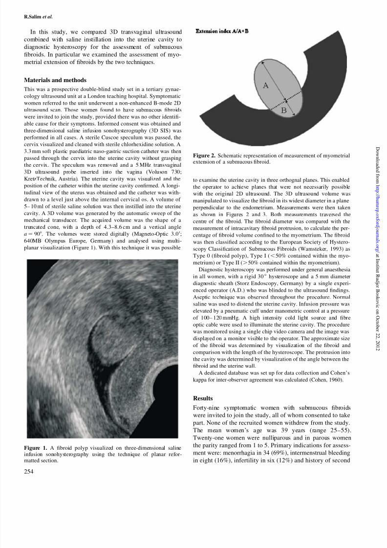
perpendicular to the endometrium. Measurements were then taken
as shown in Figures 2 and 3. Both measurements traversed the
centre of the fibroid. The fibroid diameter was compared with the
measurement of intracavitary fibroid protrusion, to calculate the per-
centage of fibroid volume confined to the myometrium. The fibroid
was then classified according to the European Society of Hystero-
scopy Classification of Submucous Fibroids (Wamsteker, 1993) as
Type 0 (fibroid polyp), Type I (,50% contained within the myo-
metrium) or Type II (.50% contained within the myometrium).
Diagnostic hysteroscopy was performed under general anaesthesia
in all women, with a rigid 308 hysteroscope and a 5 mm diameter
diagnostic sheath (Storz Endoscopy, Germany) by a single experi-
enced operator (A.D.) who was blinded to the ultrasound findings.
Aseptic technique was observed throughout the procedure. Normal
saline was used to distend the uterine cavity. Infusion pressure was
elevated by a pneumatic cuff under manometric control at a pressure
of 100– 120 mmHg. A high intensity cold light source and fibre
optic cable were used to illuminate the uterine cavity. The procedure
was monitored using a single chip video camera and the image was
displayed on a monitor visible to the operator. The approximate size
of the fibroid was determined by visualization of the fibroid and
comparison with the length of the hysteroscope. The protrusion into
the cavity was determined by visualization of the angle between the
fibroid and the uterine wall.
A dedicated database was set up for data collection and Cohen’s
kappa for inter-observer agreement was calculated (Cohen, 1960).
Results
Forty-nine symptomatic women with submucous fibroids
were invited to join the study, all of whom consented to take
part. None of the recruited women withdrew from the study.
The mean women’s age was 39 years (range 25–55).
Twenty-one women were nulliparous and in parous women
the parity ranged from 1 to 5. Primary indications for assess-
ment were: menorrhagia in 34 (69%), intermenstrual bleeding
in eight (16%), infertility in six (12%) and history of second
Figure 2. Schematic representation of measurement of myometrialextension of a submucous fibroid.
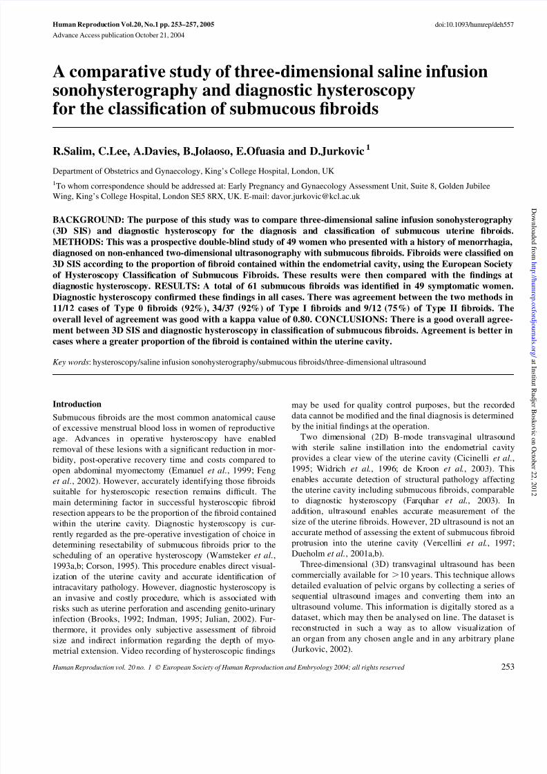
Figure 1. A fibroid polyp visualized on three-dimensional salineinfusion sonohysterography using the technique of planar refor-matted section.
R.Salim et al.
254
7/27/2019 salim.pdf
http://slidepdf.com/reader/full/salimpdf 3/5
trimester miscarriage in one (2%) woman. 3D SIS was
successful in all 49 women, providing clear views of the
uterine cavity. Mild discomfort was the only reported side-
effect of the procedure. Diagnostic hysteroscopy was also
successful in all cases; clear views of the uterine cavity were
obtained with both tubal ostia identified in each case. There
were no operative complications associated with the diagnos-
tic hysteroscopies.
A total of 61 submucous fibroids was identified by both
3D SIS and diagnostic hysteroscopy. The results are summar-
ized in Table I. Overall, there was agreement in the classifi-
cation of 54/61 (89%) fibroids (kappa 0.80). Eleven fibroids
were classified as Type 0, 34 as Type I and 9 as Type II by
both 3D SIS and hysteroscopy. One fibroid classified as Type
I on 3D SIS was described as Type 0 on hysteroscopy. In a
further six cases of discordant findings, 3D SIS indicated
deeper myometrial involvement in half of the cases and
hysteroscopy in the other half.
Discussion
This study showed good overall agreement between diagnos
tic hysteroscopy and transvaginal ultrasound in the diagnosi
of submucous fibroids. Every woman scheduled for a hys
teroscopy for suspected submucous fibroids had the diagnosis
confirmed at operation. This high accuracy is consistent with
several previous studies, which compared non-enhanced
transvaginal ultrasound with hysteroscopy (Fedele et al.1991; Cicinelli et al., 1995; Dueholm et al., 2001a,b). Simila
results were also obtained using saline infusion sonohystero
graphy (Fukuda et al., 1993; Bronz et al., 1997; Valenzano
et al., 1999; Dijkhuizen et al., 2000; Bernard et al., 2001
Dueholm et al., 2001a,b; Nanda et al., 2002; Lindheim et al.
2003).
There was also good overall agreement between diagnostic
hysteroscopy and 3D ultrasound in the assessment of myo
metrial extension of fibroids. Using the European Society
of Hysteroscopy Classification of Submucous Fibroid
(Wamsteker, 1993a) the best level of agreement was achieved
in women with fibroid polyps (Type 0). These are the fibroids
in which hysteroscopic resection is likely to be relativelysimple and successful. The level of agreement decreased with
increasing degree of myometrial involvement. This is not sur
prising as hysteroscopy can only assess the segment of the
fibroid protruding into the cavity, whilst ultrasound can also
provide information about the part of the fibroid buried within
the myometrium. In cases of discordant findings the differ
ences were random with no clear tendency of either method
to overestimate myometrial involvement. These result
are better than findings by Vercellini et al. (1997), who re
ported less agreement between non-enhanced 2D transvagina
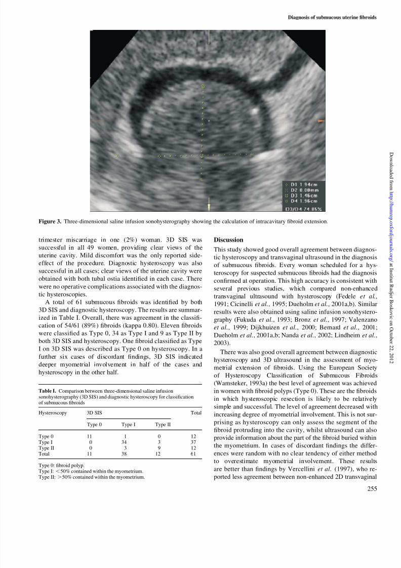
Figure 3. Three-dimensional saline infusion sonohysterography showing the calculation of intracavitary fibroid extension.
Table I. Comparison between three-dimensional saline infusion
sonohysterography (3D SIS) and diagnostic hysteroscopy for classificationof submucous fibroids
Hysteroscopy 3D SIS Total
Type 0 Type I Type II
Type 0 11 1 0 12Type I 0 34 3 37Type II 0 3 9 12Total 11 38 12 61
Type 0: fibroid polyp.Type I: ,50% contained within the myometrium.Type II: .50% contained within the myometrium.
Diagnosis of submucous uterine fibroid
255

7/27/2019 salim.pdf
http://slidepdf.com/reader/full/salimpdf 4/5
ultrasound and hysteroscopy, with the methods giving discor-
dant results in 55/228 cases (24.1%). In their study there was
also no clear pattern of differences between the methods and
discordant results were randomly distributed.
A more recent study by Leone and Lanzani (2003) using
2D SIS showed complete agreement with diagnostic hyste-
roscopy in all cases of submucous fibroids. They used the
angle formed between the intracavitary portion of the fibroid
and the endometrium to classify the fibroids. Although these
results were impressive, the reproducibility of the angle
measurements used in the study has not been tested. In
addition the angle measurement is based on the assumption
that all fibroids are spherical in shape, which is clearly not
always the case. Therefore it remains to be seen whether these
results will be successfully reproduced by other investigators.
This raises the issue of which method should be considered
as the gold standard for the evaluation of myometrial invol-
vement of submucous fibroids. Although most studies use
diagnostic hysteroscopy as the gold standard, the value of
this approach has been questioned by others. Dueholm et al.
(2001b) compared the accuracy of magnetic resonance ima-
ging (MRI), non-enhanced transvaginal ultrasound, SIS andhysteroscopy in the evaluation of abnormalities of the uterine
cavity using hysterectomy specimens as the gold standard.
Although all the methods performed reasonably well in the
detection of uterine cavity lesions, for the assessment of sub-
mucous fibroids MRI and SIS were superior to hysteroscopy.
The authors’ conclusion was that MRI, rather than hyste-
roscopy, should be used for pre-operative assessment of
submucous fibroids.
However, the critical issue when evaluating submucous
fibroids is not the relative accuracy of different diagnostic
methods, but the prediction of the success of hysteroscopic
resection in symptomatic women. This subject has been less
extensively investigated and there is only limited data on thepredictive value of hysteroscopy in the pre-operative selec-
tion of submucous fibroids for hysteroscopic resection. In a
study by Vercellini et al. (1997) only 69% of women deemed
suitable for endoscopic fibroid resection on hysteroscopic
assessment had fibroids successfully removed using this
technique. Although the cause of this discrepancy was not
addressed in the paper, it does suggest that diagnostic hys-
teroscopy may not be the optimal method for pre-operative
assessment of submucous fibroids.
A classification of submucous uterine fibroids into three
sub-types (0, I and II) depending on the degree of myometrial
extension was adopted by the European Society of Hystero-
scopy in order to improve pre-operative selection of womenfor hysteroscopic resection. However, a prospective study
conducted by the team which designed this classification
showed that the degree of myometrial involvement had very
little influence on the success of hysteroscopic resection of
submucous fibroids (Wamsteker, 1993b). In a subgroup of
women with Type I fibroids (,50% myometrial extension),
complete resection was achieved in 12/20 (60%) of the
procedures compared to 10/20 (50%) in Type II fibroids
(.50% myometrial extension). Taking into account repeated
procedures, complete resection was achieved in 12/14 cases
(85.7%) of Type I fibroids, which was almost identical to
10/12 (83.3%) in Type II fibroids.
In clinical practice it is not uncommon to find multiple
fibroids affecting the uterine cavity. Apart from the number
of fibroids, their size is also likely to be a factor in determin-
ing the success of hysteroscopic resection. None of the cur-
rent methods used in routine clinical practice is able to assess
more complex distortion of uterine cavity in sufficient detail.
Three-dimensional ultrasound may overcome some of the
limitations associated with fibroid classification using a 2D
model (Weinraub and Herman, 1998). 3D ultrasound enables
us to examine the uterus from any angle and in any arbitrary
plane and it is possible to assess both the size and the depth
of myometrial extension in each individual fibroid. The
saved volume can be manipulated in such a way as to pro-
vide measurements of the depth of myometrial extension
exactly at the widest fibroid diameter, taken in a plane per-
pendicular to the endometrium. This cannot be achieved by
using 2D ultrasound or any other conventional diagnostic
technique. Further research will show whether this increased
diagnostic capability of 3D ultrasound may be translated into
a more meaningful system of classification of submucousfibroids, which could predict the success of hysteroscopic
fibroid resection with a high degree of accuracy.
References
Bernard JP, Camatte S, Robin F, Taurelle R and Lecuru F (2001) Saline con-trast sonohysterography in the preoperative assessment of benign intrauter-ine disorders. Ultrasound Obstet Gynecol 17,145–149.
Bronz L, Suter T and Rusca T (1997) The value of transvaginal sonographywith and without saline instillation in the diagnosis of uterine pathology inpre- and postmenopausal women with abnormal bleeding or suspect sono-graphic findings. Ultrasound Obstet Gynecol 9,53–58.
Brooks PG (1992) Complications of operative hysteroscopy: how safe is it?Clin Obstet Gynecol 35,256–262.
Cicinelli E, Romano F, Silvio Anastasio P, Blasi N, Parisi C and Galantino P(1995) Transabdominal sonohysterography, transvaginal sonography andhysteroscopy in the evaluation of submucous myomas. Obstet Gynecol 85,42–47.
Cohen J (1960) A coefficient of agreement for nominal scales. Educn Psy-chol Measmt 20,37–46.
Corson SL (1995) Hysteroscopic diagnosis and operative therapy of sub-mucous myoma. Obstet Gynecol Clin North Am 22,739–755.
de Kroon CD, Willem Jansen F, Louwe LA, Dieben SWM, van HouwelingenHC and Baptist Trimbos J (2003) Technology assessment of saline con-trast hysterosonography. Am J Obstet Gynecol 188,945–949.
Dijkhuizen FP, De Vries LD, Mol BW, Brolmann HA, Peters HM,Moret E and Heintz AP (2000) Comparison of transvaginal ultrasono-graphy and saline infusion sonography for the detection of intracavitaryabnormalities in premenopausal women. Ultrasound Obstet Gynecol 15,372–376.
Dueholm M, Forman A, Jensen ML, Laursen H and Kracht P (2001a) Trans-
vaginal sonography combined with saline contrast sonohysterography inevaluating the uterine cavity in premenopausal patients with abnormaluterine bleeding. Ultrasound Obstet Gynecol 18,54–61.
Dueholm M, Lundorf E, Hansen ES, Ledertoug S and Olesen F (2001b)Evaluation of the uterine cavity with magnetic resonance imaging, transva-ginal sonography, hysteroscopic examination, and diagnostic hysteroscopy.Fertil Steril 76,350–357.
Emanuel MH, Wamsteker K, Hart AAM, Metz G and Lammes F (1999)Long-term results of hysteroscopic myomectomy for abnormal uterinebleeding. Obstet Gynecol 93,743–748.
Farquhar C, Ekeroma A, Furness S and Arroll B (2003) A systematic reviewof transvaginal ultrasonography, sonohysterography and hysteroscopy forthe investigation of abnormal uterine bleeding in premenopausal women.Acta Obstet Gynecol Scand 82,493–504.
R.Salim et al.
256

7/27/2019 salim.pdf
http://slidepdf.com/reader/full/salimpdf 5/5
Fedele L, Bianchi S, Dorta M, Brioschi D, Zanotti F and Vercellini P (1991)Transvaginal ultrasonography versus hysteroscopy in the diagnosis of uter-ine submucous myomas. Obstet Gynecol 77,745–748.
Feng ZC, Shi YP and Liu SP (2002) Hysteroscopic resection of submucousfibroids: clinical analysis of 99 cases. Gynaecol Endosc 11,127–130.
Fukuda M, Shimizu T, Fukuda K, Yomura W and Shimizu S (1993) Trans-vaginal hysterosonography for differential diagnosis between submucousand intramural myoma. Gynecol Obstet Invest 35,236–239.
Indman PD (1995) Hysteroscopic complications (editorial). J Am AssnGynecol Laparosc 3,1–2.
Julian TM (2002) Hysteroscopic complications. J Lower Gen Tract Dis 6,
39–47.Jurkovic D (2002) Three-dimensional ultrasound in gynecology: a critical
evaluation. Ultrasound Obstet Gynecol 19,109–117.
Leone FPG and Lanzani C (2003) Use of strict sonohysterographic methodsfor preoperative assessment of submucous myomas. Fertil Steril 79,998–1002.
Lindheim SR, Adsuar N, Kushner DM, Pritts EA and Olive DL (2003)Sonohysterography: a valuable tool in evaluating the female pelvis. ObstetGynecol Surv 58,770–784.
Nanda S, Chadha N, Sen J and Sangwan K (2002) Transvaginal sonographyand saline infusion sonohysterogrphy in the evaluation of abnormal uterinebleeding. Aust NZ J Obstet Gynaecol 42,530–534.
Valenzano M, Costantini S, Cucuccio S, Dugnani MC, Paoletti R anRagni N (1999) Use of hysterosonography in women with abnormal post
menopausal bleeding. Eur J Gynaecol Oncol 20,217–222.
Vercellini P, Cortesi I, Oldani S, Moschetta M, De Giorgi O and Giorgi
Crosignani P (1997) The role of transvaginal ultrasonography and outpati
ent diagnostic hysteroscopy in the evaluation of patients with menorrhagiaHum Reprod 12,1768–1771.
Wamsteker K, De Blok S, Gallinat A and Lueken RP (1993a) Fibroids
In Lewis BV and Magos AL (eds) Endometrial Ablation. Churchill Livingstone, Edinburgh, pp. 161–181.
Wamsteker K, Emanuel MH and de Kruif JH (1993b) Transcervical hystero
scopic resection of submucous fibroids for abnormal uterine bleedingresults regarding the degree of intramural extension. Obstet Gynecol 82
736–740.
Weinraub Z and Herman A (1998) Three-dimensional hysterosonography
In Merz E (ed.) 3-D Ultrasound in Obstetrics and GynecologyLippincott–Williams & Wilkins, Philadelphia, pp. 57–64.
Widrich T, Bradley LD, Mitchinson AR and Collins RL (1996) Comparison
of saline infusion sonohysterography with office hysteroscopy for thevaluation of the endometrium. Am J Obstet Gynecol 174,1327–1334.
Submitted on July 21, 2004; accepted on September 17, 2004
Diagnosis of submucous uterine fibroid
257


