Sahar Al Baroudi, L.E. Ortiz, B. Kurdi, K.E. Powers, J.M ... · Case presentation: Sahar Al...
48
A Teenager with Hypogammaglobulinemia and New Pulmonary Nodules Sahar Al Baroudi, L.E. Ortiz, B. Kurdi, K.E. Powers, J.M. Collaco, S.A. McGrath-Morrow Johns Hopkins Medical Institutions - Baltimore, MD/US Case presentation: Sahar Al Baroudi, MD Discussant: Dennis C. Stokes, MD, MPH
Transcript of Sahar Al Baroudi, L.E. Ortiz, B. Kurdi, K.E. Powers, J.M ... · Case presentation: Sahar Al...

A Teenager with Hypogammaglobulinemia
and New Pulmonary Nodules
Sahar Al Baroudi, L.E. Ortiz, B. Kurdi, K.E. Powers,
J.M. Collaco, S.A. McGrath-Morrow Johns Hopkins Medical Institutions - Baltimore, MD/US
Case presentation: Sahar Al Baroudi, MD
Discussant: Dennis C. Stokes, MD, MPH

Case presentation
• An 18 year old female with a past medical
history of hypogammaglobulinemia and
protein-losing enteropathy, who was admitted
for abdominal pain and hematochezia
• One week into her hospitalization, she
developed acute respiratory distress
requiring up to 1.5 lpm nasal cannula of
supplemental oxygen. Pulmonary service
was consulted

Case presentation
• Past Medical History:
– Previously healthy until 15 years old
– Protein-losing enteropathy (diagnosed March 2015)
– Benign brain lesion (diagnosed February 2016)
– Hypogammaglobulinemia (diagnosed March 2016)
– Other diagnoses: autoimmune hepatitis, iron deficiency
anemia, hypothyroidism, anxiety

Case presentation
• Family History:
– Negative for asthma or other lung diseases
• Social History:
– Left college due to illness
• Review of Systems:
– Developed dyspnea with activity during this admission
• Allergies:
– Amoxicillin – rash

Case presentation• Medications at time of consult:
– Hypogammaglobulinemia
• Immune Globulin (IVIG) 15g IV
on Tues and Thurs
– Hypothyroidism
• Levothyroxine
– Vitamin D Deficiency
• Cholecalciferol
– Anxiety
• Sertraline
– Protein-losing enteropathy
• Azathioprine
• Prednisone 10 mg daily
• Octreotide
• Total parenteral nutrition (TPN)
– Nausea
• Promethazine
• Dronabinol
• Ondansetron PRN
– Abdominal pain
• Clonidine patch
• Hydromorphone

Case presentation• Physical Exam:
• T 37C, HR 122, BP 107/65, RR 26, O2 Sat 94% on
1.5 LPM NC
• General: No acute distress
• Lungs: (+) Tachypnea. No grunting, flaring, or
retractions were present. Auscultation revealed clear
breath sounds. (+) Bibasilar diminished aeration
• Heart: Regular rate and rhythm, normal S1/S2, no
murmurs
• Abdomen: Normoactive bowel sounds. (+) Diffusely
tender. Soft, non-distended
• Extremities: No clubbing, cyanosis or edema.
• Neurology: Unremarkable

Case presentation
• Most recent laboratory studies at time of consult:
– VBG: pH 7.35/ pCO2 48/ bicarb 25
– CBC: WBC 9.4/HgB 6.2/Hct 29.9/Plts 398k
– CMP: Na 138/ K 3.7/ Cl 102/ Bicarb 21/ BUN 12/ Cr
0.5/ Gluc 86/ Prot 4.7/ Alb 2.4
– IgG 1250 (N), pre-transfusion IgG 355 (L)
– IgA 25 (L), IgM 16 (L)
– CD3+ 87.6% (H), CD4+ 55% (H), Absolute CD4+ 513
(L), CD8+ 32.4% (N), CD4/CD8 1.7

Case presentation• Chest radiograph one week after admission:

Audience response question 1
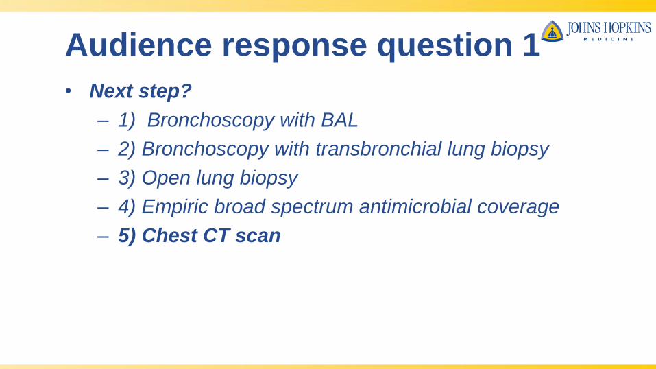
• Next step?
– 1) Bronchoscopy with BAL
– 2) Bronchoscopy with transbronchial lung biopsy
– 3) Open lung biopsy
– 4) Empiric broad spectrum antimicrobial coverage and
wait to see if she improves
– 5) Chest CT scan
Audience response question 1
• Next step?
– 1) Bronchoscopy with BAL
– 2) Bronchoscopy with transbronchial lung biopsy
– 3) Open lung biopsy
– 4) Empiric broad spectrum antimicrobial coverage
– 5) Chest CT scan
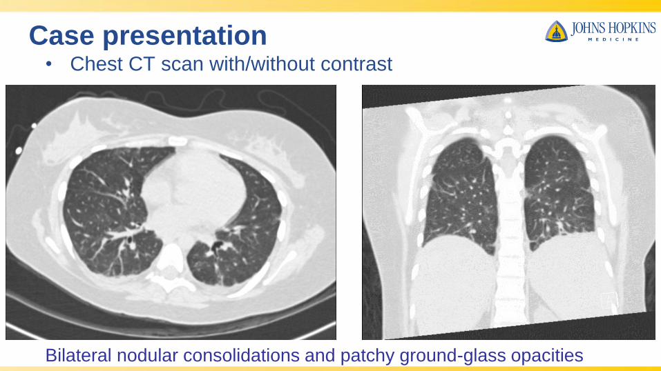
Case presentation• Chest CT scan with/without contrast
Bilateral nodular consolidations and patchy ground-glass opacities

Case discussion
• Dr. Stokes: diagnostic approach
Dennis C. Stokes MD MPHSt. Jude Children’s Research Hospital Professor of
Pediatrics (Pulmonology)
University of Tennessee Health Science Center
Diagnostic approach to the
immunocompromised
host with an unknown pulmonary
process: “pneumopathy X”

Dr. Helen Taussig: Final Meeting Harriet Lane Home Amphitheatre
1974

• Team sport (Radiology, ID, A/I, Surgery)• Likely diagnoses based on host immune defect• Review radiology
• Special thanks to Dr. Dick Heller• Make sure the non-invasive “t’s” are crossed
• Sputum, including induced sputum• Rapid antigen testing, blood work
• Invasive diagnostic studies• Bronchoscopy• Lung biopsy
My approach

• Primary immunodeficiency• CGD: Aspergillus spp, Staph, B. cepacia
• CVID: preRx: encapsulated organisms PostAbRx: Staph, fungi, viral
• Secondary/acquired immunodeficiency
• Neutropenia: H. flu, S. pneumoniae, Staph, Klebsiella
• Immunosuppressive therapies, e.g. cancer therapies• Bacterial: Staph
• Fungal: Aspergillus spp.,Mucor spp, Histoplasmosis
• Viral: CMV, PCP, VZV, HSV, RSV, hMPV
Differential dx based on underlying host defect

• HSCT• Early (<30 days): Pseudomonas other bacterial, Candida spp• Late (>30 days): Staph, Aspergillus spp, CMV, toxo, PCP, EBV, adenovirus, RSV• >100 days: Encapsulated Gram pos, VZV
• Post HSCT non-infectious complications• Edema• VOD• DAH• Idiopathic pneumonia• GVHD• Interstitial lung disease• PTLD• OB• COP
Differential dx based on underlying host defect

• Limited specificity to radiographic patterns• Airspace consolidation
• Hospital/community acquired pneumonias• Fungal pneumonia• Aspiration• Idiopathic pneumonia syndrome• Tb/atypical Tb• DAH• ARDS• Pulmonary edema• TRALI: transfusion related acute lung injury
Radiology

• Nodular lesions• Discrete
• Fungal infection• Nocardia• Metastatic calcifications• PTLD: post transplant lymphoproliferative disease• Malignancy • Septic emboli
• Tree-in-bud pattern• Viral pneumonia• Bacterial pneumonia• BOS
Radiology

• Ground glass opacities• Pulmonary edema• TRALI• ARDS• DAH• CMV• PCP• Viral: CMV, Respiratory (RSV, hMPV, parainfluenza, adenovirus)• Drug injury
Radiology

• CT more sensitive to extent of lung change• May show secondary findings: early cavitary change, pleural
effusions, splenic fungal lesions• Helps plan invasive diagnostic studies: bronchoscopy/BAL, TBB,
needle aspiration biopsy• In suspected BOS, HRCT with inspiratory/expiratory view may be
sufficient to avoid open lung biopsy
Radiology: CT versus plain radiography

• Sputum: induced, or after intubation • Rapid viral panels: RSV, influenza, parainfluenza, Chlamydia• Serum galactomanan for Aspergillus• Urinary antigen, serum antibodies for Histoplasmosis• Genetic probes: P. jirovecii, Legionella, Mycoplasma pneumoniae
Non-invasive testing

• Indications:• Failure to clear with appropriate empiric therapy• Suspicion of endobronchial obstruction (infection,
tumor)• Recurrent pneumonia in lobe or segment• Suspicion of opportunistic infection (e.g. P.
jirovecii)
Bronchoscopy
• Broncho-alveolar lavage
• Bronchoscopic biopsy techniques
• Mucosal biopsy
• “Blind” transbronchial biopsy
• “Guided” biopsy: EBUS, CT-guided/navigational methods• Limitations
• Limited availability of “guided” techniques in pediatrics• Potential application to pulmonary nodular disease• ? Less risk than IR CT guided needle aspiration biopsy
• What is the value of a “negative” bronchoscopy• Narrowing/discontinuing antimicrobial coverage• Fungal pneumonias: yield lowest when done early• May be improved by galactomannan detection BAL
Bronchoscopy

• IR: CT-guided needle aspiration biopsy• Risk of hemorrhage, pneumothorax, non-diagnostic biopsy
• Open lung biopsy
• Thoracotomy
• Thoracoscopic biopsy (VATS)
• NOW BACK TO THE CASE….
Lung biopsy

Case presentation
• She subsequently developed a fever, and was treated for
presumed bacterial pneumonia with a 14 day course of
cefepime, and was started on prophylactic pentamidine
• However, her fever did not improve on antibiotics, and an
inflammatory process was suspected

Case presentation
• Bronchoscopy with BAL and other infectious work-up were
negative
• Nodule biopsy via VATS with RUL wedge resection
showed necrotic and chronic inflammation pathology
– * insert path mage*
Case presentation• Hospital course after lung biopsy:
– Respiratory
• Supplemental oxygen requirement increased to 4 LPM via nasal cannula
– GI
• Stool output improved; were able to decrease dose of IVIG
– Immunology
• IgG levels remained stable on smaller dose of IVIG
– ID
• Fever resolved; no new growth from bronchoscopy alveolar lavage
– Neurologic
• Mental status intact, brain lesion unchanged on repeat imaging
– Endocrine
• Stable on levothyroxine

• Most common primary immunodeficiency• Prevalence: 1:25,000-1:30,000
• Definition (ESID, 2014):• Age > 4
• At least one:• Increased susceptibility to infection• Autoimmune disease• Granulomatous disease• Unexplained polyclonal lymphoproliferation• Affected family member with Ab deficiency
• AND • Marked decrease IgG, IgA (with or w/o low IgM)
• Poor functional Ab response
Common variable immunodeficiency (CVID)

• AND• Secondary causes of hypogammaglobulinemia ruled out
• AND • no evidence of profound T-cell deficiency
Common variable immunodeficiency (CVID)

• Chronic and recurrent infections in 32 children with CVID• Bronchitis 88%• Pneumonia 78%• Sinusitis 78%• OM 69%• Fungal infections (including skin) 47%• GI infections 34%• Skin infections 22%• Parasites 16%• Conjunctivitis 9%• Oral infections 9%
Common variable immunodeficiency (CVID)
Urschel, S et al J. Pediatr 2009;154:888

• Noninfectious pulmonary disease• More common in adolescence, young adulthood• “Granulomatous-lymphocytic interstitial lung disease”
• Granulomatous lung disease• Lymphocytic interstitial lung disease (LIP)• Follicular bronchiolitis• Lymphoid hypeplasia
• Risk of progression to B-cell lymphomas
Common variable immunodeficiency (CVID)
Ambruso, DR, Johnston, RB Primary immunodeficincy (Kendig and Chernick’s Disorders of the
Respiratory Tract in Children, 8th ed, 2012

• Multiple genetic disorders associated with the CVID phenotype• Majority of familial cases appear autosomal dominant
• NFKB2• CD19• TACI• ICOS• PIK3CD gain of function mutations• CTLA-4 loss of function mutations
• LATAIE: similar phenotype but autosomal recessive inheritance• Biallelic mutations in LRPA gene• “LRBA deficiency with autoantibodies, regulatory T (Treg) cell defects,
autoimmune infiltration, and enteropathy”
Emerging genetic basis of CVID
Lo, B et al Blood 2016;128:1037

Audience response question 2
• What diagnostic test would you do next?
– 1) Repeat lung biopsy at a more pathologic area
– 2) Additional targeted immunologic testing
– 3) Whole exome sequencing with targeted genetic
testing
– 4) Stop antibiotics and repeat broncho-alveolar lavage
off antibiotics
Audience response question 2
• What diagnostic test would you do next?
– 1) Repeat lung biopsy at a more pathologic area
– 2) Additional targeted immunologic testing
– 3) Whole exome sequencing with targeted genetic
testing
– 4) Repeat bronchoscopy alveolar lavage off antibiotics

Case presentation
• She had whole exome sequencing that revealed a
missense mutation (c.140 T>C, p.Leu47Pro) in CTLA-4
gene. This was confirmed by targeted genetic sequencing
Case discussion
• CTLA-4 haploinsufficiency as a new model of
immunodeficiency and autoimmunity

Discussion• Cytotoxic T-Lymphocyte associated antigen 4 (CTLA-4)
sends an inhibitory signal to T-cells
NIH: The National Center for Biotechnology Information, 2016; Orencia, 2017

Discussion
• CTLA-4: cytotoxic T lymphocyte antigen 4: critical “checkpoint” of immune response
• Ctla4 knockout mice: lethal multiorganlymphocytic infiltration
• CHAI: syndrome of CTLA-4 haploinsufficiencywith autoimmune infiltration– Heterozygous loss of function mutations
associated with lymphocytic organ infiltrations including lung
NIH:U.S National Library of Medicine, 2016
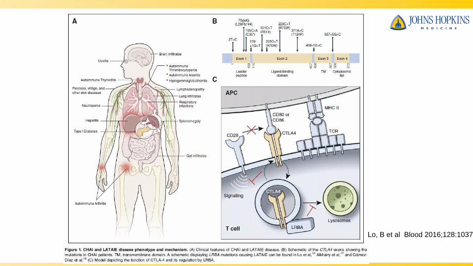
Lo, B et al Blood 2016;128:1037

Case presentation: treatment• Abatacept contains Fc region of immunoglobulin attached
to the CTLA-4. This can replace the CTLA-4 in providing
an inhibitory signal for T-cell activation
Lo et al, 2015; Orencia, 2017
Abatacept
Case presentation: later course
• She was treated with abatacept and sirolimus for CTLA-4
deficiency for additional immunosuppression. She was
continued on IVIG due to hypogammaglobulinemia
• Her symptoms improved, and she was discharged home
on supplemental oxygen 2 LPM via nasal cannula
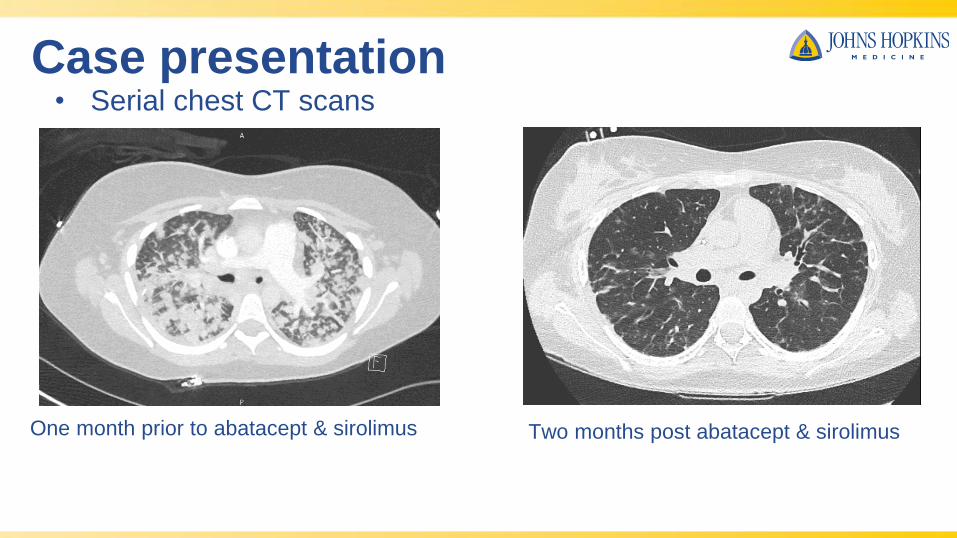
Case presentation
One month prior to abatacept & sirolimus
• Serial chest CT scans
Two months post abatacept & sirolimus

Case presentation: outpatient follow-up
• Ambulating better on room air
• Shortness of breath with walking longer than 30 minutes
or going up stairs
• Albuterol used once over the past month

Pulmonary function tests
Sirolimus started here; abatacept started 2 weeks prior
August 2016 April 2017

Case presentation: outpatient follow-up• Immunology:
– Continued on abatacept, sirolimus, and IVIG. Planning for
bone marrow transplant
• ID:
– Mycobacterium avium-intracellulare treatment: Started on
ethambutol, azithromycin, and rifampin for positive sputum
culture
– Pneumocystis prophylaxis: Switched from pentamidine to
sulfamethoxazole-trimethoprim
• GI:
– Continued on TPN

Questions?

References• Buchbinder, Elizabeth, and F. Stephen Hodi. "Cytotoxic T Lymphocyte Antigen-4 and Immune
Checkpoint Blockade." Journal of Clinical Investigation 125.9 (2015): 3377-383. Web. 3 May
2017. <https://www.jci.org/articles/view/80012/pdf>.
• "CTLA4 Cytotoxic T-lymphocyte Associated Protein 4 [Homo Sapiens (human)] - Gene -
NCBI." National Center for Biotechnology Information. U.S. National Library of Medicine, n.d.
Web. 26 Oct. 2016. <https://www.ncbi.nlm.nih.gov/gene/1493>.
• "CTLA4 Deficiency | NIH: National Institute of Allergy and Infectious Diseases." U.S National
Library of Medicine. U.S. National Library of Medicine, n.d. Web. 27 Oct. 2016.
<https://www.niaid.nih.gov/diseases-conditions/ctla4-deficiency>.
• Lo, B., J. M. Fritz, H. C. Su, G. Uzel, M. B. Jordan, and M. J. Lenardo. "CHAI and LATAIE:
New Genetic Diseases of CTLA-4 Checkpoint Insufficiency." Blood 128.8 (2016): 1037-042.
Web. 1 May 2017. <http://www.bloodjournal.org/content/128/8/1037?sso-checked=true>.
• Lo, Bernice, Kejian Zhang, Wei Lu, et al. "Patients with LRBA Deficiency Show CTLA4 Loss
and Immune Dysregulation Responsive to Abatacept Therapy." Science 349.6246 (2015):
436-40. 24 July 2015. Web. 25 Oct. 2016.
http://science.sciencemag.org/highwire/citation/633231/reference-manager>.
• "ORENCIA." Role of T Cells in Rheumatoid Arthritis. Bristol-Myers Squibb Company, Feb.
2017. Web. 07 May 2017. <http://www.orenciahcp.com/role-of-t-cells-in-rheumatoid-arthritis>.
• Pandit, C, Hsu, P, van Asperen, P et al. Respiratory manifestations and management in
children with common variable immunodeficiency Paediatr Resp Rev 2016;19:56-61.
• Urshel, S, Kayikci, L, Wintergerst, U, et al. Common variable immunodeficiency disorders in
children: delayed diagnosis despite typical clinical presentation. J. Pediatr 2009:154:888-94.