Sagittal growth of the craniofacial complex in normal embryonic mice
4
Archs oral Bid. Vol. 14, pp. 995-997, 1969. Pergamon Press. Printed in Ct. Britain. SAGITTAL GROWTH OF THE CRANIOFACIAL COMPLEX IN NORMAL EMBRYONIC MICE J. C. HART, G. R. SMILEY and A. D. DIXON Dental Research Center, School of Dentistry, University of North Carolina, Chapel Hill, North Carolina 27514, U.S.A. THERE have been many investigations into the aetiology of cleft palate formation from which several theories have evolved regarding normal and abnormal formation of the secondary palate. The majority of observations associated with cleft palate formation have been purely qualitative in nature. The purpose of the study was to quantitatively appraise some of the growth parameters of the craniofacial complex prior to, during, and after palatal closure in normal embryonic mice. Mice of the C57BL strain were chosen for this study since, according to investi- gators such as KALTER (1965), this strain has no spontaneous cleft lip-palate formation and only a 15-20 per cent incidence of cortisone-induced clefts of the palate alone. Inbred C57BL mice were mated twice a week from 9-12 a.m. and checked for vaginal plugs. Mice exhibiting a copulation plug were segregated and that day denoted as the zero day of pregnancy. Gravid females were sacrificed at 10.30 a.m. on the 13th, 14th, 15th, and 16th days of gestation. The uterine horns were removed by Caesarean section and fixed in Bouin’s solution before the embryos were recovered, decapitated, and prepared for paraffin embedding. After careful orientation in the sagittal plane, serial section of embryonic heads were cut at 10 p and stained with a modified Mallory’s trichrome stain. The section most closely representing the midsagittal plane of each head was identified and these sections were photographically enlarged at a standard magnification for subsequent tracing of angular and linear measurements on acetate matte paper (Fig. 1). A series of craniofacial triangles was constructed on the tracings of the photo- graphic enlargements using the basion, the centre of Meckel’s cartilage, the midpoint on the anterior convexity of the nasal septum, and the centre of the hypophysis cerebri as landmarks which could be identified at all ages (Fig. 2). The posterior cranial base, defined as a line from the centre of the hypophysis to the basion, showed the least amount of growth during the time span studied. The posterior cranial base was used for orientation with the centre of the hypophysis being the point of reference. A statistical analysis on the angular and linear measurements was carried out on 15-20 embryos of each age to assess the rate, amount and direction of growth. 995
Transcript of Sagittal growth of the craniofacial complex in normal embryonic mice

Archs oral Bid. Vol. 14, pp. 995-997, 1969. Pergamon Press. Printed in Ct. Britain.
SAGITTAL GROWTH OF THE CRANIOFACIAL COMPLEX IN NORMAL EMBRYONIC MICE
J. C. HART, G. R. SMILEY and A. D. DIXON
Dental Research Center, School of Dentistry, University of North Carolina, Chapel Hill, North Carolina 27514, U.S.A.
THERE have been many investigations into the aetiology of cleft palate formation from which several theories have evolved regarding normal and abnormal formation of the secondary palate. The majority of observations associated with cleft palate formation have been purely qualitative in nature. The purpose of the study was to quantitatively appraise some of the growth parameters of the craniofacial complex prior to, during, and after palatal closure in normal embryonic mice.
Mice of the C57BL strain were chosen for this study since, according to investi- gators such as KALTER (1965), this strain has no spontaneous cleft lip-palate formation and only a 15-20 per cent incidence of cortisone-induced clefts of the palate alone.
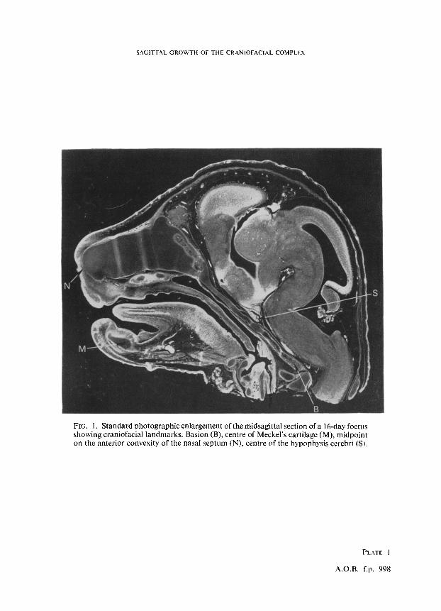
Inbred C57BL mice were mated twice a week from 9-12 a.m. and checked for vaginal plugs. Mice exhibiting a copulation plug were segregated and that day denoted as the zero day of pregnancy. Gravid females were sacrificed at 10.30 a.m. on the 13th, 14th, 15th, and 16th days of gestation. The uterine horns were removed by Caesarean section and fixed in Bouin’s solution before the embryos were recovered, decapitated, and prepared for paraffin embedding. After careful orientation in the sagittal plane, serial section of embryonic heads were cut at 10 p and stained with a modified Mallory’s trichrome stain. The section most closely representing the midsagittal plane of each head was identified and these sections were photographically enlarged at a standard magnification for subsequent tracing of angular and linear measurements on acetate matte paper (Fig. 1).
A series of craniofacial triangles was constructed on the tracings of the photo- graphic enlargements using the basion, the centre of Meckel’s cartilage, the midpoint on the anterior convexity of the nasal septum, and the centre of the hypophysis cerebri as landmarks which could be identified at all ages (Fig. 2).
The posterior cranial base, defined as a line from the centre of the hypophysis to the basion, showed the least amount of growth during the time span studied. The posterior cranial base was used for orientation with the centre of the hypophysis being the point of reference. A statistical analysis on the angular and linear measurements was carried out on 15-20 embryos of each age to assess the rate, amount and direction of growth.
995
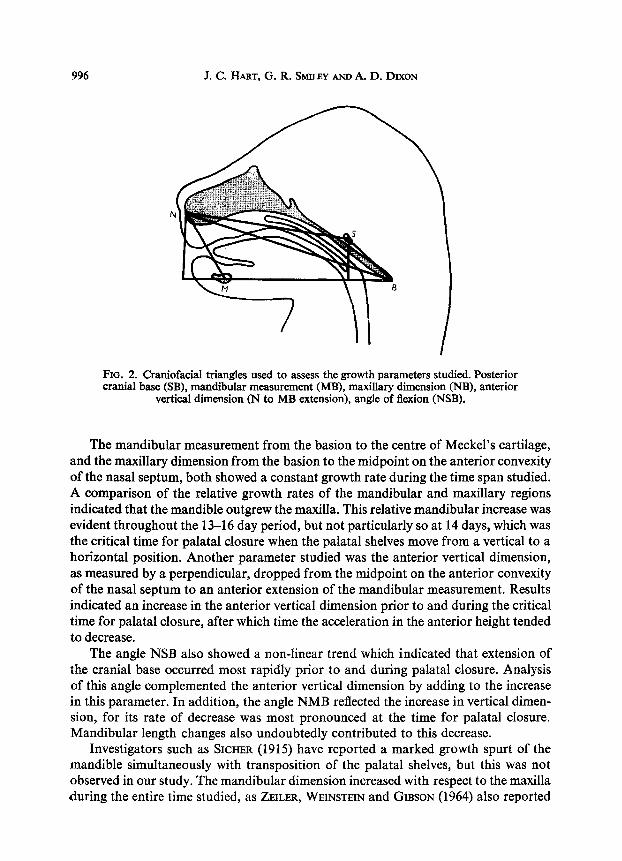
996 J. C. IIART, G. R. SMILEY AND A. D. DD(ON
N
FIO. 2. Craniofacial triangles used to assess the growth parameters studied. Posterior cranial base (SB), mandibular measurement (MB), maxillary dimension (NB), anterior
vertical dimension (N to MB extension), angle of flexion (NSB).
The mandibular measurement from the basion to the centre of Meckel’s cartilage, and the maxillary dimension from the basion to the midpoint on the anterior convexity of the nasal septum, both showed a constant growth rate during the time span studied. A comparison of the relative growth rates of the mandibular and maxillary regions indicated that the mandible outgrew the maxilla. This relative mandibular increase was evident throughout the 13-16 day period, but not particularly so at 14 days, which was the critical time for palatal closure when the palatal shelves move from a vertical to a horizontal position. Another parameter studied was the anterior vertical dimension, as measured by a perpendicular, dropped from the midpoint on the anterior convexity of the nasal septum to an anterior extension of the mandibular measurement. Results indicated an increase in the anterior vertical dimension prior to and during the critical time for palatal closure, after which time the acceleration in the anterior height tended to decrease.
The angle NSB also showed a non-linear trend which indicated that extension of the cranial base occurred most rapidly prior to and during palatal closure. Analysis of this angle complemented the anterior vertical dimension by adding to the increase in this parameter. In addition, the angle NMB reflected the increase in vertical dimen- sion, for its rate of decrease was most pronounced at the time for palatal closure. Mandibular length changes also undoubtedly contributed to this decrease.
Investigators such as SICHER (1915) have reported a marked growth spurt of the mandible simultaneously with transposition of the palatal shelves, but this was not observed in our study. The mandibular dimension increased with respect to the maxilla during the entire time studied, as ZEILER, WEINSTEIN and GIBSON (1964) also reported
SAGITTAL GROWTH OF THE CRANIOFACIAL COMPLEX 997
using morphological ratings in rats; but in our investigation the mandibular measure- ment per se was essentially linear during shelf movement. The increase of the anterior vertical dimension was the most significant finding and since this increase in height occurred prior to and during palatal closure, it suggests that changes in mandibular position and tongue posture could be important prerequisites for shelf movement. FRASER (1967) reviewed numerous processes involved in palate closure and our study emphasizes the significance of mandibular position and cranial base extension. The in- crease in the vertical dimension which occurs before shelf movement could be due to mouth opening reflexes (HUMPHREY, 1968). Height of the craniofacial complex may be more significant than mandibular length in studies which have indicated a relationship of mandibular micrognathia to cleft palate (DEUSCHLE and KALTER, 1962). Our results agreed with those of HARRIS (1967), who observed similar angular changes using morphological criteria.
Controlled breeding overcame some of the difficulties experienced in the use of chronological age (RUGH, 1968). Even with a small sample size for each age, the standard deviation was low, indicating that chronological age was quite consistent with established morphological criteria.
Results will be used to form a basis for further comparison with other normal strains of mice and with experimentally-induced clefts of the palate.
Acknowledgement-This investigation was sponsored by Grant DE-02668, U.S. Public Health Service.
REFERENCES
DEUSCHLE, F. M. and KALTER, H. 1962. Observations on the mandible in association with defects of the lip and palate. J. dent. Res. 41, 1085-1095.
FRASER, F. C. 1967. Cleft lip and cleft palate. Science, N. Y. 158, 1603-1606. HARRIS, J. W. S. 1967. Experimental studies on closure and cleft formation in the secondary palate.
The Scientific Basis of Medicine Annual Review, 357-370. HUMPHREY, T. 1968. The dynamic mechanism of palatal shelf elevation in human fetuses. Anat. Rec.
160,369 (Abs.). KAL’IzR, H. 1965. Experimental investigation of teratogenic action. Ann. N. Y. Acud. Sci. 123,287-294. RUGH, R. 1968. The Mouse: Its Reproduction and Development. Preface, p. i. Burgess, Minneapolis,
Minn., U.S.A. &HER, H. 1915. Die Entwickelung des sekundaeren Gaumens beim Menschen. Anat. Anz. 47,513-
523, 545-562. ZEILER, K. B., WEINSTEIN, S. and GIBSON, R. D. 1964. A study of the morphology and the time of
closure of the palate in the albino rat. Archs oral Biol. 9, 545-554.
SAGITTAL GROWTH OF THE CRANIOFACIAL COMPLEX
FIG. 1. Standard photographic enlargement of the midsagittal section of a 16-day foetus showing craniofacial landmarks. Basion (B), centre of Meckel’s cartilage (M), midpoint on the anterior convexity of the nasal septum (N), centre of the hypophysis cerebri (SJ.
PLATE 1
A.O.R. f.p. 998