Särkämö, Teppo; Tervaniemi, Mari; Soinila, Seppo; Autti, Taina ... · Auditory and Cognitive...
13
This is an electronic reprint of the original article. This reprint may differ from the original in pagination and typographic detail. Powered by TCPDF (www.tcpdf.org) This material is protected by copyright and other intellectual property rights, and duplication or sale of all or part of any of the repository collections is not permitted, except that material may be duplicated by you for your research use or educational purposes in electronic or print form. You must obtain permission for any other use. Electronic or print copies may not be offered, whether for sale or otherwise to anyone who is not an authorised user. Särkämö, Teppo; Tervaniemi, Mari; Soinila, Seppo; Autti, Taina; Silvennoinen, Heli M.; Laine, Matti; Hietanen, Marja; Pihko, Elina Auditory and Cognitive Deficits Associated with Acquired Amusia after Stroke: A Magnetoencephalography and Neuropsychological Follow-Up Study Published in: PloS one DOI: 10.1371/journal.pone.0015157 Published: 01/01/2010 Document Version Publisher's PDF, also known as Version of record Please cite the original version: Särkämö, T., Tervaniemi, M., Soinila, S., Autti, T., Silvennoinen, H. M., Laine, M., ... Pihko, E. (2010). Auditory and Cognitive Deficits Associated with Acquired Amusia after Stroke: A Magnetoencephalography and Neuropsychological Follow-Up Study. PloS one, 5(12), 1-12. [e15157]. https://doi.org/10.1371/journal.pone.0015157
Transcript of Särkämö, Teppo; Tervaniemi, Mari; Soinila, Seppo; Autti, Taina ... · Auditory and Cognitive...
This is an electronic reprint of the original article.This reprint may differ from the original in pagination and typographic detail.
Powered by TCPDF (www.tcpdf.org)
This material is protected by copyright and other intellectual property rights, and duplication or sale of all or part of any of the repository collections is not permitted, except that material may be duplicated by you for your research use or educational purposes in electronic or print form. You must obtain permission for any other use. Electronic or print copies may not be offered, whether for sale or otherwise to anyone who is not an authorised user.
Särkämö, Teppo; Tervaniemi, Mari; Soinila, Seppo; Autti, Taina; Silvennoinen, Heli M.; Laine,Matti; Hietanen, Marja; Pihko, ElinaAuditory and Cognitive Deficits Associated with Acquired Amusia after Stroke: AMagnetoencephalography and Neuropsychological Follow-Up Study
Published in:PloS one
DOI:10.1371/journal.pone.0015157
Published: 01/01/2010
Document VersionPublisher's PDF, also known as Version of record
Please cite the original version:Särkämö, T., Tervaniemi, M., Soinila, S., Autti, T., Silvennoinen, H. M., Laine, M., ... Pihko, E. (2010). Auditoryand Cognitive Deficits Associated with Acquired Amusia after Stroke: A Magnetoencephalography andNeuropsychological Follow-Up Study. PloS one, 5(12), 1-12. [e15157].https://doi.org/10.1371/journal.pone.0015157
Auditory and Cognitive Deficits Associated withAcquired Amusia after Stroke: AMagnetoencephalography and NeuropsychologicalFollow-Up StudyTeppo Sarkamo1,2*, Mari Tervaniemi1,2,3, Seppo Soinila4, Taina Autti5, Heli M. Silvennoinen5, Matti
Laine6, Marja Hietanen4, Elina Pihko7,8
1 Cognitive Brain Research Unit, Institute of Behavioural Sciences, University of Helsinki, Helsinki, Finland, 2 Finnish Centre of Excellence in Interdisciplinary Music
Research, University of Jyvaskyla, Jyvaskyla, Finland, 3 Department of Psychology, University of Jyvaskyla, Jyvaskyla, Finland, 4 Department of Neurology, Helsinki
University Central Hospital, Helsinki, Finland, 5 Department of Radiology, Helsinki University Central Hospital, Helsinki, Finland, 6 Department of Psychology, Abo Akademi
University, Turku, Finland, 7 BioMag Laboratory, Hospital District of Helsinki and Uusimaa, HUSLAB, Helsinki University Central Hospital, Helsinki, Finland, 8 Brain Research
Unit, Low Temperature Laboratory, Aalto University School of Science and Technology, Espoo, Finland
Abstract
Acquired amusia is a common disorder after damage to the middle cerebral artery (MCA) territory. However, itsneurocognitive mechanisms, especially the relative contribution of perceptual and cognitive factors, are still unclear. Westudied cognitive and auditory processing in the amusic brain by performing neuropsychological testing as well asmagnetoencephalography (MEG) measurements of frequency and duration discrimination using magnetic mismatchnegativity (MMNm) recordings. Fifty-three patients with a left (n = 24) or right (n = 29) hemisphere MCA stroke (MRI verified)were investigated 1 week, 3 months, and 6 months after the stroke. Amusia was evaluated using the Montreal Battery ofEvaluation of Amusia (MBEA). We found that amusia caused by right hemisphere damage (RHD), especially to temporal andfrontal areas, was more severe than amusia caused by left hemisphere damage (LHD). Furthermore, the severity of amusiawas found to correlate with weaker frequency MMNm responses only in amusic RHD patients. Additionally, within the RHDsubgroup, the amusic patients who had damage to the auditory cortex (AC) showed worse recovery on the MBEA as well asweaker MMNm responses throughout the 6-month follow-up than the non-amusic patients or the amusic patients withoutAC damage. Furthermore, the amusic patients both with and without AC damage performed worse than the non-amusicpatients on tests of working memory, attention, and cognitive flexibility. These findings suggest domain-general cognitivedeficits to be the primary mechanism underlying amusia without AC damage whereas amusia with AC damage is associatedwith both auditory and cognitive deficits.
Citation: Sarkamo T, Tervaniemi M, Soinila S, Autti T, Silvennoinen HM, et al. (2010) Auditory and Cognitive Deficits Associated with Acquired Amusia after Stroke:A Magnetoencephalography and Neuropsychological Follow-Up Study. PLoS ONE 5(12): e15157. doi:10.1371/journal.pone.0015157
Editor: Ted M. Dawson, Johns Hopkins Institute for Cell Engineering, United States of America
Received August 11, 2010; Accepted October 22, 2010; Published December 2, 2010
Copyright: � 2010 Sarkamo et al. This is an open-access article distributed under the terms of the Creative Commons Attribution License, which permitsunrestricted use, distribution, and reproduction in any medium, provided the original author and source are credited.
Funding: This work has been funded by the Academy of Finland (project no. 77322) (http://www.aka.fi/fi/A/); the Jenny and Antti Wihuri Foundation, Helsinki,Finland (http://www.wihurinrahasto.fi/); the National Graduate School of Psychology, Finland (http://www.gspsy.fi/index.php?id = 64); and the Finnish BrainFoundation, Finland (http://www.aivosaatio.fi/). The funders had no role in study design, data collection and analysis, decision to publish, or preparation of themanuscript.
Competing Interests: The authors have declared that no competing interests exist.
* E-mail: [email protected]
Introduction
Abnormal brain development or brain damage can lead to a
deficit in perceiving music, a condition known as amusia.
Although studies of both congenital and acquired forms of amusia
have surged during the past 20 years (for a recent review, see [1]),
the neural mechanisms as well as the cognitive consequences
associated with the condition are still unclear. Converging
evidence from lesion studies [2] and modern structural MRI
studies in individuals with congenital amusia [3–5] points to a
network of temporal and frontal lobe areas, especially in the right
hemisphere, as the critical brain substrate of amusia. However, the
relative contribution of perceptual and cognitive factors to amusia
is still under debate. Do amusic persons have difficulty already in
discriminating low-level acoustical features, such as sound
frequency and duration, that are crucial to music or is the deficit
more related to an impaired cognitive analysis of music
information?
Based on observed double dissociations between acquired
amusia and impairment in the perception of speech (aphasia)
and other non-musical sounds (auditory agnosia) (e.g. [6–8]), it has
been proposed that there are mental modules in the brain that are
specific to music perception [9]. However, recent behavioural
evidence from congenital amusia suggests that amusic people can
also have deficits in basic auditory discrimination [10], pitch
memory [11–13], phonological and phonemic awareness [14],
speech intonation processing [15–18], emotional prosody percep-
tion [19], and spatial processing ([20] but see also [21] for
discrepant results). These findings suggest that the behavioural
impairment in amusia may not be entirely specific to music
PLoS ONE | www.plosone.org 1 December 2010 | Volume 5 | Issue 12 | e15157
perception. This view is also supported by evidence from studies of
healthy subjects showing that music listening evokes widespread
activation of many frontal, temporal, parietal, and subcortical
areas related to for example attention, working memory, semantic
and episodic memory, and emotional processing rather than being
an endeavour of the auditory cortices alone (e.g. [22–25]).
Previously, the neural ability of amusic individuals to process
music information has been studied with electroencephalography
(EEG) by recording event-related potentials (ERPs) to acoustic
changes within tone sequences or melodies. Especially the
mismatch negativity (MMN) component is well-suited for this
purpose because it is an early ERP response elicited preattentively
to any acoustical change in a repetitive sound stream [26,27].
Current ERP evidence indicates that individuals with congenital
amusia have relatively normal early responses (N2, MMN) but
abnormal later attention-modulated responses (P3, P600) to small
pitch changes within tone sequences or melodies ([28–30] but see
also [31] for conflicting results). Also in a recent functional
magnetic resonance imaging (fMRI) study, individuals with
congenital amusia showed reduced activity in the right inferior
frontal gyrus (IFG) to small pitch changes whereas the activity in
their left and right auditory cortex (AC) was comparable to control
subjects [32]. This is in line with the fMRI studies of healthy
subjects showing that judging or making decisions about auditory
stimuli recruits domain-general frontal areas [33,34], such as the
inferior frontal gyrus (IFG), which are involved in processing
response conflict, perceptual difficulty, novelty, and working
memory [35]. Collectively, these results suggest that domain-
general auditory and cognitive processes, mediated by neural
structures beyond the AC, are linked to the music perception
deficit in congenital amusia. However, very little is currently
known about the contribution of auditory and cognitive deficits in
acquired amusia.
Previously, there have been only two relatively small auditory
ERP studies of acquired amusia (both with 12 patients). Munte
and colleagues reported that stroke patients with acquired amusia
had grossly reduced MMN responses to pitch changes [36] as well
as decreased P3a responses to novel environmental sounds [37]
compared with non-amusic patients and healthy control subjects.
Interestingly, the amusic patients also showed worse performance
on a behavioural auditory alertness test [37]. Although based on a
relatively small number of patients, these results suggest that
deficits in automatic sound-change detection and attention-
orienting could be associated with acquired amusia. However, to
date no study has systematically and directly explored which brain
areas underlie acquired amusia and how it is related to other
auditory and cognitive deficits.
In a previous study [38], we performed repeated neuropsycho-
logical testing in amusic and non-amusic patients (total n = 53)
with a middle cerebral artery (MCA) stroke during a 6-month
post-stroke period. We found that acquired amusia and its
recovery were associated with a wide range of cognitive functions,
especially attention, executive functioning, and working memory
[38]. By using the same patient sample, the aim of the present
magnetoencephalography (MEG) study was to explore whether
the amusic and the non-amusic patients would differ in auditory
discrimination, as indicated by the cortically generated magnetic
mismatch negativity (MMNm) response to changes in basic
acoustic features, such as sound pitch and duration, during
recovery. Previous studies using almost identical stimuli have
verified that healthy subjects are easily able to discriminate the
changes in pitch (500 to 575 Hz) and duration (75 to 25 ms) used
in the present study and also show robust MMN (MMNm)
responses to these changes [39,40]. Moreover, we sought to
determine how the laterality of the cerebral damage as well as the
presence of AC damage would influence the recovery of music
perception and auditory discrimination in amusia. Specifically, we
hypothesized that amusic patients with and without AC damage
would show different patterns of MMNm and cognitive deficits
(especially in tests of attention, executive functioning, and working
memory) compared with non-amusic patients during the 6-month
recovery period.
Methods
Subjects and procedureSubjects (n = 53) were non-musician stroke patients recruited
from the Department of Neurology of the Helsinki University
Central Hospital (HUCH) to a randomized clinical trial about the
effectiveness of music and audio book listening on stroke recovery
(for a more detailed description of patient characteristics and
methodology, see [41,42]). All patients had an acute left (n = 24) or
right (n = 29) MCA territory ischemic stroke verified by MRI, no
prior neurological or psychiatric disease, drug or alcohol abuse
and they were right-handed, #75 years old, Finnish-speaking, and
able to co-operate. In addition, also patients who reported any
problems in basic auditory perception (e.g., clearly worsened
hearing, presbycusis, use of hearing aids, tinnitus, or Meniere’s
disease) before the stroke were excluded. As a part of the trial, all
patients underwent a neuropsychological assessment of cognitive
recovery and an MEG measurement of auditory discrimination 1
week, 3 months, and 6 months after the stroke as well as a
structural 1.5 T MRI scan with routine sequences for stroke 2
weeks and 6 months post-stroke. The size and location of the
lesion(s) were classified by neuroradiologists (authors T.A. and
H.M.S) as previously described [41]. In addition, lesions of the
auditory cortex were recorded. The study was approved by the
Ethics Committee of the Hospital District of Helsinki and
Uusimaa. The ethical permission granted the use of this data for
both basic research related to auditory and music processing after
stroke and for applied research related to the therapeutic effects of
music listening. Since this study falls in the former category, it is
clearly within the bounds of the ethical permission. All patients
signed an informed consent stating that all information gathered
from them during the study can be stored by the researchers
according to the Finnish legislation on the concealment of
confidential information and used anonymously for research
purposes.
The patient sample and the data corpus in the present study is
the same that was previously used to study the effects of music and
audio book listening on stroke recovery [41,42]. In those studies,
music listening was found to enhance verbal memory and focused
attention, prevent depressed and confused mood, and increase the
amplitude of the frequency MMNm in the right hemisphere
whereas audio book listening had no effect on cognition or mood
but increased duration and frequency MMNm amplitudes in the
right hemisphere [41,42]. However, as we reported previously
[38], the music, audio book, and control groups did not differ in
the recovery of music perception. Moreover, the number of non-
amusic and amusic patients was approximately the same in the
music (10 vs. 8; x2 = 0.22, p = 0.637) and audio book (7 vs. 11;
x2 = 0.89, p = 0.346) groups and their recovery did not differ in
verbal memory and focused attention [38] or in the amplitude of
the frequency MMNm [mixed-model ANOVA Time x Group
interaction F(2, 30) = 0.20, p = 0.823 and F(2, 32) = 0.19,
p = 0.829, respectively] or the duration MMNm [F(2, 30)
= 1.09, p = 0.351 and F(2, 32) = 0.42, p = 0.659, respectively] in
the right hemisphere. Thus, we can conclude that comparing
Auditory and Cognitive Deficits in Acquired Amusia
PLoS ONE | www.plosone.org 2 December 2010 | Volume 5 | Issue 12 | e15157
amusic and non-amusic patients using the MMNm and neuro-
psychological data from the three time points (1 week, 3 months,
and 6 months post-stroke) was not in any way biased by the
intervention.
Assessment of cognition and music perceptionCognitive performance was assessed with an extensive (duration
about 3 h) neuropsychological testing battery, which included tests
of working memory, verbal learning and memory, verbal
expression and comprehension, visuospatial cognition, executive
functioning, and attention. Details concerning the neuropsycho-
logical tests are presented in Table 1. Parallel test versions of the
memory tests were used in different testing occasions to minimize
practice effects. Reaction time tests were always performed using
the better, non-paretic hand. All assessments were carried out in a
quiet room reserved for clinical neuropsychological assessments.
The 1-week post-stroke assessment was carried out in two or three
testing sessions to avoid interference due to fatigue. On the
average, the assessments were spread over 2.98 days (range 2–7
days).
As a part of the neuropsychological testing, also music
perception was evaluated 1 week and 3 months post-stroke by
using a shortened version [38] of the Montreal Battery of
Evaluation of Amusia (MBEA) [50]. The original MBEA includes
six subtests (each with 30 items) that measure different components
of music cognition (scale, contour, interval, rhythm, and meter
perception, and recognition memory). Using the same stimuli and
structure as in the original MBEA, we created a shortened version,
which included only 14 items per subtest and thereby reduced the
length of the test from 1.5 h to 45 min. The use of a shorter
version was crucial in the present study due to time constraints as
well as patient fatigue and the severity of cognitive deficits in the
acute post-stroke stage. The stimuli were presented using a
portable CD player and head-arch headphones. Before the test,
sound volume was adjusted to a clearly audible but comfortable
level individually for each patient. Due to time constraints, we
were unfortunately not able perform audiometry to verify the basic
hearing ability of the patients. However, no particular auditory
difficulties were observed during normal conversation or during
the neuropsychological assessment.
As it turned out, all of the 53 patients were able to perform the
Scale and Rhythm subtests but only 44 were able to complete all
the six subtests at the 1-week post-stroke stage. Since the Scale and
Rhythm subtests were highly correlated with the other subtests
and with each other [38], we opted to use their average score
(referred to hereafter as the MBEA average score) in determining
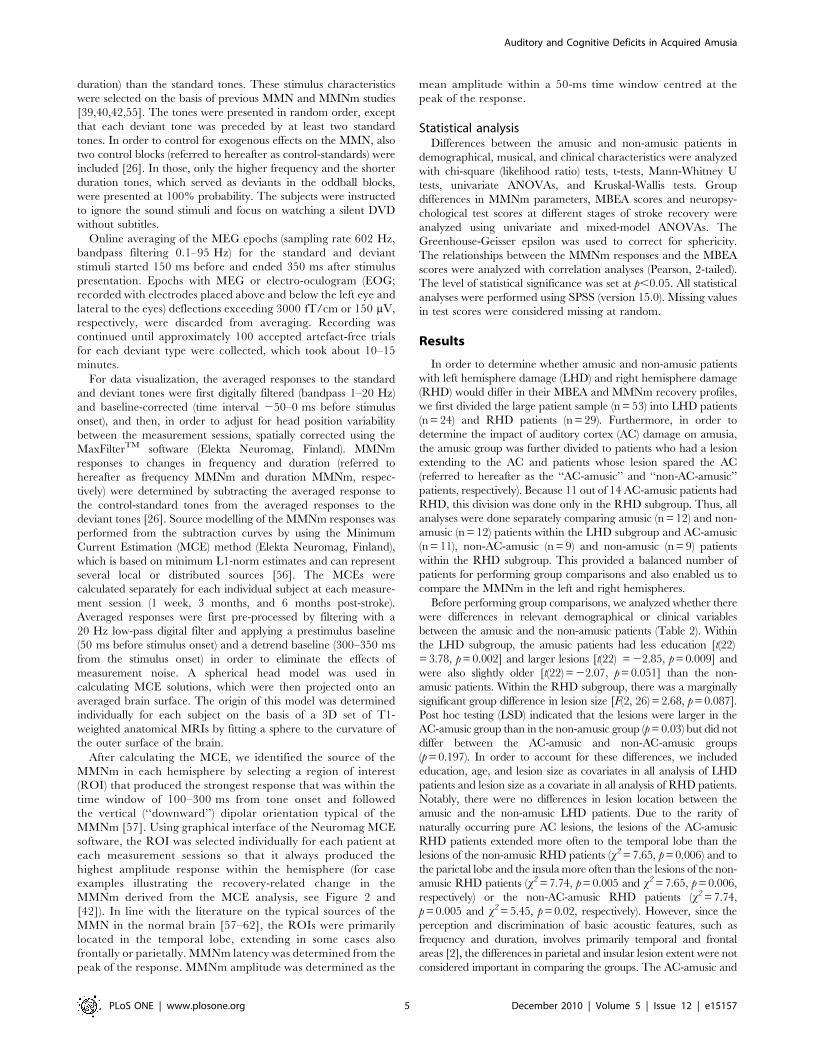
Table 1. Neuropsychological tests performed 1 week, 3 months, and 6 months post-stroke.
Test Task of the subject Reference
Working memory
Digit span (WMS-R) Recall number sequences [43]
Memory interference Recall sets of 3 words after interfering tasks [44]
Verbal learning and memory
Auditory list learning Recall a list of 10 words (3 trials + delayed recall) [44]
Story recall (RBMT) Immediate and delayed recall of a narrated story [45]
Verbal expression and comprehension
Repetition (BDAE) Repeat heard words and sentences [46]
Reading (BDAE) Read out words and sentences [46]
Semantic fluency (CERAD) Say words in the animal category in 60 s. [47]
Naming (CERAD) Name objects from line drawings [47]
Short Token test Comprehension of verbal instructions [48]
Visuospatial cognition
Clock task Recognize time and draw clock hands [44]
Copying designs Draw copies of 4 geometric designs [44]
Shortened BVRT Draw 5 geometric designs from memory [49]
Music cognition
Shortened MBEA Detect changes in musical melodies [38,50]
Executive functions and attention
FAB Perform a set of short mental and motor tasks [51]
Phonemic fluency Say words beginning with letter ‘‘s’’ in 60 s. [44]
Balloons test Cancel targets in a visuospatial array [52]
Simple reaction time (CS) Press key when visual target appears [53]
Subtraction task (CS) Press key after mental subtraction [53]
Stroop task (CS) Press key in a colour response conflict situation [53]
Vigilance task (CS) Press key when target letter appears (15 min) [53]
Abbreviations: BDAE: Boston Diagnostic Aphasia Examination, BVRT: Benton Visual Retention Test, CERAD: The Consortium to Establish a Registry for Alzheimer’sDisease, CS: CogniSpeed� reaction time software, FAB: Frontal Assessment Battery, MBEA: Montreal Battery of Evaluation of Amusia, RBMT: Rivermead BehavioralMemory Test, WMS-R: Wechsler Memory Scale - Revised.doi:10.1371/journal.pone.0015157.t001
Auditory and Cognitive Deficits in Acquired Amusia
PLoS ONE | www.plosone.org 3 December 2010 | Volume 5 | Issue 12 | e15157
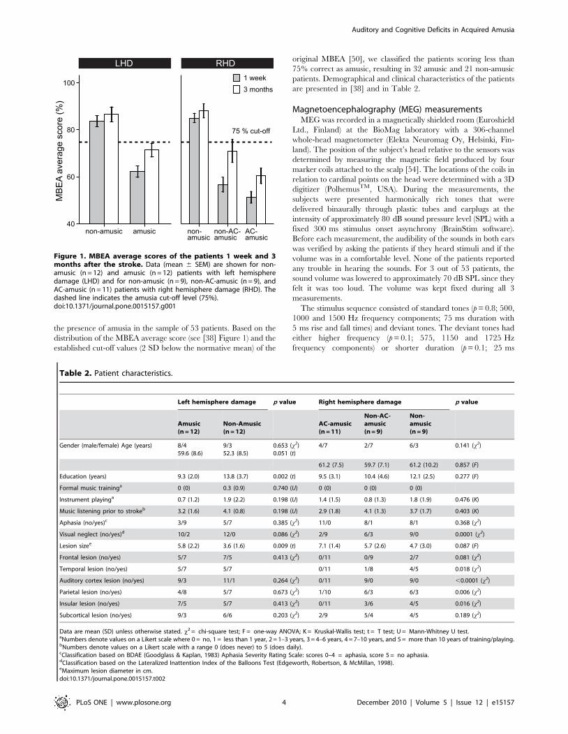
the presence of amusia in the sample of 53 patients. Based on the
distribution of the MBEA average score (see [38] Figure 1) and the
established cut-off values (2 SD below the normative mean) of the
original MBEA [50], we classified the patients scoring less than
75% correct as amusic, resulting in 32 amusic and 21 non-amusic
patients. Demographical and clinical characteristics of the patients
are presented in [38] and in Table 2.
Magnetoencephalography (MEG) measurementsMEG was recorded in a magnetically shielded room (Euroshield
Ltd., Finland) at the BioMag laboratory with a 306-channel
whole-head magnetometer (Elekta Neuromag Oy, Helsinki, Fin-
land). The position of the subject’s head relative to the sensors was
determined by measuring the magnetic field produced by four
marker coils attached to the scalp [54]. The locations of the coils in
relation to cardinal points on the head were determined with a 3D
digitizer (PolhemusTM, USA). During the measurements, the
subjects were presented harmonically rich tones that were
delivered binaurally through plastic tubes and earplugs at the
intensity of approximately 80 dB sound pressure level (SPL) with a
fixed 300 ms stimulus onset asynchrony (BrainStim software).
Before each measurement, the audibility of the sounds in both ears
was verified by asking the patients if they heard stimuli and if the
volume was in a comfortable level. None of the patients reported
any trouble in hearing the sounds. For 3 out of 53 patients, the
sound volume was lowered to approximately 70 dB SPL since they
felt it was too loud. The volume was kept fixed during all 3
measurements.
The stimulus sequence consisted of standard tones (p = 0.8; 500,
1000 and 1500 Hz frequency components; 75 ms duration with
5 ms rise and fall times) and deviant tones. The deviant tones had
either higher frequency (p = 0.1; 575, 1150 and 1725 Hz
frequency components) or shorter duration (p = 0.1; 25 ms
Figure 1. MBEA average scores of the patients 1 week and 3months after the stroke. Data (mean 6 SEM) are shown for non-amusic (n = 12) and amusic (n = 12) patients with left hemispheredamage (LHD) and for non-amusic (n = 9), non-AC-amusic (n = 9), andAC-amusic (n = 11) patients with right hemisphere damage (RHD). Thedashed line indicates the amusia cut-off level (75%).doi:10.1371/journal.pone.0015157.g001
Table 2. Patient characteristics.
Left hemisphere damage p value Right hemisphere damage p value
Amusic(n = 12)
Non-Amusic(n = 12)
AC-amusic(n = 11)
Non-AC-amusic(n = 9)
Non-amusic(n = 9)
Gender (male/female) Age (years) 8/459.6 (8.6)
9/352.3 (8.5)
0.653 (x2)0.051 (t)
4/7 2/7 6/3 0.141 (x2)
61.2 (7.5) 59.7 (7.1) 61.2 (10.2) 0.857 (F)
Education (years) 9.3 (2.0) 13.8 (3.7) 0.002 (t) 9.5 (3.1) 10.4 (4.6) 12.1 (2.5) 0.277 (F)
Formal music traininga 0 (0) 0.3 (0.9) 0.740 (U) 0 (0) 0 (0) 0 (0)
Instrument playinga 0.7 (1.2) 1.9 (2.2) 0.198 (U) 1.4 (1.5) 0.8 (1.3) 1.8 (1.9) 0.476 (K)
Music listening prior to strokeb 3.2 (1.6) 4.1 (0.8) 0.198 (U) 2.9 (1.8) 4.1 (1.3) 3.7 (1.7) 0.403 (K)
Aphasia (no/yes)c 3/9 5/7 0.385 (x2) 11/0 8/1 8/1 0.368 (x2)
Visual neglect (no/yes)d 10/2 12/0 0.086 (x2) 2/9 6/3 9/0 0.0001 (x2)
Lesion sizee 5.8 (2.2) 3.6 (1.6) 0.009 (t) 7.1 (1.4) 5.7 (2.6) 4.7 (3.0) 0.087 (F)
Frontal lesion (no/yes) 5/7 7/5 0.413 (x2) 0/11 0/9 2/7 0.081 (x2)
Temporal lesion (no/yes) 5/7 5/7 0/11 1/8 4/5 0.018 (x2)
Auditory cortex lesion (no/yes) 9/3 11/1 0.264 (x2) 0/11 9/0 9/0 ,0.0001 (x2)
Parietal lesion (no/yes) 4/8 5/7 0.673 (x2) 1/10 6/3 6/3 0.006 (x2)
Insular lesion (no/yes) 7/5 5/7 0.413 (x2) 0/11 3/6 4/5 0.016 (x2)
Subcortical lesion (no/yes) 9/3 6/6 0.203 (x2) 2/9 5/4 4/5 0.189 (x2)
Data are mean (SD) unless otherwise stated. x2 = chi-square test; F = one-way ANOVA; K = Kruskal-Wallis test; t = T test; U = Mann-Whitney U test.aNumbers denote values on a Likert scale where 0 = no, 1 = less than 1 year, 2 = 1–3 years, 3 = 4–6 years, 4 = 7–10 years, and 5 = more than 10 years of training/playing.bNumbers denote values on a Likert scale with a range 0 (does never) to 5 (does daily).cClassification based on BDAE (Goodglass & Kaplan, 1983) Aphasia Severity Rating Scale: scores 0–4 = aphasia, score 5 = no aphasia.dClassification based on the Lateralized Inattention Index of the Balloons Test (Edgeworth, Robertson, & McMillan, 1998).eMaximum lesion diameter in cm.doi:10.1371/journal.pone.0015157.t002
Auditory and Cognitive Deficits in Acquired Amusia
PLoS ONE | www.plosone.org 4 December 2010 | Volume 5 | Issue 12 | e15157
duration) than the standard tones. These stimulus characteristics
were selected on the basis of previous MMN and MMNm studies
[39,40,42,55]. The tones were presented in random order, except
that each deviant tone was preceded by at least two standard
tones. In order to control for exogenous effects on the MMN, also
two control blocks (referred to hereafter as control-standards) were
included [26]. In those, only the higher frequency and the shorter
duration tones, which served as deviants in the oddball blocks,
were presented at 100% probability. The subjects were instructed
to ignore the sound stimuli and focus on watching a silent DVD
without subtitles.
Online averaging of the MEG epochs (sampling rate 602 Hz,
bandpass filtering 0.1–95 Hz) for the standard and deviant
stimuli started 150 ms before and ended 350 ms after stimulus
presentation. Epochs with MEG or electro-oculogram (EOG;
recorded with electrodes placed above and below the left eye and
lateral to the eyes) deflections exceeding 3000 fT/cm or 150 mV,
respectively, were discarded from averaging. Recording was
continued until approximately 100 accepted artefact-free trials
for each deviant type were collected, which took about 10–15
minutes.
For data visualization, the averaged responses to the standard
and deviant tones were first digitally filtered (bandpass 1–20 Hz)
and baseline-corrected (time interval 250–0 ms before stimulus
onset), and then, in order to adjust for head position variability
between the measurement sessions, spatially corrected using the
MaxFilterTM software (Elekta Neuromag, Finland). MMNm
responses to changes in frequency and duration (referred to
hereafter as frequency MMNm and duration MMNm, respec-
tively) were determined by subtracting the averaged response to
the control-standard tones from the averaged responses to the
deviant tones [26]. Source modelling of the MMNm responses was
performed from the subtraction curves by using the Minimum
Current Estimation (MCE) method (Elekta Neuromag, Finland),
which is based on minimum L1-norm estimates and can represent
several local or distributed sources [56]. The MCEs were
calculated separately for each individual subject at each measure-
ment session (1 week, 3 months, and 6 months post-stroke).
Averaged responses were first pre-processed by filtering with a
20 Hz low-pass digital filter and applying a prestimulus baseline
(50 ms before stimulus onset) and a detrend baseline (300–350 ms
from the stimulus onset) in order to eliminate the effects of
measurement noise. A spherical head model was used in
calculating MCE solutions, which were then projected onto an
averaged brain surface. The origin of this model was determined
individually for each subject on the basis of a 3D set of T1-
weighted anatomical MRIs by fitting a sphere to the curvature of
the outer surface of the brain.
After calculating the MCE, we identified the source of the
MMNm in each hemisphere by selecting a region of interest
(ROI) that produced the strongest response that was within the
time window of 100–300 ms from tone onset and followed
the vertical (‘‘downward’’) dipolar orientation typical of the
MMNm [57]. Using graphical interface of the Neuromag MCE
software, the ROI was selected individually for each patient at
each measurement sessions so that it always produced the
highest amplitude response within the hemisphere (for case
examples illustrating the recovery-related change in the
MMNm derived from the MCE analysis, see Figure 2 and
[42]). In line with the literature on the typical sources of the
MMN in the normal brain [57–62], the ROIs were primarily
located in the temporal lobe, extending in some cases also
frontally or parietally. MMNm latency was determined from the
peak of the response. MMNm amplitude was determined as the
mean amplitude within a 50-ms time window centred at the
peak of the response.
Statistical analysisDifferences between the amusic and non-amusic patients in
demographical, musical, and clinical characteristics were analyzed
with chi-square (likelihood ratio) tests, t-tests, Mann-Whitney U
tests, univariate ANOVAs, and Kruskal-Wallis tests. Group
differences in MMNm parameters, MBEA scores and neuropsy-
chological test scores at different stages of stroke recovery were
analyzed using univariate and mixed-model ANOVAs. The
Greenhouse-Geisser epsilon was used to correct for sphericity.
The relationships between the MMNm responses and the MBEA
scores were analyzed with correlation analyses (Pearson, 2-tailed).
The level of statistical significance was set at p,0.05. All statistical
analyses were performed using SPSS (version 15.0). Missing values
in test scores were considered missing at random.
Results
In order to determine whether amusic and non-amusic patients
with left hemisphere damage (LHD) and right hemisphere damage
(RHD) would differ in their MBEA and MMNm recovery profiles,
we first divided the large patient sample (n = 53) into LHD patients
(n = 24) and RHD patients (n = 29). Furthermore, in order to
determine the impact of auditory cortex (AC) damage on amusia,
the amusic group was further divided to patients who had a lesion
extending to the AC and patients whose lesion spared the AC
(referred to hereafter as the ‘‘AC-amusic’’ and ‘‘non-AC-amusic’’
patients, respectively). Because 11 out of 14 AC-amusic patients had
RHD, this division was done only in the RHD subgroup. Thus, all
analyses were done separately comparing amusic (n = 12) and non-
amusic (n = 12) patients within the LHD subgroup and AC-amusic
(n = 11), non-AC-amusic (n = 9) and non-amusic (n = 9) patients
within the RHD subgroup. This provided a balanced number of
patients for performing group comparisons and also enabled us to
compare the MMNm in the left and right hemispheres.
Before performing group comparisons, we analyzed whether there
were differences in relevant demographical or clinical variables
between the amusic and the non-amusic patients (Table 2). Within
the LHD subgroup, the amusic patients had less education [t(22)
= 3.78, p = 0.002] and larger lesions [t(22) = 22.85, p = 0.009] and
were also slightly older [t(22) = 22.07, p = 0.051] than the non-
amusic patients. Within the RHD subgroup, there was a marginally
significant group difference in lesion size [F(2, 26) = 2.68, p = 0.087].
Post hoc testing (LSD) indicated that the lesions were larger in the
AC-amusic group than in the non-amusic group (p = 0.03) but did not
differ between the AC-amusic and non-AC-amusic groups
(p = 0.197). In order to account for these differences, we included
education, age, and lesion size as covariates in all analysis of LHD
patients and lesion size as a covariate in all analysis of RHD patients.
Notably, there were no differences in lesion location between the
amusic and the non-amusic LHD patients. Due to the rarity of
naturally occurring pure AC lesions, the lesions of the AC-amusic
RHD patients extended more often to the temporal lobe than the
lesions of the non-amusic RHD patients (x2 = 7.65, p = 0.006) and to
the parietal lobe and the insula more often than the lesions of the non-
amusic RHD patients (x2 = 7.74, p = 0.005 and x2 = 7.65, p = 0.006,
respectively) or the non-AC-amusic RHD patients (x2 = 7.74,
p = 0.005 and x2 = 5.45, p = 0.02, respectively). However, since the
perception and discrimination of basic acoustic features, such as
frequency and duration, involves primarily temporal and frontal
areas [2], the differences in parietal and insular lesion extent were not
considered important in comparing the groups. The AC-amusic and
Auditory and Cognitive Deficits in Acquired Amusia
PLoS ONE | www.plosone.org 5 December 2010 | Volume 5 | Issue 12 | e15157
Auditory and Cognitive Deficits in Acquired Amusia
PLoS ONE | www.plosone.org 6 December 2010 | Volume 5 | Issue 12 | e15157
the non-AC-amusic RHD patients did not differ in the proportion of
frontal (100% in both) or temporal lobe lesions (100% vs. 89%,
x2 = 1.66, p = 0.197), suggesting that any differences between them
could be attributed to the AC damage. Finally, we also compared the
amusic LHD patients (n = 12) and the amusic RHD patients (n = 20)
in demographical and clinical variables. Compared with the amusic
LHD group, the amusic RHD group had a higher proportion of
women (x2 = 4.15, p = 0.042) and their lesions extended more often to
frontal (x2 = 4.15, p = 0.042), temporal (x2 = 6.64, p = 0.01), insular
(x2 = 6.54, p = 0.011), and subcortical (x2 = 4.97, p = 0.026) areas.
However, their overall lesion sizes did not differ [t(22) = 20.84,
p = 0.408]. Thus, female gender was included as a covariate when
comparing these groups.
MBEA performance in amusic and non-amusic patientsGroup differences in the MBEA average score (Figure 1) were
analyzed first using univariate ANOVAs for the 1-week post-stroke
data and then, in order determine whether the group differences
were stable over time, also using mixed-model ANOVAs for the 1-
week and 3-month post-stroke data. Separate analyses were
performed for the LHD (amusic vs. non-amusic) and RHD (AC-
amusic vs. non-AC-amusic vs. non-amusic) subgroups. In the LHD
subgroup, the MBEA average score was significantly lower in the
amusic than in the non-amusic patients at the 1-week post-stroke
stage [F(1, 19) = 17.8, p = 0.0005] and also throughout the 3-month
post-stroke period [F(1, 19) = 17.82, p = 0.0005]. Also in the RHD
subgroup, there were significant group differences on the MBEA
average score both 1 week post-stroke [F(2, 25) = 40.36, p,0.0001]
and during the 3-month post-stroke period [F(2, 25) = 25.13,
p,0.0001]. Post hoc testing (LSD) showed that the MBEA average
score was clearly lower in both the AC-amusic and the non-AC-
amusic groups than in the non-amusic group (p,0.0001 in both
comparisons) but also lower in the AC-amusic group than in the
non-AC-amusic group (p,0.05) during the 3-month follow-up.
Using the same amusia classification criterion as in the 1-week post-
stroke stage, a significantly higher percentage of the AC-amusic
patients than the non-AC-amusic patients also remained amusic at
the 3-month post-stroke stage (91% vs. 44%, x2 = 5.37, p = 0.021).
Finally, we also compared the amusic LHD patients (n = 12) and the
amusic RHD patients (n = 20) and found that the MBEA average
score was lower in the amusic RHD patients at the 1-week stage
[F(1, 29) = 8.29, p = 0.007] and throughout the 3-month period
[F(1, 29) = 5.58, p = 0.025]. However, similar improvement in the
MBEA average score from the 1-week to the 3-month stage was
seen in both amusic LHD and RHD patients.
In summary, these results suggest that RHD causes a more severe
deficit in music perception than LHD but the laterality of cerebral
damage does not influence the behavioural recovery of amusia.
Moreover, at least in right hemisphere stroke, damage to the auditory
cortex seems to be a crucial factor limiting the recovery of amusia.
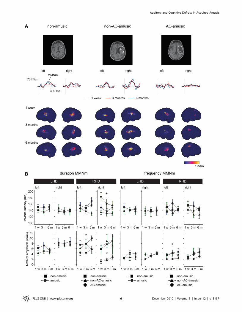
MMNm in amusic and non-amusic patientsAt the 1-week post-stroke stage, both frequency and duration
deviants elicited MMNm responses that peaked around 150 ms.
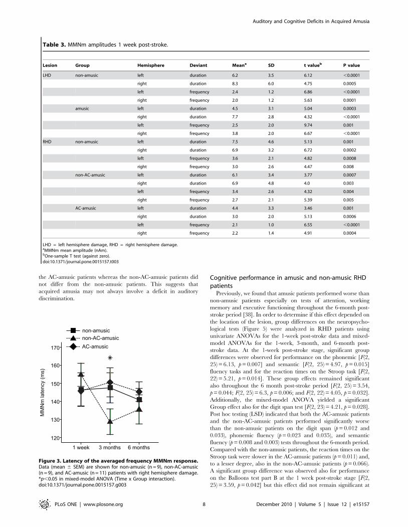
The MMNn mean amplitudes (Table 3) differed significantly from
zero in both ipsilesional and contralesional hemispheres in all
patient groups. Group differences in the latency and amplitude of
the duration MMNm and the frequency MMNm responses
(Figure 2) were analyzed using univariate ANOVAs for the 1-week
post-stroke data and mixed-model ANOVAs for the 1-week, 3-
month, and 6-month post-stroke data. In the LHD subgroup, no
significant differences between the amusic and the non-amusic
patients were observed at the 1-week post-stroke stage or during
the 6-month period. In the RHD subgroup, there were significant
group differences in right hemisphere duration MMNm latency
[F(2, 25) = 4.47, p = 0.022] and amplitude [F(2, 25) = 3.56,
p = 0.043] 1 week post-stroke with post hoc tests (LSD) showing
a longer latency and a smaller amplitude in the AC-amusic group
than in the non-amusic (p = 0.029 and 0.017) or non-AC-amusic
(p = 0.002 and 0.017) groups. These Group effects remained
significant also during the 6-month period [F(2, 25) = 3.5,
p = 0.046 and F(2, 25) = 3.47, p = 0.047]. Additionally, significant
Group effects were also observed in the amplitude of the duration
MMNm [F(2, 25) = 4.55, p = 0.021] and the frequency MMNm
[F(2, 25) = 3.84, p = 0.035] in the left hemisphere during the 6-
month period. Post hoc tests showed that the left hemisphere
duration MMNm amplitude was smaller in the AC-amusic group
than in the non-amusic group (p = 0.006) and the left hemisphere
frequency MMNm amplitude was smaller in the AC-amusic group
than in both non-amusic (p = 0.014) and non-AC-amusic
(p = 0.056) groups. Importantly, there were no significant differ-
ences between the non-AC-amusic and non-amusic groups during
the 6-month follow-up (p = 0.132–0.937).
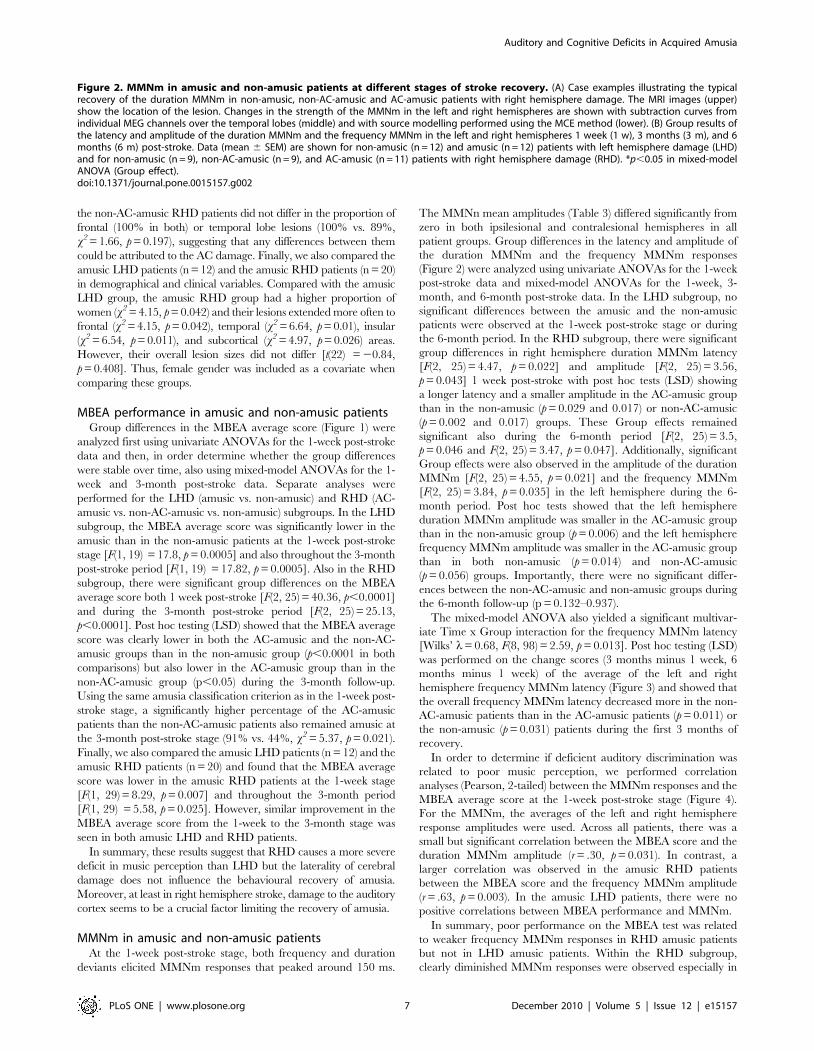
The mixed-model ANOVA also yielded a significant multivar-
iate Time x Group interaction for the frequency MMNm latency
[Wilks’ l= 0.68, F(8, 98) = 2.59, p = 0.013]. Post hoc testing (LSD)
was performed on the change scores (3 months minus 1 week, 6
months minus 1 week) of the average of the left and right
hemisphere frequency MMNm latency (Figure 3) and showed that
the overall frequency MMNm latency decreased more in the non-
AC-amusic patients than in the AC-amusic patients (p = 0.011) or
the non-amusic (p = 0.031) patients during the first 3 months of
recovery.
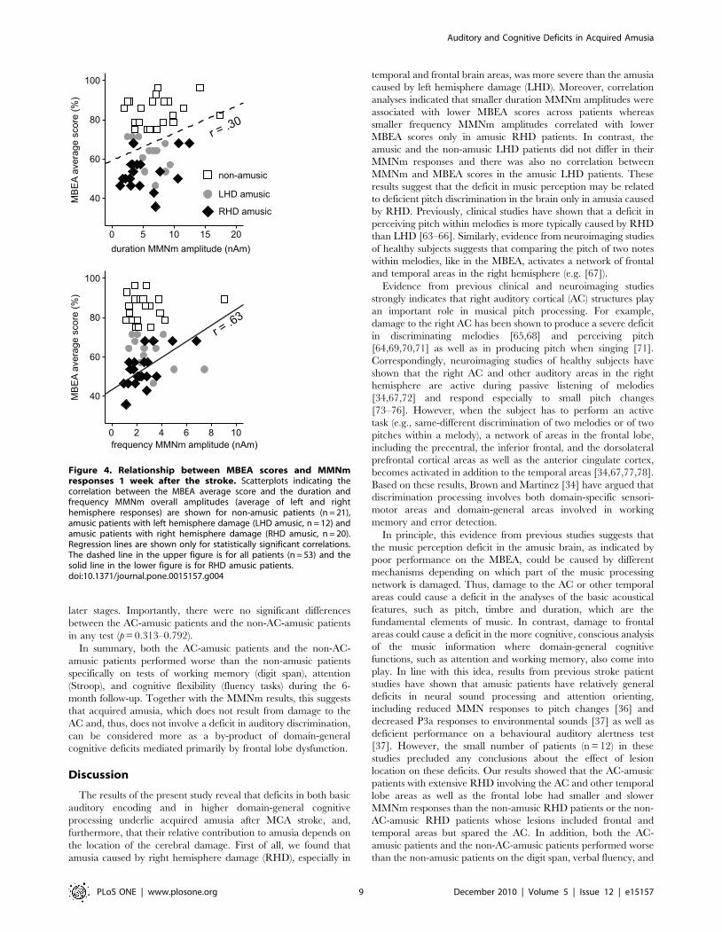
In order to determine if deficient auditory discrimination was
related to poor music perception, we performed correlation
analyses (Pearson, 2-tailed) between the MMNm responses and the
MBEA average score at the 1-week post-stroke stage (Figure 4).
For the MMNm, the averages of the left and right hemisphere
response amplitudes were used. Across all patients, there was a
small but significant correlation between the MBEA score and the
duration MMNm amplitude (r = .30, p = 0.031). In contrast, a
larger correlation was observed in the amusic RHD patients
between the MBEA score and the frequency MMNm amplitude
(r = .63, p = 0.003). In the amusic LHD patients, there were no
positive correlations between MBEA performance and MMNm.
In summary, poor performance on the MBEA test was related
to weaker frequency MMNm responses in RHD amusic patients
but not in LHD amusic patients. Within the RHD subgroup,
clearly diminished MMNm responses were observed especially in
Figure 2. MMNm in amusic and non-amusic patients at different stages of stroke recovery. (A) Case examples illustrating the typicalrecovery of the duration MMNm in non-amusic, non-AC-amusic and AC-amusic patients with right hemisphere damage. The MRI images (upper)show the location of the lesion. Changes in the strength of the MMNm in the left and right hemispheres are shown with subtraction curves fromindividual MEG channels over the temporal lobes (middle) and with source modelling performed using the MCE method (lower). (B) Group results ofthe latency and amplitude of the duration MMNm and the frequency MMNm in the left and right hemispheres 1 week (1 w), 3 months (3 m), and 6months (6 m) post-stroke. Data (mean 6 SEM) are shown for non-amusic (n = 12) and amusic (n = 12) patients with left hemisphere damage (LHD)and for non-amusic (n = 9), non-AC-amusic (n = 9), and AC-amusic (n = 11) patients with right hemisphere damage (RHD). *p,0.05 in mixed-modelANOVA (Group effect).doi:10.1371/journal.pone.0015157.g002
Auditory and Cognitive Deficits in Acquired Amusia
PLoS ONE | www.plosone.org 7 December 2010 | Volume 5 | Issue 12 | e15157
the AC-amusic patients whereas the non-AC-amusic patients did
not differ from the non-amusic patients. This suggests that
acquired amusia may not always involve a deficit in auditory
discrimination.
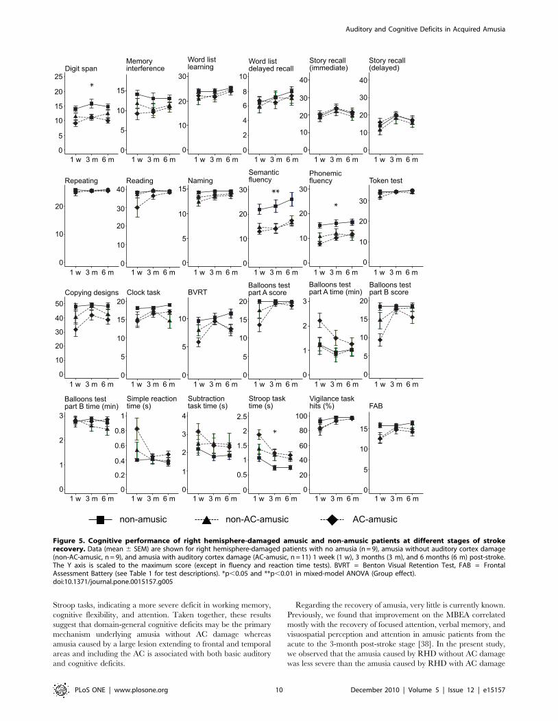
Cognitive performance in amusic and non-amusic RHDpatients
Previously, we found that amusic patients performed worse than
non-amusic patients especially on tests of attention, working
memory and executive functioning throughout the 6-month post-
stroke period [38]. In order to determine if this effect depended on
the location of the lesion, group differences on the neuropsycho-
logical tests (Figure 5) were analyzed in RHD patients using
univariate ANOVAs for the 1-week post-stroke data and mixed-
model ANOVAs for the 1-week, 3-month, and 6-month post-
stroke data. At the 1-week post-stroke stage, significant group
differences were observed for performance on the phonemic [F(2,
25) = 6.13, p = 0.007] and semantic [F(2, 25) = 4.97, p = 0.015]
fluency tasks and for the reaction times on the Stroop task [F(2,
22) = 5.21, p = 0.014]. These group effects remained significant
also throughout the 6 month post-stroke period [F(2, 25) = 3.54,
p = 0.044; F(2, 25) = 6.3, p = 0.006; and F(2, 22) = 4.05, p = 0.032].
Additionally, the mixed-model ANOVA yielded a significant
Group effect also for the digit span test [F(2, 23) = 4.21, p = 0.028].
Post hoc testing (LSD) indicated that both the AC-amusic patients
and the non-AC-amusic patients performed significantly worse
than the non-amusic patients on the digit span (p = 0.012 and
0.033), phonemic fluency (p = 0.023 and 0.035), and semantic
fluency (p = 0.008 and 0.003) tests throughout the 6-month period.
Compared with the non-amusic patients, the reaction times on the
Stroop task were slower in the AC-amusic patients (p = 0.011) and,
to a lesser degree, also in the non-AC-amusic patients (p = 0.066).
A significant group difference was observed also for performance
on the Balloons test part B at the 1 week post-stroke stage [F(2,
25) = 3.59, p = 0.042] but this effect did not remain significant at
Figure 3. Latency of the averaged frequency MMNm response.Data (mean 6 SEM) are shown for non-amusic (n = 9), non-AC-amusic(n = 9), and AC-amusic (n = 11) patients with right hemisphere damage.*p,0.05 in mixed-model ANOVA (Time x Group interaction).doi:10.1371/journal.pone.0015157.g003
Table 3. MMNm amplitudes 1 week post-stroke.
Lesion Group Hemisphere Deviant Meana SD t valueb P value
LHD non-amusic left duration 6.2 3.5 6.12 ,0.0001
right duration 8.3 6.0 4.75 0.0005
left frequency 2.4 1.2 6.86 ,0.0001
right frequency 2.0 1.2 5.63 0.0001
amusic left duration 4.5 3.1 5.04 0.0003
right duration 7.7 2.8 4.32 ,0.0001
left frequency 2.5 2.0 9.74 0.001
right frequency 3.8 2.0 6.67 ,0.0001
RHD non-amusic left duration 7.5 4.6 5.13 0.001
right duration 6.9 3.2 6.72 0.0002
left frequency 3.6 2.1 4.82 0.0008
right frequency 3.0 2.6 4.47 0.008
non-AC-amusic left duration 6.1 3.4 3.77 0.0007
right duration 6.9 4.8 4.0 0.003
left frequency 3.4 2.6 4.32 0.004
right frequency 2.7 2.1 5.39 0.005
AC-amusic left duration 4.4 3.3 3.46 0.001
right duration 3.0 2.0 5.13 0.0006
left frequency 2.1 1.0 6.55 ,0.0001
right frequency 2.2 1.4 4.91 0.0004
LHD = left hemisphere damage, RHD = right hemisphere damage.aMMNm mean amplitude (nAm).bOne-sample T test (against zero).doi:10.1371/journal.pone.0015157.t003
Auditory and Cognitive Deficits in Acquired Amusia
PLoS ONE | www.plosone.org 8 December 2010 | Volume 5 | Issue 12 | e15157
later stages. Importantly, there were no significant differences
between the AC-amusic patients and the non-AC-amusic patients
in any test (p = 0.313–0.792).
In summary, both the AC-amusic patients and the non-AC-
amusic patients performed worse than the non-amusic patients
specifically on tests of working memory (digit span), attention
(Stroop), and cognitive flexibility (fluency tasks) during the 6-
month follow-up. Together with the MMNm results, this suggests
that acquired amusia, which does not result from damage to the
AC and, thus, does not involve a deficit in auditory discrimination,
can be considered more as a by-product of domain-general
cognitive deficits mediated primarily by frontal lobe dysfunction.
Discussion
The results of the present study reveal that deficits in both basic
auditory encoding and in higher domain-general cognitive
processing underlie acquired amusia after MCA stroke, and,
furthermore, that their relative contribution to amusia depends on
the location of the cerebral damage. First of all, we found that
amusia caused by right hemisphere damage (RHD), especially in
temporal and frontal brain areas, was more severe than the amusia
caused by left hemisphere damage (LHD). Moreover, correlation
analyses indicated that smaller duration MMNm amplitudes were
associated with lower MBEA scores across patients whereas
smaller frequency MMNm amplitudes correlated with lower
MBEA scores only in amusic RHD patients. In contrast, the
amusic and the non-amusic LHD patients did not differ in their
MMNm responses and there was also no correlation between
MMNm and MBEA scores in the amusic LHD patients. These
results suggest that the deficit in music perception may be related
to deficient pitch discrimination in the brain only in amusia caused
by RHD. Previously, clinical studies have shown that a deficit in
perceiving pitch within melodies is more typically caused by RHD
than LHD [63–66]. Similarly, evidence from neuroimaging studies
of healthy subjects suggests that comparing the pitch of two notes
within melodies, like in the MBEA, activates a network of frontal
and temporal areas in the right hemisphere (e.g. [67]).
Evidence from previous clinical and neuroimaging studies
strongly indicates that right auditory cortical (AC) structures play
an important role in musical pitch processing. For example,
damage to the right AC has been shown to produce a severe deficit
in discriminating melodies [65,68] and perceiving pitch
[64,69,70,71] as well as in producing pitch when singing [71].
Correspondingly, neuroimaging studies of healthy subjects have
shown that the right AC and other auditory areas in the right
hemisphere are active during passive listening of melodies
[34,67,72] and respond especially to small pitch changes
[73–76]. However, when the subject has to perform an active
task (e.g., same-different discrimination of two melodies or of two
pitches within a melody), a network of areas in the frontal lobe,
including the precentral, the inferior frontal, and the dorsolateral
prefrontal cortical areas as well as the anterior cingulate cortex,
becomes activated in addition to the temporal areas [34,67,77,78].
Based on these results, Brown and Martinez [34] have argued that
discrimination processing involves both domain-specific sensori-
motor areas and domain-general areas involved in working
memory and error detection.
In principle, this evidence from previous studies suggests that
the music perception deficit in the amusic brain, as indicated by
poor performance on the MBEA, could be caused by different
mechanisms depending on which part of the music processing
network is damaged. Thus, damage to the AC or other temporal
areas could cause a deficit in the analyses of the basic acoustical
features, such as pitch, timbre and duration, which are the
fundamental elements of music. In contrast, damage to frontal
areas could cause a deficit in the more cognitive, conscious analysis
of the music information where domain-general cognitive
functions, such as attention and working memory, also come into
play. In line with this idea, results from previous stroke patient
studies have shown that amusic patients have relatively general
deficits in neural sound processing and attention orienting,
including reduced MMN responses to pitch changes [36] and
decreased P3a responses to environmental sounds [37] as well as
deficient performance on a behavioural auditory alertness test
[37]. However, the small number of patients (n = 12) in these
studies precluded any conclusions about the effect of lesion
location on these deficits. Our results showed that the AC-amusic
patients with extensive RHD involving the AC and other temporal
lobe areas as well as the frontal lobe had smaller and slower
MMNm responses than the non-amusic RHD patients or the non-
AC-amusic RHD patients whose lesions included frontal and
temporal areas but spared the AC. In addition, both the AC-
amusic patients and the non-AC-amusic patients performed worse
than the non-amusic patients on the digit span, verbal fluency, and
Figure 4. Relationship between MBEA scores and MMNmresponses 1 week after the stroke. Scatterplots indicating thecorrelation between the MBEA average score and the duration andfrequency MMNm overall amplitudes (average of left and righthemisphere responses) are shown for non-amusic patients (n = 21),amusic patients with left hemisphere damage (LHD amusic, n = 12) andamusic patients with right hemisphere damage (RHD amusic, n = 20).Regression lines are shown only for statistically significant correlations.The dashed line in the upper figure is for all patients (n = 53) and thesolid line in the lower figure is for RHD amusic patients.doi:10.1371/journal.pone.0015157.g004
Auditory and Cognitive Deficits in Acquired Amusia
PLoS ONE | www.plosone.org 9 December 2010 | Volume 5 | Issue 12 | e15157
Stroop tasks, indicating a more severe deficit in working memory,
cognitive flexibility, and attention. Taken together, these results
suggest that domain-general cognitive deficits may be the primary
mechanism underlying amusia without AC damage whereas
amusia caused by a large lesion extending to frontal and temporal
areas and including the AC is associated with both basic auditory
and cognitive deficits.
Regarding the recovery of amusia, very little is currently known.
Previously, we found that improvement on the MBEA correlated
mostly with the recovery of focused attention, verbal memory, and
visuospatial perception and attention in amusic patients from the
acute to the 3-month post-stroke stage [38]. In the present study,
we observed that the amusia caused by RHD without AC damage
was less severe than the amusia caused by RHD with AC damage
Figure 5. Cognitive performance of right hemisphere-damaged amusic and non-amusic patients at different stages of strokerecovery. Data (mean 6 SEM) are shown for right hemisphere-damaged patients with no amusia (n = 9), amusia without auditory cortex damage(non-AC-amusic, n = 9), and amusia with auditory cortex damage (AC-amusic, n = 11) 1 week (1 w), 3 months (3 m), and 6 months (6 m) post-stroke.The Y axis is scaled to the maximum score (except in fluency and reaction time tests). BVRT = Benton Visual Retention Test, FAB = FrontalAssessment Battery (see Table 1 for test descriptions). *p,0.05 and **p,0.01 in mixed-model ANOVA (Group effect).doi:10.1371/journal.pone.0015157.g005
Auditory and Cognitive Deficits in Acquired Amusia
PLoS ONE | www.plosone.org 10 December 2010 | Volume 5 | Issue 12 | e15157
during the 3-month post-stroke period. Overall, less than half
(44%) of the non-AC-amusic RHD patients could still be classified
as amusic (scoring below the 75% cut-off in the MBEA Scale and
Rhythm subtests) at the 3 month stage whereas a vast majority
(91%) of the AC-amusic RHD patients remained amusic at the 3
month stage. Thus, severe and persistent amusia seems to be
caused especially by extensive damage to the right hemisphere
covering the AC and other temporal and frontal lobe areas.
Interestingly, the non-AC-amusic RHD patients also showed faster
pitch discrimination, as indicated by the shortening of the
frequency MMNm latency during first the 3 months of recovery,
than the AC-amusic RHD patients or the non-amusic RHD
patients. Since the severity of cognitive deficits was comparable
between the non-AC-amusic and the AC-amusic patients
throughout the follow-up, it is plausible that the speed-up in pitch
discrimination taking place in the early post-stroke stage may be
one important mechanism underlying the recovery of music
perception in the non-AC-amusic patients.
There are a few notable methodological limitations to this study,
which should be taken into account when interpreting the results.
Firstly, due to time constraints, we were not able to include
audiometry to determine the basic auditory and hearing
capabilities of the patients in the study. However, since we (1)
excluded all patients with a history of problems in basic auditory
perception, (2) did not observe any notable hearing deficits in the
patients during normal conversation or during the neuropsycho-
logical assessment, (3) made sure that the auditory stimuli MBEA
as well as those used in the MEG were clearly audible to the
patients, and (4) verified that the amplitude of the MMNm differed
from zero in all the patient groups (including the AC-amusic RHD
patients) at the 1-week post-stroke stage, we can be confident that
the MBEA or MMNm results were not biased by potential
problems in basic auditory perception. Secondly, the small
number of amusic patients with left AC damage (n = 3) limits
the conclusions about the role of AC damage in amusia only to
RHD patients. Due to the fact that focal damage restricted to the
AC is extremely rare after an ischemic stroke, patients with left AC
damage often have large lesions extending to temporal, frontal,
parietal, and subcortical areas, and, consequently, are severely
aphasic, which naturally precludes their recruitment. Thus, studies
with larger sample sizes of LHD patients (especially with AC
damage) are still needed to provide information about the
potential neural and cognitive correlates of amusia after LHD
and also to gain more insight about the relationship between
aphasia and amusia. Thirdly, the large overlap in the lesion
locations (i.e. the lesions typically covered many cortical and
subcortical areas) of our patient sample and, consequently, the
relatively rough anatomical classification used in the present study
precludes making more precise inferences about the roles of
different brain structures in amusia. In the future, studies that use
more advanced analyses of lesion locations (for example with
voxel-based morphometry) on a larger patient sample or that focus
on patients who have specific damage to certain key brain
structures (e.g., AC and other temporal areas, IFG) would help to
shed more light on how different areas contribute to music
perception in the brain.
In conclusion, the present data indicate that the severity and
recovery of amusia caused by cerebral damage as well as the
relative contribution of perceptual and cognitive factors in amusia
depend on which parts of the large-scale neural network governing
music perception and cognition are damaged. It seems that, at
least in the right hemisphere, damage to the AC together with
damage to other temporal and frontal structures leads to a severe
and persistent form of amusia that is characterized by deficits in
both low-level auditory processing and higher cognitive functions.
In contrast, damage to temporal and frontal areas that spares the
AC results in a more transient form of amusia, which is related
primarily to cognitive deficits. Clinically, this information may be
important in helping derive a more accurate prognosis of the
musical deficit. Identifying whether the amusic patient might
benefit more from the training of musical perceptual skills (e.g.,
melody discrimination training [79]) or cognitive skills (e.g.,
attention and memory training) would also be important in
guiding the development and application of potential rehabilita-
tion interventions for amusia.
Acknowledgments
We thank the staffs of the HUCH Department of Neurology and other
rehabilitation hospitals in the Helsinki metropolitan area for their
collaboration, the patient subjects and their families for their participation
and effort, and Mikko Mikkonen, Suvi Heikkila, Minna Huotilainen, Anna
Shestakova, Irina Anourova, Jyrki Makela, and Jussi Nurminen for their
help in data collection and analysis.
Author Contributions
Conceived and designed the experiments: TS MT SS TA ML MH EP.
Performed the experiments: TS TA HMS. Analyzed the data: TS MT TA
HMS EP. Contributed reagents/materials/analysis tools: TS SS MT TA
HMS ML MH EP. Wrote the paper: TS MT SS TA ML MH EP.
References
1. Alossa N, Castelli L (2009) Amusia and musical functioning. Eur Neurol 61:
269–277.
2. Stewart L, von Kriegstein K, Warren JD, Griffiths TD (2006) Music and the
brain: Disorders of musical listening. Brain 129: 2533–2553.
3. Hyde KL, Zatorre RJ, Griffiths TD, Lerch JP, Peretz I (2006) Morphometry of
the amusic brain: A two-site study. Brain 129: 2562–2570.
4. Hyde KL, Lerch JP, Zatorre RJ, Griffiths TD, Evans AC, et al. (2007) Cortical
thickness in congenital amusia: When less is better than more. J Neurosci 27:
13028–13032.
5. Mandell J, Schulze K, Schlaug G (2007) Congenital amusia: An auditory-motor
feedback disorder? Restor Neurol Neuros 25: 323–334.
6. Griffiths TD, Rees A, Witton C, Cross PM, Shakir RA, et al. (1997) Spatial and
temporal auditory processing deficits following right hemisphere infarction: A
psychophysical study. Brain 120: 785–794.
7. Mendez M (2001) Generalized auditory agnosia with spared music recognition in
a left-hander. Analysis of a case with a right temporal stroke. Cortex 37: 139–150.
8. Peretz I, Kolinsky R, Tramo M, Labrecque R, Hublet C, et al. (1994) Functional
dissociations following bilateral lesions of auditory cortex. Brain 117:
1283–1301.
9. Peretz I, Coltheart M (2003) Modularity of music processing. Nat Neurosci 6:
688–691.
10. Jones JL, Zalewski C, Brewer C, Lucker J, Drayna D (2009) Widespread
auditory deficits in tune deafness. Ear Hearing 30: 63–72.
11. Gosselin N, Jolicoeur P, Peretz I (2009) Impaired memory for pitch in congenital
amusia. Ann NY Acad Sci 1169: 270–272.
12. Tillmann B, Schulze K, Foxton JM (2009) Congenital amusia: A short-term
memory deficit for non-verbal, but not verbal sounds. Brain Cognition 71:
259–264.
13. Williamson VJ, McDonald C, Deutsch D, Griffiths TD, Stewart L (2010) Faster
decline of pitch memory over time in congenital amusia. Adv Cogn Psychol 6:
15–22.
14. Jones JL, Lucker J, Zalewski C, Brewer C, Drayna D (2009) Phonological
processing in adults with deficits in musical pitch recognition. J Commun Disord
42: 226–234.
15. Jiang C, Hamm JP, Lim VK, Kirk IJ, Yang Y (2010) Processing melodic contour
and speech intonation in congenital amusics with Mandarin Chinese.
Neuropsychologia 48: 2630–2639.
16. Liu F, Patel AD, Fourcin A, Stewart L (2010) Intonation processing in congenital
amusia: Discrimination, identification and imitation. Brain 133: 1682–1693.
17. Patel AD, Foxton JM, Griffiths TD (2005) Musically tone-deaf individuals have
difficulty discriminating intonation contours extracted from speech. Brain
Cognition 59: 310–313.
Auditory and Cognitive Deficits in Acquired Amusia
PLoS ONE | www.plosone.org 11 December 2010 | Volume 5 | Issue 12 | e15157
18. Patel AD, Wong M, Foxton J, Lochy A, Peretz I (2008) Speech intonation
perception deficits in musical tone deafness (congenital amusia). Music Percept25: 357–368.
19. Thompson WF (2007) Exploring variants of amusia: Tone deafness, rhythm
impairment, and intonation insensitivity. In: Schubert E, Buckley K, Eliott R,Koboroff B, Chen J, et al. (2007) Proceedings of the International Conference on
Music Communication Science. Sydney: HCSNet. pp 159–163.20. Douglas KM, Bilkey DK (2007) Amusia is associated with deficits in spatial
processing. Nat Neurosci 10: 915–921.
21. Tillmann B, Jolicoeur P, Ishihara M, Gosselin N, Bertrand O, et al. (2010) Theamusic brain: Lost in music, but not in space. PLoS ONE 5: e10173.
doi:10.1371/journal.pone.0010173.22. Janata P, Tillmann B, Bharucha JJ (2002) Listening to polyphonic music recruits
domain-general attention and working memory circuits. Cogn Aff BehavNeurosci 2: 121–140.
23. Koelsch S, Siebel WA (2005) Towards a neural basis of music perception.
Trends Cogn Sci 9: 578–584.24. Koelsch S (2010) Towards a neural basis of music-evoked emotions. Trends
Cogn Sci 14: 131–137.25. Platel H, Baron JC, Desgranges B, Bernard F, Eustache F (2003) Semantic and
episodic memory of music are subserved by distinct neural networks. Neuro-
image 20: 244–256.26. Kujala T, Tervaniemi M, Schroger E (2007) The mismatch negativity in
cognitive and clinical neuroscience: Theoretical and methodological consider-ations. Biol Psychol 74: 1–19.
27. Naatanen R, Paavilainen P, Rinne T, Alho K (2007) The mismatch negativity(MMN) in basic research of central auditory processing: A review. Clin
Neurophysiol 118: 2544–2590.
28. Moreau P, Jolicoeur P, Peretz I (2009) Automatic brain responses to pitchchanges in congenital amusia. Ann NY Acad Sci 1169: 191–194.
29. Peretz I, Brattico E, Tervaniemi M (2005) Abnormal electrical brain responsesto pitch in congenital amusia. Ann Neurol 58: 478–482.
30. Peretz I, Brattico E, Jarvenpaa M, Tervaniemi M (2009) The amusic brain: In
tune, out of key, and unaware. Brain 132: 1277–1286.31. Braun A, McArdle J, Jones J, Nechaev V, Zalewski C (2008) Tune deafness:
Processing melodic errors outside of conscious awareness as reflected bycomponents of the auditory ERP. PLoS ONE 3: e2349. doi:10.1371/journal.
pone.0002349.32. Hyde KL, Zatorre RJ, Peretz I (in press) Functional MRI evidence of an
abnormal neural network for pitch processing in congenital amusia. Cereb
Cortex;in press.33. Binder JR, Liebenthal E, Possing ET, Medler DA, Ward BD (2004) Neural
correlates of sensory and decision processes in auditory object identification. NatNeurosci 7: 295–301.
34. Brown S, Martinez MJ (2007) Activation of premotor vocal areas during musical
discrimination. Brain Cognition 63: 59–69.35. Duncan J, Owen AM (2000) Common regions of the human frontal lobe
recruited by diverse cognitive demands. Trends Neurosci 23: 475–483.36. Kohlmetz C, Altenmuller E, Schuppert M, Wieringa BM, Munte TF (2001)
Deficit in automatic sound-change detection may underlie some musicperception deficits after acute hemispheric stroke. Neuropsychologia 39:
1121–1124.
37. Munte TF, Schuppert M, Johannes S, Wieringa BM, Kohlmetz C, et al. (1998)Brain potentials in patients with music perception deficits: Evidence for an early
locus. Neurosci Lett 256: 85–88.38. Sarkamo T, Tervaniemi M, Soinila S, Autti T, Silvennoinen HM, et al. (2009)
Cognitive deficits associated with acquired amusia after stroke: A neuropsycho-
logical follow-up study. Neuropsychologia 47: 2642–2651.39. Tervaniemi M, Lehtokoski A, Sinkkonen J, Virtanen J, Ilmoniemi RJ, et al.
(1999) Test-retest reliability of mismatch negativity for duration, frequency andintensity changes. Clin Neurophysiol 110: 1388–1393.
40. Tervaniemi M, Sinkkonen J, Virtanen J, Kallio J, Ilmoniemi RJ, et al. (2005)
Test-retest stability of the magnetic mismatch response (MMNm). ClinNeurophysiol 116: 1897–1905.
41. Sarkamo T, Tervaniemi M, Laitinen S, Forsblom A, Soinila S, et al. (2008)Music listening enhances cognitive recovery and mood after middle cerebral
artery stroke. Brain 131: 866–876.42. Sarkamo T, Pihko E, Laitinen S, Forsblom A, Soinila S, et al. (2010) Music and
speech listening enhance the recovery of early sensory processing after stroke.
J Cogn Neurosci 22: 2716–2727.43. Wechsler D (1987) Wechsler Memory Scale -Revised manual. San Antonio: The
Psychological Corporation.44. Lezak MD, Howieson DB, Loring DW (2004) Neuropsychological assessment
(4th ed.). Oxford: University Press.
45. Wilson BA, Cockburn J, Baddeley A (1985) The Rivermead BehaviouralMemory Test. Bury St. Edmunds, UK: Thames Valley Test Company.
46. Goodglass H, Kaplan E (1983) Boston Diagnostic Aphasia Examination(BDAE). Philadelphia: Lea and Febiger.
47. Morris JC, Heyman A, Mohs RC, Hughes JP, van Belle G, et al. (1989) TheConsortium to Establish a Registry for Alzheimer’s Disease (CERAD). Part I.
Clinical and neuropsychological assessment of Alzheimer’s disease. Neurology
39: 1159–1165.
48. De Renzi E, Faglioni P (1978) Normative data and screening power of a
shortened version of the Token Test. Cortex 14: 41–49.
49. Benton AL (1974) Revised Visual Retention Test. New York: The Psychological
Corporation.
50. Peretz I, Champod AS, Hyde K (2003) Varieties of musical disorders. TheMontreal Battery of Evaluation of Amusia. Ann NY Acad Sci 999: 58–75.
51. Dubois B, Slachevsky A, Litvan I, Pillon B (2000) The FAB: A frontal assessment
battery at bedside. Neurology 55: 1621–1626.
52. Edgeworth JA, Robertson IH, McMillan TM (1998) The Balloons Test. Bury St
Edmunds, UK: Thames Valley Test Company.
53. Revonsuo A, Portin R (1995) CogniSpeed: The computer based measurement ofcognitive processing. TurkuFinland: AboaTech.
54. Ahlfors S, Ilmoniemi RJ (1989) Magnetometer position indicator for
multichannel MEG. Proceedings of the 7th International Conference onBiomagnetism. pp 693–696.
55. Ilvonen TM, Kujala T, Kiesilainen A, Salonen O, Kozou H, et al. (2003)
Auditory discrimination after left-hemisphere stroke: A mismatch negativity
follow-up study. Stroke 34: 1746–1751.
56. Uutela K, Hamalainen M, Somersalo E (1999) Visualization of magnetoence-phalographic data using minimum current estimates. Neuroimage 10: 173–180.
57. Alho K (1995) Cerebral generators of mismatch negativity (MMN) and its
magnetic counterpart (MMNm) elicited by sound changes. Ear Hearing 16:38–51.
58. Giard MH, Perrin F, Pernier J, Bouchet P (1990) Brain generators implicated inthe processing of auditory stimulus deviance: A topographic event-related
potential study. Psychophysiology 27: 627–640.
59. Levanen S, Ahonen A, Hari R, McEvoy L, Sams M (1996) Deviant auditorystimuli activate human left and right auditory cortex differently. Cereb Cortex 6:
288–296.
60. Molholm S, Martinez A, Ritter W, Javitt DC, Foxe JJ (2005) The neural
circuitry of pre-attentive auditory change detection: An fMRI study of pitch andduration mismatch negativity generators. Cereb Cortex 15: 545–551.
61. Opitz B, Rinne T, Mecklinger A, von Cramon DY, Schroger E (2002)
Differential contribution of frontal and temporal cortices to auditory changedetection: fMRI and ERP results. Neuroimage 15: 167–174.
62. Rinne T, Alho K, Ilmoniemi RJ, Virtanen J, Naatanen R (2000) Separate time
behaviors of the temporal and frontal mismatch negativity sources. Neuroimage
12: 14–19.
63. Ayotte J, Peretz I, Rousseau I, Bard C, Bojanowski M (2000) Patterns of musicagnosia associated with middle cerebral artery infarcts. Brain 123: 1926–1938.
64. Liegeois-Chauvel C, Peretz I, Babaı M, Laguitton V, Chauvel P (1998)
Contribution of different cortical areas in the temporal lobes to music processing.Brain 121: 1853–1867.
65. Peretz I (1990) Processing of local and global musical information by unilateral
brain-damaged patients. Brain 113: 1185–1205.
66. Schuppert M, Munte TF, Wieringa BM, Altenmuller E (2000) Receptive
amusia: Evidence for cross-hemispheric neural networks underlying musicprocessing strategies. Brain 123: 546–559.
67. Zatorre RJ, Evans AC, Meyer E (1994) Neural mechanisms underlying melodic
perception and memory for pitch. J Neurosci 14: 1908–1919.
68. Milner BA (1962) Laterality effects in audition. In: Mountcastle V, ed.
Interhemispheric relations and cerebral dominance Johns Hopkins Press. pp177–195.
69. Johnsrude IS, Penhune VB, Zatorre RJ (2000) Functional specificity in the right
human auditory cortex for perceiving pitch direction. Brain 123: 155–163.
70. Zatorre RJ (1988) Pitch perception of complex tones and human temporal-lobefunction. J Acoust Soc Am 84: 566–572.
71. Terao Y, Mizuno T, Shindoh M, Sakurai Y, Ugawa Y, et al. (2006) Vocal
amusia in a professional tango singer due to a right superior temporal cortex
infarction. Neuropsychologia 44: 479–488.
72. Patterson RD, Uppenkamp S, Johnsrude IS, Griffiths TD (2002) The processingof temporal pitch and melody information in auditory cortex. Neuron 36:
767–776.
73. Hyde KL, Peretz I, Zatorre RJ (2008) Evidence for the role of the right auditorycortex in fine pitch resolution. Neuropsychologia 46: 632–639.
74. Jamison HL, Watkins KE, Bishop DV, Matthews PM (2006) Hemispheric
specialization for processing auditory nonspeech stimuli. Cereb Cortex 16:
1266–1275.
75. Schonwiesner M, Rubsamen R, von Cramon DY (2005) Hemisphericasymmetry for spectral and temporal processing in the human antero-lateral
auditory belt cortex. Eur J Neurosci 22: 1521–1528.
76. Zatorre RJ, Belin P (2001) Spectral and temporal processing in human auditorycortex. Cereb Cortex 11: 946–953.
77. Gaab N, Gaser C, Zaehle T, Jancke L, Schlaug G (2003) Functional anatomy ofpitch memory: An fMRI study with sparse temporal sampling. Neuroimage 19:
1417–1426.
78. Griffiths TD, Johnsrude I, Dean JL, Green GG (1999) A common neuralsubstrate for the analysis of pitch and duration pattern in segmented sound?
Neuroreport 10: 3825–3830.
79. Weill-Chounlamountry A, Soyez-Gayout L, Tessier C, Pradat-Diehl P (2008)
Cognitive rehabilitation of amusia. Ann Readapt Med Phys 51: 332–341.
Auditory and Cognitive Deficits in Acquired Amusia
PLoS ONE | www.plosone.org 12 December 2010 | Volume 5 | Issue 12 | e15157