S. Oak Ridge National Laboratory, Oak Ridge, Tennessee
14
THE RELATIVE EFFECTS OF X-RAYS, GAMMA RAYS AND BETA RAYS ON CHROMOSOMAL BREAKAGE IN TRADESCANTIA J. S. KIRBY-SMITH AND D. S. DANIELS Biology Division, Oak Ridge National Laboratory, Oak Ridge, Tennessee Received January 29, 1953 OR some time it has been generally considered that the production of F chromosomal aberrations in Tradescantia and many other materials is essentially independent of the wave length of the exciting radiation in the region shorter than 1.5 A. This view has not been seriously questioned in recent years and to date has remained one of the fundamental tenets of radio- biology. A number of isolated instances have been noted in this laboratory in which the effects of 250 kvp X-rays and mixed 1.17 and 1.33 Mev y-rays from Cos0 did not conform to this expected biological equivalence. In the course of, a series of experiments on the relative effects of Co60 y-rays and P32 /3-rays on Tradescantia, we have extended our measurements to include a comparison in the X-ray region. A considerable difference between the effects of X-rays and CoS0 y-rays or P32 p-rays has been observed. In quantitative radiobiological work of this type, precise and reliable physical dosimetry is a major requirement. For this reason the physical methods used have been described in somewhat more detail than is customary. Due in large part to the continuing program carried out in this laboratory f,or the past three years on the standardization and calibration of our X-, y- and p-ray sources, any errors in our absolute radiation measurements should be less than 5 percent. EXPERIMENTAL METHODS Biological technique Tradescantia paludosa (clone 5 of SAX) was used as a source of material in both the pollen tube and microspore experiments. In all the pollen-tube work, the material was collected several hours before irradiation, thoroughly mixed, and desiccated over Drierite for approximately one hour before use to facilitate handling. Storage before desiccation was in glass vials at room temperature. Temperatures during treatment were maintained within 1 or 2 degrees for the different irradiations. Immediately following irradiation the dry pollen was sowed on slides coated with a lactose-agar medium including colchicine after the method of BISHOP (1949). The slides were cultured in moist boxes at 20°C for 17 to 20 hours and finally fixed in 3 : 1 alcohol-acetic acid. Permanent 1 Work performed under Contract No. W-7405-eng-26 for the Atomic Energy Commission. GENETICS 38: 375 July 1953.
Transcript of S. Oak Ridge National Laboratory, Oak Ridge, Tennessee
THE RELATIVE EFFECTS OF X-RAYS, GAMMA RAYS AND BETA RAYS O N CHROMOSOMAL BREAKAGE
IN TRADESCANTIA
J. S. KIRBY-SMITH AND D. S. DANIELS Biology Division, Oak Ridge National Laboratory, Oak Ridge, Tennessee
Received January 29, 1953
OR some time it has been generally considered that the production of F chromosomal aberrations in Tradescantia and many other materials is essentially independent of the wave length of the exciting radiation in the region shorter than 1.5 A. This view has not been seriously questioned in recent years and to date has remained one of the fundamental tenets of radio- biology.
A number of isolated instances have been noted in this laboratory in which the effects of 250 kvp X-rays and mixed 1.17 and 1.33 Mev y-rays from Cos0 did not conform to this expected biological equivalence. In the course of, a series of experiments on the relative effects of Co60 y-rays and P32 /3-rays on Tradescantia, we have extended our measurements to include a comparison in the X-ray region. A considerable difference between the effects of X-rays and CoS0 y-rays or P32 p-rays has been observed.
In quantitative radiobiological work of this type, precise and reliable physical dosimetry is a major requirement. For this reason the physical methods used have been described in somewhat more detail than is customary. Due in large part to the continuing program carried out in this laboratory f,or the past three years on the standardization and calibration of our X-, y- and p-ray sources, any errors in our absolute radiation measurements should be less than 5 percent.
EXPERIMENTAL METHODS
Biological technique
Tradescantia paludosa (clone 5 of SAX) was used as a source of material in both the pollen tube and microspore experiments. In all the pollen-tube work, the material was collected several hours before irradiation, thoroughly mixed, and desiccated over Drierite for approximately one hour before use to facilitate handling. Storage before desiccation was in glass vials at room temperature.
Temperatures during treatment were maintained within 1 or 2 degrees for the different irradiations. Immediately following irradiation the dry pollen was sowed on slides coated with a lactose-agar medium including colchicine after the method of BISHOP (1949). The slides were cultured in moist boxes at 20°C for 17 to 20 hours and finally fixed in 3 : 1 alcohol-acetic acid. Permanent
1 Work performed under Contract No. W-7405-eng-26 for the Atomic Energy Commission. GENETICS 38: 375 July 1953.
376 J. S. KIRBY-SMITH AND D. S. DANIELS
slides were made after staining by a modification of the Feulgen technique. In the microspore studies, slides were made four days following irradiation of the inflorescences. Acetocarmine-stained smears were prepared in these cases.
From four to six slides were prepared and usually three or four of these were scored for each dose point in all experiments. All slides in both pollen- tube and microspore studies were scored under code. With the single exception of one microspore experiment, the scoring was carried out by one observer (D. S. DANIELS). Spot checks of the slides by other persons were made from time to time and in no instances were any significant variations in scoring noted, with the exception of one or two of the highest doses. Only those ex- periments were scored in which good pollen-tube growth was achieved and in which the general appearance of the chromosomes in metaphase was clear and distinct. This selection was made on the basis of the condition of the slides as a whole for each experiment. The choice was not made by the individual who scored the slides.
Although the response of the material to radiation is consistent within one experiment performed on one day, a large random day-to-day variation in the radiosensitivity of Tradescantia pollen has been observed repeatedly in this labratory. This finding has been confirmed in the present work. Due to such variation in the material, valid comparisons of the effects of different radiations can be made only on representative samples of pollen collected at the same time on the same day. Each experiment reported in the present studies has been carried out on material collected under these conditions.
Radiation sources and dosimetry X-rays. A11 X-ray exposures were made using a General Electric Maxitron
250 machine operated at 250 kvp. The X-ray tube was equipped with a beryl- lium window, affording little inherent filtration. In the present work additional external filtration of the beam by 3 mm of aluminum or 4 mm of copper backed by 1 mm of aluminum was employed in all irradiations. The approximate half- value layers for the filtered beam were found experimentally to be 0.5 mm of copper in the case of the 3 mm aluminum filter and 3.5 mm of copper for the 4 mm copper plus 1 mm aluminum filter. These values correspond approxi- mately to effective wave lengths of 0.2 and 0.06 A for the different X-ray beams.
Victoreen ionization chambers were used to measure all X-ray dosages. Throughout the course of the work these dosimeters were repeatedly checked against other chambers recently calibrated by the manufacturer, and at the termination of the experiments they were recalibrated at the U. S. Bureau of Standards in terms of a standard air chamber. The instruments used were initially compared with chambers whose response had been measured previ- ously in terms of a standard i,onization chamber in this laboratory. These vari- ous comparisons and cross checks were at all times internally consistent. Addi- tional evidence supporting the validity of the dose determinations was given
CHROMOSOME BREAKAGE IN TRADESCANTIA 377
by the constant response of the chambers to a standard Cow radiation field. In view of all these measurements and check calibrations, it is very unlikely that the X-ray dosimetry in absolute units can be in error by more than 5 percent.
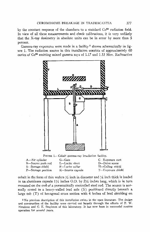
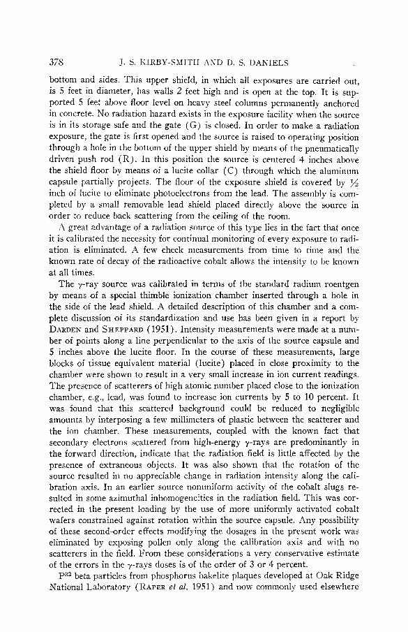
shown schematically in fig- ure 1. The radiation source in this installation consists of approximately 60 curies of Coeo emitting mixed gamma rays of 1.17 and 1.33 Mev. Radioactive
Gamma-ray exposures were made in a facility
FIGURE 1.-Cobalt gamma-ray irradiation facility. A-Air cylinder G-Gate C-Exposure cart R-Source push rod L-Lucite sheet D-Drive screw S-Storage shield F-Lucite collar H-Ceiling shield P-Storage position K-Source capsule T-Exposure shield
cobalt in the form of thin wafers inch in diameter and % inch thick is loaded in an aluminum capsule 1% inches O.D. by 2% inches long, which is in turn mounted on the end of a pneumatically controlled steel rod. The source is nor- mally stored in a heavy-walled lead safe (S) positioned directly beneath a large tub (T) of hexagonal cross section with 4 inches of lead shielding on
2 No previous description of this installation exists in the open literature. The design and construction of the facility were carried out largely through the efforts of I?. w. Hembree and G. E. Stapleton of this laboratory. I t has now been in successful routine operation for several years.
378 J. S. KIRBY-SMITH AND D. S. DANIELS
bottom and sides. This upper shield, in which all exposures are carried out, is 5 feet in diameter, has walls 2 feet high and is open at the top. I t is sup- ported 5 feet above floor level on heavy steel columns permanently anchored in concrete. N o radiation hazard exists in the exposure facility when bhe source is in its storage safe and the gate (G) is closed. In order to make a radiation exposure, the gate is first opened and the source is raised to operating position through a hole in the bottom of the upper shield by means of the pneumatically driven push rod (R) . In this position the source is centered 4 inches above the shield floor by means of a lucite collar (C) through which the aluminum capsule partially projects. The floor of the exposure shield is covered by inch of lucite to eliminate photoelectrons from the lead. The assembly is coni- pleted by a small removable lead shield placed directly above the source in order to reduce back scattering from the ceiling of the room.
A great advantage of a radiation source of this type lies in the fact that once it is calibrated the necessity for continual monitoring of every exposure to radi- ation is eliminated. A few check measurements froni time to time and the known rate of decay of the radioactive cobalt allows the intensity to be known at all times.
The 7-ray source was calibrated in terms of the standard radium roentgen by means of a special thimble ionization chamber inserted through a hole in the side of the lead shield. A detailed description of this chamber and a com- plete discussion of its standardization and use has been given in a report by DARDEN and SHEPPARD (1951). Intensity measurements were made at a num- ber of points along a line perpendicular to the axis of the source capsule and 5 inches above the lucite floor. In the course of these measurements, large blocks of tissue equivalent material (lucite) placed in close proximity to the chamber were shown to result in a very small increase in ion current readings. The presence of scatterers of high atomic number placed close to the ionization chamber, e.g., lead, was found to increase ion currents by 5 to 10 percent. It was found that this scattered background could be reduced to negligible amounts by interposing a few millimeters of plastic between the scatterer and the ion chamber. These measurements, coupled with the known fact that secondary electrons scattered from high-energy y-rays are predominantly in the forward direction, indicate that the radiation field is little affected by the presence of extraneous objects. I t was also shown that the rotation of the source resulted in no appreciable change in radiation intensity along the cali- bration axis. In an earlier source nonuniform activity of the cobalt slugs re- sulted in some azimuthal inhomogeneities in the radiation field. This was cor- rected in the present loading by the use of more uniformly activated cobalt wafers constrained against rotation within the source capsule. Any possibility of these second-order effects modifying the dosages in the present work was eliminated by exposing pollen only along the calibration axis and with no scatterers in the field. From these considerations a very conservative estimate of the errors in the 7-rays doses is of the order of 3 or 4 percent.
P32 beta particles from phosphorus bakelite plaques developed at Oak Ridge National Laboratory (RAPER et al. 1951) and now commonly used elsewhere
CHROMOSOME BREAKAGE I N TRADESCANTIA 379
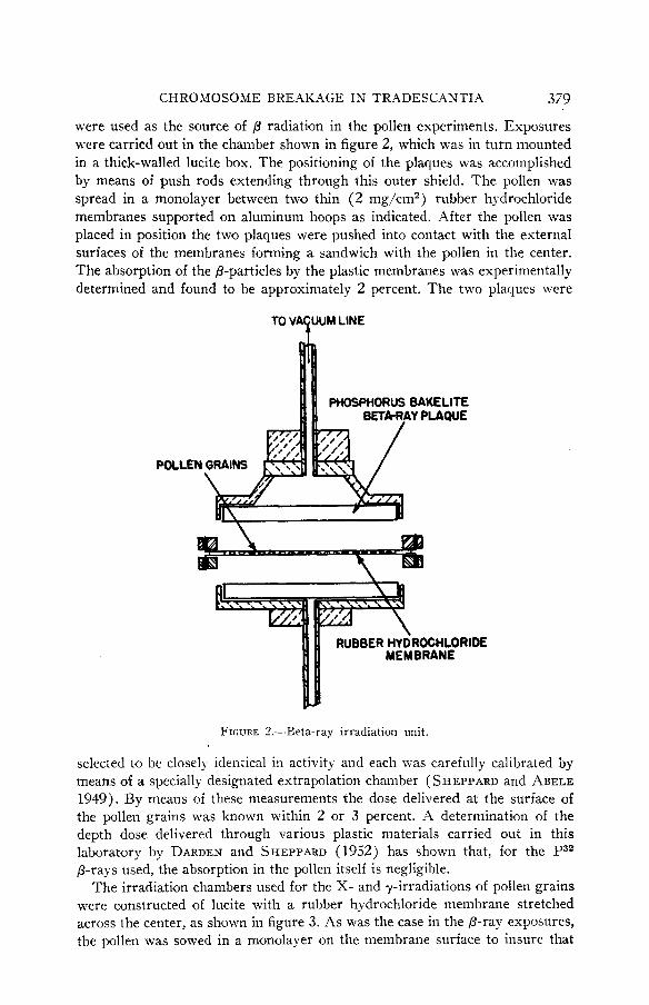
were used as the source of /3 radiation in the pollen experiments. Exposures were carried out in the chamber shown in figure 2, which was in turn mounted in a thick-walled lucite box. The positioning of the plaques was accomplished by means of push rods extending through this outer shield. The pollen was spread in a monolayer between two thin (2 mg/cm2) rubber hydrochloride membranes supported on aluminum hoops as indicated. After the pollen was placed in position the two plaques were pushed into contact with the external surfaces of the membranes forming a sandwich with the pollen in the center. The absorption of the p-particles by the plastic membranes was experimentally determined and found to be approximately 2 percent. The two plaques were
TOVA UUMLINE F
POLLEN
RIDE
FIGURE 7.-Beta-ray irradiation unit.
selected to be closely identical in activity and each was carefully calibrated by means of a specially designated extrapolation chamber ( SHEPPARD and ABELE 1949). By means of these measurements the dose delivered at the surface of the pollen grains was known within 2 or 3 percent. A determination of the depth dose delivered through various plastic materials carried out in this laboratory by DARDEN and SHEPPARD (1952) has shown that, for the P32 P-rays used, the absorption in the pollen itself is negligible.
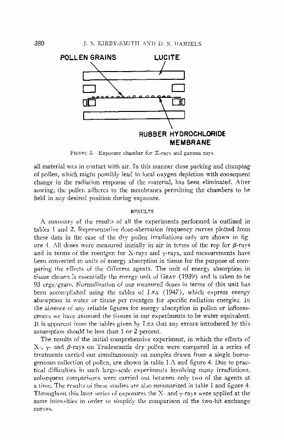
The irradiation chambers used for the X- and 7-irradiations of pollen grains were constructed of lucite with a rubber hydrochloride membrane stretched across the center, as shown in figure 3, As was the case in the /3-ray exposures, the pollen was sowed in a monolayer on the membrane surface to insure that
380 J. S. KIRBY-SMITH A S D U. S. DA4NIELS
POLLEN GRAINS LUCITE
RUBBER HY DRQCHLORIDE MEMBRANE
FIGURE 3.-Exposure chamber for X-rays and gamma rays.
all material was in contact with air. In this manner close packing and clumping of pollen, which might possibly lead to local oxygen depletion with consequent change in the radiation response of the material, has been eliminated. After sowing, the pollen adheres to the membranes permitting the chambers to be held in any desired position during exposure.
RESULTS
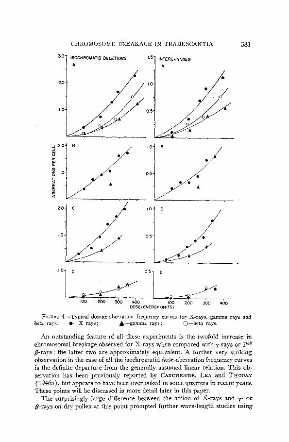
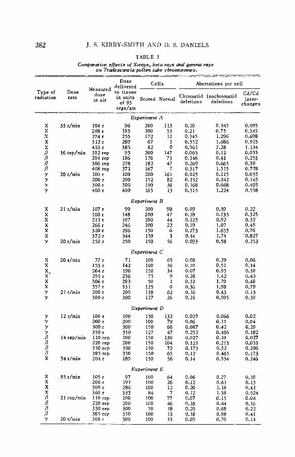
A summary of the results of all the experiments performed is outlined in tables 1 and 2. Representative dose-aberration frequency curves plotted from these data in the case of the dry pollen irradiations only are shown in fig- ure 4. All doses n7e1-e measured initially in air in terms of the rep for /3-rays and in terms of the roentgen for X-rays and y-rays, and measurements have been converted to units of energy absorption in tissue for the purpose of com- paring the effects of the different agents. The unit of energy absorption in tissue chosen is essentially the energy unit of GRAY (1939) and is taken to be 93 ergs/gram. Kormalization ,of our measured doses in terms of this unit has been accomplished using the tables of LEA (1947), which express energy absorption in water or tissue per roentgen for specific radiation energies. In the absence of any reliable figures for energy absorption in pollen or inflores- cences we have assumed the tissues in our experiments to be water equivalent. I t is apparent from the tables given by LEA that any errors introduced by this assumption should be less than 1 or 2 percent.
The results of the initial comprehensive experiment, in which the effects of X-, 7- and ,&rays on Tradescantia dry pollen were compared in a series of treatments carried out simultaneously on samples drawn from a single homo- geneous collection of pollen, are shown in table 1 A and figure 4. Due to prac- tical difficulties in such large-scale experiments involving many irradiations, subsequent comparisons were carried out between only two of the agents at a time. The results of these studies are also summarized in table 1 and figure 4. Throughout this later series of exposures the X- and y-rays were applied at the same intensities in order to simplify the comparison of the two-hit exchange curves.
CHROMOSOME BREAKAGE IN TRADESCANTIA 381
IS- INTERCHANGES 30- ISOCHROMATID DELETIONS
A A
2.0-
I .o
FIGURE 4.-Typical dosage-aberration frequency curves for X-rays, gamma rays and beta rays. 0-X rays; A-gamma rays ; 0-beta rays.
An outstanding feature of all these experiments is the twofold increase in chromosomal breakage observed for X-rays when compared with y-rays or P32 prays ; the latter two are approximately equivalent. A further very striking observation in the case of all the isochromatid dose-aberration frequency curves is the definite departure from the generally assumed linear relation. This ob- servation has been previously reported by CATCHESIDE, LEA and THODAY (1946a), but appears to have been overlooked in some quarters in recent years. These points will be discussed in more detail later in this paper.
The surprisingly large difference between the action of X-rays and y- or 8-rays on dry pollen at this p i n t prompted further wave-length studies using
382 J. S. KIRBY-SMITH AND D. S. DANIELS
TABLE I Compmaiiw e / /ec ts o/ X-rays, beta rays and gamma rays
on Tradescaniia pollen tube chromosomes. - -
________I__- - -- -- Dose Cel l s Aberrations per cel l Measured delivered
Type of Dose dose to t issue Cd/Cd radiation rate in air in units scored " w ~ deletions deletions c ~ ~ ~ ; s
Chromatid Isochromatid of 93
e r g d m
X x X X X P P P P Y Y Y Y
x X X X X X Y
X X
X X X Y Y
x.
Y Y Y
; P P ,o X
X X X X P P P P Y
35 r/min
16 rep/min
20 r/min
21 r/min
20 r/min
20 r/min
21 r/min
12 r/min
14 rep/min
34 r/min
35 r/min
21 rep/min
104 r 208 r 274 r 312 r 416 r 102 rep 204 rep 306 rep 408 rep 100 r 200 r 300 r 400 r
107 r 160 I 213 r 266 r 320 r 372 r 250 r
77 r 153 r 204 r 255 r 306 r 357 r 200 r 300 I
100 r 200 r 300 r 350 r 110 rep 220 rep 330 rep 385 rep 204 r
105 r 206 r 309 r 360 t 110 reD 220 re; 330 reb 385 rep
20 r/min 300 r
Experiment A
96 200 113 193 200 53 255 172 12 289 67 1 385 82 0 93 200 147 186 178 73 278, 182 47 371 167 7 100 200 161 200 152 82 300 190 36 400 165 13
Experiment B 99 200 98 148 200 47 197 200 44 246 200 22 296 150 6 344 I50 1 250 150 56
Experiment C 71 100 65 142 100 36 190 150 34
9 236 283 50 331 I25 0 200 138 62 300 127 26
75 1
Experiment D 100 150 132 200 100 79 300 150 66 350 127 47 100 150 130 200 150 104 300 150 72 350 150 65 189 150 56
Experiment E 97 100 64 191 100 26 286 100 12 333 84 7 100 100 77 200 100 46 300 SO 18 350 100 I3 300 100 33
0.10 0.21 0.345 0.552 0.561 0.065 0.146 0.269 0.317 0.025 0.132 0.168 0.315
0.09 0.18 0.125 0.19 0.273 0.44 0.093
0.08 0.10 0.07 0.28 0.32 0.36 0.16 0.21
0.027 0.06 0.087 0.25 2 0.027 0.113 0.173 0.12 0.14
0.06 0.12 0.20 0.12 0.07 0.16 0.20 0.18 0.09
0.345 0.75 1.296 1.686 2.28 0.12 0.41 0.665 1.515 0.115 0.342 0.668 1.224
0.39 0.735 0.92 1.07 1.653 ,
1.74 0.58
0.29 0.51 0.95 1.42 1.70 1.98 0.43 0.905
0.066 0.11 0.42 0.496 0.10 0.213 0.32 0.465 0.534
0.27 0.61 1.18 1.38 0.15 0.44 0.68 0.98 0.70
0.095 0.345 0.698 0.925 1.134 0.055 0.253 0.39 0.778 0.035 0.145 0.405 0.558
0.22 0.325 0.32 0.45 0.76 0.827 0.253
0.06 0.14 0.30 0.43 0.48 0.79 0.13 0.30
0.02 0.04 0.20 0.182 0.027 0.053 0.206 0.173 0.246
0.10 0.15 0.41 0.524 0.04 0.16 0.22 0.41 0.14
CHROMOSOME BREAKAGE I N TRADESCANTIA 353
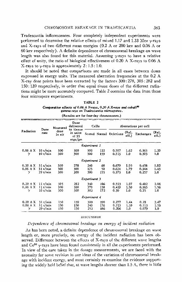
Tradescantia inflorescences. Four completely independent experiments were performed to determine the relative effects of mixed 1.17 and 1.33 Mev y-rays and X-rays of two different mean energies (0.2 A or 200 kev and 0.06 A or 60 kev respectively). A definite dependence of chromosomal breakage on wave length was also found for this material. Assuming y-rays to have a relative effect of unity, the ratio of biological effectiveness of 0.20 A X-rays to 0.06 A X-rays to y-rays is approximately 2 : 1.5 : 1.0.
I t should be noted that comparisons are made in all cases between doses expressed in energy units. The measured aberration frequencies at the 0.2 A X-ray dose points have been corrected by the factors 300 : 278, 305 : 282 and 150: 139 respectively, in order that equal tissue doses of the different radia- tions might be more accurately compared. Table 2 contains the data from these four microspore experiments.
TABLE 2 Comparative effects of 0.06 A X-rays , 0.20 A X-rays and cobaltCQ
garnma rays on Tradescant ia microspores. (Results are for four-day chromosomes.) -- -~~ ---
Dose Measwed delivered Cells Aberrations per cell
of 93 eff . ) Radiation :’,9”,’ dose to tissue
erndam
in air in units Scored Normal Deletions Exchanges z;.\ Experiment I
0.06 A X IOr/min 300 300 300 122 0.507 1.62 0.363 1.20 ’Y 10 r/min 300 300 300 159 0.313 1.0 0.303 1.0
Experiment 2 0.20 A X 31 r/min 300 278 240 69 0.679 1.95 0.458 1.92 0.06 A X 31 r/min 300 300 125 50 0.624 1.79 0.344 1.43
Y 29 r/min 300 300 300 155 0.373 1.0 0.257 1.0
Experiment 3 0.20 A X 11 r/min 305 282 340 106 0.553 1.97 0.482 2.06 0.06 A X 1 1 r/min 300 300 370 158 0.419 1.50 0.365 1.56
Y 10 r/min 300 300 302 172 0.30 1.0 0.25 1.0
Experiment 4 0.20 A X 31 r/min 150 139 300 190 0.277 1.44 0.18 2.47 0.06 A X 31 r/min 150 150 240 176 0.213 1.10 0.113 1.53
y 29 r/min 150 150 252 186 0.206 1.0 0.079 1.0
DISCUSSION
Dependence of chromosomal breakage on energy of incident radiation As has been noted, a definite dependence of chromosomal breakage on wave
length or, more precisely, on energy of the incident radiation has been ob- served. Difference between the effects of X-rays of the different wave lengths and Coeo 7-rays have been found consistently in all the experiments performed. In view of the care taken in the dosage measurements, we are faced with the necessity for some revision in our ideas of the variation of chromosomal break- age with incident energy, and must certainly re-examine the evidence support- ing the widely held belief that, at wave lengths shorter than 1.5 A, there is little
384 J. S. KIRBY-SMITH AND D. S. DANIELS
or no dependence of these biological effects on the frequency of the incident radiation.
The biological effects of ionizing radiation depends to a great extent upon the distribution of ionization produced in the tissue under study. A well-known curve showing the theoretical wave-length dependence of chromosomal break- age in the 8 A to 1.5 A region has been derived by LEA and CATCHESIDE (1942) from a consideration of the calculated ionization distribution in tissue as a function of the wave length of the primary radiation. This curve shows a steady diminution in chromosomal aberration production as the wave length of the incident radiation is decreased below 4 A. The general shape of the curve in the X-ray region has been studied experimentally in the case of Tradescantia pollen-tube chromosomes by CATCHESIDE and LEA (1943), who found chromosomal breakage produced by X-rays of 0.15 A to be only slightly less than that produced by radiations of 1.5 A wave length. Similar results for Tradescantia microspores were obtained by KOTVAL (unpublished work cited by LEA 1947) in experiments comparing the effects of 0.15 A X-rays and 0.015 A y-rays from radium. These deviations from the predictions of the simple theory have been qualitatively explained by the production of secondary electrons (&rays) and by the effects of the less densely ionizing portions of the electron tracks. At the present time it is not possible to determine theo- retically the exact value of the wave length at which such a leveling off of aberration production should occur. FANO (1943) has shown that this situa- tion should be attained ultimately at some value of the energy of the incident radiation beyond which the ratio of ionization in clusters to that distributed along the electron tracks should remain a constant as the wave length is de- creased. From this evidence it has been generally considered for some time that chromosomal breakage in Tradescantia and many other materials is essen- tially independent of the wave length of the exciting radiation in the region shorter than 1.5 A. A closer inspection of the experimental evidence shows this conclusion to be less reliable than it is often assumed. The pollen-tube work of CATCHESIDE and LEA (1943) cited above is subject to some criticism due to the small number of cells studied. The recent demonstration of large variations in pollen sensitivity from day to day further complicates the inter- pretation of early pollen-tube work.
The effect of X-rays of effective wave lengths 0.44 and 0.07 A, respectively, have been studied by FABERGB (1940), who found no significant differences in the numbers of chromosomal fragments produced in Tradescantia microspores. FABERGB applied doses of 1500 r in these experiments and analyzed for “ num- ber of fragments ” only instead of scoring specific types of aberrations as was the case in our present work. Unless some saturation effects may have been brought about by the high doses, the disagreement between FABERG~’S results and those results shown in table 2 cannot be explained at the present time. LEA and CATCHESIDE (1943) have pointed out that both points chosen by FABERCE are in a wave-length region in which the mean energy of electrons produced in tissue changes only slightly, and hence little or no effect should be observed.
CHROMOSOME BREAKAGE IiX TRADESCANTIA 385
The present work is not so greatly subject to this same objection, since the mean electron energies for our X-ray points, taken from LEA'S (1947) tables, are 11 and 45 kv, respectively, compared with 15 and 29 kv in the work of FABERGE.
There are previous results supporting our present measurements. CATCHE- SIDE, LEA and THODAY (1946a), in some usually ignored results, have shown for Tradescantia microspores that the ratio of biological effectiveness of 200 kv X-rays (mean wave length 0.15 A) to radium y-rays is 1.0: 0.77. This is not greatly different from the ratio of 1.0 : 0.70 found in our work for 0.060 A X-rays compared with y-rays. In another plant material there is some confir- mation in the work of GRAY, REED and POYNTER (1943) on the lethal action of X- and y-rays on the broad-bean root. Here the relative efficiency of 0.16 A X-rays to radium y-rays is in the ratio of 1.5: 1.0. This is not too different from our results of 2 : 1 for 0.20 A X-rays compared to cobalt y-rays.
Little more than a tabulation of the wave-length aberration rate data can be made until a quantitative theory of the effects of ionization density in the high energy electron range, i.e., in the short wave-length range, has been con- structed, or until more precise knowledge of the energy distribution in the X-ray beams is available. Even with filtration, the X-ray output from either constant potential or self-rectified machines is a wide smear of many different wave lengths. Some hope for accurate measurement of these X-ray energy distributions is now possible with the development of scintillation spectrome- ters. A second approach to a precise determination of wave-length effects con- sists in the use of monochromatic y-rays from radioactive sources at a number of energies in the l-Mev to 250-kv region. Consideration of both these methods is now underway.
Biological equivalence of P32 beta rays and Co60 gamma rays oia Tradescantia polleiz
The equivalence of these radiations on the production of chromosomal break- age is clearly shown in table 1 and figure 4. A simple qualitative explanation of this fact may be found in an examination of the electron energy distribution in these different sources. The y-ray spectrum from CoSo consists ideally in two lines of 1.17 and 1.33 Mev energy, respectively. This situation is not realized in the practical case, due to the presence of some degraded scattered y-rays as well as secondary electrons from the Compton process. In the present argument the exact value of the mean 7-ray energy is .not critical and, for sim- plicity, we will assume a mean energy of 1 MeV. Using the tables computed by LEA ( 1947), the mean energy of electrons produced in tissues by 1 Mev y-rays is found to be approximately 450 kv. These electrons are the biologically effec- tive agents in the present experiments, and if the distribution in energy of the P32 electrons can be obtained, a valid comparison of the expected effects may be made.
The initial distribution of P32 /3-rays from a thin source is well known, but these data cannot be applied directly to the thick P32 plaques used in the pres-
386 J . S. KlRBY-SMITH AND D. S. DANIELS
ent work. In the absence of a well-established theory of the degraded electron energy distribution in a thick source, the needed information must be found from experiment. A direct determination of the energy distribution from the P32 plaques, using a scintillation spectrometer, has been made recently by DARDEN (unpublished observations) in this laboratory and a mean electron energy of approximately 400 kv was found. Some additional confirmation of this result is found in a recent paper by BROWNELL (1952) on the energy distribution of p-rays from a thin P32 source after traversal through appreci- able thicknesses of plastic.
It is thus apparent that little difference in ionization density, and conse- quently in biological effect, should result in a comparison of the effects of 450 kv electron from the CoSo y-rays and the 400 kv P32 electrons. It should be noted however that these considerations are strictly valid only for thin tissues in which further degradation in energy of the P-rays is slight. This condition is satisfied in the case of pollen.
Isochromatid dose-breakage relations
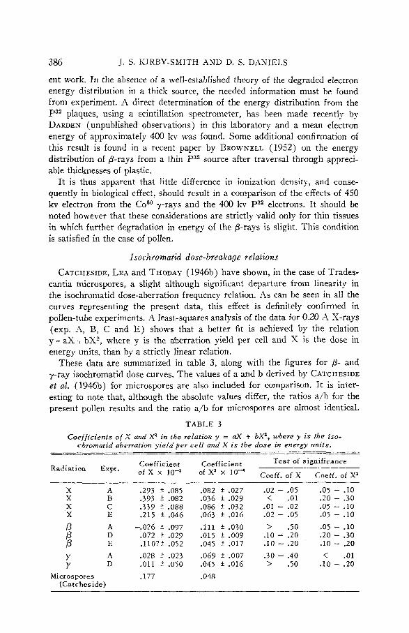
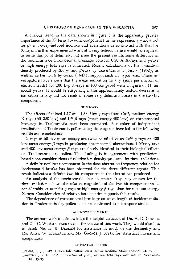
CATCHESIDE, LEA and THODAY (1946b) have shown, in the case of Trades- cantia microspores, a slight although significant departure from linearity in the isochromatid dose-aberration frequency relation. As can be seen in all the curves representing the present data, this effect is definitely confirmed in pollen-tube experiments. A least-squares analysis of the data for 0.20 X X-rays (exp. -4, B, C and E) shows that a better fit is achieved by the relation y = aX t bX2, where y is the aberration yield per cell and X is the dose in energy units, than by a strictly linear relation.
These data are summarized in table 3, along with the figures for p- and 7-ray isochromatid dose curves. The values of a and b derived by CATCHESIDE et al. (1946b) for microspores are also included for comparison. It is inter- esting to note that, although the absolute values differ, the ratios a/h for the present pollen results and the ratio a/b for microspores are almost identical.
TABLE 3
Coefficients of X and X in the relation y = aX + bXa, where y is the iso- chromatid aberration y i e ld per ce l l and X i s the dose in energy units.
_____ -_ Test of significance Coefficient Coefficient
of X x IO-* of X' x IO-' Coeff. of x Coeff. of X' Radiation Expt.
x A x B X C X E
A D E A D
; P Y Y
Microspores (Catches ide)
-293 f .085 .393 * .082 .339 f .088 .215 t .046
-.026 .097 .072 f .029 .1107* .052
.028 .023
.011 * .050
.177
.082 * .027
.036 * .029
.086 * .032
.063 * .016
. I l l + .030
.015 t . O D 9
.045 * .017
.069 * .007
.045 * .016
.048
.02 - .05 < .01
.01 - .02
.02 - .05
> .50 .10 2 .20 .10 - .20
.30 - .40 > .50
.05 - .10 -20 - .30 .05 - .IO .05 - .10
.05 - .10
.20 - .30
.10 - .20
< .01 .10 - .20
CHROMOSOME BREAKAGE I N TRADESCANTIA 387
A curious trend in the data shown in figure 3 is the apparently greater importance of the X term (two-hit component) in the expression y = aX + bx2 for P- and y-ray-induced isochromatid aberrations as contrasted with that for X-rays. Further experimental work of a very tedious nature would be required to settle this point definitely, but from the present results some difference in the mechanism of chromosomal breakage between 0.20 A X-rays and y-rays or high energy beta rays is indicated. Recent calculations of the ionization density produced by X-, y- and ,&rays by CORMACK and JOHNS (1952), as well as earlier work by GRAY (1947), support such an hypothesis, These in- vestigators have shown that the mean ionization density (ions per micron of electron track) for 200 kvp X-rays is 100 compared with a figure of 11 for cobalt y-rays. It would be surprising if this approximately tenfold decrease in ionization density did not result in some very definite increase in the two-hit component.
SUMMARY
The effects of mixed 1.17 and 1.33 Mev y-rays from Co60, medium energy X-rays (60-250 kev) and P32 P-rays (mean energy 400 kev) on chromosomal breakage in Tradescantia have been compared. A number of independent irradiations of Tradescantia pollen using these agents have led to the following results and conclusions :
X-rays of 60 kev mean energy are twice as effective as COs0 y-rays or 400 kev mean energy P-rays in producing chromosomal aberrations. 1 Mev y-rays and 400 kev mean energy p-rays are closely identical in their biological effects on Tradescantia dry pollen. This finding is in agreement with predictions based upon considerations of relative ion density produced by these radiations.
A definite nonlinear component in the dose-aberration frequency relation for isochromatid breaks has been observed for the three different agents. This result indicates a definite two-hit component in the aberrations produced.
An analysis of the isochromatid dose-aberration frequency curves for the three radiations shows the relative magnitude of the two-hit component to be considerably greater for y-rays or high-energy p-rays than for medium energy X-rays. Consideration of relative ion densities supports this result.
The dependence of chromosomal breakage on wave length of incident radia- tion in Tradescantia dry pollen has been confirmed in microspore studies.
ACKNOWLEDGMENTS
The authors wish to acknowledge the helpful criticism of DR. A. D. CONGER and DR. C. W. SHEPPARD during the course of this work. They would also like to thank MR. E. B. DARDEN for assistance in much of the dosimetry and DR. ALAN W. KIMBALL and MR. GEORGE J. ATTA for statistical advice and computation.
LITERATURE CITED
BISHOP, C. J., 1949 Pollen tube culture on a lactose medium. Stain Technol. 24: 9-12. BROWNELL, G. L., 1952
10: 30-35. Interaction of phosphorus-32 beta rays with matter. Nucleonics

388 J. S. KIRBY-SMITH AND D. S. DANIELS
CATCHESIDE, D. G., and D. E. LEA, 1943 The effect of ionization distribution on chromosome breakage by X-rays. J. Genet. 45: 186-196.
CATCHESIDE, D. G., D. E. LEA and J. M. THODAY, 1946a Types of chromosomal struc- tural change induced by irradiation of Tradescantia microspores. J. Genet 47: 113-137. 1946b The production of cromosomal structural changes in Tradescantia micro- spores in relation to dosage, intensity and temperature. J. Genet. 47: 137-149.
CORMACK, D. V., and H. E. JOHNS, 1952 Electron energies and ion densities in water irradiated with 200 Kev, 1 MeV, and 25 Mev radiations. Brit. J. Radiol, 25: 3693-381.
DARDEN, E. B., and C. W. SHEPPARD, 1951 A thimble type gamma-ray dosimeter and the measurement of radiation from lumped and distributed type sources. Report, ORNL-1002. Office of Technical Services, Department of Commerce, Washington, D. C., 45 cents.
DARDEN, E. B., JR., and C. W. SHEPPARD, 1952 Depth dose relation for biological materials exposed to beta rays. J. Tenn. hcad. Sci. 27: 210-211.
FABERG~, A. C., 1940 Equivalent effects of X-rays of different wave length on Trades- cantia chromosomes. J. Genet. 40: 379-384.
FANO, U., 1943 Production of ion clusters by X-rays. Nature 151: 698. GRAY, L. H., 1939 Measurement of neutron dose in biological experiments. Nature 144:
439-440. 1947 The distribution of the ions resulting from the irradiation of living cells. Brit. J. Radiol., Suppl. 1 : 7-15.
GRAY, L. H., J. READ and M. POYNTER, 1943 The effect of ionizing radiation on the broad bean root. Brit. J Radiol. 16: 125-128.
LEA, D. E., 1947 LEA, D. E., and D. G. CATCHESIDE, 1942 The mechanism of the induction of chromosome
aberrations in Tradescantia. J. Genet. 44: 216-245. RAPER, J. R., R. E. ZIRKLE and K. K. BARNES, 1951 Techniques of external irradiation
with beta rays. Nat. Nuclear Energy Series, Div. IV, Vol. 22E. McGraw-Hill Bwk Co., Inc., New York.
SHEPPARD, C. W., and R. K. ABELE, 1949 Construction and calibration of equipment for measuring the surface exposure of phosphorus-bakelite beta ray sources. Report, AECU-655. Office of Technical Services, Department of Commerce, Washington, D. C. 15 cents.
Action of radiations on living cells. The Macmillan Co., New York.