RXUFRPSXWHU WKHYLGHROLQNVZLOOEHFRPHGLVDEOHG CHAPTER …€¦ · CHAPTER e33 Atlas of Percutaneous...
9
33-1 CHAPTER e33 Atlas of Percutaneous Revascularization CHAPTER e33 Atlas of Percutaneous Revascularization Jane A. Leopold Deepak L. Bhatt David P. Faxon Percutaneous coronary intervention (PCI) is the most widely employed coronary revascularization procedure worldwide (Chap. 246). It is now applied to patients with stable angina, acute coronary syndromes, including unstable angina and non- ST-segment elevation myocardial infarction (NSTEMI), and as a primary treatment strategy in patients with ST-segment elevation myocardial infarction (STEMI). PCI is also applicable to patients with either single- or multi-vessel disease. In this chapter, the use of PCI will be illustrated in a variety of commonly encountered clinical and anatomic situations such as chronic total occlusion of a coronary artery, bifurcation disease, acute STEMI, saphenous vein graft disease, left main coronary artery disease, multivessel disease, and stent thrombosis. In addi- tion, the use of interventional techniques to treat structural heart disease will be shown, including closure of an atrial septal defect (ASD) and percutaneous aortic valve implantation; the latter is approved in Europe but is under active investigation in clinical tri- als in the United States and not yet approved for use. CASE 1: CHRONIC TOTAL OCCLUSION (Videos e33-1 to e33-7) An 81-year-old man with angina, NYHA Class IV congestive • heart failure and inferior-apical-posterior ischemia on an exer- cise technetium-99m scan. Diagnostic cardiac catheterization revealed a left dominant • system with a totally occluded left circumflex (LCx) artery. The distal LCx filled via collaterals from the left anterior descending (LAD) artery, indicating chronicity of the total occlusion. Video e33-1 (Play video) Baseline left coronary angiogram shows an oc- cluded LCx with left-to-left collaterals originating from LAD septal vessels. Video e33-2 (Play video) Attempts to cross the total occlusion in the LCx using a hydrophilic wire and an antegrade approach were not successful, with the wire tracking to the right of the trajectory. Video e33-3 (Play video) The LAD septal collateral is accessed with a guidewire and directed toward the distal LCx to cross the total occlusion retrograde. Video e33-4 (Play video) The total occlusion is crossed retrograde. The wire is snared in the guide, exteriorized, and used to provide antegrade access to the LCx. Video e33-5 (Play video) Antegrade flow in the LCx is restored after balloon inflation. Video e33-6 (Play video) Following stenting of the total occlusion, blood flow in the distal vessel is improved and a second significant stenosis is seen. Video e33-7 (Play video) Final result after LCx stenting. SUMMARY Approximately 15–30% of all patients referred for cardiac • catheterization will have a chronic total occlusion (CTO) of a coronary artery. CTO often leads to a surgical referral for complete revascularization. • Incomplete revascularization due to an untreated CTO is associ- • ated with an increased mortality rate (Hazard Ratio = 1.36, 95% CI = 1.12–1.66, p < 0.05). Successful PCI of a CTO leads to a 3.8–8.4% absolute reduction in • mortality, symptom relief, and improved left ventricular function. Newer techniques, such as the retrograde approach to crossing • total occlusions, are useful when the antegrade approach fails, or is not feasible, and there are well-developed collateral vessels. (Case contributed with permission by Dr. Frederick G. P. Welt) CASE 2: BIFURCATION STENTING (Fig. e33-1; Videos e33-8 to e33-16) Figure e33-1 Schematic representation of 1-stent and 2-stent techniques to treat bifurcation lesions. PTCA, Percutaneous transluminal coronary angioplasty. (Reprinted with permission from SK Sharma, A Kini: Cardiol Clin 24:233, 2006.) Bifurcation lesion Stent+PTCA Stent+stent (“T stenting”) Stent+stent (“reverse-T”) Stent+stent (“Culotte”) Stent+stent (“Y” or “V”) Stent+stent (“Crush”) Stent+stent (“Kissing”) Main vessel Side- branch No stent coverage No stent coverage 1 1 1 1 1 1 1 1 1 2 2 2 2 2 2 “V” Copyright © 2012 The McGraw-Hill Companies, Inc. All rights reserved. Note: Once this PDF is downloaded to your computer, the video links will become disabled.
Transcript of RXUFRPSXWHU WKHYLGHROLQNVZLOOEHFRPHGLVDEOHG CHAPTER …€¦ · CHAPTER e33 Atlas of Percutaneous...

33-1
CHA
PTER e33
Atlas of Percutaneous Revascularization
CHAPTER e33
Atlas of Percutaneous Revascularization Jane A. Leopold Deepak L. Bhatt David P. Faxon
Percutaneous coronary intervention (PCI) is the most widely employed coronary revascularization procedure worldwide (Chap. 246). It is now applied to patients with stable angina, acute coronary syndromes, including unstable angina and non-ST-segment elevation myocardial infarction (NSTEMI), and as a primary treatment strategy in patients with ST-segment elevation myocardial infarction (STEMI). PCI is also applicable to patients with either single- or multi-vessel disease.
In this chapter, the use of PCI will be illustrated in a variety of commonly encountered clinical and anatomic situations such as chronic total occlusion of a coronary artery, bifurcation disease, acute STEMI, saphenous vein graft disease, left main coronary artery disease, multivessel disease, and stent thrombosis. In addi-tion, the use of interventional techniques to treat structural heart disease will be shown, including closure of an atrial septal defect (ASD) and percutaneous aortic valve implantation; the latter is approved in Europe but is under active investigation in clinical tri-als in the United States and not yet approved for use.
CASE 1: CHRONIC TOTAL OCCLUSION (Videos e33-1 to e33-7)
An 81-year-old man with angina, NYHA Class IV congestive • heart failure and inferior-apical-posterior ischemia on an exer-cise technetium-99m scan.
Diagnostic cardiac catheterization revealed a left dominant • system with a totally occluded left circumflex (LCx) artery. The distal LCx filled via collaterals from the left anterior descending (LAD) artery, indicating chronicity of the total occlusion.
Video e33-1 (Play video) Baseline left coronary angiogram shows an oc-cluded LCx with left-to-left collaterals originating from LAD septal vessels. Video e33-2 (Play video) Attempts to cross the total occlusion in the LCx using a hydrophilic wire and an antegrade approach were not successful, with the wire tracking to the right of the trajectory. Video e33-3 (Play video) The LAD septal collateral is accessed with a guidewire and directed toward the distal LCx to cross the total occlusion retrograde. Video e33-4 (Play video) The total occlusion is crossed retrograde. The wire is snared in the guide, exteriorized, and used to provide antegrade access to the LCx. Video e33-5 (Play video) Antegrade flow in the LCx is restored after balloon inflation. Video e33-6 (Play video) Following stenting of the total occlusion, blood flow in the distal vessel is improved and a second significant stenosis is seen. Video e33-7 (Play video) Final result after LCx stenting.
SUMMARY
Approximately 15–30% of all patients referred for cardiac • catheterization will have a chronic total occlusion (CTO) of a coronary artery. CTO often leads to a surgical referral for complete revascularization. • Incomplete revascularization due to an untreated CTO is associ-• ated with an increased mortality rate (Hazard Ratio = 1.36, 95% CI = 1.12–1.66, p < 0.05). Successful PCI of a CTO leads to a 3.8–8.4% absolute reduction in • mortality, symptom relief, and improved left ventricular function. Newer techniques, such as the retrograde approach to crossing • total occlusions, are useful when the antegrade approach fails, or is not feasible, and there are well-developed collateral vessels.
(Case contributed with permission by Dr. Frederick G. P. Welt)
CASE 2: BIFURCATION STENTING (Fig. e33-1; Videos e33-8 to e33-16)
Figure e33-1 Schematic representation of 1-stent and 2-stent techniques to treat bifurcation lesions. PTCA, Percutaneous transluminal coronary angioplasty. (Reprinted with permission from SK Sharma, A Kini: Cardiol Clin 24:233, 2006.)
Bifurcationlesion
Stent+PTCA Stent+stent(“T stenting”)
Stent+stent(“reverse-T”)
Stent+stent(“Culotte”)
Stent+stent(“Y” or “V”)
Stent+stent(“Crush”)
Stent+stent(“Kissing”)
Mainvessel
Side-branch
No stentcoverage
No stentcoverage
1 1
1
111
11
1
2 2
2
2
2
2
“V”
Copyright © 2012 The McGraw-Hill Companies, Inc. All rights reserved.
Note: Once this PDF is downloaded to your computer, the video links will become disabled.
33-2
PART 10
Disorders of the Cardiovascular System
A 52-year-old man with an acute coronary syndrome and a • troponin I = 0.18 (upper limit normal ≤ 0.04). Diagnostic cardiac catheterization showed single-vessel coronary • artery disease with a significant stenosis in the mid-LAD and a bifurcation lesion involving a large diagonal branch.
Video e33-8 (Play video) Baseline angiogram of the left coronary cir-culation shows the significant stenosis in the mid-LAD and the bifurcation lesion involving a large diagonal branch. Video e33-9 (Play video) Both vessels are accessed with guidewires and pretreated with balloon angioplasty. Video e33-10 (Play video) Result after balloon angioplasty. Video e33-11 (Play video) Stent being positioned in the LAD. Video e33-12 (Play video) LAD post-stent result. Video e33-13 (Play video) Stent deployed in diagonal branch through the stent struts in the LAD using the “culotte” technique. Video e33-14 (Play video) Diagonal branch post-stent result. Video e33-15 (Play video) Simultaneous inflation of two 2.5-mm “kiss-ing” balloons. Video e33-16 (Play video) Final postbifurcation stenting result.
SUMMARY
Approximately 15–20% of PCIs will involve the treatment of • bifurcation lesions. Bifurcation lesions require consideration of PCI strategies that • protect side-branch patency. There are both one-stent and two-stent techniques to treat • bifurcation lesions; the selection of technique depends upon anatomic considerations, including plaque burden, angle of side-branch take-off, plaque shift during angioplasty, and side-branch distribution. Rates of target lesion revascularization and stent thrombosis are • similar between one-stent and two-stent procedures.
CASE 3. INFERIOR MYOCARDIAL INFARCTION—THROMBUS AND MANUAL THROMBECTOMY
(Figs. e33-2 to e33-4; Videos e33-17 to e33-22)
A 59-year-old man presented to the emergency room with 2 h of • severe midsternal chest pressure. His systolic blood pressure was 100 mmHg and he was tachy-• cardic in sinus rhythm with a heart rate of 90–100 bpm.
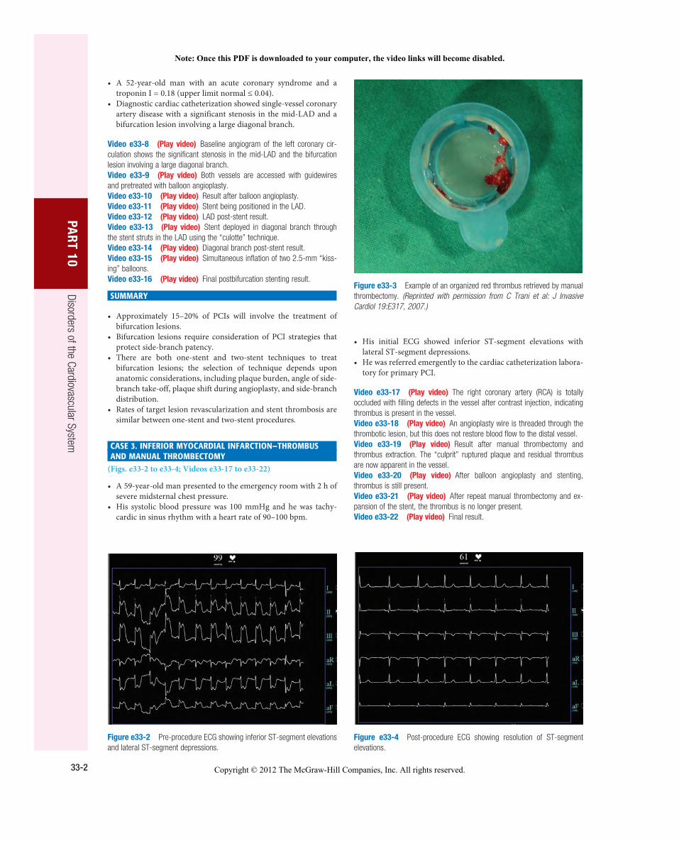
Figure e33-2 Pre-procedure ECG showing inferior ST-segment elevations and lateral ST-segment depressions.
Figure e33-3 Example of an organized red thrombus retrieved by manual thrombectomy. (Reprinted with permission from C Trani et al: J Invasive Cardiol 19:E317, 2007.)
Figure e33-4 Post-procedure ECG showing resolution of ST-segment elevations.
His initial ECG showed inferior ST-segment elevations with • lateral ST-segment depressions. He was referred emergently to the cardiac catheterization labora-• tory for primary PCI.
Video e33-17 (Play video) The right coronary artery (RCA) is totally occluded with filling defects in the vessel after contrast injection, indicating thrombus is present in the vessel. Video e33-18 (Play video) An angioplasty wire is threaded through the thrombotic lesion, but this does not restore blood flow to the distal vessel. Video e33-19 (Play video) Result after manual thrombectomy and thrombus extraction. The “culprit” ruptured plaque and residual thrombus are now apparent in the vessel. Video e33-20 (Play video) After balloon angioplasty and stenting, thrombus is still present. Video e33-21 (Play video) After repeat manual thrombectomy and ex-pansion of the stent, the thrombus is no longer present. Video e33-22 (Play video) Final result.
Copyright © 2012 The McGraw-Hill Companies, Inc. All rights reserved.
Note: Once this PDF is downloaded to your computer, the video links will become disabled.

33-3
CHA
PTER e33
Atlas of Percutaneous Revascularization SUMMARY
An acute STEMI occurs following plaque rupture that promotes • thrombotic occlusion of a coronary artery. Despite successful revascularization of the epicardial coronary • artery, microemboli liberated during balloon angioplasty and stent-ing may lead to persistent microvascular dysfunction. When pres-ent, microvascular dysfunction is associated with a larger infarct size, heart failure, malignant ventricular arrhythmias, and death. Manual thrombectomy is used to aspirate or remove thrombus • in the vessel and limit distal embolization during angioplasty and stenting. Manual thrombectomy in primary PCI is associated with • improved myocardial perfusion and a reduction in mortality. Adjunctive antiplatelet and antithrombin agents are important to • aid in the resolution of intracoronary thrombus.
CASE 4. SAPHENOUS VEIN GRAFT INTERVENTION WITH DISTAL PROTECTION
(Fig. e33-5; Videos e33-23 to e33-26)
A 62-year-old man with a history of chronic stable angina. • A four-vessel coronary artery bypass grafting (CABG) surgery • was performed 17 years earlier with a left internal mammary artery graft to the LAD, a right internal mammary artery graft to the RCA, a saphenous vein graft to the first obtuse marginal branch, and a saphenous vein graft to the first diagonal branch. The patient had a recent increase in angina with exertion and was • found to have lateral ischemia on an exercise technetium 99m scan. Diagnostic cardiac catheterization revealed a significant stenosis • in the body of the saphenous vein graft to the first obtuse mar-ginal branch.
Video e33-23 (Play video) Saphenous vein graft to a first obtuse mar-ginal branch with an 80% eccentric stenosis in the midgraft. Video e33-24 (Play video) A distal protection device is deployed past the lesion. Video e33-25 (Play video) Angioplasty balloon inflation with the distal protection device in place. Video e33-26 (Play video) Final result after stent placement.
SUMMARY
Saphenous vein grafts have a failure rate of up to 20% after 1 year • and as high as 50% by 5 years. Graft failure (>1 month) results from intimal hyperplasia and • atherosclerosis. Saphenous vein graft PCI is associated with distal emboliza-• tion of atherosclerotic debris and microthrombi, leading to microvascular occlusion, reduced antegrade blood flow (the “no-reflow” phenomenon), and myocardial infarction.
Embolic distal protection devices decrease the risk of dis-• tal embolization, as well as the incidence of no-reflow and myocardial infarction associated with saphenous vein graft interventions.
CASE 5: UNPROTECTED LEFT MAIN PCI IN A HIGH-RISK PATIENT
(Figs. e33-6 and e33-7; Videos e33-27 to e33-34)
An 89-year-old woman presented with an NSTEMI associated • with 5-mm ST-segment depression in the apical leads occurring 2 weeks after hospitalization for an NSTEMI that was treated conservatively. Chronic obstructive lung disease, elderly age, and the patient’s • refusal to consider cardiac surgery restricted the choice of thera-peutic options to medical and/or percutaneous interventions. Diagnostic catheterization revealed a left dominant circulation • with a heavily calcified 80% distal left main coronary artery stenosis extending into the LAD and into the proximal LCx coro-nary arteries. A 70% proximal LAD lesion was also present. After consultation with the patient, family, and a cardiac surgeon, • PCI was performed with intraaortic balloon pump support and a temporary pacemaker in the right ventricle.
Video e33-27 (Play video) Baseline left coronary artery injection in right anterior oblique (RAO) cranial projection shows a high-grade calcified stenosis in the left main coronary artery and a significant stenosis in the proximal LAD. Video e33-28 (Play video) In the left anterior oblique (LAO) caudal view, the left main coronary artery lesion can be seen to extend into the ostia of both the LCx and the LAD. Video e33-29 (Play video) Guidewires were placed into both the LCx and LAD. After the left main coronary artery and LCx are dilated with balloon angioplasty, the proximal LAD is dilated and a long drug-eluting stent is placed to cover a lesion dissection that occurred with wiring of the vessel. Video e33-30 (Play video) The bifurcation lesion in the left main coro-nary artery extending into the LCx and LAD ostia is treated using a “culotte” technique. First, a drug-eluting stent is placed in the left main coronary artery and into the proximal LCx. Video e33-31 (Play video) Next, the LAD wire is removed and passed through the stent into the distal LAD. A second drug-eluting stent is deployed through the struts of the left main coronary artery/LCx stent. Video e33-32 (Play video) Following rewiring of the LCx, both stents are redilated simultaneously (“kissing” balloons). Video e33-33 (Play video) The final result in the LAO caudal view. Video e33-34 (Play video) The final result in the RAO cranial view show-ing patent left main, LCx, and LAD coronary arteries.
SUMMARY
Left main coronary artery disease occurs in 5–10% of patients • with symptomatic coronary artery disease. In patients with left main coronary artery disease, revasculariza-• tion with CABG has been shown to decrease mortality signifi-cantly over 5–10 years of follow-up. PCI with drug-eluting stents in selected cases has been shown • to have equal in-hospital and 1-year death and myocardial infarction rates as compared to CABG in the Synergy between PCI with Taxus and Cardiac Surgery (SYNTAX) trial. Long-term outcome differences between the two treatment strategies are not known. Indications for PCI of left main coronary artery lesions are • high-risk surgical patients and patients with protected left main coronary artery disease (i.e., prior CABG with patent bypass
Figure e33-5 Distal protection device showing captured atherosclerotic debris liberated by initial balloon dilation. (Reprinted with permission from RA Aqel et al: J Invasive Cardiol 19:E104, 2007.)
Copyright © 2012 The McGraw-Hill Companies, Inc. All rights reserved.
Note: Once this PDF is downloaded to your computer, the video links will become disabled.
33-4
PART 10
Disorders of the Cardiovascular System
grafts). Patients who are good candidates for bypass surgery may also undergo a stenting procedure, but discussion with the patient, the interventional cardiologist, and the cardiac surgeon should be undertaken to determine the best treatment option in an individual case. Outcomes are better for patients with an isolated lesion in the • ostium and body of the left main coronary artery where a single stent can be placed compared to bifurcation lesions that involve the ostium of the LAD and LCx.
CASE 6: MULTIVESSEL PCI IN A DIABETIC PATIENT (Videos e33-35 to e33-42)
A 58-year-old man presented with an NSTEMI. • The patient has hyperlipidemia and type 2 diabetes mellitus • treated with oral hypoglycemic agents. Diagnostic catheterization revealed two-vessel disease with a • total occlusion of the second obtuse marginal branch that was felt to be responsible for the patient’s symptoms (culprit lesion).
I aVR V1 V4
II
III
VI
aVL
aVF
V2
V3 V6
V5
Figure e33-6 During chest pain, the ECG showed diffuse ST-segment depression of up to 5 mm in the inferior and lateral leads.
Figure e33-7 Following resolution of the chest pain, the ST-segment depression is less marked.
III
VI
II
I
aVF
aVL
aVR
V3
V2
V1
V6
V5
V4
Copyright © 2012 The McGraw-Hill Companies, Inc. All rights reserved.
Note: Once this PDF is downloaded to your computer, the video links will become disabled.

33-5
CHA
PTER e33
Atlas of Percutaneous Revascularization In addition, there was a high-grade stenosis in a large ramus • intermedius branch and the RCA had a significant stenosis in the midsegment of the vessel.
Video e33-35 (Play video) Baseline angiogram of the left coronary circulation in the RAO view shows the total occlusion of the second obtuse marginal branch with delayed retrograde filling via collateral vessels and a high-grade stenosis in the ramus intermedius. Video e33-36 (Play video) A guidewire is passed through the total oc-clusion and the lesion is pretreated with balloon angioplasty. Video e33-37 (Play video) Following placement of a drug-eluting stent in the lesion, the vessel is widely patent. A third obtuse marginal vessel, not previ-ously seen, now fills faintly (TIMI 1 flow) with contrast but was not treated. Video e33-38 (Play video) The ramus intermedius lesion was crossed with a guidewire and pretreated with balloon angioplasty. Video e33-39 (Play video) A drug-eluting stent is placed across the ramus lesion and deployed. The final result shows no residual stenosis in either the ramus or second obtuse marginal vessels. Video e33-40 (Play video) Baseline angiogram of the RCA shows a high-grade lesion in the midsegment of the vessel. Video e33-41 (Play video) The lesion was pretreated with balloon dila-tion followed by stent deployment. Video e33-42 (Play video) The final result shows no residual stenosis in the mid-RCA.
SUMMARY
Multivessel PCI is performed commonly and may be done in one • setting or staged with two or more procedures. Acute and long-term studies of multivessel PCI have shown com-• parable rates of death and myocardial infarction when compared to CABG but a higher incidence of repeat revascularization as a result of restenosis associated with PCI. In the randomized Bypass Angioplasty Revascularization • Investigation (BARI) trial, diabetic patients treated with PCI had a worse long-term mortality than diabetic patients treated with CABG. However, the BARI registry found that in selected diabetic patients with favorable anatomy, PCI can result in out-comes equal to that observed with CABG.
Figure e33-8 Optical coherence tomography image following initial balloon dilation. Residual thrombus that is adherent to the stent struts is seen. (Reprinted with permission from AF Schinkel et al: JACC Cardiovasc Interv 1:449, 2008.)
CASE 7: VERY LATE STENT THROMBOSIS OF A PROXIMAL LAD DRUG-ELUTING STENT
(Figs. e33-8 and e33-9; Videos e33-43 to e33-46)
A 62-year-old man had a drug-eluting stent placed in a proximal • LAD lesion to treat severe angina. He received dual antiplatelet therapy with aspirin and clopidogrel for 1 year and then discon-tinued clopidogrel per protocol. He remained asymptomatic until 15 months after the initial stent • placement, when he presented with severe chest pain due to an acute anterior STEMI. He was taken to the catheterization laboratory within 70 min of • presentation and his initial angiogram showed a total occlusion of the proximal LAD stent.
Video e33-43 (Play video) Baseline angiogram showing a total occlusion of the proximal LAD within the drug-eluting stent and a significant stenosis at the origin of the LCx. Video e33-44 (Play video) The LAO view shows the LCx stenosis with a filling defect, indicating that thrombus is present in the vessel lumen. Video e33-45 (Play video) The LAD lesion was crossed with a guidewire, which resulted in slow filling of the mid-LAD (TIMI 2 flow), and revealed thrombus filling the stent. Video e33-46 (Play video) The final result after LAD and LCx stenting. The LAD lesion was pretreated with balloon angioplasty and a bare metal stent was deployed to cover the proximal lesion. The LCx ostial lesion was dilated with balloon angioplasty and a bare metal stent was placed using a “V stenting” technique.
SUMMARY
Stent thrombosis is an infrequent (1–2%) but serious complication • of stent placement. It occurs most commonly within the first month, but rarely, as late as 1 year (0.2–0.6%) with bare metal stents. Very late stent thrombosis (VLST), which occurs after 1 year, is very rare with bare metal stents but can occur with drug-eluting stents. Premature discontinuation of dual antiplatelet therapy is the • most common cause of early and late stent thrombosis; however, the etiology of VLST is not clear.
Copyright © 2012 The McGraw-Hill Companies, Inc. All rights reserved.
Note: Once this PDF is downloaded to your computer, the video links will become disabled.
33-6
PART 10
Disorders of the Cardiovascular System
The majority of patients with stent thrombosis present with acute • coronary syndromes or STEMI; this presentation is associated with a high mortality rate (10%). Treatment is immediate PCI with balloon angioplasty or restenting. •
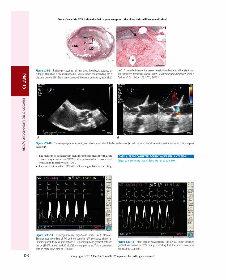
Figure e33-10 Transesophageal echocardiogram shows a calcified trileaflet aortic valve ( A ) with reduced leaflet excursion and a narrowed orifice in peak systole ( B ).
A B
Figure e33-11 Hemodynamically significant aortic (AO) stenosis. Simultaneous recording of AO and left ventricle (LV) pressures shows an 82 mmHg peak-to-peak gradient and a 63.3 mmHg mean gradient between the LV (154/9 mmHg) and AO (72/29 mmHg) pressures. This is consistent with an aortic valve area of 0.58 cm 2 .
Figure e33-12 After balloon valvuloplasty, the LV–AO mean pressure gradient decreased to 37.3 mmHg, indicating that the aortic valve area increased to 0.95 cm 2 .
Figure e33-9 Pathologic specimen of late stent thrombosis obtained at autopsy. Thrombus is seen filling the LAD vessel lumen and extending into a diagonal branch (LD). Stent struts occupied the space denoted by asterisk (∗)
( left ). A magnified view of the vessel reveals thrombus around the stent strut and neointima formation ( arrow ) ( right ). (Reprinted with permission from A Farb et al: Circulation 108:1701, 2003.)
CASE 8: TRANSCATHETER AORTIC VALVE IMPLANTATION (Figs. e33-10 to e33-14; Videos e33-47 to e33-50)
Copyright © 2012 The McGraw-Hill Companies, Inc. All rights reserved.
Note: Once this PDF is downloaded to your computer, the video links will become disabled.
33-7
CHA
PTER e33
Atlas of Percutaneous Revascularization
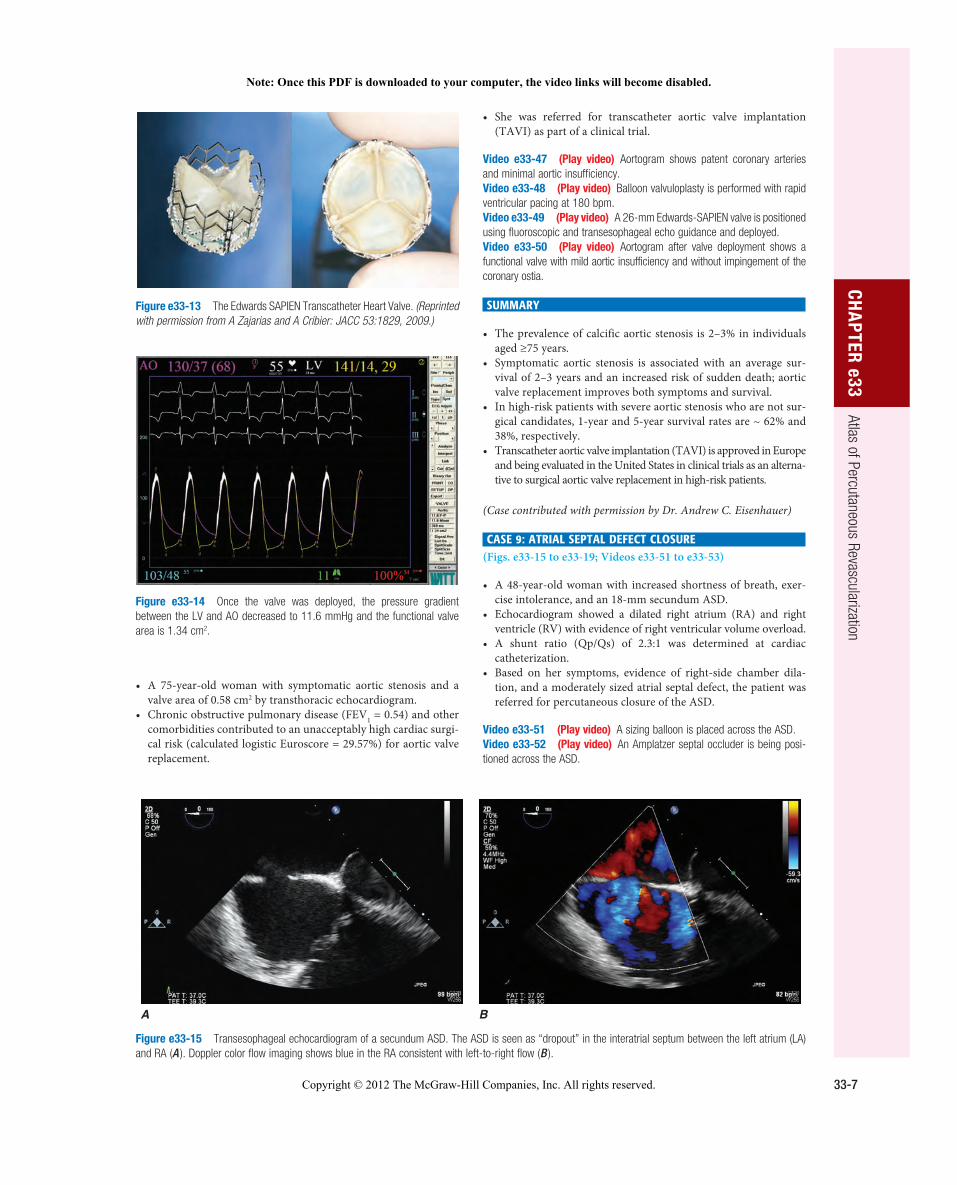
Figure e33-13 The Edwards SAPIEN Transcatheter Heart Valve. (Reprinted with permission from A Zajarias and A Cribier: JACC 53:1829, 2009.)
Figure e33-14 Once the valve was deployed, the pressure gradient between the LV and AO decreased to 11.6 mmHg and the functional valve area is 1.34 cm 2 .
She was referred for transcatheter aortic valve implantation • (TAVI) as part of a clinical trial.
Video e33-47 (Play video) Aortogram shows patent coronary arteries and minimal aortic insufficiency. Video e33-48 (Play video) Balloon valvuloplasty is performed with rapid ventricular pacing at 180 bpm. Video e33-49 (Play video) A 26-mm Edwards-SAPIEN valve is positioned using fluoroscopic and transesophageal echo guidance and deployed. Video e33-50 (Play video) Aortogram after valve deployment shows a functional valve with mild aortic insufficiency and without impingement of the coronary ostia.
SUMMARY
The prevalence of calcific aortic stenosis is 2–3% in individuals • aged ≥75 years. Symptomatic aortic stenosis is associated with an average sur-• vival of 2–3 years and an increased risk of sudden death; aortic valve replacement improves both symptoms and survival. In high-risk patients with severe aortic stenosis who are not sur-• gical candidates, 1-year and 5-year survival rates are ~ 62% and 38%, respectively. Transcatheter aortic valve implantation (TAVI) is approved in Europe • and being evaluated in the United States in clinical trials as an alterna-tive to surgical aortic valve replacement in high-risk patients.
(Case contributed with permission by Dr. Andrew C. Eisenhauer)
CASE 9: ATRIAL SEPTAL DEFECT CLOSURE (Figs. e33-15 to e33-19; Videos e33-51 to e33-53)
A 48-year-old woman with increased shortness of breath, exer-• cise intolerance, and an 18-mm secundum ASD. Echocardiogram showed a dilated right atrium (RA) and right • ventricle (RV) with evidence of right ventricular volume overload. A shunt ratio (Qp/Qs) of 2.3:1 was determined at cardiac • catheterization. Based on her symptoms, evidence of right-side chamber dila-• tion, and a moderately sized atrial septal defect, the patient was referred for percutaneous closure of the ASD.
Video e33-51 (Play video) A sizing balloon is placed across the ASD. Video e33-52 (Play video) An Amplatzer septal occluder is being posi-tioned across the ASD.
A B
Figure e33-15 Transesophageal echocardiogram of a secundum ASD. The ASD is seen as “dropout” in the interatrial septum between the left atrium (LA) and RA ( A ). Doppler color flow imaging shows blue in the RA consistent with left-to-right flow ( B ).
A 75-year-old woman with symptomatic aortic stenosis and a • valve area of 0.58 cm 2 by transthoracic echocardiogram. Chronic obstructive pulmonary disease (FEV• 1 = 0.54) and other comorbidities contributed to an unacceptably high cardiac surgi-cal risk (calculated logistic Euroscore = 29.57%) for aortic valve replacement.
Copyright © 2012 The McGraw-Hill Companies, Inc. All rights reserved.
Note: Once this PDF is downloaded to your computer, the video links will become disabled.

33-8
PART 10
Disorders of the Cardiovascular System
Figure e33-16 Three-dimensional echocardiographic reconstruction of the secundum ASD. The ASD is round and has an acceptable margin of tissue to seat a septal occluder device.
Figure e33-17 Transesophageal echocardiogram showing sizing balloon ( A ) and no flow ( B ) across the atrial septal defect.
A B
Figure e33-18 Amplatzer septal occluder in place ( A ). There is no blood flow across the device ( B ).
A B
Copyright © 2012 The McGraw-Hill Companies, Inc. All rights reserved.
Note: Once this PDF is downloaded to your computer, the video links will become disabled.

33-9
CHA
PTER e33
Atlas of Percutaneous Revascularization
Figure e33-19 Postprocedure lateral chest x-ray showing the Amplatzer septal occluder in place.
Video e33-53 (Play video) The two discs of the device in place across the ASD.
SUMMARY
Unrepaired ASDs lead to signs and symptoms of increased • pulmonary blood flow and right heart failure; dyspnea, exercise intolerance, fatigue, palpitations and atrial arrhythmias, and pul-monary infections. Percutaneous closure of an ASD may be recommended for • individuals with a secundum ASD and evidence of RA and RV enlargement with or without symptoms. Percutaneous closure is contraindicated in patients with irrevers-• ible pulmonary arterial hypertension and no left-to-right shunt. It is not recommended for closure of sinus venosus, coronary sinus, or primum ASDs. After the atrial septal occluder device is placed, patients are • treated with antiplatelet agents and use antibiotic prophylaxis for certain procedures for 6 months. Follow-up echocardiograms to assess for device migration or erosion, residual shunting, throm-bus or pericardial effusion are recommended at 1 day, 1 month, 6 months, 1 year, and periodically thereafter.
(Case contributed with permission by Dr. Andrew C. Eisenhauer)
Copyright © 2012 The McGraw-Hill Companies, Inc. All rights reserved.
Note: Once this PDF is downloaded to your computer, the video links will become disabled.