RUNNING HEAD: CYP71E7: The oxime-metabolizing P450 in ...4 ABSTRACT Cassava (Manihot esculenta...
35
1 RUNNING HEAD: CYP71E7: The oxime-metabolizing P450 in Manihot esculenta Crantz, cassava CORRESPONDING AUTHOR: Søren Bak, [email protected] ADDRESS: Plant Biochemistry Laboratory, Department of Plant Biology and Biotechnology, VKR Research Centre “Pro-Active Plants”, Faculty of Life Sciences, University of Copenhagen, 40 Thorvaldsensvej, DK-1871 Frederiksberg C, Copenhagen, Denmark TELEPHONE: +45 3533 3346 FAX: +45 3533 3333 JOURNAL RESEARCH AREA: Biochemical Processes and Macromolecular Structures Plant Physiology Preview. Published on November 2, 2010, as DOI:10.1104/pp.110.164053 Copyright 2010 by the American Society of Plant Biologists www.plantphysiol.org on March 1, 2020 - Published by Downloaded from Copyright © 2010 American Society of Plant Biologists. All rights reserved.
Transcript of RUNNING HEAD: CYP71E7: The oxime-metabolizing P450 in ...4 ABSTRACT Cassava (Manihot esculenta...

1
RUNNING HEAD: CYP71E7: The oxime-metabolizing P450 in Manihot esculenta Crantz,
cassava
CORRESPONDING AUTHOR: Søren Bak, [email protected]
ADDRESS: Plant Biochemistry Laboratory, Department of Plant Biology and
Biotechnology, VKR Research Centre “Pro-Active Plants”, Faculty of Life Sciences,
University of Copenhagen, 40 Thorvaldsensvej, DK-1871 Frederiksberg C, Copenhagen,
Denmark
TELEPHONE: +45 3533 3346
FAX: +45 3533 3333
JOURNAL RESEARCH AREA: Biochemical Processes and Macromolecular Structures
Plant Physiology Preview. Published on November 2, 2010, as DOI:10.1104/pp.110.164053
Copyright 2010 by the American Society of Plant Biologists
www.plantphysiol.orgon March 1, 2020 - Published by Downloaded from Copyright © 2010 American Society of Plant Biologists. All rights reserved.
2
TITLE: Biosynthesis of the cyanogenic glucosides linamarin and lotaustralin in cassava
(Manihot esculenta Crantz): Isolation, biochemical characterization and expression pattern of
CYP71E7, the oxime-metabolizing cytochrome P450 enzyme
AUTHORS: Kirsten Jørgensen, Anne Vinther Moranta, Marc Morantb, Niels Bjerg Jensen,
Carl Erik Olsen, Rubini Kannangara, Mohammed Saddik Motawia, Birger Lindberg Møller
and Søren Bak.
ADDRESS: Plant Biochemistry Laboratory, Department of Plant Biology and
Biotechnology, VKR Research Centre “Pro-Active Plants” (A.V.M., M.M., K.J., N. B. J,
M.S.M., B.L.M. and S.B.), Center for Synthetic Biology (K.J., M.S.M., B.L.M. and S.B) and
Department of Basic Sciences and Environment (C.E.O.), University of Copenhagen, 40
Thorvaldsensvej, DK-1871 Frederiksberg C, Copenhagen, Denmark
www.plantphysiol.orgon March 1, 2020 - Published by Downloaded from Copyright © 2010 American Society of Plant Biologists. All rights reserved.

3
FOOTNOTES
FINANCIAL SOURCES: This work has been supported by grants from the Danish
International Development Agency (DANIDA), by the Danish Research Council for
Technology and Production (FTP), the Danish National Research Foundation, the Villum
Foundation and from the UNIK Synthetic Biology Research Initiative, funded by the Danish
Ministry of Science, Technology and Innovation.
a) Current address: H. Lundbeck A/S, Ottiliavej 9, 2500 Valby, Denmark
b) Current address: Novozymes A/S Krogshøjvej 36, 2880 Bagsværd Denmark
CORRESPONDING AUTHOR: Søren Bak, email: [email protected], fax: +45 3533 3346
www.plantphysiol.orgon March 1, 2020 - Published by Downloaded from Copyright © 2010 American Society of Plant Biologists. All rights reserved.
4
ABSTRACT
Cassava (Manihot esculenta Crantz) is a eudicotyledonous plant, which produces the
valine- and isoleucine-derived cyanogenic glucosides linamarin and lotaustralin with the
corresponding oximes and cyanohydrins as key intermediates. CYP79 enzymes catalyzing
amino acid to oxime conversion in cyanogenic glucoside biosynthesis are known from
several plants including M. esculenta. The enzyme system converting oxime into
cyanohydrin has previously only been identified in the monocotyledonous plant Sorghum
bicolor Moench. Using this S. bicolor CYP71E1 sequence as a query in a BLASTp search, a
putative functional homologue which exhibited a ~50% amino acid sequence identity was
found in M. esculenta. The corresponding full length cDNA clone was obtained from a
plasmid library prepared from M. esculenta shoot tips and was assigned CYP71E7.
Heterologous expression of CYP71E7 in yeast afforded microsomes converting 2-
methylpropanal oxime (valine-derived oxime) and 2-methylbutanal oxime (isoleucine-
derived oxime) to the corresponding cyanohydrins which dissociate into hydrogen cyanide
and and acetone and 2-butanone, respectively. The volatile ketones were detected as 2.4-
dinitrophenylhydrazone derivatives by LC-MS. A KS of ~0.9 μM was determined for 2-
methylbutanal oxime based on substrate binding spectra. CYP71E7 exhibits low specificity
for the side chain of the substrate and catalyzes conversion of aliphatic and aromatic oximes
with turnovers of ~21, 17, 8 and 1 min-1 for the oximes derived from valine, isoleucine,
tyrosine and phenylalanine, respectively. A second paralog of CYP71E7 was identified by
database searches and showed ~90% amino acid sequence identity. In tube in situ PCR
showed that in nearly unfolded leaves the CYP71E7 paralogs are preferentially expressed in
specific cells in the endodermis and in most cells in the first cortex cell layer. In fully
unfolded leaves, the expression is pronounced in the cortex cell layer just beside the
epidermis and in specific cells in the vascular tissue cortex cells. Thus the transcripts of the
CYP71E7 paralogs co-localize with CYP79D1 and CYP79D2. We conclude that CYP71E7 is
the oxime-metabolizing enzyme in cyanogenic glucoside biosynthesis in M. esculenta.
www.plantphysiol.orgon March 1, 2020 - Published by Downloaded from Copyright © 2010 American Society of Plant Biologists. All rights reserved.

5
INTRODUCTION
Manihot esculenta Crantz (cassava) is one of the most important root crops in the world.
The drought tolerance of M. esculenta, combined with a high yield on poor soils and the
possibility to leave the starch rich tubers in the soil for extended time periods as a food and
thus harvest on demand, renders M. esculenta a key staple food especially in Africa (Nweke
et al., 2002). All parts of the M. esculenta plant contain the valine- and isoleucine-derived
cyanogenic glucosides, linamarin and lotaustralin in an approximately 97 to 3 ratio
(Lykkesfeldt and Møller, 1994). The cyanogenic glucosides are primarily synthesized in the
shoot apex (Andersen et al., 2000) and transported to the tubers where they accumulate up to
1.5 g/kg dry weight (Bokanga, 1994; Jørgensen et al., 2005a). In planta, cyanogenic
glucosides and specific β-glucosidases able to cleave the β-glucosidic linkage are
compartmentalized into different tissues or subcellular compartments including the apoplast
(Morant et al., 2008). This provides a two-component system which is activated upon cellular
disruption e.g. during food processing or by chewing insects. The cyanohydrin formed by the
action of the β-glucosidase subsequently dissociates into a ketone and toxic HCN.
The presence of cyanogenic glucosides in cassava poses a major nutritive drawback in
rural areas where cassava constitutes the sole staple crop available. Ingestion of incompletely
processed cassava derived products in combination with an unbalanced diet deficient in
sulphur amino acids may give rise to chronic cyanide intoxication, as sulphur amino acids are
required for cyanide detoxification (Rosling, 1994). In extreme cases, this may result in
severe diseases such as tropical ataxic neuropathy and konzo (Banea-Mayambu et al., 1997;
Oluwole et al., 2000). Acute cyanide intoxication inactivates the mitochondrial cytochrome
aa3 oxidase and thereby blocks cellular respiration (Nelson, 2006). Unfortunately, careful
processing of cassava tubers to remove the cyanide generating constituents results in
concomitant loss of proteins, vitamins and minerals and thus significantly reduces the
nutritional value of this important crop (Maziya-Dixon et al., 2007).
The enzymes and corresponding genes for the entire cyanogenic glucoside biosynthetic
pathway have only been identified in the monocotyledonous crop Sorghum bicolor Moench
(great millet; Jones et al., 1999). S. bicolor contains the aromatic cyanogenic glucoside
dhurrin. The dhurrin pathway is genetically simple, as only three structural genes encode the
entire pathway. Two membrane bound, multifunctional cytochromes P450, CYP79A1 and
CYP71E1, and a soluble UDPG-glucosyltransferase (UGT), UGT85B1, catalyze the
conversion of tyrosine into dhurrin (Halkier et al., 1995; Bak et al., 1998; Jones et al., 1999).
www.plantphysiol.orgon March 1, 2020 - Published by Downloaded from Copyright © 2010 American Society of Plant Biologists. All rights reserved.

6
CYP79A1 catalyzes the conversion of the amino acid tyrosine into (Z)-p-hydroxyphenyl
acetaldoxime in a reaction sequence that involves two consecutive N-hydroxylations, a
dehydration and a decarboxylation reaction, and a final isomerization reaction (Halkier et al.,
1995; Sibbesen et al., 1995). The next enzyme in the pathway, CYP71E1, converts (Z)-p-
hydroxyphenyl acetaldoxime into p-hydroxymandelonitrile via a dehydration and a C-
hydroxylation reaction (Halkier et al., 1989; Kahn et al., 1997; Bak et al., 1998). Finally, the
p-hydroxymandelonitrile is glucosylated by UGT85B1 to yield dhurrin (Jones et al., 1999).
The biosynthetic pathway is known to proceed as a highly channelled process (Møller and
Conn, 1980), and the three biosynthetic enzymes are thought to be organized within a
multienzyme complex, a metabolon (Nielsen and Møller, 2005; Nielsen et al., 2007), to
secure highly efficient conversion of the parent amino acid into the cyanogenic glucoside and
to facilitate channelling of toxic and reactive intermediates (reviewed in Jørgensen et al.,
2005b and Winkel, 2004).
Enzymes of the CYP79 family catalyze the first and rate limiting steps in cyanogenic
glucoside biosynthesis and have been identified from several cyanogenic plants (reviewed in
Bak et al., 2006) including M. esculenta (Andersen et al., 2000). CYP79 enzymes are clearly
evolutionarily conserved between monocotyledons and eudicotyledons and can be easily
distinguished from other cytochromes P450 by unique amino acid substitutions in the
otherwise generally conserved heme binding domain and in the so-called “PERF” motif in
the meander region (Bak et al., 2006, Paquette et al., 2009). CYP79 enzymes also catalyze
the first committed step in biosynthesis of the crucifer specific aliphatic, aromatic and indole
glucosinolates and camalexin (Rauhut and Glawischnig 2009). The general assumption is that
the glucosinolate pathway has evolved from the ancient cyanogenic glucoside pathway, and
that the amino acid to oxime step is catalyzed by orthologous enzymes and the two pathways
branch at the oxime (Bak et al., 2005). In addition, the CYP79 biochemical activity is readily
detected using radio labeled amino acids as a substrate (Andersen et al., 2000). In contrast,
identification of candidate CYP71E1 functional homologues or orthologs that catalyze the
oxime to cyanohydrin step remains a major challenge because of multiple gene duplications
and rearrangements resulting in numerous neo- and sub-functionalizations within the CYP71
family. This is exemplified by the presence of 52 members of the CYP71 family in the
diploid eudicotyledon Arabidopsis thaliana (thale cress), covering only two subfamilies,
CYP71A and CYP71B, with amino acid sequence identities above 55% in each subfamily
(Paquette et al., 2000; Nelson et al., 2004, Paquette et al., 2009). In addition, CYP71E1 lacks
obvious unique substitutions in otherwise conserved motifs in the amino acid sequence.
www.plantphysiol.orgon March 1, 2020 - Published by Downloaded from Copyright © 2010 American Society of Plant Biologists. All rights reserved.
7
Identification of a functional CYP71E1 homologue from plants producing aliphatic
cyanogenic glucosides is further complicated by the lack of a sensitive method to detect the
volatile reaction products. The lack of known CYP71E1 functional homologues raises the
question of whether the oxime-metabolizing enzyme is evolutionarily conserved between
monocotyledons and eudicotyledons as well, or if the post oxime step is a case of convergent
rather than divergent evolution.
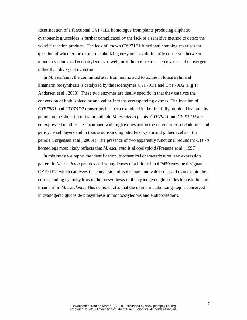
In M. esculenta, the committed step from amino acid to oxime in lotaustralin and
linamarin biosynthesis is catalyzed by the isoenzymes CYP79D1 and CYP79D2 (Fig 1;
Andersen et al., 2000). These two enzymes are dually specific in that they catalyze the
conversion of both isoleucine and valine into the corresponding oximes. The location of
CYP79D1 and CYP79D2 transcripts has been examined in the first fully unfolded leaf and its
petiole in the shoot tip of two month old M. esculenta plants. CYP79D1 and CYP79D2 are
co-expressed in all tissues examined with high expression in the outer cortex, endodermis and
pericycle cell layers and in tissues surrounding laticifers, xylem and phloem cells in the
petiole (Jørgensen et al., 2005a). The presence of two apparently functional redundant CYP79
homeologs most likely reflects that M. esculenta is allopolyploid (Fregene et al., 1997).
In this study we report the identification, biochemical characterization, and expression
pattern in M. esculenta petioles and young leaves of a bifunctional P450 enzyme designated
CYP71E7, which catalyzes the conversion of isoleucine- and valine-derived oximes into their
corresponding cyanohydrins in the biosynthesis of the cyanogenic glucosides lotaustralin and
linamarin in M. esculenta. This demonstrates that the oxime-metabolizing step is conserved
in cyanogenic glucoside biosynthesis in monocotyledons and eudicotyledons.
www.plantphysiol.orgon March 1, 2020 - Published by Downloaded from Copyright © 2010 American Society of Plant Biologists. All rights reserved.

8
RESULTS
Isolation and heterologous expression of CYP71E7 cDNA
The biosynthetic pathway for the cyanogenic glucosides lotaustralin and linamarin in M.
esculenta is illustrated in Figure 1 (Andersen et al., 2000; Koch et al., 1995). The enzyme
system(s) catalyzing conversion of the aliphatic oximes (Z)-2-methylbutanal oxime (ileox)
and (Z)-2-methylpropanal oxime (valox) into the corresponding cyanohydrins has remained
elusive. In S. bicolor, the CYP71E1 enzyme has been shown to catalyze conversion of the
aromatic oxime p-hydroxyphenyl acetaldoxime (tyrox) into the corresponding cyanohydrin,
p-hydroxymandelonitrile (Bak et al., 1998). Using the S. bicolor CYP71E1 amino acid
sequence as a query in a BLASTp search, an M. esculenta sequence with ~50% identity and
~68% similarity at the amino acid level was identified (Zhang et al., 2003; AY217351).
Phylogenetic analysis grouped the M. esculenta cytochrome P450 sequence with the
CYP71E1 sequence from S. bicolor as well as with cytochrome P450 sequences from rice,
wheat and sugarcane (data not shown), four other species known to synthesize cyanogenic
glucosides (Jones, 1998). Based on the cytochrome P450 nomenclature rules
(http://drnelson.utmem.edu/CytochromeP450.html), the M. esculenta P450 sequence has been
assigned as CYP71E7. Analysis of the genome sequence of the M. esculenta line AM-560-2
(Cassava Genome Project 2009, http:://www.phytozome.net/cassava), revealed the presence
of a CYP71E7 paralog on scaffold 02340 located within 12.000 bp of CYP71E7. The two
paralogs are 90% identical and 94% similar on the amino acid level, and the ORFs are 92%
identical.
To investigate whether CYP71E7 is the M. esculenta oxime-metabolizing enzyme, a
CYP71E7 cDNA was isolated by PCR from a cDNA library prepared from top shoots of M.
esculenta. This library had previously provided CYP79D1 and CYP79D2 (Andersen et al.,
2000) encoding the multifunctional isoenzymes CYP79D1 and CYP79D2 which catalyze the
first committed and rate limiting step in the pathway (Fig. 1; Andersen et al., 2000).
Recombinant CYP71E7 was produced in Saccharomyces cerevisiae WAT11 yeast cells that
co-express the A. thaliana NADPH:cytochrome P450 reductase, ATR1 (At4g24520). ATR1 is
a diflavin protein catalyzing electron transfer from NADPH to the heme iron during the P450
reaction cycle (Pompon et al., 1996, Jensen and Møller 2010, Laursen et al., 2010). Isolated
yeast microsomes harboring recombinant CYP71E7 produced the characteristic Soret peak at
450 nm upon CO binding (Fig. 2). This indicated that the heme group was correctly
www.plantphysiol.orgon March 1, 2020 - Published by Downloaded from Copyright © 2010 American Society of Plant Biologists. All rights reserved.

9
positioned in the active site and that CYP71E7 was produced in a correctly folded and active
form.
CYP71E7 is the oxime-metabolizing cytochrome P450 in biosynthesis of lotaustralin
and linamarin
Yeast microsomes harboring CYP71E7 were assayed for their ability to convert ileox and
valox into the corresponding cyanohydrins, 2-hydroxy-2-methylbutyronitrile and acetone
cyanohydrin. The design of the assay was based on dissociation of the labile cyanohydrins
formed into hydrogen cyanide and ketones by alkalinization of the reaction mixture at the end
of the incubation period (Fig. 3A) and subsequent trapping of the volatile ketones (2-
butanone and acetone) as 2,4-dinitrophenylhydrazones (Fig. 3B). After extraction, the 2,4-
dinitrophenylhydrazones formed were identified and quantified by LC-MS. To reduce
background levels due to contaminating aldehydes and ketones in the surrounding air and to
retain the 2-butanone and acetone produced, incubations were carried out in closed glass vials
with an acidified solution of 2,4-dinitrophenylhydrazine placed in a centre well.
In the presence of NADPH and oxygen, CYP71E7 converted ileox into 2-hydroxy-2-
methylbutyronitrile with a turnover of 17 ± 1 min-1 and a Km of 21 ± 11 µM. In agreement
with this, product formation was detected already at 10 μM ileox and increased with substrate
concentration up to 100 μM ileox (Fig. 3C, EIC 253). In the absence of NADPH or in assay
mixtures using microsomes isolated from yeast transformed with an empty expression vector,
hydrazone formation above background was not observed (Fig. 3C, EIC 253). The 2,4-
dinitrophenylhydrazone assay enables detection of as little as 50 pmol 2-butanone with
negligible background interference (Fig. 3C, EIC 253). The 2,4-dinitrophenylhydrazone of 2-
butanone was detected as two components with the main component (83%) eluting at 10.6
min and representing the (E)-isomer and the minor (17%) at 9.8 min the (Z)-isomer (Figure
3C, EIC 253).
CYP71E7 catalyzed conversion of valox into acetone cyanohydrin at a turnover of 21 ± 2
min-1 (Table 1). The acetone obtained by dissociation of the acetone cyanohydrin produced a
2,4-dinitrophenylhydrazone that eluted as a single component at 6.4 min (Fig. 3C, EIC 239)
as the carbonyl functional group of acetone carries two identical substituents. As for ileox,
product formation was detected already at a substrate concentration of 10 μM (Fig. 3C, EIC
239). Acetone is omnipresent in air and absorbed to glassware in sufficient amounts to
interfere with this highly sensitive assay. Accordingly, a high and slightly variable
background level was observed even in blank samples including only buffer (Fig. 3C, EIC
www.plantphysiol.orgon March 1, 2020 - Published by Downloaded from Copyright © 2010 American Society of Plant Biologists. All rights reserved.

10
239). The variable background level prevented calculation of an accurate Km for valox. In
addition to metabolizing the two aliphatic oximes, CYP71E7 was also able to convert tyrox
and the phenylalanine-derived phenylacetaldoxime (pheox) into the corresponding
cyanohydrins, albeit with lower turnovers 8.1 ± 0.3 and 1.3 ± 0.2 min-1, respectively.
Spectral analysis of substrate binding to CYP71E7
Binding of substrates within the active site of cytochromes P450 induces spin state
changes of the heme iron which can be recorded spectroscopically (Jefcoate, 1978). Yeast
microsomes harboring CYP71E7 produced a substrate binding spectrum with a trough at 415
nm upon addition of ileox (Fig. 4A) which reflects binding of ileox to the CYP71E7 active
site. To quantify the affinity of CYP71E7 for ileox, increasing concentrations of ileox (0.2 to
11.0 μM) were added to the yeast microsomes harboring CYP71E7. From this a KS of 0.9 ±
0.2 μM was calculated. The amplitude of the substrate binding spectrum reached maximum at
3 μM ileox (Fig. 4A). This demonstrated that ileox is a high affinity ligand of CYP71E7.
Cytochromes P450 are known to produce type II spectra upon binding of nitrogen containing
inhibitors such as n-octylamine. This spectral change is indicative of proximity of the
inhibitor amine to the active site heme iron (Jefcoate, 1978). Saturation of CYP71E7 with n-
octylamine (100 μM) resulted in the formation of a typical type IIb binding spectrum with a
trough at 410 nm and a peak at 435 nm [Fig. 4B(1)]. A new baseline was recorded by
addition of 100 μM n-octylamine to the CYP71E7 microsomes in the reference cuvette [Fig.
4B(2)]. To measure the ability of ileox to displace n-octylamine from the active site of
CYP71E7, increasing concentrations of ileox were added to the n-octylamine saturated
CYP71E7 microsome solution. Addition of ileox produced a reverse type IIb spectrum with a
peak at 410 nm and a trough at 430 nm [Fig. 4B (3, 4, 5)]. The amplitude of the reverse type
IIb spectrum reached maximum upon addition of 30 μM ileox. Addition of valox and pheox
to the yeast microsomes harboring CYP71E7 likewise resulted in substrate binding spectra
with a trough at 415 nm (results not shown), although higher substrate concentrations were
required to produce the spectral shift compared to ileox. Only upon addition of >30 μM
substrate did tyrox result in a weak trough at 415 nm (results not shown). The broad substrate
affinity of CYP71E7 is therefore reflected in the ability to bind both aliphatic and aromatic
oximes. The ability of ileox to efficiently replace n-octylamine shows that ileox is a high
affinity ligand to CYP71E7 and that n-octylamine is a competitive inhibitor that also is able
to bind to the active site of CYP71E7.
www.plantphysiol.orgon March 1, 2020 - Published by Downloaded from Copyright © 2010 American Society of Plant Biologists. All rights reserved.

11
CYP71E7 and CYP79D1 are co-expressed in M. esculenta leaf petioles
In tube in situ PCR was used to determine the cellular location of CYP71E7 transcripts in
M. esculenta. The analyses were performed on tissue sections from the petiole and leaf blade
of the nearly unfolded leaf and of the leaf blade of the first fully unfolded leaf using two
month old M. esculenta plants, and with primers that enabled detection of both CYP71E7
paralogs. The young leaf stages were selected because they contain the highest concentration
of cyanogenic glucosides (Jørgensen et al., 2005a) and hence were expected to exhibit high
expression levels of the mRNAs that encode the biosynthetic enzymes. Strong expression of
CYP71E7 in the petiole was found in outer cortex cells and in the cell layer corresponding to
the endodermis, around phloem cells and laticifers as well as in cells surrounding the xylem
as visualized by alkaline phosphatase staining (Fig. 5 CYP71E7). The same expression
pattern is seen for CYP79D1 transcripts in sections from the same petiole (Fig. 5 CYP79D1).
Alkaline phosphatase staining could not be detected in the absence of CYP71E7 or CYP79D1
primers in the reverse transciptase reaction (negative control; Fig. 5 Control). In a separate set
of experiments (Fig. 5), the in situ co-localization of the CYP71E7 and CYP79D1 transcripts
in the petiole were visualized using FITC (Fluorescein isothiocyanate) labeled anti-bodies
against digoxigenin (DIG) incorporated during the PCR reactions. As in the experiment with
alkaline phosphatase, the outer cortex cells, endodermis cells, the cells surrounding the
phloem and laticifers, and cells surrounding the xylem showed strong labeling. Negative
control sections showed no background staining.
In the corresponding leaf blade of the first nearly unfolded leaf, expression of CYP71E7 and
CYP79D1 is observed in most cells in the cortex and in the epidermis (fig.6 A & B,
respectively). In fully unfolded leaves, strong expression of CYP79D1 is observed in the
cortex cell layer just beside the epidermis and in specific cells in the vascular tissue (fig.6 C
& D). Especially in the leaf section red fluorescence is observed due to auto-fluorescence
from the chloroplast. In a previous study, CYP79D1 and CYP79D2 were shown to have
similar expression patterns (Jørgensen et al. 2005a).
The co-expression of CYP71E7 and CYP79D1 further substantiates that CYP71E7 is the
oxime-metabolizing enzyme in cyanogenic glucoside biosynthesis in M. esculenta.
DISCUSSION
A P450 enzyme, designated CYP71E7, catalyzing the conversion of isoleucine- and
valine-derived oximes into the corresponding cyanohydrins in the biosynthesis of cyanogenic
www.plantphysiol.orgon March 1, 2020 - Published by Downloaded from Copyright © 2010 American Society of Plant Biologists. All rights reserved.

12
glucosides in M. esculenta was identified based on amino acid sequence homology to
CYP71E1, functional expression in yeast, substrate binding spectra and transcriptional co-
localization with CYP79D1 and CYP79D2, the genes encoding the two enzymes that catalyze
the initial steps in cyanogenic glucoside synthesis in M. esculenta. The cyanohydrins (2-
hydroxy-2-methylbutyronitrile and acetone cyanohydrin) produced by CYP71E7 are labile
and decompose into volatile ketones (2-butanone and acetone) and HCN. To enable
identification and quantification, the ketones were detected as 2,4-dinitrophenylhydrazone
derivatives by LC-MS. This method enabled detection of pmolar amounts of ketones formed
in the CYP71E7 containing reaction, and is thus more sensitive than the previously used
colorimetric cyanide assay which requires the presence of nmolar amounts (Halkier and
Møller, 1991).
The oxime-metabolizing P450s in cyanogenic glucoside biosynthesis are highly efficient
enzymes
Ileox is a high affinity substrate for CYP71E7 with KS ~0.9 ± 0.2 μM which is reflected in
the conversion of ileox into the corresponding cyanohydrin with a Km of 21 ± 2 μM and a
turnover rate of 17 ± 1 min-1. A similar conversion rate was observed for valox with a
turnover of 21 ± 2 min-1. The higher turnover of valox as compared to ileox is in agreement
with a 97:3 ratio of linamarin and lotaustralin in cassava (Lykkesfeldt and Møller, 1994). The
KS , Km and turnover rates are in the same order of magnitude as those observed for the other
known plant oxime metabolizing cytochrome P450 involved in biosynthesis of cyanogenic
glucosides (CYP71E1) and as those for the structurally related glucosinolates (CYP83A1 and
CYP83B1) (Møller and Conn, 1979; Koch et al., 1992; Bak et al., 2001, Bak and Feyereisen
2001, Hansen et al., 2001, Naur et al., 2003). In contrast, the Km values for CYP79D1 towards
isoleucine and valine are several magnitudes higher, namely 1.3 mM and 2.2 mM,
respectively (Andersen et al., 2000). The Km values for Lotus japonicus (bird’s foot trefoil)
CYP79D3 and CYP79D4 towards isoleucine and valine are also in the 1-3 mM range
(Forslund et al., 2004). Earlier experiments using microsomes prepared from M. esculenta,
Linum usitatissimum (flax) and Triglochin maritima (seaside arrow grass) likewise
demonstrated a KM in the mM range towards the amino acid and a KM in the μM range for the
oxime (summarized in Koch et al., 1992). However, in S. bicolor KM values in the μM range
were obtained for both tyrosine and tyrox (Møller and Conn, 1979). It has been argued that an
enzyme that catalyzes the first committed step in biosynthesis of a cyanogenic glucoside
should have an elevated Km towards its substrate amino acid, to avoid depleting the free
www.plantphysiol.orgon March 1, 2020 - Published by Downloaded from Copyright © 2010 American Society of Plant Biologists. All rights reserved.

13
amino acid pool in the plant (Andersen et al., 2000). In contrast, the oxime-metabolizing
enzyme is expected to have a low Km because the oximes must be metabolized efficiently in
order to prevent release of free oximes which are known to be toxic to the plant
(Grootwassink et al., 1990; Bak et al., 1999; Hemm et al., 2003; Bak et al., 2001; Bak and
Feyereisen 2001; Morant et al., 2007, Morant et al., 2010). The combination of the
biosynthetic enzymes organized in a metabolon and a highly efficient oxime-metabolizing
enzyme explains why no oxime intermediates, or derivatives therof, are detected in
cyanogenic plants (Bak et al., 1999; Tattersall et al., 2001; Kristensen et al., 2005).
Alternatively, a functioning dependent dissociation of the metabolon might result in oxime
production and serve to combat fungal attack (Møller, 2010).
In S. bicolor, the CYP71E1 catalyzed oxime to cyanohydrin conversion proceeds via an
initial dehydration of the oxime to produce a nitrile which is then C-hydroxylated to yield the
cyanohydrin (Fig 1; Kahn et al., 1997; Bak et al., 1998). The CYP71E1 catalyzed
dehydration is an unusual cytochrome P450 reaction and may represent a relict reaction type
reflecting that P450s are ancient enzymes that originally were operating in an anaerobic
environment (Nebert and Feyereisen, 1994). P450 catalyzed dehydrations of oximes are also
known from human CYP3A4 (Boucher et al., 1994), where the reaction catalyzed by
CYP3A4 has been shown to involve direct binding of the nitrogen atom of the oxime
function to the heme iron of the P450 (Hart-Davis et al., 1998). The iron needs to be in its
reduced state (FeII) to bind the substrate, and this may explain why this reaction is NADPH
dependent. M. esculenta CYP71E7 and S. bicolor CYP71E1 catalyze the same reaction and
we propose that CYP71E7 is also bi-functional and catalyzes an initial dehydration of the
aldoxime followed by a C-hydroxylation to yield the cyanohydrin which, by glucosylation, is
converted into a cyanogenic glucoside.
CYP71E7 has broad substrate specificity
Cyanogenic glucosides are produced from the protein amino acids isoleucine, valine,
leucine, tyrosine and phenylalanine and from the non-protein amino acid cyclopentenyl
glycine.
In addition to the aliphatic oxime intermediates in lotaustralin and linamarin biosynthesis,
CYP71E7 catalyzes the conversion of tyrox and pheox, albeit with lower efficiency as
compared to valox and ileox. This property is in agreement with results of previous studies
using M. esculenta microsomes that indicated that these had the ability to metabolize
aliphatic as well as aromatic oximes into the corresponding cyanohydrins (Koch et al., 1992).
www.plantphysiol.orgon March 1, 2020 - Published by Downloaded from Copyright © 2010 American Society of Plant Biologists. All rights reserved.

14
Low substrate specificity at the oxime level was also observed using microsomes from L.
japonicus (Forslund et al., 2004). Studies in transgenic plants likewise support low substrate
specificity at the oxime level (Morant et al., 2007). These studies all provided evidence that
the substrate specificity for cyanogenic glucosides and glucosinolate biosynthesis is
determined at the parent amino acid level and that in both pathways the post oxime enzymes
have low specificity for the side chain of the substrate. The lack of need for high substrate
specificity of the post oxime enzymes can be explained by assembly of the biosynthetic
enzymes into a metabolon and by absence of other oximes (Jørgensen et al., 2005; Kristensen
et al., 2005; Nielsen et al., 2008). The organization of the biosynthetic pathway within a
metabolon imposes an evolutionary constraint for narrow substrate specificity only on the
first enzyme in the pathway.
Evolutionary origin of the two CYP71E7 paralogs
M. esculenta CYP79D1 and CYP79D2 catalyze the first committed steps in lotaustralin
and linamarin biosynthesis and exhibit the same catalytic parameters (Andersen et al., 2000).
CYP79D1 and CYP79D2 possess ~85% amino acid sequence identity, and CYP79D1 and
CYP79D2 show identical expression patterns based on in tube in situ PCR (Jørgensen et al.,
2005a). M. esculenta is an allopolyploid, and the relatively low sequence identity between
CYP79D1 and CYP79D2 most likely reflects that they are homeologs that originate from
separate ancestral diploid parental genomes, rather than via a gene or genome duplication
event. In support of this CYP79D1 and CYP79D2 are located on separate scaffolds. In
contrast, the two CYP71E7 paralogs are located on scaffold 02340 and are 90% identical and
94% similar on the amino acid level. In addition, both genes are located within a 12 kbp-
region and in close proximity to CYP79D2 which is on the same scaffold. This indicates that
the two paralogs have arisen by a recent gene duplication event and thus originate from the
same ancestral diploid parental genotype. Hence, it might be expected that a homeolog
CYP71E7 could exist in the M. esculenta genome originating from a parental genome
different to the one harboring the two CYP71E7 paralogs. However we have not been able to
identify such a homeolog in the current 1.1 version of the cassava genome .
The L. japonicus genome also contains two CYP79D paralogs. The sequence identity
between these paralogs is ~95% at the amino acid level and they catalyze the same enzyme
reaction. However, their promoters have diverged and their expression patterns differ
(Forslund et al., 2004). This indicates that the two CYP79D paralogs in L. japonicus have
originated from a gene or whole genome duplication event and that subsequent
www.plantphysiol.orgon March 1, 2020 - Published by Downloaded from Copyright © 2010 American Society of Plant Biologists. All rights reserved.

15
subfunctionalization has led to differences in expression patterns while catalysis is
maintained. The fact that L. japonicus is a paleopolyploid where the most recent
autopolyploidy event happened more than 40 million years ago preceding the speciation of L.
japonicus and Medicago truncatula (barrel clover; Cannon et al., 2006) is in support of this
assumption.
CYP71E7 and CYP79D1 transcripts co-localize in M. esculenta
CYP71E7 mRNA co-localizes with CYP79D1 mRNA in M. esculenta petioles and leaves.
This substantiates that CYP71E7 is the oxime-metabolizing enzyme in cyanogenic glucoside
biosynthesis in M. esculenta. The cyanogenic glucoside degrading β-glucosidase is located in
the cell walls and in the laticifers in M. esculenta (Elias et al., 1997), and thus expression of
the biosynthetic genes in outer cortex, in endodermis and in cells adjacent to the laticifers
ensures that the cyanogenic glucosides and their bio-activators are in close proximity, yet
separated into different cell types or subcellular compartments. The observed expression of
CYP71E7 and CYP79D1 in cells surrounding phloem could indicate that the cyanogenic
glucosides synthesized in these cells are destined for transport.
Cyanogenic glucosides are synthesized via an evolutionarily conserved pathway in
monocotyledons and eudicotyledons
S. bicolor is monocotyledonous and produces the aromatic cyanogenic glucoside dhurrin
while M. esculenta is a eudicotyledon and synthesizes aliphatic cyanogenic glucosides. The
identification of CYP71E7 and CYP71E1 as functional ortholog confirms that cyanogenic
glucosides are synthesized via an evolutionarily conserved biosynthetic pathway (Jones et al.,
2000, Bak et al., 2006). This will aid identification of CYP71E1 orthologs from other
eudicotyledonous cyanogenic plants such as the model legume L. japonicus and crop plants
like Trifolium repens (white clover), Prunus dulcis (almond), Prunus avium (sweet cherry),
Phaseolus lunatus (lima bean) and Phaseolus vulgaris (kidney bean). These plants all
produce aliphatic cyanogenic glucosides and CYP71E7 would be expected to serve as an
ideal probe for isolation of paralogs in those plants.
Identification and characterization of the enzymes involved in biosynthesis, transport and
degradation of cyanogenic glucosides in M. esculenta will provide the necessary molecular
tools to enable production of transgenic M. esculenta lines in which the cyanogenic glucoside
content of various tissues is optimized to achieve pest resistance while retaining acyanogenic
www.plantphysiol.orgon March 1, 2020 - Published by Downloaded from Copyright © 2010 American Society of Plant Biologists. All rights reserved.

16
tubers. Such acyanogenic M. esculenta tubers will provide a healthier diet for millions of
people especially in developing countries.
www.plantphysiol.orgon March 1, 2020 - Published by Downloaded from Copyright © 2010 American Society of Plant Biologists. All rights reserved.

17
MATERIALS AND METHODS
Isolation and expression of CYP71E7 in Saccharomyces cerevisiae
The M. esculenta CYP71E7 cDNA (accession no. AY217351) was PCR amplified using a
plasmid cDNA library made from shoot tips of M. esculenta plants of MCol22 (Andersen et
al., 2000) as a template and primers 5’-
ggtggtggtggatccATGTCCGTAGCCATTCTAACCTCACTG-3’ and 5’-
ccaccaccagaattcTCAGTCCCATTTGTGCTTTTTAGGAAT-3’ harboring EcoRI and BamHI
restriction sites (underlined). PCR reactions (total volume: 50 μL) were carried out in 10 mM
Tris-HCl (pH 8.3), 50 mM KCl and 2 mM MgSO4 containing 2.5 U Pwo DNA polymerase
(Roche Diagnostics A/S, Denmark), 50 μM dATP, 50 μM dCTP, 50 μM dGTP, 50 μM dTTP
and 10 pmoles of each of the primers listed above. Thermal cycling parameters were: 94ºC
for 2 min followed by 30 cycles of (94ºC for 30 s, 65ºC for 60 s and 72ºC for 90 s) and a final
72ºC for 5 min. The purified PCR product was ligated into the EcoRI and BamHI sites of
pYeDP60 (Pompon et al., 1996) to yield pYeCYP71E7. The authenticity of the insert was
verified by DNA sequencing. The unknown nucleotide at position 189 from the ATG start
codon (Zhang et al., 2003) was hereby shown to be a T. pYeCYP71E7 was transformed into
S. cerevisiae WAT11, expression of CYP71E7 was induced by galactose addition, and
microsomes were obtained as previously described (Pompon et al., 1996). Recombinant
CYP71E7 was quantified by CO difference spectroscopy (Omura and Sato, 1964).
Catalytic activity of CYP71E7
Yeast microsomes harboring recombinant CYP71E7 were assayed for the ability to
convert ileox and valox into the corresponding cyanohydrins using assay mixtures (total
volume: 100 μL) containing: ~0.20 μM CYP71E7, 10 mM Tricine (pH 7.9), 1 mM NADPH
and 0-100 μM 2-methylbutanal oxime or 2-methylpropanal oxime. The ileox and valox
administered to CYP71E7 are mixtures of approximately 70% (E)-isomers and 30% (Z)-
isomers as determined by 1H NMR. Assay mixtures without NADPH or containing
microsomes from S. cerevisiae WAT11 transformed with an empty pYeDP60 vector served
as negative controls. Incubation (30 min, 28ºC, 300 rpm) was performed in glass vials (1.5
mL) fitted with a gas tight silicone stopper and a centre well composed of a plastic pipette tip
melted at the end to form a closed cone inside the glass vial. Enzyme reaction was stopped by
direct injection of NaOH (10 μL, 2.5 N). To trap volatile ketones, an acidified 2,4-di-
www.plantphysiol.orgon March 1, 2020 - Published by Downloaded from Copyright © 2010 American Society of Plant Biologists. All rights reserved.

18
nitrophenylhydrazine (DNPH) solution [50 μL, 6.66 mg DNPH dissolved in 1.25 ml HCl,
3.125 ml H2O and 0.625 mL acetonitrile (Zwiener et al., 2002), thrice extracted with n-
pentane to remove aldehyde and ketone contaminants] was injected into the center well and
incubation continued (2h, 50ºC, 300 rpm) to trap the ketones as hydrazones in the center well.
For LC-MS analysis, hydrazones were extracted into n-pentane (600 μL) and the pellet
obtained after removal of the n-pentane dissolved in methanol (85%, 60 μL). The amount of
hydrazone present was determined from the extracted ion chromatogram (EIC) based on the
area under the appropriate signals compared to those generated by known standards (100 μL
of 0, 1, 5, 10, 25, 50 and 100 μM solutions of acetone or 2-butanone) applied to the
incubation vials and trapped by diffusion into the DNPH containing center well. For
calculation of turnover numbers, assays were performed in four replicates as described above,
except that 0.026 μM CYP71E7 was applied with 100 μM substrate in the presence or
absence of NADPH, and incubation reduced to 10 min. Turnover numbers were calculated
based on the difference of oxime conversion in the presence and absence of NADPH.
Standard deviations were calculated as the difference between the average turnover in the
presence and absence of NADPH. The KM value was calculated based on 10 different Ileox
concentrations ranging from 1 to 100 µM.
Assays to determine turnovers for tyrox and pheox were carried out as described above,
except that 0.26 μM CYP71E7 was applied, incubation was reduced to 10 min, and
termination of the reaction by NaOH (10 μL 2.5 N NaOH, 10 min, 28oC) was followed by
direct injection of DNPH solution (80 μL) into the assay mixture. After incubation (50ºC, 1 h,
300 rpm), hydrazone products formed were extracted as above. Quantification of tyrox and
pheox derived cyanohydrins was based on injection of standards (100 μL of 0, 1, 5, 10, 25, 50
and 100 μM solutions of p-hydroxymandelonitrile or mandelonitrile dissociated by NaOH
addition and reacted with DNPH as described above). Authentic standards were produced by
reacting p-hydroxybenzaldehyde and benzaldehyde (100 μL 100 μM) directly with acidified
DNPH.
LC-MS analysis
LC-MS was performed using a HP1100 HPLC (Agilent Technologies, Germany) coupled
to a Bruker HCT-Ultra ion trap mass spectrometer (Bruker Daltonics, Bremen, Germany). An
XTerra MS C18 column (Waters, Milford, MA; 3.5 μM, 2.1 × 100mm) was used at a flow
www.plantphysiol.orgon March 1, 2020 - Published by Downloaded from Copyright © 2010 American Society of Plant Biologists. All rights reserved.

19
rate of 0.2 mL min-1. The mobile phases were: A, 2% (v/v) formic acid in water; B, methanol.
The gradient program was: 0 to 2 min, isocratic 65% B; 2 to 10min, linear gradient 65 to
100% B; 10 to 12 min, isocratic 100% B, 12.1 to 18 min, isocratic 65% B. The spectrometer
was run in positive APCI mode using a vaporizer temperature of 400°.
Substrate binding monitored by optical difference spectroscopy
All spectra were obtained using a Perkin Elmer spectrometer (Lambda 800) and
microsomes harboring ~0.24 μM CYP71E7 resuspended in TEG buffer [50 mM Tris HCl
(pH 7.5), 1 mM EDTA, 30% glycerol, total volume: 500 μL] in sample and reference
cuvettes (1 cm light path). To monitor spectral changes upon substrate binding, a base line
was recorded using CYP71E7 expressing microsomes alone in both cuvettes. Binding spectra
were recorded following addition of ileox (0.5, 1 and 3 µM) or n-octylamine (100 μM) to the
sample cuvette. A KS for ileox was calculated using SigmaPlot (Systat Software Inc., CA)
from the increase in magnitude between trough and peak in the binding spectra recorded
following addition of increasing concentrations of ileox (9 concentrations between 0.2 and 11
μM). Displacement of the inhibitor n-octylamine from the CYP71E7 active site by ileox was
measured as the spectral shifts observed upon addition of increasing amounts of ileox (1, 10
and 30 μM) to CYP71E7 harboring microsomes in the presence of n-octylamine (100 μM)
after recording a new base line upon addition of n-octylamine (100 μM) to the reference
cuvette.
In tube in situ PCR on tissue sections of M. esculenta leaf petioles
In tube in situ PCR was carried out on sections of nearly unfolded leaves, fully unfolded
leaves and on petioles from the first fully unfolded leaf from two months old M. esculenta
plants of MCol22 as previously described (Jørgensen et al., 2005a) using primers 5’-
GGATGGTGCATATCCAAACC-3’ and 5’-GCCTTGACATGATCCTTGGT-3’ for
CYP71E7 and 5’-CTTCTTCAGGATTTCTGGTTGATT-3’ and 5’-
AATTTGTGCTTGATGCAAATAAGA-3’ for CYP79D1. The specificity of the primers was
verified by DNA sequencing of the PCR products. Prior to sectioning, all tissues were fixed
in FAA (2% formaldehyde; 5% acetic acid; 63% ethanol in PBS) for 5 h at 4ºC and then
washed 3 times with washing buffer (60% ethanol, 5% acetic acid in PBS). The tissue was
embedded in 5% agarose in PBS to enable sectioning into 80µm tissue sections on a Leica
vibratome. The sections were treated overnight with 30 µl RNase free DNAse (2u/µl) and 30
µl RNasin (0.8mu/µl) at 37ºC, and subsequently washed twice with H2O. To partly release
www.plantphysiol.orgon March 1, 2020 - Published by Downloaded from Copyright © 2010 American Society of Plant Biologists. All rights reserved.

20
the co-agulation of the fixative, the sections were incubated in 30 µl Pepsin (2mg /l, 0.1M
HCl) for 12 min. prior to the reverse transcriptase reaction, the sections were washed twice in
H2O. The mRNA was translated to cDNA using the specific reverse primers for CYP71E7
and CYP79D1, respectively, using the protocol from Sensiscript® (Qiagen 1017746). Before
the cDNA was amplified, the reverse transcriptase solution was removed and the PCR
reagents were added. DIG labeled dUTP was incorporated as basis for visualization. The
expression was subsequently visualized using either FITC or alkaline phosphatase labeled
antibodies recognizing DIG. The sections were mounted on glass slides and examined using a
Leica DM microscope. The sections stained with the FITC labeled antibodies recognizing
DIG were examined using a FI/RH filter cube from Leica.
www.plantphysiol.orgon March 1, 2020 - Published by Downloaded from Copyright © 2010 American Society of Plant Biologists. All rights reserved.

21
ACKNOWLEDGEMENTS
We are very grateful to Dr. René Feyereisen for helpful discussions on the detection of
CYP71E7 products. We thank Dr. Nanna Bjarnholt for helpful discussions and Mr. Steen
Malmmose is thanked for maintaining the M. esculenta plants. Susanne Bidstrup and Ziff
Hansen are thanked for their excellent technical assistance. Dr. Claus Thorn Ekstrøm is
thanked for help on statistical calculations. Emma O’Callahan is thanked for proof reading
the manuscript.
www.plantphysiol.orgon March 1, 2020 - Published by Downloaded from Copyright © 2010 American Society of Plant Biologists. All rights reserved.
22
LITERATURE CITED
Andersen MD, Busk PK, Svendsen I, Møller BL (2000) Cytochromes P-450 from cassava (Manihot esculenta Crantz) catalyzing the first steps in the biosynthesis of the cyanogenic glucosides linamarin and lotaustralin - Cloning, functional expression in Pichia pastoris, and substrate specificity of the isolated recombinant enzymes. J Biol Chem 275: 1966-1975
Bak S, Feyereisen R (2001) The involvement of two P450 enzymes, CYP83B1 and CYP83A1, in auxin homeostasis and glucosinolate biosynthesis. Plant Physiol 127: 108-118
Bak S, Kahn RA, Nielsen HL, Møller BL, Halkier BA (1998) Cloning of three A-type cytochromes P450, CYP71E1, CYP98, and CYP99 from Sorghum bicolor (L.) Moench by a PCR approach and identification by expression in Escherichia coli of CYP71E1 as a multifunctional cytochrome P450 in the biosynthesis of the cyanogenic glucoside dhurrin. Plant Mol Biol 36: 393-405
Bak S, Olsen CE, Petersen BL, Møller BL, Halkier BA (1999) Metabolic engineering of p-hydroxybenzylglucosinolate in Arabidopsis by expression of the cyanogenic CYP79A1 from Sorghum bicolor. Plant J 20: 663-671
Bak S, Paquette S, Morant M, Morant AV, Saito S, Bjarnholt N, Zagrobelny M, Jørgensen K, Osmani S, Hamann T, Simonsen HT, Perez RS, van Heeswijck TB, Jørgensen B, Møller BL (2006) Cyanogenic glucosides: A case study for evolution and application of cytochromes P450. Phytochem Rev 5: 309-329
Bak S, Tax FE, Feldmann KA, Galbraith DW, Feyereisen R (2001) CYP83B1, a cytochrome P450 at the metabolic branch paint in auxin and indole glucosinolate biosynthesis in Arabidopsis. Plant Cell 13: 101-111
Banea-Mayambu J, Tylleskär T, Gitebo N, Matadi N, Gebre-Medhin M, Rosling H (1997) Geographical and seasonal association between linamarin and cyanide exposure from cassava and the upper motor neurone disease konzo in former Zaire. Trop Med Intl Health 2: 1143-1151
Bokanga M (1994) Distribution of Cyanogenic Potential in Cassava Germplasm. In Bokanga M, Essers AJA, Poulter N, Rosling H,.and Tewe O, eds, International Workshop on Cassava Safety. WOCAS, Ibadan, Nigeria, 117-123
Boucher JL, Delaforge M, Mansuy D (1994) Dehydration of Alkylaldoxime and Arylaldoxime As A New Cytochrome-P450 Catalyzed Reaction - Mechanism and Stereochemical Characteristics. Biochemistry 33: 7811-7818
Cannon SB, Sterck L, Rombauts S, Sato S, Cheung F, Gouzy J, Wang XH, Mudge J, Vasdewani J, Scheix T, Spannagl M, Monaghan E, Nicholson C, Humphray SJ, Schoof H, Mayer KFX, Rogers J, Quetier F, Oldroyd GE, Debelle F, Cook DR, Retzel EF, Roe BA, Town CD, Tabata S, Van de Peer Y, Young ND (2006) Legume genome evolution viewed through the Medicago truncatula and Lotus japonicus genomes. Proc Natl Acad Sci USA 103: 14959-14964
www.plantphysiol.orgon March 1, 2020 - Published by Downloaded from Copyright © 2010 American Society of Plant Biologists. All rights reserved.

23
Carlsson L, Mlingi N, Juma A, Ronquist G, Rosling H (1999) Metabolic fates in humans of linamarin in cassava flour ingested as stiff porridge. Food Chem Toxicol 37: 307-312
Elias M, Nambisan B, Sudhakaran PR (1997) Characterization of linamarase of latex and its localization in petioles in cassava. Arch Biochem Biophys 341: 222-228
Forslund K, Morant M, Jørgensen B, Olsen CE, Asamizu E, Sato S, Tabata S, Bak S (2004) Biosynthesis of the nitrile glucosides rhodiocyanoside A and D and the cyanogenic glucosides lotaustralin and linamarin in Lotus japonicus. Plant Physiol 135: 71-84
Fregene M, Angel F, Gomez R, Rodriguez F, Chavarriaga P, Roca W, Tohme J, Bonierbale M (1997) A molecular genetic map of cassava (Manihot esculenta Crantz). Theoret Appl Genet 95: 431-441
Grootwassink JWD, Balsevich JJ, Kolenovsky AD (1990) Formation of Sulfatoglucosides from Exogenous Aldoximes in Plant-Cell Cultures and Organs. Plant Sci 66: 11-20
Halkier BA, Gershenzon J (2006) Biology and biochemistry of glucosinolates. Annu Rev Plant Biol 57: 303-333
Halkier BA, Møller BL (1991) Involvement of Cytochrome P-450 in the Biosynthesis of Dhurrin in Sorghum bicolor (L.) Moench. Plant Physiol 96: 10-17
Halkier BA, Nielsen HL, Koch B, Møller BL (1995) Purification and Characterization of Recombinant Cytochrome P450(Tyr) Expressed at High Levels in Escherichia coli. Arch Biochem Biophys 322: 369-377
Halkier BA, Olsen CE, Møller BL (1989) The Biosynthesis of Cyanogenic Glucosides in Higher Plants - the (E)-Isomers and (Z)-Isomers of p-Hydroxyphenylacetaldehyde Oxime As Intermediates in the Biosynthesis of Dhurrin in Sorghum bicolor (L.) Moench. J Biol Chem 264: 19487-19494
Hansen CH, Du LC, Naur P, Olsen CE, Axelsen KB, Hick AJ, Pickett JA, Halkier BA (2001) CYP83B1 is the oxime-metabolizing enzyme in the glucosinolate pathway in Arabidopsis. J Biol Chem 276: 24790-24796
Hart-Davis J, Battioni P, Boucher JL, Mansuy D (1998) New catalytic properties of iron porphyrins: Model systems for cytochrome P450-catalyzed dehydration of aldoximes. J Am Chem Soc 120: 12524-12530
Hemm MR, Ruegger MO, Chapple C (2003) The Arabidopsis ref2 mutant is defective in the gene encoding CYP83A1 and shows both phenylpropanoid and glucosinolate phenotypes. Plant Cell 15: 179-194
Jefcoate CR (1978) Substrate and inhibitor binding to cytochrome P450 by optical difference spectroscopy. Methods Enzymol 27: 258-279
Jensen K, Møller BL. (2010) Plant NADPH-cytochrome P450 oxidoreductases. Phytochemistry. 2009 Nov 18. [Epub ahead of print] PubMed PMID: 19931102.
Jones PR, Andersen MD, Nielsen JS, Høj PB, Møller BL (2000) The Biosynthesis, Degradation, Transport and Possible Function of Cyanogenic Glucosides. In Romeo JT,
www.plantphysiol.orgon March 1, 2020 - Published by Downloaded from Copyright © 2010 American Society of Plant Biologists. All rights reserved.

24
Ibrahim R, Varin L,.and De Luca V, eds, Evolution of Metabolic Pathways. Elsevier Science Ltd., New York, 191-247
Jones PR, Møller BL, Høj PB (1999) The UDP-glucose : p-hydroxymandelonitrile-O-glucosyltransferase that catalyzes the last step in synthesis of the cyanogenic glucoside dhurrin in Sorghum bicolor: Isolation, cloning, heterologous expression, and substrate specificity. J Biol Chem 274: 35483-35491
Jørgensen K, Bak S, Busk PK, Sørensen C, Olsen CE, Puonti-Kaerlas J, Møller BL (2005a) Cassava plants with a depleted cyanogenic glucoside content in leaves and tubers. Distribution of cyanogenic glucosides, their site of synthesis and transport, and blockage of the biosynthesis by RNA interference technology. Plant Physiol 139: 363-374
Jørgensen K, Rasmussen AV, Morant M, Nielsen AH, Bjarnholt N, Zagrobelny M, Bak S, Møller BL (2005b) Metabolon formation and metabolic channeling in the biosynthesis of plant natural products. Curr Opin Plant Biol 8: 280-291
Kahn RA, Bak S, Svendsen I, Halkier BA, Møller BL (1997) Isolation and reconstitution of cytochrome P450ox and in vitro reconstitution of the entire biosynthetic pathway of the cyanogenic glucoside dhurrin from sorghum. Plant Physiol 115: 1661-1670
Koch B, Nielsen VS, Halkier BA, Olsen CE, Møller BL (1992) The Biosynthesis of Cyanogenic Glucosides in Seedlings of Cassava (Manihot esculenta Crantz). Arch Biochem Biophys 292: 141-150
Koch BM, Sibbesen O, Halkier BA, Svendsen I, Moller BL (1995) The Primary Sequence of Cytochrome P450tyr, the Multifunctional N-Hydroxylase Catalyzing the Conversion of L-Tyrosine to P-Hydroxyphenylacetaldehyde Oxime in the Biosynthesis of the Cyanogenic Glucoside Dhurrin in Sorghum-Bicolor (L) Moench. Archives of Biochemistry and Biophysics 323: 177-186
Kristensen C, Morant M, Olsen CE, Ekstrøm CT, Galbraith DW, Møller BL, Bak S
(2005) Metabolic engineering of dhurrin in transgenic Arabidopsis plants with marginal inadvertent effects on the metabolome and transcriptome. Proc Natl Acad Sci USA 102: 1779-1784
Laursen T, Jensen, K., and Møller, B.L. (2010) Conformational changes of the NADPH-dependent cytochrome P450 reductase in the course of electron transfer to cytochromes P450. BBA - Proteins and Proteomics in press
Lykkesfeldt J, Moller BL (1994) Cyanogenic Glycosides in Cassava, Manihot-Esculenta
Crantz. Acta Chemica Scandinavica 48: 178-180
Maziya-Dixon, B, Dixon, AGO, Ssemakula, G (2007) Changes in total carotenoid content at different stages of traditional processing of yellow-fleshed cassav genotypes. Intl J Food Sci Technol, In press
Morant AV, Jørgensen K, Jørgensen B, Dam W, Olsen CE, Møller BL, Bak S (2007). Lessons learned from metabolic engineering of cyanogenic glucosides METABOLOMICS 3 383-398
www.plantphysiol.orgon March 1, 2020 - Published by Downloaded from Copyright © 2010 American Society of Plant Biologists. All rights reserved.
25
Morant M, Ekstrøm C, Ulvskov P, Kristensen C, Rudemo M, Olsen CE, Hansen J, Jørgensen K, Jørgensen B, Møller BL, Bak S (2010). Metabolomic, transcriptional, hormonal, and signaling cross-talk in superroot2. Mol Plant. 3:192-211.
Morant AV, Jørgensen K, Jørgensen C, Paquette SM, Sánchez-Pérez R, Møller BL, Bak S. beta-Glucosidases as detonators of plant chemical defense. Phytochemistry. 2008 Jun;69(9):1795-813.
Møller BL. Functioning dependent metabolons. Science (in press)
Møller BL, Conn EE (1979) Biosynthesis of Cyanogenic Glucosides in Higher-Plants - N-Hydroxytyrosine As An Intermediate in the Biosynthesis of Dhurrin by Sorghum bicolor (Linn) Moench. J Biol Chem 254: 8575-8583
Møller BL, Conn EE (1980) The Biosynthesis of Cyanogenic Glucosides in Higher Plants - Channeling of Intermediates in Dhurrin Biosynthesis by A Microsomal System from Sorghum bicolor (Linn) Moench. J Biol Chem 255: 3049-3056
Naur P, Petersen BL, Mikkelsen MD, Bak S, Rasmussen H, Olsen CE, Halkier BA (2003) CYP83A1 and CYP83B1, two nonredundant cytochrome P450 enzymes metabolizing oximes in the biosynthesis of glucosinolates in Arabidopsis. Plant Physiol 133: 63-72
Nebert DW, Feyereisen R (1994) Evolutionary argument for a connection between drug metabolism and signal transduction. Cytochrome P450.8th International Conference. 3-13
Nelson DR, Schuler MA, Paquette SM, Werck-Reichhart D, Bak S (2004) Comparative genomics of rice and Arabidopsis. Analysis of 727 cytochrome P450 genes and pseudogenes from a monocot and a dicot. Plant Physiol 135: 756-772
Nelson L (2006) Acute cyanide toxicity: Mechanisms and manifestations. J Emerg Nurs 32: S8-S11
Nielsen KA, Møller BL (2005) Cytochrome P450s in Plants. In Ortiz de Montellano PR, eds, Cytochrome P450. Structure, Mechanism, and Biochemistry, Third Edition. Kluwer Academic, New York, 553-583
Nielsen KA, Tattersall DB, Jones PR, Moller BL (2008) Metabolon formation in dhurrin biosynthesis. Phytochemistry 69: 88-98
Nweke FI, Spencer DSC, Lynam JK (2002) The Cassava Transformation. Michigan State University Press, East Lansing, MI
Oluwole OSA, Onabolu AO, Link H, Rosling H (2000) Persistence of tropical ataxic neuropathy in a Nigerian community. J Neurol Neurosurg Psychiatry 69: 96-101
Omura T, Sato R (1964) Carbon Monoxide-Binding Pigment of Liver Microsomes II. Solubilization, Purification and Properties. J Biol Chem 239: 2379-&
www.plantphysiol.orgon March 1, 2020 - Published by Downloaded from Copyright © 2010 American Society of Plant Biologists. All rights reserved.

26
Paquette SM, Jensen K, Bak S. (2009) A web-based resource for the Arabidopsis P450, cytochromes b5, NADPH-cytochrome P450 reductases, and family 1 glycosyltransferases (http://www.P450.kvl.dk). Phytochemistry. 70:1940-7.
Paquette SM, Bak S, Feyereisen R (2000) Intron-exon organization and phylogeny in a large superfamily, the paralogous cytochrome P450 genes of Arabidopsis thaliana. DNA Cell Biol 19: 307-317
Pompon D, Louerat B, Bronine A, Urban P (1996) Yeast expression of animal and plant P450s in optimized redox environments. Cytochrome P450, Pt B 272: 51-64
Rauhut T, Glawischnig E (2009) Evolution of camalexin and structurally related indolic compounds. Phytochemistry 70: 1638-1644
Rosling H (1994) Measuring effects in humans of dietary cyanide exposure from cassava. Acta Hortic 375: 271-283
Sibbesen O, Koch B, Halkier BA, Møller BL (1995) Cytochrome P-450(Tyr) Is A Multifunctional Heme-Thiolate Enzyme Catalyzing the Conversion of L-Tyrosine to p-Hydroxyphenylacetaldehyde Oxime in the Biosynthesis of the Cyanogenic Glucoside Dhurrin in Sorghum bicolor (L) Moench. J Biol Chem 270: 3506-3511
Sreeja VG, Nagahara N, Li Q, Minami M (2003) New aspects in pathogenesis of konzo: neural cell damage directly caused by linamarin contained in cassava (Manihot esculenta Crantz). Br J Nutr 90: 467-472
Tattersall DB, Bak S, Jones PR, Olsen CE, Nielsen JK, Hansen ML, Høj PB, Møller BL (2001) Resistance to an herbivore through engineered cyanogenic glucoside synthesis. Science 293: 1826-1828
Teuscher E, Lindequist U (1994) Cyanogene Verbindungen. In Teuscher E, Lindequist U, eds, Biogene Gifte: Biologi - Chemie - Pharmakologie. Gustav Fischer Verlag, Stuttgart, 332-353
Winkel BSJ (2004) Metabolic channeling in plants. Annu Rev Plant Biol 55: 85-107
Zhang P, Bohl-Zenger S, Puonti-Kaerlas J, Potrykus I, Gruissem W (2003) Two cassava promoters related to vascular expression and storage root formation. Planta 218: 192-203
Zwiener C, Glauner T, Frimmel FH (2002) Method optimization for the determination of carbonyl compounds in disinfected water by DNPH derivatization and LC-ESI-MS-MS. Anal Bioanal Chem 372: 615-621
www.plantphysiol.orgon March 1, 2020 - Published by Downloaded from Copyright © 2010 American Society of Plant Biologists. All rights reserved.
27
FIGURE LEGENDS
Figure 1. Biosynthesis of the isoleucine- and valine-derived cyanogenic glucosides
lotaustralin and linamarin in M. esculenta with emphasis on the CYP71E7 catalyzed
reaction. The conversion of isoleucine and valine via N-hydroxyamino acids, N,N-
dihydroxyamino acids and (E)-oximes to (Z)-oximes is catalyzed by the multifunctional and
dually specific CYP79D1 and CYP79D2. CYP71E7 catalyzes the conversion of the two (Z)-
oximes to cyanohydrins by consecutive dehydration and C-hydroxylation reactions.
Glucosylation of the cyanohydrins by a putative UDPG-glucosyl transferase (UGT) provides
lotaustralin and linamarin.
Figure 2. Carbon monoxide difference spectrum of yeast microsomes harboring
CYP71E7. The Fe2+.CO versus Fe2+ difference spectrum was recorded on the microsomal
fraction of yeast expressing CYP71E7.
Figure 3. Analysis of CYP71E7 produced cyanohydrins following dissociation into
ketones, derivatization and LC-MS analysis. A: The cyanohydrins produced in the enzyme
reaction mixtures were dissociated into ketones and CN- at alkaline pH. B: The volatile
ketones were trapped in a center well containing an acidified solution of DNPH. C: LC-MS
extracted ion chromatograms (EIC) of the 2,4-dinitrophenylhydrazones of 2-butanone (EIC
253) and acetone (EIC 239). The six chromatograms shown in each panel represent assay
mixtures containing: Buffer only (black, solid); void vector yeast microsomes, 50 μM oxime
+ 1 mM NADPH (light green, solid); CYP71E7 containing yeast microsomes, 10 μM oxime
– NADPH (red, dotted); CYP71E7 containing yeast microsomes, 10 μM oxime + 1 mM
NADPH (blue, solid); CYP71E7 containing yeast microsomes, 50 μM oxime + NADPH
(magenta, solid); CYP71E7 containing yeast microsomes, 100 μM oxime + 1 mM NADPH
(dark green, dotted).
Figure 4. Substrate binding properties of CYP71E7 as analyzed by optical difference
spectroscopy. A: (1) Baseline recorded with CYP71E7 harboring microsomes in the absence
of substrate in both sample and reference cuvettes; (2,3,4) Spectra after addition of ileox (0.5,
1 and 3 μM, respectively) to sample cuvette. B: (1) Spectrum of CYP71E7 harboring
microsomes saturated with the cytochrome P450 inhibitor n-octylamine (100 μM); (2)
Baseline recorded upon addition of equal amounts of n-octylamine (100 μM) to CYP71E7
www.plantphysiol.orgon March 1, 2020 - Published by Downloaded from Copyright © 2010 American Society of Plant Biologists. All rights reserved.

28
harboring microsomes in both sample and reference cuvette; (3,4,5) Displacement of n-
octylamine by addition of ileox (1,10 and 30 μM, respectively) to sample cuvette.
Figure 5. Cellular localization of the expression of CYP79D1 and CYP71E7 in the petiole of
the first unfolded leaf using in tube in situ RT-PCR analysis of 80 µm transverse sections. c:
cortex, e:endodermis, l: laticifers and x: xylem. Bar: 100 µm. The expression of the
transcripts has been visualized using either alkaline phosphatase labeled (black staining) or
FITC labeled (bright light green staining) antibodies recognizing DIG.
Figure 6: Cellular localization of the expression of CYP79D1 and CYP71E7 in two leaf
stages, A nearly unfolded leaf and of the first fully unfolded leaf, using in tube in situ RT-
PCR analysis of 80 µm transverse sections. A and B localization of respectively CYP79D1
and CYP71E7 expression in a nearly unfolded upper leaf. C and D localization of CYP79D1
expression in an unfolded leaf. e: epidermis, cp: pallisade tissue, v: vascular tissue. Bar
100µm. The expression of the transcripts has been visualized using FITC labeled (bright light
green staining) antibodies recognizing DIG.
www.plantphysiol.orgon March 1, 2020 - Published by Downloaded from Copyright © 2010 American Society of Plant Biologists. All rights reserved.
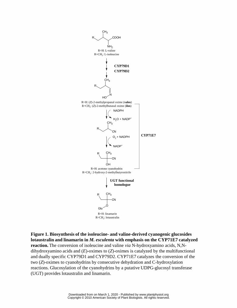
Figure 1. Biosynthesis of the isoleucine- and valine-derived cyanogenic glucosides lotaustralin and linamarin in M. esculenta with emphasis on the CYP71E7 catalyzed reaction. The conversion of isoleucine and valine via N-hydroxyamino acids, N,N-dihydroxyamino acids and (E)-oximes to (Z)-oximes is catalyzed by the multifunctional and dually specific CYP79D1 and CYP79D2. CYP71E7 catalyzes the conversion of the two (Z)-oximes to cyanohydrins by consecutive dehydration and C-hydroxylation reactions. Glucosylation of the cyanohydrins by a putative UDPG-glucosyl transferase (UGT) provides lotaustralin and linamarin.
COOH
NH2
CH3
R
N
CH3
R
OH
CH3
RCN
R
OH
CH3
CN
R
O
CH3
CN
Glu
R=H: L-valineR=CH3: L-isoleucine
CYP79D1
CYP79D2
NADPH
O2 + NADPH
NADP+
R=H: acetone cyanohydrinR=CH3: 2-hydroxy-2-methylbutyronitrile
R=H: linamarinR=CH3: lotaustralin
CYP71E7
UGT functional homologue
H2O + NADP+
R=H: (Z)-2-methylpropanal oxime (valox)R=CH3: (Z)-2-methylbutanal oxime (ilox)
www.plantphysiol.orgon March 1, 2020 - Published by Downloaded from Copyright © 2010 American Society of Plant Biologists. All rights reserved.

390 420 450 480 500
-0,02
0,00
0,02
0,04
nm
A
Figure 2. Carbon monoxide difference spectrum of yeast microsomes harbouring CYP71E7. The Fe2+.CO versus Fe2+
difference spectrum was recorded on the microsomal fraction of yeast expressing CYP71E7.
Abs
orba
nce
Wavelength (nm)
www.plantphysiol.orgon March 1, 2020 - Published by Downloaded from Copyright © 2010 American Society of Plant Biologists. All rights reserved.

O
CH3
R
+NO2
NO2
NHNH2
NO2
NO2
NHN
CH3
R
pH<1
50oC
R=H: acetone
R=CH3: 2-butanone
2,4-dinitrophenylhydrazine (DNPH)
R=H: acetone 2,4-dinitrophenylhydrazone
R=CH3: 2-butanone 2,4-dinitrophenylhydrazone
OH
CH3
CN
R
R=H: acetone cyanohydrin
R=CH3: 2-hydroxy-2-methylbutyronitrile
CN- + H2O
(g)
(aq)
(aq)
O
CH3
R
R=H: acetone
R=CH3: 2-butanone
(g)
(aq)
OH-
+ H2O
A
EIC 253 EIC 239
5.5 6.5 7.59.0 10.0 11.00.0
0.5
1.0
1.5
C
B
Inte
nsit
y x
10-5
Retention time (min)
Figure 3. Analysis of CYP71E1 produced cyanohydrins following dissociation into ketones, derivatization and LC-MS analysis. A: The cyanohydrins produced in the enzyme reaction mixtures were dissociated into ketones and CN- at alkaline pH. B: The volatile ketones were trapped in a center well containing an acidified solution of DNPH. C: LC-MS extracted ion chromatograms (EIC) of the 2,4-dinitrophenylhydrazones of 2-butanone (EIC 253) and acetone (EIC 239). The six chromatograms shown in each panel represent assay mixtures containing: Buffer only (black, solid); void vector yeast microsomes, 50 μM oxime + 1 mM NADPH (light green, solid); CYP71E7 containing yeast microsomes, 10 μM oxime – NADPH (red, dotted); CYP71E7 containing yeast microsomes, 10 μM oxime + 1 mM NADPH (blue, solid); CYP71E7 containing yeast microsomes, 50 μM oxime + NADPH (magenta, solid); CYP71E7 containing yeast microsomes, 100 μM oxime + 1 mM NADPH (dark green, dotted).
www.plantphysiol.orgon March 1, 2020 - Published by Downloaded from Copyright © 2010 American Society of Plant Biologists. All rights reserved.

2
-2
0
8
0
-4
4
370 400 420 450 380 420 460
Abs
orba
nce
x 10
3
nm nm
A
1
1
2 2
3
3
4
4
5
BA
Wavelength (nm)
Figure 4. Substrate binding properties of CYP71E7 as analyzed by optical difference spectroscopy. A: (1) Baseline recorded with CYP71E7 harbouring microsomes in the absence of substrate in both sample and reference cuvettes; (2,3,4) Spectra after addition of ilox (0.5, 1 and 3 μM, respectively) to sample cuvette. B: (1) Spectrum of CYP71E7 harbouring microsomes saturated with the cytochrome P450 inhibitor n-octylamine (100 μM); (2) Baseline recorded upon addition of equal amounts of n-octylamine (100 μM) to CYP71E7 harbouring microsomes in both sample and reference cuvette; (3,4,5) Displacement of n-octylamine by addition of ilox (1,10 and 30 μM, respectively) to sample cuvette.
www.plantphysiol.orgon March 1, 2020 - Published by Downloaded from Copyright © 2010 American Society of Plant Biologists. All rights reserved.

Figure 5: Cellular localisation of the expression of CYP79D1and CYP71E7 in the petiole of the first unfolded leaf using in tube in situ RT-PCR analysis of 80 µm transverse sections. c: cortex, e:endodermis, l: laticifers and x: xylem. Bar: 100 µm. The expression of the transcripts has been visualised using either alkaline phosphatase labelled (black staining) or FITC labelled (bright light green staining) antibodies recognising DIG.
l
c
l
c
x
c
l
x
c
x
l
c
l
x
c
x
l
c
xl
c
x
l
c
x
l
Expression of mRNA monitored by
Alkaline Phosphatase
Light microscopy FITC control
CYP79D1 expression monitored by FITC
CYP71E7 expression monitored by FITC
CYP79D1 Control CYP71E7
e e e
e e
e e
e e
www.plantphysiol.orgon March 1, 2020 - Published by Downloaded from Copyright © 2010 American Society of Plant Biologists. All rights reserved.

A
DC
Be
e
cp
cp
cp
cp
e
v
v
Figure 6: Cellular localization of the expression of CYP79D1 and CYP71E7 in two leaf stages, a nearly unfolded leaf and of the first fully unfolded leaf, using in tube in situ RT-PCR analysis of 80 µm transverse sections. A and B localization of respectively CYP79D1and CYP71E7 expression in a nearly unfolded upper leaf. C and D localization of CYP79D1 expression in un unfolded leaf. e: epidermis, cp: pallisade tissue, v: vascular tissue. Bar 100µm. The expression of the transcripts has been visualised using FITC labelled (bright light green staining) antibodies recognising DIG.
www.plantphysiol.orgon March 1, 2020 - Published by Downloaded from Copyright © 2010 American Society of Plant Biologists. All rights reserved.

Ilox Valox Tyrox Pheox
Turnover (min-1) 17.1 ± 1.0 21.2 ± 2.2 8.1 ± 0.3 1.3 ± 0.2
Km (µM) 21 ± 11 n.d. n.d. n.d.
Table 1
Table 1. Kinetic parameters of CYP71E7 catalyzed oxime to cyanohydrin conversions.
www.plantphysiol.orgon March 1, 2020 - Published by Downloaded from Copyright © 2010 American Society of Plant Biologists. All rights reserved.