rransfonnation of Medicago by Agrobacterium … · Plant Cell Reports (1986) 5: 97- I00...
4
Plant Cell Reports(1986) 5: 97- I00 rransfonnation of Medicago by Agrobacterium mediated gene transfer Maria Deak, Gyorgy B. Kiss, Csaba Koncz ', and Denes Dudits nstitute ofGenetics.Biological Research Center ofthe Hungarian Academy ofSciences.P.O. Box 521.H-6701Szeged.Hungary :eceivedOctober 10. 1985 / Revised version received January 16. 1986 - Communicated by A.Szalay ABSTRACT Shoot segments of Medicago varia genotype A2 ere co-cultivated with Agrobacterium tumefaciens train bo42 carrying pGA471, a plasmid coding for 'be kanamycin resistant determinant as ransferable positive selection marker in plant :lls (An et al., 1985). Resistant plants were :generated at high frequency from green calli weloped on inoculated stem cuttings under mamycin selection. DNA-DNA hybridization analysis %wed the presence of the structural gene of the pamycin resistant determinant in total DNA dated from several independent transformants. All 3ta presented clearly demonstrate the transfer, :able maintenance and functional express,ion of the ~namycin resistance marker in Medicago & cells ~ich retain their morphogenic property. ,breviations:Km = kanamycin KmR = kanamycin resistant Cb = carbenicillin 2,4-D = 2,4 dicblorophenoxyacetic acid BA = 6-benzyladenine T-DNA = transferred DNA into plants INTRODUCTION Leguminous plants have been in the center of ientific interest for at least two main reasons. ey are very important constituents of both human 9t and animal feed, and as compared to other crop ants legumes are capable to supply their nitrogen mnd from symbiotic associations with Rhizobia. s introduction of new genetic information into ese plants would be of great impact on both crop eld improvement and the molecular understanding of mbiotic nitrogen fixation. Fortunately,recent ientific accomplishments of the natural gene ansfer of Agrobacterium established the aboration of the transformation system for Ealfa. Virulent strains of Agrobacterium tumefaciens c capable to introduce a specific segment (T-DNA) its tumor-inducing (Ti) plasmid into the cells of ny dicotyledonous plants (DeCleene and DeLey, .76; Chilton et al., 1977; Yadav et al., 1980; ster et al., 1984) including Medicago species lariotti et al., 1984; M. Deak unpublished jservations). The transferred T-DNA contains Presenr address: Max-Planck-Insiitut for ichtungsforschung. D-5000 Koln 30. FRG Grin[ requesrs to: M. Deak several genetic loci ol which only ihc righi border is required for integration into the plant genome (Lemmers et al., 1980; Thomashow et al., 1980; Zambryski et al., 1980; Leemans et al., 1982; Yadav et al., 1982; Ursic et al., 1983; Caplan et al., 1985). The other regions are dispensable in this respect and gives room for in vivo and vitro genetic manipulations (Hernalsteens et al., 1980; Bevan et al., 1983). Thus, several structural modifications of the T-DNA led to the construction of versatile cloning vectors which can be used to transform different plant species (Bevan et al., 1983; Fraley et al., 1983; Herrera Estrella et al., 1983; Horsch et al., 1984). In this paper we describe the transfer of the KmR determinant into alfalfa cells and the regeneration of whole plant from KmR tissues. The transformation procedure is based on the recently developed binary vector system of Agrobacterium (An et al., 1985) and the use of a Medicago genotype with high embryogenic potential. MATERIALS AND METHODS Plant material Medicago varia genotype A2 exhibiting good plant regeneration capability in tissue cultures was kindly provided by Dr A. Atanassov, Tissue Culture Laboratory, Institute of Genetics, Bulgarian Academy of Sciences, Sofia, Bulgaria. Plants were propagated as sterile shoot cultures on hormone free UM medium (Uchimiya and Murashige, 1974). Young shoots of this material were used in transformation experiments. Bacterial strains Agrobacterium tumefaciens strain bo42 was kindly provided by Dr G. An, Institute of Biological Chemistrv, Washington State University, Pullman, USA. p his strain- is tumefaciens ~ 2 8 1 carrying olasmid ~GA471 (An et al.. 1985). Plasmid pGA471 contains 'a mini 'RK~ replicon, a transferable ' T-DNA carrying KmR determinant fused to the nopaline synthase promoter, ColEl type origine of replication, lambda cohesive site and restriction endonuclease recognition sequences for cloning. Media and Culture conditions A. tumefaciens strain bo42 was grown at 28O~ in LF medium containing tetracvcline-HC1 (SIGMA), 12.5 mg/l and kanamycii sulphate (SIGMA). 160 mgji for selection of plasmid pGA471. Callus tissues were induced on UM medium supplemented with 2,4-D (SIGMA), 0.5 mg/l , BA (SIGMA), 0.2 mg/l and Bacto Tryptone (DIFC0),250
-
Upload
nguyenthuy -
Category
Documents
-
view
212 -
download
0
Transcript of rransfonnation of Medicago by Agrobacterium … · Plant Cell Reports (1986) 5: 97- I00...

Plant Cell Reports (1986) 5: 97- I00
rransfonnation of Medicago by Agrobacterium mediated gene transfer
Maria Deak, Gyorgy B. Kiss, Csaba Koncz ', and Denes Dudits
nstitute of Genetics. Biological Research Center of the Hungarian Academy of Sciences. P.O. Box 521. H-6701 Szeged. Hungary
:eceived October 10. 1985 / Revised version received January 16. 1986 - Communicated by A.Szalay
ABSTRACT
Shoot segments of Medicago varia genotype A2 ere co-cultivated with Agrobacterium tumefaciens train bo42 carrying pGA471, a plasmid coding for 'be kanamycin resistant determinant as ransferable positive selection marker in plant :lls (An et al., 1985). Resistant plants were :generated at high frequency from green calli weloped on inoculated stem cuttings under mamycin selection. DNA-DNA hybridization analysis %wed the presence of the structural gene of the pamycin resistant determinant in total DNA dated from several independent transformants. All 3ta presented clearly demonstrate the transfer, :able maintenance and functional express,ion of the ~namycin resistance marker in Medicago & cells ~ich retain their morphogenic property.
,breviations: Km = kanamycin KmR = kanamycin resistant Cb = carbenicillin 2,4-D = 2,4 dicblorophenoxyacetic
acid BA = 6-benzyladenine T-DNA = transferred DNA into plants
INTRODUCTION
Leguminous plants have been in the center of ientific interest for at least two main reasons. ey are very important constituents of both human 9t and animal feed, and as compared to other crop ants legumes are capable to supply their nitrogen mnd from symbiotic associations with Rhizobia. s introduction of new genetic information into ese plants would be of great impact on both crop eld improvement and the molecular understanding of mbiotic nitrogen fixation. Fortunately,recent ientif ic accomplishments of the natural gene ansfer of Agrobacterium established the aboration of the transformation system for Ealfa.
Virulent strains of Agrobacterium tumefaciens c capable to introduce a specific segment (T-DNA) its tumor-inducing (Ti) plasmid into the cells of
ny dicotyledonous plants (DeCleene and DeLey, .76; Chilton et al., 1977; Yadav et al., 1980; ster et al., 1984) including Medicago species lariotti et al., 1984; M. Deak unpublished jservations). The transferred T-DNA contains
Presenr address: Max-Planck-Insiitut for ichtungsforschung. D-5000 Koln 30. FRG Grin[ requesrs to: M. Deak
several genetic loci o l which only ihc righi border is required for integration into the plant genome (Lemmers et al., 1980; Thomashow et al., 1980; Zambryski et al., 1980; Leemans et al., 1982; Yadav et al., 1982; Ursic et al., 1983; Caplan et al., 1985). The other regions are dispensable in this respect and gives room for in vivo and vitro genetic manipulations (Hernalsteens et al., 1980; Bevan et al., 1983). Thus, several structural modifications of the T-DNA led to the construction of versatile cloning vectors which can be used to transform different plant species (Bevan et al., 1983; Fraley et al., 1983; Herrera Estrella et al., 1983; Horsch et al., 1984).
In this paper we describe the transfer of the KmR determinant into alfalfa cells and the regeneration of whole plant from KmR tissues. The transformation procedure is based on the recently developed binary vector system of Agrobacterium (An et al., 1985) and the use of a Medicago genotype with high embryogenic potential.
MATERIALS AND METHODS
Plant material
Medicago varia genotype A2 exhibiting good plant regeneration capability in tissue cultures was kindly provided by Dr A. Atanassov, Tissue Culture Laboratory, Institute of Genetics, Bulgarian Academy of Sciences, Sofia, Bulgaria. Plants were propagated as sterile shoot cultures on hormone free UM medium (Uchimiya and Murashige, 1974). Young shoots of this material were used in transformation experiments.
Bacterial strains
Agrobacterium tumefaciens strain bo42 was kindly provided by Dr G. An, Institute of Biological Chemistrv, Washington State University, Pullman, USA. p his strain- is tumefaciens ~ 2 8 1 carrying olasmid ~GA471 (An et al.. 1985). Plasmid pGA471 contains 'a mini ' R K ~ replicon, a transferable ' T-DNA carrying KmR determinant fused to the nopaline synthase promoter, ColEl type origine of replication, lambda cohesive site and restriction endonuclease recognition sequences for cloning.
Media and Culture conditions
A. tumefaciens strain bo42 was grown at 2 8 O ~ in LF medium containing tetracvcline-HC1 (SIGMA), 12.5 mg/l and kanamycii sulphate (SIGMA). 160 mgji for selection of plasmid pGA471.
Callus tissues were induced on UM medium supplemented with 2,4-D (SIGMA), 0.5 mg/l , BA (SIGMA), 0.2 mg/l and Bacto Tryptone (DIFC0),250

mg/l. For embryo induction a liquid B5N medium was used as modified B5 (Gamborg et al., 1968) supplemented with 3% sucrose, meso-inositol (SIGMA), 500 mg/l, adenine (SIGMA), 1 mg/l, glutathione (SIGMA), 10 mg/l, 2,4-D (SIGMA), 1 mg/l and BA (SIGMA), 0.2 mg/l. All the other components of media were purchased from REANAL (BUDAPEST).
Media were solidified by adding 9,g/l of Bacto Agar. All media were autoclaved at 121 C for 20 mi8. Calli were grown in sterile petri dishes at 25 C under a 16 hrs photoperiod of fluorescent light.
Stem segments of M. varia genotype A2 produced dark green calli with several embryos on solid UM medium. Plantlets developed after transferring embryos onto UM medium with BA, while whole plants were recovered on the same medium without hormone.
The KmR phenotype of the transformed plants were tested as follow: small segments of the root were cut and placed on UM medium containing 100 mg/l Km. KmR plant material produced calli with green embryos in three weeks, while control, sensitive tissues died.
DNA isolation, restriction digestion and hybridization
Total DNA was isolated from 200-600 mg of plant material according to Dellaporta et al., (1983). Hind111 and EcoRI restriction digestion was carried out according to the supplyers instructions (Bethesda Research Laboratories). Restriction fragments were separated in 0.7 % agarose gel. Isolated BglII-SmaI fragment of plasmid pSUPlOl1 (Simon, 1984) covering the structural gene of the KmR determinant of Tn5 (Jgrgensen et al., 1979) was labelled to 4.8-11.0 x 10 dpm/ug specific activity according to Feinberg and Vogelstein (1983). Hybridization was carried out according to Purello and Balazs (1983) and Singho and Jones (1984). Autoradiography was done at -70 C under Kodak XR5 X- ray film with Kodak X-Omatic intensifying screen.
Neomycin phosphotransferase assay
The activity of the enzyme neomycin phosphotransferase was measured according to Schreier et a1.,1985.
RESULTS
Transformation of Medicago varia by Agrobacterium
The co-cultivation method originally described by Marton et al. (1979) for protoplasts was adapted to inoculate stem cuttings of alfalfa with Agrobacterium. 0.5-1.0 cm long shoot segments of steril plants of Medicago varia genotype A2 were placed into an Erlenmeyer flask (100 ml in size) containing 40 ml of liquid UM medium and 1 ml of appropriately deluted overnight culture of A, tumefaciens strain bo42 (5x10' cells/ml). The tissues were incubated on a rotary shaker (150 rpm/min) at 25'~ for three days. The stem segments were then washed three times with sterile destilled water and transferred onto solid UM medium containing 'Cb, 300 mg/l and Km, 100 mg/l. After three weeks of culture in average 3-4 green growing spots appeared on each of the treated stem segments in the presence of Km. The high efficiency in production of responsive growing tissues was repeatedly reproducable in large number of inoculation experiments. As control, shoot segments were treated as above without bacterial inoculation. In these cases the initial proliferation of the tissues was stoped by Km and the development of green calli was never detected.
Embryogenesis and plant regeneration from KmR calli
After several transfer. on UM medim supplemented with Cb, 300 mg/l and Km, 100 mg/l somatic embryos developed from the green growing calli with low frequency. To improve the efficiency of plant redifferentiation several conditions were tested for embryo induction. Among others the following protocol was found to be the most successful:
First, the primary green KmR tissues were multiplied on fresh solid UM medium supplementeci with Cb, 300 mg/l and Km, 100 mg/l for three weeks. Tissues with the proper size were then transferre, into 40 ml B5N medium containing Km, 50 mg/l in 10( ml Erlenmeyer flasks an$ incubated on rotary shake: (150 rpm/min) at 25 C. The suspensions wen subcultured twice a week in the same medium. In thr KmR suspension cultures, after several weeks large number of green globular and torpedo embryos appeared in addition to the unorganized cel aggregates. The suspension culture maintained thei embryo-producing capability for more then thref months. These embryos were transferred onto solid U' medium containing BAS 0.2 mg/l and Km, 100 mg/ where they developed further and after 3-6 weeks plants started to regenerate.
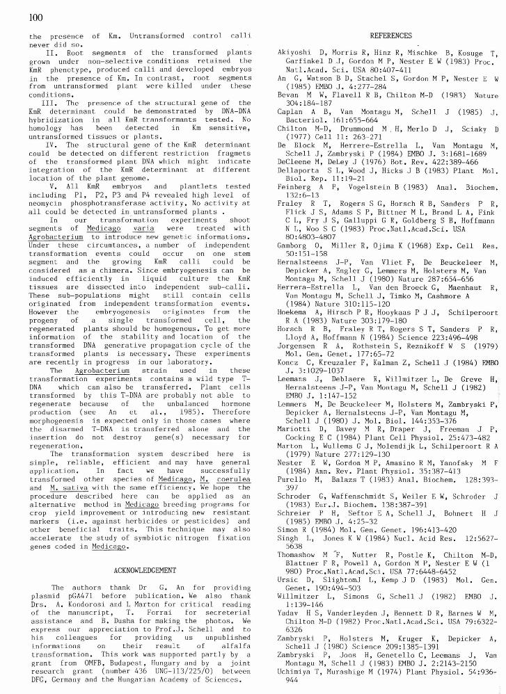
%our developing plantlets designated P1, P2 P3 and P4 were randomly selected and characterise further (see below). The photos of six regenerate plants are shown in Figure 1. The stability of th KmR phenotype was tested on the roots of th transformants as follows: root segments were place on UM medium containing Km, 100 mg/l and they forme green calli with embryos while control root segment never did so.
Figure 1. The picture of six randomly selected kanamycin resistant plants.
Detection of the KmR structural gene and its functional expression in the transformants
Three developing KmR plantlets P1, P2 and (see above) and a KmS control plant were devided ir two parts. One part was used to detect DNA homolog) to the structural gene of the KmR determinant while the other part was used to measure the activity of the neomycin phosphotransferase.
After total DNA isolation, Hind I11 digestior and agarose gel electrophoresis, DNA-DNA hybridization was performed using labelled probe specific to the structural gene of the Kd determinant (see Materials and methods). As seen on Figure 2. Panel B, hybridization signal could only be detected in the transformed plant material (lane

2 and 3). No hybridization could be observed with A from untreated calli (lane 4) and from whole ant of M. varia, M. falcata, M. sativa, ' tobacco, rrot and wheat (data not shown). The DNA of sntlet P1 contained multiple hybridization agments from which one band is very intense. mtlet P2 had one hybridizing band. Though the gestion of the DNA from plantlet P3 is partial the bridization is obvious.
Similar experiments were done with eight ndomly selected calli as well as from untreated sensitive control material except that digestion
s with enzyme EcoR I. Specific hybridization was tected to different EcoRI fragments of the DNA om the eight KmR calli, while no hybridization was und with DNA from Km sensitive callus (data not own).
A Hind , S B
Marker
M. Deak (unpublished results) demonstrated that transformed tissue of alfalfa could grow on hormone free medium but no plant regeneration could be achieved. In these experiments wild type Ti plasmids were used where the integration of T-DNA resulted in unbalanced phytohormone production (Akiyoshi et al., 1983; Schroder et al., 1983; Nester et al., 1984). As a consequence the highly morphogenic lines of Medicago species also failed to produce plants from hormone autotrophic tissues. Elegant series of experiments showed that the phytohormone genes of the T-DNA were dispensable for gene transfer, and could be replaced by selectable markers (e.g. KmR determinant) as well as other genes to be transferred into plants (Herrera-Estrella et al., 1983; Fraley et al., 1983; Herrera-Estrella et al., 1984; Koncz et al., 1984). By creating "disarmed" (hormone gene-deleted) T-DNA, the transferred genetic material has no influence any longer on the hormone metabolism of the recipient cell, therefore it does not interfere with the morphogenic potential (Bevan et al., 1983; Zambryski et al., 1983; De Block et al., 1984).It has also been shown that the
:ure 2. HindIII restriction fragments of total Int DNA was separated on 0.7 % agarose gel, tined with ethidium bromide and photographed me1 A). The same gel was dried and hybridized -h a probe specific to the structural gene of the t determinant (Panel 8). Autoradiography was done -70 C for 9 days. Lane 1, 2 and 3 represents DNA
)rn three randomly selected regenerating plantlets iignated P1, P2 and P3 (see text). Lane 4 is DNA )m untransformed control tissue. HindIII digested nbda DNA as indicated was used for molecular ight marker (kb, kilobase pairs).
The activity of . the enzyme neomycin )sophotransferase of the KmR plantlets P1, P2 P3 I P4 as well as of the KmS control plant was meas- -?d as described in the Materials and Methods. As ?n from Figure 2, the transformed material showed ':ivity (lkine 2, 3, 4, and 5). No activity at all .~ld be detected in the control plant (lane 1).
DISCUSSION
In this paper we demonstrate the transfer of e KmR determinant into Medicago varia cells by robacterium, and that the transformed tissues tained their morphogenic property and regenerated KmR plants at high frequency. It was shown earlier that A, tumefaciens causes
own gall tumors on Medicago species (De Cleene and Ley, 1976). Recently !lariotti et al. (1984) and
Figure 3. Detection of the neomycin phosphotransferase activity of the transformants. The signal on the autoradiogram shows the presence of phosphorylated kanamycin as the consequence of the activity of neomycin phosphotransferase from extracts of untransformed plant (lane l), plantlet P1 (lane 2), P2 (lane 4) ,P3 (lane 5),and P4 (lane 3). The assay was carried out as described in Materials and Methods.
vir genes (necessary for transfer and integration - process) can act & trans making possible the development of binary vector system, i.e. 2 genes and T-DNA are on different replicon.This allows, T- DNA being on a plasmid of small size, to carry out in vitro manipulations more easy (Leemans et al., -- 1982; Hoekema et al., 1983).
As shown in this paper a recently developed binary vector system (An et al., 1985) fulfilled these expectations and made possible to transform alfalfa cells which retained their morphogenic potential. The co-cultivation procedure outlined in the text resulted in KmR calli which in turn could be regenerated to KmR plants. There are several indicatibns that the KmR phenotype is the consequence of the functional expression of the transferred and stably maintained neomycin phosphotransferase gene:
I. Only the co-cultivated shoot segments could grow, form green embryos and regenerate plants in
the presence of Km. Untransformed control calli never did so.
11. Root segments of the transformed plants grown under non-selective conditions retained the KmR phenotype, produced calli and developed embryos in the presence of Km. In contrast, root segments from untransformed plant were killed under these conditions.
111. The presence of the structural gene of the KmR determinant could be demonstrated by DNA-DNA hybridization in all KmR transformants tested. No homology has been detected in Km sensitive, untransformed tissues or plants.
IV. The structural gene of the KmR determinant could be detected on different restriction fragmets of the transformed plant DNA which might indicate integration of the KmR determinant at different location of the plant genome.
V. All KmR embryos and plantlets tested including P1, P2, P3 and P4 revealed high level of neomycin phosphotransferase activity. No activity at all could be detected in,untransformed plants .
In our transformatlon experiments shoot segments of Medicago varia were treated with A~robacterium to introduce new genetic informations. Under these circumtances, a number of independent transformation events could occur on one stem segment and the growing KmR calli could be considered as a chimera. Since embryogenesis can be induced efficiently in liquid culture the KmR tissues are dissected into independent sub-calli. These sub-populations might still contain cells originated from independent transformation events. However the embryogenesis originates from the progeny of a single transformed cell, the regenerated plants should be homogenous. To get more information of the stability and location of the transformed DNA generative propagation cycle of the transformed plants is necessary. These experiments are recently in progress in our laboratory.
The A~robacterium strain used in these transformation experiments contains a wild type T- DNA which can also be transferred. Plant cells transformed by this T-DNA are probably not able to regenerate because of the unbalanced hormone production (see An et al., 1985). Therefore morphogenesis is expected only in those cases where the disarmed T-DNA is transferred alone and the insertion do not destroy gene(s) necessary for regeneration.
The transformation system described here is simple, reliable, efficient and may have general application. In fact we have successfully transformed other species of Medicago, M, coerulea and M. sativa with the same efficiency. We hope the procedure described here can be applied as an alternative method in Medicago breeding programs for crop yield improvement or introducing new resistant markers (i.e. against herbicides or pesticides) and other beneficial traits. This technique may also accelerate the study of symbiotic nitrogen fixation genes coded in Medicago.
ACKNOWLEDGEMENT
The authors thank Dr G. An for providing plasmid pGA471 before publication. We also thank Drs. A. Kondorosi and L Marton for critical reading of the manuscript, T. Forrai for secreterial assistance and B. Dusha for making the photos. We express our appreciation to Pr0f.J. Schell and to his colleagues for providing us unpublished informations on their result of alfalfa transformation. This work was supported partly by a grant from OMFB. Budapest, Hungary and by a joint research grant (number 436 UNG-113/225/0) between DFG, Germany and the Hungarian Academy of Sciences.
REFERENCES
Akiyoshi D, Morris R, Hinz R, Mischke B, Kosuge T, Garfinkel D J, Gordon M P, Nester E W (1983) Proc. Natl.Acad. Sci. USA 80:407-411
An G, Watson B D, Stachel S, Gordon M P, Nester E W (1985) EMBO J. 4:277-284
Bevan M W, Flavell R B, Chilton M-D (1983) Nature 304:184-187
Caplan A B, Van Montagu M, Schell J (1985) J. Bacteriol. 161:655-664
Chilton M-D, Drummond M . H, Merlo D J, Sciaky D (1977) Cell 11: 263-271
De Block M, Herrere-Estrella L, Van Montagu M, Schell J, Zambryski P (1984) EMBO J. 3:1681-1689
DeCleene M, DeLey J (1976) Bot. Rev. 422:389-466 Dellaporta S L, Wood J, Hicks J B (1983) Plant Mol. Biol. Rep. 11:19-21
Feinberg A P, Vogelstein B (1983) Anal. Biochem. 132:6-13
Fraley R T, Rogers S G, Horsch R B, Sanders P R, Flick J S, Adams S P, Bittner M L, Brand L A , Fink C L, Fry J S, Galluppi G R, Goldberg S B, Hoffmann N L, Woo S C (1983) Proc.Natl.Acad.Sci. USA 80 : 4803-4807
Gamborg 0, Miller R, Ojima K (1968) Exp. Cell Res. 50:151-158
Hernalsteens J-P, Van Vliet F, De Beuckeleer M, Depicker A, Engler G, Lemmers M, Holsters M, Van Montagu M, Schell J (1980) Nature 287:654-656
Herrera-Estrella L, Van den Broeck G, Maenhaut R, Van Montagu M, Schell J, Timko M, Cashmore A (1984) Nature 310:115-120
Hoekema A, Hirsch P R, Hooykaas P J J, Schilperoort R A (1983) Nature 303:179-180
Horsch R B, Fraley R T, Rogers S T, Sanders P R, Lloyd A, Hoffmann N (1984) Science 223:496-498
Jorgensen R A, Rothstein S, Reznikoff W S (1979) Mol. Gen. Genet. 177:65-72
Koncz C, Kreuzaler F, Kalman Z, Schell J (1984) PMBO J. 3:1029-1037
Leemans J, Deblaere R, Willmitzer L, De Greve H, Hernalsteens J-P, Van Montagu M, Schell J (1982) EMBO J. 1:147-152
Lemmers M, De Beuckeleer M, Holsters M, Zambryski P, Depicker A, Hernalsteens J-P, Van Montagu M, Schell J (1980) J. Mol. Biol. 144:353-376
Mariotti D, Davey M R, Draper J, Freeman J P, Cocking E C (1984) Plant Cell Physiol. 25:473-482
Marton L, Wullems G J, Molendijk L, Schilperoort R A (1979) Nature 277: 129-130
Nester E W, Gordon M P. Amasino R M, Yanofsky M F (1984) Ann. Rev. Plant Physiol. 35:387-413
Pure110 M, Balazs T (1983) Anal. Biochem. 128:393- 397
Schroder G, Waffenschmidt S, Weiler E W, Schroder J (1983) Eur.J. Biochem. 138:387-391
Schreier P H, Seftor E A, Schell J,, Bohnert H J (1985) EMBO J. 4:25-32
Simon R (1984) Mol. Gen. Genet. 196:413-420 Singh L, Jones K W (1984) Nucl. Acid Res. 12:5627- 5638
Thomashow M -F, Nutter R. Postle K, Chilton M-D, Blattner F R, Powell A, Gordon M P, Nester E W (1 980) Proc.Natl.Acad.Sci. USA 77:6448-6452
Ursic D, SlightomJ L, Kemp J D (1983) Mol. Gen. Genet. 190:494-503
Willmitzer L, Simons G, Schell J (1982) EMBO J. 1 : 139-146
Yadav H S, Vanderleyden J, Bennett D R, Barnes W M, Chilton M-D (1982) Proc.Natl.Acad.Sci. USA 79:6322- 6326
Zambryski P, Holsters M, Kruger K, Depicker A, Schell J (1980) Science 209:1385-1391
Zambryski P, Joos H, Genetello C, Leemans J, Van Montagu M, Schell J (1983) EMBO J. 2:2143-2150
Uchimiya T, Murashige M (1974) Plant Physiol. 54:936- 944