
Round Immature Spermatogenic Cells in Semen Fluids … · Spermatological assay includes the...
9
15 Institute of Experimental Morphology, Pathology and Anthropology with Museum Bulgarian Anatomical Society Acta morphologica et anthropologica, 22 Sofia • 2015 Round Immature Spermatogenic Cells in Semen Fluids of Infertile Men with Diagnosis “Migrating Testis”. Two Casuistic Cases in Adults I. Ilieva¹, P. Tzvetkova², M. Kacarov³, I. Sainova¹, P. Taushanova², I. Vladov¹, E. Zvetkova 4 ¹Institute of Experimental Morphology, Pathology and Anthropology with Museum, Bulgarian Academy of Sciences, Sofia, Bulgaria ²Institute of Biology and Immunology of Reproduction “Acad. Kiril Bratanov”, Bulgarian Academy of Sciences, Sofia, Bulgaria ³Medical Center “Doverie”, Sofia, Bulgaria 4 Bulgarian Biorheological Society The spermatological analysis of ejaculates from two patients suffering of low male fertility and with diagnosis “migrating” testis syndrome (casuistic cases in adults), reveals cytological characteristics of different immature spermatogenic cells (“round cells”) as precursors of spermatozoa. The high quantity of undifferentiated spermatocytes/spermatides in ejaculates is important for early diagnoses of andro- logical diseases related to male fertility (sub-fertility, infertility). The results from semen assays showed a high percent (7.3% and 13.5%) of immature (“round”) spermatogenic cells in the ejaculates of patients – in correlation with the elevated number of abnormal spermatozoa in probes. The morphological analy- sis of sperm samples in patients with ascending testis could serve as additional diagnostic and prognostic tool in the routine everyday andrological practice. Key words: spermatogenesis, immature spermatogenic cells (“round cells”), ascending testis syndrome. Introduction In the process of spermatogenesis, spontaneous degenerative changes in the separated zones of the seminiferous tubules are possible. These features affect individual cell types – spermatogonia, spermatocytes (primary and/or secondary), spermatids and spermatozoa, and some of them pass in the seminal fluid. In pathology of the genital tract, which could be a result of congenital factors (cryptorhidism, anorhidism, etc.) or of the influence of different diseases, these changes can include larger regions of the tes- ticular tissue [7, 14, 20]. In normal conditions, the amount of the degenerated gametes in the ejaculate is approximately 25% (WHO, 2010). However, in stress conditions, sig- nificant increase is possible, which could influence the inseminal qualities of the fluid. The morphological assessment of the seminiferous assay includes the determination of the mature spermatozoa, but also of other – both cytologically and functionally different
-
Upload
nguyenkhanh -
Category
Documents
-
view
215 -
download
0
Transcript of Round Immature Spermatogenic Cells in Semen Fluids … · Spermatological assay includes the...
15
Institute of Experimental Morphology, Pathology and Anthropology with Museum Bulgarian Anatomical SocietyActa morphologica et anthropologica, 22Sofia • 2015
Round Immature Spermatogenic Cells in Semen Fluids of Infertile Men with Diagnosis “Migrating Testis”. Two Casuistic Cases in AdultsI. Ilieva¹, P. Tzvetkova², M. Kacarov³, I. Sainova¹, P. Taushanova²,I. Vladov¹, E. Zvetkova4
¹Institute of Experimental Morphology, Pathology and Anthropology with Museum, Bulgarian Academy of Sciences, Sofia, Bulgaria²Institute of Biology and Immunology of Reproduction “Acad. Kiril Bratanov”, Bulgarian Academy of Sciences, Sofia, Bulgaria³Medical Center “Doverie”, Sofia, Bulgaria4Bulgarian Biorheological Society
The spermatological analysis of ejaculates from two patients suffering of low male fertility and with diagnosis “migrating” testis syndrome (casuistic cases in adults), reveals cytological characteristics of different immature spermatogenic cells (“round cells”) as precursors of spermatozoa. The high quantity of undifferentiated spermatocytes/spermatides in ejaculates is important for early diagnoses of andro-logical diseases related to male fertility (sub-fertility, infertility). The results from semen assays showed a high percent (7.3% and 13.5%) of immature (“round”) spermatogenic cells in the ejaculates of patients – in correlation with the elevated number of abnormal spermatozoa in probes. The morphological analy-sis of sperm samples in patients with ascending testis could serve as additional diagnostic and prognostic tool in the routine everyday andrological practice.
Key words: spermatogenesis, immature spermatogenic cells (“round cells”), ascending testis syndrome.
Introduction
In the process of spermatogenesis, spontaneous degenerative changes in the separated zones of the seminiferous tubules are possible. These features affect individual cell types – spermatogonia, spermatocytes (primary and/or secondary), spermatids and spermatozoa, and some of them pass in the seminal fluid. In pathology of the genital tract, which could be a result of congenital factors (cryptorhidism, anorhidism, etc.) or of the influence of different diseases, these changes can include larger regions of the tes-ticular tissue [7, 14, 20]. In normal conditions, the amount of the degenerated gametes in the ejaculate is approximately 25% (WHO, 2010). However, in stress conditions, sig-nificant increase is possible, which could influence the inseminal qualities of the fluid. The morphological assessment of the seminiferous assay includes the determination of the mature spermatozoa, but also of other – both cytologically and functionally different
16
cell types. In most cases these are immature germ cells – precursors of spermatozoa, but also leucocytes (e.g. granulocytes, monocytes/macrophages) or epithelial cells from the urogenital tract. The immature germ cells, together with the leucocytes, usually are beyond the general group of so called “round cells” [4, 11, 14]. The exact determination of the cell types and their differentiation in spermatogenic or non-spermatogenic mature cells is important not only for a correct diagnosis, but also for the therapeutic approach. According to the recommendations of the World Health Organization (WHO), if the “round cells” in the ejaculate probe are more than 1 × 10⁶/ml, they should be determined as “presenting leucocytes”, because their increased amount could be a sign for inflam-mation process in the urogenital tract [6, 19]. In this connection, it is of interest to study the cytological content of the seminal fluid in patients with diagnosis “migrating” testis – two casuistic cases in adult men. In the scientific literature, the term “migrating” (elevator) testis (“retractile tests”) [2, 12, 23] is also described as a “retractile testis” and/or as “a syndrome of ascending testis” [12, 20, 23]. This abnormal gonad state is usually diagnosed in boys (child and juvenile age – most often around 10 years), but it is extremely rare in adult men. Unlike in cases of cryptorhidism, despite the scrotal position of the testes there is a risk of migrating testes (they remain in inguinal duct and later descend into the scrotum) due to external conditions like cold, nerve stress of cremaste muscle. As a result, abnormalities in the spermatogenesis are possible, which are similar to those, established in diagnosed cryptorhidism and/or varicocelle, due to the abnormal gonad thermoregulation [20, 23].
In this connection, the aim of the present study is to evaluate the quantity and to characterize the morphology of “round cells” in the samples from semen fluids of pa-tients with more rare diagnosis ascending testis. The methods of qualitative and quanti-tative semen analysis, applied by us, after in situ staining of “round cells” in ejaculates, could be an important diagnostic tool applicable in the treatment of patients with distur-bances in spermatogenesis and subfertility/infertility-related problems.
Materials and Methods
Semen liquids (ejaculates) from two andrological patients (average 23 and 30 years) with ascending testis, are examined, and the results are compared with data, obtained from a control group of 20 fertile healthy men (average 32.45 ± 1.59 years).
For a precise clinical diagnosis, in each case the patient’s data (anamnesis), to-gether with his local clinical status, are carefully investigated.
Sperm AnalysisSpermatological assay includes the qualitative and quantitative analyses of ejaculates and investigations of individual cells (spermatogonia, spermatocytes, spermatids, sper-matozoa and other, so-called “round cells”, in seminal plasma – assessed according to WHO criteria, 2010).
For cytological analysis, the smears from ejaculates are stained by the standard methods of Papanicollaou and Hematoxyllin/Eosin. The slides are examined light-mi-croscopically with microscope Leica DM 5000B (at different magnifications).
On the basis of morphological characteristics, the spermatogenic and non-sperma-togenic (“round”) cells in ejaculates are identified, as:
• spermatogonia (Spg);• primary spermatocytes (Spc-I);• secondary spermatocytes (Spc-II);• spermatids (Spt);
17
– round spermatids – rSpt; – elongated spermatids – elSpt;• other (non-spermatogenic) round cells: – granulocytes; – monocytes/macrophages; – lymphocytes.
Statistical AnalysisStatistical significance is verified by Student’s t-test. The results are given as MEAN ± SD.
Results
In the course of the spermatological analysis we obtained a reduced number of mor-phologically normal spermatozoa in the ejaculates of two andrological patients (with ascending testis), in comparison with the normal values for the control group. The quan-titative results are presented in Table 1.
Table 1. Cell number and presence of spermatozoa (including abnormal), as well as undifferentiated (immature) spermatogenic cells in ejaculates of patients with ascending testis, versus data for the control group
Number ofspermatozoa (million/ml)
Normal spermatozoa
(%)
Abnormal spermatozoa
(%)
Immaturegerm cells
(million/ml)
Immaturegerm cells
(%)
Ascending testis
(n = 2)
(n = 1) Normospermia67 38.3 54 5.59 7.3
(n = 1) Oligospermia19 22.95 63.45 2.97 13.5
Control group(n = 20) 84.79 ± 13.57 80.63 18.37 0.86 ± 0.09 1.0
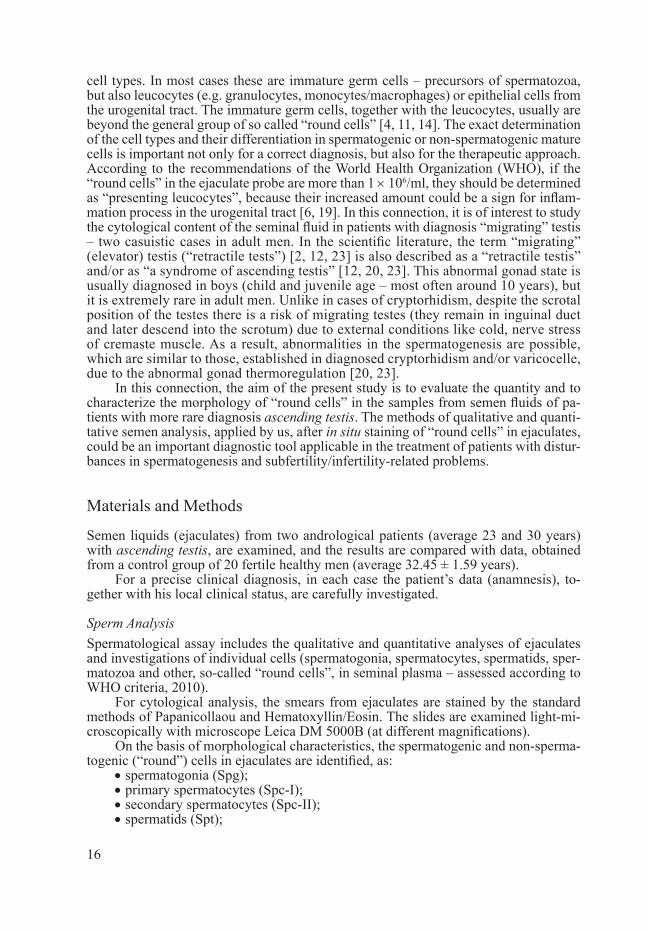
In one of the patients, the results show oligospermia – significantly decreased total spermatozoa number (19 million/ml) in the ejaculate, and in the other – normospermia (67 million/ml). In both cases, especially in that with oligospermia we assessed increased (> 50%) percent of the spermatozoa with abnormal morphology (Table 1). The high-est percent is established for the spermatozoa (18.46%), possessing deformations in the head, but gametes with combined abnormalities also reach significant percent (25.71%) (Fig. 1).
According to our results, simultaneously with the decrease of total number of sper-matozoa, an increase in the percent of the spermatogenic “round cells” in the ejaculates was found, which correlates with the higher percent of the abnormal gametes. The per-centage of the immature “round” spermatogenic cells in the samples of both patients is
2 Acta morphologica et anthropologica, 22
18
significantly increased (7.3% and 13.5%, respectively), in comparison with the control group (1%). In Table 2 is presented the percent distribution of the morphologically immature spermatogenic cells on cytological smears from the patients with “ascending testis”.
Table 2. Percent (%) distribution of immature spermatogenic cells in the ejaculates of patients with “ascending testis” (Patient 1– normospermia, Patient 2 – oligospermia)
% Spg Spc-I-II Spt
Patient 1 3.6 20.7 75.7
Patient 2 3.7 21.4 74.9
Control group (n = 20) – 6.8 93.2
The distribution in Table 2 shows higher percent of the spermatids in all cases, but unlike in the control group, in both patients we established increased number of spermatogonia, and spermatocytes. In all ejaculates tested, the leucocytes amounts vary (0:0.4%).
The microscopical investigations of semen fluids resulted in a precise morphologi-cal characterization and identification of spermatogenic cells at different stages of cell maturiration/differentiation: from undiferentiated spermatogonia to mature spermato-zoa (Figs. 2, 3).
Spermatids (with round or oval nuclear shape) and spermatocytes are the most of-ten spermatogenic “round cells” identified in the ejaculates of patients with “ascending testis“ (Fig. 2).
Fig. 1. Percent (%) distribution of the morphological spermatozoa abnormali-ties (in head, tail, mixed anomalies) in the two patients with ascending testis (Patient 1 – normospermia, Patient 2 – oligospermia)
19
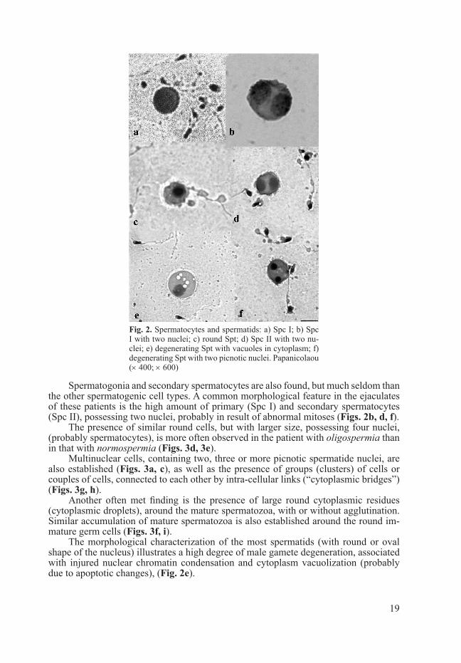
Spermatogonia and secondary spermatocytes are also found, but much seldom than the other spermatogenic cell types. A common morphological feature in the ejaculates of these patients is the high amount of primary (Spc I) and secondary spermatocytes (Spc II), possessing two nuclei, probably in result of abnormal mitoses (Figs. 2b, d, f).
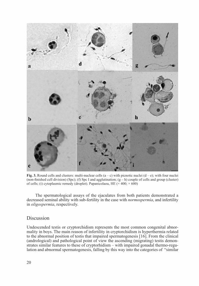
The presence of similar round cells, but with larger size, possessing four nuclei, (probably spermatocytes), is more often observed in the patient with oligospermia than in that with normospermia (Figs. 3d, 3e).
Multinuclear cells, containing two, three or more picnotic spermatide nuclei, are also established (Figs. 3a, c), as well as the presence of groups (clusters) of cells or couples of cells, connected to each other by intra-cellular links (“cytoplasmic bridges”) (Figs. 3g, h).
Another often met finding is the presence of large round cytoplasmic residues (cytoplasmic droplets), around the mature spermatozoa, with or without agglutination. Similar accumulation of mature spermatozoa is also established around the round im-mature germ cells (Figs. 3f, i).
The morphological characterization of the most spermatids (with round or oval shape of the nucleus) illustrates a high degree of male gamete degeneration, associated with injured nuclear chromatin condensation and cytoplasm vacuolization (probably due to apoptotic changes), (Fig. 2е).
Fig. 2. Spermatocytes and spermatids: a) Spc I; b) Spc I with two nuclei; c) round Spt; d) Spc II with two nu-clei; e) degenerating Spt with vacuoles in cytoplasm; f) degenerating Spt with two picnotic nuclei. Papanicolaou (× 400; × 600)
20
The spermatological assays of the ejaculates from both patients demonstrated a decreased seminal ability with sub-fertility in the case with normospermia, and infertility in oligospermia, respectively.
Discussion
Undescended testis or cryptorchidism represents the most common congenital abnor-mality in boys. The main reason of infertility in cryptorchidism is hyperthermia related to the abnormal position of testis that impaired spermatogenesis [16]. From the clinical (andrological) and pathological point of view the ascending (migrating) testis demon-strates similar features to these of cryptorhidism – with impaired gonadal thermo-regu-lation and abnormal spermatogenesis, falling by this way into the categories of “similar
Fig. 3. Round cells and clusters: multi-nuclear cells (a – c) with picnotic nuclei (d – e); with four nuclei (non-finished cell division) (Spc); (f) Spc I and agglutination; (g – h) couple of cells and group (cluster) of cells; (i) cytoplasmic remedy (droplet). Papanicolaou, HЕ (× 400; × 600)

21
diseases” [13, 14, 20]. If these disorders are not treated for a prolonged time period, they could lead to an increased risk for infertility, but also to development of testicular tumors in adults [3, 5].
In the current spermatological study, a high percent of immature spermatogenic cells in the ejaculates of both patients with “migrating testis” diagnosis, is established – 7.3% and 13.5%, respectively. Although we obtained results from two patients only, the increased amounts of spermatids and spermatocytes in the seminal probes might be due to pathological seminiferous tubules. The observed groups (clusters) of two, three or more cells, connected to each other by cytoplasmic connections (“bridges”), but also the presence of two- and multi-nuclear spermatids (most often with picnotic nuclei), show an interesting cytological pattern in both oligospermia- and normospermia-diag-nosed patients. In both cases, the reduced inseminal possibility is associated with the increased percentage of abnormal spermatozoa, in particular such with deformations in the head, as well as with mixed anomalies. These results are similar to our previous data from investigations of patients with pathologies of the male reproductive system [9, 10], where we also established increased amount of gametes with head anomalies. In cases with cryptorchidism the spermatozoa with elongated and round heads were prevailed, but increased number of cells with two heads was also observed. However, in comparison with the patients with ascending testis, significantly lower amount of the gametes with mixed anomalies was assessed in the cases with cryptorchidism.
According to many authors [8, 20, 22], the results from histological studies in-dicate that the spermatogenesis could be affected and injured in all stages of the male reproductive system development (ontogenesis, puberty and adult age). Different eti-ological factors could lead to the same and/or to similar structural abnormalities in the testicular tissue, manifested by decreased germ cells proliferation, abnormal differen-tiation and hence appearance of “teratological forms” among the mature spermatozoa as well as elimination of many immature spermatogenic cells. The gametogenesis abnormalities in cases with cryptorhidism, are often characterized by blocking of the process at the different phases of spermatocytes and/or spermatids’ development in the different regions of the seminiferous ducts. The blocking is depending on the sus-ceptibility of the respective cell populations to the increased temperature in the scro-tum, as well as to the subsequent hypoxia and oxidative stress [3, 14, 18]. Depending on the pathological conditions and factors, however, changes in the spermatogenesis are possible not to occur or they could be in non-equal degree in all tubules, and intact spermatogenesis might also be established. On the other hand, the changes could be transitive, in non-constant hypospermatogenesis, associated with intra-tubular disor-ganization, suppression and/or arrest of the germ cells maturation process. Moreover, changes could be definitive, characterized by germinal aplasia, tubular sclerosis and/or progressive peritubular fibrosis in the testis, leading to azoospermia [1, 17]. In the patients with ascending testis, the cytological observations suggested cell apoptosis activation in early stages of germ cells development (depending on the time period of the gonads in hyperthermia conditions), rather than a concrete affection in any spermatogenic stage.
The established in the current study high percentage of abnormal spermatozoa, as well as in other disorders of the male reproductive system, suggests altered function of the epididymis, responsible for further proceses of the spermatozoa capacitation, and acquisition of motility. The defects in the spermatozoa formation are often connected with injuries in the cellular DNA and nuclear chromatin structure [15, 24], important in application of technologies for in vitro-insemination.
Assisted reproduction technologies required precise identification of immature germ cells sub-populations, for subsequent application in the intra-cytoplasmic injec-
22
tions on ICSI technique [21]. The results from the current investigation indicate an increased content of degenerating spermatids with picnotic nuclei and vacuolized cy-toplasm, which are not appropriate for such purposes. The nuclear/chromatin defects that might occur in germ cells and their subpopulations as well as in mature sperma-tozoa demand careful choice in their using for ICSI procedures.
Additional studies are necessary in cases with ascending testis or similar patholo-gies (hyperthermia, cryptorhidism, varicocele), for a better clarification of the seminal fluid cytology and, hence, of the injuries in the human spermatogenesis.
Conclusion
The increased secretion of germ cells (spermatogonia, spermatocytes and spermatids) in the seminal fluid might be good indicators for the abnormal functions of the testis, and in this way, could provide information for the stage, in which the arrest in the germ cells development occurs. Further investigations are necessary due to limited informa-tion in the literature about the “round cells” presence in the seminal fluid, which also play a prognostic role for determination of the spermatogenesis defects and could be useful in the choice of appropriate therapeutic strategies in andrology.Acknowledgments: This work was supported by the European Social Fund and Republic of Bulgaria, Operational Programme “Human Resources Development” 2007-2013 framework, Grant No BG-051PO001-3.3.06-0048 from 04.10.2012.
R e f e r e n c e s
1. Agoulnik, A. I., Z. Huang, L. Ferguson. Spermatogenesis in cryptorchidism. – Meth. Mol. Biol., 825, 2012, 127-147.
2. Clarnette, T. D., D. Rowe, S. Hasthorpe, J. Huston. Incomplete disappearance of the processus Vaginalis as a cause of ascending testis. – J. Urol., 157, 1997, 1889-1891.
3. Cobellins, C. Spermatogenesis and cryptorchidism. – Front. Endocrinol. (Lausanne), 5, 2014, 63. 4. Fedder, J. Nonsperm cells in human semen: with special reference to human leucocytes and their
possible influence on fertility. – Arch. Androl., 36, 1996, 41-65. 5. Ferguson, L., A. I. Agoulnik. Testicular cancer and cryptorchidism. – Front. Endocrinol. (Lau-
sanne), 4, 2013, 32. 6. Gandini, L., A. Lenzi, F. Lombardo, F. Pacifici, F. Dondero. Immature germ cell separation using
a modified discontinuous Percoll gradient technique in human semen. – Hum. Reprod., 14, 1999, 1022-1027.
7. Giwercman, A., J. Bonde. Declining male fertility and environmental factors. – Endocr. Metab. Clin. of North America, 27, 1998, 807-830.
8. Holstein, A., W. Schulze, M. Davidoff. Understanding spermatogenesis is a prerequisite for treat-ment. – Reprod. Biol. Endocrinol., 1, 2003, 16 p.
9. Ilieva, I., P. Tzvetkova. Morphological and ultrastructural changes in the tail of the sperm abnor-malities of mixed type and infertility. – Andrology, 16, 2007, 14-19.
10. Ilieva, I., St. Ivanova, I. Chavdarov, S. Rangelov, P. Tzvetkova. Scanning electron microscopic studies of abnormal sperm in the pathology of the male reproductive system. – Compt. rend. Acad. bulg. Sci., 65, 2012, 1095-1098.
11. Johansson, E., A. Campana, R. Luthi, A. de Agostini. Evaluation of “round cells” in semen analy-sis: a comparative study. – Hum. Reprod. Update, 6, 2000, 404-412.
12. Mouriquand, P. The nomad testis. – Arch. Dis. Childhood, 92, 2007, 3.13. Nierderberger, C. Re: Oral administration of retinoic acid receptor antagonist reversibly inhibits
spermatogenesis in mice. – J. Urol., 187, 2012, 1509 p.
23
14. Paul, C., S. Teng, T. Saunders. A single, mild, transient scrotal heat stress causes hypoxia and oxi-dative stress in mouse testes, which induces germ cell death. – Biol. Reprod., 80, 2009, 913-919.
15. Ringertz, N., B. Gledhill, Z. Darzynkiewicz. Changes in deoxyribonucleoproteins during sper-matogenesis in the bull. – Exp. Cell Res., 62, 1970, 204-218.
16. Robin, G., F. Boitrelle, X. Leroy, M. C. Peers, F. Marcelli, J. Rigot, V. Mitchell. Assessment of azoospermia and histological evaluation of spermatogenesis. – Ann. Pathol., 30(3), 2010, 182-95.
17. Ross, M., W. Pawlina. Histology: a text and atlas: with correlated cell and molecular biology. – Bal-timore MD: Lippincott Williams & Wilkins, 2006.
18. Thorp, J., K. Kvist, E. Clasen-Linde, B. Petersen, D. Cortes. The relation between adult dark spermatogonia and other parameters of fertility potential in cryptorchid testes. – J. Urol., 190, 2013, 1566-1571.
19. Tomilson, M., A. Whitte, C. Barratt, A. Bolton, L. Cooke. The removal of morphologically ab-normal sperm forms by phagocytes: a positive role for seminal leukocytes? – Hum. Reprod., 7, 1992, 517-522.
20. Tzvetkov, D., P. Tzvetkova. Congenital disorders of ducts deferens. – In: Cogenital malformation of male reproductive system. – Bulg. Sci. Found. Androl., Sofia, 1999, 93-100.
21. Vernaeve, V., G. Verheyen, A. Goossens, A. Van Steirteghem, P. Devroey, H. Tournaye. How successful is repeat testicular sperm extraction in patients with azoospermia? – Hum. Reprod., 21, 2006, 1551-1554.
22. Yin, Y., W. De Wolf, A. Morgentaler. Experimental cryptorchidism induces testicular germ cell apoptosis by p53-dependent and-independent pathways in mice. – Biol. Reprod., 58, 492-496.
23. Yoshida, T., K. Ohno, Y. Morotomi, T. Nakamura, T. Azuma, H. Yamada, H. Hayashi, S. Sue-hiro. Clinical and pathological features of ascending testis. – Osaka City Med., 55, 2009, 81-87.
24. Цветкова, П. Морфо-функционални характеристики на епидидима и мъжки инфертилитет. – Андрология, 10, 2001, 21–30.


![Bio-Active Mineroplex Bio... · 2019-08-12 · • Anti-inflammatory, antioxidant, memory enhancer, neuroprotective, and synergistic enhancer of drugs.[8] • Spermatogenic effects:](https://static.fdocuments.us/doc/165x107/5e4f78858694391e6537b94a/bio-active-mineroplex-bio-2019-08-12-a-anti-inflammatory-antioxidant.jpg)