ROMICAT II Protocol - National Institutes of Health · ROMICAT II Study Protocol Version 6.0...
57
ROMICAT II Rule Out Myocardial Ischemia/Infarction Using Computer Assisted Tomography A Randomized, Controlled, Multicenter, Diagnostic Trial Study Protocol Version: 6.0 Amendment 5 Date: May 17, 2011 Principal Investigator Clinical Coordinating Center Udo Hoffmann, MD, MPH Massachusetts General Hospital Cardiac MR PET CT Program 165 Cambridge Street, Suite 400 Boston, MA 02114 Phone: 617-726-5954 Fax: 617-724-4152 Email: [email protected] Co-Investigator James Udelson, MD Tufts Medical Center 800 Washington Street Boston, MA 02111 Email: [email protected] Co-Investigator Clinical Coordinating Center Quynh Truong, MD, MPH Massachusetts General Hospital 165 Cambridge Street, Suite 400 Boston, MA 02114 Phone: 617-726-0798 Fax: 617-724-4152 Email: [email protected] CONFIDENTIAL
Transcript of ROMICAT II Protocol - National Institutes of Health · ROMICAT II Study Protocol Version 6.0...

ROMICAT II
Rule Out Myocardial IschemiaInfarction Using Computer Assisted
Tomography
A Randomized Controlled Multicenter Diagnostic Trial
Study Protocol Version 60 Amendment 5 Date May 17 2011
Principal Investigator Clinical Coordinating Center Udo Hoffmann MD MPH Massachusetts General Hospital Cardiac MR PET CT Program 165 Cambridge Street Suite 400 Boston MA 02114 Phone 617-726-5954 Fax 617-724-4152 Email uhoffmannpartnersorg
Co-Investigator James Udelson MD Tufts Medical Center 800 Washington Street Boston MA 02111 Email judelsontuftsmedicalcenterorg
Co-Investigator Clinical Coordinating Center Quynh Truong MD MPH Massachusetts General Hospital 165 Cambridge Street Suite 400 Boston MA 02114 Phone 617-726-0798 Fax 617-724-4152 Email qtruongpartnersorg
CONFIDENTIAL
ROMICAT II Study Protocol Version 60 Amendment 5 dated May 17 2011
Principal Investigator Data Coordinating and Statistical Center (DCSC) David Schoenfeld PhD Massachusetts General Hospital 50 Staniford St Suite 560 Boston MA 02114 Phone 617-726-6111 Fax 617-724-9878 Email dschoenfeldpartnersorg
Principal Investigator Clinical Events Committee Stephen Wiviott MD Brigham and Womenrsquos Hospital 75 Francis Street Boston MA 02115 Email swiviottpartnersorg
Principal Investigator Cost Effectiveness and Decision Analysis (DACE) Scott Gazelle MD MPH PhD Massachusetts General Hospital Email scottmgh-itaorg
Cardiovascular Imaging Core Lab Administrative Manager Nathan Sciortino Massachusetts General Hospital 25 New Chardon Street Suite 400B Boston MA 02114 Phone 617-643-5308 Fax 617-643-0111 Email nsciortinopartnersorg
Study Project Manager Pearl Zakroysky Massachusetts General Hospital 165 Cambridge Street Suite 400 Boston MA 02114 Phone 617-643-0954 Fax 617-724-4152 Email pzakroyskypartnersorg
Page 2 of 57 Confidential
__________________________________
__________________________________ ________________________
ROMICAT II Study Protocol Version 60 Amendment 5 dated May 17 2011
Investigator Protocol Signature Page
I have read and understand the protocol and agree that it contains all the ethical legal and scientific information necessary to conduct this study I will personally conduct the study as described I will provide copies of the protocol to all physicians nurses and other professional personnel responsible to me who will participate in the study I am aware that this protocol must be approved by the Institutional Review Board or Ethics Committee I agree to provide all patients with informed consent forms as required by government regulations and International Conference on Harmonization guidelines
Principal Investigator (print name)
Principal Investigator (signature) Date
Page 3 of 57 Confidential
ROMICAT II Study Protocol Version 60 Amendment 5 dated May 17 2011
TABLE OF CONTENTS
Amendment 1 Summary of Changes 6 Amendment 2 Summary of Changes 7 Amendment 3 Summary of Changes 9 Amendment 4 Summary of Changes 10 Amendment 5 Summary of Changes 12 Study Synopsis 13 Abbreviations 16 Part I Study Overview 17 Part II Study Description 19 1 Background and Significance 19
11 Standard Evaluation of Participants with Acute Chest Pain 19 12 Accuracy of Cardiac CT for Detection of Coronary Artery Disease and LV Function helliphelliphelliphelliphelliphelliphelliphelliphelliphelliphelliphelliphelliphelliphelliphelliphelliphelliphelliphelliphelliphelliphelliphelliphelliphelliphelliphelliphelliphelliphelliphelliphelliphellip19 13 Accuracy of Cardiac CT to Detect ACS in Participants with Acute Chest Pain 20 14 Cost Effectiveness of Diagnostic Testing in Subjects with Acute Chest Pain 21 15 Significance of the Proposed Clinical Trial 21
2 Preliminary Data 22 21 Final Results of ROMICAT I 22 211 Cardiac CT Based Detection of Coronary Stenosis and Plaque in the Assessment of
Participants with Acute Chest Pain 22 212 Incremental Diagnostic Value of Regional LV Function Over Coronary Assessment of
Cardiac CT for the Detection of ACS in Participants with Acute Chest Pain 24 213 Initial Decision Model to Assess the Cost Effectiveness of Coronary CT in Triage of
Subjects with Acute Chest Pain 24 3 Study ObjectiveSpecific Aims 25
31 Primary Aim 25 32 Secondary Aims 26 33 Tertiary Aims 27
4 Participant SelectionEligibility Criteria 27 41 Inclusion Criteria 28 42 Exclusion Criteria 28 43 Recruitment Screening and Consent 29 44 Randomization 30
5 Study Procedures 30 51 Index Hospitalization 30 52 48-72 Hour Follow up after ED discharge 31 53 28 Day Follow up 31
6 Data Collection 32 7 Statistical Considerations 32
71 Primary Aim 32
Page 4 of 57 Confidential
ROMICAT II Study Protocol Version 60 Amendment 5 dated May 17 2011
72 Secondary Aims 33 73 Tertiary Aims 36
8 Data Entry and Site Monitoring 37 81 Data Collection 37 82 Site Monitoring 37
9 Risk and Benefit Assessment 37 91 Risks of Nonionic Contrast Agent 37 92 Risks Associated With Radiation 38 93 Other Risks Additional Risks of CT Scans 40 94 Potential Benefits 40
10 Ethical Considerations 40 101 Inclusion of Women and Minorities 41
11 Adverse Event Reporting 42 12 References 43 Appendix A Cardiac CT Imaging Protocol Guidelines 49 Appendix B Adjudication of Events by Clinical Events Committee 53 Appendix C Data and Safety Monitoring Board 57
Page 5 of 57 Confidential
ROMICAT II Study Protocol Version 60 Amendment 5 dated May 17 2011
Amendment 1 Summary of Changes
1 Changes to InclusionExclusion Criteria (page 19) bull In order to ensure targeted population is enrolled the following inclusion criteria
was added Participant must have 2 or more cardiac risk factors (diabetes hypertension hyperlipidemia current smoker and family history of coronary artery disease)
2 Clarification of consent and randomization procedures (pages 21-22) bull Patients can be consented but not randomized before the first troponin test result
is available Patients whose initial troponin level is markedly elevated will not be randomized and will not be included in the intent-to-treat population Patients whose subsequent troponin testing is markedly elevated will be included in the intent to treat population
bull Removed the phrase lsquobased upon blocked assignment in groups of 30rsquo from the end of the sentence lsquoStudy subjects will be randomized into the standard care arm or the intervention arm by means randomization stratified by institution and gender using a central computerized randomization systemrsquo
3 Clarification of the Statistical Considerations section (pages 5 18 24 27-28) bull Removed sentence lsquoLength of hospital stay between study arms will be compared
in subgroups of patients directly discharged from the ED and patients admitted to the hospitalrsquo from primary endpoint analysis
bull Minor clarification to emergency department triage accuracy assumption discussion
bull Minor clarification to secondary aim of health care utilization
bull Removed sentence lsquoWe will use an uncorrected chi-squared statistic to evaluate this null hypothesisrsquo from discussion of secondary aim for comparison between two groups of rate of MACE at I and 2 years
Page 6 of 57 Confidential
ROMICAT II Study Protocol Version 60 Amendment 5 dated May 17 2011
Amendment 2 Summary of Changes
1 Changes to InclusionExclusion Criteria (page 20)
bull Page 20 The baseline population of ROMICAT I (age gt40) had an ACS rate of 93 For subjects with gt= 1 risk factor the ACS rate rose to 100 at the cost of losing 17 of the population while for subjects with gt=2 risk factors the ACS rate rose to 139 with a loss of 46 eligible subjects As the role of risk factors in determination of ACS is controversial and based on DSMB recommendations this criterion will be removed from the inclusion criteria
bull Page 21 The exclusion criteria of subjects who present to the ED more than 4 hours ago will now be extended to more than 6 hours ago This is based on DSMB recommendations as a useful technique to capture subjects who present with chest pain early in the morning
2 Clarification of Study Protocol
bull Pages 31 43 Currently the CT protocol only covers retrospective gating This will be amended to extend to allow prospective triggering also as this is a pragmatic trial which should reflect current clinical practice This also has the potential benefit of reduced radiation exposure (lt5mSv)
bull Page 24 Subjects discharged within 24 hours of presentation to the hospital will be followed up at the 48-72 hour period This was mentioned as 48 hours in the protocol and was a typographical error
bull Page 25 We will remove all language suggestive of facilitated handling of patients in the CT arm (including scheduling the CT scan immediately after randomization and notification of the ED caregivers about the results of the CT scan in a given time frame) The sentences ldquoTo facilitate timely CT imaging the cardiac CT exam will be scheduled immediately after randomizationrdquo rdquo The results of cardiac CT including the presence and extent of coronary atherosclerotic plaque coronary artery stenosis and global and regional LV dysfunction will be provided to the ED caregivers immediatelyldquo and rdquoIn subjects randomized to receive a cardiac CT patient management will be additionally informed and guided by the cardiac CT findings on coronary plaque and stenosis and LV function CT will be discussed with the ED physicians caring for the subject ldquo have been removed from page 23 ldquoThe CT reader will contact the Emergency Department (ED) treating physician and report the results of the cardiac CTrdquo has been removed from page 49 of Appendix B
bull Page 24 Gender stratification will not be used in the randomization schema for the simplicity and to use it as a covariate in further statistical analysis
Page 7 of 57 Confidential
ROMICAT II Study Protocol Version 60 Amendment 5 dated May 17 2011
bull Page 33 MD over read will be performed for internal quality control assessment of findings related to CAD and significant cardiac findings such as pulmonary embolism and aortic dissection to increase awareness to sites of non-coronary findings
3 Clarification of Aims
bull Page 18 The language of the aims and related hypotheses has been expressed in a more clarified manner
Page 8 of 57 Confidential
ROMICAT II Study Protocol Version 60 Amendment 5 dated May 17 2011
Amendment 3 Summary of Changes
1 Changes to InclusionExclusion Criteria (page 25)
bull Page 25 Participant must have at least five minutes of chest pain or equivalent (chest tightness pain radiating to left right or both arms or shoulders back neck epigastrium jawthroat or unexplained shortness of breath syncopepresyncope generalized weakness nausea or vomiting thought to be of cardiac origin) at rest or during exercise within 24 hours of ED presentation warranting further inpatient risk stratification as determined by an ED attending
bull Page 26 9 Currently symptomatic asthma (or taking daily medication for asthma) 10 Contraindication to beta blockers (taking daily antiasthmatic medication)
This exclusion only applies to patients with a heart rate gt 65 bpm at sites using a non-dual source CT scanner
2 Clarification of Study Protocol
bull Page 28 Interventional Arm In subjects randomized to the intervention arm a contrast enhanced cardiac CT will be performed (after initial troponin level is available) in addition to the standard evaluation initial ED evaluation
3 Extension of follow-up time windows
bull Page 28 The 48-72 hour follow-up phone call has been extended to accommodate calls that fall either during the weekend or during a holiday
bull Page 29 The 12 month and 24 month follow-up phone call windows have been extended from +- 2 weeks to +- 4 weeks
Page 9 of 57 Confidential
ROMICAT II Study Protocol Version 60 Amendment 5 dated May 17 2011
Amendment 4 Summary of Changes
1 Study Synopsis (page 13) Duration of Study Participation Participants will be followed during index ED visithospitalization There will be a phone call at 48-72 hours for those discharged within 24 hours All participants will receive a follow up phone call at 28 days 1 year and 2 year post emergency room discharge
2 Changes to Inclusion Criteria (page 27)
bull Participant must be in normal sinus rhythm
3 Elimination of the 1 and 2 year follow-up interviews
bull Page 16 All participants will receive a 28-day a one year and two year follow-up phone interview The efficiency of each strategy will be determined by comparing 1) the length of hospital stay (primary endpoint) 2) the time to diagnosis (secondary endpoint) 3) the rates of ED discharge (secondary endpoint) 4) utilization of other diagnostic testing ie invasive coronary angiograms and resulting interventions ie coronary revascularization procedures during index hospitalization and 28 days one year and two year (secondary endpoint) The safety of the two strategies will be compared by determining the occurrence of adverse events within 48-72 hours in directly discharged patients as well as determination of major adverse cardiovascular events (MACE) within 28 days one year and two years after discharge from the index hospitalization
bull Page 26 To compare the two study arms in terms of rates of MACE (major adverse cardiac eventsmdashcardiac death AMI revascularization unstable angina and rehospitalization) within one and two years
Onetwo Years Defined as onetwo years post the day of discharge from index hospitalization
Hypothesis The rates of MACE in both arms during the follow up period will be lower in the CT arm
bull Page 30 A reminder letter will be sent or phone call will be made to participants two weeks prior to the 28-day follow up phone call If unable to contact the participant by phone mortality will be assessed online using the SSDI website
bull Page 30 54 12 Month Follow up Follow up one year phone call All subjects will be interviewed via a telephone call at one year post ED discharge (12 months +- 4 weeks) to determine the occurrence of MACE and readmissions to the hospital using a standardized questionnaire All cases of recurrent chest pain hospital admissions and diagnostic testing will be verified by review of medical records
Page 10 of 57 Confidential
ROMICAT II Study Protocol Version 60 Amendment 5 dated May 17 2011
A reminder letter will be sent or phone call will be made to participants two weeks prior to the 12-month follow up phone call
55 24 Month Follow up Follow up two year phone call All subjects will be interviewed via a telephone call at two years post ED discharge (24 months +- 4 weeks) to determine the occurrence of MACE using a standardized questionnaire All cases of hospital admissions and diagnostic testing will be verified by review of medical records
Note A reminder letter will be sent or phone call will be made to participants two weeks prior to the two year follow up phone call A minimum of five (5) phone attempts will be to contact each participant If unable to contact the participant by phone mortality will be assessed online using the SSDI website
bull Page 34 To compare the two study arms in terms of rates of MACE (cardiac death AMI revascularization unstable angina and rehospitalization) within one and two years This analysis will be performed from the intent to treat perspective The occurrence of MACE will be assessed via patient follow-up and medical chart review The primary analysis for this aim will be performed at the patient level by determining whether any MACE occurred or not Event rates will be estimated and compared between study groups using exact procedures Prior data estimate a MACE rate of 5 over two years We anticipate a MACE rate between and 2 and 5 in both groups and have 80 power to detect a difference between 2 and 5 between the two groups The Type I error probability associated with this test of this null hypothesis is 005 In a secondary analysis the rates of each type of MACE will be examined separately
4 Extension of 28-Day follow-up time window bull Page 30 The 28 day follow-up phone call window has been extended from +2
weeks to +4 weeks
Page 11 of 57 Confidential
ROMICAT II Study Protocol Version 60 Amendment 5 dated May 17 2011
Amendment 5 Summary of Changes
1 Study Synopsis (page 15) Number of sites 9 7
2 Part I Study Overview (page 17) The study is designed as a randomized controlled diagnostic multicenter trial and will enroll 1000 participants at nine seven sites over a 15 month period
3 Study Design (page 18) The study is designed as a randomized controlled diagnostic multicenter trial and will enroll 1000 participants at nine seven sites over a 15 month period
Page 12 of 57 Confidential
ROMICAT II Study Protocol Version 60 Amendment 5 dated May 17 2011
Study Synopsis Sponsor(s) National Heart Lung and Blood Institute (NHLBI)
Protocol Title Rule Out Myocardial IschemiaInfarction Using Computer Assisted Tomography - A Diagnostic Randomized Multicenter Trial
Diagnosis and Main Adults (ge40-75 yrs) without knowndocumented history of coronary artery Criterion for Inclusion disease who have come to the emergency department (ED) presenting
with a lead symptom of acute chest pain suggestive of acute cardiac ischemia defined as chest pain lasting for at least 5 minutes and occurring within the last 24 hours but without diagnostic ECG changes of acute myocardial ischemia are eligible for participation Only patients in whom the ED attending feels that further inpatient risk stratification is required will be included
Primary Study Objective To determine whether length of hospital stay is significantly shortened in the interventional arm vs standard of care
Secondary Study Objectives bull To determine whether time to diagnosis (for ACS and no ACS) is significantly shortened and rates of direct discharge from the emergency department are increased in the interventional arm vs standard of care
bull To determine a) the additional number of invasive coronary angiograms in both arms and b) to determine the number of coronary revascularization procedures
bull To determine the safety of immediate ED discharge after a normal cardiac CT defined as the occurrence of ACS within 48-72 hours after discharge
bull To determine whether subsequent testing and follow-up including invasive coronary angiograms diagnostic (imaging) tests interventions repeat ED visits for cardiac related problems and repeat hospitalizations for chest pain or equivalent during 28 days after hospital discharge is decreased in the interventional arm compared to SOC
bull To determine the two study arms in terms of rates of MACE within 28 days
bull To compare the two study arms in terms of rates of MACE within one and two years
bull To estimate and compare the sensitivity specificity NPV and PPV of contrast-enhanced CT and non-contrast CT with clinical diagnosis of ACS as the reference standard
bull To determine whether the cost of care (per TSI) for index hospitalization and after 28 days is decreased by implementing cardiac CT into the early ED evaluation of patients with acute chest pain
bull To determine the cost-effectiveness of incorporating cardiac CT into
Page 13 of 57 Confidential
ROMICAT II Study Protocol Version 60 Amendment 5 dated May 17 2011
the standard ED evaluation of patients with acute chest pain over a 28shyday and lifetime horizon as compared to SOC
Tertiary Study Objectives bull To determine whether CT leads to increased test burden after the initial tests during index hospitalization
bull To compare the consistency of physician decisions management depending on the results of diagnostic testing (ie normal CT ndash what fraction is immediately discharged)
bull To determine whether hospital setting availability of observation unit CT experience of hospital and readers and reader performance are associated with primary or secondary outcomes
Primary Endpoint The primary endpoint is time from ED presentation to hospital discharge The length of hospital stay is defined as the time from ED presentation to the time of discharge note or order This includes time in the ED time in any institution-specific specialized chest pain unit and time as an inpatient on the floor
Secondary Endpoints bull The time to diagnosis For ACS defined as the time from ED presentation until the first test during index hospitalization that leads to the diagnosis of ACS (tests may be biomarkers CTA ETT nuclear imaging stress ECHO or cardiac catheterization) for no ACS defined as the time from ED presentation to the final test during index hospitalization
bull Admission to hospital Participants are considered to be admitted if the ED attending admitted the subject to a hospital floor (medical floor step down unit telemetry hospital floor or intensive care unit)
bull Direct ED discharge will include all subjects who are not admitted to the hospital
bull Health care utilization number of invasive coronary angiograms diagnostic (imaging) tests interventions repeat ED visits
bull Rates of MACE major adverse cardiac events ndash cardiac death acute myocardial infarction revascularization unstable angina and rehospitalization
bull Cost of care derived from the cost-accounting database (ie TSI)
Tertiary Endpoints Increased test burden Further imaging tests performed after index hospitalization
Primary Hypothesis Adding cardiac CT to the initial ED evaluation of subjects with acute chest pain significantly decreases length of hospital stay
Page 14 of 57 Confidential
ROMICAT II Study Protocol Version 60 Amendment 5 dated May 17 2011
Secondary Hypothesis Addition of cardiac CT to the initial ED evaluation of subjects with acute chest pain will significantly increase the ED discharge rate and shorten the time to diagnosis and it will be a cost-saving and cost-effective modality There will not be an increase in the number of invasive angiograms or coronary revascularization procedures as compared to the SOC arm With a high negative predictive value patient safety will not be compromised There will not be an increase in subsequent further testing and the rate of MACE in both arms in the follow-up period will be similar The test characteristics of contrast enhanced CT will be significantly better than that of a non-contrast CT
Study Design The study is designed as a randomized diagnostic multicenter trial and will enroll 1000 participants at seven sites over a 15 month period Participants presenting with acute chest pain in the ED will be randomized to standard of care (SOC) or to an interventional arm Patients in the interventional arm will receive standard evaluation supplemented by contrast-enhanced cardiac CT imaging
Duration of Study Participants will be followed during index ED visithospitalization There Participation will be a phone call at 48-72 hours for those discharged within 24 hours
All participants will receive a follow up phone call at 28 days post emergency room discharge
Number of Subjects 1000 subjects will be enrolled with 500 each in the standard of care and interventional arms
Number of Sites 9
Statistical Considerations The primary aim of the study is to compare the two study arms in terms of a difference in mean length of hospital stay (LOS) between the two arms We have conducted a power evaluation using the estimated mean and standard deviation based on observed data from ROMICAT I and simulated data based on health outcomes We will have 95 power to detect a difference in 10 hours of LOS at a Type-1 error rate of 5 for an independent samples t-test using 500 patients in each arm
Page 15 of 57 Confidential
ROMICAT II Study Protocol Version 60 Amendment 5 dated May 17 2011
Abbreviations
ACS Acute Coronary Syndrome CABG Coronary artery bypass graft CAD Coronary Artery Disease CK-MB Creatinine kinase MB type CRC Clinical Research Coordinator CT Computed Tomography ECG Electrocardiogram ED Emergency Department ETT Exercise treadmill ECG testing MACE Major adverse cardiac event MI Myocardial Infarction NPV Negative predictive value PPV Positive predictive value SOC Standard of care SPECT Rest or stress myocardial perfusion imaging
Page 16 of 57 Confidential
ROMICAT II Study Protocol Version 60 Amendment 5 dated May 17 2011
Part I Study Overview The study is designed as a randomized controlled diagnostic multicenter trial and will enroll 1000 participants at nine sites over a 15 month period Participants presenting with acute chest pain in the ED will be randomized to a standard of care arm (SOC) or interventional arm patients in the interventional arm will receive standard evaluation supplemented by contrast-enhanced cardiac computed tomography (CT) imaging (Figure 1)
In both arms the initial Emergency Department (ED) evaluation (electrocardiogram [ECG] physical examination clinical presentation renal function lab and medical history) will determine eligibility of patients After randomization standard of care will be performed in both arms However for patients randomized to the interventional arm a CT will be performed The following decisions made by the caregiver team will be tracked admission or discharge after initial ED evaluation plus CTother initial diagnostic test in admitted participants the length of hospital stay the extent of further testing including serial cardiac markers and ECG and other diagnostic tests or interventions For safety reasons participants discharged in less than 24 hours of ED arrival will receive a phone call 48-72 hours after discharge All participants will receive a 28-day follow-up phone interview The efficiency of each strategy will be determined by comparing 1) the length of hospital stay (primary endpoint) 2) the time to diagnosis (secondary endpoint) 3) the rates of ED discharge (secondary endpoint) 4) utilization of other diagnostic testing ie invasive coronary angiograms and resulting interventions ie coronary revascularization procedures during index hospitalization and28 days (secondary endpoint) The safety of the two strategies will be compared by determining the occurrence of adverse events within 48-72 hours in directly discharged patients as well as determination of major adverse cardiovascular events (MACE) within 28 days after discharge from the index hospitalization We will also perform a cost and cost effectiveness analysis In subsequent analyses we will determine the patient physician and institutional factors that affect the primary endpoint or secondary endpoints
Primary Objective To determine whether length of hospital stay is significantly shortened in the interventional arm vs SOC
Hypothesis
Adding cardiac CT to the initial ED evaluation of subjects with acute chest pain decreases length of hospital stay
Page 17 of 57 Confidential
Secondary Endpoints
Primary Endpoint
Screening
Consent
Length of Hospital Stay
Tertiary Endpoints
Cost and Cost Effectiveness
Incremental Value of CTA over a CAC scan
Rates of Direct ED Discharge
Time to Diagnosis
No of invasive coronary angiograms and revascularizations
Rates of MACE after immediate ED discharge after 28 days
Health care utilization after 28 days
Institutional and Caregiver Characteristics associated with primary and secondary outcomes
Incremental Value of LV function over a CTA
Radiation Exposure during index hospitalization and follow up
ROMICAT II Study Protocol Version 60 Amendment 5 dated May 17 2011
Study Design
The study is designed as a randomized controlled diagnostic multicenter trial and will enroll 1000 participants at nine sites over a 15 month period
Patients with Acute Chest Pain at Low to Intermediate Risk for ACS Screening
Consent
StStandaandard ord off C Caarere CaCarrddiiaac Cc CTT RanRanddomomizizatatioionn
Length of Hospital Stay Primary Endpoint
Rates of Direct ED Discharge
Time to Diagnosis
No of invasive coronary angiograms and revascularizations
Rates of MACE after immediate ED discharge after 28 days Secondary Endpoints
Health care utilization after 28 days
Cost and Cost--Effectiveness
Institutional and Caregiver Characteristics associated with primary and secondary outcomes
Incremental Value of CTA over a CAC scan
Incremental Value of LV function over a CTA Tertiary Endpoints
Radiation Exposure during index hospitalization and follow--up
Page 18 of 57 Confidential
Patients with Acute Chest Pain at Low to Intermediate Risk for ACS
ROMICAT II Study Protocol Version 60 Amendment 5 dated May 17 2011
Part II Study Description
1 Background and Significance
11 Standard Evaluation of Participants with Acute Chest Pain Accurate triage of participants presenting with acute chest pain to the ED remains difficult because neither the chest pain history12 a single set of established biochemical markers for myocardial necrosis (troponin I troponin T creatine kinase MB-type [CK-MB])34 nor initial 12-lead ECG alone or in combination (acute cardiac ischemia time-insensitive predictive instrument) identifies a group of participants that can be safely discharged home without further diagnostic testing5ndash7 As a result the threshold to admit chest pain participants remains low and over six million are admitted annually to US hospitals28ndash10
The standard ldquorule outrdquo myocardial infarction (MI) protocol consists of serial ECG and cardiac biomarker measurements and usually requires a noninvasive stress test to rule out MI These tests (exercise treadmill ECG testing [ETT] stress echocardiography [Echo] and rest or stress myocardial perfusion imaging with Tc-99m [SPECT]) require the exclusion of myocardial necrosis with negative serial biomarkers and are performed to rule out the presence of a hemodynamically significant coronary stenosis While they have good sensitivities (ETT 76 SPECT 83 and Echo 85) for detecting the presence of significant coronary artery stenosis when compared to coronary angiography11ndash13 their specificities are only moderate (ETT 60 SPECT 64 and Echo 77) Moreover these tests are time consuming (ie SPECT 2ndash3 hours) and usually not available 247 Thus the standard evaluation of patients with chest pain to rule out myocardial ischemia requires hospital admission for 24 to 36 hours in over 90 of US hospitals14 Because of the moderate specificities of standard diagnostic testing 33 to 44 of patients with suspected acute coronary syndrome (ACS) who undergo cardiac catheterization have no significant coronary artery disease (CAD)1516 The remarkable inefficiency of current evaluation strategies is also documented by the fact that only lt10 of the six million admitted each year in the US ultimately receive a diagnosis of ACS at discharge1718 The inpatient care for the negative evaluations imparts a significant economic burden in excess of $8 billion annually19ndash21 for the US health care system
Thus an improvement of the ED triage of participants with acute chest pain particularly the increase in ED discharge rates in participants who ultimately do not have ACS would have enormous clinical and economic implications This research will determine if noninvasive cardiac CT that accurately visualizes the coronary arteries for plaques andor stenosis can be an effective strategy to facilitate early ED discharge in this patient population
12 Accuracy of Cardiac CT for Detection of Coronary Artery Disease and LV Function Advances in CT technology with an improvement of volume coverage (ge64 parallel detectors) and temporal (lt210 ms) and spatial (lt06 mm) resolution enables a nearly motion free contrast-enhanced imaging of the coronary artery tree and the heart chambers during a single short breath
Page 19 of 57 Confidential
ROMICAT II Study Protocol Version 60 Amendment 5 dated May 17 2011
hold (8ndash13 second scan)22 The data acquired during an ECG-gated contrast-enhanced multi-detector CT scan allows for 1) accurate assessment of the coronary arteries for plaque and stenosis and 2) the assessment of global and regional LV function in a cine mode
CAD Over the last several years we and others have demonstrated that cardiac CT has excellent diagnostic test characteristics for the detection of significant coronary artery stenosis as compared to invasive coronary angiography23ndash28 There is strong consensus that the strength of cardiac CT is its ability to reliably rule out the presence of stenosis and relevant coronary plaque (negative predictive value [NPV] 95 to 98 positive predictive value [PPV] 92 to 97)22
In addition cardiac CT also accurately detects the presence and extent of calcified and non-calcified coronary atherosclerotic plaque in good agreement with intravascular ultrasound (IVUS) especially in proximal coronary segments (sensitivity 84 to 92)29 30
Global and Regional LV Function Cardiac CT also permits the assessment of global regional LV function in a cine mode Studies have shown excellent reproducibility (κ=086) and agreement to cine magnetic resonance imaging (CMRI) for the detection of global and regional LV dysfunction (r=091)31ndash36
13 Accuracy of Cardiac CT to Detect ACS in Participants with Acute Chest Pain Preliminary data generated by the investigators suggest that cardiac CT using at least 64-slice technology can be performed safely and with excellent image quality in the acute care setting37
In addition our observational data in 368 patients demonstrated that the presence of significant coronary artery stenosis as assessed by cardiac CT can be definitively ruled out in 70 of participants presenting with acute chest pain to the ED (see Sections 31 and 32) This finding has a 100 NPV for ACS during index hospitalization and major adverse cardiac event (MACE) during follow-up (see Section 31)38 There are now several studies that support these results and suggest that cardiac CT has the potential to improve the management of participants with acute chest pain in the ED Rubinshtein et al in a study of 58 participants with acute chest pain but negative initial cardiac troponin levels and non-diagnostic ECG demonstrated a NPV of 100 and specificity of 92 (n=3538) for a diagnosis of ACS in the ED by cardiac CT39 Participants were discharged (n=3558) without stress testing if obstructive CAD was excluded by cardiac CT and if serial troponin measurements were negative There were no deaths or myocardial infarctions during a 15-month follow-up Earlier Goldstein et al40 randomized 200 participants at very low risk for ACS into a cardiac CT based triage system or a stress SPECT based triage system They found no or minimal CAD in 68 of patients which was a safe predictor for the absence of ACS and MACE over 6 months The study further shows the potential of cardiac CT to shorten length of hospital stay and decrease costs of evaluation of patients with acute chest pain However a potential drawback of CT was noted as the CT arm had more invasive angiograms and revascularization procedures (n=11 vs 3 and 4 vs 1 respectively) However this most likely represents that at this time the association between CT findings and clinical outcomes was unknown
In addition studies have suggested that the absence of coronary artery calcification (CAC) as detected in a non-contrast low radiation CT scan also has a high negative predictive value for
Page 20 of 57 Confidential
ROMICAT II Study Protocol Version 60 Amendment 5 dated May 17 2011
ACS in these participants4142 However the clinical utility of CAC alone for the assessment of ACS is inadequate because it cannot exclude the presence of high-grade non-calcified plaquestenosis
14 Cost Effectiveness of Diagnostic Testing in Subjects with Acute Chest Pain Current management of subjects with suspected ACS is managed by a sequential strategy (decision rule) involving quality and duration of chest pain risk factors biomarkers ECGrsquos and diagnostic tests such as SPECT echo or ETT However cost-effective utilization of these tests remains poor43 44 and hospitalization rates for ACS have increased while length of hospitalization has been constant over the last 10 years9 45 The introduction of chest pain observation units may have further decreased the threshold to admit patients Acute appendicitis and pulmonary embolism are excellent examples of the effects of the implementation of advanced imaging technology on patient management in the ED Because of the costs associated with unnecessary appendectomy and one day of in patient observation the implementation of the CT scan resulted in significant cost savings46 A similar approach is warranted to determine whether cardiac CT reduces cost and is cost effective in subjects with acute chest pain Moreover a formal model-based cost-effectiveness analysis is essential to project possible costs and benefits from a broader perspective (including the assessment of potential risks associated with CT) and longer time horizon Modeling can also help to answer questions and perform comparisons not possible within the confines of the clinical trial The current proposal critically investigates these issues
15 Significance of the Proposed Clinical Trial Evaluation of the appropriate clinical use of novel diagnostic imaging modalities is now more important than ever as the cost of these examinations steeply rises and now exceeds the cost spent on prescribed drugs All major professional medical societies have publicly demanded that clinical use of diagnostic imaging should be dictated by evidence-based medicine to not only control costs but also to minimize additional extraneous testing as it is currently unclear whether their usage will result in improvement in the current standard of care
Advanced cardiac CT is one of these novel diagnostic imaging technologies that may potentially enhance standard of care in patients presenting to the ED with acute chest pain but non-diagnostic ECG and negative initial biomarkers a population that remains diagnostically challenging and currently causes a significant clinical burden on each hospital every day Preliminary studies demonstrate that cardiac CT accurately detects absence of coronary atherosclerotic plaque Levels of atherosclerotic plaque as detected by cardiac CT is a powerful predictor of the absence of ACS and can be detected in a significant fraction of participants with acute chest pain Thus we anticipate the implementation of cardiac CT in the early ED triage process will (1) enable earlier and safe discharge of significant fraction of patients directly from the ED who currently would have been unnecessarily admitted as well as (2) allow for earlier detection and better treatment of CAD compared to the current standard of care
The proposed research will constitute the largest randomized multicenter clinical trial using cardiac CT to date and will provide an unbiased assessment of the above stated hypotheses It
Page 21 of 57 Confidential
ROMICAT II Study Protocol Version 60 Amendment 5 dated May 17 2011
will be conducted by a group of experienced clinical researchers consisting of CT imagers ED physicians and cardiologist with clinical expertise in the management of chest pain syndromes The results from this trial will determine the clinical utility of cardiac CT in patients with acute chest pain and may lead to a change in the management of these patients
2 Preliminary Data In support of the aim of the proposed research we provide data from a double blinded observational cohort study (ldquoCardiac CT for triage of participants with intermediate likelihood of ACSrdquo R01 HL080053-03) demonstrating the association between cardiac CT findings on plaque stenosis and LV function with clinical outcomes in patients with acute chest pain These data demonstrate 1) the absence of any CAD in up to 50 of patients with acute chest pain 2) the high NPV of absence of plaque and stenosis for ACS and thus the safety to discharge patients from the ED and the incremental diagnostic value of LV function assessment for risk stratification
21 Final Results of ROMICAT I
211 Cardiac CT Based Detection of Coronary Stenosis and Plaque in the Assessment of Participants with Acute Chest Pain Objective To determine the usefulness of cardiac CT angiography in patients with acute chest pain Methods Observational cohort study in chest pain patients with normal initial troponin and non-ischemic ECG 64-slice coronary CT was performed prior to admission to detect coronary plaque and stenosis (gt 50 luminal narrowing) Results were not disclosed Endpoints were ACS during index hospitalization and MACE during 6- month follow up Results Among 368 participants (mean age 53 plusmn 12 years 61 male) 31 had ACS (8) By cardiac CT 50 of these participants were free of CAD 31 had nonobstructive disease and 19 had inconclusive or positive CT for significant stenosis Sensitivity and NPV for ACS were 100 (n=183368 95 confidence interval [CI] 98ndash100) and 100 (95 CI 089ndash100) with the absence of CAD and 77 (95 CI 59ndash90) and 98 (n=300368 95 CI 95ndash99) with significant stenosis by cardiac CT Specificity of presence of plaque and stenosis for ACS were 54 (95 CI 049ndash060) and 87 (95 CI 083ndash090) respectively Only one ACS occurred in the absence of calcified plaque Both the extent of coronary plaque and presence of stenosis predicted ACS independently and incrementally to TIMI risk score (AUC 088 082 vs 063 respectively all plt00001) Conclusion Fifty percent of participants with acute chest pain and low to intermediate likelihood of ACS are free of CAD by CT and have no ACS Given the large number of such patients early cardiac CT may significantly improve patient management in the ED
Page 22 of 57 Confidential
ROMICAT II Study Protocol Version 60 Amendment 5 dated May 17 2011
Figure 1 A-C 67 year old female 3 hours of substernal chest pain radiating to the back negative initial troponin and CK-MB ECG sinus bradycardia admitted to rule out MI All coronary arteries are widely patent with no evidence of coronary atherosclerosis A Volume rendered 3-dimensional CT image (surface weighted volume rendering technique) of the heart depicting the left circumflex coronary artery (LCX arrowhead) obtuse marginal branch (dashed arrow) and left anterior descending (LAD arrow) two hours after presentation to the ED B CT based evaluation of segments 1 to 3 of the right coronary artery (RCA arrowheads) and conus branch (dashed arrow) using a 5mm thick maximum intensity projection (MIP) C Curved multiplaner reformatted (MPR) image of the LAD (arrowheads) and proximal LCX (dashed arrow)
Figure 2 A-C 40-year old male with hypercholesterolemia and a family history of diabetes woke with chest pressure and noticed intermittent chest pressure during his 1 mile walk to work He had similar symptoms a week prior to admission Initial troponin and CK-MB were negative and ECG showed a normal sinus rhythm without ST-segment or T-wave abnormalities Coronary CT was performed two hours after presentation to the ED and revealed a significant mid LAD stenosis Eight hours after ED presentation cardiac biomarkers became positive The mid LAD stenosis was confirmed on coronary angiography the next day and the participant was treated with a stent placement A Volume rendered 3-dimensional CT image of the heart depicting the right coronary artery (RCA arrow) and the left anterior descending coronary artery (LAD arrowhead) Discontinuation of the contrast-enhanced coronary lumen in the mid section of the LAD (dashed arrow) was detected B CT based evaluation of the right coronary artery (RCA arrowheads) demonstrates calcified and non-calcified plaque in segments 1 to 3 no significant coronary stenosis was detected C Curved MPR image of the LAD reveals good luminal contrast enhancement without any plaque in the distal LAD and calcified plaque in the proximal
Page 23 of 57 Confidential
ROMICAT II Study Protocol Version 60 Amendment 5 dated May 17 2011
segment (arrowheads) A significant luminal narrowing (arrow) was detected in the mid portion of the LAD
212 Incremental Diagnostic Value of Regional LV Function Over Coronary Assessment of Cardiac CT for the Detection of ACS in Participants with Acute Chest Pain Objective To determine incremental value of regional left ventricular function (LVF) over coronary assessment for the diagnosis and prediction of acute coronary (ACS) in patients with acute chest pain but inconclusive initial emergency department (ED) evaluation Methods We enrolled 356 consecutive patients (mean age 53plusmn12 years 62 male) with acute chest pain and inconclusive initial ED evaluation These patients underwent 64-slice contrast-enhanced cardiac CT prior to hospital admission Regional LVF and presence of coronary atherosclerotic plaque and significant stenosis (gt50) were separately assessed by two experienced readers blinded to the clinical course and other CT findings Caregivers and patients remained blinded to the CT results Incremental diagnostic accuracy and predictive value of regional LVF to predict ACS was determined in the entire cohort and in subgroups of patients with inconclusive or positive coronary assessment Results Regional LVF was impaired in 46 patients (129) and 31 patients developed ACS (87) Adding regional LVF resulted in a 10 increase in sensitivity to detect ACS (87 95CI 70shy96) when compared to detection of significant stenosis In the subgroup of 33 patients with significant stenosis impaired regional LVF correctly predicted ACS in 1719 patients (PPV 895 95CI 67-99) In the subgroup of patients with inconclusive coronary CTA (n=33) normal regional LVF correctly predicted the absence of ACS in 2426 patients (NPV 923 95CI 75-99) Impaired regional LVF independent of the extent of plaque and the presence of stenosis was associated with a 25 and 20 fold increased risk for ACS (RR 2023 95CI 747 to 5482 and RR 2534 95CI 936 to 6857 respectively) Moreover c-statistics improved significantly after the addition of regional LVF to both the extent of plaque and the presence of stenosis (c-statistic 088 vs 094 and 082 vs 090 respectively both plt003) Conclusions Regional LVF assessment at rest improves overall sensitivity of coronary CTA for ACS in patients with acute chest pain Combined assessment of coronary morphology and regional LVF is especially helpful to guide further testing and interventions if coronary assessment is inconclusive or positive
213 Initial Decision Model to Assess the Cost Effectiveness of Coronary CT in Triage of Subjects with Acute Chest Pain Objective To develop a preliminary decision-analytic Markov model to predict the long-term consequences of four competing strategies 47 in the ED evaluation of subjects with acute chest pain Methods The model consists of the six health states chest pain healthy early heart disease undiagnosed CAD diagnosed CAD and death Subjects start in the chest pain state The probability that the chest pain is caused by CAD depends on the underlying risk of CAD as
Page 24 of 57 Confidential
ROMICAT II Study Protocol Version 60 Amendment 5 dated May 17 2011
determined by age gender and cardiovascular risk factors As simulated subjects progress through the model CAD (if present) can remain stable or progress Subjects may die from cardiovascular disease or other causes at rates determined by disease stage and demographics By simulating hypothetical cohorts of subjects we determined quality-adjusted life expectancy (QALE) quality-adjusted life year (QALY) and cost-effectiveness ratios for competing strategies We considered four competing strategies for subjects presenting with chest pain to the ED 1) admission in which all subjects are admitted to the hospital for further evaluation 2) discharge in which all subjects are discharged directly home from the ED 3) cardiac CT with conservative treatment in which all subjects without any evidence of CAD are discharged and all other patients are admitted and undergo stress testing and 4) cardiac CT with aggressive treatment which is similar to strategy 3 but sends subjects with evidence of significant stenosis directly for invasive coronary angiography It is important to note that the model permits an evaluation of each strategy using a similar cohort of subjects The model also permits an evaluation of estimate life expectancy and lifetime costs Results Strategies involving cardiac CT were associated with lower ED costs and incremental improvements in QALE when compared to a strategy of admission for all subjects Relative to admitting all subjects the use of cardiac CT yielded savings in ED and hospital costs ranging from $1800-$2200 in men and women ages 30-40 and from $500-$1200 men and women ages 60-70 respectively The incremental cost-effectiveness of strategies involving cardiac CT compared with a strategy of discharging all subjects ranged from $94100-$267200 per QALY in men and $74800-$326100 per QALY in women Conclusions This preliminary decision model suggests that strategies involving cardiac CT are associated with lower ED costs and incremental improvements in QALE in both men and women across all ages The precision of the model will be greatly enhanced by input of real clinical scenarios derived from this proposal
3 Study ObjectiveSpecific Aims This primary objective of this protocol is to assess the efficiency of implementing cardiac CT into the diagnostic workup of patients with acute chest pain normal initial biomarkers and normal or non-diagnostic ECGs Study participants will be patients with acute chest pain and low to intermediate likelihood of ACS and will be randomly assigned to the SOC arm or the interventional arm patients in the interventional arm will receive the standard of care supplemented by cardiac CT
31 Primary Aim The primary aim is to determine whether length of hospital stay is significantly reduced in the interventional arm compared to standard of care The primary endpoint is time from ED presentation to hospital discharge
The length of hospital stay is defined as the time from ED presentation to the time of discharge note or order This includes time in the ED time in any institution-specific
Page 25 of 57 Confidential
ROMICAT II Study Protocol Version 60 Amendment 5 dated May 17 2011
specialized chest pain unit and time as an inpatient on the floor All time sequences must be documented and reported to comprise total length of hospital stay Hypothesis Adding cardiac CT to the initial ED evaluation of subjects with acute chest pain significantly decreases length of hospital stay
32 Secondary Aims 1 To determine whether time to diagnosis is significantly shortened and rates of direct
discharge from ED are increased in the interventional arm vs SOC
A) Time to Diagnosis For ACS Defined as the time from ED presentation until the first test during index hospitalization that leads to the diagnosis of ACS (tests may be biomarkers CTA ETT nuclear imaging stress ECHO or cardiac catheterization) For no ACS Defined as the time from ED presentation defined as the final test during index hospitalization
B) Admission to Hospital Participants are considered to be admitted if the ED attending admitted the subject to a hospital floor (medical floor step down unit telemetry hospital floor or intensive care unit)
Direct ED Discharge will include all subjects who are not included in (B)
Hypothesis ED evaluation of subjects with acute chest pain supplemented by cardiac CT significantly increases direct ED discharge rates and shortens time to diagnosis for both ACS positive and negative patients
2 To determine a) the additional number of invasive coronary angiograms both arms and b) to determine the number of coronary revascularization procedures
Hypothesis ED evaluation of subjects with acute chest pain supplemented by cardiac CT will not increase the number of invasive coronary angiograms without revascularization or revascularizations during index hospitalization
3 To determine the safety of immediate ED discharge after a normal cardiac CT defined as the occurrence of ACS within 48-72 hours after discharge
Hypothesis CTA has a high negative predictive value based on which immediate ED discharge should not compromise patient safety
4 To determine whether subsequent testing and follow up including invasive coronary angiograms diagnostic (imaging) tests interventions repeat ED visits for cardiac related problems and repeat hospitalizations for chest pain or equivalent during 28 days after hospital discharge is decreased in the interventional arm compared to SOC
Hypothesis Incorporation of CTA in the evaluation of acute chest pain patients in the ED will not lead to decreased further testing to manage chest pain when as compared to the SOC arm
5 To compare the two study arms in terms of rates of MACE (major adverse cardiac eventsmdashcardiac death AMI revascularization unstable angina and rehospitalization) within 28 days
28 Days Defined as 28 days post the day of discharge from index hospitalization
Page 26 of 57 Confidential
ROMICAT II Study Protocol Version 60 Amendment 5 dated May 17 2011
Hypothesis The rates of MACE in both arms during the 28 day follow up period will be similar
6 To estimate and compare the sensitivity specificity NPV and PPV of contrast-enhanced CT and non-contrast CT with clinical diagnosis of ACS as the reference standard
Hypothesis The test characteristics of contrast enhanced CT will be significantly better than those of a non-contrast CT for the diagnosis of ACS
7 Determine whether cost of care (per TSI) for index hospitalization and after 28 days is decreased by implementing cardiac CT into the early ED evaluation of patients with acute chest pain
Hypothesis Incorporation of cardiac CT into standard ED evaluation of patients with acute chest pain will decrease the cost of care
8 Determine the cost-effectiveness of incorporating cardiac CT into the standard ED evaluation of patients with acute chest pain over a 28-day and lifetime horizon as compared to SOC
Hypothesis Incorporating cardiac CT into the standard evaluation of patients with acute chest pain will be cost-saving and cost-effective over a 28-day and lifetime horizon
33 Tertiary Aims 1 To determine whether CT leads to increased test burden after the initial tests during index
hospitalization
Hypothesis Cardiac CT may diagnose coronary artery disease at a more incipient stage than determined by imaging tests in the SOC arm which may increase test burden during the follow up period
2 Compare the consistency of physician decisions management depending on the results of diagnostic testing (ie normal CT - what fraction is immediately discharged)
Hypothesis There will be comparable consistency between the two arms
3 Determine whether hospital setting availability of Observation Unit CT experience of hospital and readers and reader performance are associated with primary or secondary outcomes
Hypothesis Enhanced CT experience of the hospital and readers better reader performance during CT certification and hospital setting will be associated with improved primary or secondary outcomes
4 Participant SelectionEligibility Criteria
Adult ED participants presenting with a lead symptom of acute chest pain suggestive of acute cardiac ischemia - defined as chest pain lasting for at least 5 minutes and occurring within the last 24 hours but without diagnostic ECG changes of acute myocardial ischemia - will be eligible for participation
Page 27 of 57 Confidential
ROMICAT II Study Protocol Version 60 Amendment 5 dated May 17 2011
Only patients in whom the ED attending feels that further inpatient testing is required will be included Patients with knowndocumented history of CAD will not be included since such patients most often have an abnormal or non-diagnostic cardiac CT In addition men and women younger than 40 years will be excluded due to the low pre-test likelihood for CAD and the increased radiation risk Detailed inclusion and exclusion criteria are presented below
41 Inclusion Criteria 1 Participant must have at least five minutes of chest pain or equivalent (chest tightness
pain radiating to left right or both arms or shoulders back neck epigastrium jawthroat or unexplained shortness of breath syncopepresyncope generalized weakness nausea or vomiting thought to be of cardiac origin) at rest or during exercise within 24 hours of ED presentation warranting further risk stratification as determined by an ED attending
2 Participant must be able to provide a written informed consent
3 Participants must be lt75 years of age but gt40 years of age
4 Participant must be able to hold breath for at least 10 seconds
5 Participant must be in sinus rhythm
42 Exclusion Criteria 1 New diagnostic ischemic ECG changes (ST-segment elevation or depression gt 1 mm or
T-wave inversion gt 4 mm) in more than two anatomically adjacent leads or left bundle branch block
2 Documented or self-reported history of CAD (MI percutaneous coronary interventions [PCIs] coronary artery bypass graft [CABG] known significant coronary stenosis [gt50])
3 Greater than 6 hours since presentation to ED to time of consent
4 BMI gt40 kgm2
5 Impaired renal function as defined by local standard of care - for example measured serum creatinine gt15 mgdL
6 Markedly elevated troponin as defined by local standard of care
7 Hemodynamically or clinically unstable condition (BP systolic lt 80 mm Hg atrial or ventricular arrhythmias persistent chest pain despite adequate therapy)
8 Known allergy to iodinated contrast agent
9 Currently symptomatic asthma
10 Documented or self-reported cocaine use within the past 48 hours (acute)
11 On Metformin therapy and unable or unwilling to discontinue for 48 hours after the CT scan
Page 28 of 57 Confidential
ROMICAT II Study Protocol Version 60 Amendment 5 dated May 17 2011
12 Contraindication to beta blockers (taking daily antiasthmatic medication) This exclusion only applies to patients with a heart rate gt 65 bpm at sites using a non-dual source CT scanner
13 Participant with no telephone or cell phone numbers (preventing follow-up)
14 Participant with positive pregnancy test Women of childbearing potential defined as lt2 years of menopause in the absence of hysterectomy or tube ligation must have a pregnancy test performed within 24 hours before the CT scan
15 Participant unwilling to provide a written informed consent
43 Recruitment Screening and Consent
The research team at each participating site includes the ED attending physician CT technologist and research associate(s) The investigator and the research staff will be responsible for the screening review of participant medical records and investigator-designated data submission All clinical sites will perform screening Monday through Friday during daytime hours (9am-5pm) Potential subjects will be identified by research coordinatorstudy nurse staff shortly after admission to the ED usually after the initial ED evaluation consisting of ECG physical examination clinical symptoms and presentation immediate and past medical history has been concluded and screened for eligibility based on a checklist providing inclusion and exclusion criteria
Eligible subjects will be approached and if the subject considers participation the Clinical Research Coordinator (CRC) will describe the rationale of the study the study procedures with associated risks and benefits A study physicianstudy nurseCRC will speak directly with an ED physician caring for the patient to obtain an assessment of pretest probability for ACS and will verify that the ED physician feels that further observation or diagnostic testing is warranted in this patient
If the subject agrees and further testing or observation is planned as SOC key study personnel typically a study physician or the attending ED physician will review the study procedures as well as all potential risks and discomforts and the opportunity to decline or cease participation in the study at any time answer any remaining questions and consent the subject
Patients can be consented but not randomized before the first troponin test result is available Patients whose initial troponin level is markedly elevated will not be randomized and will not be included in the intent-to-treat population Patients whose subsequent troponin testing is markedly elevated will be included in the intent to treat population
Patients can be consented before a pregnancy test is available However patients with a positive pregnancy test will not be randomized
Patients who are not randomized will not be considered part of the intent to treat population
Informed Consent
Page 29 of 57 Confidential
ROMICAT II Study Protocol Version 60 Amendment 5 dated May 17 2011
bull A potential subject will then be afforded as much time as clinically feasible to review the consent form and decide upon their participation without adversely effecting their ED care which is typically 1 hour
bull If the ED staff caring for the patient determines that the potential subjectrsquos previously determined plan of care must proceed before the potential subject has had adequate time to decide upon hisher participation without undue pressure the potential subject will be excluded from enrollment
bull Subjects will be provided with a copy of the informed consent form
44 Randomization
After the patient has been consented by the study physician the study subject will be randomized into the standard care arm or the intervention arm (cardiac CT) by means randomization stratified by institution using a central computerized randomization system
5 Study Procedures
51 Index Hospitalization Standard of Care Arm Subjects will be evaluated according to each hospitalrsquos specific protocol to evaluate and manage patients with acute chest pain Typically the standard evaluation in the ED will include the past and current medical history a physical examination an ECG and a cardiac biomarker (Troponin and CK-MB) as well as other routinely obtained blood testing Patients may undergo cardiac CT as part of SOC but only as a secondary diagnostic test Patients in the CT arm may undergo further diagnostic testing as well
All admitted subjects will undergo each hospitalrsquos standard rule out myocardial ischemia protocol This protocol typically consists of observation and monitoring including serial ECGs and repeated cardiac biomarker measurements as well as a noninvasive stress test (often imaging based) to evaluate for myocardial ischemia The participating clinical sites perform routinely either nuclear perfusion imaging [SPECT] at rest and stress andor stress echocardiography andor exercise treadmill test [ETT] Depending on the results subjects may undergo additional noninvasive or invasive testing (coronary angiography) andor coronary revascularization during their hospital stay
Interventional Arm In subjects randomized to the intervention arm a contrast enhanced cardiac CT will be performed (after initial troponin level is available) in addition to the initial ED evaluation The ED physicians will resume care of the patient and make all clinical decisions for further care of the patient (eg need for further evaluation or admission) based upon their cumulative clinical assessment of the patient including findings revealed on the CT scan
Page 30 of 57 Confidential
ROMICAT II Study Protocol Version 60 Amendment 5 dated May 17 2011
In addition in patients from both study arms who consented to a blood draw we will obtain three fifteen (15) mL venous blood samples over a six-hour period less than forty-five (45) mL in total Blood samples will be centrifuged and frozen for future analysis for cardiac-related markers
All diagnostic tests will be performed per Society Guidelines Cardiac Computed Tomography is performed in accordance with best practice standards as delineated in the imaging guidelines of the Society of Cardiovascular Computed Tomography by competent63 and appropriately credentialed physicians This includes the optimization of the scan protocol to limit radiation exposure
52 48-72 Hour Follow up after ED discharge Follow-up phone call at 48-72 hours Subjects who are discharged from the ED within 24 hours of presentation (for both randomization arms) will be contacted via telephone between 48 and 72 hours after discharge to avoid ascertainment bias During this call subjects will be interviewed to determine whether they have had any recurrent symptoms suggestive of myocardial ischemia and andor whether they needed to seek further medical attention following their emergency department visit If these phone attempts at 48 and 72 hours are not successful a final attempt will be made at 96 hours to contact the subject
NOTE If a subject is discharged on a Thursday the 48-72 hour phone call may be extended to 96 hours In the rare circumstance that a subject is discharged on a Thursday and the following Monday is a holiday the follow-up call can be made during the next working business day
53 28 Day Follow up Follow up 28 day phone call All subjects will be interviewed via a telephone call 28 days post ED discharge (+4 weeks) to determine the occurrence of MACE and healthcare utilization using a standardized questionnaire All cases of recurrent chest pain hospital admissions and diagnostic testing will be verified by review of medical records
A reminder letter will be sent or phone call will be made to participants two weeks prior to the 28-day follow up phone call If unable to contact the participant by phone mortality will be assessed online using the SSDI website
Page 31 of 57 Confidential
ROMICAT II Study Protocol Version 60 Amendment 5 dated May 17 2011
6 Data Collection The following subject data will be collected during index ED visithospitalization and follow up phone interviews bull Subject demographic information bull Medical history including cardiac risk factors and medications bull Description of recent pain episodes bull Vitals signs ECG cardiac biomarker results bull Cardiac CT imaging and other cardiac-related diagnostic testing data bull Adverse events
7 Statistical Considerations A total of 1000 participants will be enrolled in this study Enrollment is planned to be completed in 15 months
71 Primary Aim To compare the two study arms in terms of the time from ED presentation to hospital discharge The primary aim of the study is to compare the two study arms in terms of a difference in mean length of hospital stay (LOS) between the two arms Based on the results of ROMICAT I we estimate the mean (plusmn standard deviation) hospital LOS (including ED) of these patients in standard evaluation to be 405 plusmn 432 hours The observed frequency distribution appeared to fit well to a log-normal distribution (Median was 77 hours and Range was 275 ndash 3815 hours) We anticipate that the observed distributions of both Group A and Group B follow such a pattern Because the independent samples t-test is robust to a departure from the data normality when applied to a large sample size we will employ the t-test for this inference
We have conducted a power evaluation using the estimated mean and standard deviation based on ROMICAT I for Group A and using the estimated the mean and standard deviation based on a simulated data for Group B The simulated data consisted of 500 LOS values resulted as a mixture of three subsets randomly drawn from log-normal distributions of which the first subgroup was to represent the patients with normal LV function and without CAD (486 Mean plusmn SD of 6 plusmn 12 hours) the second was to represent the patients with normal LV function and with non-obstructive CAD (288 Mean plusmn SD of 10 plusmn 20 hours) and the third group was to represent all other possible conditions (226 Mean plusmn SD of 579 plusmn 604 hours which was estimated from ROMICAT 1) We also incorporated in the possibility that some patients without CAD will not be correctly triaged The triage accuracy rate in the following table is the rate at which a patient without CAD would be correctly triaged The following table summarizes for each ED triage accuracy rate assumption the estimated mean LOS and the power at a Type-1 error rate of 5 for an independent samples t-test using 500 patients in each arm The proposed sample size will attain a sufficient power to detect a clinically meaningful difference in mean LOS between the two groups even in the presence some degree of ED triage inaccuracy
Page 32 of 57 Confidential
ROMICAT II Study Protocol Version 60 Amendment 5 dated May 17 2011
Powers to Detect Estimated Differences in Mean LOS values at Assumed ED Triage Accuracy Rates (Type-1 error rate = 005)
Assumed ED Triage Accuracy Rate
Estimated Mean (plusmn SD) LOS Estimated Difference in Mean (hours) Power
Group A (Standard Eval)
N=500
Group B (CT Eval)
N=500 65 405 (plusmn 432) 328 (plusmn 467) -83 83 70 405 (plusmn 432) 305 (plusmn 455) -101 95 80 405 (plusmn 432) 263 (plusmn 417) -142 99 90 405 (plusmn 432) 224 (plusmn 373) -181 gt99
100 405 (plusmn 432) 188 (plusmn 335) -217 gt99
72 Secondary Aims To compare the two study arms in terms of time to diagnosis and rates of direct discharge The time to discharge (defined as time from ED presentation until discharge to home) will be plotted using Kaplan-Meier curves because some patients will be in the hospital at 30 days post day of presentationrandomization The curves for the two groups will be compared using a test based on the difference in the average length of stay in the two groups (truncated at 30 days) divided by the standard error of this difference60 We choose this test because the average length of stay is most closely associated with cost It will be also instructive to compare the curves themselves as they will provide information on the effect of cardiac CT on different groups of patients For instance one group of patients those who are discharged due to cardiac CT may have a shorter length of stay while others may have a longer length of stay due to waiting for the results of the CT or other procedures that the CT indicates
Sample Size The planned sample size will also provide adequate power to detect the rates of direct discharge from ED Screening data from a site survey of our clinical sites as well as published data indicate that the standard ED discharge rate is approximately 15 in the target population According to our preliminary data and data from other groups we expect that 40 to 50 of participants have no detectable CAD by cardiac CT The negative predictive value of this finding for ACS has been shown to be 100 Our guidelines suggest that these participants will be discharged directly from the ED However realistically we expect that because of administrative issues and remaining individual variation of the decision to discharge depending on the clinical presentation of participants actually only 60 to 80 of these participants will be discharged Thus anywhere from 24 to 40 participants could be discharged directly from the ED in the interventional arm For the sample size analysis we assume that 32 of participants will be discharged
Based on the aforementioned key assumptions for the Standard Evaluation we anticipate that 15 of all participants will be discharged and 85 will be admitted (discharge rate 15) For the Cardiac CT Evaluation we assume a discharge rate of 32 For the objective of superiority
Page 33 of 57 Confidential
ROMICAT II Study Protocol Version 60 Amendment 5 dated May 17 2011
of discharge rates we will have a 100 chance at a significance level of 005 to detect a 17 increase in discharge rates from 15 to 32
The comparison of time to discharge among patients who are admitted to the hospital is complicated by the fact that the two groups will not be the same group of patients For instance patients who would have been discharged in the cardiac CT group but admitted in the usual care group may have a relatively shorter time post admission than patients who would have been admitted in both groups We have developed methods for analyzing these types of data using causal inference that will be applied to this analysis61 A similar analysis will be applied to other subgroups such as those with and without ACS
To compare the two study arms in terms of cardiac health care utilization during the index visit Health care utilization components will include invasive coronary angiograms and coronary revascularization
The analysis for this secondary aim will be performed from the intent to treat perspective Health care utilization will be assessed using medical record review Measures of key components of utilization will be compared across the two study groups using procedures appropriate for each measure (rate or count)
In particular the two arms will be compared in terms of the number of invasive coronary angiograms This secondary endpoint will be analyzed in terms of a non-inferiority trial In a non-inferiority trial the null hypothesis is that cardiac CT is worse than usual care in that the rate of coronary angiograms is increased and the alternative hypothesis is that it is not in worse and the rate of angiograms is the same The reason for reversing the usual null and alternative hypothesis is that the purpose of this analysis is to prove that cardiac CT is not worse than usual care this would not be accomplished by just showing that the difference between the two was not significant
Screening data from a survey of clinical sites as well as published data indicate that the standard rate of invasive coronary angiograms in the target population is approximately 10 According to our study guidelines we assume that most but not all participants with significant stenosis in CT (10 of participants) undergo invasive coronary angiography (notably those who also have a positive stress test and those who have high grade proximal stenosis or LAD stenosis) In addition we assume that a minority of participants with indeterminate stenosis (lt5) and positive stress testing undergo invasive coronary angiography Because of the moderate specificities of standard diagnostic testing up to 40 of participants who undergo cardiac catheterization have no significant CAD1516 Because the diagnostic accuracy of cardiac CT for the detection of coronary stenosis is comparable or better than standard diagnostic testing we believe that there will be no increase in the rates of invasive coronary angiograms If we define a 5 difference as an ldquoacceptablerdquo difference in cardiac catheterization rates than we will have an 84 chance of showing that the CT arm is significantly less than 5 worse than the standard arm at a one sided p=005 significance level
Page 34 of 57 Confidential
ROMICAT II Study Protocol Version 60 Amendment 5 dated May 17 2011
Another key measure of utilization is the number of coronary revascularization procedures In ROMICAT I there were 18 revascularization procedures performed in 368 patients (5) Thus we expect 5 procedures in the standard evaluation If we assume that not all participants with significant stenosis will undergo coronary revascularization (because if no MI it can be medically managed) and that cardiac CT may also avoid some angiograms by excluding disease than we can realistically assume that there is not a difference between the two study arms If we consider this in terms of a non-inferiority trial as above we will have 80 power to rule a difference of 35
To compare the two study arms in terms of cardiac health care utilization within one year after index hospitalization Health care utilization components will include invasive coronary angiograms cardiac related diagnostic and lab testing and ED visits
This analysis will be performed from the intent to treat perspective CP and CT related health care utilization will be assessed using medical record review Measures of key components of utilization will be compared across the two study groups using procedures appropriate for each measure (rate or count) We expect that the rate of additional cardiac work-up will be 29 in the SOC arm and will be 14 in the interventional arm We will have gt95 power to detect this difference We expect that the rate of hospital admissions with additional diagnostic cardiac testing will be 93 in the SOC arm and will be 4 in the interventional arm We will have gt95 power to detect this difference We expect that the rates of these measures will be associated with the CT findings at index hospitalization
To evaluate the cost and cost-effectiveness of incorporating cardiac CT into the standard ED evaluation of patients with acute chest pain These analyses will be performed by the Decision Analysis and Cost-Effectiveness Center (DACE) Therefore the sites will collect and provide the necessary data to the DACE and the costs will be derived from the cost-accounting database (TSI) As a first step incorporating cardiac CT into the standard evaluation of patients with acute chest pain will be tested for cost-saving over a 28-day and lifetime span as compared to standard care Further on cost-effectiveness of incorporating cardiac CT into the standard ED evaluation of patients with acute chest pain will be determined using first-order Monte Carlo microsimulations
To compare the two study arms in terms of rates of MACE (cardiac death AMI revascularization unstable angina and rehospitalization) within 28 days The analysis for this secondary aim will be performed from the intent to treat perspective The occurrence of MACE will be assessed via patient follow-up and medical chart review The primary analysis for this aim will be performed at the patient level by determining whether any MACE occurred or not Event rates will be estimated and compared between study groups using exact procedures In a secondary analysis the rates of each type of MACE will be examined separately
Sample Size- Safety Considerations- Missed ACS Mistaken discharges are the primary safety issue in this study All available data indicate that the absence of any detectable CAD (absence
Page 35 of 57 Confidential
ROMICAT II Study Protocol Version 60 Amendment 5 dated May 17 2011
of any plaque and stenosis) has a very high NPV for ACS (100 or close to 100)62 Thus we anticipate that the occurrence of ACS in participants discharged home directly from the ED in the interventional arm will be also minimal or even zero and similar to the standard of care If as demonstrated in ROMICAT I none of the participants without CAD will have MACE defined as ACS within 30 days after direct ED discharge this observation has 90 power to ensure that the true event rate is less than 1 These assumptions are supported by the results of the ERASE trial which reported similar and very minimal event rates in each group (only 2 pts with acute MI were mistakenly discharged 1 in each randomization group out of 1200 [event rate 00008]) In this trial we would have an 80 chance of detecting at least one occurrence of an inappropriately discharged participant if the true rate where over 1
To estimate and compare the sensitivity specificity NPV and PPV of contrast-enhanced CT and non-contrast CT with clinical diagnosis of ACS as the reference standard The analysis for this aim will utilize data on participants randomized to Group B In this arm each participant will undergo non-contrast CT and contrast-enhanced CT which will be interpreted as positive or negative for ACS Clinical determination of presence or absence of ACS will be used as the reference standard For each of the two types of CT we will estimate the sensitivity specificity and positive and negative predictive value of the modality for ACS Each measure of diagnostic or predictive performance will be compared between the two modalities using statistical techniques that account for the correlation due to the paired test design In addition to these measures we will estimate the proportion of cases with a clinical diagnosis of ACS and a negative non-contrast CT in which contrasted enhanced CT was positive for ACS This proportion provides a measure of the incremental value of contrast-enhanced CT for diagnosis ACS
73 Tertiary Aims To determine whether CT leads to increased test burden after the initial tests during index hospitalization
To compare the consistency of physician decision management depending on the results of the diagnostic testing (ie normal CT ndash what fraction is immediately discharged)
To determine whether hospital setting availability of Observation Unit CT experience of hospital and readers and reader performance are associated with primary or secondary outcomes
Analysis of Data on Women and Minorities We will analyze the effect of the intervention on women and minorities as outlined in the NIH guidelines Thus we include testing for gender-treatment and minority status-treatment interactions with the discharge rate and the angiogram rate
Page 36 of 57 Confidential
ROMICAT II Study Protocol Version 60 Amendment 5 dated May 17 2011
8 Data Entry and Site Monitoring
81 Data Collection Subject data will be collected by the research associates and paper records will be maintained at individual clinical sites The Data Coordinating and Statistical Center (DCSC) will develop a web based electronic case report form The de-identified study data will be entered into this electronic data capture system (EDC) and sent to the Data Coordinating and Statistical Center (DCSC) at Massachusetts General Hospital for further analysis
82 Site Monitoring Site visits will be performed on a regular basis by the Data Coordinating Center to ensure that all regulatory requirements are being met and to monitor the quality of the data collected Records of Institutional Review Board approvals and patientsrsquo charts will be examined on a spot check basis to evaluate the accuracy of the data entered into the database
9 Risk and Benefit Assessment
91 Risks of Nonionic Contrast Agent
Rare risks of the nonionic contrast agent bull Itchiness bull Rash bull Mild allergic reaction
Very rare risks of the nonionic contrast agent bull Kidney diseaseContrast-induced nephropathy bull Death
Regarding the risks of contrast material the rate of adverse reactions with nonionic contrast reagents is 0250 More than 90 of such adverse reactions are allergic-like very mild (itching rush) and can be very effectively treated with available drugs (ie antihistaminic) Severe reactions occur in 002 and one death occurred in 57739 injections of nonionic contrast material50
Contrast-induced nephropathy (CIN) CIN is expected to be a very rare event among this patient population because patients with already impaired renal function (the strongest predictor of CIN) will not be enrolled) This assumption is strongly supported by our preliminary experience from ROMICAT I In this study none of the participants had such an event (0368)
The very low risk associated with contrast administration has been documented in large scale trials previously In a randomized trial in 1196 participants the incidence rate of CIN defined as an increase in serum creatinine ge1mgdL from baseline within 48 to 72 hours following the injection of contrast material was 0 in 359 participants (0) who had no renal insufficiency and no diabetes melliltus51 The risk was 2 in 315 (06) in participants with no renal insufficiency but who had diabetes mellitus51 In this study the mean volume of contrast-agent administered
Page 37 of 57 Confidential
ROMICAT II Study Protocol Version 60 Amendment 5 dated May 17 2011
was 140 plusmn 57 mL which is much higher than the 80 mL of contrast that is administered for a coronary CTA The risk of CIN may increase if participants in the cardiac interventional arm will be admitted and subsequently undergo further contrast-related diagnostic testing However among 209 participants who had both a pulmonary angiogram and a contrast-enhanced spiral CT in the Prospective Investigation of Pulmonary Embolism Diagnosis II only one participant with diabetes mellitus had a transient episode of acute renal failure characterized by an increase in the serum creatinine level from 13 to 29 mgdL52 In this study the contrast-enhanced CT and pulmonary angiogram were performed within 22 hours The elevated creatinine returned to normal after administration of intravenous fluids Thus overall we expect that the occurrence of CIN will be a rare event in this study
92 Risks Associated With Radiation While the radiation dosage for CT scanning varies with the part of the body being scanned the exposure for this examination is approximately between 7 and 17 mSv This exposure is comparable or less than stress nuclear myocardial perfusion imaging (14ndash22 mSv) (personal communication with Prof Ronald Callahan Chairman of the MGH Radiation Safety Committee) In comparison the average yearly effective dose of natural background radiation is 25 mSv53 the allowed annual exposure of radiation workers is 50 mSv the allowed exposure in five years is 100 mSv (Table 3)54 Thus even if an enrolled participant would undergo cardiac CT followed by a conventional stress test and selective coronary angiography the participant would be well below the allowable annual effective dose for radiation workers The radiation dose from cardiac CT has not been shown to have any adverse effects
Radiation Exposure
Cardiac CT scanning results in a measurable radiation exposure55 In order to minimize risk for participants in this study we have implemented the following measures
bull Exclude women lt40 years of age bull Exclude pregnant and breast feeding women bull Include only women who have a negative pregnancy test bull Application of cardiac CT protocols with the goal to minimize radiation exposure (ie
use of tube modulationmdashdecrease of radiation exposure by 30 to 50 and prospective triggering in appropriate conditions)
bull Review and approval of these CT protocols by the CVCT Core Lab
General Risk Associated with Radiation Exposure
The risk of inducing a malignancy by exposure to radiation is estimated to be 5 cases of malignancy for each 100 individuals exposed to an effective dose of 1 Sv54 The risk is estimated to be higher in infants and children and lower in the elderly The data which these estimates are based on are weak and apply to relatively high levels of radiation exposure56 and it remains unclear whether there is a linear relation in lower levels of radiation Assuming a linear proportion with a low level of radiation exposure to 1 mSv would lead to 5 malignancies in 100000 individuals exposed and a dose of 8 mSV would lead to 40 malignancies in 100000 individuals56 Small amounts of radiation exposure less than 100 mSv have not been clearly
Page 38 of 57 Confidential
ROMICAT II Study Protocol Version 60 Amendment 5 dated May 17 2011
shown to have harmful effects The amount of radiation to which patients will be exposed in this study is less than the amount that is known to cause a measurable harmful effect Even so it is generally assumed that a small amount of radiation will have a small chance of producing a harmful effect These extrapolated risks of the effects of small amounts of radiation are probably overestimates because the estimates ignore the fact that the body can repair the damage done by radiation if the repair mechanisms are not overwhelmed Radiation is a well-known teratogenic agent and the developing fetus is much more sensitive to radiation than the mother
Table 3 Estimated radiation exposure for diagnostic tests background radiation and exposure limits
Examination Effective Dose (mSv) Chest PA and lateral 007 Myocardial Perfusion Imaging (Rest and 14ndash22Stress) Thorax CT for the Exclusion of Pulmonary 7-12Embolism 64-Slice CT coronary angiogram 7ndash17 2-view mammogram 30 Natural yearly background radiation 25 Max exposure in radiation workers 50
Lifetime Cancer Risk Attributable to Cardiac CT
The absolute cancer risk has been described recently and recommendations have been made57
The life time cancer risk from cardiac CT decreases with age and is higher in women than in men in particular exposure of younger women should be avoided In our study these recommendations have been incorporated In comparison the absolute cancer risk for women to develop breast cancer is 13 and for men to develop colon cancer is 7 the risk attributed to a cardiac CT scan is 1466 vs 1715 for a 60 year old women and 11241 vs 11911 for 60 year old men (for CT without and with tube modulation (female 21 and 14 mSv male 15 and 9 mSv respectively)57 Thus the incremental risk induced by cardiac CT compared to the overall risk of the population to develop cancer seems moderate
Effect of the Implementation of Cardiac CT in the ED Evaluation of Patients with Acute Chest Pain on a Population Basis
The number of CT scans performed in the US was 62 million in 200658 Imaging of the abdomen (50) and the head (33) were major single contributors Specific concern was given to the rise of pediatric CT imaging (from 6 to now 11) and screening the adult asymptomatic population (ie lung heart) Implementation of cardiac CT in the ED could lead to a small increase of the number of overall CT procedures However the effects of the implementation of cardiac CT in the ED would be minimal if 1) other procedures associated with radiation doses would be saved 2) repeated testing currently performed in 25 of patients would be minimized and 3) rates of unnecessary purely diagnostic cardiac catheterizations would
Page 39 of 57 Confidential
ROMICAT II Study Protocol Version 60 Amendment 5 dated May 17 2011
decrease Furthermore already cardiac CT scanning could be performed using single phase acquisition with prospective triggering which would decrease the dose to lt5 mSv In addition MD over-read will be performed for internal quality control assessment of findings related to CAD and significant cardiac findings such as pulmonary embolism and aortic dissection to increase awareness to sites of non-coronary findings
93 Other Risks Additional Risks of CT Scans bull Discomfort bull Claustrophobia
Risks of IV Needle Placement
bull Hemorrhage (hematoma at the injection site) bull Infection (catheter related infection) at the injection site bull Minor discomfort bull Bleeding bull Infection bull Bruising
94 Potential Benefits bull Subjects may benefit from randomization into the interventional arm of trial from detection
of pathology that may not have been otherwise detected during SOC (eg a non-significant coronary stenosis or non-obstructive coronary artery disease that would likely not have been detected by a stress test)
bull Data from prior studies indicate that patients in the interventional arm may be earlier discharged and may not have to spend the night in the hospital there is also the chance that alternative diagnosis such as pneumonia or Pulmonary embolism may be detected - patients may avoid the dual radiation exposure of a CT protocol solely to evaluate for pulmonary embolus followed by a SPECT myocardial perfusion scan
bull Subjects may also benefit in their assessment for long term prognosis and risk for adverse cardiac events and may receive preventive medication as a result of CT
10 Ethical Considerations This study is to be conducted according to US and international standards of Good Clinical Practice (International Conference of Harmonization [ICH] guidelines) applicable government regulations and NIH research policies and procedures
This protocol and any amendments will be submitted to a properly constituted independent Institutional Review Board (IRB) for formal approval of the study conduct The decision of the IRB concerning the conduct of the study will be made in writing to the investigator and a copy of this decision will be provided to the ROMICAT PI before implementation of the study
Page 40 of 57 Confidential
ROMICAT II Study Protocol Version 60 Amendment 5 dated May 17 2011
All study participants in this study will be given an IRB-approved site-specific ICF describing the study and providing sufficient information for participants to make informed decisions about their participation in this study The study participant MUST be consented with the ECIRBshyapproved ICF before the participant is subjected to any study procedures The IRB-approved ICF MUST be signed and dated by the study participant or legally acceptable representative and the investigator-designated research staff obtaining the consent before the participant is subjected to any study procedures
101 Inclusion of Women and Minorities The participating institutions will not exclude potential participants from participating in this or any study solely based on ethnic origin or socioeconomic status Every attempt will be made to enter all eligible participants into this protocol and therefore address the study objectives in a patient population representative of the entire English-speaking population at risk for ACS treated by the institution
Both men and women and members of all ethnic groups are eligible for this trial In conformance with the National Institutes of Health (NIH) Revitalization Act of 1993 with regard to inclusion of women and minorities in clinical research the projected gender and minority accruals are shown below
Table 1 Gender and Minority Accrual Estimates
Ethnic Category SexGender Females Males Total
Hispanic or Latino 66 79 145 Not Hispanic or Latino 384 471 855
Ethnic Category Total of all participants
450 550 1000
Racial Category American Indian or Alaskan Native 2 2 4 Asian 35 42 77 Black or African American 125 153 278 Native Hawaiian or other Pacific
Islander 2 2 4
White 254 312 566 OtherUnknown 32 39 71
Racial Category Total of allparticipants
450 550 1000
Page 41 of 57 Confidential
ROMICAT II Study Protocol Version 60 Amendment 5 dated May 17 2011
11 Adverse Event Reporting Mistaken discharges are the primary safety issue in this study All available data indicate that the absence of any detectable CAD (absence of any plaque and stenosis) has a very high NPV for ACS (100 or close to 100)62 Thus we anticipate that the occurrence of ACS in participants discharged home directly from the ED in the cardiac interventional arm will be also minimal or even zero and similar to the standard of care If as demonstrated in ROMICAT I none of the participants without CAD will have MACE defined as ACS within 30 days after direct ED discharge this observation has 90 power to ensure that the true event rate is less than 1 These assumptions are supported by the results of the ERASE trial which reported similar and very minimal event rates in each group (only 2 pts with acute MI were mistakenly discharged 1 in each randomization group out of 1200 [event rate 00008]) In this trial we would have an 80 chance of detecting at least one occurrence of an inappropriately discharged participant if the true rate where over 1
At the time of ED discharge investigators will determine at if any adverse events have occurred An Adverse Event (AE) is any untoward medical occurrence in a study participant that may or may not have a causal relationship with the study intervention An AE can therefore be any unfavorable and unintended sign (including an abnormal laboratory or physiological finding) symptom or disease temporally associated with the use of a medical treatment or procedure regardless of whether it is considered related to the medical treatment or procedure (attribution of unrelated unlikely possible probable or definite) Abnormal results of diagnostic procedures are considered to be AEs if the abnormality
bull results in study withdrawal bull is associated with a serious adverse event (SAE) bull is associated with clinical signs or symptoms bull leads to additional treatment or to further diagnostic tests bull is considered by the investigator to be of clinical significance
Serious Adverse Event (SAE) An SAE is defined as any untoward medical occurrence that
bull results in death or bull is life-threatening (at the time of the event) or bull requires inpatient hospitalization or prolongation of an existing hospitalization or bull results in persistent or significant disability or incapacity or bull is a congenital anomalybirth defect
Reportable Adverse Events Any untoward medical occurrence that occurs after randomization and prior to discharge from the ED should be reported to the CCC by completing the electronic adverse event case report form
Immediately Reportable Adverse Events
Page 42 of 57 Confidential
ROMICAT II Study Protocol Version 60 Amendment 5 dated May 17 2011
Any adverse event that is judged by the investigator to be serious AND unexpected AND related to study procedures should be reported within 24 hours of discovery by phone or email to the Coordinating Center The local Institutional Review Board must also be notified in a timely manner
Investigators must also report Unanticipated Problems regardless of severity associated with the study procedures within 24 hours An unanticipated problem is defined as follows
Unanticipated Problem (UP) any incident experience or outcome that meets all of the following criteria
bull Unexpected in terms of nature severity or frequency given the research procedures that are described in the protocol-related documents such as the IRB-approved research protocol and informed consent document and the characteristics of the subject population being studied
bull Related or possibly related to participation in the research in this guidance document possibly related means there is a reasonable possibility that the incident experience or outcome may have been caused by the procedures involved in the research
bull Suggests that the research places subjects or others at a greater risk of harm (including physical psychological economic or social harm) than was previously known or recognized
12 References 1 Lee TH Rouan GW Weisberg MC Brand DA et al Clinical characteristics and natural
history of patients with acute myocardial infarction sent home from the emergency room Am J Cardio 198760(4)219-224
2 Swap CJ Nagurney JT Value and limitations of chest pain history in the evaluation of patients with suspected acute coronary syndromes JAMA 2005294(20)2623-2629
3 Limkakeng A Jr Gibler WB Pollack C et al Combination of Goldman risk and initial cardiac troponin I for emergency department chest pain patient risk stratification Acad Emerg Med 20018(7)696-702
4 Zimmerman J Fromm R Meyer D Boudreaux A et al Diagnostic marker cooperative study for the diagnosis of myocardial infarction Circulation 199999(13)1671-1677
5 Fesmire FM Hughes AD Fody EP et al The Erlanger chest pain evaluation protocol a one-year experience with serial 12-lead ECG monitoring two-hour delta serum marker measurements and selective nuclear stress testing to identify and exclude acute coronary syndromes Ann Emerg Med 200240(6)584-594
6 Hedges JR Young GP Henkel GF et al Serial ECGs are less accurate than serial CKshyMB results for emergency department diagnosis of myocardial infarction Ann Emerg Med 199221(12)1445-1450
Page 43 of 57 Confidential
ROMICAT II Study Protocol Version 60 Amendment 5 dated May 17 2011
7 Selker HP Beshansky JR Griffith JL et al Use of the acute cardiac ischemia time-insensitive predictive instrument (ACI-TIPI) to assist with triage of patients with chest pain or other symptoms suggestive of acute cardiac ischemia A multicenter controlled clinical trial Ann Intern Med 1998129(11)845-855
8 Hollander JE Sease KL Sparano DM et al Effects of neural network feedback to physicians on admitdischarge decision for emergency department patients with chest pain Ann Emerg Med 200444(3)199-205
9 McCaig LF Burt CW National Hospital Ambulatory Medical Care Survey 2002 emergency department summary Adv Data Mar 18 2004(340)1-34
10 McCaig LF Nawar EW National Hospital Ambulatory Medical Care Survey 2004 emergency department summary Adv Data 20063721-29
11 Fleischmann KE Hunink MG Kuntz KM Douglas PS Exercise echocardiography or exercise SPECT imaging A meta-analysis of diagnostic test performance J Nucl Cardiol 20029(1)133-134
12 Klocke FJ Baird MG Lorell BH et al ACCAHAASNC guidelines for the clinical use of cardiac radionuclide imaging--executive summary a report of the American College of CardiologyAmerican Heart Association Task Force on Practice Guidelines (ACCAHAASNC Committee to Revise the 1995 Guidelines for the Clinical Use of Cardiac Radionuclide Imaging) Circulation 2003108(11)1404-1418
13 Cheitlin MD Armstrong WF Aurigemma GP et al ACCAHAASE 2003 guideline update for the clinical application of echocardiography summary article a report of the American College of CardiologyAmerican Heart Association Task Force on Practice Guidelines (ACCAHAASE Committee to Update the 1997 Guidelines for the Clinical Application of Echocardiography) Circulation 2003108(9)1146-1162
14 Zalenski RJ Rydman RJ Ting S et al A national survey of emergency department chest pain centers in the United States Am J Cardio 199881(11)1305-1309
15 Amsterdam EA Kirk JD Diercks DB et al Immediate exercise testing to evaluate low-risk patients presenting to the emergency department with chest pain J Am Coll Cardiol 200240(2)251-256
16 Jeetley P Burden L Greaves K Senior R Prognostic value of myocardial contrast echocardiography in patients presenting to hospital with acute chest pain and negative troponin Am J Cardiol 200799(10)1369-1373
17 Burt CW Summary statistics for acute cardiac ischemia and chest pain visits to United States EDs 1995-1996 Am J Emerg Med 199917(6)552-559
18 Lee TH Goldman L Evaluation of the patient with acute chest pain N Engl J Med 2000342(16)1187-1195
Page 44 of 57 Confidential
ROMICAT II Study Protocol Version 60 Amendment 5 dated May 17 2011
19 Tosteson AN Goldman L Udvarhelyi IS Lee TH Cost-effectiveness of a coronary care unit versus an intermediate care unit for emergency department patients with chest pain Circulation 199694(2)143-150
20 Conti A Paladini B Magazzini S Toccafondi S et al Chest pain unit management of patients at low and not low-risk for coronary artery disease in the emergency department A 5-year experience in the Florence area Eur J Emerg Med 20029(1)31-36
21 Solinas L Raucci R Terrazzino S et al Prevalence clinical characteristics resource utilization and outcome of patients with acute chest pain in the emergency department A multicenter prospective observational study in north-eastern Italy Ital Heart J 20034(5)318-324
22 Budoff MJ Achenbach S Blumenthal RS et al Assessment of coronary artery disease by cardiac computed tomography a scientific statement from the American Heart Association Committee on Cardiovascular Imaging and Intervention Council on Cardiovascular Radiology and Intervention and Committee on Cardiac Imaging Council on Clinical Cardiology Circulation 2006114(16)1761-1791
23 Hamon M Biondi-Zoccai GG Malagutti P et al Diagnostic performance of multislice spiral computed tomography of coronary arteries as compared with conventional invasive coronary angiography a meta-analysis J Am Coll Cardiol 200648(9)1896-1910
24 Stein PD Beemath A Kayali F et al Multidetector computed tomography for the diagnosis of coronary artery disease a systematic review Am J Med 2006119(3)203shy216
25 Scheffel H Alkadhi H Plass A et al Accuracy of dual-source CT coronary angiography First experience in a high pre-test probability population without heart rate control Eur Radiol 200616(12)2739-2747
26 Dewey M Teige F Schnapauff D et al Noninvasive detection of coronary artery stenoses with multislice computed tomography or magnetic resonance imaging Ann Intern Med 2006145(6)407-415
27 Martuscelli E Romagnoli A DEliseo A et al Accuracy of thin-slice computed tomography in the detection of coronary stenoses Eur Heart J 200425(12)1043-1048
28 Hoffmann MH Shi H Schmitz BL et al Noninvasive coronary angiography with multislice computed tomography JAMA 2005293(20)2471-2478
29 Achenbach S Moselewski F Ropers D et al Detection of calcified and noncalcified coronary atherosclerotic plaque by contrast-enhanced submillimeter multidetector spiral computed tomography a segment-based comparison with intravascular ultrasound Circulation 2004109(1)14-17
30 Leber AW Knez A von Ziegler F et al Quantification of Obstructive and Nonobstructive Coronary Lesions by 64-Slice Computed Tomography A Comparative
Page 45 of 57 Confidential
ROMICAT II Study Protocol Version 60 Amendment 5 dated May 17 2011
Study With Quantitative Coronary Angiography and Intravascular Ultrasound JACC 200546(1)154
31 Belge B Coche E Pasquet A et al Accurate estimation of global and regional cardiac function by retrospectively gated multidetector row computed tomography comparison with cine magnetic resonance imaging Eur Radiol 200616(7)1424-1433
32 Raman SV Shah M McCarthy B et al Multi-detector row cardiac computed tomography accurately quantifies right and left ventricular size and function compared with cardiac magnetic resonance Am Heart J 2006151(3)736-744
33 Salm LP Schuijf JD de Roos A et al Global and regional left ventricular function assessment with 16-detector row CT comparison with echocardiography and cardiovascular magnetic resonance Eur J Echocardiogr 20067(4)308-314
34 Schuijf JD Bax JJ Salm LP et al Noninvasive coronary imaging and assessment of left ventricular function using 16-slice computed tomography Am J Cardiol 200595(5)571shy574
35 Mahnken AH Koos R Katoh M et al Sixteen-slice spiral CT versus MR imaging for the assessment of left ventricular function in acute myocardial infarction Eur Radiol 200515(4)714-720
36 Dirksen MS Jukema JW Bax JJ et al Cardiac multidetector-row computed tomography in patients with unstable angina Am J Cardiol 200595(4)457-461
37 Hoffmann U Pena AJ Moselewski F ettt al MDCT in early triage of patients with acute chest pain Am J Roentgenol 2006187(5)1240-1247
38 Hoffmann U Nagurney JT Moselewski F et al Coronary multidetector computed tomography in the assessment of patients with acute chest pain Circulation 2006114(21)2251-2260
39 Rubinshtein R Halon DA Gaspar T et al Usefulness of 64-Slice Cardiac Computed Tomographic Angiography for Diagnosing Acute Coronary Syndromes and Predicting Clinical Outcome in Emergency Department Patients With Chest Pain of Uncertain Origin Circulation 2007
40 Goldstein JA Gallagher MJ ONeill WW et al A randomized controlled trial of multi-slice coronary computed tomography for evaluation of acute chest pain JACC 200749(8)863-871
41 Laudon DA Vukov LF Breen JF et al Use of electron-beam computed tomography in the evaluation of chest pain patients in the emergency department Ann Emerg Med 199933(1)15-21
Page 46 of 57 Confidential
ROMICAT II Study Protocol Version 60 Amendment 5 dated May 17 2011
42 Georgiou D Budoff MJ Kaufer E et al Screening patients with chest pain in the emergency department using electron beam tomography a follow-up study JACC 200138(1)105-110
43 Dewey M Hoffmann H Hamm B Multislice CT coronary angiography effect of sublingual nitroglycerine on the diameter of coronary arteries Rofo 2006178(6)600shy604
44 Hong C Bae KT Pilgram TK Coronary artery calcium accuracy and reproducibility of measurements with multi-detector row CT--assessment of effects of different thresholds and quantification methods Radiology 2003227(3)795-801
45 Agatston AS Janowitz WR Hildner FJ et al Quantification of coronary artery calcium using ultrafast computed tomography JACC 199015(4)827-832
46 Ropers D Baum U Pohle K et al Detection of coronary artery stenoses with thin-slice multi-detector row spiral computed tomography and multiplanar reconstruction Circulation 2003107(5)664-666
47 Nieman K Oudkerk M Rensing BJ et al Coronary angiography with multi-slice computed tomography Lancet 2001357(9256)599-603
48 Hoffmann U Moselewski F Cury RC et al Predictive value of 16-slice multidetector spiral computed tomography to detect significant obstructive coronary artery disease in patients at high risk for coronary artery disease patient-versus segment-based analysis Circulation 2004110(17)2638-2643
49 Cerqueira MD Weissman NJ Dilsizian V ettt al Standardized myocardial segmentation and nomenclature for tomographic imaging of the heart a statement for healthcare professionals from the Cardiac Imaging Committee of the Council on Clinical Cardiology of the American Heart Association Circulation 2002105(4)539-542
50 Cochran ST Bomyea K Sayre JW Trends in adverse events after IV administration of contrast media Ajr 2001176(6)1385-1388
51 Rudnick MR Goldfarb S Wexler L et al Nephrotoxicity of ionic and nonionic contrast media in 1196 patients a randomized trial The Iohexol Cooperative Study Kidney Int 199547(1)254-261
52 Stein PD Fowler SE Goodman LR et al Multidetector computed tomography for acute pulmonary embolism New Engl J Med 2006354(22)2317-2327
53 Robinson A Protection and Patient doses in diagnostic radiology New York Churchill Livingstone 1997
54 Radiological protection and safety in medicine A report of the International Commission on Radiological Protection Ann ICRP 199626(2)1-47
Page 47 of 57 Confidential
ROMICAT II Study Protocol Version 60 Amendment 5 dated May 17 2011
55 Hunold P Vogt FM Schmermund A et al Radiation exposure during cardiac CT effective doses at multi-detector row CT and electron-beam CT Radiology 2003226(1)145-152
56 Diederich S Lenzen H Radiation exposure associated with imaging of the chest comparison of different radiographic and computed tomography techniques Cancer 200089(11 Suppl)2457-2460
57 Einstein AJ Moser KW Thompson RC et al Radiation dose to patients from cardiac diagnostic imaging Circulation2007116(11)1290-1305
58 Brenner DJ Hall EJ Computed tomography--an increasing source of radiation exposure N Engl J Med 2007357(22)2277-2284
59 Udelson JE Beshansky JR Ballin DS et al Myocardial perfusion imaging for evaluation and triage of patients with suspected acute cardiac ischemia a randomized controlled trial JAMA 2002288(21)2693-2700
60 Hastie T Tibshirani R Friedman J Additive Models Trees and Related Methods The Elements of Statistical Learning New York Springer 2001266-275
61 Hayden D Pauler D Schoenfeld D An Estimator for Treatment Comparisons among Survivors in Randomized Trials Biometrics 200561(1)305-310
62 Hoffmann U Shapiro M Coronary multidetector computed tomography a new standard for preoperative risk assessment J Am Coll Cardiol 200647(10)2025-2026
63 Budoff MJ Cohen MC Garcia MJ Hodgson JMcB Hundley WG Lima AC Manning WJ Pohost GM Raggi PM Rodgers GP Rumberger JA Taylor AJ ACCAHA clinical competence statement on cardiac imaging with computed tomography and magnetic resonance a report of the American College of Cardiology FoundationAmerican Heart Association American College of Physicians Task Force on Clinical Competence (ACCAHA Committee on CV Tomography) J Am Coll Cardiol 200546383ndash402 PMID 16046290
Page 48 of 57 Confidential
ROMICAT II Study Protocol Version 60 Amendment 5 dated May 17 2011
Appendix A Cardiac CT Imaging Protocol Guidelinesdagger
Cardiac CT Imaging Diagnostic Intervention to Be Tested
Equipment Cardiac CT imaging will be performed using at least 64-slice CT scanner technology according to the vendor specific protocol for the evaluation of the coronary arteries and LV function (Table 2)
Table 2 Overview of vender-specific protocols as used in the proposed trial CT Scanner Spatial
Resolution Slice Collimation
Gantry Rotation Time Temporal Resolution
Tube Voltage Tube Current
Estimated Radiation Exposure (mSv)
Siemens Definition Dual Source 64-row MDCT 2 x 64 x 06 mm 330 ms83 ms 100-120 Kvp 850
effective mAs 7-15 mSv Toshiba Acquillion 64-row MDCT 64 x 045 mm 400 ms200 ms 100-120 Kvp 700
mAs 10-14 mSv Siemens Somatom 64-row MDCT 64 x 06 mm 330 ms165 ms 100-120 Kvp 850
mAs 7-14 mSv GE Lightspeed VCT 64-row MDCT 64 x 06 mm 350 ms175 ms 100-120 Kvp 750
mAs 11-17 mSv Philips Brilliance iCT 256-row MDCT 64 x 0624mm 270 ms135 ms 100-120 kVp 850
mAs 7-13 mSv
includes radiation exposure from CAC imaging assumes the use of tube modulation Actual radiation doses will depend on the subjectsrsquo body habitus as well as the rate and regularity of the heart during acquisition
Image Acquisition Based on preliminary data from the clinical sites the image acquisition takes approximately 20 minutes The participant will be transported to the CT suite and returned by qualified study personnel Continuous heart rate and rhythm will be monitored during the study exam with 4shylead ECG electrodes Blood pressure and heart rate will be measured before and after the study exam
General recommendations include bull Minimal flow rate 5mlsec
bull Retrospective ECG triggering with use of tube modulation techniques to permit assessment of LV function
dagger This appendix is intended as example of a cardiac CT imaging protocol Please refer to the current version of the Massachusetts General Hospital Cardiovascular Imaging Core Laboratory (CVIC) Study Imaging Procedure for ROMICAT II study imaging procedures
Page 49 of 57 Confidential
ROMICAT II Study Protocol Version 60 Amendment 5 dated May 17 2011
bull Prospective triggering in subjects with appropriate conditions to further minimize radiation exposure
bull 100 kvp if BMI lt30 kgm2
bull 120 kvp if BMI gt 30 kgm2
In general the image acquisition protocol consists of the following 3 steps
Step 1mdashParticipant Preparation Participants will be instructed and trained in breath holding ECG electrodes will be mounted on the chest to enable co-registration of image acquisition and ECG signal At sites WITHOUT a dual source computed tomography (DSCT) scanner all participants with a heart rate gt65 beats per minute will receive beta blocker (5 to 20 mg Metoprolol IV) to optimize image quality and 600 mcg sublingual nitroglycerin to maximally dilate the coronary arteries unless their systolic blood pressure is lt100 mm Hg or other contraindications are present43 At sites with a DSCT scanner due to the improved temporal resolution beta blockers are not required though may be given and participants will receive sublingual nitroglycerin if there are no contraindications
Step 2mdashImage Acquisition All image acquisitions will be performed during a breath hold on inspiration with the following acquisitions
a TopogrammdashThe cardiac CT imaging protocol will start with a topogram of the chest permitting the localization of the position of the heart
b Noncontrast calcium score scanmdashA prospective gated low dose non-contrast CT scan (100 kVp for BMIlt30 or 120 kVp for BMI gt30 320 mA) with scan initiation at 70 (Siemens scanners) or 75 (all other manufacturers) of the RR-interval will be performed44 Contiguous 25 to 3 mm images will be acquired to quantify the presence and amount of coronary artery calcification
c Contrast-enhanced CT angiographymdashImage acquisition will conclude with a contrast-enhanced CT scan to evaluate the presence of coronary atherosclerotic plaque significant coronary artery stenosis and global and regional LV dysfunction For this scan on average 90 mL of contrast will be injected as a bolus at a rate of 5-6 mLs to opacify the lumen of the coronary arteries the aorta and the left ventricle Appropriate timing of the contrast bolus will be ensured by either the determination of the transit time or the bolus trigger technique Scanning will be performed continuously throughout the cardiac cycle Using ECG gating the tube current will be reduced during systole according to each manufacturerrsquos specific protocol to minimize radiation exposure Alternatively in subjects where prospective triggering is appropriate it may be performed per the manufacturerrsquos specific protocol to further reduce radiation exposure
Step 3mdashImage Reconstruction The following data sets will be reconstructed immediately after the scanning on a dedicated offline work station 1) axial images from the non-contrast-enhanced scan for the quantification of CAC (pixel matrix 512 x 512 field of view [FOV] 25 cm) 2) two end-diastolic and one end-systolic data set (05-075 mm thick axial images 50 overlap) from the contrast-enhanced CT scan for the
Page 50 of 57 Confidential
ROMICAT II Study Protocol Version 60 Amendment 5 dated May 17 2011
detection of coronary plaque and stenosis (pixel matrix 512 x 512 FOV 25 cm) 3) one multiphase reformatted dataset of 15 mm thick axial images at 10 increments (10 phases) for single source CT scanners or 5 increments (20 phases) for dual-source CT scanners throughout the cardiac cycle from the onset of the R-wave for the assessment of global and regional LV function in a cine mode and 4) a full field of view with 3 mm thick axial images covering the portions of the thorax acquired during the cardiac CT scan for the assessment of major incidental findings All CT data sets will be transmitted to the CVIC Labrsquos picture archiving and communication system (PACS) via internet using a dedicated routing system (Code of Federal Regulatory compliant) and stored
Image Evaluation Reconstructed data sets will be evaluated for the presence of CAC coronary atherosclerotic plaque and stenosis and global and regional LV function by dedicated highly trained cardiac CT imagers using established criteria22
Step 1mdashCoronary Artery Calcification All non-contrast enhanced CT scans will be assessed for the presence of CAC If present the amount of CAC will be quantified using a dedicated semiautomatic software and expressed as an Agatston Score (AS) as described elsewhere45
Step 2mdashAssessment for Stenosis Contrast-enhanced CT images will be qualitatively evaluated for the presence of coronary atherosclerotic plaque and significant coronary artery stenosis using axial data sets thin slice (5 mm) reconstructions orthogonal and perpendicular to the vessel centerline and short axis cross-sectional reconstructions Assessment will include each of the major epicardial vessels (left main left anterior descending left circumflex and right coronary artery) and their side branches46ndash48
Step 3mdashGlobal and Regional LV Function Global and regional LV function will be assessed visually with the multiphase reformatted dataset in cine mode to correspond to those typically used in echocardiography including LV horizontal long-axis (4-chamber view) vertical long-axis (2-chamber view) LV outflow tract long-axis (parasternal long axis) and short-axis view Global LV function will be reported as normal or impaired (mild moderate or severe) Regional wall motion (RWM) of the myocardium will be assessed to be normal or abnormal (hypokinesia akinesia dyskinesia aneurysm) in short-axis segments of the LV corresponding to the AHAACCASE 17-segment model Hypokinesia will be defined as a segment with impaired thickening and motion akinesia by absent thickening and motion dyskinesia as a paradoxical outward motion of the segment during systole and an aneurysm as a segment with wall thinning and diastolic deformation The abnormality has to be present in at least two contiguous myocardial segments or in one segment visualized in two different views
Step 4mdashMajor Incidental Findings The full field of view will be assessed by a CT reader for the presence of major cardiac and non-cardiac incidental findings such as aortic dissection pericardial effusion pulmonary embolism pneumothorax or mediastinal masses
Page 51 of 57 Confidential
ROMICAT II Study Protocol Version 60 Amendment 5 dated May 17 2011
CT Reporting Cardiac CT Findings
The Cardiac CT Report will be reported and included in the hospitalrsquos electronic medical record which is accessible to all treating physicians and will be the basis for in-hospital patient management The triage and management of the patient will be at the discretion of the ED physician
Image Submissions The protocol-required images must be in DICOM format and submitted to the Cardiovascular Imaging Core Laboratory (CVIC) via the internet using secure File Transfer Protocol (FTP) along with an Imaging Transmittal Worksheet The submission procedure will be detailed in the CVIC site operations manual
Page 52 of 57 Confidential
ROMICAT II Study Protocol Version 60 Amendment 5 dated May 17 2011
Appendix B Adjudication of Events by Clinical Events Committee
Introduction
Because patient safety is one of the concerns in a trial assessing new technology clinical events and serious adverse events will be independently adjudicated by the Clinical Events Committee under the direction of Stephen Wiviott MD This committee will adjudicate events in all subjects who potentially could have a major adverse cardiovascular event (MACE) such as death unstable angina pectoris (UA) myocardial infarction (MI) or coronary revascularization that occur after symptom assessment at patientrsquos initial emergency department (ED) presentation There is a possibility that MACE will occur during index hospitalization For example a second episode of chest pain diagnosed as acute coronary syndrome (ACS) occurring after the chest pain the patient presented with has resolved and all diagnostic tests were negative or recurrence of symptoms with or without hospitalization and re-hospitalization with or without testing
Definitions of Adverse Cardiovascular Events Adverse cardiovascular events comprise cardiovascular death MI unstable angina pectoris (UAP) coronary revascularization andor re-hospitalization that are distinct from the qualifying event (after patientrsquos initial ED presentation)
Definitions of events are listed below
Cardiovascular Death Any sudden cardiac death death due to acute myocardial infarction death due to heart failure death due to stroke and death due to other cardiovascular causes In addition any death without a clear non-cardiovascular cause or a death without known cause will be considered cardiovascular death
MI In Subjects with no recent revascularization in whom normal biomarkers were never elevated or have been documented to return to normal after a qualifying (or recent) MI who meet the following criteria
1) Typical cardiac biomarker rise andor fall AND at least one of the following a) Ischemic discomfort at rest lasing ge10 minutes b) ECG changes indicative of ischemia (ST elevation ge01 mV or ST
depression ge005 mV or new T-wave inversions OR
2) Development of new abnormal Q waves (ge30 msec in duration and ge1 mm in depth) in gt2 contiguous precordial leads or ge2 adjacent limb leads or increase R amplitude in V1-V3 consistent with posterior infarction
OR
3) Pathologic findings of an acute MI
Page 53 of 57 Confidential
ROMICAT II Study Protocol Version 60 Amendment 5 dated May 17 2011
4) Sudden unexpected cardiac death including cardiac arrest often with symptoms suggestive of myocardial ischemia accompanied by presumably new ST elevation or new LBBB or evidence of fresh thrombus in a coronary artery by angiography andor at autopsy but death occurring before blood samples could be obtained or at a time before the appearance of cardiac biomarkers in the blood
In subjects with percutaneous coronary intervention within 48 hours an elevation of CK-MB gt3x ULN distinct from a prior event will be considered to be a procedural MI
In subjects with CABG within 48 hours an elevation of CK-MB gt 10x ULN distinct from a prior event will be considered to be a procedural MI
UA An event not meeting the definition of myocardial infarction and with the following characteristics Chest pain or anginal equivalent at rest or in accelerating pattern AND at least one of the following objective signs
a) New ST-segment changes
b) New TWI
c) Positive stress test with imaging showing ischemia
d) Positive stress test without imaging resulting in increased anginal medication
e) Cath ge70 stenosis or thrombus
Urgent Coronary Revascularization Ischemic discomfort or equivalent meeting the following criteria
a) Lasting ge10 minutes at rest or repeated episodes at rest lasting ge5 minutes considered to be myocardial ischemia upon final diagnosis
AND
b) Prompting coronary revascularization during an unscheduled visit to healthcare facility or during an unplanned (or prolonged) hospitalization for these symptoms
Note Attempted revascularization procedures even if not successful will be counted Potential ischemic events meeting the criteria for myocardial infarction will not be adjudicated as urgent coronary revascularization
Re-hospitalization Coronary ischemia requiring re-hospitalization is defined as an event not meeting the definitions of myocardial infarction or urgent coronary revascularization ischemic discomfort or equivalent meeting the following criteria
a) Lasting ge10 minutes at rest or repeated episodes at rest lasting ge5 minutes or an accelerating pattern of ischemic discomfort (episodes that
Page 54 of 57 Confidential
ROMICAT II Study Protocol Version 60 Amendment 5 dated May 17 2011
are more frequent severe longer in duration and precipitated by minimal exertion) considered to be myocardial ischemia upon final diagnosis
AND
b) Prompting hospitalization (including overnight stay on an inpatient unit) within 48 hours of the most recent symptoms or prolonging hospitalization if occurring during existing hospitalization
AND
c) At least one of the following additional criteria for coronary artery disease andor ischemia
New andor dynamic ST-depression ge005 mV ST-elevation ge01 mV or symmetric T wave inversion ge02 mV on a resting ECG
Definite evidence of ischemia on stress sechocardiography myocardial scintigraphy (eg an area of clear reversible ischemia) or ECG-only stress test (eg significant dynamic ST shift horizontal or downsloping)
Angiographic evidence of epicardial coronary stenosis of ge70 diameter reduction andor evidence for intraluminal arterial thrombus
Note If subjects are admitted with suspected myocardial ischemia and subsequent testing reveals non-cardiac or non-ischemic etiology this will not be adjudicated as meeting this definition Potential ischemic events meeting the criteria for myocardial infarction will not be adjudicated as ischemia requiring hospitalization
Adjudication Process Adverse cardiovascular events (both during index hospitalization and within 28 days) will be independently adjudicated by the Clinical Events Committee (CEC) Potential events will be identified by the sites during a follow-up phone interview at 48-72 hours and 28 days The information collected during the call will be recorded on an ldquoAssessment of MACErdquo Case Report Form which will be submitted to the Data Coordinating Center (DCC) The DCC will request de-identified medical records and source documents supporting the adjudication of the events including but not limited to as discharge summary cardiac consultation notes ECG reports cardiac catheterization reports biomarker laboratory values death certificates autopsy reports etc The DCC will provide copies of the medical record pertaining to the index hospitalization and the potential adverse event for adjudication to the CEC Based on the medical record review and site reported information the adjudicator will complete an ldquoAssessment of MACErdquo Case Report Form which in turn will be submitted to the DCC
Adjudicators are independent board certified or board eligible cardiologists who have received formal documented training based on a CEC Charter and will perform a composite review of the medical records to ascertain the presence or absence of MACE as defined in the CEC charter The adjudication is performed by the TIMI (Thrombolysis In Myocardial Infarction) Study
Page 55 of 57 Confidential
ROMICAT II Study Protocol Version 60 Amendment 5 dated May 17 2011
Group CEC which provides adjudication services for cardiovascular trials performed worldwide Cases will be adjudicated by two independent cardiologists with an experience in cardiovascular event adjudication In case of disagreement the adjudicators will meet discuss the case and attempt to come to consensus If consensus cannot be reached a third adjudicator will cast a tie breaking vote This is a standard accepted technique to assess the presence of clinical events in cardiovascular trials 18 55-58
Discharge Diagnosis from Index Hospitalization The CEC will adjudicate the accuracy of the discharge diagnosis during index hospitalization for the first eight patients for each site followed by a random selection of 10 of the participants Discharge diagnoses are ACS as defined above and consistent with the ACCAHA Guidelines59ndash
61 including MI and UA chest pain of cardiac origin and chest pain of non-cardiac origin with or without a clear differential diagnosis
Clinical sites use different cut-off values and tests to determine MI diagnosis The DCC will provide each sitersquos biomarker cutoffs for each individual case The CEC therefore will adjudicate events in a manner consistent application of site-specific criteria
For each MI identified by the CEC a Type of MI will be assigned using the following guidelines
Type 1 Spontaneous myocardial infarction related to ischemia due to a primary coronary event such as plaque erosion andor rupture fissuring or dissection
Type 2 Myocardial infarction secondary to ischemia due to either increased oxygen demand or decreased supply eg coronary artery spasm coronary embolism anemia arrhythmias hypertension or hypotension
Type 3 Sudden unexpected cardiac death including cardiac arrest often with symptoms suggestive of myocardial ischemia accompanied by presumably new ST elevation or new LBBB or evidence of fresh thrombus in a coronary artery by angiography andor at autopsy but death occurring before blood samples could be obtained or at a time before the appearance of cardiac biomarkers in the blood
Type 4a Myocardial infarction associated with PCI
Type 4b Myocardial infarction associated with stent thrombosis as documented by angiography or at autopsy
Type 5 Myocardial infarction associated with CABG
Page 56 of 57 Confidential
ROMICAT II Study Protocol Version 60 Amendment 5 dated May 17 2011
Appendix C Data and Safety Monitoring Board
The DSMB is responsible for safeguarding the interests of study participants assessing the safety and efficacy of study procedures and for monitoring the overall conduct of the study
The DSMB is an independent group advisory to the Director NHLBI and is required to provide recommendations about starting continuing and stopping the study In addition the DSMB is asked to make recommendations as appropriate to the NHLBI about bull Efficacy of the study intervention (DSMB only) bull Benefitrisk ratio of procedures and participant burden bull Selection recruitment and retention of participants bull Adherence to protocol requirements bull Completeness quality and analysis of measurements bull Amendments to the study protocol and consent forms bull Performance of individual centers and core labs bull Participant safety bull Notification of and referral for abnormal findings
Meetings are usually held approximately twice a year with additional meetings or conference calls scheduled as needed
Page 57 of 57 Confidential
- TABLE OF CONTENTS
- Amendment 1 Summary of Changes
- Amendment 2 Summary of Changes
- Amendment 3 Summary of Changes
- Amendment 4 Summary of Changes
- Amendment 5 Summary of Changes
- Study Synopsis
- Abbreviations
- Part I Study Overview
- Part II Study Description
-
- 1 Background and Significance
-
- 11 Standard Evaluation of Participants with Acute Chest Pain
- 12 Accuracy of Cardiac CT for Detection of Coronary Artery Disease and LV Function
- 13 Accuracy of Cardiac CT to Detect ACS in Participants with Acute Chest Pain
- 14 Cost Effectiveness of Diagnostic Testing in Subjects with Acute Chest Pain
- 15 Significance of the Proposed Clinical Trial
-
- 2 Preliminary Data
-
- 21 Final Results of ROMICAT I
-
- 3 Study ObjectiveSpecific Aims
-
- 31 Primary Aim
- 32 Secondary Aims
- 33 Tertiary Aims
-
- 4 Participant SelectionEligibility Criteria
-
- 41 Inclusion Criteria
- 42 Exclusion Criteria
- 43 Recruitment Screening and Consent
- 44 Randomization
-
- 5 Study Procedures
-
- 51 Index Hospitalization
- 52 48-72 Hour Follow up after ED discharge
- 53 28 Day Follow up
-
- 6 Data Collection
- 7 Statistical Considerations
-
- 71 Primary Aim
- 72 Secondary Aims
- 73 Tertiary Aims
-
- 8 Data Entry and Site Monitoring
-
- 81 Data Collection
- 82 Site Monitoring
-
- 9 Risk and Benefit Assessment
-
- 91 Risks of Nonionic Contrast Agent
- 92 Risks Associated With Radiation
- 93 Other Risks Additional Risks of CT Scans
- 94 Potential Benefits
-
- 10 Ethical Considerations
-
- 101 Inclusion of Women and Minorities
-
- 11 Adverse Event Reporting
- 12 References
-
- Appendix A Cardiac CT Imaging Protocol Guidelinesdagger
- Appendix B Adjudication of Events by Clinical Events Committee
- Appendix C Data and Safety Monitoring Board
-

ROMICAT II Study Protocol Version 60 Amendment 5 dated May 17 2011
Principal Investigator Data Coordinating and Statistical Center (DCSC) David Schoenfeld PhD Massachusetts General Hospital 50 Staniford St Suite 560 Boston MA 02114 Phone 617-726-6111 Fax 617-724-9878 Email dschoenfeldpartnersorg
Principal Investigator Clinical Events Committee Stephen Wiviott MD Brigham and Womenrsquos Hospital 75 Francis Street Boston MA 02115 Email swiviottpartnersorg
Principal Investigator Cost Effectiveness and Decision Analysis (DACE) Scott Gazelle MD MPH PhD Massachusetts General Hospital Email scottmgh-itaorg
Cardiovascular Imaging Core Lab Administrative Manager Nathan Sciortino Massachusetts General Hospital 25 New Chardon Street Suite 400B Boston MA 02114 Phone 617-643-5308 Fax 617-643-0111 Email nsciortinopartnersorg
Study Project Manager Pearl Zakroysky Massachusetts General Hospital 165 Cambridge Street Suite 400 Boston MA 02114 Phone 617-643-0954 Fax 617-724-4152 Email pzakroyskypartnersorg
Page 2 of 57 Confidential
__________________________________
__________________________________ ________________________
ROMICAT II Study Protocol Version 60 Amendment 5 dated May 17 2011
Investigator Protocol Signature Page
I have read and understand the protocol and agree that it contains all the ethical legal and scientific information necessary to conduct this study I will personally conduct the study as described I will provide copies of the protocol to all physicians nurses and other professional personnel responsible to me who will participate in the study I am aware that this protocol must be approved by the Institutional Review Board or Ethics Committee I agree to provide all patients with informed consent forms as required by government regulations and International Conference on Harmonization guidelines
Principal Investigator (print name)
Principal Investigator (signature) Date
Page 3 of 57 Confidential
ROMICAT II Study Protocol Version 60 Amendment 5 dated May 17 2011
TABLE OF CONTENTS
Amendment 1 Summary of Changes 6 Amendment 2 Summary of Changes 7 Amendment 3 Summary of Changes 9 Amendment 4 Summary of Changes 10 Amendment 5 Summary of Changes 12 Study Synopsis 13 Abbreviations 16 Part I Study Overview 17 Part II Study Description 19 1 Background and Significance 19
11 Standard Evaluation of Participants with Acute Chest Pain 19 12 Accuracy of Cardiac CT for Detection of Coronary Artery Disease and LV Function helliphelliphelliphelliphelliphelliphelliphelliphelliphelliphelliphelliphelliphelliphelliphelliphelliphelliphelliphelliphelliphelliphelliphelliphelliphelliphelliphelliphelliphelliphelliphelliphelliphellip19 13 Accuracy of Cardiac CT to Detect ACS in Participants with Acute Chest Pain 20 14 Cost Effectiveness of Diagnostic Testing in Subjects with Acute Chest Pain 21 15 Significance of the Proposed Clinical Trial 21
2 Preliminary Data 22 21 Final Results of ROMICAT I 22 211 Cardiac CT Based Detection of Coronary Stenosis and Plaque in the Assessment of
Participants with Acute Chest Pain 22 212 Incremental Diagnostic Value of Regional LV Function Over Coronary Assessment of
Cardiac CT for the Detection of ACS in Participants with Acute Chest Pain 24 213 Initial Decision Model to Assess the Cost Effectiveness of Coronary CT in Triage of
Subjects with Acute Chest Pain 24 3 Study ObjectiveSpecific Aims 25
31 Primary Aim 25 32 Secondary Aims 26 33 Tertiary Aims 27
4 Participant SelectionEligibility Criteria 27 41 Inclusion Criteria 28 42 Exclusion Criteria 28 43 Recruitment Screening and Consent 29 44 Randomization 30
5 Study Procedures 30 51 Index Hospitalization 30 52 48-72 Hour Follow up after ED discharge 31 53 28 Day Follow up 31
6 Data Collection 32 7 Statistical Considerations 32
71 Primary Aim 32
Page 4 of 57 Confidential
ROMICAT II Study Protocol Version 60 Amendment 5 dated May 17 2011
72 Secondary Aims 33 73 Tertiary Aims 36
8 Data Entry and Site Monitoring 37 81 Data Collection 37 82 Site Monitoring 37
9 Risk and Benefit Assessment 37 91 Risks of Nonionic Contrast Agent 37 92 Risks Associated With Radiation 38 93 Other Risks Additional Risks of CT Scans 40 94 Potential Benefits 40
10 Ethical Considerations 40 101 Inclusion of Women and Minorities 41
11 Adverse Event Reporting 42 12 References 43 Appendix A Cardiac CT Imaging Protocol Guidelines 49 Appendix B Adjudication of Events by Clinical Events Committee 53 Appendix C Data and Safety Monitoring Board 57
Page 5 of 57 Confidential
ROMICAT II Study Protocol Version 60 Amendment 5 dated May 17 2011
Amendment 1 Summary of Changes
1 Changes to InclusionExclusion Criteria (page 19) bull In order to ensure targeted population is enrolled the following inclusion criteria
was added Participant must have 2 or more cardiac risk factors (diabetes hypertension hyperlipidemia current smoker and family history of coronary artery disease)
2 Clarification of consent and randomization procedures (pages 21-22) bull Patients can be consented but not randomized before the first troponin test result
is available Patients whose initial troponin level is markedly elevated will not be randomized and will not be included in the intent-to-treat population Patients whose subsequent troponin testing is markedly elevated will be included in the intent to treat population
bull Removed the phrase lsquobased upon blocked assignment in groups of 30rsquo from the end of the sentence lsquoStudy subjects will be randomized into the standard care arm or the intervention arm by means randomization stratified by institution and gender using a central computerized randomization systemrsquo
3 Clarification of the Statistical Considerations section (pages 5 18 24 27-28) bull Removed sentence lsquoLength of hospital stay between study arms will be compared
in subgroups of patients directly discharged from the ED and patients admitted to the hospitalrsquo from primary endpoint analysis
bull Minor clarification to emergency department triage accuracy assumption discussion
bull Minor clarification to secondary aim of health care utilization
bull Removed sentence lsquoWe will use an uncorrected chi-squared statistic to evaluate this null hypothesisrsquo from discussion of secondary aim for comparison between two groups of rate of MACE at I and 2 years
Page 6 of 57 Confidential
ROMICAT II Study Protocol Version 60 Amendment 5 dated May 17 2011
Amendment 2 Summary of Changes
1 Changes to InclusionExclusion Criteria (page 20)
bull Page 20 The baseline population of ROMICAT I (age gt40) had an ACS rate of 93 For subjects with gt= 1 risk factor the ACS rate rose to 100 at the cost of losing 17 of the population while for subjects with gt=2 risk factors the ACS rate rose to 139 with a loss of 46 eligible subjects As the role of risk factors in determination of ACS is controversial and based on DSMB recommendations this criterion will be removed from the inclusion criteria
bull Page 21 The exclusion criteria of subjects who present to the ED more than 4 hours ago will now be extended to more than 6 hours ago This is based on DSMB recommendations as a useful technique to capture subjects who present with chest pain early in the morning
2 Clarification of Study Protocol
bull Pages 31 43 Currently the CT protocol only covers retrospective gating This will be amended to extend to allow prospective triggering also as this is a pragmatic trial which should reflect current clinical practice This also has the potential benefit of reduced radiation exposure (lt5mSv)
bull Page 24 Subjects discharged within 24 hours of presentation to the hospital will be followed up at the 48-72 hour period This was mentioned as 48 hours in the protocol and was a typographical error
bull Page 25 We will remove all language suggestive of facilitated handling of patients in the CT arm (including scheduling the CT scan immediately after randomization and notification of the ED caregivers about the results of the CT scan in a given time frame) The sentences ldquoTo facilitate timely CT imaging the cardiac CT exam will be scheduled immediately after randomizationrdquo rdquo The results of cardiac CT including the presence and extent of coronary atherosclerotic plaque coronary artery stenosis and global and regional LV dysfunction will be provided to the ED caregivers immediatelyldquo and rdquoIn subjects randomized to receive a cardiac CT patient management will be additionally informed and guided by the cardiac CT findings on coronary plaque and stenosis and LV function CT will be discussed with the ED physicians caring for the subject ldquo have been removed from page 23 ldquoThe CT reader will contact the Emergency Department (ED) treating physician and report the results of the cardiac CTrdquo has been removed from page 49 of Appendix B
bull Page 24 Gender stratification will not be used in the randomization schema for the simplicity and to use it as a covariate in further statistical analysis
Page 7 of 57 Confidential
ROMICAT II Study Protocol Version 60 Amendment 5 dated May 17 2011
bull Page 33 MD over read will be performed for internal quality control assessment of findings related to CAD and significant cardiac findings such as pulmonary embolism and aortic dissection to increase awareness to sites of non-coronary findings
3 Clarification of Aims
bull Page 18 The language of the aims and related hypotheses has been expressed in a more clarified manner
Page 8 of 57 Confidential
ROMICAT II Study Protocol Version 60 Amendment 5 dated May 17 2011
Amendment 3 Summary of Changes
1 Changes to InclusionExclusion Criteria (page 25)
bull Page 25 Participant must have at least five minutes of chest pain or equivalent (chest tightness pain radiating to left right or both arms or shoulders back neck epigastrium jawthroat or unexplained shortness of breath syncopepresyncope generalized weakness nausea or vomiting thought to be of cardiac origin) at rest or during exercise within 24 hours of ED presentation warranting further inpatient risk stratification as determined by an ED attending
bull Page 26 9 Currently symptomatic asthma (or taking daily medication for asthma) 10 Contraindication to beta blockers (taking daily antiasthmatic medication)
This exclusion only applies to patients with a heart rate gt 65 bpm at sites using a non-dual source CT scanner
2 Clarification of Study Protocol
bull Page 28 Interventional Arm In subjects randomized to the intervention arm a contrast enhanced cardiac CT will be performed (after initial troponin level is available) in addition to the standard evaluation initial ED evaluation
3 Extension of follow-up time windows
bull Page 28 The 48-72 hour follow-up phone call has been extended to accommodate calls that fall either during the weekend or during a holiday
bull Page 29 The 12 month and 24 month follow-up phone call windows have been extended from +- 2 weeks to +- 4 weeks
Page 9 of 57 Confidential
ROMICAT II Study Protocol Version 60 Amendment 5 dated May 17 2011
Amendment 4 Summary of Changes
1 Study Synopsis (page 13) Duration of Study Participation Participants will be followed during index ED visithospitalization There will be a phone call at 48-72 hours for those discharged within 24 hours All participants will receive a follow up phone call at 28 days 1 year and 2 year post emergency room discharge
2 Changes to Inclusion Criteria (page 27)
bull Participant must be in normal sinus rhythm
3 Elimination of the 1 and 2 year follow-up interviews
bull Page 16 All participants will receive a 28-day a one year and two year follow-up phone interview The efficiency of each strategy will be determined by comparing 1) the length of hospital stay (primary endpoint) 2) the time to diagnosis (secondary endpoint) 3) the rates of ED discharge (secondary endpoint) 4) utilization of other diagnostic testing ie invasive coronary angiograms and resulting interventions ie coronary revascularization procedures during index hospitalization and 28 days one year and two year (secondary endpoint) The safety of the two strategies will be compared by determining the occurrence of adverse events within 48-72 hours in directly discharged patients as well as determination of major adverse cardiovascular events (MACE) within 28 days one year and two years after discharge from the index hospitalization
bull Page 26 To compare the two study arms in terms of rates of MACE (major adverse cardiac eventsmdashcardiac death AMI revascularization unstable angina and rehospitalization) within one and two years
Onetwo Years Defined as onetwo years post the day of discharge from index hospitalization
Hypothesis The rates of MACE in both arms during the follow up period will be lower in the CT arm
bull Page 30 A reminder letter will be sent or phone call will be made to participants two weeks prior to the 28-day follow up phone call If unable to contact the participant by phone mortality will be assessed online using the SSDI website
bull Page 30 54 12 Month Follow up Follow up one year phone call All subjects will be interviewed via a telephone call at one year post ED discharge (12 months +- 4 weeks) to determine the occurrence of MACE and readmissions to the hospital using a standardized questionnaire All cases of recurrent chest pain hospital admissions and diagnostic testing will be verified by review of medical records
Page 10 of 57 Confidential
ROMICAT II Study Protocol Version 60 Amendment 5 dated May 17 2011
A reminder letter will be sent or phone call will be made to participants two weeks prior to the 12-month follow up phone call
55 24 Month Follow up Follow up two year phone call All subjects will be interviewed via a telephone call at two years post ED discharge (24 months +- 4 weeks) to determine the occurrence of MACE using a standardized questionnaire All cases of hospital admissions and diagnostic testing will be verified by review of medical records
Note A reminder letter will be sent or phone call will be made to participants two weeks prior to the two year follow up phone call A minimum of five (5) phone attempts will be to contact each participant If unable to contact the participant by phone mortality will be assessed online using the SSDI website
bull Page 34 To compare the two study arms in terms of rates of MACE (cardiac death AMI revascularization unstable angina and rehospitalization) within one and two years This analysis will be performed from the intent to treat perspective The occurrence of MACE will be assessed via patient follow-up and medical chart review The primary analysis for this aim will be performed at the patient level by determining whether any MACE occurred or not Event rates will be estimated and compared between study groups using exact procedures Prior data estimate a MACE rate of 5 over two years We anticipate a MACE rate between and 2 and 5 in both groups and have 80 power to detect a difference between 2 and 5 between the two groups The Type I error probability associated with this test of this null hypothesis is 005 In a secondary analysis the rates of each type of MACE will be examined separately
4 Extension of 28-Day follow-up time window bull Page 30 The 28 day follow-up phone call window has been extended from +2
weeks to +4 weeks
Page 11 of 57 Confidential
ROMICAT II Study Protocol Version 60 Amendment 5 dated May 17 2011
Amendment 5 Summary of Changes
1 Study Synopsis (page 15) Number of sites 9 7
2 Part I Study Overview (page 17) The study is designed as a randomized controlled diagnostic multicenter trial and will enroll 1000 participants at nine seven sites over a 15 month period
3 Study Design (page 18) The study is designed as a randomized controlled diagnostic multicenter trial and will enroll 1000 participants at nine seven sites over a 15 month period
Page 12 of 57 Confidential
ROMICAT II Study Protocol Version 60 Amendment 5 dated May 17 2011
Study Synopsis Sponsor(s) National Heart Lung and Blood Institute (NHLBI)
Protocol Title Rule Out Myocardial IschemiaInfarction Using Computer Assisted Tomography - A Diagnostic Randomized Multicenter Trial
Diagnosis and Main Adults (ge40-75 yrs) without knowndocumented history of coronary artery Criterion for Inclusion disease who have come to the emergency department (ED) presenting
with a lead symptom of acute chest pain suggestive of acute cardiac ischemia defined as chest pain lasting for at least 5 minutes and occurring within the last 24 hours but without diagnostic ECG changes of acute myocardial ischemia are eligible for participation Only patients in whom the ED attending feels that further inpatient risk stratification is required will be included
Primary Study Objective To determine whether length of hospital stay is significantly shortened in the interventional arm vs standard of care
Secondary Study Objectives bull To determine whether time to diagnosis (for ACS and no ACS) is significantly shortened and rates of direct discharge from the emergency department are increased in the interventional arm vs standard of care
bull To determine a) the additional number of invasive coronary angiograms in both arms and b) to determine the number of coronary revascularization procedures
bull To determine the safety of immediate ED discharge after a normal cardiac CT defined as the occurrence of ACS within 48-72 hours after discharge
bull To determine whether subsequent testing and follow-up including invasive coronary angiograms diagnostic (imaging) tests interventions repeat ED visits for cardiac related problems and repeat hospitalizations for chest pain or equivalent during 28 days after hospital discharge is decreased in the interventional arm compared to SOC
bull To determine the two study arms in terms of rates of MACE within 28 days
bull To compare the two study arms in terms of rates of MACE within one and two years
bull To estimate and compare the sensitivity specificity NPV and PPV of contrast-enhanced CT and non-contrast CT with clinical diagnosis of ACS as the reference standard
bull To determine whether the cost of care (per TSI) for index hospitalization and after 28 days is decreased by implementing cardiac CT into the early ED evaluation of patients with acute chest pain
bull To determine the cost-effectiveness of incorporating cardiac CT into
Page 13 of 57 Confidential
ROMICAT II Study Protocol Version 60 Amendment 5 dated May 17 2011
the standard ED evaluation of patients with acute chest pain over a 28shyday and lifetime horizon as compared to SOC
Tertiary Study Objectives bull To determine whether CT leads to increased test burden after the initial tests during index hospitalization
bull To compare the consistency of physician decisions management depending on the results of diagnostic testing (ie normal CT ndash what fraction is immediately discharged)
bull To determine whether hospital setting availability of observation unit CT experience of hospital and readers and reader performance are associated with primary or secondary outcomes
Primary Endpoint The primary endpoint is time from ED presentation to hospital discharge The length of hospital stay is defined as the time from ED presentation to the time of discharge note or order This includes time in the ED time in any institution-specific specialized chest pain unit and time as an inpatient on the floor
Secondary Endpoints bull The time to diagnosis For ACS defined as the time from ED presentation until the first test during index hospitalization that leads to the diagnosis of ACS (tests may be biomarkers CTA ETT nuclear imaging stress ECHO or cardiac catheterization) for no ACS defined as the time from ED presentation to the final test during index hospitalization
bull Admission to hospital Participants are considered to be admitted if the ED attending admitted the subject to a hospital floor (medical floor step down unit telemetry hospital floor or intensive care unit)
bull Direct ED discharge will include all subjects who are not admitted to the hospital
bull Health care utilization number of invasive coronary angiograms diagnostic (imaging) tests interventions repeat ED visits
bull Rates of MACE major adverse cardiac events ndash cardiac death acute myocardial infarction revascularization unstable angina and rehospitalization
bull Cost of care derived from the cost-accounting database (ie TSI)
Tertiary Endpoints Increased test burden Further imaging tests performed after index hospitalization
Primary Hypothesis Adding cardiac CT to the initial ED evaluation of subjects with acute chest pain significantly decreases length of hospital stay
Page 14 of 57 Confidential
ROMICAT II Study Protocol Version 60 Amendment 5 dated May 17 2011
Secondary Hypothesis Addition of cardiac CT to the initial ED evaluation of subjects with acute chest pain will significantly increase the ED discharge rate and shorten the time to diagnosis and it will be a cost-saving and cost-effective modality There will not be an increase in the number of invasive angiograms or coronary revascularization procedures as compared to the SOC arm With a high negative predictive value patient safety will not be compromised There will not be an increase in subsequent further testing and the rate of MACE in both arms in the follow-up period will be similar The test characteristics of contrast enhanced CT will be significantly better than that of a non-contrast CT
Study Design The study is designed as a randomized diagnostic multicenter trial and will enroll 1000 participants at seven sites over a 15 month period Participants presenting with acute chest pain in the ED will be randomized to standard of care (SOC) or to an interventional arm Patients in the interventional arm will receive standard evaluation supplemented by contrast-enhanced cardiac CT imaging
Duration of Study Participants will be followed during index ED visithospitalization There Participation will be a phone call at 48-72 hours for those discharged within 24 hours
All participants will receive a follow up phone call at 28 days post emergency room discharge
Number of Subjects 1000 subjects will be enrolled with 500 each in the standard of care and interventional arms
Number of Sites 9
Statistical Considerations The primary aim of the study is to compare the two study arms in terms of a difference in mean length of hospital stay (LOS) between the two arms We have conducted a power evaluation using the estimated mean and standard deviation based on observed data from ROMICAT I and simulated data based on health outcomes We will have 95 power to detect a difference in 10 hours of LOS at a Type-1 error rate of 5 for an independent samples t-test using 500 patients in each arm
Page 15 of 57 Confidential
ROMICAT II Study Protocol Version 60 Amendment 5 dated May 17 2011
Abbreviations
ACS Acute Coronary Syndrome CABG Coronary artery bypass graft CAD Coronary Artery Disease CK-MB Creatinine kinase MB type CRC Clinical Research Coordinator CT Computed Tomography ECG Electrocardiogram ED Emergency Department ETT Exercise treadmill ECG testing MACE Major adverse cardiac event MI Myocardial Infarction NPV Negative predictive value PPV Positive predictive value SOC Standard of care SPECT Rest or stress myocardial perfusion imaging
Page 16 of 57 Confidential
ROMICAT II Study Protocol Version 60 Amendment 5 dated May 17 2011
Part I Study Overview The study is designed as a randomized controlled diagnostic multicenter trial and will enroll 1000 participants at nine sites over a 15 month period Participants presenting with acute chest pain in the ED will be randomized to a standard of care arm (SOC) or interventional arm patients in the interventional arm will receive standard evaluation supplemented by contrast-enhanced cardiac computed tomography (CT) imaging (Figure 1)
In both arms the initial Emergency Department (ED) evaluation (electrocardiogram [ECG] physical examination clinical presentation renal function lab and medical history) will determine eligibility of patients After randomization standard of care will be performed in both arms However for patients randomized to the interventional arm a CT will be performed The following decisions made by the caregiver team will be tracked admission or discharge after initial ED evaluation plus CTother initial diagnostic test in admitted participants the length of hospital stay the extent of further testing including serial cardiac markers and ECG and other diagnostic tests or interventions For safety reasons participants discharged in less than 24 hours of ED arrival will receive a phone call 48-72 hours after discharge All participants will receive a 28-day follow-up phone interview The efficiency of each strategy will be determined by comparing 1) the length of hospital stay (primary endpoint) 2) the time to diagnosis (secondary endpoint) 3) the rates of ED discharge (secondary endpoint) 4) utilization of other diagnostic testing ie invasive coronary angiograms and resulting interventions ie coronary revascularization procedures during index hospitalization and28 days (secondary endpoint) The safety of the two strategies will be compared by determining the occurrence of adverse events within 48-72 hours in directly discharged patients as well as determination of major adverse cardiovascular events (MACE) within 28 days after discharge from the index hospitalization We will also perform a cost and cost effectiveness analysis In subsequent analyses we will determine the patient physician and institutional factors that affect the primary endpoint or secondary endpoints
Primary Objective To determine whether length of hospital stay is significantly shortened in the interventional arm vs SOC
Hypothesis
Adding cardiac CT to the initial ED evaluation of subjects with acute chest pain decreases length of hospital stay
Page 17 of 57 Confidential
Secondary Endpoints
Primary Endpoint
Screening
Consent
Length of Hospital Stay
Tertiary Endpoints
Cost and Cost Effectiveness
Incremental Value of CTA over a CAC scan
Rates of Direct ED Discharge
Time to Diagnosis
No of invasive coronary angiograms and revascularizations
Rates of MACE after immediate ED discharge after 28 days
Health care utilization after 28 days
Institutional and Caregiver Characteristics associated with primary and secondary outcomes
Incremental Value of LV function over a CTA
Radiation Exposure during index hospitalization and follow up
ROMICAT II Study Protocol Version 60 Amendment 5 dated May 17 2011
Study Design
The study is designed as a randomized controlled diagnostic multicenter trial and will enroll 1000 participants at nine sites over a 15 month period
Patients with Acute Chest Pain at Low to Intermediate Risk for ACS Screening
Consent
StStandaandard ord off C Caarere CaCarrddiiaac Cc CTT RanRanddomomizizatatioionn
Length of Hospital Stay Primary Endpoint
Rates of Direct ED Discharge
Time to Diagnosis
No of invasive coronary angiograms and revascularizations
Rates of MACE after immediate ED discharge after 28 days Secondary Endpoints
Health care utilization after 28 days
Cost and Cost--Effectiveness
Institutional and Caregiver Characteristics associated with primary and secondary outcomes
Incremental Value of CTA over a CAC scan
Incremental Value of LV function over a CTA Tertiary Endpoints
Radiation Exposure during index hospitalization and follow--up
Page 18 of 57 Confidential
Patients with Acute Chest Pain at Low to Intermediate Risk for ACS
ROMICAT II Study Protocol Version 60 Amendment 5 dated May 17 2011
Part II Study Description
1 Background and Significance
11 Standard Evaluation of Participants with Acute Chest Pain Accurate triage of participants presenting with acute chest pain to the ED remains difficult because neither the chest pain history12 a single set of established biochemical markers for myocardial necrosis (troponin I troponin T creatine kinase MB-type [CK-MB])34 nor initial 12-lead ECG alone or in combination (acute cardiac ischemia time-insensitive predictive instrument) identifies a group of participants that can be safely discharged home without further diagnostic testing5ndash7 As a result the threshold to admit chest pain participants remains low and over six million are admitted annually to US hospitals28ndash10
The standard ldquorule outrdquo myocardial infarction (MI) protocol consists of serial ECG and cardiac biomarker measurements and usually requires a noninvasive stress test to rule out MI These tests (exercise treadmill ECG testing [ETT] stress echocardiography [Echo] and rest or stress myocardial perfusion imaging with Tc-99m [SPECT]) require the exclusion of myocardial necrosis with negative serial biomarkers and are performed to rule out the presence of a hemodynamically significant coronary stenosis While they have good sensitivities (ETT 76 SPECT 83 and Echo 85) for detecting the presence of significant coronary artery stenosis when compared to coronary angiography11ndash13 their specificities are only moderate (ETT 60 SPECT 64 and Echo 77) Moreover these tests are time consuming (ie SPECT 2ndash3 hours) and usually not available 247 Thus the standard evaluation of patients with chest pain to rule out myocardial ischemia requires hospital admission for 24 to 36 hours in over 90 of US hospitals14 Because of the moderate specificities of standard diagnostic testing 33 to 44 of patients with suspected acute coronary syndrome (ACS) who undergo cardiac catheterization have no significant coronary artery disease (CAD)1516 The remarkable inefficiency of current evaluation strategies is also documented by the fact that only lt10 of the six million admitted each year in the US ultimately receive a diagnosis of ACS at discharge1718 The inpatient care for the negative evaluations imparts a significant economic burden in excess of $8 billion annually19ndash21 for the US health care system
Thus an improvement of the ED triage of participants with acute chest pain particularly the increase in ED discharge rates in participants who ultimately do not have ACS would have enormous clinical and economic implications This research will determine if noninvasive cardiac CT that accurately visualizes the coronary arteries for plaques andor stenosis can be an effective strategy to facilitate early ED discharge in this patient population
12 Accuracy of Cardiac CT for Detection of Coronary Artery Disease and LV Function Advances in CT technology with an improvement of volume coverage (ge64 parallel detectors) and temporal (lt210 ms) and spatial (lt06 mm) resolution enables a nearly motion free contrast-enhanced imaging of the coronary artery tree and the heart chambers during a single short breath
Page 19 of 57 Confidential
ROMICAT II Study Protocol Version 60 Amendment 5 dated May 17 2011
hold (8ndash13 second scan)22 The data acquired during an ECG-gated contrast-enhanced multi-detector CT scan allows for 1) accurate assessment of the coronary arteries for plaque and stenosis and 2) the assessment of global and regional LV function in a cine mode
CAD Over the last several years we and others have demonstrated that cardiac CT has excellent diagnostic test characteristics for the detection of significant coronary artery stenosis as compared to invasive coronary angiography23ndash28 There is strong consensus that the strength of cardiac CT is its ability to reliably rule out the presence of stenosis and relevant coronary plaque (negative predictive value [NPV] 95 to 98 positive predictive value [PPV] 92 to 97)22
In addition cardiac CT also accurately detects the presence and extent of calcified and non-calcified coronary atherosclerotic plaque in good agreement with intravascular ultrasound (IVUS) especially in proximal coronary segments (sensitivity 84 to 92)29 30
Global and Regional LV Function Cardiac CT also permits the assessment of global regional LV function in a cine mode Studies have shown excellent reproducibility (κ=086) and agreement to cine magnetic resonance imaging (CMRI) for the detection of global and regional LV dysfunction (r=091)31ndash36
13 Accuracy of Cardiac CT to Detect ACS in Participants with Acute Chest Pain Preliminary data generated by the investigators suggest that cardiac CT using at least 64-slice technology can be performed safely and with excellent image quality in the acute care setting37
In addition our observational data in 368 patients demonstrated that the presence of significant coronary artery stenosis as assessed by cardiac CT can be definitively ruled out in 70 of participants presenting with acute chest pain to the ED (see Sections 31 and 32) This finding has a 100 NPV for ACS during index hospitalization and major adverse cardiac event (MACE) during follow-up (see Section 31)38 There are now several studies that support these results and suggest that cardiac CT has the potential to improve the management of participants with acute chest pain in the ED Rubinshtein et al in a study of 58 participants with acute chest pain but negative initial cardiac troponin levels and non-diagnostic ECG demonstrated a NPV of 100 and specificity of 92 (n=3538) for a diagnosis of ACS in the ED by cardiac CT39 Participants were discharged (n=3558) without stress testing if obstructive CAD was excluded by cardiac CT and if serial troponin measurements were negative There were no deaths or myocardial infarctions during a 15-month follow-up Earlier Goldstein et al40 randomized 200 participants at very low risk for ACS into a cardiac CT based triage system or a stress SPECT based triage system They found no or minimal CAD in 68 of patients which was a safe predictor for the absence of ACS and MACE over 6 months The study further shows the potential of cardiac CT to shorten length of hospital stay and decrease costs of evaluation of patients with acute chest pain However a potential drawback of CT was noted as the CT arm had more invasive angiograms and revascularization procedures (n=11 vs 3 and 4 vs 1 respectively) However this most likely represents that at this time the association between CT findings and clinical outcomes was unknown
In addition studies have suggested that the absence of coronary artery calcification (CAC) as detected in a non-contrast low radiation CT scan also has a high negative predictive value for
Page 20 of 57 Confidential
ROMICAT II Study Protocol Version 60 Amendment 5 dated May 17 2011
ACS in these participants4142 However the clinical utility of CAC alone for the assessment of ACS is inadequate because it cannot exclude the presence of high-grade non-calcified plaquestenosis
14 Cost Effectiveness of Diagnostic Testing in Subjects with Acute Chest Pain Current management of subjects with suspected ACS is managed by a sequential strategy (decision rule) involving quality and duration of chest pain risk factors biomarkers ECGrsquos and diagnostic tests such as SPECT echo or ETT However cost-effective utilization of these tests remains poor43 44 and hospitalization rates for ACS have increased while length of hospitalization has been constant over the last 10 years9 45 The introduction of chest pain observation units may have further decreased the threshold to admit patients Acute appendicitis and pulmonary embolism are excellent examples of the effects of the implementation of advanced imaging technology on patient management in the ED Because of the costs associated with unnecessary appendectomy and one day of in patient observation the implementation of the CT scan resulted in significant cost savings46 A similar approach is warranted to determine whether cardiac CT reduces cost and is cost effective in subjects with acute chest pain Moreover a formal model-based cost-effectiveness analysis is essential to project possible costs and benefits from a broader perspective (including the assessment of potential risks associated with CT) and longer time horizon Modeling can also help to answer questions and perform comparisons not possible within the confines of the clinical trial The current proposal critically investigates these issues
15 Significance of the Proposed Clinical Trial Evaluation of the appropriate clinical use of novel diagnostic imaging modalities is now more important than ever as the cost of these examinations steeply rises and now exceeds the cost spent on prescribed drugs All major professional medical societies have publicly demanded that clinical use of diagnostic imaging should be dictated by evidence-based medicine to not only control costs but also to minimize additional extraneous testing as it is currently unclear whether their usage will result in improvement in the current standard of care
Advanced cardiac CT is one of these novel diagnostic imaging technologies that may potentially enhance standard of care in patients presenting to the ED with acute chest pain but non-diagnostic ECG and negative initial biomarkers a population that remains diagnostically challenging and currently causes a significant clinical burden on each hospital every day Preliminary studies demonstrate that cardiac CT accurately detects absence of coronary atherosclerotic plaque Levels of atherosclerotic plaque as detected by cardiac CT is a powerful predictor of the absence of ACS and can be detected in a significant fraction of participants with acute chest pain Thus we anticipate the implementation of cardiac CT in the early ED triage process will (1) enable earlier and safe discharge of significant fraction of patients directly from the ED who currently would have been unnecessarily admitted as well as (2) allow for earlier detection and better treatment of CAD compared to the current standard of care
The proposed research will constitute the largest randomized multicenter clinical trial using cardiac CT to date and will provide an unbiased assessment of the above stated hypotheses It
Page 21 of 57 Confidential
ROMICAT II Study Protocol Version 60 Amendment 5 dated May 17 2011
will be conducted by a group of experienced clinical researchers consisting of CT imagers ED physicians and cardiologist with clinical expertise in the management of chest pain syndromes The results from this trial will determine the clinical utility of cardiac CT in patients with acute chest pain and may lead to a change in the management of these patients
2 Preliminary Data In support of the aim of the proposed research we provide data from a double blinded observational cohort study (ldquoCardiac CT for triage of participants with intermediate likelihood of ACSrdquo R01 HL080053-03) demonstrating the association between cardiac CT findings on plaque stenosis and LV function with clinical outcomes in patients with acute chest pain These data demonstrate 1) the absence of any CAD in up to 50 of patients with acute chest pain 2) the high NPV of absence of plaque and stenosis for ACS and thus the safety to discharge patients from the ED and the incremental diagnostic value of LV function assessment for risk stratification
21 Final Results of ROMICAT I
211 Cardiac CT Based Detection of Coronary Stenosis and Plaque in the Assessment of Participants with Acute Chest Pain Objective To determine the usefulness of cardiac CT angiography in patients with acute chest pain Methods Observational cohort study in chest pain patients with normal initial troponin and non-ischemic ECG 64-slice coronary CT was performed prior to admission to detect coronary plaque and stenosis (gt 50 luminal narrowing) Results were not disclosed Endpoints were ACS during index hospitalization and MACE during 6- month follow up Results Among 368 participants (mean age 53 plusmn 12 years 61 male) 31 had ACS (8) By cardiac CT 50 of these participants were free of CAD 31 had nonobstructive disease and 19 had inconclusive or positive CT for significant stenosis Sensitivity and NPV for ACS were 100 (n=183368 95 confidence interval [CI] 98ndash100) and 100 (95 CI 089ndash100) with the absence of CAD and 77 (95 CI 59ndash90) and 98 (n=300368 95 CI 95ndash99) with significant stenosis by cardiac CT Specificity of presence of plaque and stenosis for ACS were 54 (95 CI 049ndash060) and 87 (95 CI 083ndash090) respectively Only one ACS occurred in the absence of calcified plaque Both the extent of coronary plaque and presence of stenosis predicted ACS independently and incrementally to TIMI risk score (AUC 088 082 vs 063 respectively all plt00001) Conclusion Fifty percent of participants with acute chest pain and low to intermediate likelihood of ACS are free of CAD by CT and have no ACS Given the large number of such patients early cardiac CT may significantly improve patient management in the ED
Page 22 of 57 Confidential
ROMICAT II Study Protocol Version 60 Amendment 5 dated May 17 2011
Figure 1 A-C 67 year old female 3 hours of substernal chest pain radiating to the back negative initial troponin and CK-MB ECG sinus bradycardia admitted to rule out MI All coronary arteries are widely patent with no evidence of coronary atherosclerosis A Volume rendered 3-dimensional CT image (surface weighted volume rendering technique) of the heart depicting the left circumflex coronary artery (LCX arrowhead) obtuse marginal branch (dashed arrow) and left anterior descending (LAD arrow) two hours after presentation to the ED B CT based evaluation of segments 1 to 3 of the right coronary artery (RCA arrowheads) and conus branch (dashed arrow) using a 5mm thick maximum intensity projection (MIP) C Curved multiplaner reformatted (MPR) image of the LAD (arrowheads) and proximal LCX (dashed arrow)
Figure 2 A-C 40-year old male with hypercholesterolemia and a family history of diabetes woke with chest pressure and noticed intermittent chest pressure during his 1 mile walk to work He had similar symptoms a week prior to admission Initial troponin and CK-MB were negative and ECG showed a normal sinus rhythm without ST-segment or T-wave abnormalities Coronary CT was performed two hours after presentation to the ED and revealed a significant mid LAD stenosis Eight hours after ED presentation cardiac biomarkers became positive The mid LAD stenosis was confirmed on coronary angiography the next day and the participant was treated with a stent placement A Volume rendered 3-dimensional CT image of the heart depicting the right coronary artery (RCA arrow) and the left anterior descending coronary artery (LAD arrowhead) Discontinuation of the contrast-enhanced coronary lumen in the mid section of the LAD (dashed arrow) was detected B CT based evaluation of the right coronary artery (RCA arrowheads) demonstrates calcified and non-calcified plaque in segments 1 to 3 no significant coronary stenosis was detected C Curved MPR image of the LAD reveals good luminal contrast enhancement without any plaque in the distal LAD and calcified plaque in the proximal
Page 23 of 57 Confidential
ROMICAT II Study Protocol Version 60 Amendment 5 dated May 17 2011
segment (arrowheads) A significant luminal narrowing (arrow) was detected in the mid portion of the LAD
212 Incremental Diagnostic Value of Regional LV Function Over Coronary Assessment of Cardiac CT for the Detection of ACS in Participants with Acute Chest Pain Objective To determine incremental value of regional left ventricular function (LVF) over coronary assessment for the diagnosis and prediction of acute coronary (ACS) in patients with acute chest pain but inconclusive initial emergency department (ED) evaluation Methods We enrolled 356 consecutive patients (mean age 53plusmn12 years 62 male) with acute chest pain and inconclusive initial ED evaluation These patients underwent 64-slice contrast-enhanced cardiac CT prior to hospital admission Regional LVF and presence of coronary atherosclerotic plaque and significant stenosis (gt50) were separately assessed by two experienced readers blinded to the clinical course and other CT findings Caregivers and patients remained blinded to the CT results Incremental diagnostic accuracy and predictive value of regional LVF to predict ACS was determined in the entire cohort and in subgroups of patients with inconclusive or positive coronary assessment Results Regional LVF was impaired in 46 patients (129) and 31 patients developed ACS (87) Adding regional LVF resulted in a 10 increase in sensitivity to detect ACS (87 95CI 70shy96) when compared to detection of significant stenosis In the subgroup of 33 patients with significant stenosis impaired regional LVF correctly predicted ACS in 1719 patients (PPV 895 95CI 67-99) In the subgroup of patients with inconclusive coronary CTA (n=33) normal regional LVF correctly predicted the absence of ACS in 2426 patients (NPV 923 95CI 75-99) Impaired regional LVF independent of the extent of plaque and the presence of stenosis was associated with a 25 and 20 fold increased risk for ACS (RR 2023 95CI 747 to 5482 and RR 2534 95CI 936 to 6857 respectively) Moreover c-statistics improved significantly after the addition of regional LVF to both the extent of plaque and the presence of stenosis (c-statistic 088 vs 094 and 082 vs 090 respectively both plt003) Conclusions Regional LVF assessment at rest improves overall sensitivity of coronary CTA for ACS in patients with acute chest pain Combined assessment of coronary morphology and regional LVF is especially helpful to guide further testing and interventions if coronary assessment is inconclusive or positive
213 Initial Decision Model to Assess the Cost Effectiveness of Coronary CT in Triage of Subjects with Acute Chest Pain Objective To develop a preliminary decision-analytic Markov model to predict the long-term consequences of four competing strategies 47 in the ED evaluation of subjects with acute chest pain Methods The model consists of the six health states chest pain healthy early heart disease undiagnosed CAD diagnosed CAD and death Subjects start in the chest pain state The probability that the chest pain is caused by CAD depends on the underlying risk of CAD as
Page 24 of 57 Confidential
ROMICAT II Study Protocol Version 60 Amendment 5 dated May 17 2011
determined by age gender and cardiovascular risk factors As simulated subjects progress through the model CAD (if present) can remain stable or progress Subjects may die from cardiovascular disease or other causes at rates determined by disease stage and demographics By simulating hypothetical cohorts of subjects we determined quality-adjusted life expectancy (QALE) quality-adjusted life year (QALY) and cost-effectiveness ratios for competing strategies We considered four competing strategies for subjects presenting with chest pain to the ED 1) admission in which all subjects are admitted to the hospital for further evaluation 2) discharge in which all subjects are discharged directly home from the ED 3) cardiac CT with conservative treatment in which all subjects without any evidence of CAD are discharged and all other patients are admitted and undergo stress testing and 4) cardiac CT with aggressive treatment which is similar to strategy 3 but sends subjects with evidence of significant stenosis directly for invasive coronary angiography It is important to note that the model permits an evaluation of each strategy using a similar cohort of subjects The model also permits an evaluation of estimate life expectancy and lifetime costs Results Strategies involving cardiac CT were associated with lower ED costs and incremental improvements in QALE when compared to a strategy of admission for all subjects Relative to admitting all subjects the use of cardiac CT yielded savings in ED and hospital costs ranging from $1800-$2200 in men and women ages 30-40 and from $500-$1200 men and women ages 60-70 respectively The incremental cost-effectiveness of strategies involving cardiac CT compared with a strategy of discharging all subjects ranged from $94100-$267200 per QALY in men and $74800-$326100 per QALY in women Conclusions This preliminary decision model suggests that strategies involving cardiac CT are associated with lower ED costs and incremental improvements in QALE in both men and women across all ages The precision of the model will be greatly enhanced by input of real clinical scenarios derived from this proposal
3 Study ObjectiveSpecific Aims This primary objective of this protocol is to assess the efficiency of implementing cardiac CT into the diagnostic workup of patients with acute chest pain normal initial biomarkers and normal or non-diagnostic ECGs Study participants will be patients with acute chest pain and low to intermediate likelihood of ACS and will be randomly assigned to the SOC arm or the interventional arm patients in the interventional arm will receive the standard of care supplemented by cardiac CT
31 Primary Aim The primary aim is to determine whether length of hospital stay is significantly reduced in the interventional arm compared to standard of care The primary endpoint is time from ED presentation to hospital discharge
The length of hospital stay is defined as the time from ED presentation to the time of discharge note or order This includes time in the ED time in any institution-specific
Page 25 of 57 Confidential
ROMICAT II Study Protocol Version 60 Amendment 5 dated May 17 2011
specialized chest pain unit and time as an inpatient on the floor All time sequences must be documented and reported to comprise total length of hospital stay Hypothesis Adding cardiac CT to the initial ED evaluation of subjects with acute chest pain significantly decreases length of hospital stay
32 Secondary Aims 1 To determine whether time to diagnosis is significantly shortened and rates of direct
discharge from ED are increased in the interventional arm vs SOC
A) Time to Diagnosis For ACS Defined as the time from ED presentation until the first test during index hospitalization that leads to the diagnosis of ACS (tests may be biomarkers CTA ETT nuclear imaging stress ECHO or cardiac catheterization) For no ACS Defined as the time from ED presentation defined as the final test during index hospitalization
B) Admission to Hospital Participants are considered to be admitted if the ED attending admitted the subject to a hospital floor (medical floor step down unit telemetry hospital floor or intensive care unit)
Direct ED Discharge will include all subjects who are not included in (B)
Hypothesis ED evaluation of subjects with acute chest pain supplemented by cardiac CT significantly increases direct ED discharge rates and shortens time to diagnosis for both ACS positive and negative patients
2 To determine a) the additional number of invasive coronary angiograms both arms and b) to determine the number of coronary revascularization procedures
Hypothesis ED evaluation of subjects with acute chest pain supplemented by cardiac CT will not increase the number of invasive coronary angiograms without revascularization or revascularizations during index hospitalization
3 To determine the safety of immediate ED discharge after a normal cardiac CT defined as the occurrence of ACS within 48-72 hours after discharge
Hypothesis CTA has a high negative predictive value based on which immediate ED discharge should not compromise patient safety
4 To determine whether subsequent testing and follow up including invasive coronary angiograms diagnostic (imaging) tests interventions repeat ED visits for cardiac related problems and repeat hospitalizations for chest pain or equivalent during 28 days after hospital discharge is decreased in the interventional arm compared to SOC
Hypothesis Incorporation of CTA in the evaluation of acute chest pain patients in the ED will not lead to decreased further testing to manage chest pain when as compared to the SOC arm
5 To compare the two study arms in terms of rates of MACE (major adverse cardiac eventsmdashcardiac death AMI revascularization unstable angina and rehospitalization) within 28 days
28 Days Defined as 28 days post the day of discharge from index hospitalization
Page 26 of 57 Confidential
ROMICAT II Study Protocol Version 60 Amendment 5 dated May 17 2011
Hypothesis The rates of MACE in both arms during the 28 day follow up period will be similar
6 To estimate and compare the sensitivity specificity NPV and PPV of contrast-enhanced CT and non-contrast CT with clinical diagnosis of ACS as the reference standard
Hypothesis The test characteristics of contrast enhanced CT will be significantly better than those of a non-contrast CT for the diagnosis of ACS
7 Determine whether cost of care (per TSI) for index hospitalization and after 28 days is decreased by implementing cardiac CT into the early ED evaluation of patients with acute chest pain
Hypothesis Incorporation of cardiac CT into standard ED evaluation of patients with acute chest pain will decrease the cost of care
8 Determine the cost-effectiveness of incorporating cardiac CT into the standard ED evaluation of patients with acute chest pain over a 28-day and lifetime horizon as compared to SOC
Hypothesis Incorporating cardiac CT into the standard evaluation of patients with acute chest pain will be cost-saving and cost-effective over a 28-day and lifetime horizon
33 Tertiary Aims 1 To determine whether CT leads to increased test burden after the initial tests during index
hospitalization
Hypothesis Cardiac CT may diagnose coronary artery disease at a more incipient stage than determined by imaging tests in the SOC arm which may increase test burden during the follow up period
2 Compare the consistency of physician decisions management depending on the results of diagnostic testing (ie normal CT - what fraction is immediately discharged)
Hypothesis There will be comparable consistency between the two arms
3 Determine whether hospital setting availability of Observation Unit CT experience of hospital and readers and reader performance are associated with primary or secondary outcomes
Hypothesis Enhanced CT experience of the hospital and readers better reader performance during CT certification and hospital setting will be associated with improved primary or secondary outcomes
4 Participant SelectionEligibility Criteria
Adult ED participants presenting with a lead symptom of acute chest pain suggestive of acute cardiac ischemia - defined as chest pain lasting for at least 5 minutes and occurring within the last 24 hours but without diagnostic ECG changes of acute myocardial ischemia - will be eligible for participation
Page 27 of 57 Confidential
ROMICAT II Study Protocol Version 60 Amendment 5 dated May 17 2011
Only patients in whom the ED attending feels that further inpatient testing is required will be included Patients with knowndocumented history of CAD will not be included since such patients most often have an abnormal or non-diagnostic cardiac CT In addition men and women younger than 40 years will be excluded due to the low pre-test likelihood for CAD and the increased radiation risk Detailed inclusion and exclusion criteria are presented below
41 Inclusion Criteria 1 Participant must have at least five minutes of chest pain or equivalent (chest tightness
pain radiating to left right or both arms or shoulders back neck epigastrium jawthroat or unexplained shortness of breath syncopepresyncope generalized weakness nausea or vomiting thought to be of cardiac origin) at rest or during exercise within 24 hours of ED presentation warranting further risk stratification as determined by an ED attending
2 Participant must be able to provide a written informed consent
3 Participants must be lt75 years of age but gt40 years of age
4 Participant must be able to hold breath for at least 10 seconds
5 Participant must be in sinus rhythm
42 Exclusion Criteria 1 New diagnostic ischemic ECG changes (ST-segment elevation or depression gt 1 mm or
T-wave inversion gt 4 mm) in more than two anatomically adjacent leads or left bundle branch block
2 Documented or self-reported history of CAD (MI percutaneous coronary interventions [PCIs] coronary artery bypass graft [CABG] known significant coronary stenosis [gt50])
3 Greater than 6 hours since presentation to ED to time of consent
4 BMI gt40 kgm2
5 Impaired renal function as defined by local standard of care - for example measured serum creatinine gt15 mgdL
6 Markedly elevated troponin as defined by local standard of care
7 Hemodynamically or clinically unstable condition (BP systolic lt 80 mm Hg atrial or ventricular arrhythmias persistent chest pain despite adequate therapy)
8 Known allergy to iodinated contrast agent
9 Currently symptomatic asthma
10 Documented or self-reported cocaine use within the past 48 hours (acute)
11 On Metformin therapy and unable or unwilling to discontinue for 48 hours after the CT scan
Page 28 of 57 Confidential
ROMICAT II Study Protocol Version 60 Amendment 5 dated May 17 2011
12 Contraindication to beta blockers (taking daily antiasthmatic medication) This exclusion only applies to patients with a heart rate gt 65 bpm at sites using a non-dual source CT scanner
13 Participant with no telephone or cell phone numbers (preventing follow-up)
14 Participant with positive pregnancy test Women of childbearing potential defined as lt2 years of menopause in the absence of hysterectomy or tube ligation must have a pregnancy test performed within 24 hours before the CT scan
15 Participant unwilling to provide a written informed consent
43 Recruitment Screening and Consent
The research team at each participating site includes the ED attending physician CT technologist and research associate(s) The investigator and the research staff will be responsible for the screening review of participant medical records and investigator-designated data submission All clinical sites will perform screening Monday through Friday during daytime hours (9am-5pm) Potential subjects will be identified by research coordinatorstudy nurse staff shortly after admission to the ED usually after the initial ED evaluation consisting of ECG physical examination clinical symptoms and presentation immediate and past medical history has been concluded and screened for eligibility based on a checklist providing inclusion and exclusion criteria
Eligible subjects will be approached and if the subject considers participation the Clinical Research Coordinator (CRC) will describe the rationale of the study the study procedures with associated risks and benefits A study physicianstudy nurseCRC will speak directly with an ED physician caring for the patient to obtain an assessment of pretest probability for ACS and will verify that the ED physician feels that further observation or diagnostic testing is warranted in this patient
If the subject agrees and further testing or observation is planned as SOC key study personnel typically a study physician or the attending ED physician will review the study procedures as well as all potential risks and discomforts and the opportunity to decline or cease participation in the study at any time answer any remaining questions and consent the subject
Patients can be consented but not randomized before the first troponin test result is available Patients whose initial troponin level is markedly elevated will not be randomized and will not be included in the intent-to-treat population Patients whose subsequent troponin testing is markedly elevated will be included in the intent to treat population
Patients can be consented before a pregnancy test is available However patients with a positive pregnancy test will not be randomized
Patients who are not randomized will not be considered part of the intent to treat population
Informed Consent
Page 29 of 57 Confidential
ROMICAT II Study Protocol Version 60 Amendment 5 dated May 17 2011
bull A potential subject will then be afforded as much time as clinically feasible to review the consent form and decide upon their participation without adversely effecting their ED care which is typically 1 hour
bull If the ED staff caring for the patient determines that the potential subjectrsquos previously determined plan of care must proceed before the potential subject has had adequate time to decide upon hisher participation without undue pressure the potential subject will be excluded from enrollment
bull Subjects will be provided with a copy of the informed consent form
44 Randomization
After the patient has been consented by the study physician the study subject will be randomized into the standard care arm or the intervention arm (cardiac CT) by means randomization stratified by institution using a central computerized randomization system
5 Study Procedures
51 Index Hospitalization Standard of Care Arm Subjects will be evaluated according to each hospitalrsquos specific protocol to evaluate and manage patients with acute chest pain Typically the standard evaluation in the ED will include the past and current medical history a physical examination an ECG and a cardiac biomarker (Troponin and CK-MB) as well as other routinely obtained blood testing Patients may undergo cardiac CT as part of SOC but only as a secondary diagnostic test Patients in the CT arm may undergo further diagnostic testing as well
All admitted subjects will undergo each hospitalrsquos standard rule out myocardial ischemia protocol This protocol typically consists of observation and monitoring including serial ECGs and repeated cardiac biomarker measurements as well as a noninvasive stress test (often imaging based) to evaluate for myocardial ischemia The participating clinical sites perform routinely either nuclear perfusion imaging [SPECT] at rest and stress andor stress echocardiography andor exercise treadmill test [ETT] Depending on the results subjects may undergo additional noninvasive or invasive testing (coronary angiography) andor coronary revascularization during their hospital stay
Interventional Arm In subjects randomized to the intervention arm a contrast enhanced cardiac CT will be performed (after initial troponin level is available) in addition to the initial ED evaluation The ED physicians will resume care of the patient and make all clinical decisions for further care of the patient (eg need for further evaluation or admission) based upon their cumulative clinical assessment of the patient including findings revealed on the CT scan
Page 30 of 57 Confidential
ROMICAT II Study Protocol Version 60 Amendment 5 dated May 17 2011
In addition in patients from both study arms who consented to a blood draw we will obtain three fifteen (15) mL venous blood samples over a six-hour period less than forty-five (45) mL in total Blood samples will be centrifuged and frozen for future analysis for cardiac-related markers
All diagnostic tests will be performed per Society Guidelines Cardiac Computed Tomography is performed in accordance with best practice standards as delineated in the imaging guidelines of the Society of Cardiovascular Computed Tomography by competent63 and appropriately credentialed physicians This includes the optimization of the scan protocol to limit radiation exposure
52 48-72 Hour Follow up after ED discharge Follow-up phone call at 48-72 hours Subjects who are discharged from the ED within 24 hours of presentation (for both randomization arms) will be contacted via telephone between 48 and 72 hours after discharge to avoid ascertainment bias During this call subjects will be interviewed to determine whether they have had any recurrent symptoms suggestive of myocardial ischemia and andor whether they needed to seek further medical attention following their emergency department visit If these phone attempts at 48 and 72 hours are not successful a final attempt will be made at 96 hours to contact the subject
NOTE If a subject is discharged on a Thursday the 48-72 hour phone call may be extended to 96 hours In the rare circumstance that a subject is discharged on a Thursday and the following Monday is a holiday the follow-up call can be made during the next working business day
53 28 Day Follow up Follow up 28 day phone call All subjects will be interviewed via a telephone call 28 days post ED discharge (+4 weeks) to determine the occurrence of MACE and healthcare utilization using a standardized questionnaire All cases of recurrent chest pain hospital admissions and diagnostic testing will be verified by review of medical records
A reminder letter will be sent or phone call will be made to participants two weeks prior to the 28-day follow up phone call If unable to contact the participant by phone mortality will be assessed online using the SSDI website
Page 31 of 57 Confidential
ROMICAT II Study Protocol Version 60 Amendment 5 dated May 17 2011
6 Data Collection The following subject data will be collected during index ED visithospitalization and follow up phone interviews bull Subject demographic information bull Medical history including cardiac risk factors and medications bull Description of recent pain episodes bull Vitals signs ECG cardiac biomarker results bull Cardiac CT imaging and other cardiac-related diagnostic testing data bull Adverse events
7 Statistical Considerations A total of 1000 participants will be enrolled in this study Enrollment is planned to be completed in 15 months
71 Primary Aim To compare the two study arms in terms of the time from ED presentation to hospital discharge The primary aim of the study is to compare the two study arms in terms of a difference in mean length of hospital stay (LOS) between the two arms Based on the results of ROMICAT I we estimate the mean (plusmn standard deviation) hospital LOS (including ED) of these patients in standard evaluation to be 405 plusmn 432 hours The observed frequency distribution appeared to fit well to a log-normal distribution (Median was 77 hours and Range was 275 ndash 3815 hours) We anticipate that the observed distributions of both Group A and Group B follow such a pattern Because the independent samples t-test is robust to a departure from the data normality when applied to a large sample size we will employ the t-test for this inference
We have conducted a power evaluation using the estimated mean and standard deviation based on ROMICAT I for Group A and using the estimated the mean and standard deviation based on a simulated data for Group B The simulated data consisted of 500 LOS values resulted as a mixture of three subsets randomly drawn from log-normal distributions of which the first subgroup was to represent the patients with normal LV function and without CAD (486 Mean plusmn SD of 6 plusmn 12 hours) the second was to represent the patients with normal LV function and with non-obstructive CAD (288 Mean plusmn SD of 10 plusmn 20 hours) and the third group was to represent all other possible conditions (226 Mean plusmn SD of 579 plusmn 604 hours which was estimated from ROMICAT 1) We also incorporated in the possibility that some patients without CAD will not be correctly triaged The triage accuracy rate in the following table is the rate at which a patient without CAD would be correctly triaged The following table summarizes for each ED triage accuracy rate assumption the estimated mean LOS and the power at a Type-1 error rate of 5 for an independent samples t-test using 500 patients in each arm The proposed sample size will attain a sufficient power to detect a clinically meaningful difference in mean LOS between the two groups even in the presence some degree of ED triage inaccuracy
Page 32 of 57 Confidential
ROMICAT II Study Protocol Version 60 Amendment 5 dated May 17 2011
Powers to Detect Estimated Differences in Mean LOS values at Assumed ED Triage Accuracy Rates (Type-1 error rate = 005)
Assumed ED Triage Accuracy Rate
Estimated Mean (plusmn SD) LOS Estimated Difference in Mean (hours) Power
Group A (Standard Eval)
N=500
Group B (CT Eval)
N=500 65 405 (plusmn 432) 328 (plusmn 467) -83 83 70 405 (plusmn 432) 305 (plusmn 455) -101 95 80 405 (plusmn 432) 263 (plusmn 417) -142 99 90 405 (plusmn 432) 224 (plusmn 373) -181 gt99
100 405 (plusmn 432) 188 (plusmn 335) -217 gt99
72 Secondary Aims To compare the two study arms in terms of time to diagnosis and rates of direct discharge The time to discharge (defined as time from ED presentation until discharge to home) will be plotted using Kaplan-Meier curves because some patients will be in the hospital at 30 days post day of presentationrandomization The curves for the two groups will be compared using a test based on the difference in the average length of stay in the two groups (truncated at 30 days) divided by the standard error of this difference60 We choose this test because the average length of stay is most closely associated with cost It will be also instructive to compare the curves themselves as they will provide information on the effect of cardiac CT on different groups of patients For instance one group of patients those who are discharged due to cardiac CT may have a shorter length of stay while others may have a longer length of stay due to waiting for the results of the CT or other procedures that the CT indicates
Sample Size The planned sample size will also provide adequate power to detect the rates of direct discharge from ED Screening data from a site survey of our clinical sites as well as published data indicate that the standard ED discharge rate is approximately 15 in the target population According to our preliminary data and data from other groups we expect that 40 to 50 of participants have no detectable CAD by cardiac CT The negative predictive value of this finding for ACS has been shown to be 100 Our guidelines suggest that these participants will be discharged directly from the ED However realistically we expect that because of administrative issues and remaining individual variation of the decision to discharge depending on the clinical presentation of participants actually only 60 to 80 of these participants will be discharged Thus anywhere from 24 to 40 participants could be discharged directly from the ED in the interventional arm For the sample size analysis we assume that 32 of participants will be discharged
Based on the aforementioned key assumptions for the Standard Evaluation we anticipate that 15 of all participants will be discharged and 85 will be admitted (discharge rate 15) For the Cardiac CT Evaluation we assume a discharge rate of 32 For the objective of superiority
Page 33 of 57 Confidential
ROMICAT II Study Protocol Version 60 Amendment 5 dated May 17 2011
of discharge rates we will have a 100 chance at a significance level of 005 to detect a 17 increase in discharge rates from 15 to 32
The comparison of time to discharge among patients who are admitted to the hospital is complicated by the fact that the two groups will not be the same group of patients For instance patients who would have been discharged in the cardiac CT group but admitted in the usual care group may have a relatively shorter time post admission than patients who would have been admitted in both groups We have developed methods for analyzing these types of data using causal inference that will be applied to this analysis61 A similar analysis will be applied to other subgroups such as those with and without ACS
To compare the two study arms in terms of cardiac health care utilization during the index visit Health care utilization components will include invasive coronary angiograms and coronary revascularization
The analysis for this secondary aim will be performed from the intent to treat perspective Health care utilization will be assessed using medical record review Measures of key components of utilization will be compared across the two study groups using procedures appropriate for each measure (rate or count)
In particular the two arms will be compared in terms of the number of invasive coronary angiograms This secondary endpoint will be analyzed in terms of a non-inferiority trial In a non-inferiority trial the null hypothesis is that cardiac CT is worse than usual care in that the rate of coronary angiograms is increased and the alternative hypothesis is that it is not in worse and the rate of angiograms is the same The reason for reversing the usual null and alternative hypothesis is that the purpose of this analysis is to prove that cardiac CT is not worse than usual care this would not be accomplished by just showing that the difference between the two was not significant
Screening data from a survey of clinical sites as well as published data indicate that the standard rate of invasive coronary angiograms in the target population is approximately 10 According to our study guidelines we assume that most but not all participants with significant stenosis in CT (10 of participants) undergo invasive coronary angiography (notably those who also have a positive stress test and those who have high grade proximal stenosis or LAD stenosis) In addition we assume that a minority of participants with indeterminate stenosis (lt5) and positive stress testing undergo invasive coronary angiography Because of the moderate specificities of standard diagnostic testing up to 40 of participants who undergo cardiac catheterization have no significant CAD1516 Because the diagnostic accuracy of cardiac CT for the detection of coronary stenosis is comparable or better than standard diagnostic testing we believe that there will be no increase in the rates of invasive coronary angiograms If we define a 5 difference as an ldquoacceptablerdquo difference in cardiac catheterization rates than we will have an 84 chance of showing that the CT arm is significantly less than 5 worse than the standard arm at a one sided p=005 significance level
Page 34 of 57 Confidential
ROMICAT II Study Protocol Version 60 Amendment 5 dated May 17 2011
Another key measure of utilization is the number of coronary revascularization procedures In ROMICAT I there were 18 revascularization procedures performed in 368 patients (5) Thus we expect 5 procedures in the standard evaluation If we assume that not all participants with significant stenosis will undergo coronary revascularization (because if no MI it can be medically managed) and that cardiac CT may also avoid some angiograms by excluding disease than we can realistically assume that there is not a difference between the two study arms If we consider this in terms of a non-inferiority trial as above we will have 80 power to rule a difference of 35
To compare the two study arms in terms of cardiac health care utilization within one year after index hospitalization Health care utilization components will include invasive coronary angiograms cardiac related diagnostic and lab testing and ED visits
This analysis will be performed from the intent to treat perspective CP and CT related health care utilization will be assessed using medical record review Measures of key components of utilization will be compared across the two study groups using procedures appropriate for each measure (rate or count) We expect that the rate of additional cardiac work-up will be 29 in the SOC arm and will be 14 in the interventional arm We will have gt95 power to detect this difference We expect that the rate of hospital admissions with additional diagnostic cardiac testing will be 93 in the SOC arm and will be 4 in the interventional arm We will have gt95 power to detect this difference We expect that the rates of these measures will be associated with the CT findings at index hospitalization
To evaluate the cost and cost-effectiveness of incorporating cardiac CT into the standard ED evaluation of patients with acute chest pain These analyses will be performed by the Decision Analysis and Cost-Effectiveness Center (DACE) Therefore the sites will collect and provide the necessary data to the DACE and the costs will be derived from the cost-accounting database (TSI) As a first step incorporating cardiac CT into the standard evaluation of patients with acute chest pain will be tested for cost-saving over a 28-day and lifetime span as compared to standard care Further on cost-effectiveness of incorporating cardiac CT into the standard ED evaluation of patients with acute chest pain will be determined using first-order Monte Carlo microsimulations
To compare the two study arms in terms of rates of MACE (cardiac death AMI revascularization unstable angina and rehospitalization) within 28 days The analysis for this secondary aim will be performed from the intent to treat perspective The occurrence of MACE will be assessed via patient follow-up and medical chart review The primary analysis for this aim will be performed at the patient level by determining whether any MACE occurred or not Event rates will be estimated and compared between study groups using exact procedures In a secondary analysis the rates of each type of MACE will be examined separately
Sample Size- Safety Considerations- Missed ACS Mistaken discharges are the primary safety issue in this study All available data indicate that the absence of any detectable CAD (absence
Page 35 of 57 Confidential
ROMICAT II Study Protocol Version 60 Amendment 5 dated May 17 2011
of any plaque and stenosis) has a very high NPV for ACS (100 or close to 100)62 Thus we anticipate that the occurrence of ACS in participants discharged home directly from the ED in the interventional arm will be also minimal or even zero and similar to the standard of care If as demonstrated in ROMICAT I none of the participants without CAD will have MACE defined as ACS within 30 days after direct ED discharge this observation has 90 power to ensure that the true event rate is less than 1 These assumptions are supported by the results of the ERASE trial which reported similar and very minimal event rates in each group (only 2 pts with acute MI were mistakenly discharged 1 in each randomization group out of 1200 [event rate 00008]) In this trial we would have an 80 chance of detecting at least one occurrence of an inappropriately discharged participant if the true rate where over 1
To estimate and compare the sensitivity specificity NPV and PPV of contrast-enhanced CT and non-contrast CT with clinical diagnosis of ACS as the reference standard The analysis for this aim will utilize data on participants randomized to Group B In this arm each participant will undergo non-contrast CT and contrast-enhanced CT which will be interpreted as positive or negative for ACS Clinical determination of presence or absence of ACS will be used as the reference standard For each of the two types of CT we will estimate the sensitivity specificity and positive and negative predictive value of the modality for ACS Each measure of diagnostic or predictive performance will be compared between the two modalities using statistical techniques that account for the correlation due to the paired test design In addition to these measures we will estimate the proportion of cases with a clinical diagnosis of ACS and a negative non-contrast CT in which contrasted enhanced CT was positive for ACS This proportion provides a measure of the incremental value of contrast-enhanced CT for diagnosis ACS
73 Tertiary Aims To determine whether CT leads to increased test burden after the initial tests during index hospitalization
To compare the consistency of physician decision management depending on the results of the diagnostic testing (ie normal CT ndash what fraction is immediately discharged)
To determine whether hospital setting availability of Observation Unit CT experience of hospital and readers and reader performance are associated with primary or secondary outcomes
Analysis of Data on Women and Minorities We will analyze the effect of the intervention on women and minorities as outlined in the NIH guidelines Thus we include testing for gender-treatment and minority status-treatment interactions with the discharge rate and the angiogram rate
Page 36 of 57 Confidential
ROMICAT II Study Protocol Version 60 Amendment 5 dated May 17 2011
8 Data Entry and Site Monitoring
81 Data Collection Subject data will be collected by the research associates and paper records will be maintained at individual clinical sites The Data Coordinating and Statistical Center (DCSC) will develop a web based electronic case report form The de-identified study data will be entered into this electronic data capture system (EDC) and sent to the Data Coordinating and Statistical Center (DCSC) at Massachusetts General Hospital for further analysis
82 Site Monitoring Site visits will be performed on a regular basis by the Data Coordinating Center to ensure that all regulatory requirements are being met and to monitor the quality of the data collected Records of Institutional Review Board approvals and patientsrsquo charts will be examined on a spot check basis to evaluate the accuracy of the data entered into the database
9 Risk and Benefit Assessment
91 Risks of Nonionic Contrast Agent
Rare risks of the nonionic contrast agent bull Itchiness bull Rash bull Mild allergic reaction
Very rare risks of the nonionic contrast agent bull Kidney diseaseContrast-induced nephropathy bull Death
Regarding the risks of contrast material the rate of adverse reactions with nonionic contrast reagents is 0250 More than 90 of such adverse reactions are allergic-like very mild (itching rush) and can be very effectively treated with available drugs (ie antihistaminic) Severe reactions occur in 002 and one death occurred in 57739 injections of nonionic contrast material50
Contrast-induced nephropathy (CIN) CIN is expected to be a very rare event among this patient population because patients with already impaired renal function (the strongest predictor of CIN) will not be enrolled) This assumption is strongly supported by our preliminary experience from ROMICAT I In this study none of the participants had such an event (0368)
The very low risk associated with contrast administration has been documented in large scale trials previously In a randomized trial in 1196 participants the incidence rate of CIN defined as an increase in serum creatinine ge1mgdL from baseline within 48 to 72 hours following the injection of contrast material was 0 in 359 participants (0) who had no renal insufficiency and no diabetes melliltus51 The risk was 2 in 315 (06) in participants with no renal insufficiency but who had diabetes mellitus51 In this study the mean volume of contrast-agent administered
Page 37 of 57 Confidential
ROMICAT II Study Protocol Version 60 Amendment 5 dated May 17 2011
was 140 plusmn 57 mL which is much higher than the 80 mL of contrast that is administered for a coronary CTA The risk of CIN may increase if participants in the cardiac interventional arm will be admitted and subsequently undergo further contrast-related diagnostic testing However among 209 participants who had both a pulmonary angiogram and a contrast-enhanced spiral CT in the Prospective Investigation of Pulmonary Embolism Diagnosis II only one participant with diabetes mellitus had a transient episode of acute renal failure characterized by an increase in the serum creatinine level from 13 to 29 mgdL52 In this study the contrast-enhanced CT and pulmonary angiogram were performed within 22 hours The elevated creatinine returned to normal after administration of intravenous fluids Thus overall we expect that the occurrence of CIN will be a rare event in this study
92 Risks Associated With Radiation While the radiation dosage for CT scanning varies with the part of the body being scanned the exposure for this examination is approximately between 7 and 17 mSv This exposure is comparable or less than stress nuclear myocardial perfusion imaging (14ndash22 mSv) (personal communication with Prof Ronald Callahan Chairman of the MGH Radiation Safety Committee) In comparison the average yearly effective dose of natural background radiation is 25 mSv53 the allowed annual exposure of radiation workers is 50 mSv the allowed exposure in five years is 100 mSv (Table 3)54 Thus even if an enrolled participant would undergo cardiac CT followed by a conventional stress test and selective coronary angiography the participant would be well below the allowable annual effective dose for radiation workers The radiation dose from cardiac CT has not been shown to have any adverse effects
Radiation Exposure
Cardiac CT scanning results in a measurable radiation exposure55 In order to minimize risk for participants in this study we have implemented the following measures
bull Exclude women lt40 years of age bull Exclude pregnant and breast feeding women bull Include only women who have a negative pregnancy test bull Application of cardiac CT protocols with the goal to minimize radiation exposure (ie
use of tube modulationmdashdecrease of radiation exposure by 30 to 50 and prospective triggering in appropriate conditions)
bull Review and approval of these CT protocols by the CVCT Core Lab
General Risk Associated with Radiation Exposure
The risk of inducing a malignancy by exposure to radiation is estimated to be 5 cases of malignancy for each 100 individuals exposed to an effective dose of 1 Sv54 The risk is estimated to be higher in infants and children and lower in the elderly The data which these estimates are based on are weak and apply to relatively high levels of radiation exposure56 and it remains unclear whether there is a linear relation in lower levels of radiation Assuming a linear proportion with a low level of radiation exposure to 1 mSv would lead to 5 malignancies in 100000 individuals exposed and a dose of 8 mSV would lead to 40 malignancies in 100000 individuals56 Small amounts of radiation exposure less than 100 mSv have not been clearly
Page 38 of 57 Confidential
ROMICAT II Study Protocol Version 60 Amendment 5 dated May 17 2011
shown to have harmful effects The amount of radiation to which patients will be exposed in this study is less than the amount that is known to cause a measurable harmful effect Even so it is generally assumed that a small amount of radiation will have a small chance of producing a harmful effect These extrapolated risks of the effects of small amounts of radiation are probably overestimates because the estimates ignore the fact that the body can repair the damage done by radiation if the repair mechanisms are not overwhelmed Radiation is a well-known teratogenic agent and the developing fetus is much more sensitive to radiation than the mother
Table 3 Estimated radiation exposure for diagnostic tests background radiation and exposure limits
Examination Effective Dose (mSv) Chest PA and lateral 007 Myocardial Perfusion Imaging (Rest and 14ndash22Stress) Thorax CT for the Exclusion of Pulmonary 7-12Embolism 64-Slice CT coronary angiogram 7ndash17 2-view mammogram 30 Natural yearly background radiation 25 Max exposure in radiation workers 50
Lifetime Cancer Risk Attributable to Cardiac CT
The absolute cancer risk has been described recently and recommendations have been made57
The life time cancer risk from cardiac CT decreases with age and is higher in women than in men in particular exposure of younger women should be avoided In our study these recommendations have been incorporated In comparison the absolute cancer risk for women to develop breast cancer is 13 and for men to develop colon cancer is 7 the risk attributed to a cardiac CT scan is 1466 vs 1715 for a 60 year old women and 11241 vs 11911 for 60 year old men (for CT without and with tube modulation (female 21 and 14 mSv male 15 and 9 mSv respectively)57 Thus the incremental risk induced by cardiac CT compared to the overall risk of the population to develop cancer seems moderate
Effect of the Implementation of Cardiac CT in the ED Evaluation of Patients with Acute Chest Pain on a Population Basis
The number of CT scans performed in the US was 62 million in 200658 Imaging of the abdomen (50) and the head (33) were major single contributors Specific concern was given to the rise of pediatric CT imaging (from 6 to now 11) and screening the adult asymptomatic population (ie lung heart) Implementation of cardiac CT in the ED could lead to a small increase of the number of overall CT procedures However the effects of the implementation of cardiac CT in the ED would be minimal if 1) other procedures associated with radiation doses would be saved 2) repeated testing currently performed in 25 of patients would be minimized and 3) rates of unnecessary purely diagnostic cardiac catheterizations would
Page 39 of 57 Confidential
ROMICAT II Study Protocol Version 60 Amendment 5 dated May 17 2011
decrease Furthermore already cardiac CT scanning could be performed using single phase acquisition with prospective triggering which would decrease the dose to lt5 mSv In addition MD over-read will be performed for internal quality control assessment of findings related to CAD and significant cardiac findings such as pulmonary embolism and aortic dissection to increase awareness to sites of non-coronary findings
93 Other Risks Additional Risks of CT Scans bull Discomfort bull Claustrophobia
Risks of IV Needle Placement
bull Hemorrhage (hematoma at the injection site) bull Infection (catheter related infection) at the injection site bull Minor discomfort bull Bleeding bull Infection bull Bruising
94 Potential Benefits bull Subjects may benefit from randomization into the interventional arm of trial from detection
of pathology that may not have been otherwise detected during SOC (eg a non-significant coronary stenosis or non-obstructive coronary artery disease that would likely not have been detected by a stress test)
bull Data from prior studies indicate that patients in the interventional arm may be earlier discharged and may not have to spend the night in the hospital there is also the chance that alternative diagnosis such as pneumonia or Pulmonary embolism may be detected - patients may avoid the dual radiation exposure of a CT protocol solely to evaluate for pulmonary embolus followed by a SPECT myocardial perfusion scan
bull Subjects may also benefit in their assessment for long term prognosis and risk for adverse cardiac events and may receive preventive medication as a result of CT
10 Ethical Considerations This study is to be conducted according to US and international standards of Good Clinical Practice (International Conference of Harmonization [ICH] guidelines) applicable government regulations and NIH research policies and procedures
This protocol and any amendments will be submitted to a properly constituted independent Institutional Review Board (IRB) for formal approval of the study conduct The decision of the IRB concerning the conduct of the study will be made in writing to the investigator and a copy of this decision will be provided to the ROMICAT PI before implementation of the study
Page 40 of 57 Confidential
ROMICAT II Study Protocol Version 60 Amendment 5 dated May 17 2011
All study participants in this study will be given an IRB-approved site-specific ICF describing the study and providing sufficient information for participants to make informed decisions about their participation in this study The study participant MUST be consented with the ECIRBshyapproved ICF before the participant is subjected to any study procedures The IRB-approved ICF MUST be signed and dated by the study participant or legally acceptable representative and the investigator-designated research staff obtaining the consent before the participant is subjected to any study procedures
101 Inclusion of Women and Minorities The participating institutions will not exclude potential participants from participating in this or any study solely based on ethnic origin or socioeconomic status Every attempt will be made to enter all eligible participants into this protocol and therefore address the study objectives in a patient population representative of the entire English-speaking population at risk for ACS treated by the institution
Both men and women and members of all ethnic groups are eligible for this trial In conformance with the National Institutes of Health (NIH) Revitalization Act of 1993 with regard to inclusion of women and minorities in clinical research the projected gender and minority accruals are shown below
Table 1 Gender and Minority Accrual Estimates
Ethnic Category SexGender Females Males Total
Hispanic or Latino 66 79 145 Not Hispanic or Latino 384 471 855
Ethnic Category Total of all participants
450 550 1000
Racial Category American Indian or Alaskan Native 2 2 4 Asian 35 42 77 Black or African American 125 153 278 Native Hawaiian or other Pacific
Islander 2 2 4
White 254 312 566 OtherUnknown 32 39 71
Racial Category Total of allparticipants
450 550 1000
Page 41 of 57 Confidential
ROMICAT II Study Protocol Version 60 Amendment 5 dated May 17 2011
11 Adverse Event Reporting Mistaken discharges are the primary safety issue in this study All available data indicate that the absence of any detectable CAD (absence of any plaque and stenosis) has a very high NPV for ACS (100 or close to 100)62 Thus we anticipate that the occurrence of ACS in participants discharged home directly from the ED in the cardiac interventional arm will be also minimal or even zero and similar to the standard of care If as demonstrated in ROMICAT I none of the participants without CAD will have MACE defined as ACS within 30 days after direct ED discharge this observation has 90 power to ensure that the true event rate is less than 1 These assumptions are supported by the results of the ERASE trial which reported similar and very minimal event rates in each group (only 2 pts with acute MI were mistakenly discharged 1 in each randomization group out of 1200 [event rate 00008]) In this trial we would have an 80 chance of detecting at least one occurrence of an inappropriately discharged participant if the true rate where over 1
At the time of ED discharge investigators will determine at if any adverse events have occurred An Adverse Event (AE) is any untoward medical occurrence in a study participant that may or may not have a causal relationship with the study intervention An AE can therefore be any unfavorable and unintended sign (including an abnormal laboratory or physiological finding) symptom or disease temporally associated with the use of a medical treatment or procedure regardless of whether it is considered related to the medical treatment or procedure (attribution of unrelated unlikely possible probable or definite) Abnormal results of diagnostic procedures are considered to be AEs if the abnormality
bull results in study withdrawal bull is associated with a serious adverse event (SAE) bull is associated with clinical signs or symptoms bull leads to additional treatment or to further diagnostic tests bull is considered by the investigator to be of clinical significance
Serious Adverse Event (SAE) An SAE is defined as any untoward medical occurrence that
bull results in death or bull is life-threatening (at the time of the event) or bull requires inpatient hospitalization or prolongation of an existing hospitalization or bull results in persistent or significant disability or incapacity or bull is a congenital anomalybirth defect
Reportable Adverse Events Any untoward medical occurrence that occurs after randomization and prior to discharge from the ED should be reported to the CCC by completing the electronic adverse event case report form
Immediately Reportable Adverse Events
Page 42 of 57 Confidential
ROMICAT II Study Protocol Version 60 Amendment 5 dated May 17 2011
Any adverse event that is judged by the investigator to be serious AND unexpected AND related to study procedures should be reported within 24 hours of discovery by phone or email to the Coordinating Center The local Institutional Review Board must also be notified in a timely manner
Investigators must also report Unanticipated Problems regardless of severity associated with the study procedures within 24 hours An unanticipated problem is defined as follows
Unanticipated Problem (UP) any incident experience or outcome that meets all of the following criteria
bull Unexpected in terms of nature severity or frequency given the research procedures that are described in the protocol-related documents such as the IRB-approved research protocol and informed consent document and the characteristics of the subject population being studied
bull Related or possibly related to participation in the research in this guidance document possibly related means there is a reasonable possibility that the incident experience or outcome may have been caused by the procedures involved in the research
bull Suggests that the research places subjects or others at a greater risk of harm (including physical psychological economic or social harm) than was previously known or recognized
12 References 1 Lee TH Rouan GW Weisberg MC Brand DA et al Clinical characteristics and natural
history of patients with acute myocardial infarction sent home from the emergency room Am J Cardio 198760(4)219-224
2 Swap CJ Nagurney JT Value and limitations of chest pain history in the evaluation of patients with suspected acute coronary syndromes JAMA 2005294(20)2623-2629
3 Limkakeng A Jr Gibler WB Pollack C et al Combination of Goldman risk and initial cardiac troponin I for emergency department chest pain patient risk stratification Acad Emerg Med 20018(7)696-702
4 Zimmerman J Fromm R Meyer D Boudreaux A et al Diagnostic marker cooperative study for the diagnosis of myocardial infarction Circulation 199999(13)1671-1677
5 Fesmire FM Hughes AD Fody EP et al The Erlanger chest pain evaluation protocol a one-year experience with serial 12-lead ECG monitoring two-hour delta serum marker measurements and selective nuclear stress testing to identify and exclude acute coronary syndromes Ann Emerg Med 200240(6)584-594
6 Hedges JR Young GP Henkel GF et al Serial ECGs are less accurate than serial CKshyMB results for emergency department diagnosis of myocardial infarction Ann Emerg Med 199221(12)1445-1450
Page 43 of 57 Confidential
ROMICAT II Study Protocol Version 60 Amendment 5 dated May 17 2011
7 Selker HP Beshansky JR Griffith JL et al Use of the acute cardiac ischemia time-insensitive predictive instrument (ACI-TIPI) to assist with triage of patients with chest pain or other symptoms suggestive of acute cardiac ischemia A multicenter controlled clinical trial Ann Intern Med 1998129(11)845-855
8 Hollander JE Sease KL Sparano DM et al Effects of neural network feedback to physicians on admitdischarge decision for emergency department patients with chest pain Ann Emerg Med 200444(3)199-205
9 McCaig LF Burt CW National Hospital Ambulatory Medical Care Survey 2002 emergency department summary Adv Data Mar 18 2004(340)1-34
10 McCaig LF Nawar EW National Hospital Ambulatory Medical Care Survey 2004 emergency department summary Adv Data 20063721-29
11 Fleischmann KE Hunink MG Kuntz KM Douglas PS Exercise echocardiography or exercise SPECT imaging A meta-analysis of diagnostic test performance J Nucl Cardiol 20029(1)133-134
12 Klocke FJ Baird MG Lorell BH et al ACCAHAASNC guidelines for the clinical use of cardiac radionuclide imaging--executive summary a report of the American College of CardiologyAmerican Heart Association Task Force on Practice Guidelines (ACCAHAASNC Committee to Revise the 1995 Guidelines for the Clinical Use of Cardiac Radionuclide Imaging) Circulation 2003108(11)1404-1418
13 Cheitlin MD Armstrong WF Aurigemma GP et al ACCAHAASE 2003 guideline update for the clinical application of echocardiography summary article a report of the American College of CardiologyAmerican Heart Association Task Force on Practice Guidelines (ACCAHAASE Committee to Update the 1997 Guidelines for the Clinical Application of Echocardiography) Circulation 2003108(9)1146-1162
14 Zalenski RJ Rydman RJ Ting S et al A national survey of emergency department chest pain centers in the United States Am J Cardio 199881(11)1305-1309
15 Amsterdam EA Kirk JD Diercks DB et al Immediate exercise testing to evaluate low-risk patients presenting to the emergency department with chest pain J Am Coll Cardiol 200240(2)251-256
16 Jeetley P Burden L Greaves K Senior R Prognostic value of myocardial contrast echocardiography in patients presenting to hospital with acute chest pain and negative troponin Am J Cardiol 200799(10)1369-1373
17 Burt CW Summary statistics for acute cardiac ischemia and chest pain visits to United States EDs 1995-1996 Am J Emerg Med 199917(6)552-559
18 Lee TH Goldman L Evaluation of the patient with acute chest pain N Engl J Med 2000342(16)1187-1195
Page 44 of 57 Confidential
ROMICAT II Study Protocol Version 60 Amendment 5 dated May 17 2011
19 Tosteson AN Goldman L Udvarhelyi IS Lee TH Cost-effectiveness of a coronary care unit versus an intermediate care unit for emergency department patients with chest pain Circulation 199694(2)143-150
20 Conti A Paladini B Magazzini S Toccafondi S et al Chest pain unit management of patients at low and not low-risk for coronary artery disease in the emergency department A 5-year experience in the Florence area Eur J Emerg Med 20029(1)31-36
21 Solinas L Raucci R Terrazzino S et al Prevalence clinical characteristics resource utilization and outcome of patients with acute chest pain in the emergency department A multicenter prospective observational study in north-eastern Italy Ital Heart J 20034(5)318-324
22 Budoff MJ Achenbach S Blumenthal RS et al Assessment of coronary artery disease by cardiac computed tomography a scientific statement from the American Heart Association Committee on Cardiovascular Imaging and Intervention Council on Cardiovascular Radiology and Intervention and Committee on Cardiac Imaging Council on Clinical Cardiology Circulation 2006114(16)1761-1791
23 Hamon M Biondi-Zoccai GG Malagutti P et al Diagnostic performance of multislice spiral computed tomography of coronary arteries as compared with conventional invasive coronary angiography a meta-analysis J Am Coll Cardiol 200648(9)1896-1910
24 Stein PD Beemath A Kayali F et al Multidetector computed tomography for the diagnosis of coronary artery disease a systematic review Am J Med 2006119(3)203shy216
25 Scheffel H Alkadhi H Plass A et al Accuracy of dual-source CT coronary angiography First experience in a high pre-test probability population without heart rate control Eur Radiol 200616(12)2739-2747
26 Dewey M Teige F Schnapauff D et al Noninvasive detection of coronary artery stenoses with multislice computed tomography or magnetic resonance imaging Ann Intern Med 2006145(6)407-415
27 Martuscelli E Romagnoli A DEliseo A et al Accuracy of thin-slice computed tomography in the detection of coronary stenoses Eur Heart J 200425(12)1043-1048
28 Hoffmann MH Shi H Schmitz BL et al Noninvasive coronary angiography with multislice computed tomography JAMA 2005293(20)2471-2478
29 Achenbach S Moselewski F Ropers D et al Detection of calcified and noncalcified coronary atherosclerotic plaque by contrast-enhanced submillimeter multidetector spiral computed tomography a segment-based comparison with intravascular ultrasound Circulation 2004109(1)14-17
30 Leber AW Knez A von Ziegler F et al Quantification of Obstructive and Nonobstructive Coronary Lesions by 64-Slice Computed Tomography A Comparative
Page 45 of 57 Confidential
ROMICAT II Study Protocol Version 60 Amendment 5 dated May 17 2011
Study With Quantitative Coronary Angiography and Intravascular Ultrasound JACC 200546(1)154
31 Belge B Coche E Pasquet A et al Accurate estimation of global and regional cardiac function by retrospectively gated multidetector row computed tomography comparison with cine magnetic resonance imaging Eur Radiol 200616(7)1424-1433
32 Raman SV Shah M McCarthy B et al Multi-detector row cardiac computed tomography accurately quantifies right and left ventricular size and function compared with cardiac magnetic resonance Am Heart J 2006151(3)736-744
33 Salm LP Schuijf JD de Roos A et al Global and regional left ventricular function assessment with 16-detector row CT comparison with echocardiography and cardiovascular magnetic resonance Eur J Echocardiogr 20067(4)308-314
34 Schuijf JD Bax JJ Salm LP et al Noninvasive coronary imaging and assessment of left ventricular function using 16-slice computed tomography Am J Cardiol 200595(5)571shy574
35 Mahnken AH Koos R Katoh M et al Sixteen-slice spiral CT versus MR imaging for the assessment of left ventricular function in acute myocardial infarction Eur Radiol 200515(4)714-720
36 Dirksen MS Jukema JW Bax JJ et al Cardiac multidetector-row computed tomography in patients with unstable angina Am J Cardiol 200595(4)457-461
37 Hoffmann U Pena AJ Moselewski F ettt al MDCT in early triage of patients with acute chest pain Am J Roentgenol 2006187(5)1240-1247
38 Hoffmann U Nagurney JT Moselewski F et al Coronary multidetector computed tomography in the assessment of patients with acute chest pain Circulation 2006114(21)2251-2260
39 Rubinshtein R Halon DA Gaspar T et al Usefulness of 64-Slice Cardiac Computed Tomographic Angiography for Diagnosing Acute Coronary Syndromes and Predicting Clinical Outcome in Emergency Department Patients With Chest Pain of Uncertain Origin Circulation 2007
40 Goldstein JA Gallagher MJ ONeill WW et al A randomized controlled trial of multi-slice coronary computed tomography for evaluation of acute chest pain JACC 200749(8)863-871
41 Laudon DA Vukov LF Breen JF et al Use of electron-beam computed tomography in the evaluation of chest pain patients in the emergency department Ann Emerg Med 199933(1)15-21
Page 46 of 57 Confidential
ROMICAT II Study Protocol Version 60 Amendment 5 dated May 17 2011
42 Georgiou D Budoff MJ Kaufer E et al Screening patients with chest pain in the emergency department using electron beam tomography a follow-up study JACC 200138(1)105-110
43 Dewey M Hoffmann H Hamm B Multislice CT coronary angiography effect of sublingual nitroglycerine on the diameter of coronary arteries Rofo 2006178(6)600shy604
44 Hong C Bae KT Pilgram TK Coronary artery calcium accuracy and reproducibility of measurements with multi-detector row CT--assessment of effects of different thresholds and quantification methods Radiology 2003227(3)795-801
45 Agatston AS Janowitz WR Hildner FJ et al Quantification of coronary artery calcium using ultrafast computed tomography JACC 199015(4)827-832
46 Ropers D Baum U Pohle K et al Detection of coronary artery stenoses with thin-slice multi-detector row spiral computed tomography and multiplanar reconstruction Circulation 2003107(5)664-666
47 Nieman K Oudkerk M Rensing BJ et al Coronary angiography with multi-slice computed tomography Lancet 2001357(9256)599-603
48 Hoffmann U Moselewski F Cury RC et al Predictive value of 16-slice multidetector spiral computed tomography to detect significant obstructive coronary artery disease in patients at high risk for coronary artery disease patient-versus segment-based analysis Circulation 2004110(17)2638-2643
49 Cerqueira MD Weissman NJ Dilsizian V ettt al Standardized myocardial segmentation and nomenclature for tomographic imaging of the heart a statement for healthcare professionals from the Cardiac Imaging Committee of the Council on Clinical Cardiology of the American Heart Association Circulation 2002105(4)539-542
50 Cochran ST Bomyea K Sayre JW Trends in adverse events after IV administration of contrast media Ajr 2001176(6)1385-1388
51 Rudnick MR Goldfarb S Wexler L et al Nephrotoxicity of ionic and nonionic contrast media in 1196 patients a randomized trial The Iohexol Cooperative Study Kidney Int 199547(1)254-261
52 Stein PD Fowler SE Goodman LR et al Multidetector computed tomography for acute pulmonary embolism New Engl J Med 2006354(22)2317-2327
53 Robinson A Protection and Patient doses in diagnostic radiology New York Churchill Livingstone 1997
54 Radiological protection and safety in medicine A report of the International Commission on Radiological Protection Ann ICRP 199626(2)1-47
Page 47 of 57 Confidential
ROMICAT II Study Protocol Version 60 Amendment 5 dated May 17 2011
55 Hunold P Vogt FM Schmermund A et al Radiation exposure during cardiac CT effective doses at multi-detector row CT and electron-beam CT Radiology 2003226(1)145-152
56 Diederich S Lenzen H Radiation exposure associated with imaging of the chest comparison of different radiographic and computed tomography techniques Cancer 200089(11 Suppl)2457-2460
57 Einstein AJ Moser KW Thompson RC et al Radiation dose to patients from cardiac diagnostic imaging Circulation2007116(11)1290-1305
58 Brenner DJ Hall EJ Computed tomography--an increasing source of radiation exposure N Engl J Med 2007357(22)2277-2284
59 Udelson JE Beshansky JR Ballin DS et al Myocardial perfusion imaging for evaluation and triage of patients with suspected acute cardiac ischemia a randomized controlled trial JAMA 2002288(21)2693-2700
60 Hastie T Tibshirani R Friedman J Additive Models Trees and Related Methods The Elements of Statistical Learning New York Springer 2001266-275
61 Hayden D Pauler D Schoenfeld D An Estimator for Treatment Comparisons among Survivors in Randomized Trials Biometrics 200561(1)305-310
62 Hoffmann U Shapiro M Coronary multidetector computed tomography a new standard for preoperative risk assessment J Am Coll Cardiol 200647(10)2025-2026
63 Budoff MJ Cohen MC Garcia MJ Hodgson JMcB Hundley WG Lima AC Manning WJ Pohost GM Raggi PM Rodgers GP Rumberger JA Taylor AJ ACCAHA clinical competence statement on cardiac imaging with computed tomography and magnetic resonance a report of the American College of Cardiology FoundationAmerican Heart Association American College of Physicians Task Force on Clinical Competence (ACCAHA Committee on CV Tomography) J Am Coll Cardiol 200546383ndash402 PMID 16046290
Page 48 of 57 Confidential
ROMICAT II Study Protocol Version 60 Amendment 5 dated May 17 2011
Appendix A Cardiac CT Imaging Protocol Guidelinesdagger
Cardiac CT Imaging Diagnostic Intervention to Be Tested
Equipment Cardiac CT imaging will be performed using at least 64-slice CT scanner technology according to the vendor specific protocol for the evaluation of the coronary arteries and LV function (Table 2)
Table 2 Overview of vender-specific protocols as used in the proposed trial CT Scanner Spatial
Resolution Slice Collimation
Gantry Rotation Time Temporal Resolution
Tube Voltage Tube Current
Estimated Radiation Exposure (mSv)
Siemens Definition Dual Source 64-row MDCT 2 x 64 x 06 mm 330 ms83 ms 100-120 Kvp 850
effective mAs 7-15 mSv Toshiba Acquillion 64-row MDCT 64 x 045 mm 400 ms200 ms 100-120 Kvp 700
mAs 10-14 mSv Siemens Somatom 64-row MDCT 64 x 06 mm 330 ms165 ms 100-120 Kvp 850
mAs 7-14 mSv GE Lightspeed VCT 64-row MDCT 64 x 06 mm 350 ms175 ms 100-120 Kvp 750
mAs 11-17 mSv Philips Brilliance iCT 256-row MDCT 64 x 0624mm 270 ms135 ms 100-120 kVp 850
mAs 7-13 mSv
includes radiation exposure from CAC imaging assumes the use of tube modulation Actual radiation doses will depend on the subjectsrsquo body habitus as well as the rate and regularity of the heart during acquisition
Image Acquisition Based on preliminary data from the clinical sites the image acquisition takes approximately 20 minutes The participant will be transported to the CT suite and returned by qualified study personnel Continuous heart rate and rhythm will be monitored during the study exam with 4shylead ECG electrodes Blood pressure and heart rate will be measured before and after the study exam
General recommendations include bull Minimal flow rate 5mlsec
bull Retrospective ECG triggering with use of tube modulation techniques to permit assessment of LV function
dagger This appendix is intended as example of a cardiac CT imaging protocol Please refer to the current version of the Massachusetts General Hospital Cardiovascular Imaging Core Laboratory (CVIC) Study Imaging Procedure for ROMICAT II study imaging procedures
Page 49 of 57 Confidential
ROMICAT II Study Protocol Version 60 Amendment 5 dated May 17 2011
bull Prospective triggering in subjects with appropriate conditions to further minimize radiation exposure
bull 100 kvp if BMI lt30 kgm2
bull 120 kvp if BMI gt 30 kgm2
In general the image acquisition protocol consists of the following 3 steps
Step 1mdashParticipant Preparation Participants will be instructed and trained in breath holding ECG electrodes will be mounted on the chest to enable co-registration of image acquisition and ECG signal At sites WITHOUT a dual source computed tomography (DSCT) scanner all participants with a heart rate gt65 beats per minute will receive beta blocker (5 to 20 mg Metoprolol IV) to optimize image quality and 600 mcg sublingual nitroglycerin to maximally dilate the coronary arteries unless their systolic blood pressure is lt100 mm Hg or other contraindications are present43 At sites with a DSCT scanner due to the improved temporal resolution beta blockers are not required though may be given and participants will receive sublingual nitroglycerin if there are no contraindications
Step 2mdashImage Acquisition All image acquisitions will be performed during a breath hold on inspiration with the following acquisitions
a TopogrammdashThe cardiac CT imaging protocol will start with a topogram of the chest permitting the localization of the position of the heart
b Noncontrast calcium score scanmdashA prospective gated low dose non-contrast CT scan (100 kVp for BMIlt30 or 120 kVp for BMI gt30 320 mA) with scan initiation at 70 (Siemens scanners) or 75 (all other manufacturers) of the RR-interval will be performed44 Contiguous 25 to 3 mm images will be acquired to quantify the presence and amount of coronary artery calcification
c Contrast-enhanced CT angiographymdashImage acquisition will conclude with a contrast-enhanced CT scan to evaluate the presence of coronary atherosclerotic plaque significant coronary artery stenosis and global and regional LV dysfunction For this scan on average 90 mL of contrast will be injected as a bolus at a rate of 5-6 mLs to opacify the lumen of the coronary arteries the aorta and the left ventricle Appropriate timing of the contrast bolus will be ensured by either the determination of the transit time or the bolus trigger technique Scanning will be performed continuously throughout the cardiac cycle Using ECG gating the tube current will be reduced during systole according to each manufacturerrsquos specific protocol to minimize radiation exposure Alternatively in subjects where prospective triggering is appropriate it may be performed per the manufacturerrsquos specific protocol to further reduce radiation exposure
Step 3mdashImage Reconstruction The following data sets will be reconstructed immediately after the scanning on a dedicated offline work station 1) axial images from the non-contrast-enhanced scan for the quantification of CAC (pixel matrix 512 x 512 field of view [FOV] 25 cm) 2) two end-diastolic and one end-systolic data set (05-075 mm thick axial images 50 overlap) from the contrast-enhanced CT scan for the
Page 50 of 57 Confidential
ROMICAT II Study Protocol Version 60 Amendment 5 dated May 17 2011
detection of coronary plaque and stenosis (pixel matrix 512 x 512 FOV 25 cm) 3) one multiphase reformatted dataset of 15 mm thick axial images at 10 increments (10 phases) for single source CT scanners or 5 increments (20 phases) for dual-source CT scanners throughout the cardiac cycle from the onset of the R-wave for the assessment of global and regional LV function in a cine mode and 4) a full field of view with 3 mm thick axial images covering the portions of the thorax acquired during the cardiac CT scan for the assessment of major incidental findings All CT data sets will be transmitted to the CVIC Labrsquos picture archiving and communication system (PACS) via internet using a dedicated routing system (Code of Federal Regulatory compliant) and stored
Image Evaluation Reconstructed data sets will be evaluated for the presence of CAC coronary atherosclerotic plaque and stenosis and global and regional LV function by dedicated highly trained cardiac CT imagers using established criteria22
Step 1mdashCoronary Artery Calcification All non-contrast enhanced CT scans will be assessed for the presence of CAC If present the amount of CAC will be quantified using a dedicated semiautomatic software and expressed as an Agatston Score (AS) as described elsewhere45
Step 2mdashAssessment for Stenosis Contrast-enhanced CT images will be qualitatively evaluated for the presence of coronary atherosclerotic plaque and significant coronary artery stenosis using axial data sets thin slice (5 mm) reconstructions orthogonal and perpendicular to the vessel centerline and short axis cross-sectional reconstructions Assessment will include each of the major epicardial vessels (left main left anterior descending left circumflex and right coronary artery) and their side branches46ndash48
Step 3mdashGlobal and Regional LV Function Global and regional LV function will be assessed visually with the multiphase reformatted dataset in cine mode to correspond to those typically used in echocardiography including LV horizontal long-axis (4-chamber view) vertical long-axis (2-chamber view) LV outflow tract long-axis (parasternal long axis) and short-axis view Global LV function will be reported as normal or impaired (mild moderate or severe) Regional wall motion (RWM) of the myocardium will be assessed to be normal or abnormal (hypokinesia akinesia dyskinesia aneurysm) in short-axis segments of the LV corresponding to the AHAACCASE 17-segment model Hypokinesia will be defined as a segment with impaired thickening and motion akinesia by absent thickening and motion dyskinesia as a paradoxical outward motion of the segment during systole and an aneurysm as a segment with wall thinning and diastolic deformation The abnormality has to be present in at least two contiguous myocardial segments or in one segment visualized in two different views
Step 4mdashMajor Incidental Findings The full field of view will be assessed by a CT reader for the presence of major cardiac and non-cardiac incidental findings such as aortic dissection pericardial effusion pulmonary embolism pneumothorax or mediastinal masses
Page 51 of 57 Confidential
ROMICAT II Study Protocol Version 60 Amendment 5 dated May 17 2011
CT Reporting Cardiac CT Findings
The Cardiac CT Report will be reported and included in the hospitalrsquos electronic medical record which is accessible to all treating physicians and will be the basis for in-hospital patient management The triage and management of the patient will be at the discretion of the ED physician
Image Submissions The protocol-required images must be in DICOM format and submitted to the Cardiovascular Imaging Core Laboratory (CVIC) via the internet using secure File Transfer Protocol (FTP) along with an Imaging Transmittal Worksheet The submission procedure will be detailed in the CVIC site operations manual
Page 52 of 57 Confidential
ROMICAT II Study Protocol Version 60 Amendment 5 dated May 17 2011
Appendix B Adjudication of Events by Clinical Events Committee
Introduction
Because patient safety is one of the concerns in a trial assessing new technology clinical events and serious adverse events will be independently adjudicated by the Clinical Events Committee under the direction of Stephen Wiviott MD This committee will adjudicate events in all subjects who potentially could have a major adverse cardiovascular event (MACE) such as death unstable angina pectoris (UA) myocardial infarction (MI) or coronary revascularization that occur after symptom assessment at patientrsquos initial emergency department (ED) presentation There is a possibility that MACE will occur during index hospitalization For example a second episode of chest pain diagnosed as acute coronary syndrome (ACS) occurring after the chest pain the patient presented with has resolved and all diagnostic tests were negative or recurrence of symptoms with or without hospitalization and re-hospitalization with or without testing
Definitions of Adverse Cardiovascular Events Adverse cardiovascular events comprise cardiovascular death MI unstable angina pectoris (UAP) coronary revascularization andor re-hospitalization that are distinct from the qualifying event (after patientrsquos initial ED presentation)
Definitions of events are listed below
Cardiovascular Death Any sudden cardiac death death due to acute myocardial infarction death due to heart failure death due to stroke and death due to other cardiovascular causes In addition any death without a clear non-cardiovascular cause or a death without known cause will be considered cardiovascular death
MI In Subjects with no recent revascularization in whom normal biomarkers were never elevated or have been documented to return to normal after a qualifying (or recent) MI who meet the following criteria
1) Typical cardiac biomarker rise andor fall AND at least one of the following a) Ischemic discomfort at rest lasing ge10 minutes b) ECG changes indicative of ischemia (ST elevation ge01 mV or ST
depression ge005 mV or new T-wave inversions OR
2) Development of new abnormal Q waves (ge30 msec in duration and ge1 mm in depth) in gt2 contiguous precordial leads or ge2 adjacent limb leads or increase R amplitude in V1-V3 consistent with posterior infarction
OR
3) Pathologic findings of an acute MI
Page 53 of 57 Confidential
ROMICAT II Study Protocol Version 60 Amendment 5 dated May 17 2011
4) Sudden unexpected cardiac death including cardiac arrest often with symptoms suggestive of myocardial ischemia accompanied by presumably new ST elevation or new LBBB or evidence of fresh thrombus in a coronary artery by angiography andor at autopsy but death occurring before blood samples could be obtained or at a time before the appearance of cardiac biomarkers in the blood
In subjects with percutaneous coronary intervention within 48 hours an elevation of CK-MB gt3x ULN distinct from a prior event will be considered to be a procedural MI
In subjects with CABG within 48 hours an elevation of CK-MB gt 10x ULN distinct from a prior event will be considered to be a procedural MI
UA An event not meeting the definition of myocardial infarction and with the following characteristics Chest pain or anginal equivalent at rest or in accelerating pattern AND at least one of the following objective signs
a) New ST-segment changes
b) New TWI
c) Positive stress test with imaging showing ischemia
d) Positive stress test without imaging resulting in increased anginal medication
e) Cath ge70 stenosis or thrombus
Urgent Coronary Revascularization Ischemic discomfort or equivalent meeting the following criteria
a) Lasting ge10 minutes at rest or repeated episodes at rest lasting ge5 minutes considered to be myocardial ischemia upon final diagnosis
AND
b) Prompting coronary revascularization during an unscheduled visit to healthcare facility or during an unplanned (or prolonged) hospitalization for these symptoms
Note Attempted revascularization procedures even if not successful will be counted Potential ischemic events meeting the criteria for myocardial infarction will not be adjudicated as urgent coronary revascularization
Re-hospitalization Coronary ischemia requiring re-hospitalization is defined as an event not meeting the definitions of myocardial infarction or urgent coronary revascularization ischemic discomfort or equivalent meeting the following criteria
a) Lasting ge10 minutes at rest or repeated episodes at rest lasting ge5 minutes or an accelerating pattern of ischemic discomfort (episodes that
Page 54 of 57 Confidential
ROMICAT II Study Protocol Version 60 Amendment 5 dated May 17 2011
are more frequent severe longer in duration and precipitated by minimal exertion) considered to be myocardial ischemia upon final diagnosis
AND
b) Prompting hospitalization (including overnight stay on an inpatient unit) within 48 hours of the most recent symptoms or prolonging hospitalization if occurring during existing hospitalization
AND
c) At least one of the following additional criteria for coronary artery disease andor ischemia
New andor dynamic ST-depression ge005 mV ST-elevation ge01 mV or symmetric T wave inversion ge02 mV on a resting ECG
Definite evidence of ischemia on stress sechocardiography myocardial scintigraphy (eg an area of clear reversible ischemia) or ECG-only stress test (eg significant dynamic ST shift horizontal or downsloping)
Angiographic evidence of epicardial coronary stenosis of ge70 diameter reduction andor evidence for intraluminal arterial thrombus
Note If subjects are admitted with suspected myocardial ischemia and subsequent testing reveals non-cardiac or non-ischemic etiology this will not be adjudicated as meeting this definition Potential ischemic events meeting the criteria for myocardial infarction will not be adjudicated as ischemia requiring hospitalization
Adjudication Process Adverse cardiovascular events (both during index hospitalization and within 28 days) will be independently adjudicated by the Clinical Events Committee (CEC) Potential events will be identified by the sites during a follow-up phone interview at 48-72 hours and 28 days The information collected during the call will be recorded on an ldquoAssessment of MACErdquo Case Report Form which will be submitted to the Data Coordinating Center (DCC) The DCC will request de-identified medical records and source documents supporting the adjudication of the events including but not limited to as discharge summary cardiac consultation notes ECG reports cardiac catheterization reports biomarker laboratory values death certificates autopsy reports etc The DCC will provide copies of the medical record pertaining to the index hospitalization and the potential adverse event for adjudication to the CEC Based on the medical record review and site reported information the adjudicator will complete an ldquoAssessment of MACErdquo Case Report Form which in turn will be submitted to the DCC
Adjudicators are independent board certified or board eligible cardiologists who have received formal documented training based on a CEC Charter and will perform a composite review of the medical records to ascertain the presence or absence of MACE as defined in the CEC charter The adjudication is performed by the TIMI (Thrombolysis In Myocardial Infarction) Study
Page 55 of 57 Confidential
ROMICAT II Study Protocol Version 60 Amendment 5 dated May 17 2011
Group CEC which provides adjudication services for cardiovascular trials performed worldwide Cases will be adjudicated by two independent cardiologists with an experience in cardiovascular event adjudication In case of disagreement the adjudicators will meet discuss the case and attempt to come to consensus If consensus cannot be reached a third adjudicator will cast a tie breaking vote This is a standard accepted technique to assess the presence of clinical events in cardiovascular trials 18 55-58
Discharge Diagnosis from Index Hospitalization The CEC will adjudicate the accuracy of the discharge diagnosis during index hospitalization for the first eight patients for each site followed by a random selection of 10 of the participants Discharge diagnoses are ACS as defined above and consistent with the ACCAHA Guidelines59ndash
61 including MI and UA chest pain of cardiac origin and chest pain of non-cardiac origin with or without a clear differential diagnosis
Clinical sites use different cut-off values and tests to determine MI diagnosis The DCC will provide each sitersquos biomarker cutoffs for each individual case The CEC therefore will adjudicate events in a manner consistent application of site-specific criteria
For each MI identified by the CEC a Type of MI will be assigned using the following guidelines
Type 1 Spontaneous myocardial infarction related to ischemia due to a primary coronary event such as plaque erosion andor rupture fissuring or dissection
Type 2 Myocardial infarction secondary to ischemia due to either increased oxygen demand or decreased supply eg coronary artery spasm coronary embolism anemia arrhythmias hypertension or hypotension
Type 3 Sudden unexpected cardiac death including cardiac arrest often with symptoms suggestive of myocardial ischemia accompanied by presumably new ST elevation or new LBBB or evidence of fresh thrombus in a coronary artery by angiography andor at autopsy but death occurring before blood samples could be obtained or at a time before the appearance of cardiac biomarkers in the blood
Type 4a Myocardial infarction associated with PCI
Type 4b Myocardial infarction associated with stent thrombosis as documented by angiography or at autopsy
Type 5 Myocardial infarction associated with CABG
Page 56 of 57 Confidential
ROMICAT II Study Protocol Version 60 Amendment 5 dated May 17 2011
Appendix C Data and Safety Monitoring Board
The DSMB is responsible for safeguarding the interests of study participants assessing the safety and efficacy of study procedures and for monitoring the overall conduct of the study
The DSMB is an independent group advisory to the Director NHLBI and is required to provide recommendations about starting continuing and stopping the study In addition the DSMB is asked to make recommendations as appropriate to the NHLBI about bull Efficacy of the study intervention (DSMB only) bull Benefitrisk ratio of procedures and participant burden bull Selection recruitment and retention of participants bull Adherence to protocol requirements bull Completeness quality and analysis of measurements bull Amendments to the study protocol and consent forms bull Performance of individual centers and core labs bull Participant safety bull Notification of and referral for abnormal findings
Meetings are usually held approximately twice a year with additional meetings or conference calls scheduled as needed
Page 57 of 57 Confidential
- TABLE OF CONTENTS
- Amendment 1 Summary of Changes
- Amendment 2 Summary of Changes
- Amendment 3 Summary of Changes
- Amendment 4 Summary of Changes
- Amendment 5 Summary of Changes
- Study Synopsis
- Abbreviations
- Part I Study Overview
- Part II Study Description
-
- 1 Background and Significance
-
- 11 Standard Evaluation of Participants with Acute Chest Pain
- 12 Accuracy of Cardiac CT for Detection of Coronary Artery Disease and LV Function
- 13 Accuracy of Cardiac CT to Detect ACS in Participants with Acute Chest Pain
- 14 Cost Effectiveness of Diagnostic Testing in Subjects with Acute Chest Pain
- 15 Significance of the Proposed Clinical Trial
-
- 2 Preliminary Data
-
- 21 Final Results of ROMICAT I
-
- 3 Study ObjectiveSpecific Aims
-
- 31 Primary Aim
- 32 Secondary Aims
- 33 Tertiary Aims
-
- 4 Participant SelectionEligibility Criteria
-
- 41 Inclusion Criteria
- 42 Exclusion Criteria
- 43 Recruitment Screening and Consent
- 44 Randomization
-
- 5 Study Procedures
-
- 51 Index Hospitalization
- 52 48-72 Hour Follow up after ED discharge
- 53 28 Day Follow up
-
- 6 Data Collection
- 7 Statistical Considerations
-
- 71 Primary Aim
- 72 Secondary Aims
- 73 Tertiary Aims
-
- 8 Data Entry and Site Monitoring
-
- 81 Data Collection
- 82 Site Monitoring
-
- 9 Risk and Benefit Assessment
-
- 91 Risks of Nonionic Contrast Agent
- 92 Risks Associated With Radiation
- 93 Other Risks Additional Risks of CT Scans
- 94 Potential Benefits
-
- 10 Ethical Considerations
-
- 101 Inclusion of Women and Minorities
-
- 11 Adverse Event Reporting
- 12 References
-
- Appendix A Cardiac CT Imaging Protocol Guidelinesdagger
- Appendix B Adjudication of Events by Clinical Events Committee
- Appendix C Data and Safety Monitoring Board
-
ROMICAT II Study Protocol Version 60 Amendment 5 dated May 17 2011
TABLE OF CONTENTS
Amendment 1 Summary of Changes 6 Amendment 2 Summary of Changes 7 Amendment 3 Summary of Changes 9 Amendment 4 Summary of Changes 10 Amendment 5 Summary of Changes 12 Study Synopsis 13 Abbreviations 16 Part I Study Overview 17 Part II Study Description 19 1 Background and Significance 19
11 Standard Evaluation of Participants with Acute Chest Pain 19 12 Accuracy of Cardiac CT for Detection of Coronary Artery Disease and LV Function helliphelliphelliphelliphelliphelliphelliphelliphelliphelliphelliphelliphelliphelliphelliphelliphelliphelliphelliphelliphelliphelliphelliphelliphelliphelliphelliphelliphelliphelliphelliphelliphelliphellip19 13 Accuracy of Cardiac CT to Detect ACS in Participants with Acute Chest Pain 20 14 Cost Effectiveness of Diagnostic Testing in Subjects with Acute Chest Pain 21 15 Significance of the Proposed Clinical Trial 21
2 Preliminary Data 22 21 Final Results of ROMICAT I 22 211 Cardiac CT Based Detection of Coronary Stenosis and Plaque in the Assessment of
Participants with Acute Chest Pain 22 212 Incremental Diagnostic Value of Regional LV Function Over Coronary Assessment of
Cardiac CT for the Detection of ACS in Participants with Acute Chest Pain 24 213 Initial Decision Model to Assess the Cost Effectiveness of Coronary CT in Triage of
Subjects with Acute Chest Pain 24 3 Study ObjectiveSpecific Aims 25
31 Primary Aim 25 32 Secondary Aims 26 33 Tertiary Aims 27
4 Participant SelectionEligibility Criteria 27 41 Inclusion Criteria 28 42 Exclusion Criteria 28 43 Recruitment Screening and Consent 29 44 Randomization 30
5 Study Procedures 30 51 Index Hospitalization 30 52 48-72 Hour Follow up after ED discharge 31 53 28 Day Follow up 31
6 Data Collection 32 7 Statistical Considerations 32
71 Primary Aim 32
Page 4 of 57 Confidential
ROMICAT II Study Protocol Version 60 Amendment 5 dated May 17 2011
72 Secondary Aims 33 73 Tertiary Aims 36
8 Data Entry and Site Monitoring 37 81 Data Collection 37 82 Site Monitoring 37
9 Risk and Benefit Assessment 37 91 Risks of Nonionic Contrast Agent 37 92 Risks Associated With Radiation 38 93 Other Risks Additional Risks of CT Scans 40 94 Potential Benefits 40
10 Ethical Considerations 40 101 Inclusion of Women and Minorities 41
11 Adverse Event Reporting 42 12 References 43 Appendix A Cardiac CT Imaging Protocol Guidelines 49 Appendix B Adjudication of Events by Clinical Events Committee 53 Appendix C Data and Safety Monitoring Board 57
Page 5 of 57 Confidential
ROMICAT II Study Protocol Version 60 Amendment 5 dated May 17 2011
Amendment 1 Summary of Changes
1 Changes to InclusionExclusion Criteria (page 19) bull In order to ensure targeted population is enrolled the following inclusion criteria
was added Participant must have 2 or more cardiac risk factors (diabetes hypertension hyperlipidemia current smoker and family history of coronary artery disease)
2 Clarification of consent and randomization procedures (pages 21-22) bull Patients can be consented but not randomized before the first troponin test result
is available Patients whose initial troponin level is markedly elevated will not be randomized and will not be included in the intent-to-treat population Patients whose subsequent troponin testing is markedly elevated will be included in the intent to treat population
bull Removed the phrase lsquobased upon blocked assignment in groups of 30rsquo from the end of the sentence lsquoStudy subjects will be randomized into the standard care arm or the intervention arm by means randomization stratified by institution and gender using a central computerized randomization systemrsquo
3 Clarification of the Statistical Considerations section (pages 5 18 24 27-28) bull Removed sentence lsquoLength of hospital stay between study arms will be compared
in subgroups of patients directly discharged from the ED and patients admitted to the hospitalrsquo from primary endpoint analysis
bull Minor clarification to emergency department triage accuracy assumption discussion
bull Minor clarification to secondary aim of health care utilization
bull Removed sentence lsquoWe will use an uncorrected chi-squared statistic to evaluate this null hypothesisrsquo from discussion of secondary aim for comparison between two groups of rate of MACE at I and 2 years
Page 6 of 57 Confidential
ROMICAT II Study Protocol Version 60 Amendment 5 dated May 17 2011
Amendment 2 Summary of Changes
1 Changes to InclusionExclusion Criteria (page 20)
bull Page 20 The baseline population of ROMICAT I (age gt40) had an ACS rate of 93 For subjects with gt= 1 risk factor the ACS rate rose to 100 at the cost of losing 17 of the population while for subjects with gt=2 risk factors the ACS rate rose to 139 with a loss of 46 eligible subjects As the role of risk factors in determination of ACS is controversial and based on DSMB recommendations this criterion will be removed from the inclusion criteria
bull Page 21 The exclusion criteria of subjects who present to the ED more than 4 hours ago will now be extended to more than 6 hours ago This is based on DSMB recommendations as a useful technique to capture subjects who present with chest pain early in the morning
2 Clarification of Study Protocol
bull Pages 31 43 Currently the CT protocol only covers retrospective gating This will be amended to extend to allow prospective triggering also as this is a pragmatic trial which should reflect current clinical practice This also has the potential benefit of reduced radiation exposure (lt5mSv)
bull Page 24 Subjects discharged within 24 hours of presentation to the hospital will be followed up at the 48-72 hour period This was mentioned as 48 hours in the protocol and was a typographical error
bull Page 25 We will remove all language suggestive of facilitated handling of patients in the CT arm (including scheduling the CT scan immediately after randomization and notification of the ED caregivers about the results of the CT scan in a given time frame) The sentences ldquoTo facilitate timely CT imaging the cardiac CT exam will be scheduled immediately after randomizationrdquo rdquo The results of cardiac CT including the presence and extent of coronary atherosclerotic plaque coronary artery stenosis and global and regional LV dysfunction will be provided to the ED caregivers immediatelyldquo and rdquoIn subjects randomized to receive a cardiac CT patient management will be additionally informed and guided by the cardiac CT findings on coronary plaque and stenosis and LV function CT will be discussed with the ED physicians caring for the subject ldquo have been removed from page 23 ldquoThe CT reader will contact the Emergency Department (ED) treating physician and report the results of the cardiac CTrdquo has been removed from page 49 of Appendix B
bull Page 24 Gender stratification will not be used in the randomization schema for the simplicity and to use it as a covariate in further statistical analysis
Page 7 of 57 Confidential
ROMICAT II Study Protocol Version 60 Amendment 5 dated May 17 2011
bull Page 33 MD over read will be performed for internal quality control assessment of findings related to CAD and significant cardiac findings such as pulmonary embolism and aortic dissection to increase awareness to sites of non-coronary findings
3 Clarification of Aims
bull Page 18 The language of the aims and related hypotheses has been expressed in a more clarified manner
Page 8 of 57 Confidential
ROMICAT II Study Protocol Version 60 Amendment 5 dated May 17 2011
Amendment 3 Summary of Changes
1 Changes to InclusionExclusion Criteria (page 25)
bull Page 25 Participant must have at least five minutes of chest pain or equivalent (chest tightness pain radiating to left right or both arms or shoulders back neck epigastrium jawthroat or unexplained shortness of breath syncopepresyncope generalized weakness nausea or vomiting thought to be of cardiac origin) at rest or during exercise within 24 hours of ED presentation warranting further inpatient risk stratification as determined by an ED attending
bull Page 26 9 Currently symptomatic asthma (or taking daily medication for asthma) 10 Contraindication to beta blockers (taking daily antiasthmatic medication)
This exclusion only applies to patients with a heart rate gt 65 bpm at sites using a non-dual source CT scanner
2 Clarification of Study Protocol
bull Page 28 Interventional Arm In subjects randomized to the intervention arm a contrast enhanced cardiac CT will be performed (after initial troponin level is available) in addition to the standard evaluation initial ED evaluation
3 Extension of follow-up time windows
bull Page 28 The 48-72 hour follow-up phone call has been extended to accommodate calls that fall either during the weekend or during a holiday
bull Page 29 The 12 month and 24 month follow-up phone call windows have been extended from +- 2 weeks to +- 4 weeks
Page 9 of 57 Confidential
ROMICAT II Study Protocol Version 60 Amendment 5 dated May 17 2011
Amendment 4 Summary of Changes
1 Study Synopsis (page 13) Duration of Study Participation Participants will be followed during index ED visithospitalization There will be a phone call at 48-72 hours for those discharged within 24 hours All participants will receive a follow up phone call at 28 days 1 year and 2 year post emergency room discharge
2 Changes to Inclusion Criteria (page 27)
bull Participant must be in normal sinus rhythm
3 Elimination of the 1 and 2 year follow-up interviews
bull Page 16 All participants will receive a 28-day a one year and two year follow-up phone interview The efficiency of each strategy will be determined by comparing 1) the length of hospital stay (primary endpoint) 2) the time to diagnosis (secondary endpoint) 3) the rates of ED discharge (secondary endpoint) 4) utilization of other diagnostic testing ie invasive coronary angiograms and resulting interventions ie coronary revascularization procedures during index hospitalization and 28 days one year and two year (secondary endpoint) The safety of the two strategies will be compared by determining the occurrence of adverse events within 48-72 hours in directly discharged patients as well as determination of major adverse cardiovascular events (MACE) within 28 days one year and two years after discharge from the index hospitalization
bull Page 26 To compare the two study arms in terms of rates of MACE (major adverse cardiac eventsmdashcardiac death AMI revascularization unstable angina and rehospitalization) within one and two years
Onetwo Years Defined as onetwo years post the day of discharge from index hospitalization
Hypothesis The rates of MACE in both arms during the follow up period will be lower in the CT arm
bull Page 30 A reminder letter will be sent or phone call will be made to participants two weeks prior to the 28-day follow up phone call If unable to contact the participant by phone mortality will be assessed online using the SSDI website
bull Page 30 54 12 Month Follow up Follow up one year phone call All subjects will be interviewed via a telephone call at one year post ED discharge (12 months +- 4 weeks) to determine the occurrence of MACE and readmissions to the hospital using a standardized questionnaire All cases of recurrent chest pain hospital admissions and diagnostic testing will be verified by review of medical records
Page 10 of 57 Confidential
ROMICAT II Study Protocol Version 60 Amendment 5 dated May 17 2011
A reminder letter will be sent or phone call will be made to participants two weeks prior to the 12-month follow up phone call
55 24 Month Follow up Follow up two year phone call All subjects will be interviewed via a telephone call at two years post ED discharge (24 months +- 4 weeks) to determine the occurrence of MACE using a standardized questionnaire All cases of hospital admissions and diagnostic testing will be verified by review of medical records
Note A reminder letter will be sent or phone call will be made to participants two weeks prior to the two year follow up phone call A minimum of five (5) phone attempts will be to contact each participant If unable to contact the participant by phone mortality will be assessed online using the SSDI website
bull Page 34 To compare the two study arms in terms of rates of MACE (cardiac death AMI revascularization unstable angina and rehospitalization) within one and two years This analysis will be performed from the intent to treat perspective The occurrence of MACE will be assessed via patient follow-up and medical chart review The primary analysis for this aim will be performed at the patient level by determining whether any MACE occurred or not Event rates will be estimated and compared between study groups using exact procedures Prior data estimate a MACE rate of 5 over two years We anticipate a MACE rate between and 2 and 5 in both groups and have 80 power to detect a difference between 2 and 5 between the two groups The Type I error probability associated with this test of this null hypothesis is 005 In a secondary analysis the rates of each type of MACE will be examined separately
4 Extension of 28-Day follow-up time window bull Page 30 The 28 day follow-up phone call window has been extended from +2
weeks to +4 weeks
Page 11 of 57 Confidential
ROMICAT II Study Protocol Version 60 Amendment 5 dated May 17 2011
Amendment 5 Summary of Changes
1 Study Synopsis (page 15) Number of sites 9 7
2 Part I Study Overview (page 17) The study is designed as a randomized controlled diagnostic multicenter trial and will enroll 1000 participants at nine seven sites over a 15 month period
3 Study Design (page 18) The study is designed as a randomized controlled diagnostic multicenter trial and will enroll 1000 participants at nine seven sites over a 15 month period
Page 12 of 57 Confidential
ROMICAT II Study Protocol Version 60 Amendment 5 dated May 17 2011
Study Synopsis Sponsor(s) National Heart Lung and Blood Institute (NHLBI)
Protocol Title Rule Out Myocardial IschemiaInfarction Using Computer Assisted Tomography - A Diagnostic Randomized Multicenter Trial
Diagnosis and Main Adults (ge40-75 yrs) without knowndocumented history of coronary artery Criterion for Inclusion disease who have come to the emergency department (ED) presenting
with a lead symptom of acute chest pain suggestive of acute cardiac ischemia defined as chest pain lasting for at least 5 minutes and occurring within the last 24 hours but without diagnostic ECG changes of acute myocardial ischemia are eligible for participation Only patients in whom the ED attending feels that further inpatient risk stratification is required will be included
Primary Study Objective To determine whether length of hospital stay is significantly shortened in the interventional arm vs standard of care
Secondary Study Objectives bull To determine whether time to diagnosis (for ACS and no ACS) is significantly shortened and rates of direct discharge from the emergency department are increased in the interventional arm vs standard of care
bull To determine a) the additional number of invasive coronary angiograms in both arms and b) to determine the number of coronary revascularization procedures
bull To determine the safety of immediate ED discharge after a normal cardiac CT defined as the occurrence of ACS within 48-72 hours after discharge
bull To determine whether subsequent testing and follow-up including invasive coronary angiograms diagnostic (imaging) tests interventions repeat ED visits for cardiac related problems and repeat hospitalizations for chest pain or equivalent during 28 days after hospital discharge is decreased in the interventional arm compared to SOC
bull To determine the two study arms in terms of rates of MACE within 28 days
bull To compare the two study arms in terms of rates of MACE within one and two years
bull To estimate and compare the sensitivity specificity NPV and PPV of contrast-enhanced CT and non-contrast CT with clinical diagnosis of ACS as the reference standard
bull To determine whether the cost of care (per TSI) for index hospitalization and after 28 days is decreased by implementing cardiac CT into the early ED evaluation of patients with acute chest pain
bull To determine the cost-effectiveness of incorporating cardiac CT into
Page 13 of 57 Confidential
ROMICAT II Study Protocol Version 60 Amendment 5 dated May 17 2011
the standard ED evaluation of patients with acute chest pain over a 28shyday and lifetime horizon as compared to SOC
Tertiary Study Objectives bull To determine whether CT leads to increased test burden after the initial tests during index hospitalization
bull To compare the consistency of physician decisions management depending on the results of diagnostic testing (ie normal CT ndash what fraction is immediately discharged)
bull To determine whether hospital setting availability of observation unit CT experience of hospital and readers and reader performance are associated with primary or secondary outcomes
Primary Endpoint The primary endpoint is time from ED presentation to hospital discharge The length of hospital stay is defined as the time from ED presentation to the time of discharge note or order This includes time in the ED time in any institution-specific specialized chest pain unit and time as an inpatient on the floor
Secondary Endpoints bull The time to diagnosis For ACS defined as the time from ED presentation until the first test during index hospitalization that leads to the diagnosis of ACS (tests may be biomarkers CTA ETT nuclear imaging stress ECHO or cardiac catheterization) for no ACS defined as the time from ED presentation to the final test during index hospitalization
bull Admission to hospital Participants are considered to be admitted if the ED attending admitted the subject to a hospital floor (medical floor step down unit telemetry hospital floor or intensive care unit)
bull Direct ED discharge will include all subjects who are not admitted to the hospital
bull Health care utilization number of invasive coronary angiograms diagnostic (imaging) tests interventions repeat ED visits
bull Rates of MACE major adverse cardiac events ndash cardiac death acute myocardial infarction revascularization unstable angina and rehospitalization
bull Cost of care derived from the cost-accounting database (ie TSI)
Tertiary Endpoints Increased test burden Further imaging tests performed after index hospitalization
Primary Hypothesis Adding cardiac CT to the initial ED evaluation of subjects with acute chest pain significantly decreases length of hospital stay
Page 14 of 57 Confidential
ROMICAT II Study Protocol Version 60 Amendment 5 dated May 17 2011
Secondary Hypothesis Addition of cardiac CT to the initial ED evaluation of subjects with acute chest pain will significantly increase the ED discharge rate and shorten the time to diagnosis and it will be a cost-saving and cost-effective modality There will not be an increase in the number of invasive angiograms or coronary revascularization procedures as compared to the SOC arm With a high negative predictive value patient safety will not be compromised There will not be an increase in subsequent further testing and the rate of MACE in both arms in the follow-up period will be similar The test characteristics of contrast enhanced CT will be significantly better than that of a non-contrast CT
Study Design The study is designed as a randomized diagnostic multicenter trial and will enroll 1000 participants at seven sites over a 15 month period Participants presenting with acute chest pain in the ED will be randomized to standard of care (SOC) or to an interventional arm Patients in the interventional arm will receive standard evaluation supplemented by contrast-enhanced cardiac CT imaging
Duration of Study Participants will be followed during index ED visithospitalization There Participation will be a phone call at 48-72 hours for those discharged within 24 hours
All participants will receive a follow up phone call at 28 days post emergency room discharge
Number of Subjects 1000 subjects will be enrolled with 500 each in the standard of care and interventional arms
Number of Sites 9
Statistical Considerations The primary aim of the study is to compare the two study arms in terms of a difference in mean length of hospital stay (LOS) between the two arms We have conducted a power evaluation using the estimated mean and standard deviation based on observed data from ROMICAT I and simulated data based on health outcomes We will have 95 power to detect a difference in 10 hours of LOS at a Type-1 error rate of 5 for an independent samples t-test using 500 patients in each arm
Page 15 of 57 Confidential
ROMICAT II Study Protocol Version 60 Amendment 5 dated May 17 2011
Abbreviations
ACS Acute Coronary Syndrome CABG Coronary artery bypass graft CAD Coronary Artery Disease CK-MB Creatinine kinase MB type CRC Clinical Research Coordinator CT Computed Tomography ECG Electrocardiogram ED Emergency Department ETT Exercise treadmill ECG testing MACE Major adverse cardiac event MI Myocardial Infarction NPV Negative predictive value PPV Positive predictive value SOC Standard of care SPECT Rest or stress myocardial perfusion imaging
Page 16 of 57 Confidential
ROMICAT II Study Protocol Version 60 Amendment 5 dated May 17 2011
Part I Study Overview The study is designed as a randomized controlled diagnostic multicenter trial and will enroll 1000 participants at nine sites over a 15 month period Participants presenting with acute chest pain in the ED will be randomized to a standard of care arm (SOC) or interventional arm patients in the interventional arm will receive standard evaluation supplemented by contrast-enhanced cardiac computed tomography (CT) imaging (Figure 1)
In both arms the initial Emergency Department (ED) evaluation (electrocardiogram [ECG] physical examination clinical presentation renal function lab and medical history) will determine eligibility of patients After randomization standard of care will be performed in both arms However for patients randomized to the interventional arm a CT will be performed The following decisions made by the caregiver team will be tracked admission or discharge after initial ED evaluation plus CTother initial diagnostic test in admitted participants the length of hospital stay the extent of further testing including serial cardiac markers and ECG and other diagnostic tests or interventions For safety reasons participants discharged in less than 24 hours of ED arrival will receive a phone call 48-72 hours after discharge All participants will receive a 28-day follow-up phone interview The efficiency of each strategy will be determined by comparing 1) the length of hospital stay (primary endpoint) 2) the time to diagnosis (secondary endpoint) 3) the rates of ED discharge (secondary endpoint) 4) utilization of other diagnostic testing ie invasive coronary angiograms and resulting interventions ie coronary revascularization procedures during index hospitalization and28 days (secondary endpoint) The safety of the two strategies will be compared by determining the occurrence of adverse events within 48-72 hours in directly discharged patients as well as determination of major adverse cardiovascular events (MACE) within 28 days after discharge from the index hospitalization We will also perform a cost and cost effectiveness analysis In subsequent analyses we will determine the patient physician and institutional factors that affect the primary endpoint or secondary endpoints
Primary Objective To determine whether length of hospital stay is significantly shortened in the interventional arm vs SOC
Hypothesis
Adding cardiac CT to the initial ED evaluation of subjects with acute chest pain decreases length of hospital stay
Page 17 of 57 Confidential
Secondary Endpoints
Primary Endpoint
Screening
Consent
Length of Hospital Stay
Tertiary Endpoints
Cost and Cost Effectiveness
Incremental Value of CTA over a CAC scan
Rates of Direct ED Discharge
Time to Diagnosis
No of invasive coronary angiograms and revascularizations
Rates of MACE after immediate ED discharge after 28 days
Health care utilization after 28 days
Institutional and Caregiver Characteristics associated with primary and secondary outcomes
Incremental Value of LV function over a CTA
Radiation Exposure during index hospitalization and follow up
ROMICAT II Study Protocol Version 60 Amendment 5 dated May 17 2011
Study Design
The study is designed as a randomized controlled diagnostic multicenter trial and will enroll 1000 participants at nine sites over a 15 month period
Patients with Acute Chest Pain at Low to Intermediate Risk for ACS Screening
Consent
StStandaandard ord off C Caarere CaCarrddiiaac Cc CTT RanRanddomomizizatatioionn
Length of Hospital Stay Primary Endpoint
Rates of Direct ED Discharge
Time to Diagnosis
No of invasive coronary angiograms and revascularizations
Rates of MACE after immediate ED discharge after 28 days Secondary Endpoints
Health care utilization after 28 days
Cost and Cost--Effectiveness
Institutional and Caregiver Characteristics associated with primary and secondary outcomes
Incremental Value of CTA over a CAC scan
Incremental Value of LV function over a CTA Tertiary Endpoints
Radiation Exposure during index hospitalization and follow--up
Page 18 of 57 Confidential
Patients with Acute Chest Pain at Low to Intermediate Risk for ACS
ROMICAT II Study Protocol Version 60 Amendment 5 dated May 17 2011
Part II Study Description
1 Background and Significance
11 Standard Evaluation of Participants with Acute Chest Pain Accurate triage of participants presenting with acute chest pain to the ED remains difficult because neither the chest pain history12 a single set of established biochemical markers for myocardial necrosis (troponin I troponin T creatine kinase MB-type [CK-MB])34 nor initial 12-lead ECG alone or in combination (acute cardiac ischemia time-insensitive predictive instrument) identifies a group of participants that can be safely discharged home without further diagnostic testing5ndash7 As a result the threshold to admit chest pain participants remains low and over six million are admitted annually to US hospitals28ndash10
The standard ldquorule outrdquo myocardial infarction (MI) protocol consists of serial ECG and cardiac biomarker measurements and usually requires a noninvasive stress test to rule out MI These tests (exercise treadmill ECG testing [ETT] stress echocardiography [Echo] and rest or stress myocardial perfusion imaging with Tc-99m [SPECT]) require the exclusion of myocardial necrosis with negative serial biomarkers and are performed to rule out the presence of a hemodynamically significant coronary stenosis While they have good sensitivities (ETT 76 SPECT 83 and Echo 85) for detecting the presence of significant coronary artery stenosis when compared to coronary angiography11ndash13 their specificities are only moderate (ETT 60 SPECT 64 and Echo 77) Moreover these tests are time consuming (ie SPECT 2ndash3 hours) and usually not available 247 Thus the standard evaluation of patients with chest pain to rule out myocardial ischemia requires hospital admission for 24 to 36 hours in over 90 of US hospitals14 Because of the moderate specificities of standard diagnostic testing 33 to 44 of patients with suspected acute coronary syndrome (ACS) who undergo cardiac catheterization have no significant coronary artery disease (CAD)1516 The remarkable inefficiency of current evaluation strategies is also documented by the fact that only lt10 of the six million admitted each year in the US ultimately receive a diagnosis of ACS at discharge1718 The inpatient care for the negative evaluations imparts a significant economic burden in excess of $8 billion annually19ndash21 for the US health care system
Thus an improvement of the ED triage of participants with acute chest pain particularly the increase in ED discharge rates in participants who ultimately do not have ACS would have enormous clinical and economic implications This research will determine if noninvasive cardiac CT that accurately visualizes the coronary arteries for plaques andor stenosis can be an effective strategy to facilitate early ED discharge in this patient population
12 Accuracy of Cardiac CT for Detection of Coronary Artery Disease and LV Function Advances in CT technology with an improvement of volume coverage (ge64 parallel detectors) and temporal (lt210 ms) and spatial (lt06 mm) resolution enables a nearly motion free contrast-enhanced imaging of the coronary artery tree and the heart chambers during a single short breath
Page 19 of 57 Confidential
ROMICAT II Study Protocol Version 60 Amendment 5 dated May 17 2011
hold (8ndash13 second scan)22 The data acquired during an ECG-gated contrast-enhanced multi-detector CT scan allows for 1) accurate assessment of the coronary arteries for plaque and stenosis and 2) the assessment of global and regional LV function in a cine mode
CAD Over the last several years we and others have demonstrated that cardiac CT has excellent diagnostic test characteristics for the detection of significant coronary artery stenosis as compared to invasive coronary angiography23ndash28 There is strong consensus that the strength of cardiac CT is its ability to reliably rule out the presence of stenosis and relevant coronary plaque (negative predictive value [NPV] 95 to 98 positive predictive value [PPV] 92 to 97)22
In addition cardiac CT also accurately detects the presence and extent of calcified and non-calcified coronary atherosclerotic plaque in good agreement with intravascular ultrasound (IVUS) especially in proximal coronary segments (sensitivity 84 to 92)29 30
Global and Regional LV Function Cardiac CT also permits the assessment of global regional LV function in a cine mode Studies have shown excellent reproducibility (κ=086) and agreement to cine magnetic resonance imaging (CMRI) for the detection of global and regional LV dysfunction (r=091)31ndash36
13 Accuracy of Cardiac CT to Detect ACS in Participants with Acute Chest Pain Preliminary data generated by the investigators suggest that cardiac CT using at least 64-slice technology can be performed safely and with excellent image quality in the acute care setting37
In addition our observational data in 368 patients demonstrated that the presence of significant coronary artery stenosis as assessed by cardiac CT can be definitively ruled out in 70 of participants presenting with acute chest pain to the ED (see Sections 31 and 32) This finding has a 100 NPV for ACS during index hospitalization and major adverse cardiac event (MACE) during follow-up (see Section 31)38 There are now several studies that support these results and suggest that cardiac CT has the potential to improve the management of participants with acute chest pain in the ED Rubinshtein et al in a study of 58 participants with acute chest pain but negative initial cardiac troponin levels and non-diagnostic ECG demonstrated a NPV of 100 and specificity of 92 (n=3538) for a diagnosis of ACS in the ED by cardiac CT39 Participants were discharged (n=3558) without stress testing if obstructive CAD was excluded by cardiac CT and if serial troponin measurements were negative There were no deaths or myocardial infarctions during a 15-month follow-up Earlier Goldstein et al40 randomized 200 participants at very low risk for ACS into a cardiac CT based triage system or a stress SPECT based triage system They found no or minimal CAD in 68 of patients which was a safe predictor for the absence of ACS and MACE over 6 months The study further shows the potential of cardiac CT to shorten length of hospital stay and decrease costs of evaluation of patients with acute chest pain However a potential drawback of CT was noted as the CT arm had more invasive angiograms and revascularization procedures (n=11 vs 3 and 4 vs 1 respectively) However this most likely represents that at this time the association between CT findings and clinical outcomes was unknown
In addition studies have suggested that the absence of coronary artery calcification (CAC) as detected in a non-contrast low radiation CT scan also has a high negative predictive value for
Page 20 of 57 Confidential
ROMICAT II Study Protocol Version 60 Amendment 5 dated May 17 2011
ACS in these participants4142 However the clinical utility of CAC alone for the assessment of ACS is inadequate because it cannot exclude the presence of high-grade non-calcified plaquestenosis
14 Cost Effectiveness of Diagnostic Testing in Subjects with Acute Chest Pain Current management of subjects with suspected ACS is managed by a sequential strategy (decision rule) involving quality and duration of chest pain risk factors biomarkers ECGrsquos and diagnostic tests such as SPECT echo or ETT However cost-effective utilization of these tests remains poor43 44 and hospitalization rates for ACS have increased while length of hospitalization has been constant over the last 10 years9 45 The introduction of chest pain observation units may have further decreased the threshold to admit patients Acute appendicitis and pulmonary embolism are excellent examples of the effects of the implementation of advanced imaging technology on patient management in the ED Because of the costs associated with unnecessary appendectomy and one day of in patient observation the implementation of the CT scan resulted in significant cost savings46 A similar approach is warranted to determine whether cardiac CT reduces cost and is cost effective in subjects with acute chest pain Moreover a formal model-based cost-effectiveness analysis is essential to project possible costs and benefits from a broader perspective (including the assessment of potential risks associated with CT) and longer time horizon Modeling can also help to answer questions and perform comparisons not possible within the confines of the clinical trial The current proposal critically investigates these issues
15 Significance of the Proposed Clinical Trial Evaluation of the appropriate clinical use of novel diagnostic imaging modalities is now more important than ever as the cost of these examinations steeply rises and now exceeds the cost spent on prescribed drugs All major professional medical societies have publicly demanded that clinical use of diagnostic imaging should be dictated by evidence-based medicine to not only control costs but also to minimize additional extraneous testing as it is currently unclear whether their usage will result in improvement in the current standard of care
Advanced cardiac CT is one of these novel diagnostic imaging technologies that may potentially enhance standard of care in patients presenting to the ED with acute chest pain but non-diagnostic ECG and negative initial biomarkers a population that remains diagnostically challenging and currently causes a significant clinical burden on each hospital every day Preliminary studies demonstrate that cardiac CT accurately detects absence of coronary atherosclerotic plaque Levels of atherosclerotic plaque as detected by cardiac CT is a powerful predictor of the absence of ACS and can be detected in a significant fraction of participants with acute chest pain Thus we anticipate the implementation of cardiac CT in the early ED triage process will (1) enable earlier and safe discharge of significant fraction of patients directly from the ED who currently would have been unnecessarily admitted as well as (2) allow for earlier detection and better treatment of CAD compared to the current standard of care
The proposed research will constitute the largest randomized multicenter clinical trial using cardiac CT to date and will provide an unbiased assessment of the above stated hypotheses It
Page 21 of 57 Confidential
ROMICAT II Study Protocol Version 60 Amendment 5 dated May 17 2011
will be conducted by a group of experienced clinical researchers consisting of CT imagers ED physicians and cardiologist with clinical expertise in the management of chest pain syndromes The results from this trial will determine the clinical utility of cardiac CT in patients with acute chest pain and may lead to a change in the management of these patients
2 Preliminary Data In support of the aim of the proposed research we provide data from a double blinded observational cohort study (ldquoCardiac CT for triage of participants with intermediate likelihood of ACSrdquo R01 HL080053-03) demonstrating the association between cardiac CT findings on plaque stenosis and LV function with clinical outcomes in patients with acute chest pain These data demonstrate 1) the absence of any CAD in up to 50 of patients with acute chest pain 2) the high NPV of absence of plaque and stenosis for ACS and thus the safety to discharge patients from the ED and the incremental diagnostic value of LV function assessment for risk stratification
21 Final Results of ROMICAT I
211 Cardiac CT Based Detection of Coronary Stenosis and Plaque in the Assessment of Participants with Acute Chest Pain Objective To determine the usefulness of cardiac CT angiography in patients with acute chest pain Methods Observational cohort study in chest pain patients with normal initial troponin and non-ischemic ECG 64-slice coronary CT was performed prior to admission to detect coronary plaque and stenosis (gt 50 luminal narrowing) Results were not disclosed Endpoints were ACS during index hospitalization and MACE during 6- month follow up Results Among 368 participants (mean age 53 plusmn 12 years 61 male) 31 had ACS (8) By cardiac CT 50 of these participants were free of CAD 31 had nonobstructive disease and 19 had inconclusive or positive CT for significant stenosis Sensitivity and NPV for ACS were 100 (n=183368 95 confidence interval [CI] 98ndash100) and 100 (95 CI 089ndash100) with the absence of CAD and 77 (95 CI 59ndash90) and 98 (n=300368 95 CI 95ndash99) with significant stenosis by cardiac CT Specificity of presence of plaque and stenosis for ACS were 54 (95 CI 049ndash060) and 87 (95 CI 083ndash090) respectively Only one ACS occurred in the absence of calcified plaque Both the extent of coronary plaque and presence of stenosis predicted ACS independently and incrementally to TIMI risk score (AUC 088 082 vs 063 respectively all plt00001) Conclusion Fifty percent of participants with acute chest pain and low to intermediate likelihood of ACS are free of CAD by CT and have no ACS Given the large number of such patients early cardiac CT may significantly improve patient management in the ED
Page 22 of 57 Confidential
ROMICAT II Study Protocol Version 60 Amendment 5 dated May 17 2011
Figure 1 A-C 67 year old female 3 hours of substernal chest pain radiating to the back negative initial troponin and CK-MB ECG sinus bradycardia admitted to rule out MI All coronary arteries are widely patent with no evidence of coronary atherosclerosis A Volume rendered 3-dimensional CT image (surface weighted volume rendering technique) of the heart depicting the left circumflex coronary artery (LCX arrowhead) obtuse marginal branch (dashed arrow) and left anterior descending (LAD arrow) two hours after presentation to the ED B CT based evaluation of segments 1 to 3 of the right coronary artery (RCA arrowheads) and conus branch (dashed arrow) using a 5mm thick maximum intensity projection (MIP) C Curved multiplaner reformatted (MPR) image of the LAD (arrowheads) and proximal LCX (dashed arrow)
Figure 2 A-C 40-year old male with hypercholesterolemia and a family history of diabetes woke with chest pressure and noticed intermittent chest pressure during his 1 mile walk to work He had similar symptoms a week prior to admission Initial troponin and CK-MB were negative and ECG showed a normal sinus rhythm without ST-segment or T-wave abnormalities Coronary CT was performed two hours after presentation to the ED and revealed a significant mid LAD stenosis Eight hours after ED presentation cardiac biomarkers became positive The mid LAD stenosis was confirmed on coronary angiography the next day and the participant was treated with a stent placement A Volume rendered 3-dimensional CT image of the heart depicting the right coronary artery (RCA arrow) and the left anterior descending coronary artery (LAD arrowhead) Discontinuation of the contrast-enhanced coronary lumen in the mid section of the LAD (dashed arrow) was detected B CT based evaluation of the right coronary artery (RCA arrowheads) demonstrates calcified and non-calcified plaque in segments 1 to 3 no significant coronary stenosis was detected C Curved MPR image of the LAD reveals good luminal contrast enhancement without any plaque in the distal LAD and calcified plaque in the proximal
Page 23 of 57 Confidential
ROMICAT II Study Protocol Version 60 Amendment 5 dated May 17 2011
segment (arrowheads) A significant luminal narrowing (arrow) was detected in the mid portion of the LAD
212 Incremental Diagnostic Value of Regional LV Function Over Coronary Assessment of Cardiac CT for the Detection of ACS in Participants with Acute Chest Pain Objective To determine incremental value of regional left ventricular function (LVF) over coronary assessment for the diagnosis and prediction of acute coronary (ACS) in patients with acute chest pain but inconclusive initial emergency department (ED) evaluation Methods We enrolled 356 consecutive patients (mean age 53plusmn12 years 62 male) with acute chest pain and inconclusive initial ED evaluation These patients underwent 64-slice contrast-enhanced cardiac CT prior to hospital admission Regional LVF and presence of coronary atherosclerotic plaque and significant stenosis (gt50) were separately assessed by two experienced readers blinded to the clinical course and other CT findings Caregivers and patients remained blinded to the CT results Incremental diagnostic accuracy and predictive value of regional LVF to predict ACS was determined in the entire cohort and in subgroups of patients with inconclusive or positive coronary assessment Results Regional LVF was impaired in 46 patients (129) and 31 patients developed ACS (87) Adding regional LVF resulted in a 10 increase in sensitivity to detect ACS (87 95CI 70shy96) when compared to detection of significant stenosis In the subgroup of 33 patients with significant stenosis impaired regional LVF correctly predicted ACS in 1719 patients (PPV 895 95CI 67-99) In the subgroup of patients with inconclusive coronary CTA (n=33) normal regional LVF correctly predicted the absence of ACS in 2426 patients (NPV 923 95CI 75-99) Impaired regional LVF independent of the extent of plaque and the presence of stenosis was associated with a 25 and 20 fold increased risk for ACS (RR 2023 95CI 747 to 5482 and RR 2534 95CI 936 to 6857 respectively) Moreover c-statistics improved significantly after the addition of regional LVF to both the extent of plaque and the presence of stenosis (c-statistic 088 vs 094 and 082 vs 090 respectively both plt003) Conclusions Regional LVF assessment at rest improves overall sensitivity of coronary CTA for ACS in patients with acute chest pain Combined assessment of coronary morphology and regional LVF is especially helpful to guide further testing and interventions if coronary assessment is inconclusive or positive
213 Initial Decision Model to Assess the Cost Effectiveness of Coronary CT in Triage of Subjects with Acute Chest Pain Objective To develop a preliminary decision-analytic Markov model to predict the long-term consequences of four competing strategies 47 in the ED evaluation of subjects with acute chest pain Methods The model consists of the six health states chest pain healthy early heart disease undiagnosed CAD diagnosed CAD and death Subjects start in the chest pain state The probability that the chest pain is caused by CAD depends on the underlying risk of CAD as
Page 24 of 57 Confidential
ROMICAT II Study Protocol Version 60 Amendment 5 dated May 17 2011
determined by age gender and cardiovascular risk factors As simulated subjects progress through the model CAD (if present) can remain stable or progress Subjects may die from cardiovascular disease or other causes at rates determined by disease stage and demographics By simulating hypothetical cohorts of subjects we determined quality-adjusted life expectancy (QALE) quality-adjusted life year (QALY) and cost-effectiveness ratios for competing strategies We considered four competing strategies for subjects presenting with chest pain to the ED 1) admission in which all subjects are admitted to the hospital for further evaluation 2) discharge in which all subjects are discharged directly home from the ED 3) cardiac CT with conservative treatment in which all subjects without any evidence of CAD are discharged and all other patients are admitted and undergo stress testing and 4) cardiac CT with aggressive treatment which is similar to strategy 3 but sends subjects with evidence of significant stenosis directly for invasive coronary angiography It is important to note that the model permits an evaluation of each strategy using a similar cohort of subjects The model also permits an evaluation of estimate life expectancy and lifetime costs Results Strategies involving cardiac CT were associated with lower ED costs and incremental improvements in QALE when compared to a strategy of admission for all subjects Relative to admitting all subjects the use of cardiac CT yielded savings in ED and hospital costs ranging from $1800-$2200 in men and women ages 30-40 and from $500-$1200 men and women ages 60-70 respectively The incremental cost-effectiveness of strategies involving cardiac CT compared with a strategy of discharging all subjects ranged from $94100-$267200 per QALY in men and $74800-$326100 per QALY in women Conclusions This preliminary decision model suggests that strategies involving cardiac CT are associated with lower ED costs and incremental improvements in QALE in both men and women across all ages The precision of the model will be greatly enhanced by input of real clinical scenarios derived from this proposal
3 Study ObjectiveSpecific Aims This primary objective of this protocol is to assess the efficiency of implementing cardiac CT into the diagnostic workup of patients with acute chest pain normal initial biomarkers and normal or non-diagnostic ECGs Study participants will be patients with acute chest pain and low to intermediate likelihood of ACS and will be randomly assigned to the SOC arm or the interventional arm patients in the interventional arm will receive the standard of care supplemented by cardiac CT
31 Primary Aim The primary aim is to determine whether length of hospital stay is significantly reduced in the interventional arm compared to standard of care The primary endpoint is time from ED presentation to hospital discharge
The length of hospital stay is defined as the time from ED presentation to the time of discharge note or order This includes time in the ED time in any institution-specific
Page 25 of 57 Confidential
ROMICAT II Study Protocol Version 60 Amendment 5 dated May 17 2011
specialized chest pain unit and time as an inpatient on the floor All time sequences must be documented and reported to comprise total length of hospital stay Hypothesis Adding cardiac CT to the initial ED evaluation of subjects with acute chest pain significantly decreases length of hospital stay
32 Secondary Aims 1 To determine whether time to diagnosis is significantly shortened and rates of direct
discharge from ED are increased in the interventional arm vs SOC
A) Time to Diagnosis For ACS Defined as the time from ED presentation until the first test during index hospitalization that leads to the diagnosis of ACS (tests may be biomarkers CTA ETT nuclear imaging stress ECHO or cardiac catheterization) For no ACS Defined as the time from ED presentation defined as the final test during index hospitalization
B) Admission to Hospital Participants are considered to be admitted if the ED attending admitted the subject to a hospital floor (medical floor step down unit telemetry hospital floor or intensive care unit)
Direct ED Discharge will include all subjects who are not included in (B)
Hypothesis ED evaluation of subjects with acute chest pain supplemented by cardiac CT significantly increases direct ED discharge rates and shortens time to diagnosis for both ACS positive and negative patients
2 To determine a) the additional number of invasive coronary angiograms both arms and b) to determine the number of coronary revascularization procedures
Hypothesis ED evaluation of subjects with acute chest pain supplemented by cardiac CT will not increase the number of invasive coronary angiograms without revascularization or revascularizations during index hospitalization
3 To determine the safety of immediate ED discharge after a normal cardiac CT defined as the occurrence of ACS within 48-72 hours after discharge
Hypothesis CTA has a high negative predictive value based on which immediate ED discharge should not compromise patient safety
4 To determine whether subsequent testing and follow up including invasive coronary angiograms diagnostic (imaging) tests interventions repeat ED visits for cardiac related problems and repeat hospitalizations for chest pain or equivalent during 28 days after hospital discharge is decreased in the interventional arm compared to SOC
Hypothesis Incorporation of CTA in the evaluation of acute chest pain patients in the ED will not lead to decreased further testing to manage chest pain when as compared to the SOC arm
5 To compare the two study arms in terms of rates of MACE (major adverse cardiac eventsmdashcardiac death AMI revascularization unstable angina and rehospitalization) within 28 days
28 Days Defined as 28 days post the day of discharge from index hospitalization
Page 26 of 57 Confidential
ROMICAT II Study Protocol Version 60 Amendment 5 dated May 17 2011
Hypothesis The rates of MACE in both arms during the 28 day follow up period will be similar
6 To estimate and compare the sensitivity specificity NPV and PPV of contrast-enhanced CT and non-contrast CT with clinical diagnosis of ACS as the reference standard
Hypothesis The test characteristics of contrast enhanced CT will be significantly better than those of a non-contrast CT for the diagnosis of ACS
7 Determine whether cost of care (per TSI) for index hospitalization and after 28 days is decreased by implementing cardiac CT into the early ED evaluation of patients with acute chest pain
Hypothesis Incorporation of cardiac CT into standard ED evaluation of patients with acute chest pain will decrease the cost of care
8 Determine the cost-effectiveness of incorporating cardiac CT into the standard ED evaluation of patients with acute chest pain over a 28-day and lifetime horizon as compared to SOC
Hypothesis Incorporating cardiac CT into the standard evaluation of patients with acute chest pain will be cost-saving and cost-effective over a 28-day and lifetime horizon
33 Tertiary Aims 1 To determine whether CT leads to increased test burden after the initial tests during index
hospitalization
Hypothesis Cardiac CT may diagnose coronary artery disease at a more incipient stage than determined by imaging tests in the SOC arm which may increase test burden during the follow up period
2 Compare the consistency of physician decisions management depending on the results of diagnostic testing (ie normal CT - what fraction is immediately discharged)
Hypothesis There will be comparable consistency between the two arms
3 Determine whether hospital setting availability of Observation Unit CT experience of hospital and readers and reader performance are associated with primary or secondary outcomes
Hypothesis Enhanced CT experience of the hospital and readers better reader performance during CT certification and hospital setting will be associated with improved primary or secondary outcomes
4 Participant SelectionEligibility Criteria
Adult ED participants presenting with a lead symptom of acute chest pain suggestive of acute cardiac ischemia - defined as chest pain lasting for at least 5 minutes and occurring within the last 24 hours but without diagnostic ECG changes of acute myocardial ischemia - will be eligible for participation
Page 27 of 57 Confidential
ROMICAT II Study Protocol Version 60 Amendment 5 dated May 17 2011
Only patients in whom the ED attending feels that further inpatient testing is required will be included Patients with knowndocumented history of CAD will not be included since such patients most often have an abnormal or non-diagnostic cardiac CT In addition men and women younger than 40 years will be excluded due to the low pre-test likelihood for CAD and the increased radiation risk Detailed inclusion and exclusion criteria are presented below
41 Inclusion Criteria 1 Participant must have at least five minutes of chest pain or equivalent (chest tightness
pain radiating to left right or both arms or shoulders back neck epigastrium jawthroat or unexplained shortness of breath syncopepresyncope generalized weakness nausea or vomiting thought to be of cardiac origin) at rest or during exercise within 24 hours of ED presentation warranting further risk stratification as determined by an ED attending
2 Participant must be able to provide a written informed consent
3 Participants must be lt75 years of age but gt40 years of age
4 Participant must be able to hold breath for at least 10 seconds
5 Participant must be in sinus rhythm
42 Exclusion Criteria 1 New diagnostic ischemic ECG changes (ST-segment elevation or depression gt 1 mm or
T-wave inversion gt 4 mm) in more than two anatomically adjacent leads or left bundle branch block
2 Documented or self-reported history of CAD (MI percutaneous coronary interventions [PCIs] coronary artery bypass graft [CABG] known significant coronary stenosis [gt50])
3 Greater than 6 hours since presentation to ED to time of consent
4 BMI gt40 kgm2
5 Impaired renal function as defined by local standard of care - for example measured serum creatinine gt15 mgdL
6 Markedly elevated troponin as defined by local standard of care
7 Hemodynamically or clinically unstable condition (BP systolic lt 80 mm Hg atrial or ventricular arrhythmias persistent chest pain despite adequate therapy)
8 Known allergy to iodinated contrast agent
9 Currently symptomatic asthma
10 Documented or self-reported cocaine use within the past 48 hours (acute)
11 On Metformin therapy and unable or unwilling to discontinue for 48 hours after the CT scan
Page 28 of 57 Confidential
ROMICAT II Study Protocol Version 60 Amendment 5 dated May 17 2011
12 Contraindication to beta blockers (taking daily antiasthmatic medication) This exclusion only applies to patients with a heart rate gt 65 bpm at sites using a non-dual source CT scanner
13 Participant with no telephone or cell phone numbers (preventing follow-up)
14 Participant with positive pregnancy test Women of childbearing potential defined as lt2 years of menopause in the absence of hysterectomy or tube ligation must have a pregnancy test performed within 24 hours before the CT scan
15 Participant unwilling to provide a written informed consent
43 Recruitment Screening and Consent
The research team at each participating site includes the ED attending physician CT technologist and research associate(s) The investigator and the research staff will be responsible for the screening review of participant medical records and investigator-designated data submission All clinical sites will perform screening Monday through Friday during daytime hours (9am-5pm) Potential subjects will be identified by research coordinatorstudy nurse staff shortly after admission to the ED usually after the initial ED evaluation consisting of ECG physical examination clinical symptoms and presentation immediate and past medical history has been concluded and screened for eligibility based on a checklist providing inclusion and exclusion criteria
Eligible subjects will be approached and if the subject considers participation the Clinical Research Coordinator (CRC) will describe the rationale of the study the study procedures with associated risks and benefits A study physicianstudy nurseCRC will speak directly with an ED physician caring for the patient to obtain an assessment of pretest probability for ACS and will verify that the ED physician feels that further observation or diagnostic testing is warranted in this patient
If the subject agrees and further testing or observation is planned as SOC key study personnel typically a study physician or the attending ED physician will review the study procedures as well as all potential risks and discomforts and the opportunity to decline or cease participation in the study at any time answer any remaining questions and consent the subject
Patients can be consented but not randomized before the first troponin test result is available Patients whose initial troponin level is markedly elevated will not be randomized and will not be included in the intent-to-treat population Patients whose subsequent troponin testing is markedly elevated will be included in the intent to treat population
Patients can be consented before a pregnancy test is available However patients with a positive pregnancy test will not be randomized
Patients who are not randomized will not be considered part of the intent to treat population
Informed Consent
Page 29 of 57 Confidential
ROMICAT II Study Protocol Version 60 Amendment 5 dated May 17 2011
bull A potential subject will then be afforded as much time as clinically feasible to review the consent form and decide upon their participation without adversely effecting their ED care which is typically 1 hour
bull If the ED staff caring for the patient determines that the potential subjectrsquos previously determined plan of care must proceed before the potential subject has had adequate time to decide upon hisher participation without undue pressure the potential subject will be excluded from enrollment
bull Subjects will be provided with a copy of the informed consent form
44 Randomization
After the patient has been consented by the study physician the study subject will be randomized into the standard care arm or the intervention arm (cardiac CT) by means randomization stratified by institution using a central computerized randomization system
5 Study Procedures
51 Index Hospitalization Standard of Care Arm Subjects will be evaluated according to each hospitalrsquos specific protocol to evaluate and manage patients with acute chest pain Typically the standard evaluation in the ED will include the past and current medical history a physical examination an ECG and a cardiac biomarker (Troponin and CK-MB) as well as other routinely obtained blood testing Patients may undergo cardiac CT as part of SOC but only as a secondary diagnostic test Patients in the CT arm may undergo further diagnostic testing as well
All admitted subjects will undergo each hospitalrsquos standard rule out myocardial ischemia protocol This protocol typically consists of observation and monitoring including serial ECGs and repeated cardiac biomarker measurements as well as a noninvasive stress test (often imaging based) to evaluate for myocardial ischemia The participating clinical sites perform routinely either nuclear perfusion imaging [SPECT] at rest and stress andor stress echocardiography andor exercise treadmill test [ETT] Depending on the results subjects may undergo additional noninvasive or invasive testing (coronary angiography) andor coronary revascularization during their hospital stay
Interventional Arm In subjects randomized to the intervention arm a contrast enhanced cardiac CT will be performed (after initial troponin level is available) in addition to the initial ED evaluation The ED physicians will resume care of the patient and make all clinical decisions for further care of the patient (eg need for further evaluation or admission) based upon their cumulative clinical assessment of the patient including findings revealed on the CT scan
Page 30 of 57 Confidential
ROMICAT II Study Protocol Version 60 Amendment 5 dated May 17 2011
In addition in patients from both study arms who consented to a blood draw we will obtain three fifteen (15) mL venous blood samples over a six-hour period less than forty-five (45) mL in total Blood samples will be centrifuged and frozen for future analysis for cardiac-related markers
All diagnostic tests will be performed per Society Guidelines Cardiac Computed Tomography is performed in accordance with best practice standards as delineated in the imaging guidelines of the Society of Cardiovascular Computed Tomography by competent63 and appropriately credentialed physicians This includes the optimization of the scan protocol to limit radiation exposure
52 48-72 Hour Follow up after ED discharge Follow-up phone call at 48-72 hours Subjects who are discharged from the ED within 24 hours of presentation (for both randomization arms) will be contacted via telephone between 48 and 72 hours after discharge to avoid ascertainment bias During this call subjects will be interviewed to determine whether they have had any recurrent symptoms suggestive of myocardial ischemia and andor whether they needed to seek further medical attention following their emergency department visit If these phone attempts at 48 and 72 hours are not successful a final attempt will be made at 96 hours to contact the subject
NOTE If a subject is discharged on a Thursday the 48-72 hour phone call may be extended to 96 hours In the rare circumstance that a subject is discharged on a Thursday and the following Monday is a holiday the follow-up call can be made during the next working business day
53 28 Day Follow up Follow up 28 day phone call All subjects will be interviewed via a telephone call 28 days post ED discharge (+4 weeks) to determine the occurrence of MACE and healthcare utilization using a standardized questionnaire All cases of recurrent chest pain hospital admissions and diagnostic testing will be verified by review of medical records
A reminder letter will be sent or phone call will be made to participants two weeks prior to the 28-day follow up phone call If unable to contact the participant by phone mortality will be assessed online using the SSDI website
Page 31 of 57 Confidential
ROMICAT II Study Protocol Version 60 Amendment 5 dated May 17 2011
6 Data Collection The following subject data will be collected during index ED visithospitalization and follow up phone interviews bull Subject demographic information bull Medical history including cardiac risk factors and medications bull Description of recent pain episodes bull Vitals signs ECG cardiac biomarker results bull Cardiac CT imaging and other cardiac-related diagnostic testing data bull Adverse events
7 Statistical Considerations A total of 1000 participants will be enrolled in this study Enrollment is planned to be completed in 15 months
71 Primary Aim To compare the two study arms in terms of the time from ED presentation to hospital discharge The primary aim of the study is to compare the two study arms in terms of a difference in mean length of hospital stay (LOS) between the two arms Based on the results of ROMICAT I we estimate the mean (plusmn standard deviation) hospital LOS (including ED) of these patients in standard evaluation to be 405 plusmn 432 hours The observed frequency distribution appeared to fit well to a log-normal distribution (Median was 77 hours and Range was 275 ndash 3815 hours) We anticipate that the observed distributions of both Group A and Group B follow such a pattern Because the independent samples t-test is robust to a departure from the data normality when applied to a large sample size we will employ the t-test for this inference
We have conducted a power evaluation using the estimated mean and standard deviation based on ROMICAT I for Group A and using the estimated the mean and standard deviation based on a simulated data for Group B The simulated data consisted of 500 LOS values resulted as a mixture of three subsets randomly drawn from log-normal distributions of which the first subgroup was to represent the patients with normal LV function and without CAD (486 Mean plusmn SD of 6 plusmn 12 hours) the second was to represent the patients with normal LV function and with non-obstructive CAD (288 Mean plusmn SD of 10 plusmn 20 hours) and the third group was to represent all other possible conditions (226 Mean plusmn SD of 579 plusmn 604 hours which was estimated from ROMICAT 1) We also incorporated in the possibility that some patients without CAD will not be correctly triaged The triage accuracy rate in the following table is the rate at which a patient without CAD would be correctly triaged The following table summarizes for each ED triage accuracy rate assumption the estimated mean LOS and the power at a Type-1 error rate of 5 for an independent samples t-test using 500 patients in each arm The proposed sample size will attain a sufficient power to detect a clinically meaningful difference in mean LOS between the two groups even in the presence some degree of ED triage inaccuracy
Page 32 of 57 Confidential
ROMICAT II Study Protocol Version 60 Amendment 5 dated May 17 2011
Powers to Detect Estimated Differences in Mean LOS values at Assumed ED Triage Accuracy Rates (Type-1 error rate = 005)
Assumed ED Triage Accuracy Rate
Estimated Mean (plusmn SD) LOS Estimated Difference in Mean (hours) Power
Group A (Standard Eval)
N=500
Group B (CT Eval)
N=500 65 405 (plusmn 432) 328 (plusmn 467) -83 83 70 405 (plusmn 432) 305 (plusmn 455) -101 95 80 405 (plusmn 432) 263 (plusmn 417) -142 99 90 405 (plusmn 432) 224 (plusmn 373) -181 gt99
100 405 (plusmn 432) 188 (plusmn 335) -217 gt99
72 Secondary Aims To compare the two study arms in terms of time to diagnosis and rates of direct discharge The time to discharge (defined as time from ED presentation until discharge to home) will be plotted using Kaplan-Meier curves because some patients will be in the hospital at 30 days post day of presentationrandomization The curves for the two groups will be compared using a test based on the difference in the average length of stay in the two groups (truncated at 30 days) divided by the standard error of this difference60 We choose this test because the average length of stay is most closely associated with cost It will be also instructive to compare the curves themselves as they will provide information on the effect of cardiac CT on different groups of patients For instance one group of patients those who are discharged due to cardiac CT may have a shorter length of stay while others may have a longer length of stay due to waiting for the results of the CT or other procedures that the CT indicates
Sample Size The planned sample size will also provide adequate power to detect the rates of direct discharge from ED Screening data from a site survey of our clinical sites as well as published data indicate that the standard ED discharge rate is approximately 15 in the target population According to our preliminary data and data from other groups we expect that 40 to 50 of participants have no detectable CAD by cardiac CT The negative predictive value of this finding for ACS has been shown to be 100 Our guidelines suggest that these participants will be discharged directly from the ED However realistically we expect that because of administrative issues and remaining individual variation of the decision to discharge depending on the clinical presentation of participants actually only 60 to 80 of these participants will be discharged Thus anywhere from 24 to 40 participants could be discharged directly from the ED in the interventional arm For the sample size analysis we assume that 32 of participants will be discharged
Based on the aforementioned key assumptions for the Standard Evaluation we anticipate that 15 of all participants will be discharged and 85 will be admitted (discharge rate 15) For the Cardiac CT Evaluation we assume a discharge rate of 32 For the objective of superiority
Page 33 of 57 Confidential
ROMICAT II Study Protocol Version 60 Amendment 5 dated May 17 2011
of discharge rates we will have a 100 chance at a significance level of 005 to detect a 17 increase in discharge rates from 15 to 32
The comparison of time to discharge among patients who are admitted to the hospital is complicated by the fact that the two groups will not be the same group of patients For instance patients who would have been discharged in the cardiac CT group but admitted in the usual care group may have a relatively shorter time post admission than patients who would have been admitted in both groups We have developed methods for analyzing these types of data using causal inference that will be applied to this analysis61 A similar analysis will be applied to other subgroups such as those with and without ACS
To compare the two study arms in terms of cardiac health care utilization during the index visit Health care utilization components will include invasive coronary angiograms and coronary revascularization
The analysis for this secondary aim will be performed from the intent to treat perspective Health care utilization will be assessed using medical record review Measures of key components of utilization will be compared across the two study groups using procedures appropriate for each measure (rate or count)
In particular the two arms will be compared in terms of the number of invasive coronary angiograms This secondary endpoint will be analyzed in terms of a non-inferiority trial In a non-inferiority trial the null hypothesis is that cardiac CT is worse than usual care in that the rate of coronary angiograms is increased and the alternative hypothesis is that it is not in worse and the rate of angiograms is the same The reason for reversing the usual null and alternative hypothesis is that the purpose of this analysis is to prove that cardiac CT is not worse than usual care this would not be accomplished by just showing that the difference between the two was not significant
Screening data from a survey of clinical sites as well as published data indicate that the standard rate of invasive coronary angiograms in the target population is approximately 10 According to our study guidelines we assume that most but not all participants with significant stenosis in CT (10 of participants) undergo invasive coronary angiography (notably those who also have a positive stress test and those who have high grade proximal stenosis or LAD stenosis) In addition we assume that a minority of participants with indeterminate stenosis (lt5) and positive stress testing undergo invasive coronary angiography Because of the moderate specificities of standard diagnostic testing up to 40 of participants who undergo cardiac catheterization have no significant CAD1516 Because the diagnostic accuracy of cardiac CT for the detection of coronary stenosis is comparable or better than standard diagnostic testing we believe that there will be no increase in the rates of invasive coronary angiograms If we define a 5 difference as an ldquoacceptablerdquo difference in cardiac catheterization rates than we will have an 84 chance of showing that the CT arm is significantly less than 5 worse than the standard arm at a one sided p=005 significance level
Page 34 of 57 Confidential
ROMICAT II Study Protocol Version 60 Amendment 5 dated May 17 2011
Another key measure of utilization is the number of coronary revascularization procedures In ROMICAT I there were 18 revascularization procedures performed in 368 patients (5) Thus we expect 5 procedures in the standard evaluation If we assume that not all participants with significant stenosis will undergo coronary revascularization (because if no MI it can be medically managed) and that cardiac CT may also avoid some angiograms by excluding disease than we can realistically assume that there is not a difference between the two study arms If we consider this in terms of a non-inferiority trial as above we will have 80 power to rule a difference of 35
To compare the two study arms in terms of cardiac health care utilization within one year after index hospitalization Health care utilization components will include invasive coronary angiograms cardiac related diagnostic and lab testing and ED visits
This analysis will be performed from the intent to treat perspective CP and CT related health care utilization will be assessed using medical record review Measures of key components of utilization will be compared across the two study groups using procedures appropriate for each measure (rate or count) We expect that the rate of additional cardiac work-up will be 29 in the SOC arm and will be 14 in the interventional arm We will have gt95 power to detect this difference We expect that the rate of hospital admissions with additional diagnostic cardiac testing will be 93 in the SOC arm and will be 4 in the interventional arm We will have gt95 power to detect this difference We expect that the rates of these measures will be associated with the CT findings at index hospitalization
To evaluate the cost and cost-effectiveness of incorporating cardiac CT into the standard ED evaluation of patients with acute chest pain These analyses will be performed by the Decision Analysis and Cost-Effectiveness Center (DACE) Therefore the sites will collect and provide the necessary data to the DACE and the costs will be derived from the cost-accounting database (TSI) As a first step incorporating cardiac CT into the standard evaluation of patients with acute chest pain will be tested for cost-saving over a 28-day and lifetime span as compared to standard care Further on cost-effectiveness of incorporating cardiac CT into the standard ED evaluation of patients with acute chest pain will be determined using first-order Monte Carlo microsimulations
To compare the two study arms in terms of rates of MACE (cardiac death AMI revascularization unstable angina and rehospitalization) within 28 days The analysis for this secondary aim will be performed from the intent to treat perspective The occurrence of MACE will be assessed via patient follow-up and medical chart review The primary analysis for this aim will be performed at the patient level by determining whether any MACE occurred or not Event rates will be estimated and compared between study groups using exact procedures In a secondary analysis the rates of each type of MACE will be examined separately
Sample Size- Safety Considerations- Missed ACS Mistaken discharges are the primary safety issue in this study All available data indicate that the absence of any detectable CAD (absence
Page 35 of 57 Confidential
ROMICAT II Study Protocol Version 60 Amendment 5 dated May 17 2011
of any plaque and stenosis) has a very high NPV for ACS (100 or close to 100)62 Thus we anticipate that the occurrence of ACS in participants discharged home directly from the ED in the interventional arm will be also minimal or even zero and similar to the standard of care If as demonstrated in ROMICAT I none of the participants without CAD will have MACE defined as ACS within 30 days after direct ED discharge this observation has 90 power to ensure that the true event rate is less than 1 These assumptions are supported by the results of the ERASE trial which reported similar and very minimal event rates in each group (only 2 pts with acute MI were mistakenly discharged 1 in each randomization group out of 1200 [event rate 00008]) In this trial we would have an 80 chance of detecting at least one occurrence of an inappropriately discharged participant if the true rate where over 1
To estimate and compare the sensitivity specificity NPV and PPV of contrast-enhanced CT and non-contrast CT with clinical diagnosis of ACS as the reference standard The analysis for this aim will utilize data on participants randomized to Group B In this arm each participant will undergo non-contrast CT and contrast-enhanced CT which will be interpreted as positive or negative for ACS Clinical determination of presence or absence of ACS will be used as the reference standard For each of the two types of CT we will estimate the sensitivity specificity and positive and negative predictive value of the modality for ACS Each measure of diagnostic or predictive performance will be compared between the two modalities using statistical techniques that account for the correlation due to the paired test design In addition to these measures we will estimate the proportion of cases with a clinical diagnosis of ACS and a negative non-contrast CT in which contrasted enhanced CT was positive for ACS This proportion provides a measure of the incremental value of contrast-enhanced CT for diagnosis ACS
73 Tertiary Aims To determine whether CT leads to increased test burden after the initial tests during index hospitalization
To compare the consistency of physician decision management depending on the results of the diagnostic testing (ie normal CT ndash what fraction is immediately discharged)
To determine whether hospital setting availability of Observation Unit CT experience of hospital and readers and reader performance are associated with primary or secondary outcomes
Analysis of Data on Women and Minorities We will analyze the effect of the intervention on women and minorities as outlined in the NIH guidelines Thus we include testing for gender-treatment and minority status-treatment interactions with the discharge rate and the angiogram rate
Page 36 of 57 Confidential
ROMICAT II Study Protocol Version 60 Amendment 5 dated May 17 2011
8 Data Entry and Site Monitoring
81 Data Collection Subject data will be collected by the research associates and paper records will be maintained at individual clinical sites The Data Coordinating and Statistical Center (DCSC) will develop a web based electronic case report form The de-identified study data will be entered into this electronic data capture system (EDC) and sent to the Data Coordinating and Statistical Center (DCSC) at Massachusetts General Hospital for further analysis
82 Site Monitoring Site visits will be performed on a regular basis by the Data Coordinating Center to ensure that all regulatory requirements are being met and to monitor the quality of the data collected Records of Institutional Review Board approvals and patientsrsquo charts will be examined on a spot check basis to evaluate the accuracy of the data entered into the database
9 Risk and Benefit Assessment
91 Risks of Nonionic Contrast Agent
Rare risks of the nonionic contrast agent bull Itchiness bull Rash bull Mild allergic reaction
Very rare risks of the nonionic contrast agent bull Kidney diseaseContrast-induced nephropathy bull Death
Regarding the risks of contrast material the rate of adverse reactions with nonionic contrast reagents is 0250 More than 90 of such adverse reactions are allergic-like very mild (itching rush) and can be very effectively treated with available drugs (ie antihistaminic) Severe reactions occur in 002 and one death occurred in 57739 injections of nonionic contrast material50
Contrast-induced nephropathy (CIN) CIN is expected to be a very rare event among this patient population because patients with already impaired renal function (the strongest predictor of CIN) will not be enrolled) This assumption is strongly supported by our preliminary experience from ROMICAT I In this study none of the participants had such an event (0368)
The very low risk associated with contrast administration has been documented in large scale trials previously In a randomized trial in 1196 participants the incidence rate of CIN defined as an increase in serum creatinine ge1mgdL from baseline within 48 to 72 hours following the injection of contrast material was 0 in 359 participants (0) who had no renal insufficiency and no diabetes melliltus51 The risk was 2 in 315 (06) in participants with no renal insufficiency but who had diabetes mellitus51 In this study the mean volume of contrast-agent administered
Page 37 of 57 Confidential
ROMICAT II Study Protocol Version 60 Amendment 5 dated May 17 2011
was 140 plusmn 57 mL which is much higher than the 80 mL of contrast that is administered for a coronary CTA The risk of CIN may increase if participants in the cardiac interventional arm will be admitted and subsequently undergo further contrast-related diagnostic testing However among 209 participants who had both a pulmonary angiogram and a contrast-enhanced spiral CT in the Prospective Investigation of Pulmonary Embolism Diagnosis II only one participant with diabetes mellitus had a transient episode of acute renal failure characterized by an increase in the serum creatinine level from 13 to 29 mgdL52 In this study the contrast-enhanced CT and pulmonary angiogram were performed within 22 hours The elevated creatinine returned to normal after administration of intravenous fluids Thus overall we expect that the occurrence of CIN will be a rare event in this study
92 Risks Associated With Radiation While the radiation dosage for CT scanning varies with the part of the body being scanned the exposure for this examination is approximately between 7 and 17 mSv This exposure is comparable or less than stress nuclear myocardial perfusion imaging (14ndash22 mSv) (personal communication with Prof Ronald Callahan Chairman of the MGH Radiation Safety Committee) In comparison the average yearly effective dose of natural background radiation is 25 mSv53 the allowed annual exposure of radiation workers is 50 mSv the allowed exposure in five years is 100 mSv (Table 3)54 Thus even if an enrolled participant would undergo cardiac CT followed by a conventional stress test and selective coronary angiography the participant would be well below the allowable annual effective dose for radiation workers The radiation dose from cardiac CT has not been shown to have any adverse effects
Radiation Exposure
Cardiac CT scanning results in a measurable radiation exposure55 In order to minimize risk for participants in this study we have implemented the following measures
bull Exclude women lt40 years of age bull Exclude pregnant and breast feeding women bull Include only women who have a negative pregnancy test bull Application of cardiac CT protocols with the goal to minimize radiation exposure (ie
use of tube modulationmdashdecrease of radiation exposure by 30 to 50 and prospective triggering in appropriate conditions)
bull Review and approval of these CT protocols by the CVCT Core Lab
General Risk Associated with Radiation Exposure
The risk of inducing a malignancy by exposure to radiation is estimated to be 5 cases of malignancy for each 100 individuals exposed to an effective dose of 1 Sv54 The risk is estimated to be higher in infants and children and lower in the elderly The data which these estimates are based on are weak and apply to relatively high levels of radiation exposure56 and it remains unclear whether there is a linear relation in lower levels of radiation Assuming a linear proportion with a low level of radiation exposure to 1 mSv would lead to 5 malignancies in 100000 individuals exposed and a dose of 8 mSV would lead to 40 malignancies in 100000 individuals56 Small amounts of radiation exposure less than 100 mSv have not been clearly
Page 38 of 57 Confidential
ROMICAT II Study Protocol Version 60 Amendment 5 dated May 17 2011
shown to have harmful effects The amount of radiation to which patients will be exposed in this study is less than the amount that is known to cause a measurable harmful effect Even so it is generally assumed that a small amount of radiation will have a small chance of producing a harmful effect These extrapolated risks of the effects of small amounts of radiation are probably overestimates because the estimates ignore the fact that the body can repair the damage done by radiation if the repair mechanisms are not overwhelmed Radiation is a well-known teratogenic agent and the developing fetus is much more sensitive to radiation than the mother
Table 3 Estimated radiation exposure for diagnostic tests background radiation and exposure limits
Examination Effective Dose (mSv) Chest PA and lateral 007 Myocardial Perfusion Imaging (Rest and 14ndash22Stress) Thorax CT for the Exclusion of Pulmonary 7-12Embolism 64-Slice CT coronary angiogram 7ndash17 2-view mammogram 30 Natural yearly background radiation 25 Max exposure in radiation workers 50
Lifetime Cancer Risk Attributable to Cardiac CT
The absolute cancer risk has been described recently and recommendations have been made57
The life time cancer risk from cardiac CT decreases with age and is higher in women than in men in particular exposure of younger women should be avoided In our study these recommendations have been incorporated In comparison the absolute cancer risk for women to develop breast cancer is 13 and for men to develop colon cancer is 7 the risk attributed to a cardiac CT scan is 1466 vs 1715 for a 60 year old women and 11241 vs 11911 for 60 year old men (for CT without and with tube modulation (female 21 and 14 mSv male 15 and 9 mSv respectively)57 Thus the incremental risk induced by cardiac CT compared to the overall risk of the population to develop cancer seems moderate
Effect of the Implementation of Cardiac CT in the ED Evaluation of Patients with Acute Chest Pain on a Population Basis
The number of CT scans performed in the US was 62 million in 200658 Imaging of the abdomen (50) and the head (33) were major single contributors Specific concern was given to the rise of pediatric CT imaging (from 6 to now 11) and screening the adult asymptomatic population (ie lung heart) Implementation of cardiac CT in the ED could lead to a small increase of the number of overall CT procedures However the effects of the implementation of cardiac CT in the ED would be minimal if 1) other procedures associated with radiation doses would be saved 2) repeated testing currently performed in 25 of patients would be minimized and 3) rates of unnecessary purely diagnostic cardiac catheterizations would
Page 39 of 57 Confidential
ROMICAT II Study Protocol Version 60 Amendment 5 dated May 17 2011
decrease Furthermore already cardiac CT scanning could be performed using single phase acquisition with prospective triggering which would decrease the dose to lt5 mSv In addition MD over-read will be performed for internal quality control assessment of findings related to CAD and significant cardiac findings such as pulmonary embolism and aortic dissection to increase awareness to sites of non-coronary findings
93 Other Risks Additional Risks of CT Scans bull Discomfort bull Claustrophobia
Risks of IV Needle Placement
bull Hemorrhage (hematoma at the injection site) bull Infection (catheter related infection) at the injection site bull Minor discomfort bull Bleeding bull Infection bull Bruising
94 Potential Benefits bull Subjects may benefit from randomization into the interventional arm of trial from detection
of pathology that may not have been otherwise detected during SOC (eg a non-significant coronary stenosis or non-obstructive coronary artery disease that would likely not have been detected by a stress test)
bull Data from prior studies indicate that patients in the interventional arm may be earlier discharged and may not have to spend the night in the hospital there is also the chance that alternative diagnosis such as pneumonia or Pulmonary embolism may be detected - patients may avoid the dual radiation exposure of a CT protocol solely to evaluate for pulmonary embolus followed by a SPECT myocardial perfusion scan
bull Subjects may also benefit in their assessment for long term prognosis and risk for adverse cardiac events and may receive preventive medication as a result of CT
10 Ethical Considerations This study is to be conducted according to US and international standards of Good Clinical Practice (International Conference of Harmonization [ICH] guidelines) applicable government regulations and NIH research policies and procedures
This protocol and any amendments will be submitted to a properly constituted independent Institutional Review Board (IRB) for formal approval of the study conduct The decision of the IRB concerning the conduct of the study will be made in writing to the investigator and a copy of this decision will be provided to the ROMICAT PI before implementation of the study
Page 40 of 57 Confidential
ROMICAT II Study Protocol Version 60 Amendment 5 dated May 17 2011
All study participants in this study will be given an IRB-approved site-specific ICF describing the study and providing sufficient information for participants to make informed decisions about their participation in this study The study participant MUST be consented with the ECIRBshyapproved ICF before the participant is subjected to any study procedures The IRB-approved ICF MUST be signed and dated by the study participant or legally acceptable representative and the investigator-designated research staff obtaining the consent before the participant is subjected to any study procedures
101 Inclusion of Women and Minorities The participating institutions will not exclude potential participants from participating in this or any study solely based on ethnic origin or socioeconomic status Every attempt will be made to enter all eligible participants into this protocol and therefore address the study objectives in a patient population representative of the entire English-speaking population at risk for ACS treated by the institution
Both men and women and members of all ethnic groups are eligible for this trial In conformance with the National Institutes of Health (NIH) Revitalization Act of 1993 with regard to inclusion of women and minorities in clinical research the projected gender and minority accruals are shown below
Table 1 Gender and Minority Accrual Estimates
Ethnic Category SexGender Females Males Total
Hispanic or Latino 66 79 145 Not Hispanic or Latino 384 471 855
Ethnic Category Total of all participants
450 550 1000
Racial Category American Indian or Alaskan Native 2 2 4 Asian 35 42 77 Black or African American 125 153 278 Native Hawaiian or other Pacific
Islander 2 2 4
White 254 312 566 OtherUnknown 32 39 71
Racial Category Total of allparticipants
450 550 1000
Page 41 of 57 Confidential
ROMICAT II Study Protocol Version 60 Amendment 5 dated May 17 2011
11 Adverse Event Reporting Mistaken discharges are the primary safety issue in this study All available data indicate that the absence of any detectable CAD (absence of any plaque and stenosis) has a very high NPV for ACS (100 or close to 100)62 Thus we anticipate that the occurrence of ACS in participants discharged home directly from the ED in the cardiac interventional arm will be also minimal or even zero and similar to the standard of care If as demonstrated in ROMICAT I none of the participants without CAD will have MACE defined as ACS within 30 days after direct ED discharge this observation has 90 power to ensure that the true event rate is less than 1 These assumptions are supported by the results of the ERASE trial which reported similar and very minimal event rates in each group (only 2 pts with acute MI were mistakenly discharged 1 in each randomization group out of 1200 [event rate 00008]) In this trial we would have an 80 chance of detecting at least one occurrence of an inappropriately discharged participant if the true rate where over 1
At the time of ED discharge investigators will determine at if any adverse events have occurred An Adverse Event (AE) is any untoward medical occurrence in a study participant that may or may not have a causal relationship with the study intervention An AE can therefore be any unfavorable and unintended sign (including an abnormal laboratory or physiological finding) symptom or disease temporally associated with the use of a medical treatment or procedure regardless of whether it is considered related to the medical treatment or procedure (attribution of unrelated unlikely possible probable or definite) Abnormal results of diagnostic procedures are considered to be AEs if the abnormality
bull results in study withdrawal bull is associated with a serious adverse event (SAE) bull is associated with clinical signs or symptoms bull leads to additional treatment or to further diagnostic tests bull is considered by the investigator to be of clinical significance
Serious Adverse Event (SAE) An SAE is defined as any untoward medical occurrence that
bull results in death or bull is life-threatening (at the time of the event) or bull requires inpatient hospitalization or prolongation of an existing hospitalization or bull results in persistent or significant disability or incapacity or bull is a congenital anomalybirth defect
Reportable Adverse Events Any untoward medical occurrence that occurs after randomization and prior to discharge from the ED should be reported to the CCC by completing the electronic adverse event case report form
Immediately Reportable Adverse Events
Page 42 of 57 Confidential
ROMICAT II Study Protocol Version 60 Amendment 5 dated May 17 2011
Any adverse event that is judged by the investigator to be serious AND unexpected AND related to study procedures should be reported within 24 hours of discovery by phone or email to the Coordinating Center The local Institutional Review Board must also be notified in a timely manner
Investigators must also report Unanticipated Problems regardless of severity associated with the study procedures within 24 hours An unanticipated problem is defined as follows
Unanticipated Problem (UP) any incident experience or outcome that meets all of the following criteria
bull Unexpected in terms of nature severity or frequency given the research procedures that are described in the protocol-related documents such as the IRB-approved research protocol and informed consent document and the characteristics of the subject population being studied
bull Related or possibly related to participation in the research in this guidance document possibly related means there is a reasonable possibility that the incident experience or outcome may have been caused by the procedures involved in the research
bull Suggests that the research places subjects or others at a greater risk of harm (including physical psychological economic or social harm) than was previously known or recognized
12 References 1 Lee TH Rouan GW Weisberg MC Brand DA et al Clinical characteristics and natural
history of patients with acute myocardial infarction sent home from the emergency room Am J Cardio 198760(4)219-224
2 Swap CJ Nagurney JT Value and limitations of chest pain history in the evaluation of patients with suspected acute coronary syndromes JAMA 2005294(20)2623-2629
3 Limkakeng A Jr Gibler WB Pollack C et al Combination of Goldman risk and initial cardiac troponin I for emergency department chest pain patient risk stratification Acad Emerg Med 20018(7)696-702
4 Zimmerman J Fromm R Meyer D Boudreaux A et al Diagnostic marker cooperative study for the diagnosis of myocardial infarction Circulation 199999(13)1671-1677
5 Fesmire FM Hughes AD Fody EP et al The Erlanger chest pain evaluation protocol a one-year experience with serial 12-lead ECG monitoring two-hour delta serum marker measurements and selective nuclear stress testing to identify and exclude acute coronary syndromes Ann Emerg Med 200240(6)584-594
6 Hedges JR Young GP Henkel GF et al Serial ECGs are less accurate than serial CKshyMB results for emergency department diagnosis of myocardial infarction Ann Emerg Med 199221(12)1445-1450
Page 43 of 57 Confidential
ROMICAT II Study Protocol Version 60 Amendment 5 dated May 17 2011
7 Selker HP Beshansky JR Griffith JL et al Use of the acute cardiac ischemia time-insensitive predictive instrument (ACI-TIPI) to assist with triage of patients with chest pain or other symptoms suggestive of acute cardiac ischemia A multicenter controlled clinical trial Ann Intern Med 1998129(11)845-855
8 Hollander JE Sease KL Sparano DM et al Effects of neural network feedback to physicians on admitdischarge decision for emergency department patients with chest pain Ann Emerg Med 200444(3)199-205
9 McCaig LF Burt CW National Hospital Ambulatory Medical Care Survey 2002 emergency department summary Adv Data Mar 18 2004(340)1-34
10 McCaig LF Nawar EW National Hospital Ambulatory Medical Care Survey 2004 emergency department summary Adv Data 20063721-29
11 Fleischmann KE Hunink MG Kuntz KM Douglas PS Exercise echocardiography or exercise SPECT imaging A meta-analysis of diagnostic test performance J Nucl Cardiol 20029(1)133-134
12 Klocke FJ Baird MG Lorell BH et al ACCAHAASNC guidelines for the clinical use of cardiac radionuclide imaging--executive summary a report of the American College of CardiologyAmerican Heart Association Task Force on Practice Guidelines (ACCAHAASNC Committee to Revise the 1995 Guidelines for the Clinical Use of Cardiac Radionuclide Imaging) Circulation 2003108(11)1404-1418
13 Cheitlin MD Armstrong WF Aurigemma GP et al ACCAHAASE 2003 guideline update for the clinical application of echocardiography summary article a report of the American College of CardiologyAmerican Heart Association Task Force on Practice Guidelines (ACCAHAASE Committee to Update the 1997 Guidelines for the Clinical Application of Echocardiography) Circulation 2003108(9)1146-1162
14 Zalenski RJ Rydman RJ Ting S et al A national survey of emergency department chest pain centers in the United States Am J Cardio 199881(11)1305-1309
15 Amsterdam EA Kirk JD Diercks DB et al Immediate exercise testing to evaluate low-risk patients presenting to the emergency department with chest pain J Am Coll Cardiol 200240(2)251-256
16 Jeetley P Burden L Greaves K Senior R Prognostic value of myocardial contrast echocardiography in patients presenting to hospital with acute chest pain and negative troponin Am J Cardiol 200799(10)1369-1373
17 Burt CW Summary statistics for acute cardiac ischemia and chest pain visits to United States EDs 1995-1996 Am J Emerg Med 199917(6)552-559
18 Lee TH Goldman L Evaluation of the patient with acute chest pain N Engl J Med 2000342(16)1187-1195
Page 44 of 57 Confidential
ROMICAT II Study Protocol Version 60 Amendment 5 dated May 17 2011
19 Tosteson AN Goldman L Udvarhelyi IS Lee TH Cost-effectiveness of a coronary care unit versus an intermediate care unit for emergency department patients with chest pain Circulation 199694(2)143-150
20 Conti A Paladini B Magazzini S Toccafondi S et al Chest pain unit management of patients at low and not low-risk for coronary artery disease in the emergency department A 5-year experience in the Florence area Eur J Emerg Med 20029(1)31-36
21 Solinas L Raucci R Terrazzino S et al Prevalence clinical characteristics resource utilization and outcome of patients with acute chest pain in the emergency department A multicenter prospective observational study in north-eastern Italy Ital Heart J 20034(5)318-324
22 Budoff MJ Achenbach S Blumenthal RS et al Assessment of coronary artery disease by cardiac computed tomography a scientific statement from the American Heart Association Committee on Cardiovascular Imaging and Intervention Council on Cardiovascular Radiology and Intervention and Committee on Cardiac Imaging Council on Clinical Cardiology Circulation 2006114(16)1761-1791
23 Hamon M Biondi-Zoccai GG Malagutti P et al Diagnostic performance of multislice spiral computed tomography of coronary arteries as compared with conventional invasive coronary angiography a meta-analysis J Am Coll Cardiol 200648(9)1896-1910
24 Stein PD Beemath A Kayali F et al Multidetector computed tomography for the diagnosis of coronary artery disease a systematic review Am J Med 2006119(3)203shy216
25 Scheffel H Alkadhi H Plass A et al Accuracy of dual-source CT coronary angiography First experience in a high pre-test probability population without heart rate control Eur Radiol 200616(12)2739-2747
26 Dewey M Teige F Schnapauff D et al Noninvasive detection of coronary artery stenoses with multislice computed tomography or magnetic resonance imaging Ann Intern Med 2006145(6)407-415
27 Martuscelli E Romagnoli A DEliseo A et al Accuracy of thin-slice computed tomography in the detection of coronary stenoses Eur Heart J 200425(12)1043-1048
28 Hoffmann MH Shi H Schmitz BL et al Noninvasive coronary angiography with multislice computed tomography JAMA 2005293(20)2471-2478
29 Achenbach S Moselewski F Ropers D et al Detection of calcified and noncalcified coronary atherosclerotic plaque by contrast-enhanced submillimeter multidetector spiral computed tomography a segment-based comparison with intravascular ultrasound Circulation 2004109(1)14-17
30 Leber AW Knez A von Ziegler F et al Quantification of Obstructive and Nonobstructive Coronary Lesions by 64-Slice Computed Tomography A Comparative
Page 45 of 57 Confidential
ROMICAT II Study Protocol Version 60 Amendment 5 dated May 17 2011
Study With Quantitative Coronary Angiography and Intravascular Ultrasound JACC 200546(1)154
31 Belge B Coche E Pasquet A et al Accurate estimation of global and regional cardiac function by retrospectively gated multidetector row computed tomography comparison with cine magnetic resonance imaging Eur Radiol 200616(7)1424-1433
32 Raman SV Shah M McCarthy B et al Multi-detector row cardiac computed tomography accurately quantifies right and left ventricular size and function compared with cardiac magnetic resonance Am Heart J 2006151(3)736-744
33 Salm LP Schuijf JD de Roos A et al Global and regional left ventricular function assessment with 16-detector row CT comparison with echocardiography and cardiovascular magnetic resonance Eur J Echocardiogr 20067(4)308-314
34 Schuijf JD Bax JJ Salm LP et al Noninvasive coronary imaging and assessment of left ventricular function using 16-slice computed tomography Am J Cardiol 200595(5)571shy574
35 Mahnken AH Koos R Katoh M et al Sixteen-slice spiral CT versus MR imaging for the assessment of left ventricular function in acute myocardial infarction Eur Radiol 200515(4)714-720
36 Dirksen MS Jukema JW Bax JJ et al Cardiac multidetector-row computed tomography in patients with unstable angina Am J Cardiol 200595(4)457-461
37 Hoffmann U Pena AJ Moselewski F ettt al MDCT in early triage of patients with acute chest pain Am J Roentgenol 2006187(5)1240-1247
38 Hoffmann U Nagurney JT Moselewski F et al Coronary multidetector computed tomography in the assessment of patients with acute chest pain Circulation 2006114(21)2251-2260
39 Rubinshtein R Halon DA Gaspar T et al Usefulness of 64-Slice Cardiac Computed Tomographic Angiography for Diagnosing Acute Coronary Syndromes and Predicting Clinical Outcome in Emergency Department Patients With Chest Pain of Uncertain Origin Circulation 2007
40 Goldstein JA Gallagher MJ ONeill WW et al A randomized controlled trial of multi-slice coronary computed tomography for evaluation of acute chest pain JACC 200749(8)863-871
41 Laudon DA Vukov LF Breen JF et al Use of electron-beam computed tomography in the evaluation of chest pain patients in the emergency department Ann Emerg Med 199933(1)15-21
Page 46 of 57 Confidential
ROMICAT II Study Protocol Version 60 Amendment 5 dated May 17 2011
42 Georgiou D Budoff MJ Kaufer E et al Screening patients with chest pain in the emergency department using electron beam tomography a follow-up study JACC 200138(1)105-110
43 Dewey M Hoffmann H Hamm B Multislice CT coronary angiography effect of sublingual nitroglycerine on the diameter of coronary arteries Rofo 2006178(6)600shy604
44 Hong C Bae KT Pilgram TK Coronary artery calcium accuracy and reproducibility of measurements with multi-detector row CT--assessment of effects of different thresholds and quantification methods Radiology 2003227(3)795-801
45 Agatston AS Janowitz WR Hildner FJ et al Quantification of coronary artery calcium using ultrafast computed tomography JACC 199015(4)827-832
46 Ropers D Baum U Pohle K et al Detection of coronary artery stenoses with thin-slice multi-detector row spiral computed tomography and multiplanar reconstruction Circulation 2003107(5)664-666
47 Nieman K Oudkerk M Rensing BJ et al Coronary angiography with multi-slice computed tomography Lancet 2001357(9256)599-603
48 Hoffmann U Moselewski F Cury RC et al Predictive value of 16-slice multidetector spiral computed tomography to detect significant obstructive coronary artery disease in patients at high risk for coronary artery disease patient-versus segment-based analysis Circulation 2004110(17)2638-2643
49 Cerqueira MD Weissman NJ Dilsizian V ettt al Standardized myocardial segmentation and nomenclature for tomographic imaging of the heart a statement for healthcare professionals from the Cardiac Imaging Committee of the Council on Clinical Cardiology of the American Heart Association Circulation 2002105(4)539-542
50 Cochran ST Bomyea K Sayre JW Trends in adverse events after IV administration of contrast media Ajr 2001176(6)1385-1388
51 Rudnick MR Goldfarb S Wexler L et al Nephrotoxicity of ionic and nonionic contrast media in 1196 patients a randomized trial The Iohexol Cooperative Study Kidney Int 199547(1)254-261
52 Stein PD Fowler SE Goodman LR et al Multidetector computed tomography for acute pulmonary embolism New Engl J Med 2006354(22)2317-2327
53 Robinson A Protection and Patient doses in diagnostic radiology New York Churchill Livingstone 1997
54 Radiological protection and safety in medicine A report of the International Commission on Radiological Protection Ann ICRP 199626(2)1-47
Page 47 of 57 Confidential
ROMICAT II Study Protocol Version 60 Amendment 5 dated May 17 2011
55 Hunold P Vogt FM Schmermund A et al Radiation exposure during cardiac CT effective doses at multi-detector row CT and electron-beam CT Radiology 2003226(1)145-152
56 Diederich S Lenzen H Radiation exposure associated with imaging of the chest comparison of different radiographic and computed tomography techniques Cancer 200089(11 Suppl)2457-2460
57 Einstein AJ Moser KW Thompson RC et al Radiation dose to patients from cardiac diagnostic imaging Circulation2007116(11)1290-1305
58 Brenner DJ Hall EJ Computed tomography--an increasing source of radiation exposure N Engl J Med 2007357(22)2277-2284
59 Udelson JE Beshansky JR Ballin DS et al Myocardial perfusion imaging for evaluation and triage of patients with suspected acute cardiac ischemia a randomized controlled trial JAMA 2002288(21)2693-2700
60 Hastie T Tibshirani R Friedman J Additive Models Trees and Related Methods The Elements of Statistical Learning New York Springer 2001266-275
61 Hayden D Pauler D Schoenfeld D An Estimator for Treatment Comparisons among Survivors in Randomized Trials Biometrics 200561(1)305-310
62 Hoffmann U Shapiro M Coronary multidetector computed tomography a new standard for preoperative risk assessment J Am Coll Cardiol 200647(10)2025-2026
63 Budoff MJ Cohen MC Garcia MJ Hodgson JMcB Hundley WG Lima AC Manning WJ Pohost GM Raggi PM Rodgers GP Rumberger JA Taylor AJ ACCAHA clinical competence statement on cardiac imaging with computed tomography and magnetic resonance a report of the American College of Cardiology FoundationAmerican Heart Association American College of Physicians Task Force on Clinical Competence (ACCAHA Committee on CV Tomography) J Am Coll Cardiol 200546383ndash402 PMID 16046290
Page 48 of 57 Confidential
ROMICAT II Study Protocol Version 60 Amendment 5 dated May 17 2011
Appendix A Cardiac CT Imaging Protocol Guidelinesdagger
Cardiac CT Imaging Diagnostic Intervention to Be Tested
Equipment Cardiac CT imaging will be performed using at least 64-slice CT scanner technology according to the vendor specific protocol for the evaluation of the coronary arteries and LV function (Table 2)
Table 2 Overview of vender-specific protocols as used in the proposed trial CT Scanner Spatial
Resolution Slice Collimation
Gantry Rotation Time Temporal Resolution
Tube Voltage Tube Current
Estimated Radiation Exposure (mSv)
Siemens Definition Dual Source 64-row MDCT 2 x 64 x 06 mm 330 ms83 ms 100-120 Kvp 850
effective mAs 7-15 mSv Toshiba Acquillion 64-row MDCT 64 x 045 mm 400 ms200 ms 100-120 Kvp 700
mAs 10-14 mSv Siemens Somatom 64-row MDCT 64 x 06 mm 330 ms165 ms 100-120 Kvp 850
mAs 7-14 mSv GE Lightspeed VCT 64-row MDCT 64 x 06 mm 350 ms175 ms 100-120 Kvp 750
mAs 11-17 mSv Philips Brilliance iCT 256-row MDCT 64 x 0624mm 270 ms135 ms 100-120 kVp 850
mAs 7-13 mSv
includes radiation exposure from CAC imaging assumes the use of tube modulation Actual radiation doses will depend on the subjectsrsquo body habitus as well as the rate and regularity of the heart during acquisition
Image Acquisition Based on preliminary data from the clinical sites the image acquisition takes approximately 20 minutes The participant will be transported to the CT suite and returned by qualified study personnel Continuous heart rate and rhythm will be monitored during the study exam with 4shylead ECG electrodes Blood pressure and heart rate will be measured before and after the study exam
General recommendations include bull Minimal flow rate 5mlsec
bull Retrospective ECG triggering with use of tube modulation techniques to permit assessment of LV function
dagger This appendix is intended as example of a cardiac CT imaging protocol Please refer to the current version of the Massachusetts General Hospital Cardiovascular Imaging Core Laboratory (CVIC) Study Imaging Procedure for ROMICAT II study imaging procedures
Page 49 of 57 Confidential
ROMICAT II Study Protocol Version 60 Amendment 5 dated May 17 2011
bull Prospective triggering in subjects with appropriate conditions to further minimize radiation exposure
bull 100 kvp if BMI lt30 kgm2
bull 120 kvp if BMI gt 30 kgm2
In general the image acquisition protocol consists of the following 3 steps
Step 1mdashParticipant Preparation Participants will be instructed and trained in breath holding ECG electrodes will be mounted on the chest to enable co-registration of image acquisition and ECG signal At sites WITHOUT a dual source computed tomography (DSCT) scanner all participants with a heart rate gt65 beats per minute will receive beta blocker (5 to 20 mg Metoprolol IV) to optimize image quality and 600 mcg sublingual nitroglycerin to maximally dilate the coronary arteries unless their systolic blood pressure is lt100 mm Hg or other contraindications are present43 At sites with a DSCT scanner due to the improved temporal resolution beta blockers are not required though may be given and participants will receive sublingual nitroglycerin if there are no contraindications
Step 2mdashImage Acquisition All image acquisitions will be performed during a breath hold on inspiration with the following acquisitions
a TopogrammdashThe cardiac CT imaging protocol will start with a topogram of the chest permitting the localization of the position of the heart
b Noncontrast calcium score scanmdashA prospective gated low dose non-contrast CT scan (100 kVp for BMIlt30 or 120 kVp for BMI gt30 320 mA) with scan initiation at 70 (Siemens scanners) or 75 (all other manufacturers) of the RR-interval will be performed44 Contiguous 25 to 3 mm images will be acquired to quantify the presence and amount of coronary artery calcification
c Contrast-enhanced CT angiographymdashImage acquisition will conclude with a contrast-enhanced CT scan to evaluate the presence of coronary atherosclerotic plaque significant coronary artery stenosis and global and regional LV dysfunction For this scan on average 90 mL of contrast will be injected as a bolus at a rate of 5-6 mLs to opacify the lumen of the coronary arteries the aorta and the left ventricle Appropriate timing of the contrast bolus will be ensured by either the determination of the transit time or the bolus trigger technique Scanning will be performed continuously throughout the cardiac cycle Using ECG gating the tube current will be reduced during systole according to each manufacturerrsquos specific protocol to minimize radiation exposure Alternatively in subjects where prospective triggering is appropriate it may be performed per the manufacturerrsquos specific protocol to further reduce radiation exposure
Step 3mdashImage Reconstruction The following data sets will be reconstructed immediately after the scanning on a dedicated offline work station 1) axial images from the non-contrast-enhanced scan for the quantification of CAC (pixel matrix 512 x 512 field of view [FOV] 25 cm) 2) two end-diastolic and one end-systolic data set (05-075 mm thick axial images 50 overlap) from the contrast-enhanced CT scan for the
Page 50 of 57 Confidential
ROMICAT II Study Protocol Version 60 Amendment 5 dated May 17 2011
detection of coronary plaque and stenosis (pixel matrix 512 x 512 FOV 25 cm) 3) one multiphase reformatted dataset of 15 mm thick axial images at 10 increments (10 phases) for single source CT scanners or 5 increments (20 phases) for dual-source CT scanners throughout the cardiac cycle from the onset of the R-wave for the assessment of global and regional LV function in a cine mode and 4) a full field of view with 3 mm thick axial images covering the portions of the thorax acquired during the cardiac CT scan for the assessment of major incidental findings All CT data sets will be transmitted to the CVIC Labrsquos picture archiving and communication system (PACS) via internet using a dedicated routing system (Code of Federal Regulatory compliant) and stored
Image Evaluation Reconstructed data sets will be evaluated for the presence of CAC coronary atherosclerotic plaque and stenosis and global and regional LV function by dedicated highly trained cardiac CT imagers using established criteria22
Step 1mdashCoronary Artery Calcification All non-contrast enhanced CT scans will be assessed for the presence of CAC If present the amount of CAC will be quantified using a dedicated semiautomatic software and expressed as an Agatston Score (AS) as described elsewhere45
Step 2mdashAssessment for Stenosis Contrast-enhanced CT images will be qualitatively evaluated for the presence of coronary atherosclerotic plaque and significant coronary artery stenosis using axial data sets thin slice (5 mm) reconstructions orthogonal and perpendicular to the vessel centerline and short axis cross-sectional reconstructions Assessment will include each of the major epicardial vessels (left main left anterior descending left circumflex and right coronary artery) and their side branches46ndash48
Step 3mdashGlobal and Regional LV Function Global and regional LV function will be assessed visually with the multiphase reformatted dataset in cine mode to correspond to those typically used in echocardiography including LV horizontal long-axis (4-chamber view) vertical long-axis (2-chamber view) LV outflow tract long-axis (parasternal long axis) and short-axis view Global LV function will be reported as normal or impaired (mild moderate or severe) Regional wall motion (RWM) of the myocardium will be assessed to be normal or abnormal (hypokinesia akinesia dyskinesia aneurysm) in short-axis segments of the LV corresponding to the AHAACCASE 17-segment model Hypokinesia will be defined as a segment with impaired thickening and motion akinesia by absent thickening and motion dyskinesia as a paradoxical outward motion of the segment during systole and an aneurysm as a segment with wall thinning and diastolic deformation The abnormality has to be present in at least two contiguous myocardial segments or in one segment visualized in two different views
Step 4mdashMajor Incidental Findings The full field of view will be assessed by a CT reader for the presence of major cardiac and non-cardiac incidental findings such as aortic dissection pericardial effusion pulmonary embolism pneumothorax or mediastinal masses
Page 51 of 57 Confidential
ROMICAT II Study Protocol Version 60 Amendment 5 dated May 17 2011
CT Reporting Cardiac CT Findings
The Cardiac CT Report will be reported and included in the hospitalrsquos electronic medical record which is accessible to all treating physicians and will be the basis for in-hospital patient management The triage and management of the patient will be at the discretion of the ED physician
Image Submissions The protocol-required images must be in DICOM format and submitted to the Cardiovascular Imaging Core Laboratory (CVIC) via the internet using secure File Transfer Protocol (FTP) along with an Imaging Transmittal Worksheet The submission procedure will be detailed in the CVIC site operations manual
Page 52 of 57 Confidential
ROMICAT II Study Protocol Version 60 Amendment 5 dated May 17 2011
Appendix B Adjudication of Events by Clinical Events Committee
Introduction
Because patient safety is one of the concerns in a trial assessing new technology clinical events and serious adverse events will be independently adjudicated by the Clinical Events Committee under the direction of Stephen Wiviott MD This committee will adjudicate events in all subjects who potentially could have a major adverse cardiovascular event (MACE) such as death unstable angina pectoris (UA) myocardial infarction (MI) or coronary revascularization that occur after symptom assessment at patientrsquos initial emergency department (ED) presentation There is a possibility that MACE will occur during index hospitalization For example a second episode of chest pain diagnosed as acute coronary syndrome (ACS) occurring after the chest pain the patient presented with has resolved and all diagnostic tests were negative or recurrence of symptoms with or without hospitalization and re-hospitalization with or without testing
Definitions of Adverse Cardiovascular Events Adverse cardiovascular events comprise cardiovascular death MI unstable angina pectoris (UAP) coronary revascularization andor re-hospitalization that are distinct from the qualifying event (after patientrsquos initial ED presentation)
Definitions of events are listed below
Cardiovascular Death Any sudden cardiac death death due to acute myocardial infarction death due to heart failure death due to stroke and death due to other cardiovascular causes In addition any death without a clear non-cardiovascular cause or a death without known cause will be considered cardiovascular death
MI In Subjects with no recent revascularization in whom normal biomarkers were never elevated or have been documented to return to normal after a qualifying (or recent) MI who meet the following criteria
1) Typical cardiac biomarker rise andor fall AND at least one of the following a) Ischemic discomfort at rest lasing ge10 minutes b) ECG changes indicative of ischemia (ST elevation ge01 mV or ST
depression ge005 mV or new T-wave inversions OR
2) Development of new abnormal Q waves (ge30 msec in duration and ge1 mm in depth) in gt2 contiguous precordial leads or ge2 adjacent limb leads or increase R amplitude in V1-V3 consistent with posterior infarction
OR
3) Pathologic findings of an acute MI
Page 53 of 57 Confidential
ROMICAT II Study Protocol Version 60 Amendment 5 dated May 17 2011
4) Sudden unexpected cardiac death including cardiac arrest often with symptoms suggestive of myocardial ischemia accompanied by presumably new ST elevation or new LBBB or evidence of fresh thrombus in a coronary artery by angiography andor at autopsy but death occurring before blood samples could be obtained or at a time before the appearance of cardiac biomarkers in the blood
In subjects with percutaneous coronary intervention within 48 hours an elevation of CK-MB gt3x ULN distinct from a prior event will be considered to be a procedural MI
In subjects with CABG within 48 hours an elevation of CK-MB gt 10x ULN distinct from a prior event will be considered to be a procedural MI
UA An event not meeting the definition of myocardial infarction and with the following characteristics Chest pain or anginal equivalent at rest or in accelerating pattern AND at least one of the following objective signs
a) New ST-segment changes
b) New TWI
c) Positive stress test with imaging showing ischemia
d) Positive stress test without imaging resulting in increased anginal medication
e) Cath ge70 stenosis or thrombus
Urgent Coronary Revascularization Ischemic discomfort or equivalent meeting the following criteria
a) Lasting ge10 minutes at rest or repeated episodes at rest lasting ge5 minutes considered to be myocardial ischemia upon final diagnosis
AND
b) Prompting coronary revascularization during an unscheduled visit to healthcare facility or during an unplanned (or prolonged) hospitalization for these symptoms
Note Attempted revascularization procedures even if not successful will be counted Potential ischemic events meeting the criteria for myocardial infarction will not be adjudicated as urgent coronary revascularization
Re-hospitalization Coronary ischemia requiring re-hospitalization is defined as an event not meeting the definitions of myocardial infarction or urgent coronary revascularization ischemic discomfort or equivalent meeting the following criteria
a) Lasting ge10 minutes at rest or repeated episodes at rest lasting ge5 minutes or an accelerating pattern of ischemic discomfort (episodes that
Page 54 of 57 Confidential
ROMICAT II Study Protocol Version 60 Amendment 5 dated May 17 2011
are more frequent severe longer in duration and precipitated by minimal exertion) considered to be myocardial ischemia upon final diagnosis
AND
b) Prompting hospitalization (including overnight stay on an inpatient unit) within 48 hours of the most recent symptoms or prolonging hospitalization if occurring during existing hospitalization
AND
c) At least one of the following additional criteria for coronary artery disease andor ischemia
New andor dynamic ST-depression ge005 mV ST-elevation ge01 mV or symmetric T wave inversion ge02 mV on a resting ECG
Definite evidence of ischemia on stress sechocardiography myocardial scintigraphy (eg an area of clear reversible ischemia) or ECG-only stress test (eg significant dynamic ST shift horizontal or downsloping)
Angiographic evidence of epicardial coronary stenosis of ge70 diameter reduction andor evidence for intraluminal arterial thrombus
Note If subjects are admitted with suspected myocardial ischemia and subsequent testing reveals non-cardiac or non-ischemic etiology this will not be adjudicated as meeting this definition Potential ischemic events meeting the criteria for myocardial infarction will not be adjudicated as ischemia requiring hospitalization
Adjudication Process Adverse cardiovascular events (both during index hospitalization and within 28 days) will be independently adjudicated by the Clinical Events Committee (CEC) Potential events will be identified by the sites during a follow-up phone interview at 48-72 hours and 28 days The information collected during the call will be recorded on an ldquoAssessment of MACErdquo Case Report Form which will be submitted to the Data Coordinating Center (DCC) The DCC will request de-identified medical records and source documents supporting the adjudication of the events including but not limited to as discharge summary cardiac consultation notes ECG reports cardiac catheterization reports biomarker laboratory values death certificates autopsy reports etc The DCC will provide copies of the medical record pertaining to the index hospitalization and the potential adverse event for adjudication to the CEC Based on the medical record review and site reported information the adjudicator will complete an ldquoAssessment of MACErdquo Case Report Form which in turn will be submitted to the DCC
Adjudicators are independent board certified or board eligible cardiologists who have received formal documented training based on a CEC Charter and will perform a composite review of the medical records to ascertain the presence or absence of MACE as defined in the CEC charter The adjudication is performed by the TIMI (Thrombolysis In Myocardial Infarction) Study
Page 55 of 57 Confidential
ROMICAT II Study Protocol Version 60 Amendment 5 dated May 17 2011
Group CEC which provides adjudication services for cardiovascular trials performed worldwide Cases will be adjudicated by two independent cardiologists with an experience in cardiovascular event adjudication In case of disagreement the adjudicators will meet discuss the case and attempt to come to consensus If consensus cannot be reached a third adjudicator will cast a tie breaking vote This is a standard accepted technique to assess the presence of clinical events in cardiovascular trials 18 55-58
Discharge Diagnosis from Index Hospitalization The CEC will adjudicate the accuracy of the discharge diagnosis during index hospitalization for the first eight patients for each site followed by a random selection of 10 of the participants Discharge diagnoses are ACS as defined above and consistent with the ACCAHA Guidelines59ndash
61 including MI and UA chest pain of cardiac origin and chest pain of non-cardiac origin with or without a clear differential diagnosis
Clinical sites use different cut-off values and tests to determine MI diagnosis The DCC will provide each sitersquos biomarker cutoffs for each individual case The CEC therefore will adjudicate events in a manner consistent application of site-specific criteria
For each MI identified by the CEC a Type of MI will be assigned using the following guidelines
Type 1 Spontaneous myocardial infarction related to ischemia due to a primary coronary event such as plaque erosion andor rupture fissuring or dissection
Type 2 Myocardial infarction secondary to ischemia due to either increased oxygen demand or decreased supply eg coronary artery spasm coronary embolism anemia arrhythmias hypertension or hypotension
Type 3 Sudden unexpected cardiac death including cardiac arrest often with symptoms suggestive of myocardial ischemia accompanied by presumably new ST elevation or new LBBB or evidence of fresh thrombus in a coronary artery by angiography andor at autopsy but death occurring before blood samples could be obtained or at a time before the appearance of cardiac biomarkers in the blood
Type 4a Myocardial infarction associated with PCI
Type 4b Myocardial infarction associated with stent thrombosis as documented by angiography or at autopsy
Type 5 Myocardial infarction associated with CABG
Page 56 of 57 Confidential
ROMICAT II Study Protocol Version 60 Amendment 5 dated May 17 2011
Appendix C Data and Safety Monitoring Board
The DSMB is responsible for safeguarding the interests of study participants assessing the safety and efficacy of study procedures and for monitoring the overall conduct of the study
The DSMB is an independent group advisory to the Director NHLBI and is required to provide recommendations about starting continuing and stopping the study In addition the DSMB is asked to make recommendations as appropriate to the NHLBI about bull Efficacy of the study intervention (DSMB only) bull Benefitrisk ratio of procedures and participant burden bull Selection recruitment and retention of participants bull Adherence to protocol requirements bull Completeness quality and analysis of measurements bull Amendments to the study protocol and consent forms bull Performance of individual centers and core labs bull Participant safety bull Notification of and referral for abnormal findings
Meetings are usually held approximately twice a year with additional meetings or conference calls scheduled as needed
Page 57 of 57 Confidential
- TABLE OF CONTENTS
- Amendment 1 Summary of Changes
- Amendment 2 Summary of Changes
- Amendment 3 Summary of Changes
- Amendment 4 Summary of Changes
- Amendment 5 Summary of Changes
- Study Synopsis
- Abbreviations
- Part I Study Overview
- Part II Study Description
-
- 1 Background and Significance
-
- 11 Standard Evaluation of Participants with Acute Chest Pain
- 12 Accuracy of Cardiac CT for Detection of Coronary Artery Disease and LV Function
- 13 Accuracy of Cardiac CT to Detect ACS in Participants with Acute Chest Pain
- 14 Cost Effectiveness of Diagnostic Testing in Subjects with Acute Chest Pain
- 15 Significance of the Proposed Clinical Trial
-
- 2 Preliminary Data
-
- 21 Final Results of ROMICAT I
-
- 3 Study ObjectiveSpecific Aims
-
- 31 Primary Aim
- 32 Secondary Aims
- 33 Tertiary Aims
-
- 4 Participant SelectionEligibility Criteria
-
- 41 Inclusion Criteria
- 42 Exclusion Criteria
- 43 Recruitment Screening and Consent
- 44 Randomization
-
- 5 Study Procedures
-
- 51 Index Hospitalization
- 52 48-72 Hour Follow up after ED discharge
- 53 28 Day Follow up
-
- 6 Data Collection
- 7 Statistical Considerations
-
- 71 Primary Aim
- 72 Secondary Aims
- 73 Tertiary Aims
-
- 8 Data Entry and Site Monitoring
-
- 81 Data Collection
- 82 Site Monitoring
-
- 9 Risk and Benefit Assessment
-
- 91 Risks of Nonionic Contrast Agent
- 92 Risks Associated With Radiation
- 93 Other Risks Additional Risks of CT Scans
- 94 Potential Benefits
-
- 10 Ethical Considerations
-
- 101 Inclusion of Women and Minorities
-
- 11 Adverse Event Reporting
- 12 References
-
- Appendix A Cardiac CT Imaging Protocol Guidelinesdagger
- Appendix B Adjudication of Events by Clinical Events Committee
- Appendix C Data and Safety Monitoring Board
-
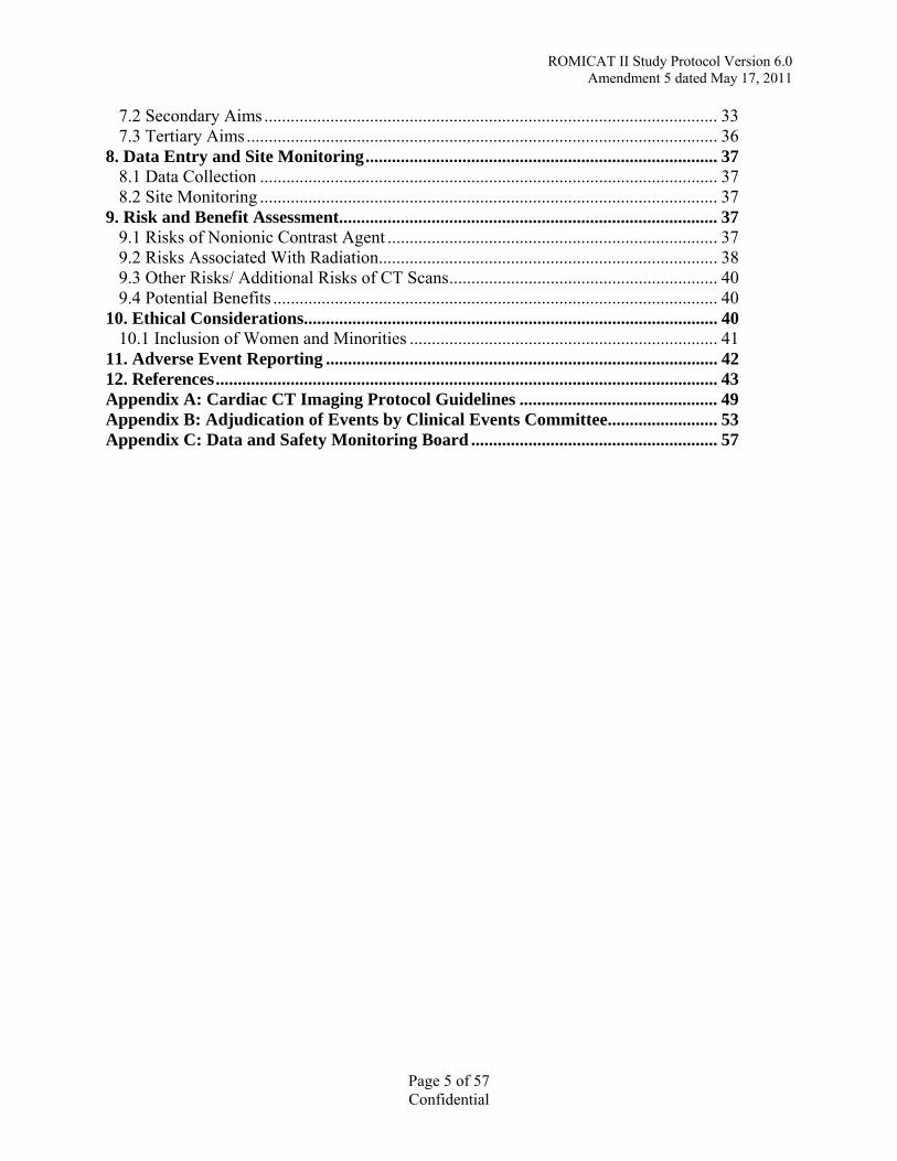
ROMICAT II Study Protocol Version 60 Amendment 5 dated May 17 2011
bull Page 33 MD over read will be performed for internal quality control assessment of findings related to CAD and significant cardiac findings such as pulmonary embolism and aortic dissection to increase awareness to sites of non-coronary findings
3 Clarification of Aims
bull Page 18 The language of the aims and related hypotheses has been expressed in a more clarified manner
Page 8 of 57 Confidential
ROMICAT II Study Protocol Version 60 Amendment 5 dated May 17 2011
Amendment 3 Summary of Changes
1 Changes to InclusionExclusion Criteria (page 25)
bull Page 25 Participant must have at least five minutes of chest pain or equivalent (chest tightness pain radiating to left right or both arms or shoulders back neck epigastrium jawthroat or unexplained shortness of breath syncopepresyncope generalized weakness nausea or vomiting thought to be of cardiac origin) at rest or during exercise within 24 hours of ED presentation warranting further inpatient risk stratification as determined by an ED attending
bull Page 26 9 Currently symptomatic asthma (or taking daily medication for asthma) 10 Contraindication to beta blockers (taking daily antiasthmatic medication)
This exclusion only applies to patients with a heart rate gt 65 bpm at sites using a non-dual source CT scanner
2 Clarification of Study Protocol
bull Page 28 Interventional Arm In subjects randomized to the intervention arm a contrast enhanced cardiac CT will be performed (after initial troponin level is available) in addition to the standard evaluation initial ED evaluation
3 Extension of follow-up time windows
bull Page 28 The 48-72 hour follow-up phone call has been extended to accommodate calls that fall either during the weekend or during a holiday
bull Page 29 The 12 month and 24 month follow-up phone call windows have been extended from +- 2 weeks to +- 4 weeks
Page 9 of 57 Confidential
ROMICAT II Study Protocol Version 60 Amendment 5 dated May 17 2011
Amendment 4 Summary of Changes
1 Study Synopsis (page 13) Duration of Study Participation Participants will be followed during index ED visithospitalization There will be a phone call at 48-72 hours for those discharged within 24 hours All participants will receive a follow up phone call at 28 days 1 year and 2 year post emergency room discharge
2 Changes to Inclusion Criteria (page 27)
bull Participant must be in normal sinus rhythm
3 Elimination of the 1 and 2 year follow-up interviews
bull Page 16 All participants will receive a 28-day a one year and two year follow-up phone interview The efficiency of each strategy will be determined by comparing 1) the length of hospital stay (primary endpoint) 2) the time to diagnosis (secondary endpoint) 3) the rates of ED discharge (secondary endpoint) 4) utilization of other diagnostic testing ie invasive coronary angiograms and resulting interventions ie coronary revascularization procedures during index hospitalization and 28 days one year and two year (secondary endpoint) The safety of the two strategies will be compared by determining the occurrence of adverse events within 48-72 hours in directly discharged patients as well as determination of major adverse cardiovascular events (MACE) within 28 days one year and two years after discharge from the index hospitalization
bull Page 26 To compare the two study arms in terms of rates of MACE (major adverse cardiac eventsmdashcardiac death AMI revascularization unstable angina and rehospitalization) within one and two years
Onetwo Years Defined as onetwo years post the day of discharge from index hospitalization
Hypothesis The rates of MACE in both arms during the follow up period will be lower in the CT arm
bull Page 30 A reminder letter will be sent or phone call will be made to participants two weeks prior to the 28-day follow up phone call If unable to contact the participant by phone mortality will be assessed online using the SSDI website
bull Page 30 54 12 Month Follow up Follow up one year phone call All subjects will be interviewed via a telephone call at one year post ED discharge (12 months +- 4 weeks) to determine the occurrence of MACE and readmissions to the hospital using a standardized questionnaire All cases of recurrent chest pain hospital admissions and diagnostic testing will be verified by review of medical records
Page 10 of 57 Confidential
ROMICAT II Study Protocol Version 60 Amendment 5 dated May 17 2011
A reminder letter will be sent or phone call will be made to participants two weeks prior to the 12-month follow up phone call
55 24 Month Follow up Follow up two year phone call All subjects will be interviewed via a telephone call at two years post ED discharge (24 months +- 4 weeks) to determine the occurrence of MACE using a standardized questionnaire All cases of hospital admissions and diagnostic testing will be verified by review of medical records
Note A reminder letter will be sent or phone call will be made to participants two weeks prior to the two year follow up phone call A minimum of five (5) phone attempts will be to contact each participant If unable to contact the participant by phone mortality will be assessed online using the SSDI website
bull Page 34 To compare the two study arms in terms of rates of MACE (cardiac death AMI revascularization unstable angina and rehospitalization) within one and two years This analysis will be performed from the intent to treat perspective The occurrence of MACE will be assessed via patient follow-up and medical chart review The primary analysis for this aim will be performed at the patient level by determining whether any MACE occurred or not Event rates will be estimated and compared between study groups using exact procedures Prior data estimate a MACE rate of 5 over two years We anticipate a MACE rate between and 2 and 5 in both groups and have 80 power to detect a difference between 2 and 5 between the two groups The Type I error probability associated with this test of this null hypothesis is 005 In a secondary analysis the rates of each type of MACE will be examined separately
4 Extension of 28-Day follow-up time window bull Page 30 The 28 day follow-up phone call window has been extended from +2
weeks to +4 weeks
Page 11 of 57 Confidential
ROMICAT II Study Protocol Version 60 Amendment 5 dated May 17 2011
Amendment 5 Summary of Changes
1 Study Synopsis (page 15) Number of sites 9 7
2 Part I Study Overview (page 17) The study is designed as a randomized controlled diagnostic multicenter trial and will enroll 1000 participants at nine seven sites over a 15 month period
3 Study Design (page 18) The study is designed as a randomized controlled diagnostic multicenter trial and will enroll 1000 participants at nine seven sites over a 15 month period
Page 12 of 57 Confidential
ROMICAT II Study Protocol Version 60 Amendment 5 dated May 17 2011
Study Synopsis Sponsor(s) National Heart Lung and Blood Institute (NHLBI)
Protocol Title Rule Out Myocardial IschemiaInfarction Using Computer Assisted Tomography - A Diagnostic Randomized Multicenter Trial
Diagnosis and Main Adults (ge40-75 yrs) without knowndocumented history of coronary artery Criterion for Inclusion disease who have come to the emergency department (ED) presenting
with a lead symptom of acute chest pain suggestive of acute cardiac ischemia defined as chest pain lasting for at least 5 minutes and occurring within the last 24 hours but without diagnostic ECG changes of acute myocardial ischemia are eligible for participation Only patients in whom the ED attending feels that further inpatient risk stratification is required will be included
Primary Study Objective To determine whether length of hospital stay is significantly shortened in the interventional arm vs standard of care
Secondary Study Objectives bull To determine whether time to diagnosis (for ACS and no ACS) is significantly shortened and rates of direct discharge from the emergency department are increased in the interventional arm vs standard of care
bull To determine a) the additional number of invasive coronary angiograms in both arms and b) to determine the number of coronary revascularization procedures
bull To determine the safety of immediate ED discharge after a normal cardiac CT defined as the occurrence of ACS within 48-72 hours after discharge
bull To determine whether subsequent testing and follow-up including invasive coronary angiograms diagnostic (imaging) tests interventions repeat ED visits for cardiac related problems and repeat hospitalizations for chest pain or equivalent during 28 days after hospital discharge is decreased in the interventional arm compared to SOC
bull To determine the two study arms in terms of rates of MACE within 28 days
bull To compare the two study arms in terms of rates of MACE within one and two years
bull To estimate and compare the sensitivity specificity NPV and PPV of contrast-enhanced CT and non-contrast CT with clinical diagnosis of ACS as the reference standard
bull To determine whether the cost of care (per TSI) for index hospitalization and after 28 days is decreased by implementing cardiac CT into the early ED evaluation of patients with acute chest pain
bull To determine the cost-effectiveness of incorporating cardiac CT into
Page 13 of 57 Confidential
ROMICAT II Study Protocol Version 60 Amendment 5 dated May 17 2011
the standard ED evaluation of patients with acute chest pain over a 28shyday and lifetime horizon as compared to SOC
Tertiary Study Objectives bull To determine whether CT leads to increased test burden after the initial tests during index hospitalization
bull To compare the consistency of physician decisions management depending on the results of diagnostic testing (ie normal CT ndash what fraction is immediately discharged)
bull To determine whether hospital setting availability of observation unit CT experience of hospital and readers and reader performance are associated with primary or secondary outcomes
Primary Endpoint The primary endpoint is time from ED presentation to hospital discharge The length of hospital stay is defined as the time from ED presentation to the time of discharge note or order This includes time in the ED time in any institution-specific specialized chest pain unit and time as an inpatient on the floor
Secondary Endpoints bull The time to diagnosis For ACS defined as the time from ED presentation until the first test during index hospitalization that leads to the diagnosis of ACS (tests may be biomarkers CTA ETT nuclear imaging stress ECHO or cardiac catheterization) for no ACS defined as the time from ED presentation to the final test during index hospitalization
bull Admission to hospital Participants are considered to be admitted if the ED attending admitted the subject to a hospital floor (medical floor step down unit telemetry hospital floor or intensive care unit)
bull Direct ED discharge will include all subjects who are not admitted to the hospital
bull Health care utilization number of invasive coronary angiograms diagnostic (imaging) tests interventions repeat ED visits
bull Rates of MACE major adverse cardiac events ndash cardiac death acute myocardial infarction revascularization unstable angina and rehospitalization
bull Cost of care derived from the cost-accounting database (ie TSI)
Tertiary Endpoints Increased test burden Further imaging tests performed after index hospitalization
Primary Hypothesis Adding cardiac CT to the initial ED evaluation of subjects with acute chest pain significantly decreases length of hospital stay
Page 14 of 57 Confidential
ROMICAT II Study Protocol Version 60 Amendment 5 dated May 17 2011
Secondary Hypothesis Addition of cardiac CT to the initial ED evaluation of subjects with acute chest pain will significantly increase the ED discharge rate and shorten the time to diagnosis and it will be a cost-saving and cost-effective modality There will not be an increase in the number of invasive angiograms or coronary revascularization procedures as compared to the SOC arm With a high negative predictive value patient safety will not be compromised There will not be an increase in subsequent further testing and the rate of MACE in both arms in the follow-up period will be similar The test characteristics of contrast enhanced CT will be significantly better than that of a non-contrast CT
Study Design The study is designed as a randomized diagnostic multicenter trial and will enroll 1000 participants at seven sites over a 15 month period Participants presenting with acute chest pain in the ED will be randomized to standard of care (SOC) or to an interventional arm Patients in the interventional arm will receive standard evaluation supplemented by contrast-enhanced cardiac CT imaging
Duration of Study Participants will be followed during index ED visithospitalization There Participation will be a phone call at 48-72 hours for those discharged within 24 hours
All participants will receive a follow up phone call at 28 days post emergency room discharge
Number of Subjects 1000 subjects will be enrolled with 500 each in the standard of care and interventional arms
Number of Sites 9
Statistical Considerations The primary aim of the study is to compare the two study arms in terms of a difference in mean length of hospital stay (LOS) between the two arms We have conducted a power evaluation using the estimated mean and standard deviation based on observed data from ROMICAT I and simulated data based on health outcomes We will have 95 power to detect a difference in 10 hours of LOS at a Type-1 error rate of 5 for an independent samples t-test using 500 patients in each arm
Page 15 of 57 Confidential
ROMICAT II Study Protocol Version 60 Amendment 5 dated May 17 2011
Abbreviations
ACS Acute Coronary Syndrome CABG Coronary artery bypass graft CAD Coronary Artery Disease CK-MB Creatinine kinase MB type CRC Clinical Research Coordinator CT Computed Tomography ECG Electrocardiogram ED Emergency Department ETT Exercise treadmill ECG testing MACE Major adverse cardiac event MI Myocardial Infarction NPV Negative predictive value PPV Positive predictive value SOC Standard of care SPECT Rest or stress myocardial perfusion imaging
Page 16 of 57 Confidential
ROMICAT II Study Protocol Version 60 Amendment 5 dated May 17 2011
Part I Study Overview The study is designed as a randomized controlled diagnostic multicenter trial and will enroll 1000 participants at nine sites over a 15 month period Participants presenting with acute chest pain in the ED will be randomized to a standard of care arm (SOC) or interventional arm patients in the interventional arm will receive standard evaluation supplemented by contrast-enhanced cardiac computed tomography (CT) imaging (Figure 1)
In both arms the initial Emergency Department (ED) evaluation (electrocardiogram [ECG] physical examination clinical presentation renal function lab and medical history) will determine eligibility of patients After randomization standard of care will be performed in both arms However for patients randomized to the interventional arm a CT will be performed The following decisions made by the caregiver team will be tracked admission or discharge after initial ED evaluation plus CTother initial diagnostic test in admitted participants the length of hospital stay the extent of further testing including serial cardiac markers and ECG and other diagnostic tests or interventions For safety reasons participants discharged in less than 24 hours of ED arrival will receive a phone call 48-72 hours after discharge All participants will receive a 28-day follow-up phone interview The efficiency of each strategy will be determined by comparing 1) the length of hospital stay (primary endpoint) 2) the time to diagnosis (secondary endpoint) 3) the rates of ED discharge (secondary endpoint) 4) utilization of other diagnostic testing ie invasive coronary angiograms and resulting interventions ie coronary revascularization procedures during index hospitalization and28 days (secondary endpoint) The safety of the two strategies will be compared by determining the occurrence of adverse events within 48-72 hours in directly discharged patients as well as determination of major adverse cardiovascular events (MACE) within 28 days after discharge from the index hospitalization We will also perform a cost and cost effectiveness analysis In subsequent analyses we will determine the patient physician and institutional factors that affect the primary endpoint or secondary endpoints
Primary Objective To determine whether length of hospital stay is significantly shortened in the interventional arm vs SOC
Hypothesis
Adding cardiac CT to the initial ED evaluation of subjects with acute chest pain decreases length of hospital stay
Page 17 of 57 Confidential
Secondary Endpoints
Primary Endpoint
Screening
Consent
Length of Hospital Stay
Tertiary Endpoints
Cost and Cost Effectiveness
Incremental Value of CTA over a CAC scan
Rates of Direct ED Discharge
Time to Diagnosis
No of invasive coronary angiograms and revascularizations
Rates of MACE after immediate ED discharge after 28 days
Health care utilization after 28 days
Institutional and Caregiver Characteristics associated with primary and secondary outcomes
Incremental Value of LV function over a CTA
Radiation Exposure during index hospitalization and follow up
ROMICAT II Study Protocol Version 60 Amendment 5 dated May 17 2011
Study Design
The study is designed as a randomized controlled diagnostic multicenter trial and will enroll 1000 participants at nine sites over a 15 month period
Patients with Acute Chest Pain at Low to Intermediate Risk for ACS Screening
Consent
StStandaandard ord off C Caarere CaCarrddiiaac Cc CTT RanRanddomomizizatatioionn
Length of Hospital Stay Primary Endpoint
Rates of Direct ED Discharge
Time to Diagnosis
No of invasive coronary angiograms and revascularizations
Rates of MACE after immediate ED discharge after 28 days Secondary Endpoints
Health care utilization after 28 days
Cost and Cost--Effectiveness
Institutional and Caregiver Characteristics associated with primary and secondary outcomes
Incremental Value of CTA over a CAC scan
Incremental Value of LV function over a CTA Tertiary Endpoints
Radiation Exposure during index hospitalization and follow--up
Page 18 of 57 Confidential
Patients with Acute Chest Pain at Low to Intermediate Risk for ACS
ROMICAT II Study Protocol Version 60 Amendment 5 dated May 17 2011
Part II Study Description
1 Background and Significance
11 Standard Evaluation of Participants with Acute Chest Pain Accurate triage of participants presenting with acute chest pain to the ED remains difficult because neither the chest pain history12 a single set of established biochemical markers for myocardial necrosis (troponin I troponin T creatine kinase MB-type [CK-MB])34 nor initial 12-lead ECG alone or in combination (acute cardiac ischemia time-insensitive predictive instrument) identifies a group of participants that can be safely discharged home without further diagnostic testing5ndash7 As a result the threshold to admit chest pain participants remains low and over six million are admitted annually to US hospitals28ndash10
The standard ldquorule outrdquo myocardial infarction (MI) protocol consists of serial ECG and cardiac biomarker measurements and usually requires a noninvasive stress test to rule out MI These tests (exercise treadmill ECG testing [ETT] stress echocardiography [Echo] and rest or stress myocardial perfusion imaging with Tc-99m [SPECT]) require the exclusion of myocardial necrosis with negative serial biomarkers and are performed to rule out the presence of a hemodynamically significant coronary stenosis While they have good sensitivities (ETT 76 SPECT 83 and Echo 85) for detecting the presence of significant coronary artery stenosis when compared to coronary angiography11ndash13 their specificities are only moderate (ETT 60 SPECT 64 and Echo 77) Moreover these tests are time consuming (ie SPECT 2ndash3 hours) and usually not available 247 Thus the standard evaluation of patients with chest pain to rule out myocardial ischemia requires hospital admission for 24 to 36 hours in over 90 of US hospitals14 Because of the moderate specificities of standard diagnostic testing 33 to 44 of patients with suspected acute coronary syndrome (ACS) who undergo cardiac catheterization have no significant coronary artery disease (CAD)1516 The remarkable inefficiency of current evaluation strategies is also documented by the fact that only lt10 of the six million admitted each year in the US ultimately receive a diagnosis of ACS at discharge1718 The inpatient care for the negative evaluations imparts a significant economic burden in excess of $8 billion annually19ndash21 for the US health care system
Thus an improvement of the ED triage of participants with acute chest pain particularly the increase in ED discharge rates in participants who ultimately do not have ACS would have enormous clinical and economic implications This research will determine if noninvasive cardiac CT that accurately visualizes the coronary arteries for plaques andor stenosis can be an effective strategy to facilitate early ED discharge in this patient population
12 Accuracy of Cardiac CT for Detection of Coronary Artery Disease and LV Function Advances in CT technology with an improvement of volume coverage (ge64 parallel detectors) and temporal (lt210 ms) and spatial (lt06 mm) resolution enables a nearly motion free contrast-enhanced imaging of the coronary artery tree and the heart chambers during a single short breath
Page 19 of 57 Confidential
ROMICAT II Study Protocol Version 60 Amendment 5 dated May 17 2011
hold (8ndash13 second scan)22 The data acquired during an ECG-gated contrast-enhanced multi-detector CT scan allows for 1) accurate assessment of the coronary arteries for plaque and stenosis and 2) the assessment of global and regional LV function in a cine mode
CAD Over the last several years we and others have demonstrated that cardiac CT has excellent diagnostic test characteristics for the detection of significant coronary artery stenosis as compared to invasive coronary angiography23ndash28 There is strong consensus that the strength of cardiac CT is its ability to reliably rule out the presence of stenosis and relevant coronary plaque (negative predictive value [NPV] 95 to 98 positive predictive value [PPV] 92 to 97)22
In addition cardiac CT also accurately detects the presence and extent of calcified and non-calcified coronary atherosclerotic plaque in good agreement with intravascular ultrasound (IVUS) especially in proximal coronary segments (sensitivity 84 to 92)29 30
Global and Regional LV Function Cardiac CT also permits the assessment of global regional LV function in a cine mode Studies have shown excellent reproducibility (κ=086) and agreement to cine magnetic resonance imaging (CMRI) for the detection of global and regional LV dysfunction (r=091)31ndash36
13 Accuracy of Cardiac CT to Detect ACS in Participants with Acute Chest Pain Preliminary data generated by the investigators suggest that cardiac CT using at least 64-slice technology can be performed safely and with excellent image quality in the acute care setting37
In addition our observational data in 368 patients demonstrated that the presence of significant coronary artery stenosis as assessed by cardiac CT can be definitively ruled out in 70 of participants presenting with acute chest pain to the ED (see Sections 31 and 32) This finding has a 100 NPV for ACS during index hospitalization and major adverse cardiac event (MACE) during follow-up (see Section 31)38 There are now several studies that support these results and suggest that cardiac CT has the potential to improve the management of participants with acute chest pain in the ED Rubinshtein et al in a study of 58 participants with acute chest pain but negative initial cardiac troponin levels and non-diagnostic ECG demonstrated a NPV of 100 and specificity of 92 (n=3538) for a diagnosis of ACS in the ED by cardiac CT39 Participants were discharged (n=3558) without stress testing if obstructive CAD was excluded by cardiac CT and if serial troponin measurements were negative There were no deaths or myocardial infarctions during a 15-month follow-up Earlier Goldstein et al40 randomized 200 participants at very low risk for ACS into a cardiac CT based triage system or a stress SPECT based triage system They found no or minimal CAD in 68 of patients which was a safe predictor for the absence of ACS and MACE over 6 months The study further shows the potential of cardiac CT to shorten length of hospital stay and decrease costs of evaluation of patients with acute chest pain However a potential drawback of CT was noted as the CT arm had more invasive angiograms and revascularization procedures (n=11 vs 3 and 4 vs 1 respectively) However this most likely represents that at this time the association between CT findings and clinical outcomes was unknown
In addition studies have suggested that the absence of coronary artery calcification (CAC) as detected in a non-contrast low radiation CT scan also has a high negative predictive value for
Page 20 of 57 Confidential
ROMICAT II Study Protocol Version 60 Amendment 5 dated May 17 2011
ACS in these participants4142 However the clinical utility of CAC alone for the assessment of ACS is inadequate because it cannot exclude the presence of high-grade non-calcified plaquestenosis
14 Cost Effectiveness of Diagnostic Testing in Subjects with Acute Chest Pain Current management of subjects with suspected ACS is managed by a sequential strategy (decision rule) involving quality and duration of chest pain risk factors biomarkers ECGrsquos and diagnostic tests such as SPECT echo or ETT However cost-effective utilization of these tests remains poor43 44 and hospitalization rates for ACS have increased while length of hospitalization has been constant over the last 10 years9 45 The introduction of chest pain observation units may have further decreased the threshold to admit patients Acute appendicitis and pulmonary embolism are excellent examples of the effects of the implementation of advanced imaging technology on patient management in the ED Because of the costs associated with unnecessary appendectomy and one day of in patient observation the implementation of the CT scan resulted in significant cost savings46 A similar approach is warranted to determine whether cardiac CT reduces cost and is cost effective in subjects with acute chest pain Moreover a formal model-based cost-effectiveness analysis is essential to project possible costs and benefits from a broader perspective (including the assessment of potential risks associated with CT) and longer time horizon Modeling can also help to answer questions and perform comparisons not possible within the confines of the clinical trial The current proposal critically investigates these issues
15 Significance of the Proposed Clinical Trial Evaluation of the appropriate clinical use of novel diagnostic imaging modalities is now more important than ever as the cost of these examinations steeply rises and now exceeds the cost spent on prescribed drugs All major professional medical societies have publicly demanded that clinical use of diagnostic imaging should be dictated by evidence-based medicine to not only control costs but also to minimize additional extraneous testing as it is currently unclear whether their usage will result in improvement in the current standard of care
Advanced cardiac CT is one of these novel diagnostic imaging technologies that may potentially enhance standard of care in patients presenting to the ED with acute chest pain but non-diagnostic ECG and negative initial biomarkers a population that remains diagnostically challenging and currently causes a significant clinical burden on each hospital every day Preliminary studies demonstrate that cardiac CT accurately detects absence of coronary atherosclerotic plaque Levels of atherosclerotic plaque as detected by cardiac CT is a powerful predictor of the absence of ACS and can be detected in a significant fraction of participants with acute chest pain Thus we anticipate the implementation of cardiac CT in the early ED triage process will (1) enable earlier and safe discharge of significant fraction of patients directly from the ED who currently would have been unnecessarily admitted as well as (2) allow for earlier detection and better treatment of CAD compared to the current standard of care
The proposed research will constitute the largest randomized multicenter clinical trial using cardiac CT to date and will provide an unbiased assessment of the above stated hypotheses It
Page 21 of 57 Confidential
ROMICAT II Study Protocol Version 60 Amendment 5 dated May 17 2011
will be conducted by a group of experienced clinical researchers consisting of CT imagers ED physicians and cardiologist with clinical expertise in the management of chest pain syndromes The results from this trial will determine the clinical utility of cardiac CT in patients with acute chest pain and may lead to a change in the management of these patients
2 Preliminary Data In support of the aim of the proposed research we provide data from a double blinded observational cohort study (ldquoCardiac CT for triage of participants with intermediate likelihood of ACSrdquo R01 HL080053-03) demonstrating the association between cardiac CT findings on plaque stenosis and LV function with clinical outcomes in patients with acute chest pain These data demonstrate 1) the absence of any CAD in up to 50 of patients with acute chest pain 2) the high NPV of absence of plaque and stenosis for ACS and thus the safety to discharge patients from the ED and the incremental diagnostic value of LV function assessment for risk stratification
21 Final Results of ROMICAT I
211 Cardiac CT Based Detection of Coronary Stenosis and Plaque in the Assessment of Participants with Acute Chest Pain Objective To determine the usefulness of cardiac CT angiography in patients with acute chest pain Methods Observational cohort study in chest pain patients with normal initial troponin and non-ischemic ECG 64-slice coronary CT was performed prior to admission to detect coronary plaque and stenosis (gt 50 luminal narrowing) Results were not disclosed Endpoints were ACS during index hospitalization and MACE during 6- month follow up Results Among 368 participants (mean age 53 plusmn 12 years 61 male) 31 had ACS (8) By cardiac CT 50 of these participants were free of CAD 31 had nonobstructive disease and 19 had inconclusive or positive CT for significant stenosis Sensitivity and NPV for ACS were 100 (n=183368 95 confidence interval [CI] 98ndash100) and 100 (95 CI 089ndash100) with the absence of CAD and 77 (95 CI 59ndash90) and 98 (n=300368 95 CI 95ndash99) with significant stenosis by cardiac CT Specificity of presence of plaque and stenosis for ACS were 54 (95 CI 049ndash060) and 87 (95 CI 083ndash090) respectively Only one ACS occurred in the absence of calcified plaque Both the extent of coronary plaque and presence of stenosis predicted ACS independently and incrementally to TIMI risk score (AUC 088 082 vs 063 respectively all plt00001) Conclusion Fifty percent of participants with acute chest pain and low to intermediate likelihood of ACS are free of CAD by CT and have no ACS Given the large number of such patients early cardiac CT may significantly improve patient management in the ED
Page 22 of 57 Confidential
ROMICAT II Study Protocol Version 60 Amendment 5 dated May 17 2011
Figure 1 A-C 67 year old female 3 hours of substernal chest pain radiating to the back negative initial troponin and CK-MB ECG sinus bradycardia admitted to rule out MI All coronary arteries are widely patent with no evidence of coronary atherosclerosis A Volume rendered 3-dimensional CT image (surface weighted volume rendering technique) of the heart depicting the left circumflex coronary artery (LCX arrowhead) obtuse marginal branch (dashed arrow) and left anterior descending (LAD arrow) two hours after presentation to the ED B CT based evaluation of segments 1 to 3 of the right coronary artery (RCA arrowheads) and conus branch (dashed arrow) using a 5mm thick maximum intensity projection (MIP) C Curved multiplaner reformatted (MPR) image of the LAD (arrowheads) and proximal LCX (dashed arrow)
Figure 2 A-C 40-year old male with hypercholesterolemia and a family history of diabetes woke with chest pressure and noticed intermittent chest pressure during his 1 mile walk to work He had similar symptoms a week prior to admission Initial troponin and CK-MB were negative and ECG showed a normal sinus rhythm without ST-segment or T-wave abnormalities Coronary CT was performed two hours after presentation to the ED and revealed a significant mid LAD stenosis Eight hours after ED presentation cardiac biomarkers became positive The mid LAD stenosis was confirmed on coronary angiography the next day and the participant was treated with a stent placement A Volume rendered 3-dimensional CT image of the heart depicting the right coronary artery (RCA arrow) and the left anterior descending coronary artery (LAD arrowhead) Discontinuation of the contrast-enhanced coronary lumen in the mid section of the LAD (dashed arrow) was detected B CT based evaluation of the right coronary artery (RCA arrowheads) demonstrates calcified and non-calcified plaque in segments 1 to 3 no significant coronary stenosis was detected C Curved MPR image of the LAD reveals good luminal contrast enhancement without any plaque in the distal LAD and calcified plaque in the proximal
Page 23 of 57 Confidential
ROMICAT II Study Protocol Version 60 Amendment 5 dated May 17 2011
segment (arrowheads) A significant luminal narrowing (arrow) was detected in the mid portion of the LAD
212 Incremental Diagnostic Value of Regional LV Function Over Coronary Assessment of Cardiac CT for the Detection of ACS in Participants with Acute Chest Pain Objective To determine incremental value of regional left ventricular function (LVF) over coronary assessment for the diagnosis and prediction of acute coronary (ACS) in patients with acute chest pain but inconclusive initial emergency department (ED) evaluation Methods We enrolled 356 consecutive patients (mean age 53plusmn12 years 62 male) with acute chest pain and inconclusive initial ED evaluation These patients underwent 64-slice contrast-enhanced cardiac CT prior to hospital admission Regional LVF and presence of coronary atherosclerotic plaque and significant stenosis (gt50) were separately assessed by two experienced readers blinded to the clinical course and other CT findings Caregivers and patients remained blinded to the CT results Incremental diagnostic accuracy and predictive value of regional LVF to predict ACS was determined in the entire cohort and in subgroups of patients with inconclusive or positive coronary assessment Results Regional LVF was impaired in 46 patients (129) and 31 patients developed ACS (87) Adding regional LVF resulted in a 10 increase in sensitivity to detect ACS (87 95CI 70shy96) when compared to detection of significant stenosis In the subgroup of 33 patients with significant stenosis impaired regional LVF correctly predicted ACS in 1719 patients (PPV 895 95CI 67-99) In the subgroup of patients with inconclusive coronary CTA (n=33) normal regional LVF correctly predicted the absence of ACS in 2426 patients (NPV 923 95CI 75-99) Impaired regional LVF independent of the extent of plaque and the presence of stenosis was associated with a 25 and 20 fold increased risk for ACS (RR 2023 95CI 747 to 5482 and RR 2534 95CI 936 to 6857 respectively) Moreover c-statistics improved significantly after the addition of regional LVF to both the extent of plaque and the presence of stenosis (c-statistic 088 vs 094 and 082 vs 090 respectively both plt003) Conclusions Regional LVF assessment at rest improves overall sensitivity of coronary CTA for ACS in patients with acute chest pain Combined assessment of coronary morphology and regional LVF is especially helpful to guide further testing and interventions if coronary assessment is inconclusive or positive
213 Initial Decision Model to Assess the Cost Effectiveness of Coronary CT in Triage of Subjects with Acute Chest Pain Objective To develop a preliminary decision-analytic Markov model to predict the long-term consequences of four competing strategies 47 in the ED evaluation of subjects with acute chest pain Methods The model consists of the six health states chest pain healthy early heart disease undiagnosed CAD diagnosed CAD and death Subjects start in the chest pain state The probability that the chest pain is caused by CAD depends on the underlying risk of CAD as
Page 24 of 57 Confidential
ROMICAT II Study Protocol Version 60 Amendment 5 dated May 17 2011
determined by age gender and cardiovascular risk factors As simulated subjects progress through the model CAD (if present) can remain stable or progress Subjects may die from cardiovascular disease or other causes at rates determined by disease stage and demographics By simulating hypothetical cohorts of subjects we determined quality-adjusted life expectancy (QALE) quality-adjusted life year (QALY) and cost-effectiveness ratios for competing strategies We considered four competing strategies for subjects presenting with chest pain to the ED 1) admission in which all subjects are admitted to the hospital for further evaluation 2) discharge in which all subjects are discharged directly home from the ED 3) cardiac CT with conservative treatment in which all subjects without any evidence of CAD are discharged and all other patients are admitted and undergo stress testing and 4) cardiac CT with aggressive treatment which is similar to strategy 3 but sends subjects with evidence of significant stenosis directly for invasive coronary angiography It is important to note that the model permits an evaluation of each strategy using a similar cohort of subjects The model also permits an evaluation of estimate life expectancy and lifetime costs Results Strategies involving cardiac CT were associated with lower ED costs and incremental improvements in QALE when compared to a strategy of admission for all subjects Relative to admitting all subjects the use of cardiac CT yielded savings in ED and hospital costs ranging from $1800-$2200 in men and women ages 30-40 and from $500-$1200 men and women ages 60-70 respectively The incremental cost-effectiveness of strategies involving cardiac CT compared with a strategy of discharging all subjects ranged from $94100-$267200 per QALY in men and $74800-$326100 per QALY in women Conclusions This preliminary decision model suggests that strategies involving cardiac CT are associated with lower ED costs and incremental improvements in QALE in both men and women across all ages The precision of the model will be greatly enhanced by input of real clinical scenarios derived from this proposal
3 Study ObjectiveSpecific Aims This primary objective of this protocol is to assess the efficiency of implementing cardiac CT into the diagnostic workup of patients with acute chest pain normal initial biomarkers and normal or non-diagnostic ECGs Study participants will be patients with acute chest pain and low to intermediate likelihood of ACS and will be randomly assigned to the SOC arm or the interventional arm patients in the interventional arm will receive the standard of care supplemented by cardiac CT
31 Primary Aim The primary aim is to determine whether length of hospital stay is significantly reduced in the interventional arm compared to standard of care The primary endpoint is time from ED presentation to hospital discharge
The length of hospital stay is defined as the time from ED presentation to the time of discharge note or order This includes time in the ED time in any institution-specific
Page 25 of 57 Confidential
ROMICAT II Study Protocol Version 60 Amendment 5 dated May 17 2011
specialized chest pain unit and time as an inpatient on the floor All time sequences must be documented and reported to comprise total length of hospital stay Hypothesis Adding cardiac CT to the initial ED evaluation of subjects with acute chest pain significantly decreases length of hospital stay
32 Secondary Aims 1 To determine whether time to diagnosis is significantly shortened and rates of direct
discharge from ED are increased in the interventional arm vs SOC
A) Time to Diagnosis For ACS Defined as the time from ED presentation until the first test during index hospitalization that leads to the diagnosis of ACS (tests may be biomarkers CTA ETT nuclear imaging stress ECHO or cardiac catheterization) For no ACS Defined as the time from ED presentation defined as the final test during index hospitalization
B) Admission to Hospital Participants are considered to be admitted if the ED attending admitted the subject to a hospital floor (medical floor step down unit telemetry hospital floor or intensive care unit)
Direct ED Discharge will include all subjects who are not included in (B)
Hypothesis ED evaluation of subjects with acute chest pain supplemented by cardiac CT significantly increases direct ED discharge rates and shortens time to diagnosis for both ACS positive and negative patients
2 To determine a) the additional number of invasive coronary angiograms both arms and b) to determine the number of coronary revascularization procedures
Hypothesis ED evaluation of subjects with acute chest pain supplemented by cardiac CT will not increase the number of invasive coronary angiograms without revascularization or revascularizations during index hospitalization
3 To determine the safety of immediate ED discharge after a normal cardiac CT defined as the occurrence of ACS within 48-72 hours after discharge
Hypothesis CTA has a high negative predictive value based on which immediate ED discharge should not compromise patient safety
4 To determine whether subsequent testing and follow up including invasive coronary angiograms diagnostic (imaging) tests interventions repeat ED visits for cardiac related problems and repeat hospitalizations for chest pain or equivalent during 28 days after hospital discharge is decreased in the interventional arm compared to SOC
Hypothesis Incorporation of CTA in the evaluation of acute chest pain patients in the ED will not lead to decreased further testing to manage chest pain when as compared to the SOC arm
5 To compare the two study arms in terms of rates of MACE (major adverse cardiac eventsmdashcardiac death AMI revascularization unstable angina and rehospitalization) within 28 days
28 Days Defined as 28 days post the day of discharge from index hospitalization
Page 26 of 57 Confidential
ROMICAT II Study Protocol Version 60 Amendment 5 dated May 17 2011
Hypothesis The rates of MACE in both arms during the 28 day follow up period will be similar
6 To estimate and compare the sensitivity specificity NPV and PPV of contrast-enhanced CT and non-contrast CT with clinical diagnosis of ACS as the reference standard
Hypothesis The test characteristics of contrast enhanced CT will be significantly better than those of a non-contrast CT for the diagnosis of ACS
7 Determine whether cost of care (per TSI) for index hospitalization and after 28 days is decreased by implementing cardiac CT into the early ED evaluation of patients with acute chest pain
Hypothesis Incorporation of cardiac CT into standard ED evaluation of patients with acute chest pain will decrease the cost of care
8 Determine the cost-effectiveness of incorporating cardiac CT into the standard ED evaluation of patients with acute chest pain over a 28-day and lifetime horizon as compared to SOC
Hypothesis Incorporating cardiac CT into the standard evaluation of patients with acute chest pain will be cost-saving and cost-effective over a 28-day and lifetime horizon
33 Tertiary Aims 1 To determine whether CT leads to increased test burden after the initial tests during index
hospitalization
Hypothesis Cardiac CT may diagnose coronary artery disease at a more incipient stage than determined by imaging tests in the SOC arm which may increase test burden during the follow up period
2 Compare the consistency of physician decisions management depending on the results of diagnostic testing (ie normal CT - what fraction is immediately discharged)
Hypothesis There will be comparable consistency between the two arms
3 Determine whether hospital setting availability of Observation Unit CT experience of hospital and readers and reader performance are associated with primary or secondary outcomes
Hypothesis Enhanced CT experience of the hospital and readers better reader performance during CT certification and hospital setting will be associated with improved primary or secondary outcomes
4 Participant SelectionEligibility Criteria
Adult ED participants presenting with a lead symptom of acute chest pain suggestive of acute cardiac ischemia - defined as chest pain lasting for at least 5 minutes and occurring within the last 24 hours but without diagnostic ECG changes of acute myocardial ischemia - will be eligible for participation
Page 27 of 57 Confidential
ROMICAT II Study Protocol Version 60 Amendment 5 dated May 17 2011
Only patients in whom the ED attending feels that further inpatient testing is required will be included Patients with knowndocumented history of CAD will not be included since such patients most often have an abnormal or non-diagnostic cardiac CT In addition men and women younger than 40 years will be excluded due to the low pre-test likelihood for CAD and the increased radiation risk Detailed inclusion and exclusion criteria are presented below
41 Inclusion Criteria 1 Participant must have at least five minutes of chest pain or equivalent (chest tightness
pain radiating to left right or both arms or shoulders back neck epigastrium jawthroat or unexplained shortness of breath syncopepresyncope generalized weakness nausea or vomiting thought to be of cardiac origin) at rest or during exercise within 24 hours of ED presentation warranting further risk stratification as determined by an ED attending
2 Participant must be able to provide a written informed consent
3 Participants must be lt75 years of age but gt40 years of age
4 Participant must be able to hold breath for at least 10 seconds
5 Participant must be in sinus rhythm
42 Exclusion Criteria 1 New diagnostic ischemic ECG changes (ST-segment elevation or depression gt 1 mm or
T-wave inversion gt 4 mm) in more than two anatomically adjacent leads or left bundle branch block
2 Documented or self-reported history of CAD (MI percutaneous coronary interventions [PCIs] coronary artery bypass graft [CABG] known significant coronary stenosis [gt50])
3 Greater than 6 hours since presentation to ED to time of consent
4 BMI gt40 kgm2
5 Impaired renal function as defined by local standard of care - for example measured serum creatinine gt15 mgdL
6 Markedly elevated troponin as defined by local standard of care
7 Hemodynamically or clinically unstable condition (BP systolic lt 80 mm Hg atrial or ventricular arrhythmias persistent chest pain despite adequate therapy)
8 Known allergy to iodinated contrast agent
9 Currently symptomatic asthma
10 Documented or self-reported cocaine use within the past 48 hours (acute)
11 On Metformin therapy and unable or unwilling to discontinue for 48 hours after the CT scan
Page 28 of 57 Confidential
ROMICAT II Study Protocol Version 60 Amendment 5 dated May 17 2011
12 Contraindication to beta blockers (taking daily antiasthmatic medication) This exclusion only applies to patients with a heart rate gt 65 bpm at sites using a non-dual source CT scanner
13 Participant with no telephone or cell phone numbers (preventing follow-up)
14 Participant with positive pregnancy test Women of childbearing potential defined as lt2 years of menopause in the absence of hysterectomy or tube ligation must have a pregnancy test performed within 24 hours before the CT scan
15 Participant unwilling to provide a written informed consent
43 Recruitment Screening and Consent
The research team at each participating site includes the ED attending physician CT technologist and research associate(s) The investigator and the research staff will be responsible for the screening review of participant medical records and investigator-designated data submission All clinical sites will perform screening Monday through Friday during daytime hours (9am-5pm) Potential subjects will be identified by research coordinatorstudy nurse staff shortly after admission to the ED usually after the initial ED evaluation consisting of ECG physical examination clinical symptoms and presentation immediate and past medical history has been concluded and screened for eligibility based on a checklist providing inclusion and exclusion criteria
Eligible subjects will be approached and if the subject considers participation the Clinical Research Coordinator (CRC) will describe the rationale of the study the study procedures with associated risks and benefits A study physicianstudy nurseCRC will speak directly with an ED physician caring for the patient to obtain an assessment of pretest probability for ACS and will verify that the ED physician feels that further observation or diagnostic testing is warranted in this patient
If the subject agrees and further testing or observation is planned as SOC key study personnel typically a study physician or the attending ED physician will review the study procedures as well as all potential risks and discomforts and the opportunity to decline or cease participation in the study at any time answer any remaining questions and consent the subject
Patients can be consented but not randomized before the first troponin test result is available Patients whose initial troponin level is markedly elevated will not be randomized and will not be included in the intent-to-treat population Patients whose subsequent troponin testing is markedly elevated will be included in the intent to treat population
Patients can be consented before a pregnancy test is available However patients with a positive pregnancy test will not be randomized
Patients who are not randomized will not be considered part of the intent to treat population
Informed Consent
Page 29 of 57 Confidential
ROMICAT II Study Protocol Version 60 Amendment 5 dated May 17 2011
bull A potential subject will then be afforded as much time as clinically feasible to review the consent form and decide upon their participation without adversely effecting their ED care which is typically 1 hour
bull If the ED staff caring for the patient determines that the potential subjectrsquos previously determined plan of care must proceed before the potential subject has had adequate time to decide upon hisher participation without undue pressure the potential subject will be excluded from enrollment
bull Subjects will be provided with a copy of the informed consent form
44 Randomization
After the patient has been consented by the study physician the study subject will be randomized into the standard care arm or the intervention arm (cardiac CT) by means randomization stratified by institution using a central computerized randomization system
5 Study Procedures
51 Index Hospitalization Standard of Care Arm Subjects will be evaluated according to each hospitalrsquos specific protocol to evaluate and manage patients with acute chest pain Typically the standard evaluation in the ED will include the past and current medical history a physical examination an ECG and a cardiac biomarker (Troponin and CK-MB) as well as other routinely obtained blood testing Patients may undergo cardiac CT as part of SOC but only as a secondary diagnostic test Patients in the CT arm may undergo further diagnostic testing as well
All admitted subjects will undergo each hospitalrsquos standard rule out myocardial ischemia protocol This protocol typically consists of observation and monitoring including serial ECGs and repeated cardiac biomarker measurements as well as a noninvasive stress test (often imaging based) to evaluate for myocardial ischemia The participating clinical sites perform routinely either nuclear perfusion imaging [SPECT] at rest and stress andor stress echocardiography andor exercise treadmill test [ETT] Depending on the results subjects may undergo additional noninvasive or invasive testing (coronary angiography) andor coronary revascularization during their hospital stay
Interventional Arm In subjects randomized to the intervention arm a contrast enhanced cardiac CT will be performed (after initial troponin level is available) in addition to the initial ED evaluation The ED physicians will resume care of the patient and make all clinical decisions for further care of the patient (eg need for further evaluation or admission) based upon their cumulative clinical assessment of the patient including findings revealed on the CT scan
Page 30 of 57 Confidential
ROMICAT II Study Protocol Version 60 Amendment 5 dated May 17 2011
In addition in patients from both study arms who consented to a blood draw we will obtain three fifteen (15) mL venous blood samples over a six-hour period less than forty-five (45) mL in total Blood samples will be centrifuged and frozen for future analysis for cardiac-related markers
All diagnostic tests will be performed per Society Guidelines Cardiac Computed Tomography is performed in accordance with best practice standards as delineated in the imaging guidelines of the Society of Cardiovascular Computed Tomography by competent63 and appropriately credentialed physicians This includes the optimization of the scan protocol to limit radiation exposure
52 48-72 Hour Follow up after ED discharge Follow-up phone call at 48-72 hours Subjects who are discharged from the ED within 24 hours of presentation (for both randomization arms) will be contacted via telephone between 48 and 72 hours after discharge to avoid ascertainment bias During this call subjects will be interviewed to determine whether they have had any recurrent symptoms suggestive of myocardial ischemia and andor whether they needed to seek further medical attention following their emergency department visit If these phone attempts at 48 and 72 hours are not successful a final attempt will be made at 96 hours to contact the subject
NOTE If a subject is discharged on a Thursday the 48-72 hour phone call may be extended to 96 hours In the rare circumstance that a subject is discharged on a Thursday and the following Monday is a holiday the follow-up call can be made during the next working business day
53 28 Day Follow up Follow up 28 day phone call All subjects will be interviewed via a telephone call 28 days post ED discharge (+4 weeks) to determine the occurrence of MACE and healthcare utilization using a standardized questionnaire All cases of recurrent chest pain hospital admissions and diagnostic testing will be verified by review of medical records
A reminder letter will be sent or phone call will be made to participants two weeks prior to the 28-day follow up phone call If unable to contact the participant by phone mortality will be assessed online using the SSDI website
Page 31 of 57 Confidential
ROMICAT II Study Protocol Version 60 Amendment 5 dated May 17 2011
6 Data Collection The following subject data will be collected during index ED visithospitalization and follow up phone interviews bull Subject demographic information bull Medical history including cardiac risk factors and medications bull Description of recent pain episodes bull Vitals signs ECG cardiac biomarker results bull Cardiac CT imaging and other cardiac-related diagnostic testing data bull Adverse events
7 Statistical Considerations A total of 1000 participants will be enrolled in this study Enrollment is planned to be completed in 15 months
71 Primary Aim To compare the two study arms in terms of the time from ED presentation to hospital discharge The primary aim of the study is to compare the two study arms in terms of a difference in mean length of hospital stay (LOS) between the two arms Based on the results of ROMICAT I we estimate the mean (plusmn standard deviation) hospital LOS (including ED) of these patients in standard evaluation to be 405 plusmn 432 hours The observed frequency distribution appeared to fit well to a log-normal distribution (Median was 77 hours and Range was 275 ndash 3815 hours) We anticipate that the observed distributions of both Group A and Group B follow such a pattern Because the independent samples t-test is robust to a departure from the data normality when applied to a large sample size we will employ the t-test for this inference
We have conducted a power evaluation using the estimated mean and standard deviation based on ROMICAT I for Group A and using the estimated the mean and standard deviation based on a simulated data for Group B The simulated data consisted of 500 LOS values resulted as a mixture of three subsets randomly drawn from log-normal distributions of which the first subgroup was to represent the patients with normal LV function and without CAD (486 Mean plusmn SD of 6 plusmn 12 hours) the second was to represent the patients with normal LV function and with non-obstructive CAD (288 Mean plusmn SD of 10 plusmn 20 hours) and the third group was to represent all other possible conditions (226 Mean plusmn SD of 579 plusmn 604 hours which was estimated from ROMICAT 1) We also incorporated in the possibility that some patients without CAD will not be correctly triaged The triage accuracy rate in the following table is the rate at which a patient without CAD would be correctly triaged The following table summarizes for each ED triage accuracy rate assumption the estimated mean LOS and the power at a Type-1 error rate of 5 for an independent samples t-test using 500 patients in each arm The proposed sample size will attain a sufficient power to detect a clinically meaningful difference in mean LOS between the two groups even in the presence some degree of ED triage inaccuracy
Page 32 of 57 Confidential
ROMICAT II Study Protocol Version 60 Amendment 5 dated May 17 2011
Powers to Detect Estimated Differences in Mean LOS values at Assumed ED Triage Accuracy Rates (Type-1 error rate = 005)
Assumed ED Triage Accuracy Rate
Estimated Mean (plusmn SD) LOS Estimated Difference in Mean (hours) Power
Group A (Standard Eval)
N=500
Group B (CT Eval)
N=500 65 405 (plusmn 432) 328 (plusmn 467) -83 83 70 405 (plusmn 432) 305 (plusmn 455) -101 95 80 405 (plusmn 432) 263 (plusmn 417) -142 99 90 405 (plusmn 432) 224 (plusmn 373) -181 gt99
100 405 (plusmn 432) 188 (plusmn 335) -217 gt99
72 Secondary Aims To compare the two study arms in terms of time to diagnosis and rates of direct discharge The time to discharge (defined as time from ED presentation until discharge to home) will be plotted using Kaplan-Meier curves because some patients will be in the hospital at 30 days post day of presentationrandomization The curves for the two groups will be compared using a test based on the difference in the average length of stay in the two groups (truncated at 30 days) divided by the standard error of this difference60 We choose this test because the average length of stay is most closely associated with cost It will be also instructive to compare the curves themselves as they will provide information on the effect of cardiac CT on different groups of patients For instance one group of patients those who are discharged due to cardiac CT may have a shorter length of stay while others may have a longer length of stay due to waiting for the results of the CT or other procedures that the CT indicates
Sample Size The planned sample size will also provide adequate power to detect the rates of direct discharge from ED Screening data from a site survey of our clinical sites as well as published data indicate that the standard ED discharge rate is approximately 15 in the target population According to our preliminary data and data from other groups we expect that 40 to 50 of participants have no detectable CAD by cardiac CT The negative predictive value of this finding for ACS has been shown to be 100 Our guidelines suggest that these participants will be discharged directly from the ED However realistically we expect that because of administrative issues and remaining individual variation of the decision to discharge depending on the clinical presentation of participants actually only 60 to 80 of these participants will be discharged Thus anywhere from 24 to 40 participants could be discharged directly from the ED in the interventional arm For the sample size analysis we assume that 32 of participants will be discharged
Based on the aforementioned key assumptions for the Standard Evaluation we anticipate that 15 of all participants will be discharged and 85 will be admitted (discharge rate 15) For the Cardiac CT Evaluation we assume a discharge rate of 32 For the objective of superiority
Page 33 of 57 Confidential
ROMICAT II Study Protocol Version 60 Amendment 5 dated May 17 2011
of discharge rates we will have a 100 chance at a significance level of 005 to detect a 17 increase in discharge rates from 15 to 32
The comparison of time to discharge among patients who are admitted to the hospital is complicated by the fact that the two groups will not be the same group of patients For instance patients who would have been discharged in the cardiac CT group but admitted in the usual care group may have a relatively shorter time post admission than patients who would have been admitted in both groups We have developed methods for analyzing these types of data using causal inference that will be applied to this analysis61 A similar analysis will be applied to other subgroups such as those with and without ACS
To compare the two study arms in terms of cardiac health care utilization during the index visit Health care utilization components will include invasive coronary angiograms and coronary revascularization
The analysis for this secondary aim will be performed from the intent to treat perspective Health care utilization will be assessed using medical record review Measures of key components of utilization will be compared across the two study groups using procedures appropriate for each measure (rate or count)
In particular the two arms will be compared in terms of the number of invasive coronary angiograms This secondary endpoint will be analyzed in terms of a non-inferiority trial In a non-inferiority trial the null hypothesis is that cardiac CT is worse than usual care in that the rate of coronary angiograms is increased and the alternative hypothesis is that it is not in worse and the rate of angiograms is the same The reason for reversing the usual null and alternative hypothesis is that the purpose of this analysis is to prove that cardiac CT is not worse than usual care this would not be accomplished by just showing that the difference between the two was not significant
Screening data from a survey of clinical sites as well as published data indicate that the standard rate of invasive coronary angiograms in the target population is approximately 10 According to our study guidelines we assume that most but not all participants with significant stenosis in CT (10 of participants) undergo invasive coronary angiography (notably those who also have a positive stress test and those who have high grade proximal stenosis or LAD stenosis) In addition we assume that a minority of participants with indeterminate stenosis (lt5) and positive stress testing undergo invasive coronary angiography Because of the moderate specificities of standard diagnostic testing up to 40 of participants who undergo cardiac catheterization have no significant CAD1516 Because the diagnostic accuracy of cardiac CT for the detection of coronary stenosis is comparable or better than standard diagnostic testing we believe that there will be no increase in the rates of invasive coronary angiograms If we define a 5 difference as an ldquoacceptablerdquo difference in cardiac catheterization rates than we will have an 84 chance of showing that the CT arm is significantly less than 5 worse than the standard arm at a one sided p=005 significance level
Page 34 of 57 Confidential
ROMICAT II Study Protocol Version 60 Amendment 5 dated May 17 2011
Another key measure of utilization is the number of coronary revascularization procedures In ROMICAT I there were 18 revascularization procedures performed in 368 patients (5) Thus we expect 5 procedures in the standard evaluation If we assume that not all participants with significant stenosis will undergo coronary revascularization (because if no MI it can be medically managed) and that cardiac CT may also avoid some angiograms by excluding disease than we can realistically assume that there is not a difference between the two study arms If we consider this in terms of a non-inferiority trial as above we will have 80 power to rule a difference of 35
To compare the two study arms in terms of cardiac health care utilization within one year after index hospitalization Health care utilization components will include invasive coronary angiograms cardiac related diagnostic and lab testing and ED visits
This analysis will be performed from the intent to treat perspective CP and CT related health care utilization will be assessed using medical record review Measures of key components of utilization will be compared across the two study groups using procedures appropriate for each measure (rate or count) We expect that the rate of additional cardiac work-up will be 29 in the SOC arm and will be 14 in the interventional arm We will have gt95 power to detect this difference We expect that the rate of hospital admissions with additional diagnostic cardiac testing will be 93 in the SOC arm and will be 4 in the interventional arm We will have gt95 power to detect this difference We expect that the rates of these measures will be associated with the CT findings at index hospitalization
To evaluate the cost and cost-effectiveness of incorporating cardiac CT into the standard ED evaluation of patients with acute chest pain These analyses will be performed by the Decision Analysis and Cost-Effectiveness Center (DACE) Therefore the sites will collect and provide the necessary data to the DACE and the costs will be derived from the cost-accounting database (TSI) As a first step incorporating cardiac CT into the standard evaluation of patients with acute chest pain will be tested for cost-saving over a 28-day and lifetime span as compared to standard care Further on cost-effectiveness of incorporating cardiac CT into the standard ED evaluation of patients with acute chest pain will be determined using first-order Monte Carlo microsimulations
To compare the two study arms in terms of rates of MACE (cardiac death AMI revascularization unstable angina and rehospitalization) within 28 days The analysis for this secondary aim will be performed from the intent to treat perspective The occurrence of MACE will be assessed via patient follow-up and medical chart review The primary analysis for this aim will be performed at the patient level by determining whether any MACE occurred or not Event rates will be estimated and compared between study groups using exact procedures In a secondary analysis the rates of each type of MACE will be examined separately
Sample Size- Safety Considerations- Missed ACS Mistaken discharges are the primary safety issue in this study All available data indicate that the absence of any detectable CAD (absence
Page 35 of 57 Confidential
ROMICAT II Study Protocol Version 60 Amendment 5 dated May 17 2011
of any plaque and stenosis) has a very high NPV for ACS (100 or close to 100)62 Thus we anticipate that the occurrence of ACS in participants discharged home directly from the ED in the interventional arm will be also minimal or even zero and similar to the standard of care If as demonstrated in ROMICAT I none of the participants without CAD will have MACE defined as ACS within 30 days after direct ED discharge this observation has 90 power to ensure that the true event rate is less than 1 These assumptions are supported by the results of the ERASE trial which reported similar and very minimal event rates in each group (only 2 pts with acute MI were mistakenly discharged 1 in each randomization group out of 1200 [event rate 00008]) In this trial we would have an 80 chance of detecting at least one occurrence of an inappropriately discharged participant if the true rate where over 1
To estimate and compare the sensitivity specificity NPV and PPV of contrast-enhanced CT and non-contrast CT with clinical diagnosis of ACS as the reference standard The analysis for this aim will utilize data on participants randomized to Group B In this arm each participant will undergo non-contrast CT and contrast-enhanced CT which will be interpreted as positive or negative for ACS Clinical determination of presence or absence of ACS will be used as the reference standard For each of the two types of CT we will estimate the sensitivity specificity and positive and negative predictive value of the modality for ACS Each measure of diagnostic or predictive performance will be compared between the two modalities using statistical techniques that account for the correlation due to the paired test design In addition to these measures we will estimate the proportion of cases with a clinical diagnosis of ACS and a negative non-contrast CT in which contrasted enhanced CT was positive for ACS This proportion provides a measure of the incremental value of contrast-enhanced CT for diagnosis ACS
73 Tertiary Aims To determine whether CT leads to increased test burden after the initial tests during index hospitalization
To compare the consistency of physician decision management depending on the results of the diagnostic testing (ie normal CT ndash what fraction is immediately discharged)
To determine whether hospital setting availability of Observation Unit CT experience of hospital and readers and reader performance are associated with primary or secondary outcomes
Analysis of Data on Women and Minorities We will analyze the effect of the intervention on women and minorities as outlined in the NIH guidelines Thus we include testing for gender-treatment and minority status-treatment interactions with the discharge rate and the angiogram rate
Page 36 of 57 Confidential
ROMICAT II Study Protocol Version 60 Amendment 5 dated May 17 2011
8 Data Entry and Site Monitoring
81 Data Collection Subject data will be collected by the research associates and paper records will be maintained at individual clinical sites The Data Coordinating and Statistical Center (DCSC) will develop a web based electronic case report form The de-identified study data will be entered into this electronic data capture system (EDC) and sent to the Data Coordinating and Statistical Center (DCSC) at Massachusetts General Hospital for further analysis
82 Site Monitoring Site visits will be performed on a regular basis by the Data Coordinating Center to ensure that all regulatory requirements are being met and to monitor the quality of the data collected Records of Institutional Review Board approvals and patientsrsquo charts will be examined on a spot check basis to evaluate the accuracy of the data entered into the database
9 Risk and Benefit Assessment
91 Risks of Nonionic Contrast Agent
Rare risks of the nonionic contrast agent bull Itchiness bull Rash bull Mild allergic reaction
Very rare risks of the nonionic contrast agent bull Kidney diseaseContrast-induced nephropathy bull Death
Regarding the risks of contrast material the rate of adverse reactions with nonionic contrast reagents is 0250 More than 90 of such adverse reactions are allergic-like very mild (itching rush) and can be very effectively treated with available drugs (ie antihistaminic) Severe reactions occur in 002 and one death occurred in 57739 injections of nonionic contrast material50
Contrast-induced nephropathy (CIN) CIN is expected to be a very rare event among this patient population because patients with already impaired renal function (the strongest predictor of CIN) will not be enrolled) This assumption is strongly supported by our preliminary experience from ROMICAT I In this study none of the participants had such an event (0368)
The very low risk associated with contrast administration has been documented in large scale trials previously In a randomized trial in 1196 participants the incidence rate of CIN defined as an increase in serum creatinine ge1mgdL from baseline within 48 to 72 hours following the injection of contrast material was 0 in 359 participants (0) who had no renal insufficiency and no diabetes melliltus51 The risk was 2 in 315 (06) in participants with no renal insufficiency but who had diabetes mellitus51 In this study the mean volume of contrast-agent administered
Page 37 of 57 Confidential
ROMICAT II Study Protocol Version 60 Amendment 5 dated May 17 2011
was 140 plusmn 57 mL which is much higher than the 80 mL of contrast that is administered for a coronary CTA The risk of CIN may increase if participants in the cardiac interventional arm will be admitted and subsequently undergo further contrast-related diagnostic testing However among 209 participants who had both a pulmonary angiogram and a contrast-enhanced spiral CT in the Prospective Investigation of Pulmonary Embolism Diagnosis II only one participant with diabetes mellitus had a transient episode of acute renal failure characterized by an increase in the serum creatinine level from 13 to 29 mgdL52 In this study the contrast-enhanced CT and pulmonary angiogram were performed within 22 hours The elevated creatinine returned to normal after administration of intravenous fluids Thus overall we expect that the occurrence of CIN will be a rare event in this study
92 Risks Associated With Radiation While the radiation dosage for CT scanning varies with the part of the body being scanned the exposure for this examination is approximately between 7 and 17 mSv This exposure is comparable or less than stress nuclear myocardial perfusion imaging (14ndash22 mSv) (personal communication with Prof Ronald Callahan Chairman of the MGH Radiation Safety Committee) In comparison the average yearly effective dose of natural background radiation is 25 mSv53 the allowed annual exposure of radiation workers is 50 mSv the allowed exposure in five years is 100 mSv (Table 3)54 Thus even if an enrolled participant would undergo cardiac CT followed by a conventional stress test and selective coronary angiography the participant would be well below the allowable annual effective dose for radiation workers The radiation dose from cardiac CT has not been shown to have any adverse effects
Radiation Exposure
Cardiac CT scanning results in a measurable radiation exposure55 In order to minimize risk for participants in this study we have implemented the following measures
bull Exclude women lt40 years of age bull Exclude pregnant and breast feeding women bull Include only women who have a negative pregnancy test bull Application of cardiac CT protocols with the goal to minimize radiation exposure (ie
use of tube modulationmdashdecrease of radiation exposure by 30 to 50 and prospective triggering in appropriate conditions)
bull Review and approval of these CT protocols by the CVCT Core Lab
General Risk Associated with Radiation Exposure
The risk of inducing a malignancy by exposure to radiation is estimated to be 5 cases of malignancy for each 100 individuals exposed to an effective dose of 1 Sv54 The risk is estimated to be higher in infants and children and lower in the elderly The data which these estimates are based on are weak and apply to relatively high levels of radiation exposure56 and it remains unclear whether there is a linear relation in lower levels of radiation Assuming a linear proportion with a low level of radiation exposure to 1 mSv would lead to 5 malignancies in 100000 individuals exposed and a dose of 8 mSV would lead to 40 malignancies in 100000 individuals56 Small amounts of radiation exposure less than 100 mSv have not been clearly
Page 38 of 57 Confidential
ROMICAT II Study Protocol Version 60 Amendment 5 dated May 17 2011
shown to have harmful effects The amount of radiation to which patients will be exposed in this study is less than the amount that is known to cause a measurable harmful effect Even so it is generally assumed that a small amount of radiation will have a small chance of producing a harmful effect These extrapolated risks of the effects of small amounts of radiation are probably overestimates because the estimates ignore the fact that the body can repair the damage done by radiation if the repair mechanisms are not overwhelmed Radiation is a well-known teratogenic agent and the developing fetus is much more sensitive to radiation than the mother
Table 3 Estimated radiation exposure for diagnostic tests background radiation and exposure limits
Examination Effective Dose (mSv) Chest PA and lateral 007 Myocardial Perfusion Imaging (Rest and 14ndash22Stress) Thorax CT for the Exclusion of Pulmonary 7-12Embolism 64-Slice CT coronary angiogram 7ndash17 2-view mammogram 30 Natural yearly background radiation 25 Max exposure in radiation workers 50
Lifetime Cancer Risk Attributable to Cardiac CT
The absolute cancer risk has been described recently and recommendations have been made57
The life time cancer risk from cardiac CT decreases with age and is higher in women than in men in particular exposure of younger women should be avoided In our study these recommendations have been incorporated In comparison the absolute cancer risk for women to develop breast cancer is 13 and for men to develop colon cancer is 7 the risk attributed to a cardiac CT scan is 1466 vs 1715 for a 60 year old women and 11241 vs 11911 for 60 year old men (for CT without and with tube modulation (female 21 and 14 mSv male 15 and 9 mSv respectively)57 Thus the incremental risk induced by cardiac CT compared to the overall risk of the population to develop cancer seems moderate
Effect of the Implementation of Cardiac CT in the ED Evaluation of Patients with Acute Chest Pain on a Population Basis
The number of CT scans performed in the US was 62 million in 200658 Imaging of the abdomen (50) and the head (33) were major single contributors Specific concern was given to the rise of pediatric CT imaging (from 6 to now 11) and screening the adult asymptomatic population (ie lung heart) Implementation of cardiac CT in the ED could lead to a small increase of the number of overall CT procedures However the effects of the implementation of cardiac CT in the ED would be minimal if 1) other procedures associated with radiation doses would be saved 2) repeated testing currently performed in 25 of patients would be minimized and 3) rates of unnecessary purely diagnostic cardiac catheterizations would
Page 39 of 57 Confidential
ROMICAT II Study Protocol Version 60 Amendment 5 dated May 17 2011
decrease Furthermore already cardiac CT scanning could be performed using single phase acquisition with prospective triggering which would decrease the dose to lt5 mSv In addition MD over-read will be performed for internal quality control assessment of findings related to CAD and significant cardiac findings such as pulmonary embolism and aortic dissection to increase awareness to sites of non-coronary findings
93 Other Risks Additional Risks of CT Scans bull Discomfort bull Claustrophobia
Risks of IV Needle Placement
bull Hemorrhage (hematoma at the injection site) bull Infection (catheter related infection) at the injection site bull Minor discomfort bull Bleeding bull Infection bull Bruising
94 Potential Benefits bull Subjects may benefit from randomization into the interventional arm of trial from detection
of pathology that may not have been otherwise detected during SOC (eg a non-significant coronary stenosis or non-obstructive coronary artery disease that would likely not have been detected by a stress test)
bull Data from prior studies indicate that patients in the interventional arm may be earlier discharged and may not have to spend the night in the hospital there is also the chance that alternative diagnosis such as pneumonia or Pulmonary embolism may be detected - patients may avoid the dual radiation exposure of a CT protocol solely to evaluate for pulmonary embolus followed by a SPECT myocardial perfusion scan
bull Subjects may also benefit in their assessment for long term prognosis and risk for adverse cardiac events and may receive preventive medication as a result of CT
10 Ethical Considerations This study is to be conducted according to US and international standards of Good Clinical Practice (International Conference of Harmonization [ICH] guidelines) applicable government regulations and NIH research policies and procedures
This protocol and any amendments will be submitted to a properly constituted independent Institutional Review Board (IRB) for formal approval of the study conduct The decision of the IRB concerning the conduct of the study will be made in writing to the investigator and a copy of this decision will be provided to the ROMICAT PI before implementation of the study
Page 40 of 57 Confidential
ROMICAT II Study Protocol Version 60 Amendment 5 dated May 17 2011
All study participants in this study will be given an IRB-approved site-specific ICF describing the study and providing sufficient information for participants to make informed decisions about their participation in this study The study participant MUST be consented with the ECIRBshyapproved ICF before the participant is subjected to any study procedures The IRB-approved ICF MUST be signed and dated by the study participant or legally acceptable representative and the investigator-designated research staff obtaining the consent before the participant is subjected to any study procedures
101 Inclusion of Women and Minorities The participating institutions will not exclude potential participants from participating in this or any study solely based on ethnic origin or socioeconomic status Every attempt will be made to enter all eligible participants into this protocol and therefore address the study objectives in a patient population representative of the entire English-speaking population at risk for ACS treated by the institution
Both men and women and members of all ethnic groups are eligible for this trial In conformance with the National Institutes of Health (NIH) Revitalization Act of 1993 with regard to inclusion of women and minorities in clinical research the projected gender and minority accruals are shown below
Table 1 Gender and Minority Accrual Estimates
Ethnic Category SexGender Females Males Total
Hispanic or Latino 66 79 145 Not Hispanic or Latino 384 471 855
Ethnic Category Total of all participants
450 550 1000
Racial Category American Indian or Alaskan Native 2 2 4 Asian 35 42 77 Black or African American 125 153 278 Native Hawaiian or other Pacific
Islander 2 2 4
White 254 312 566 OtherUnknown 32 39 71
Racial Category Total of allparticipants
450 550 1000
Page 41 of 57 Confidential
ROMICAT II Study Protocol Version 60 Amendment 5 dated May 17 2011
11 Adverse Event Reporting Mistaken discharges are the primary safety issue in this study All available data indicate that the absence of any detectable CAD (absence of any plaque and stenosis) has a very high NPV for ACS (100 or close to 100)62 Thus we anticipate that the occurrence of ACS in participants discharged home directly from the ED in the cardiac interventional arm will be also minimal or even zero and similar to the standard of care If as demonstrated in ROMICAT I none of the participants without CAD will have MACE defined as ACS within 30 days after direct ED discharge this observation has 90 power to ensure that the true event rate is less than 1 These assumptions are supported by the results of the ERASE trial which reported similar and very minimal event rates in each group (only 2 pts with acute MI were mistakenly discharged 1 in each randomization group out of 1200 [event rate 00008]) In this trial we would have an 80 chance of detecting at least one occurrence of an inappropriately discharged participant if the true rate where over 1
At the time of ED discharge investigators will determine at if any adverse events have occurred An Adverse Event (AE) is any untoward medical occurrence in a study participant that may or may not have a causal relationship with the study intervention An AE can therefore be any unfavorable and unintended sign (including an abnormal laboratory or physiological finding) symptom or disease temporally associated with the use of a medical treatment or procedure regardless of whether it is considered related to the medical treatment or procedure (attribution of unrelated unlikely possible probable or definite) Abnormal results of diagnostic procedures are considered to be AEs if the abnormality
bull results in study withdrawal bull is associated with a serious adverse event (SAE) bull is associated with clinical signs or symptoms bull leads to additional treatment or to further diagnostic tests bull is considered by the investigator to be of clinical significance
Serious Adverse Event (SAE) An SAE is defined as any untoward medical occurrence that
bull results in death or bull is life-threatening (at the time of the event) or bull requires inpatient hospitalization or prolongation of an existing hospitalization or bull results in persistent or significant disability or incapacity or bull is a congenital anomalybirth defect
Reportable Adverse Events Any untoward medical occurrence that occurs after randomization and prior to discharge from the ED should be reported to the CCC by completing the electronic adverse event case report form
Immediately Reportable Adverse Events
Page 42 of 57 Confidential
ROMICAT II Study Protocol Version 60 Amendment 5 dated May 17 2011
Any adverse event that is judged by the investigator to be serious AND unexpected AND related to study procedures should be reported within 24 hours of discovery by phone or email to the Coordinating Center The local Institutional Review Board must also be notified in a timely manner
Investigators must also report Unanticipated Problems regardless of severity associated with the study procedures within 24 hours An unanticipated problem is defined as follows
Unanticipated Problem (UP) any incident experience or outcome that meets all of the following criteria
bull Unexpected in terms of nature severity or frequency given the research procedures that are described in the protocol-related documents such as the IRB-approved research protocol and informed consent document and the characteristics of the subject population being studied
bull Related or possibly related to participation in the research in this guidance document possibly related means there is a reasonable possibility that the incident experience or outcome may have been caused by the procedures involved in the research
bull Suggests that the research places subjects or others at a greater risk of harm (including physical psychological economic or social harm) than was previously known or recognized
12 References 1 Lee TH Rouan GW Weisberg MC Brand DA et al Clinical characteristics and natural
history of patients with acute myocardial infarction sent home from the emergency room Am J Cardio 198760(4)219-224
2 Swap CJ Nagurney JT Value and limitations of chest pain history in the evaluation of patients with suspected acute coronary syndromes JAMA 2005294(20)2623-2629
3 Limkakeng A Jr Gibler WB Pollack C et al Combination of Goldman risk and initial cardiac troponin I for emergency department chest pain patient risk stratification Acad Emerg Med 20018(7)696-702
4 Zimmerman J Fromm R Meyer D Boudreaux A et al Diagnostic marker cooperative study for the diagnosis of myocardial infarction Circulation 199999(13)1671-1677
5 Fesmire FM Hughes AD Fody EP et al The Erlanger chest pain evaluation protocol a one-year experience with serial 12-lead ECG monitoring two-hour delta serum marker measurements and selective nuclear stress testing to identify and exclude acute coronary syndromes Ann Emerg Med 200240(6)584-594
6 Hedges JR Young GP Henkel GF et al Serial ECGs are less accurate than serial CKshyMB results for emergency department diagnosis of myocardial infarction Ann Emerg Med 199221(12)1445-1450
Page 43 of 57 Confidential
ROMICAT II Study Protocol Version 60 Amendment 5 dated May 17 2011
7 Selker HP Beshansky JR Griffith JL et al Use of the acute cardiac ischemia time-insensitive predictive instrument (ACI-TIPI) to assist with triage of patients with chest pain or other symptoms suggestive of acute cardiac ischemia A multicenter controlled clinical trial Ann Intern Med 1998129(11)845-855
8 Hollander JE Sease KL Sparano DM et al Effects of neural network feedback to physicians on admitdischarge decision for emergency department patients with chest pain Ann Emerg Med 200444(3)199-205
9 McCaig LF Burt CW National Hospital Ambulatory Medical Care Survey 2002 emergency department summary Adv Data Mar 18 2004(340)1-34
10 McCaig LF Nawar EW National Hospital Ambulatory Medical Care Survey 2004 emergency department summary Adv Data 20063721-29
11 Fleischmann KE Hunink MG Kuntz KM Douglas PS Exercise echocardiography or exercise SPECT imaging A meta-analysis of diagnostic test performance J Nucl Cardiol 20029(1)133-134
12 Klocke FJ Baird MG Lorell BH et al ACCAHAASNC guidelines for the clinical use of cardiac radionuclide imaging--executive summary a report of the American College of CardiologyAmerican Heart Association Task Force on Practice Guidelines (ACCAHAASNC Committee to Revise the 1995 Guidelines for the Clinical Use of Cardiac Radionuclide Imaging) Circulation 2003108(11)1404-1418
13 Cheitlin MD Armstrong WF Aurigemma GP et al ACCAHAASE 2003 guideline update for the clinical application of echocardiography summary article a report of the American College of CardiologyAmerican Heart Association Task Force on Practice Guidelines (ACCAHAASE Committee to Update the 1997 Guidelines for the Clinical Application of Echocardiography) Circulation 2003108(9)1146-1162
14 Zalenski RJ Rydman RJ Ting S et al A national survey of emergency department chest pain centers in the United States Am J Cardio 199881(11)1305-1309
15 Amsterdam EA Kirk JD Diercks DB et al Immediate exercise testing to evaluate low-risk patients presenting to the emergency department with chest pain J Am Coll Cardiol 200240(2)251-256
16 Jeetley P Burden L Greaves K Senior R Prognostic value of myocardial contrast echocardiography in patients presenting to hospital with acute chest pain and negative troponin Am J Cardiol 200799(10)1369-1373
17 Burt CW Summary statistics for acute cardiac ischemia and chest pain visits to United States EDs 1995-1996 Am J Emerg Med 199917(6)552-559
18 Lee TH Goldman L Evaluation of the patient with acute chest pain N Engl J Med 2000342(16)1187-1195
Page 44 of 57 Confidential
ROMICAT II Study Protocol Version 60 Amendment 5 dated May 17 2011
19 Tosteson AN Goldman L Udvarhelyi IS Lee TH Cost-effectiveness of a coronary care unit versus an intermediate care unit for emergency department patients with chest pain Circulation 199694(2)143-150
20 Conti A Paladini B Magazzini S Toccafondi S et al Chest pain unit management of patients at low and not low-risk for coronary artery disease in the emergency department A 5-year experience in the Florence area Eur J Emerg Med 20029(1)31-36
21 Solinas L Raucci R Terrazzino S et al Prevalence clinical characteristics resource utilization and outcome of patients with acute chest pain in the emergency department A multicenter prospective observational study in north-eastern Italy Ital Heart J 20034(5)318-324
22 Budoff MJ Achenbach S Blumenthal RS et al Assessment of coronary artery disease by cardiac computed tomography a scientific statement from the American Heart Association Committee on Cardiovascular Imaging and Intervention Council on Cardiovascular Radiology and Intervention and Committee on Cardiac Imaging Council on Clinical Cardiology Circulation 2006114(16)1761-1791
23 Hamon M Biondi-Zoccai GG Malagutti P et al Diagnostic performance of multislice spiral computed tomography of coronary arteries as compared with conventional invasive coronary angiography a meta-analysis J Am Coll Cardiol 200648(9)1896-1910
24 Stein PD Beemath A Kayali F et al Multidetector computed tomography for the diagnosis of coronary artery disease a systematic review Am J Med 2006119(3)203shy216
25 Scheffel H Alkadhi H Plass A et al Accuracy of dual-source CT coronary angiography First experience in a high pre-test probability population without heart rate control Eur Radiol 200616(12)2739-2747
26 Dewey M Teige F Schnapauff D et al Noninvasive detection of coronary artery stenoses with multislice computed tomography or magnetic resonance imaging Ann Intern Med 2006145(6)407-415
27 Martuscelli E Romagnoli A DEliseo A et al Accuracy of thin-slice computed tomography in the detection of coronary stenoses Eur Heart J 200425(12)1043-1048
28 Hoffmann MH Shi H Schmitz BL et al Noninvasive coronary angiography with multislice computed tomography JAMA 2005293(20)2471-2478
29 Achenbach S Moselewski F Ropers D et al Detection of calcified and noncalcified coronary atherosclerotic plaque by contrast-enhanced submillimeter multidetector spiral computed tomography a segment-based comparison with intravascular ultrasound Circulation 2004109(1)14-17
30 Leber AW Knez A von Ziegler F et al Quantification of Obstructive and Nonobstructive Coronary Lesions by 64-Slice Computed Tomography A Comparative
Page 45 of 57 Confidential
ROMICAT II Study Protocol Version 60 Amendment 5 dated May 17 2011
Study With Quantitative Coronary Angiography and Intravascular Ultrasound JACC 200546(1)154
31 Belge B Coche E Pasquet A et al Accurate estimation of global and regional cardiac function by retrospectively gated multidetector row computed tomography comparison with cine magnetic resonance imaging Eur Radiol 200616(7)1424-1433
32 Raman SV Shah M McCarthy B et al Multi-detector row cardiac computed tomography accurately quantifies right and left ventricular size and function compared with cardiac magnetic resonance Am Heart J 2006151(3)736-744
33 Salm LP Schuijf JD de Roos A et al Global and regional left ventricular function assessment with 16-detector row CT comparison with echocardiography and cardiovascular magnetic resonance Eur J Echocardiogr 20067(4)308-314
34 Schuijf JD Bax JJ Salm LP et al Noninvasive coronary imaging and assessment of left ventricular function using 16-slice computed tomography Am J Cardiol 200595(5)571shy574
35 Mahnken AH Koos R Katoh M et al Sixteen-slice spiral CT versus MR imaging for the assessment of left ventricular function in acute myocardial infarction Eur Radiol 200515(4)714-720
36 Dirksen MS Jukema JW Bax JJ et al Cardiac multidetector-row computed tomography in patients with unstable angina Am J Cardiol 200595(4)457-461
37 Hoffmann U Pena AJ Moselewski F ettt al MDCT in early triage of patients with acute chest pain Am J Roentgenol 2006187(5)1240-1247
38 Hoffmann U Nagurney JT Moselewski F et al Coronary multidetector computed tomography in the assessment of patients with acute chest pain Circulation 2006114(21)2251-2260
39 Rubinshtein R Halon DA Gaspar T et al Usefulness of 64-Slice Cardiac Computed Tomographic Angiography for Diagnosing Acute Coronary Syndromes and Predicting Clinical Outcome in Emergency Department Patients With Chest Pain of Uncertain Origin Circulation 2007
40 Goldstein JA Gallagher MJ ONeill WW et al A randomized controlled trial of multi-slice coronary computed tomography for evaluation of acute chest pain JACC 200749(8)863-871
41 Laudon DA Vukov LF Breen JF et al Use of electron-beam computed tomography in the evaluation of chest pain patients in the emergency department Ann Emerg Med 199933(1)15-21
Page 46 of 57 Confidential
ROMICAT II Study Protocol Version 60 Amendment 5 dated May 17 2011
42 Georgiou D Budoff MJ Kaufer E et al Screening patients with chest pain in the emergency department using electron beam tomography a follow-up study JACC 200138(1)105-110
43 Dewey M Hoffmann H Hamm B Multislice CT coronary angiography effect of sublingual nitroglycerine on the diameter of coronary arteries Rofo 2006178(6)600shy604
44 Hong C Bae KT Pilgram TK Coronary artery calcium accuracy and reproducibility of measurements with multi-detector row CT--assessment of effects of different thresholds and quantification methods Radiology 2003227(3)795-801
45 Agatston AS Janowitz WR Hildner FJ et al Quantification of coronary artery calcium using ultrafast computed tomography JACC 199015(4)827-832
46 Ropers D Baum U Pohle K et al Detection of coronary artery stenoses with thin-slice multi-detector row spiral computed tomography and multiplanar reconstruction Circulation 2003107(5)664-666
47 Nieman K Oudkerk M Rensing BJ et al Coronary angiography with multi-slice computed tomography Lancet 2001357(9256)599-603
48 Hoffmann U Moselewski F Cury RC et al Predictive value of 16-slice multidetector spiral computed tomography to detect significant obstructive coronary artery disease in patients at high risk for coronary artery disease patient-versus segment-based analysis Circulation 2004110(17)2638-2643
49 Cerqueira MD Weissman NJ Dilsizian V ettt al Standardized myocardial segmentation and nomenclature for tomographic imaging of the heart a statement for healthcare professionals from the Cardiac Imaging Committee of the Council on Clinical Cardiology of the American Heart Association Circulation 2002105(4)539-542
50 Cochran ST Bomyea K Sayre JW Trends in adverse events after IV administration of contrast media Ajr 2001176(6)1385-1388
51 Rudnick MR Goldfarb S Wexler L et al Nephrotoxicity of ionic and nonionic contrast media in 1196 patients a randomized trial The Iohexol Cooperative Study Kidney Int 199547(1)254-261
52 Stein PD Fowler SE Goodman LR et al Multidetector computed tomography for acute pulmonary embolism New Engl J Med 2006354(22)2317-2327
53 Robinson A Protection and Patient doses in diagnostic radiology New York Churchill Livingstone 1997
54 Radiological protection and safety in medicine A report of the International Commission on Radiological Protection Ann ICRP 199626(2)1-47
Page 47 of 57 Confidential
ROMICAT II Study Protocol Version 60 Amendment 5 dated May 17 2011
55 Hunold P Vogt FM Schmermund A et al Radiation exposure during cardiac CT effective doses at multi-detector row CT and electron-beam CT Radiology 2003226(1)145-152
56 Diederich S Lenzen H Radiation exposure associated with imaging of the chest comparison of different radiographic and computed tomography techniques Cancer 200089(11 Suppl)2457-2460
57 Einstein AJ Moser KW Thompson RC et al Radiation dose to patients from cardiac diagnostic imaging Circulation2007116(11)1290-1305
58 Brenner DJ Hall EJ Computed tomography--an increasing source of radiation exposure N Engl J Med 2007357(22)2277-2284
59 Udelson JE Beshansky JR Ballin DS et al Myocardial perfusion imaging for evaluation and triage of patients with suspected acute cardiac ischemia a randomized controlled trial JAMA 2002288(21)2693-2700
60 Hastie T Tibshirani R Friedman J Additive Models Trees and Related Methods The Elements of Statistical Learning New York Springer 2001266-275
61 Hayden D Pauler D Schoenfeld D An Estimator for Treatment Comparisons among Survivors in Randomized Trials Biometrics 200561(1)305-310
62 Hoffmann U Shapiro M Coronary multidetector computed tomography a new standard for preoperative risk assessment J Am Coll Cardiol 200647(10)2025-2026
63 Budoff MJ Cohen MC Garcia MJ Hodgson JMcB Hundley WG Lima AC Manning WJ Pohost GM Raggi PM Rodgers GP Rumberger JA Taylor AJ ACCAHA clinical competence statement on cardiac imaging with computed tomography and magnetic resonance a report of the American College of Cardiology FoundationAmerican Heart Association American College of Physicians Task Force on Clinical Competence (ACCAHA Committee on CV Tomography) J Am Coll Cardiol 200546383ndash402 PMID 16046290
Page 48 of 57 Confidential
ROMICAT II Study Protocol Version 60 Amendment 5 dated May 17 2011
Appendix A Cardiac CT Imaging Protocol Guidelinesdagger
Cardiac CT Imaging Diagnostic Intervention to Be Tested
Equipment Cardiac CT imaging will be performed using at least 64-slice CT scanner technology according to the vendor specific protocol for the evaluation of the coronary arteries and LV function (Table 2)
Table 2 Overview of vender-specific protocols as used in the proposed trial CT Scanner Spatial
Resolution Slice Collimation
Gantry Rotation Time Temporal Resolution
Tube Voltage Tube Current
Estimated Radiation Exposure (mSv)
Siemens Definition Dual Source 64-row MDCT 2 x 64 x 06 mm 330 ms83 ms 100-120 Kvp 850
effective mAs 7-15 mSv Toshiba Acquillion 64-row MDCT 64 x 045 mm 400 ms200 ms 100-120 Kvp 700
mAs 10-14 mSv Siemens Somatom 64-row MDCT 64 x 06 mm 330 ms165 ms 100-120 Kvp 850
mAs 7-14 mSv GE Lightspeed VCT 64-row MDCT 64 x 06 mm 350 ms175 ms 100-120 Kvp 750
mAs 11-17 mSv Philips Brilliance iCT 256-row MDCT 64 x 0624mm 270 ms135 ms 100-120 kVp 850
mAs 7-13 mSv
includes radiation exposure from CAC imaging assumes the use of tube modulation Actual radiation doses will depend on the subjectsrsquo body habitus as well as the rate and regularity of the heart during acquisition
Image Acquisition Based on preliminary data from the clinical sites the image acquisition takes approximately 20 minutes The participant will be transported to the CT suite and returned by qualified study personnel Continuous heart rate and rhythm will be monitored during the study exam with 4shylead ECG electrodes Blood pressure and heart rate will be measured before and after the study exam
General recommendations include bull Minimal flow rate 5mlsec
bull Retrospective ECG triggering with use of tube modulation techniques to permit assessment of LV function
dagger This appendix is intended as example of a cardiac CT imaging protocol Please refer to the current version of the Massachusetts General Hospital Cardiovascular Imaging Core Laboratory (CVIC) Study Imaging Procedure for ROMICAT II study imaging procedures
Page 49 of 57 Confidential
ROMICAT II Study Protocol Version 60 Amendment 5 dated May 17 2011
bull Prospective triggering in subjects with appropriate conditions to further minimize radiation exposure
bull 100 kvp if BMI lt30 kgm2
bull 120 kvp if BMI gt 30 kgm2
In general the image acquisition protocol consists of the following 3 steps
Step 1mdashParticipant Preparation Participants will be instructed and trained in breath holding ECG electrodes will be mounted on the chest to enable co-registration of image acquisition and ECG signal At sites WITHOUT a dual source computed tomography (DSCT) scanner all participants with a heart rate gt65 beats per minute will receive beta blocker (5 to 20 mg Metoprolol IV) to optimize image quality and 600 mcg sublingual nitroglycerin to maximally dilate the coronary arteries unless their systolic blood pressure is lt100 mm Hg or other contraindications are present43 At sites with a DSCT scanner due to the improved temporal resolution beta blockers are not required though may be given and participants will receive sublingual nitroglycerin if there are no contraindications
Step 2mdashImage Acquisition All image acquisitions will be performed during a breath hold on inspiration with the following acquisitions
a TopogrammdashThe cardiac CT imaging protocol will start with a topogram of the chest permitting the localization of the position of the heart
b Noncontrast calcium score scanmdashA prospective gated low dose non-contrast CT scan (100 kVp for BMIlt30 or 120 kVp for BMI gt30 320 mA) with scan initiation at 70 (Siemens scanners) or 75 (all other manufacturers) of the RR-interval will be performed44 Contiguous 25 to 3 mm images will be acquired to quantify the presence and amount of coronary artery calcification
c Contrast-enhanced CT angiographymdashImage acquisition will conclude with a contrast-enhanced CT scan to evaluate the presence of coronary atherosclerotic plaque significant coronary artery stenosis and global and regional LV dysfunction For this scan on average 90 mL of contrast will be injected as a bolus at a rate of 5-6 mLs to opacify the lumen of the coronary arteries the aorta and the left ventricle Appropriate timing of the contrast bolus will be ensured by either the determination of the transit time or the bolus trigger technique Scanning will be performed continuously throughout the cardiac cycle Using ECG gating the tube current will be reduced during systole according to each manufacturerrsquos specific protocol to minimize radiation exposure Alternatively in subjects where prospective triggering is appropriate it may be performed per the manufacturerrsquos specific protocol to further reduce radiation exposure
Step 3mdashImage Reconstruction The following data sets will be reconstructed immediately after the scanning on a dedicated offline work station 1) axial images from the non-contrast-enhanced scan for the quantification of CAC (pixel matrix 512 x 512 field of view [FOV] 25 cm) 2) two end-diastolic and one end-systolic data set (05-075 mm thick axial images 50 overlap) from the contrast-enhanced CT scan for the
Page 50 of 57 Confidential
ROMICAT II Study Protocol Version 60 Amendment 5 dated May 17 2011
detection of coronary plaque and stenosis (pixel matrix 512 x 512 FOV 25 cm) 3) one multiphase reformatted dataset of 15 mm thick axial images at 10 increments (10 phases) for single source CT scanners or 5 increments (20 phases) for dual-source CT scanners throughout the cardiac cycle from the onset of the R-wave for the assessment of global and regional LV function in a cine mode and 4) a full field of view with 3 mm thick axial images covering the portions of the thorax acquired during the cardiac CT scan for the assessment of major incidental findings All CT data sets will be transmitted to the CVIC Labrsquos picture archiving and communication system (PACS) via internet using a dedicated routing system (Code of Federal Regulatory compliant) and stored
Image Evaluation Reconstructed data sets will be evaluated for the presence of CAC coronary atherosclerotic plaque and stenosis and global and regional LV function by dedicated highly trained cardiac CT imagers using established criteria22
Step 1mdashCoronary Artery Calcification All non-contrast enhanced CT scans will be assessed for the presence of CAC If present the amount of CAC will be quantified using a dedicated semiautomatic software and expressed as an Agatston Score (AS) as described elsewhere45
Step 2mdashAssessment for Stenosis Contrast-enhanced CT images will be qualitatively evaluated for the presence of coronary atherosclerotic plaque and significant coronary artery stenosis using axial data sets thin slice (5 mm) reconstructions orthogonal and perpendicular to the vessel centerline and short axis cross-sectional reconstructions Assessment will include each of the major epicardial vessels (left main left anterior descending left circumflex and right coronary artery) and their side branches46ndash48
Step 3mdashGlobal and Regional LV Function Global and regional LV function will be assessed visually with the multiphase reformatted dataset in cine mode to correspond to those typically used in echocardiography including LV horizontal long-axis (4-chamber view) vertical long-axis (2-chamber view) LV outflow tract long-axis (parasternal long axis) and short-axis view Global LV function will be reported as normal or impaired (mild moderate or severe) Regional wall motion (RWM) of the myocardium will be assessed to be normal or abnormal (hypokinesia akinesia dyskinesia aneurysm) in short-axis segments of the LV corresponding to the AHAACCASE 17-segment model Hypokinesia will be defined as a segment with impaired thickening and motion akinesia by absent thickening and motion dyskinesia as a paradoxical outward motion of the segment during systole and an aneurysm as a segment with wall thinning and diastolic deformation The abnormality has to be present in at least two contiguous myocardial segments or in one segment visualized in two different views
Step 4mdashMajor Incidental Findings The full field of view will be assessed by a CT reader for the presence of major cardiac and non-cardiac incidental findings such as aortic dissection pericardial effusion pulmonary embolism pneumothorax or mediastinal masses
Page 51 of 57 Confidential
ROMICAT II Study Protocol Version 60 Amendment 5 dated May 17 2011
CT Reporting Cardiac CT Findings
The Cardiac CT Report will be reported and included in the hospitalrsquos electronic medical record which is accessible to all treating physicians and will be the basis for in-hospital patient management The triage and management of the patient will be at the discretion of the ED physician
Image Submissions The protocol-required images must be in DICOM format and submitted to the Cardiovascular Imaging Core Laboratory (CVIC) via the internet using secure File Transfer Protocol (FTP) along with an Imaging Transmittal Worksheet The submission procedure will be detailed in the CVIC site operations manual
Page 52 of 57 Confidential
ROMICAT II Study Protocol Version 60 Amendment 5 dated May 17 2011
Appendix B Adjudication of Events by Clinical Events Committee
Introduction
Because patient safety is one of the concerns in a trial assessing new technology clinical events and serious adverse events will be independently adjudicated by the Clinical Events Committee under the direction of Stephen Wiviott MD This committee will adjudicate events in all subjects who potentially could have a major adverse cardiovascular event (MACE) such as death unstable angina pectoris (UA) myocardial infarction (MI) or coronary revascularization that occur after symptom assessment at patientrsquos initial emergency department (ED) presentation There is a possibility that MACE will occur during index hospitalization For example a second episode of chest pain diagnosed as acute coronary syndrome (ACS) occurring after the chest pain the patient presented with has resolved and all diagnostic tests were negative or recurrence of symptoms with or without hospitalization and re-hospitalization with or without testing
Definitions of Adverse Cardiovascular Events Adverse cardiovascular events comprise cardiovascular death MI unstable angina pectoris (UAP) coronary revascularization andor re-hospitalization that are distinct from the qualifying event (after patientrsquos initial ED presentation)
Definitions of events are listed below
Cardiovascular Death Any sudden cardiac death death due to acute myocardial infarction death due to heart failure death due to stroke and death due to other cardiovascular causes In addition any death without a clear non-cardiovascular cause or a death without known cause will be considered cardiovascular death
MI In Subjects with no recent revascularization in whom normal biomarkers were never elevated or have been documented to return to normal after a qualifying (or recent) MI who meet the following criteria
1) Typical cardiac biomarker rise andor fall AND at least one of the following a) Ischemic discomfort at rest lasing ge10 minutes b) ECG changes indicative of ischemia (ST elevation ge01 mV or ST
depression ge005 mV or new T-wave inversions OR
2) Development of new abnormal Q waves (ge30 msec in duration and ge1 mm in depth) in gt2 contiguous precordial leads or ge2 adjacent limb leads or increase R amplitude in V1-V3 consistent with posterior infarction
OR
3) Pathologic findings of an acute MI
Page 53 of 57 Confidential
ROMICAT II Study Protocol Version 60 Amendment 5 dated May 17 2011
4) Sudden unexpected cardiac death including cardiac arrest often with symptoms suggestive of myocardial ischemia accompanied by presumably new ST elevation or new LBBB or evidence of fresh thrombus in a coronary artery by angiography andor at autopsy but death occurring before blood samples could be obtained or at a time before the appearance of cardiac biomarkers in the blood
In subjects with percutaneous coronary intervention within 48 hours an elevation of CK-MB gt3x ULN distinct from a prior event will be considered to be a procedural MI
In subjects with CABG within 48 hours an elevation of CK-MB gt 10x ULN distinct from a prior event will be considered to be a procedural MI
UA An event not meeting the definition of myocardial infarction and with the following characteristics Chest pain or anginal equivalent at rest or in accelerating pattern AND at least one of the following objective signs
a) New ST-segment changes
b) New TWI
c) Positive stress test with imaging showing ischemia
d) Positive stress test without imaging resulting in increased anginal medication
e) Cath ge70 stenosis or thrombus
Urgent Coronary Revascularization Ischemic discomfort or equivalent meeting the following criteria
a) Lasting ge10 minutes at rest or repeated episodes at rest lasting ge5 minutes considered to be myocardial ischemia upon final diagnosis
AND
b) Prompting coronary revascularization during an unscheduled visit to healthcare facility or during an unplanned (or prolonged) hospitalization for these symptoms
Note Attempted revascularization procedures even if not successful will be counted Potential ischemic events meeting the criteria for myocardial infarction will not be adjudicated as urgent coronary revascularization
Re-hospitalization Coronary ischemia requiring re-hospitalization is defined as an event not meeting the definitions of myocardial infarction or urgent coronary revascularization ischemic discomfort or equivalent meeting the following criteria
a) Lasting ge10 minutes at rest or repeated episodes at rest lasting ge5 minutes or an accelerating pattern of ischemic discomfort (episodes that
Page 54 of 57 Confidential
ROMICAT II Study Protocol Version 60 Amendment 5 dated May 17 2011
are more frequent severe longer in duration and precipitated by minimal exertion) considered to be myocardial ischemia upon final diagnosis
AND
b) Prompting hospitalization (including overnight stay on an inpatient unit) within 48 hours of the most recent symptoms or prolonging hospitalization if occurring during existing hospitalization
AND
c) At least one of the following additional criteria for coronary artery disease andor ischemia
New andor dynamic ST-depression ge005 mV ST-elevation ge01 mV or symmetric T wave inversion ge02 mV on a resting ECG
Definite evidence of ischemia on stress sechocardiography myocardial scintigraphy (eg an area of clear reversible ischemia) or ECG-only stress test (eg significant dynamic ST shift horizontal or downsloping)
Angiographic evidence of epicardial coronary stenosis of ge70 diameter reduction andor evidence for intraluminal arterial thrombus
Note If subjects are admitted with suspected myocardial ischemia and subsequent testing reveals non-cardiac or non-ischemic etiology this will not be adjudicated as meeting this definition Potential ischemic events meeting the criteria for myocardial infarction will not be adjudicated as ischemia requiring hospitalization
Adjudication Process Adverse cardiovascular events (both during index hospitalization and within 28 days) will be independently adjudicated by the Clinical Events Committee (CEC) Potential events will be identified by the sites during a follow-up phone interview at 48-72 hours and 28 days The information collected during the call will be recorded on an ldquoAssessment of MACErdquo Case Report Form which will be submitted to the Data Coordinating Center (DCC) The DCC will request de-identified medical records and source documents supporting the adjudication of the events including but not limited to as discharge summary cardiac consultation notes ECG reports cardiac catheterization reports biomarker laboratory values death certificates autopsy reports etc The DCC will provide copies of the medical record pertaining to the index hospitalization and the potential adverse event for adjudication to the CEC Based on the medical record review and site reported information the adjudicator will complete an ldquoAssessment of MACErdquo Case Report Form which in turn will be submitted to the DCC
Adjudicators are independent board certified or board eligible cardiologists who have received formal documented training based on a CEC Charter and will perform a composite review of the medical records to ascertain the presence or absence of MACE as defined in the CEC charter The adjudication is performed by the TIMI (Thrombolysis In Myocardial Infarction) Study
Page 55 of 57 Confidential
ROMICAT II Study Protocol Version 60 Amendment 5 dated May 17 2011
Group CEC which provides adjudication services for cardiovascular trials performed worldwide Cases will be adjudicated by two independent cardiologists with an experience in cardiovascular event adjudication In case of disagreement the adjudicators will meet discuss the case and attempt to come to consensus If consensus cannot be reached a third adjudicator will cast a tie breaking vote This is a standard accepted technique to assess the presence of clinical events in cardiovascular trials 18 55-58
Discharge Diagnosis from Index Hospitalization The CEC will adjudicate the accuracy of the discharge diagnosis during index hospitalization for the first eight patients for each site followed by a random selection of 10 of the participants Discharge diagnoses are ACS as defined above and consistent with the ACCAHA Guidelines59ndash
61 including MI and UA chest pain of cardiac origin and chest pain of non-cardiac origin with or without a clear differential diagnosis
Clinical sites use different cut-off values and tests to determine MI diagnosis The DCC will provide each sitersquos biomarker cutoffs for each individual case The CEC therefore will adjudicate events in a manner consistent application of site-specific criteria
For each MI identified by the CEC a Type of MI will be assigned using the following guidelines
Type 1 Spontaneous myocardial infarction related to ischemia due to a primary coronary event such as plaque erosion andor rupture fissuring or dissection
Type 2 Myocardial infarction secondary to ischemia due to either increased oxygen demand or decreased supply eg coronary artery spasm coronary embolism anemia arrhythmias hypertension or hypotension
Type 3 Sudden unexpected cardiac death including cardiac arrest often with symptoms suggestive of myocardial ischemia accompanied by presumably new ST elevation or new LBBB or evidence of fresh thrombus in a coronary artery by angiography andor at autopsy but death occurring before blood samples could be obtained or at a time before the appearance of cardiac biomarkers in the blood
Type 4a Myocardial infarction associated with PCI
Type 4b Myocardial infarction associated with stent thrombosis as documented by angiography or at autopsy
Type 5 Myocardial infarction associated with CABG
Page 56 of 57 Confidential
ROMICAT II Study Protocol Version 60 Amendment 5 dated May 17 2011
Appendix C Data and Safety Monitoring Board
The DSMB is responsible for safeguarding the interests of study participants assessing the safety and efficacy of study procedures and for monitoring the overall conduct of the study
The DSMB is an independent group advisory to the Director NHLBI and is required to provide recommendations about starting continuing and stopping the study In addition the DSMB is asked to make recommendations as appropriate to the NHLBI about bull Efficacy of the study intervention (DSMB only) bull Benefitrisk ratio of procedures and participant burden bull Selection recruitment and retention of participants bull Adherence to protocol requirements bull Completeness quality and analysis of measurements bull Amendments to the study protocol and consent forms bull Performance of individual centers and core labs bull Participant safety bull Notification of and referral for abnormal findings
Meetings are usually held approximately twice a year with additional meetings or conference calls scheduled as needed
Page 57 of 57 Confidential
- TABLE OF CONTENTS
- Amendment 1 Summary of Changes
- Amendment 2 Summary of Changes
- Amendment 3 Summary of Changes
- Amendment 4 Summary of Changes
- Amendment 5 Summary of Changes
- Study Synopsis
- Abbreviations
- Part I Study Overview
- Part II Study Description
-
- 1 Background and Significance
-
- 11 Standard Evaluation of Participants with Acute Chest Pain
- 12 Accuracy of Cardiac CT for Detection of Coronary Artery Disease and LV Function
- 13 Accuracy of Cardiac CT to Detect ACS in Participants with Acute Chest Pain
- 14 Cost Effectiveness of Diagnostic Testing in Subjects with Acute Chest Pain
- 15 Significance of the Proposed Clinical Trial
-
- 2 Preliminary Data
-
- 21 Final Results of ROMICAT I
-
- 3 Study ObjectiveSpecific Aims
-
- 31 Primary Aim
- 32 Secondary Aims
- 33 Tertiary Aims
-
- 4 Participant SelectionEligibility Criteria
-
- 41 Inclusion Criteria
- 42 Exclusion Criteria
- 43 Recruitment Screening and Consent
- 44 Randomization
-
- 5 Study Procedures
-
- 51 Index Hospitalization
- 52 48-72 Hour Follow up after ED discharge
- 53 28 Day Follow up
-
- 6 Data Collection
- 7 Statistical Considerations
-
- 71 Primary Aim
- 72 Secondary Aims
- 73 Tertiary Aims
-
- 8 Data Entry and Site Monitoring
-
- 81 Data Collection
- 82 Site Monitoring
-
- 9 Risk and Benefit Assessment
-
- 91 Risks of Nonionic Contrast Agent
- 92 Risks Associated With Radiation
- 93 Other Risks Additional Risks of CT Scans
- 94 Potential Benefits
-
- 10 Ethical Considerations
-
- 101 Inclusion of Women and Minorities
-
- 11 Adverse Event Reporting
- 12 References
-
- Appendix A Cardiac CT Imaging Protocol Guidelinesdagger
- Appendix B Adjudication of Events by Clinical Events Committee
- Appendix C Data and Safety Monitoring Board
-
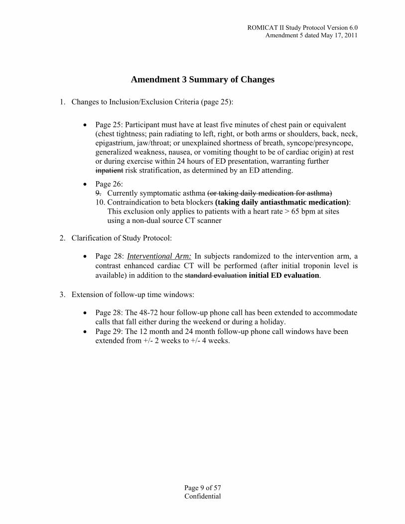
ROMICAT II Study Protocol Version 60 Amendment 5 dated May 17 2011
Study Synopsis Sponsor(s) National Heart Lung and Blood Institute (NHLBI)
Protocol Title Rule Out Myocardial IschemiaInfarction Using Computer Assisted Tomography - A Diagnostic Randomized Multicenter Trial
Diagnosis and Main Adults (ge40-75 yrs) without knowndocumented history of coronary artery Criterion for Inclusion disease who have come to the emergency department (ED) presenting
with a lead symptom of acute chest pain suggestive of acute cardiac ischemia defined as chest pain lasting for at least 5 minutes and occurring within the last 24 hours but without diagnostic ECG changes of acute myocardial ischemia are eligible for participation Only patients in whom the ED attending feels that further inpatient risk stratification is required will be included
Primary Study Objective To determine whether length of hospital stay is significantly shortened in the interventional arm vs standard of care
Secondary Study Objectives bull To determine whether time to diagnosis (for ACS and no ACS) is significantly shortened and rates of direct discharge from the emergency department are increased in the interventional arm vs standard of care
bull To determine a) the additional number of invasive coronary angiograms in both arms and b) to determine the number of coronary revascularization procedures
bull To determine the safety of immediate ED discharge after a normal cardiac CT defined as the occurrence of ACS within 48-72 hours after discharge
bull To determine whether subsequent testing and follow-up including invasive coronary angiograms diagnostic (imaging) tests interventions repeat ED visits for cardiac related problems and repeat hospitalizations for chest pain or equivalent during 28 days after hospital discharge is decreased in the interventional arm compared to SOC
bull To determine the two study arms in terms of rates of MACE within 28 days
bull To compare the two study arms in terms of rates of MACE within one and two years
bull To estimate and compare the sensitivity specificity NPV and PPV of contrast-enhanced CT and non-contrast CT with clinical diagnosis of ACS as the reference standard
bull To determine whether the cost of care (per TSI) for index hospitalization and after 28 days is decreased by implementing cardiac CT into the early ED evaluation of patients with acute chest pain
bull To determine the cost-effectiveness of incorporating cardiac CT into
Page 13 of 57 Confidential
ROMICAT II Study Protocol Version 60 Amendment 5 dated May 17 2011
the standard ED evaluation of patients with acute chest pain over a 28shyday and lifetime horizon as compared to SOC
Tertiary Study Objectives bull To determine whether CT leads to increased test burden after the initial tests during index hospitalization
bull To compare the consistency of physician decisions management depending on the results of diagnostic testing (ie normal CT ndash what fraction is immediately discharged)
bull To determine whether hospital setting availability of observation unit CT experience of hospital and readers and reader performance are associated with primary or secondary outcomes
Primary Endpoint The primary endpoint is time from ED presentation to hospital discharge The length of hospital stay is defined as the time from ED presentation to the time of discharge note or order This includes time in the ED time in any institution-specific specialized chest pain unit and time as an inpatient on the floor
Secondary Endpoints bull The time to diagnosis For ACS defined as the time from ED presentation until the first test during index hospitalization that leads to the diagnosis of ACS (tests may be biomarkers CTA ETT nuclear imaging stress ECHO or cardiac catheterization) for no ACS defined as the time from ED presentation to the final test during index hospitalization
bull Admission to hospital Participants are considered to be admitted if the ED attending admitted the subject to a hospital floor (medical floor step down unit telemetry hospital floor or intensive care unit)
bull Direct ED discharge will include all subjects who are not admitted to the hospital
bull Health care utilization number of invasive coronary angiograms diagnostic (imaging) tests interventions repeat ED visits
bull Rates of MACE major adverse cardiac events ndash cardiac death acute myocardial infarction revascularization unstable angina and rehospitalization
bull Cost of care derived from the cost-accounting database (ie TSI)
Tertiary Endpoints Increased test burden Further imaging tests performed after index hospitalization
Primary Hypothesis Adding cardiac CT to the initial ED evaluation of subjects with acute chest pain significantly decreases length of hospital stay
Page 14 of 57 Confidential
ROMICAT II Study Protocol Version 60 Amendment 5 dated May 17 2011
Secondary Hypothesis Addition of cardiac CT to the initial ED evaluation of subjects with acute chest pain will significantly increase the ED discharge rate and shorten the time to diagnosis and it will be a cost-saving and cost-effective modality There will not be an increase in the number of invasive angiograms or coronary revascularization procedures as compared to the SOC arm With a high negative predictive value patient safety will not be compromised There will not be an increase in subsequent further testing and the rate of MACE in both arms in the follow-up period will be similar The test characteristics of contrast enhanced CT will be significantly better than that of a non-contrast CT
Study Design The study is designed as a randomized diagnostic multicenter trial and will enroll 1000 participants at seven sites over a 15 month period Participants presenting with acute chest pain in the ED will be randomized to standard of care (SOC) or to an interventional arm Patients in the interventional arm will receive standard evaluation supplemented by contrast-enhanced cardiac CT imaging
Duration of Study Participants will be followed during index ED visithospitalization There Participation will be a phone call at 48-72 hours for those discharged within 24 hours
All participants will receive a follow up phone call at 28 days post emergency room discharge
Number of Subjects 1000 subjects will be enrolled with 500 each in the standard of care and interventional arms
Number of Sites 9
Statistical Considerations The primary aim of the study is to compare the two study arms in terms of a difference in mean length of hospital stay (LOS) between the two arms We have conducted a power evaluation using the estimated mean and standard deviation based on observed data from ROMICAT I and simulated data based on health outcomes We will have 95 power to detect a difference in 10 hours of LOS at a Type-1 error rate of 5 for an independent samples t-test using 500 patients in each arm
Page 15 of 57 Confidential
ROMICAT II Study Protocol Version 60 Amendment 5 dated May 17 2011
Abbreviations
ACS Acute Coronary Syndrome CABG Coronary artery bypass graft CAD Coronary Artery Disease CK-MB Creatinine kinase MB type CRC Clinical Research Coordinator CT Computed Tomography ECG Electrocardiogram ED Emergency Department ETT Exercise treadmill ECG testing MACE Major adverse cardiac event MI Myocardial Infarction NPV Negative predictive value PPV Positive predictive value SOC Standard of care SPECT Rest or stress myocardial perfusion imaging
Page 16 of 57 Confidential
ROMICAT II Study Protocol Version 60 Amendment 5 dated May 17 2011
Part I Study Overview The study is designed as a randomized controlled diagnostic multicenter trial and will enroll 1000 participants at nine sites over a 15 month period Participants presenting with acute chest pain in the ED will be randomized to a standard of care arm (SOC) or interventional arm patients in the interventional arm will receive standard evaluation supplemented by contrast-enhanced cardiac computed tomography (CT) imaging (Figure 1)
In both arms the initial Emergency Department (ED) evaluation (electrocardiogram [ECG] physical examination clinical presentation renal function lab and medical history) will determine eligibility of patients After randomization standard of care will be performed in both arms However for patients randomized to the interventional arm a CT will be performed The following decisions made by the caregiver team will be tracked admission or discharge after initial ED evaluation plus CTother initial diagnostic test in admitted participants the length of hospital stay the extent of further testing including serial cardiac markers and ECG and other diagnostic tests or interventions For safety reasons participants discharged in less than 24 hours of ED arrival will receive a phone call 48-72 hours after discharge All participants will receive a 28-day follow-up phone interview The efficiency of each strategy will be determined by comparing 1) the length of hospital stay (primary endpoint) 2) the time to diagnosis (secondary endpoint) 3) the rates of ED discharge (secondary endpoint) 4) utilization of other diagnostic testing ie invasive coronary angiograms and resulting interventions ie coronary revascularization procedures during index hospitalization and28 days (secondary endpoint) The safety of the two strategies will be compared by determining the occurrence of adverse events within 48-72 hours in directly discharged patients as well as determination of major adverse cardiovascular events (MACE) within 28 days after discharge from the index hospitalization We will also perform a cost and cost effectiveness analysis In subsequent analyses we will determine the patient physician and institutional factors that affect the primary endpoint or secondary endpoints
Primary Objective To determine whether length of hospital stay is significantly shortened in the interventional arm vs SOC
Hypothesis
Adding cardiac CT to the initial ED evaluation of subjects with acute chest pain decreases length of hospital stay
Page 17 of 57 Confidential
Secondary Endpoints
Primary Endpoint
Screening
Consent
Length of Hospital Stay
Tertiary Endpoints
Cost and Cost Effectiveness
Incremental Value of CTA over a CAC scan
Rates of Direct ED Discharge
Time to Diagnosis
No of invasive coronary angiograms and revascularizations
Rates of MACE after immediate ED discharge after 28 days
Health care utilization after 28 days
Institutional and Caregiver Characteristics associated with primary and secondary outcomes
Incremental Value of LV function over a CTA
Radiation Exposure during index hospitalization and follow up
ROMICAT II Study Protocol Version 60 Amendment 5 dated May 17 2011
Study Design
The study is designed as a randomized controlled diagnostic multicenter trial and will enroll 1000 participants at nine sites over a 15 month period
Patients with Acute Chest Pain at Low to Intermediate Risk for ACS Screening
Consent
StStandaandard ord off C Caarere CaCarrddiiaac Cc CTT RanRanddomomizizatatioionn
Length of Hospital Stay Primary Endpoint
Rates of Direct ED Discharge
Time to Diagnosis
No of invasive coronary angiograms and revascularizations
Rates of MACE after immediate ED discharge after 28 days Secondary Endpoints
Health care utilization after 28 days
Cost and Cost--Effectiveness
Institutional and Caregiver Characteristics associated with primary and secondary outcomes
Incremental Value of CTA over a CAC scan
Incremental Value of LV function over a CTA Tertiary Endpoints
Radiation Exposure during index hospitalization and follow--up
Page 18 of 57 Confidential
Patients with Acute Chest Pain at Low to Intermediate Risk for ACS
ROMICAT II Study Protocol Version 60 Amendment 5 dated May 17 2011
Part II Study Description
1 Background and Significance
11 Standard Evaluation of Participants with Acute Chest Pain Accurate triage of participants presenting with acute chest pain to the ED remains difficult because neither the chest pain history12 a single set of established biochemical markers for myocardial necrosis (troponin I troponin T creatine kinase MB-type [CK-MB])34 nor initial 12-lead ECG alone or in combination (acute cardiac ischemia time-insensitive predictive instrument) identifies a group of participants that can be safely discharged home without further diagnostic testing5ndash7 As a result the threshold to admit chest pain participants remains low and over six million are admitted annually to US hospitals28ndash10
The standard ldquorule outrdquo myocardial infarction (MI) protocol consists of serial ECG and cardiac biomarker measurements and usually requires a noninvasive stress test to rule out MI These tests (exercise treadmill ECG testing [ETT] stress echocardiography [Echo] and rest or stress myocardial perfusion imaging with Tc-99m [SPECT]) require the exclusion of myocardial necrosis with negative serial biomarkers and are performed to rule out the presence of a hemodynamically significant coronary stenosis While they have good sensitivities (ETT 76 SPECT 83 and Echo 85) for detecting the presence of significant coronary artery stenosis when compared to coronary angiography11ndash13 their specificities are only moderate (ETT 60 SPECT 64 and Echo 77) Moreover these tests are time consuming (ie SPECT 2ndash3 hours) and usually not available 247 Thus the standard evaluation of patients with chest pain to rule out myocardial ischemia requires hospital admission for 24 to 36 hours in over 90 of US hospitals14 Because of the moderate specificities of standard diagnostic testing 33 to 44 of patients with suspected acute coronary syndrome (ACS) who undergo cardiac catheterization have no significant coronary artery disease (CAD)1516 The remarkable inefficiency of current evaluation strategies is also documented by the fact that only lt10 of the six million admitted each year in the US ultimately receive a diagnosis of ACS at discharge1718 The inpatient care for the negative evaluations imparts a significant economic burden in excess of $8 billion annually19ndash21 for the US health care system
Thus an improvement of the ED triage of participants with acute chest pain particularly the increase in ED discharge rates in participants who ultimately do not have ACS would have enormous clinical and economic implications This research will determine if noninvasive cardiac CT that accurately visualizes the coronary arteries for plaques andor stenosis can be an effective strategy to facilitate early ED discharge in this patient population
12 Accuracy of Cardiac CT for Detection of Coronary Artery Disease and LV Function Advances in CT technology with an improvement of volume coverage (ge64 parallel detectors) and temporal (lt210 ms) and spatial (lt06 mm) resolution enables a nearly motion free contrast-enhanced imaging of the coronary artery tree and the heart chambers during a single short breath
Page 19 of 57 Confidential
ROMICAT II Study Protocol Version 60 Amendment 5 dated May 17 2011
hold (8ndash13 second scan)22 The data acquired during an ECG-gated contrast-enhanced multi-detector CT scan allows for 1) accurate assessment of the coronary arteries for plaque and stenosis and 2) the assessment of global and regional LV function in a cine mode
CAD Over the last several years we and others have demonstrated that cardiac CT has excellent diagnostic test characteristics for the detection of significant coronary artery stenosis as compared to invasive coronary angiography23ndash28 There is strong consensus that the strength of cardiac CT is its ability to reliably rule out the presence of stenosis and relevant coronary plaque (negative predictive value [NPV] 95 to 98 positive predictive value [PPV] 92 to 97)22
In addition cardiac CT also accurately detects the presence and extent of calcified and non-calcified coronary atherosclerotic plaque in good agreement with intravascular ultrasound (IVUS) especially in proximal coronary segments (sensitivity 84 to 92)29 30
Global and Regional LV Function Cardiac CT also permits the assessment of global regional LV function in a cine mode Studies have shown excellent reproducibility (κ=086) and agreement to cine magnetic resonance imaging (CMRI) for the detection of global and regional LV dysfunction (r=091)31ndash36
13 Accuracy of Cardiac CT to Detect ACS in Participants with Acute Chest Pain Preliminary data generated by the investigators suggest that cardiac CT using at least 64-slice technology can be performed safely and with excellent image quality in the acute care setting37
In addition our observational data in 368 patients demonstrated that the presence of significant coronary artery stenosis as assessed by cardiac CT can be definitively ruled out in 70 of participants presenting with acute chest pain to the ED (see Sections 31 and 32) This finding has a 100 NPV for ACS during index hospitalization and major adverse cardiac event (MACE) during follow-up (see Section 31)38 There are now several studies that support these results and suggest that cardiac CT has the potential to improve the management of participants with acute chest pain in the ED Rubinshtein et al in a study of 58 participants with acute chest pain but negative initial cardiac troponin levels and non-diagnostic ECG demonstrated a NPV of 100 and specificity of 92 (n=3538) for a diagnosis of ACS in the ED by cardiac CT39 Participants were discharged (n=3558) without stress testing if obstructive CAD was excluded by cardiac CT and if serial troponin measurements were negative There were no deaths or myocardial infarctions during a 15-month follow-up Earlier Goldstein et al40 randomized 200 participants at very low risk for ACS into a cardiac CT based triage system or a stress SPECT based triage system They found no or minimal CAD in 68 of patients which was a safe predictor for the absence of ACS and MACE over 6 months The study further shows the potential of cardiac CT to shorten length of hospital stay and decrease costs of evaluation of patients with acute chest pain However a potential drawback of CT was noted as the CT arm had more invasive angiograms and revascularization procedures (n=11 vs 3 and 4 vs 1 respectively) However this most likely represents that at this time the association between CT findings and clinical outcomes was unknown
In addition studies have suggested that the absence of coronary artery calcification (CAC) as detected in a non-contrast low radiation CT scan also has a high negative predictive value for
Page 20 of 57 Confidential
ROMICAT II Study Protocol Version 60 Amendment 5 dated May 17 2011
ACS in these participants4142 However the clinical utility of CAC alone for the assessment of ACS is inadequate because it cannot exclude the presence of high-grade non-calcified plaquestenosis
14 Cost Effectiveness of Diagnostic Testing in Subjects with Acute Chest Pain Current management of subjects with suspected ACS is managed by a sequential strategy (decision rule) involving quality and duration of chest pain risk factors biomarkers ECGrsquos and diagnostic tests such as SPECT echo or ETT However cost-effective utilization of these tests remains poor43 44 and hospitalization rates for ACS have increased while length of hospitalization has been constant over the last 10 years9 45 The introduction of chest pain observation units may have further decreased the threshold to admit patients Acute appendicitis and pulmonary embolism are excellent examples of the effects of the implementation of advanced imaging technology on patient management in the ED Because of the costs associated with unnecessary appendectomy and one day of in patient observation the implementation of the CT scan resulted in significant cost savings46 A similar approach is warranted to determine whether cardiac CT reduces cost and is cost effective in subjects with acute chest pain Moreover a formal model-based cost-effectiveness analysis is essential to project possible costs and benefits from a broader perspective (including the assessment of potential risks associated with CT) and longer time horizon Modeling can also help to answer questions and perform comparisons not possible within the confines of the clinical trial The current proposal critically investigates these issues
15 Significance of the Proposed Clinical Trial Evaluation of the appropriate clinical use of novel diagnostic imaging modalities is now more important than ever as the cost of these examinations steeply rises and now exceeds the cost spent on prescribed drugs All major professional medical societies have publicly demanded that clinical use of diagnostic imaging should be dictated by evidence-based medicine to not only control costs but also to minimize additional extraneous testing as it is currently unclear whether their usage will result in improvement in the current standard of care
Advanced cardiac CT is one of these novel diagnostic imaging technologies that may potentially enhance standard of care in patients presenting to the ED with acute chest pain but non-diagnostic ECG and negative initial biomarkers a population that remains diagnostically challenging and currently causes a significant clinical burden on each hospital every day Preliminary studies demonstrate that cardiac CT accurately detects absence of coronary atherosclerotic plaque Levels of atherosclerotic plaque as detected by cardiac CT is a powerful predictor of the absence of ACS and can be detected in a significant fraction of participants with acute chest pain Thus we anticipate the implementation of cardiac CT in the early ED triage process will (1) enable earlier and safe discharge of significant fraction of patients directly from the ED who currently would have been unnecessarily admitted as well as (2) allow for earlier detection and better treatment of CAD compared to the current standard of care
The proposed research will constitute the largest randomized multicenter clinical trial using cardiac CT to date and will provide an unbiased assessment of the above stated hypotheses It
Page 21 of 57 Confidential
ROMICAT II Study Protocol Version 60 Amendment 5 dated May 17 2011
will be conducted by a group of experienced clinical researchers consisting of CT imagers ED physicians and cardiologist with clinical expertise in the management of chest pain syndromes The results from this trial will determine the clinical utility of cardiac CT in patients with acute chest pain and may lead to a change in the management of these patients
2 Preliminary Data In support of the aim of the proposed research we provide data from a double blinded observational cohort study (ldquoCardiac CT for triage of participants with intermediate likelihood of ACSrdquo R01 HL080053-03) demonstrating the association between cardiac CT findings on plaque stenosis and LV function with clinical outcomes in patients with acute chest pain These data demonstrate 1) the absence of any CAD in up to 50 of patients with acute chest pain 2) the high NPV of absence of plaque and stenosis for ACS and thus the safety to discharge patients from the ED and the incremental diagnostic value of LV function assessment for risk stratification
21 Final Results of ROMICAT I
211 Cardiac CT Based Detection of Coronary Stenosis and Plaque in the Assessment of Participants with Acute Chest Pain Objective To determine the usefulness of cardiac CT angiography in patients with acute chest pain Methods Observational cohort study in chest pain patients with normal initial troponin and non-ischemic ECG 64-slice coronary CT was performed prior to admission to detect coronary plaque and stenosis (gt 50 luminal narrowing) Results were not disclosed Endpoints were ACS during index hospitalization and MACE during 6- month follow up Results Among 368 participants (mean age 53 plusmn 12 years 61 male) 31 had ACS (8) By cardiac CT 50 of these participants were free of CAD 31 had nonobstructive disease and 19 had inconclusive or positive CT for significant stenosis Sensitivity and NPV for ACS were 100 (n=183368 95 confidence interval [CI] 98ndash100) and 100 (95 CI 089ndash100) with the absence of CAD and 77 (95 CI 59ndash90) and 98 (n=300368 95 CI 95ndash99) with significant stenosis by cardiac CT Specificity of presence of plaque and stenosis for ACS were 54 (95 CI 049ndash060) and 87 (95 CI 083ndash090) respectively Only one ACS occurred in the absence of calcified plaque Both the extent of coronary plaque and presence of stenosis predicted ACS independently and incrementally to TIMI risk score (AUC 088 082 vs 063 respectively all plt00001) Conclusion Fifty percent of participants with acute chest pain and low to intermediate likelihood of ACS are free of CAD by CT and have no ACS Given the large number of such patients early cardiac CT may significantly improve patient management in the ED
Page 22 of 57 Confidential
ROMICAT II Study Protocol Version 60 Amendment 5 dated May 17 2011
Figure 1 A-C 67 year old female 3 hours of substernal chest pain radiating to the back negative initial troponin and CK-MB ECG sinus bradycardia admitted to rule out MI All coronary arteries are widely patent with no evidence of coronary atherosclerosis A Volume rendered 3-dimensional CT image (surface weighted volume rendering technique) of the heart depicting the left circumflex coronary artery (LCX arrowhead) obtuse marginal branch (dashed arrow) and left anterior descending (LAD arrow) two hours after presentation to the ED B CT based evaluation of segments 1 to 3 of the right coronary artery (RCA arrowheads) and conus branch (dashed arrow) using a 5mm thick maximum intensity projection (MIP) C Curved multiplaner reformatted (MPR) image of the LAD (arrowheads) and proximal LCX (dashed arrow)
Figure 2 A-C 40-year old male with hypercholesterolemia and a family history of diabetes woke with chest pressure and noticed intermittent chest pressure during his 1 mile walk to work He had similar symptoms a week prior to admission Initial troponin and CK-MB were negative and ECG showed a normal sinus rhythm without ST-segment or T-wave abnormalities Coronary CT was performed two hours after presentation to the ED and revealed a significant mid LAD stenosis Eight hours after ED presentation cardiac biomarkers became positive The mid LAD stenosis was confirmed on coronary angiography the next day and the participant was treated with a stent placement A Volume rendered 3-dimensional CT image of the heart depicting the right coronary artery (RCA arrow) and the left anterior descending coronary artery (LAD arrowhead) Discontinuation of the contrast-enhanced coronary lumen in the mid section of the LAD (dashed arrow) was detected B CT based evaluation of the right coronary artery (RCA arrowheads) demonstrates calcified and non-calcified plaque in segments 1 to 3 no significant coronary stenosis was detected C Curved MPR image of the LAD reveals good luminal contrast enhancement without any plaque in the distal LAD and calcified plaque in the proximal
Page 23 of 57 Confidential
ROMICAT II Study Protocol Version 60 Amendment 5 dated May 17 2011
segment (arrowheads) A significant luminal narrowing (arrow) was detected in the mid portion of the LAD
212 Incremental Diagnostic Value of Regional LV Function Over Coronary Assessment of Cardiac CT for the Detection of ACS in Participants with Acute Chest Pain Objective To determine incremental value of regional left ventricular function (LVF) over coronary assessment for the diagnosis and prediction of acute coronary (ACS) in patients with acute chest pain but inconclusive initial emergency department (ED) evaluation Methods We enrolled 356 consecutive patients (mean age 53plusmn12 years 62 male) with acute chest pain and inconclusive initial ED evaluation These patients underwent 64-slice contrast-enhanced cardiac CT prior to hospital admission Regional LVF and presence of coronary atherosclerotic plaque and significant stenosis (gt50) were separately assessed by two experienced readers blinded to the clinical course and other CT findings Caregivers and patients remained blinded to the CT results Incremental diagnostic accuracy and predictive value of regional LVF to predict ACS was determined in the entire cohort and in subgroups of patients with inconclusive or positive coronary assessment Results Regional LVF was impaired in 46 patients (129) and 31 patients developed ACS (87) Adding regional LVF resulted in a 10 increase in sensitivity to detect ACS (87 95CI 70shy96) when compared to detection of significant stenosis In the subgroup of 33 patients with significant stenosis impaired regional LVF correctly predicted ACS in 1719 patients (PPV 895 95CI 67-99) In the subgroup of patients with inconclusive coronary CTA (n=33) normal regional LVF correctly predicted the absence of ACS in 2426 patients (NPV 923 95CI 75-99) Impaired regional LVF independent of the extent of plaque and the presence of stenosis was associated with a 25 and 20 fold increased risk for ACS (RR 2023 95CI 747 to 5482 and RR 2534 95CI 936 to 6857 respectively) Moreover c-statistics improved significantly after the addition of regional LVF to both the extent of plaque and the presence of stenosis (c-statistic 088 vs 094 and 082 vs 090 respectively both plt003) Conclusions Regional LVF assessment at rest improves overall sensitivity of coronary CTA for ACS in patients with acute chest pain Combined assessment of coronary morphology and regional LVF is especially helpful to guide further testing and interventions if coronary assessment is inconclusive or positive
213 Initial Decision Model to Assess the Cost Effectiveness of Coronary CT in Triage of Subjects with Acute Chest Pain Objective To develop a preliminary decision-analytic Markov model to predict the long-term consequences of four competing strategies 47 in the ED evaluation of subjects with acute chest pain Methods The model consists of the six health states chest pain healthy early heart disease undiagnosed CAD diagnosed CAD and death Subjects start in the chest pain state The probability that the chest pain is caused by CAD depends on the underlying risk of CAD as
Page 24 of 57 Confidential
ROMICAT II Study Protocol Version 60 Amendment 5 dated May 17 2011
determined by age gender and cardiovascular risk factors As simulated subjects progress through the model CAD (if present) can remain stable or progress Subjects may die from cardiovascular disease or other causes at rates determined by disease stage and demographics By simulating hypothetical cohorts of subjects we determined quality-adjusted life expectancy (QALE) quality-adjusted life year (QALY) and cost-effectiveness ratios for competing strategies We considered four competing strategies for subjects presenting with chest pain to the ED 1) admission in which all subjects are admitted to the hospital for further evaluation 2) discharge in which all subjects are discharged directly home from the ED 3) cardiac CT with conservative treatment in which all subjects without any evidence of CAD are discharged and all other patients are admitted and undergo stress testing and 4) cardiac CT with aggressive treatment which is similar to strategy 3 but sends subjects with evidence of significant stenosis directly for invasive coronary angiography It is important to note that the model permits an evaluation of each strategy using a similar cohort of subjects The model also permits an evaluation of estimate life expectancy and lifetime costs Results Strategies involving cardiac CT were associated with lower ED costs and incremental improvements in QALE when compared to a strategy of admission for all subjects Relative to admitting all subjects the use of cardiac CT yielded savings in ED and hospital costs ranging from $1800-$2200 in men and women ages 30-40 and from $500-$1200 men and women ages 60-70 respectively The incremental cost-effectiveness of strategies involving cardiac CT compared with a strategy of discharging all subjects ranged from $94100-$267200 per QALY in men and $74800-$326100 per QALY in women Conclusions This preliminary decision model suggests that strategies involving cardiac CT are associated with lower ED costs and incremental improvements in QALE in both men and women across all ages The precision of the model will be greatly enhanced by input of real clinical scenarios derived from this proposal
3 Study ObjectiveSpecific Aims This primary objective of this protocol is to assess the efficiency of implementing cardiac CT into the diagnostic workup of patients with acute chest pain normal initial biomarkers and normal or non-diagnostic ECGs Study participants will be patients with acute chest pain and low to intermediate likelihood of ACS and will be randomly assigned to the SOC arm or the interventional arm patients in the interventional arm will receive the standard of care supplemented by cardiac CT
31 Primary Aim The primary aim is to determine whether length of hospital stay is significantly reduced in the interventional arm compared to standard of care The primary endpoint is time from ED presentation to hospital discharge
The length of hospital stay is defined as the time from ED presentation to the time of discharge note or order This includes time in the ED time in any institution-specific
Page 25 of 57 Confidential
ROMICAT II Study Protocol Version 60 Amendment 5 dated May 17 2011
specialized chest pain unit and time as an inpatient on the floor All time sequences must be documented and reported to comprise total length of hospital stay Hypothesis Adding cardiac CT to the initial ED evaluation of subjects with acute chest pain significantly decreases length of hospital stay
32 Secondary Aims 1 To determine whether time to diagnosis is significantly shortened and rates of direct
discharge from ED are increased in the interventional arm vs SOC
A) Time to Diagnosis For ACS Defined as the time from ED presentation until the first test during index hospitalization that leads to the diagnosis of ACS (tests may be biomarkers CTA ETT nuclear imaging stress ECHO or cardiac catheterization) For no ACS Defined as the time from ED presentation defined as the final test during index hospitalization
B) Admission to Hospital Participants are considered to be admitted if the ED attending admitted the subject to a hospital floor (medical floor step down unit telemetry hospital floor or intensive care unit)
Direct ED Discharge will include all subjects who are not included in (B)
Hypothesis ED evaluation of subjects with acute chest pain supplemented by cardiac CT significantly increases direct ED discharge rates and shortens time to diagnosis for both ACS positive and negative patients
2 To determine a) the additional number of invasive coronary angiograms both arms and b) to determine the number of coronary revascularization procedures
Hypothesis ED evaluation of subjects with acute chest pain supplemented by cardiac CT will not increase the number of invasive coronary angiograms without revascularization or revascularizations during index hospitalization
3 To determine the safety of immediate ED discharge after a normal cardiac CT defined as the occurrence of ACS within 48-72 hours after discharge
Hypothesis CTA has a high negative predictive value based on which immediate ED discharge should not compromise patient safety
4 To determine whether subsequent testing and follow up including invasive coronary angiograms diagnostic (imaging) tests interventions repeat ED visits for cardiac related problems and repeat hospitalizations for chest pain or equivalent during 28 days after hospital discharge is decreased in the interventional arm compared to SOC
Hypothesis Incorporation of CTA in the evaluation of acute chest pain patients in the ED will not lead to decreased further testing to manage chest pain when as compared to the SOC arm
5 To compare the two study arms in terms of rates of MACE (major adverse cardiac eventsmdashcardiac death AMI revascularization unstable angina and rehospitalization) within 28 days
28 Days Defined as 28 days post the day of discharge from index hospitalization
Page 26 of 57 Confidential
ROMICAT II Study Protocol Version 60 Amendment 5 dated May 17 2011
Hypothesis The rates of MACE in both arms during the 28 day follow up period will be similar
6 To estimate and compare the sensitivity specificity NPV and PPV of contrast-enhanced CT and non-contrast CT with clinical diagnosis of ACS as the reference standard
Hypothesis The test characteristics of contrast enhanced CT will be significantly better than those of a non-contrast CT for the diagnosis of ACS
7 Determine whether cost of care (per TSI) for index hospitalization and after 28 days is decreased by implementing cardiac CT into the early ED evaluation of patients with acute chest pain
Hypothesis Incorporation of cardiac CT into standard ED evaluation of patients with acute chest pain will decrease the cost of care
8 Determine the cost-effectiveness of incorporating cardiac CT into the standard ED evaluation of patients with acute chest pain over a 28-day and lifetime horizon as compared to SOC
Hypothesis Incorporating cardiac CT into the standard evaluation of patients with acute chest pain will be cost-saving and cost-effective over a 28-day and lifetime horizon
33 Tertiary Aims 1 To determine whether CT leads to increased test burden after the initial tests during index
hospitalization
Hypothesis Cardiac CT may diagnose coronary artery disease at a more incipient stage than determined by imaging tests in the SOC arm which may increase test burden during the follow up period
2 Compare the consistency of physician decisions management depending on the results of diagnostic testing (ie normal CT - what fraction is immediately discharged)
Hypothesis There will be comparable consistency between the two arms
3 Determine whether hospital setting availability of Observation Unit CT experience of hospital and readers and reader performance are associated with primary or secondary outcomes
Hypothesis Enhanced CT experience of the hospital and readers better reader performance during CT certification and hospital setting will be associated with improved primary or secondary outcomes
4 Participant SelectionEligibility Criteria
Adult ED participants presenting with a lead symptom of acute chest pain suggestive of acute cardiac ischemia - defined as chest pain lasting for at least 5 minutes and occurring within the last 24 hours but without diagnostic ECG changes of acute myocardial ischemia - will be eligible for participation
Page 27 of 57 Confidential
ROMICAT II Study Protocol Version 60 Amendment 5 dated May 17 2011
Only patients in whom the ED attending feels that further inpatient testing is required will be included Patients with knowndocumented history of CAD will not be included since such patients most often have an abnormal or non-diagnostic cardiac CT In addition men and women younger than 40 years will be excluded due to the low pre-test likelihood for CAD and the increased radiation risk Detailed inclusion and exclusion criteria are presented below
41 Inclusion Criteria 1 Participant must have at least five minutes of chest pain or equivalent (chest tightness
pain radiating to left right or both arms or shoulders back neck epigastrium jawthroat or unexplained shortness of breath syncopepresyncope generalized weakness nausea or vomiting thought to be of cardiac origin) at rest or during exercise within 24 hours of ED presentation warranting further risk stratification as determined by an ED attending
2 Participant must be able to provide a written informed consent
3 Participants must be lt75 years of age but gt40 years of age
4 Participant must be able to hold breath for at least 10 seconds
5 Participant must be in sinus rhythm
42 Exclusion Criteria 1 New diagnostic ischemic ECG changes (ST-segment elevation or depression gt 1 mm or
T-wave inversion gt 4 mm) in more than two anatomically adjacent leads or left bundle branch block
2 Documented or self-reported history of CAD (MI percutaneous coronary interventions [PCIs] coronary artery bypass graft [CABG] known significant coronary stenosis [gt50])
3 Greater than 6 hours since presentation to ED to time of consent
4 BMI gt40 kgm2
5 Impaired renal function as defined by local standard of care - for example measured serum creatinine gt15 mgdL
6 Markedly elevated troponin as defined by local standard of care
7 Hemodynamically or clinically unstable condition (BP systolic lt 80 mm Hg atrial or ventricular arrhythmias persistent chest pain despite adequate therapy)
8 Known allergy to iodinated contrast agent
9 Currently symptomatic asthma
10 Documented or self-reported cocaine use within the past 48 hours (acute)
11 On Metformin therapy and unable or unwilling to discontinue for 48 hours after the CT scan
Page 28 of 57 Confidential
ROMICAT II Study Protocol Version 60 Amendment 5 dated May 17 2011
12 Contraindication to beta blockers (taking daily antiasthmatic medication) This exclusion only applies to patients with a heart rate gt 65 bpm at sites using a non-dual source CT scanner
13 Participant with no telephone or cell phone numbers (preventing follow-up)
14 Participant with positive pregnancy test Women of childbearing potential defined as lt2 years of menopause in the absence of hysterectomy or tube ligation must have a pregnancy test performed within 24 hours before the CT scan
15 Participant unwilling to provide a written informed consent
43 Recruitment Screening and Consent
The research team at each participating site includes the ED attending physician CT technologist and research associate(s) The investigator and the research staff will be responsible for the screening review of participant medical records and investigator-designated data submission All clinical sites will perform screening Monday through Friday during daytime hours (9am-5pm) Potential subjects will be identified by research coordinatorstudy nurse staff shortly after admission to the ED usually after the initial ED evaluation consisting of ECG physical examination clinical symptoms and presentation immediate and past medical history has been concluded and screened for eligibility based on a checklist providing inclusion and exclusion criteria
Eligible subjects will be approached and if the subject considers participation the Clinical Research Coordinator (CRC) will describe the rationale of the study the study procedures with associated risks and benefits A study physicianstudy nurseCRC will speak directly with an ED physician caring for the patient to obtain an assessment of pretest probability for ACS and will verify that the ED physician feels that further observation or diagnostic testing is warranted in this patient
If the subject agrees and further testing or observation is planned as SOC key study personnel typically a study physician or the attending ED physician will review the study procedures as well as all potential risks and discomforts and the opportunity to decline or cease participation in the study at any time answer any remaining questions and consent the subject
Patients can be consented but not randomized before the first troponin test result is available Patients whose initial troponin level is markedly elevated will not be randomized and will not be included in the intent-to-treat population Patients whose subsequent troponin testing is markedly elevated will be included in the intent to treat population
Patients can be consented before a pregnancy test is available However patients with a positive pregnancy test will not be randomized
Patients who are not randomized will not be considered part of the intent to treat population
Informed Consent
Page 29 of 57 Confidential
ROMICAT II Study Protocol Version 60 Amendment 5 dated May 17 2011
bull A potential subject will then be afforded as much time as clinically feasible to review the consent form and decide upon their participation without adversely effecting their ED care which is typically 1 hour
bull If the ED staff caring for the patient determines that the potential subjectrsquos previously determined plan of care must proceed before the potential subject has had adequate time to decide upon hisher participation without undue pressure the potential subject will be excluded from enrollment
bull Subjects will be provided with a copy of the informed consent form
44 Randomization
After the patient has been consented by the study physician the study subject will be randomized into the standard care arm or the intervention arm (cardiac CT) by means randomization stratified by institution using a central computerized randomization system
5 Study Procedures
51 Index Hospitalization Standard of Care Arm Subjects will be evaluated according to each hospitalrsquos specific protocol to evaluate and manage patients with acute chest pain Typically the standard evaluation in the ED will include the past and current medical history a physical examination an ECG and a cardiac biomarker (Troponin and CK-MB) as well as other routinely obtained blood testing Patients may undergo cardiac CT as part of SOC but only as a secondary diagnostic test Patients in the CT arm may undergo further diagnostic testing as well
All admitted subjects will undergo each hospitalrsquos standard rule out myocardial ischemia protocol This protocol typically consists of observation and monitoring including serial ECGs and repeated cardiac biomarker measurements as well as a noninvasive stress test (often imaging based) to evaluate for myocardial ischemia The participating clinical sites perform routinely either nuclear perfusion imaging [SPECT] at rest and stress andor stress echocardiography andor exercise treadmill test [ETT] Depending on the results subjects may undergo additional noninvasive or invasive testing (coronary angiography) andor coronary revascularization during their hospital stay
Interventional Arm In subjects randomized to the intervention arm a contrast enhanced cardiac CT will be performed (after initial troponin level is available) in addition to the initial ED evaluation The ED physicians will resume care of the patient and make all clinical decisions for further care of the patient (eg need for further evaluation or admission) based upon their cumulative clinical assessment of the patient including findings revealed on the CT scan
Page 30 of 57 Confidential
ROMICAT II Study Protocol Version 60 Amendment 5 dated May 17 2011
In addition in patients from both study arms who consented to a blood draw we will obtain three fifteen (15) mL venous blood samples over a six-hour period less than forty-five (45) mL in total Blood samples will be centrifuged and frozen for future analysis for cardiac-related markers
All diagnostic tests will be performed per Society Guidelines Cardiac Computed Tomography is performed in accordance with best practice standards as delineated in the imaging guidelines of the Society of Cardiovascular Computed Tomography by competent63 and appropriately credentialed physicians This includes the optimization of the scan protocol to limit radiation exposure
52 48-72 Hour Follow up after ED discharge Follow-up phone call at 48-72 hours Subjects who are discharged from the ED within 24 hours of presentation (for both randomization arms) will be contacted via telephone between 48 and 72 hours after discharge to avoid ascertainment bias During this call subjects will be interviewed to determine whether they have had any recurrent symptoms suggestive of myocardial ischemia and andor whether they needed to seek further medical attention following their emergency department visit If these phone attempts at 48 and 72 hours are not successful a final attempt will be made at 96 hours to contact the subject
NOTE If a subject is discharged on a Thursday the 48-72 hour phone call may be extended to 96 hours In the rare circumstance that a subject is discharged on a Thursday and the following Monday is a holiday the follow-up call can be made during the next working business day
53 28 Day Follow up Follow up 28 day phone call All subjects will be interviewed via a telephone call 28 days post ED discharge (+4 weeks) to determine the occurrence of MACE and healthcare utilization using a standardized questionnaire All cases of recurrent chest pain hospital admissions and diagnostic testing will be verified by review of medical records
A reminder letter will be sent or phone call will be made to participants two weeks prior to the 28-day follow up phone call If unable to contact the participant by phone mortality will be assessed online using the SSDI website
Page 31 of 57 Confidential
ROMICAT II Study Protocol Version 60 Amendment 5 dated May 17 2011
6 Data Collection The following subject data will be collected during index ED visithospitalization and follow up phone interviews bull Subject demographic information bull Medical history including cardiac risk factors and medications bull Description of recent pain episodes bull Vitals signs ECG cardiac biomarker results bull Cardiac CT imaging and other cardiac-related diagnostic testing data bull Adverse events
7 Statistical Considerations A total of 1000 participants will be enrolled in this study Enrollment is planned to be completed in 15 months
71 Primary Aim To compare the two study arms in terms of the time from ED presentation to hospital discharge The primary aim of the study is to compare the two study arms in terms of a difference in mean length of hospital stay (LOS) between the two arms Based on the results of ROMICAT I we estimate the mean (plusmn standard deviation) hospital LOS (including ED) of these patients in standard evaluation to be 405 plusmn 432 hours The observed frequency distribution appeared to fit well to a log-normal distribution (Median was 77 hours and Range was 275 ndash 3815 hours) We anticipate that the observed distributions of both Group A and Group B follow such a pattern Because the independent samples t-test is robust to a departure from the data normality when applied to a large sample size we will employ the t-test for this inference
We have conducted a power evaluation using the estimated mean and standard deviation based on ROMICAT I for Group A and using the estimated the mean and standard deviation based on a simulated data for Group B The simulated data consisted of 500 LOS values resulted as a mixture of three subsets randomly drawn from log-normal distributions of which the first subgroup was to represent the patients with normal LV function and without CAD (486 Mean plusmn SD of 6 plusmn 12 hours) the second was to represent the patients with normal LV function and with non-obstructive CAD (288 Mean plusmn SD of 10 plusmn 20 hours) and the third group was to represent all other possible conditions (226 Mean plusmn SD of 579 plusmn 604 hours which was estimated from ROMICAT 1) We also incorporated in the possibility that some patients without CAD will not be correctly triaged The triage accuracy rate in the following table is the rate at which a patient without CAD would be correctly triaged The following table summarizes for each ED triage accuracy rate assumption the estimated mean LOS and the power at a Type-1 error rate of 5 for an independent samples t-test using 500 patients in each arm The proposed sample size will attain a sufficient power to detect a clinically meaningful difference in mean LOS between the two groups even in the presence some degree of ED triage inaccuracy
Page 32 of 57 Confidential
ROMICAT II Study Protocol Version 60 Amendment 5 dated May 17 2011
Powers to Detect Estimated Differences in Mean LOS values at Assumed ED Triage Accuracy Rates (Type-1 error rate = 005)
Assumed ED Triage Accuracy Rate
Estimated Mean (plusmn SD) LOS Estimated Difference in Mean (hours) Power
Group A (Standard Eval)
N=500
Group B (CT Eval)
N=500 65 405 (plusmn 432) 328 (plusmn 467) -83 83 70 405 (plusmn 432) 305 (plusmn 455) -101 95 80 405 (plusmn 432) 263 (plusmn 417) -142 99 90 405 (plusmn 432) 224 (plusmn 373) -181 gt99
100 405 (plusmn 432) 188 (plusmn 335) -217 gt99
72 Secondary Aims To compare the two study arms in terms of time to diagnosis and rates of direct discharge The time to discharge (defined as time from ED presentation until discharge to home) will be plotted using Kaplan-Meier curves because some patients will be in the hospital at 30 days post day of presentationrandomization The curves for the two groups will be compared using a test based on the difference in the average length of stay in the two groups (truncated at 30 days) divided by the standard error of this difference60 We choose this test because the average length of stay is most closely associated with cost It will be also instructive to compare the curves themselves as they will provide information on the effect of cardiac CT on different groups of patients For instance one group of patients those who are discharged due to cardiac CT may have a shorter length of stay while others may have a longer length of stay due to waiting for the results of the CT or other procedures that the CT indicates
Sample Size The planned sample size will also provide adequate power to detect the rates of direct discharge from ED Screening data from a site survey of our clinical sites as well as published data indicate that the standard ED discharge rate is approximately 15 in the target population According to our preliminary data and data from other groups we expect that 40 to 50 of participants have no detectable CAD by cardiac CT The negative predictive value of this finding for ACS has been shown to be 100 Our guidelines suggest that these participants will be discharged directly from the ED However realistically we expect that because of administrative issues and remaining individual variation of the decision to discharge depending on the clinical presentation of participants actually only 60 to 80 of these participants will be discharged Thus anywhere from 24 to 40 participants could be discharged directly from the ED in the interventional arm For the sample size analysis we assume that 32 of participants will be discharged
Based on the aforementioned key assumptions for the Standard Evaluation we anticipate that 15 of all participants will be discharged and 85 will be admitted (discharge rate 15) For the Cardiac CT Evaluation we assume a discharge rate of 32 For the objective of superiority
Page 33 of 57 Confidential
ROMICAT II Study Protocol Version 60 Amendment 5 dated May 17 2011
of discharge rates we will have a 100 chance at a significance level of 005 to detect a 17 increase in discharge rates from 15 to 32
The comparison of time to discharge among patients who are admitted to the hospital is complicated by the fact that the two groups will not be the same group of patients For instance patients who would have been discharged in the cardiac CT group but admitted in the usual care group may have a relatively shorter time post admission than patients who would have been admitted in both groups We have developed methods for analyzing these types of data using causal inference that will be applied to this analysis61 A similar analysis will be applied to other subgroups such as those with and without ACS
To compare the two study arms in terms of cardiac health care utilization during the index visit Health care utilization components will include invasive coronary angiograms and coronary revascularization
The analysis for this secondary aim will be performed from the intent to treat perspective Health care utilization will be assessed using medical record review Measures of key components of utilization will be compared across the two study groups using procedures appropriate for each measure (rate or count)
In particular the two arms will be compared in terms of the number of invasive coronary angiograms This secondary endpoint will be analyzed in terms of a non-inferiority trial In a non-inferiority trial the null hypothesis is that cardiac CT is worse than usual care in that the rate of coronary angiograms is increased and the alternative hypothesis is that it is not in worse and the rate of angiograms is the same The reason for reversing the usual null and alternative hypothesis is that the purpose of this analysis is to prove that cardiac CT is not worse than usual care this would not be accomplished by just showing that the difference between the two was not significant
Screening data from a survey of clinical sites as well as published data indicate that the standard rate of invasive coronary angiograms in the target population is approximately 10 According to our study guidelines we assume that most but not all participants with significant stenosis in CT (10 of participants) undergo invasive coronary angiography (notably those who also have a positive stress test and those who have high grade proximal stenosis or LAD stenosis) In addition we assume that a minority of participants with indeterminate stenosis (lt5) and positive stress testing undergo invasive coronary angiography Because of the moderate specificities of standard diagnostic testing up to 40 of participants who undergo cardiac catheterization have no significant CAD1516 Because the diagnostic accuracy of cardiac CT for the detection of coronary stenosis is comparable or better than standard diagnostic testing we believe that there will be no increase in the rates of invasive coronary angiograms If we define a 5 difference as an ldquoacceptablerdquo difference in cardiac catheterization rates than we will have an 84 chance of showing that the CT arm is significantly less than 5 worse than the standard arm at a one sided p=005 significance level
Page 34 of 57 Confidential
ROMICAT II Study Protocol Version 60 Amendment 5 dated May 17 2011
Another key measure of utilization is the number of coronary revascularization procedures In ROMICAT I there were 18 revascularization procedures performed in 368 patients (5) Thus we expect 5 procedures in the standard evaluation If we assume that not all participants with significant stenosis will undergo coronary revascularization (because if no MI it can be medically managed) and that cardiac CT may also avoid some angiograms by excluding disease than we can realistically assume that there is not a difference between the two study arms If we consider this in terms of a non-inferiority trial as above we will have 80 power to rule a difference of 35
To compare the two study arms in terms of cardiac health care utilization within one year after index hospitalization Health care utilization components will include invasive coronary angiograms cardiac related diagnostic and lab testing and ED visits
This analysis will be performed from the intent to treat perspective CP and CT related health care utilization will be assessed using medical record review Measures of key components of utilization will be compared across the two study groups using procedures appropriate for each measure (rate or count) We expect that the rate of additional cardiac work-up will be 29 in the SOC arm and will be 14 in the interventional arm We will have gt95 power to detect this difference We expect that the rate of hospital admissions with additional diagnostic cardiac testing will be 93 in the SOC arm and will be 4 in the interventional arm We will have gt95 power to detect this difference We expect that the rates of these measures will be associated with the CT findings at index hospitalization
To evaluate the cost and cost-effectiveness of incorporating cardiac CT into the standard ED evaluation of patients with acute chest pain These analyses will be performed by the Decision Analysis and Cost-Effectiveness Center (DACE) Therefore the sites will collect and provide the necessary data to the DACE and the costs will be derived from the cost-accounting database (TSI) As a first step incorporating cardiac CT into the standard evaluation of patients with acute chest pain will be tested for cost-saving over a 28-day and lifetime span as compared to standard care Further on cost-effectiveness of incorporating cardiac CT into the standard ED evaluation of patients with acute chest pain will be determined using first-order Monte Carlo microsimulations
To compare the two study arms in terms of rates of MACE (cardiac death AMI revascularization unstable angina and rehospitalization) within 28 days The analysis for this secondary aim will be performed from the intent to treat perspective The occurrence of MACE will be assessed via patient follow-up and medical chart review The primary analysis for this aim will be performed at the patient level by determining whether any MACE occurred or not Event rates will be estimated and compared between study groups using exact procedures In a secondary analysis the rates of each type of MACE will be examined separately
Sample Size- Safety Considerations- Missed ACS Mistaken discharges are the primary safety issue in this study All available data indicate that the absence of any detectable CAD (absence
Page 35 of 57 Confidential
ROMICAT II Study Protocol Version 60 Amendment 5 dated May 17 2011
of any plaque and stenosis) has a very high NPV for ACS (100 or close to 100)62 Thus we anticipate that the occurrence of ACS in participants discharged home directly from the ED in the interventional arm will be also minimal or even zero and similar to the standard of care If as demonstrated in ROMICAT I none of the participants without CAD will have MACE defined as ACS within 30 days after direct ED discharge this observation has 90 power to ensure that the true event rate is less than 1 These assumptions are supported by the results of the ERASE trial which reported similar and very minimal event rates in each group (only 2 pts with acute MI were mistakenly discharged 1 in each randomization group out of 1200 [event rate 00008]) In this trial we would have an 80 chance of detecting at least one occurrence of an inappropriately discharged participant if the true rate where over 1
To estimate and compare the sensitivity specificity NPV and PPV of contrast-enhanced CT and non-contrast CT with clinical diagnosis of ACS as the reference standard The analysis for this aim will utilize data on participants randomized to Group B In this arm each participant will undergo non-contrast CT and contrast-enhanced CT which will be interpreted as positive or negative for ACS Clinical determination of presence or absence of ACS will be used as the reference standard For each of the two types of CT we will estimate the sensitivity specificity and positive and negative predictive value of the modality for ACS Each measure of diagnostic or predictive performance will be compared between the two modalities using statistical techniques that account for the correlation due to the paired test design In addition to these measures we will estimate the proportion of cases with a clinical diagnosis of ACS and a negative non-contrast CT in which contrasted enhanced CT was positive for ACS This proportion provides a measure of the incremental value of contrast-enhanced CT for diagnosis ACS
73 Tertiary Aims To determine whether CT leads to increased test burden after the initial tests during index hospitalization
To compare the consistency of physician decision management depending on the results of the diagnostic testing (ie normal CT ndash what fraction is immediately discharged)
To determine whether hospital setting availability of Observation Unit CT experience of hospital and readers and reader performance are associated with primary or secondary outcomes
Analysis of Data on Women and Minorities We will analyze the effect of the intervention on women and minorities as outlined in the NIH guidelines Thus we include testing for gender-treatment and minority status-treatment interactions with the discharge rate and the angiogram rate
Page 36 of 57 Confidential
ROMICAT II Study Protocol Version 60 Amendment 5 dated May 17 2011
8 Data Entry and Site Monitoring
81 Data Collection Subject data will be collected by the research associates and paper records will be maintained at individual clinical sites The Data Coordinating and Statistical Center (DCSC) will develop a web based electronic case report form The de-identified study data will be entered into this electronic data capture system (EDC) and sent to the Data Coordinating and Statistical Center (DCSC) at Massachusetts General Hospital for further analysis
82 Site Monitoring Site visits will be performed on a regular basis by the Data Coordinating Center to ensure that all regulatory requirements are being met and to monitor the quality of the data collected Records of Institutional Review Board approvals and patientsrsquo charts will be examined on a spot check basis to evaluate the accuracy of the data entered into the database
9 Risk and Benefit Assessment
91 Risks of Nonionic Contrast Agent
Rare risks of the nonionic contrast agent bull Itchiness bull Rash bull Mild allergic reaction
Very rare risks of the nonionic contrast agent bull Kidney diseaseContrast-induced nephropathy bull Death
Regarding the risks of contrast material the rate of adverse reactions with nonionic contrast reagents is 0250 More than 90 of such adverse reactions are allergic-like very mild (itching rush) and can be very effectively treated with available drugs (ie antihistaminic) Severe reactions occur in 002 and one death occurred in 57739 injections of nonionic contrast material50
Contrast-induced nephropathy (CIN) CIN is expected to be a very rare event among this patient population because patients with already impaired renal function (the strongest predictor of CIN) will not be enrolled) This assumption is strongly supported by our preliminary experience from ROMICAT I In this study none of the participants had such an event (0368)
The very low risk associated with contrast administration has been documented in large scale trials previously In a randomized trial in 1196 participants the incidence rate of CIN defined as an increase in serum creatinine ge1mgdL from baseline within 48 to 72 hours following the injection of contrast material was 0 in 359 participants (0) who had no renal insufficiency and no diabetes melliltus51 The risk was 2 in 315 (06) in participants with no renal insufficiency but who had diabetes mellitus51 In this study the mean volume of contrast-agent administered
Page 37 of 57 Confidential
ROMICAT II Study Protocol Version 60 Amendment 5 dated May 17 2011
was 140 plusmn 57 mL which is much higher than the 80 mL of contrast that is administered for a coronary CTA The risk of CIN may increase if participants in the cardiac interventional arm will be admitted and subsequently undergo further contrast-related diagnostic testing However among 209 participants who had both a pulmonary angiogram and a contrast-enhanced spiral CT in the Prospective Investigation of Pulmonary Embolism Diagnosis II only one participant with diabetes mellitus had a transient episode of acute renal failure characterized by an increase in the serum creatinine level from 13 to 29 mgdL52 In this study the contrast-enhanced CT and pulmonary angiogram were performed within 22 hours The elevated creatinine returned to normal after administration of intravenous fluids Thus overall we expect that the occurrence of CIN will be a rare event in this study
92 Risks Associated With Radiation While the radiation dosage for CT scanning varies with the part of the body being scanned the exposure for this examination is approximately between 7 and 17 mSv This exposure is comparable or less than stress nuclear myocardial perfusion imaging (14ndash22 mSv) (personal communication with Prof Ronald Callahan Chairman of the MGH Radiation Safety Committee) In comparison the average yearly effective dose of natural background radiation is 25 mSv53 the allowed annual exposure of radiation workers is 50 mSv the allowed exposure in five years is 100 mSv (Table 3)54 Thus even if an enrolled participant would undergo cardiac CT followed by a conventional stress test and selective coronary angiography the participant would be well below the allowable annual effective dose for radiation workers The radiation dose from cardiac CT has not been shown to have any adverse effects
Radiation Exposure
Cardiac CT scanning results in a measurable radiation exposure55 In order to minimize risk for participants in this study we have implemented the following measures
bull Exclude women lt40 years of age bull Exclude pregnant and breast feeding women bull Include only women who have a negative pregnancy test bull Application of cardiac CT protocols with the goal to minimize radiation exposure (ie
use of tube modulationmdashdecrease of radiation exposure by 30 to 50 and prospective triggering in appropriate conditions)
bull Review and approval of these CT protocols by the CVCT Core Lab
General Risk Associated with Radiation Exposure
The risk of inducing a malignancy by exposure to radiation is estimated to be 5 cases of malignancy for each 100 individuals exposed to an effective dose of 1 Sv54 The risk is estimated to be higher in infants and children and lower in the elderly The data which these estimates are based on are weak and apply to relatively high levels of radiation exposure56 and it remains unclear whether there is a linear relation in lower levels of radiation Assuming a linear proportion with a low level of radiation exposure to 1 mSv would lead to 5 malignancies in 100000 individuals exposed and a dose of 8 mSV would lead to 40 malignancies in 100000 individuals56 Small amounts of radiation exposure less than 100 mSv have not been clearly
Page 38 of 57 Confidential
ROMICAT II Study Protocol Version 60 Amendment 5 dated May 17 2011
shown to have harmful effects The amount of radiation to which patients will be exposed in this study is less than the amount that is known to cause a measurable harmful effect Even so it is generally assumed that a small amount of radiation will have a small chance of producing a harmful effect These extrapolated risks of the effects of small amounts of radiation are probably overestimates because the estimates ignore the fact that the body can repair the damage done by radiation if the repair mechanisms are not overwhelmed Radiation is a well-known teratogenic agent and the developing fetus is much more sensitive to radiation than the mother
Table 3 Estimated radiation exposure for diagnostic tests background radiation and exposure limits
Examination Effective Dose (mSv) Chest PA and lateral 007 Myocardial Perfusion Imaging (Rest and 14ndash22Stress) Thorax CT for the Exclusion of Pulmonary 7-12Embolism 64-Slice CT coronary angiogram 7ndash17 2-view mammogram 30 Natural yearly background radiation 25 Max exposure in radiation workers 50
Lifetime Cancer Risk Attributable to Cardiac CT
The absolute cancer risk has been described recently and recommendations have been made57
The life time cancer risk from cardiac CT decreases with age and is higher in women than in men in particular exposure of younger women should be avoided In our study these recommendations have been incorporated In comparison the absolute cancer risk for women to develop breast cancer is 13 and for men to develop colon cancer is 7 the risk attributed to a cardiac CT scan is 1466 vs 1715 for a 60 year old women and 11241 vs 11911 for 60 year old men (for CT without and with tube modulation (female 21 and 14 mSv male 15 and 9 mSv respectively)57 Thus the incremental risk induced by cardiac CT compared to the overall risk of the population to develop cancer seems moderate
Effect of the Implementation of Cardiac CT in the ED Evaluation of Patients with Acute Chest Pain on a Population Basis
The number of CT scans performed in the US was 62 million in 200658 Imaging of the abdomen (50) and the head (33) were major single contributors Specific concern was given to the rise of pediatric CT imaging (from 6 to now 11) and screening the adult asymptomatic population (ie lung heart) Implementation of cardiac CT in the ED could lead to a small increase of the number of overall CT procedures However the effects of the implementation of cardiac CT in the ED would be minimal if 1) other procedures associated with radiation doses would be saved 2) repeated testing currently performed in 25 of patients would be minimized and 3) rates of unnecessary purely diagnostic cardiac catheterizations would
Page 39 of 57 Confidential
ROMICAT II Study Protocol Version 60 Amendment 5 dated May 17 2011
decrease Furthermore already cardiac CT scanning could be performed using single phase acquisition with prospective triggering which would decrease the dose to lt5 mSv In addition MD over-read will be performed for internal quality control assessment of findings related to CAD and significant cardiac findings such as pulmonary embolism and aortic dissection to increase awareness to sites of non-coronary findings
93 Other Risks Additional Risks of CT Scans bull Discomfort bull Claustrophobia
Risks of IV Needle Placement
bull Hemorrhage (hematoma at the injection site) bull Infection (catheter related infection) at the injection site bull Minor discomfort bull Bleeding bull Infection bull Bruising
94 Potential Benefits bull Subjects may benefit from randomization into the interventional arm of trial from detection
of pathology that may not have been otherwise detected during SOC (eg a non-significant coronary stenosis or non-obstructive coronary artery disease that would likely not have been detected by a stress test)
bull Data from prior studies indicate that patients in the interventional arm may be earlier discharged and may not have to spend the night in the hospital there is also the chance that alternative diagnosis such as pneumonia or Pulmonary embolism may be detected - patients may avoid the dual radiation exposure of a CT protocol solely to evaluate for pulmonary embolus followed by a SPECT myocardial perfusion scan
bull Subjects may also benefit in their assessment for long term prognosis and risk for adverse cardiac events and may receive preventive medication as a result of CT
10 Ethical Considerations This study is to be conducted according to US and international standards of Good Clinical Practice (International Conference of Harmonization [ICH] guidelines) applicable government regulations and NIH research policies and procedures
This protocol and any amendments will be submitted to a properly constituted independent Institutional Review Board (IRB) for formal approval of the study conduct The decision of the IRB concerning the conduct of the study will be made in writing to the investigator and a copy of this decision will be provided to the ROMICAT PI before implementation of the study
Page 40 of 57 Confidential
ROMICAT II Study Protocol Version 60 Amendment 5 dated May 17 2011
All study participants in this study will be given an IRB-approved site-specific ICF describing the study and providing sufficient information for participants to make informed decisions about their participation in this study The study participant MUST be consented with the ECIRBshyapproved ICF before the participant is subjected to any study procedures The IRB-approved ICF MUST be signed and dated by the study participant or legally acceptable representative and the investigator-designated research staff obtaining the consent before the participant is subjected to any study procedures
101 Inclusion of Women and Minorities The participating institutions will not exclude potential participants from participating in this or any study solely based on ethnic origin or socioeconomic status Every attempt will be made to enter all eligible participants into this protocol and therefore address the study objectives in a patient population representative of the entire English-speaking population at risk for ACS treated by the institution
Both men and women and members of all ethnic groups are eligible for this trial In conformance with the National Institutes of Health (NIH) Revitalization Act of 1993 with regard to inclusion of women and minorities in clinical research the projected gender and minority accruals are shown below
Table 1 Gender and Minority Accrual Estimates
Ethnic Category SexGender Females Males Total
Hispanic or Latino 66 79 145 Not Hispanic or Latino 384 471 855
Ethnic Category Total of all participants
450 550 1000
Racial Category American Indian or Alaskan Native 2 2 4 Asian 35 42 77 Black or African American 125 153 278 Native Hawaiian or other Pacific
Islander 2 2 4
White 254 312 566 OtherUnknown 32 39 71
Racial Category Total of allparticipants
450 550 1000
Page 41 of 57 Confidential
ROMICAT II Study Protocol Version 60 Amendment 5 dated May 17 2011
11 Adverse Event Reporting Mistaken discharges are the primary safety issue in this study All available data indicate that the absence of any detectable CAD (absence of any plaque and stenosis) has a very high NPV for ACS (100 or close to 100)62 Thus we anticipate that the occurrence of ACS in participants discharged home directly from the ED in the cardiac interventional arm will be also minimal or even zero and similar to the standard of care If as demonstrated in ROMICAT I none of the participants without CAD will have MACE defined as ACS within 30 days after direct ED discharge this observation has 90 power to ensure that the true event rate is less than 1 These assumptions are supported by the results of the ERASE trial which reported similar and very minimal event rates in each group (only 2 pts with acute MI were mistakenly discharged 1 in each randomization group out of 1200 [event rate 00008]) In this trial we would have an 80 chance of detecting at least one occurrence of an inappropriately discharged participant if the true rate where over 1
At the time of ED discharge investigators will determine at if any adverse events have occurred An Adverse Event (AE) is any untoward medical occurrence in a study participant that may or may not have a causal relationship with the study intervention An AE can therefore be any unfavorable and unintended sign (including an abnormal laboratory or physiological finding) symptom or disease temporally associated with the use of a medical treatment or procedure regardless of whether it is considered related to the medical treatment or procedure (attribution of unrelated unlikely possible probable or definite) Abnormal results of diagnostic procedures are considered to be AEs if the abnormality
bull results in study withdrawal bull is associated with a serious adverse event (SAE) bull is associated with clinical signs or symptoms bull leads to additional treatment or to further diagnostic tests bull is considered by the investigator to be of clinical significance
Serious Adverse Event (SAE) An SAE is defined as any untoward medical occurrence that
bull results in death or bull is life-threatening (at the time of the event) or bull requires inpatient hospitalization or prolongation of an existing hospitalization or bull results in persistent or significant disability or incapacity or bull is a congenital anomalybirth defect
Reportable Adverse Events Any untoward medical occurrence that occurs after randomization and prior to discharge from the ED should be reported to the CCC by completing the electronic adverse event case report form
Immediately Reportable Adverse Events
Page 42 of 57 Confidential
ROMICAT II Study Protocol Version 60 Amendment 5 dated May 17 2011
Any adverse event that is judged by the investigator to be serious AND unexpected AND related to study procedures should be reported within 24 hours of discovery by phone or email to the Coordinating Center The local Institutional Review Board must also be notified in a timely manner
Investigators must also report Unanticipated Problems regardless of severity associated with the study procedures within 24 hours An unanticipated problem is defined as follows
Unanticipated Problem (UP) any incident experience or outcome that meets all of the following criteria
bull Unexpected in terms of nature severity or frequency given the research procedures that are described in the protocol-related documents such as the IRB-approved research protocol and informed consent document and the characteristics of the subject population being studied
bull Related or possibly related to participation in the research in this guidance document possibly related means there is a reasonable possibility that the incident experience or outcome may have been caused by the procedures involved in the research
bull Suggests that the research places subjects or others at a greater risk of harm (including physical psychological economic or social harm) than was previously known or recognized
12 References 1 Lee TH Rouan GW Weisberg MC Brand DA et al Clinical characteristics and natural
history of patients with acute myocardial infarction sent home from the emergency room Am J Cardio 198760(4)219-224
2 Swap CJ Nagurney JT Value and limitations of chest pain history in the evaluation of patients with suspected acute coronary syndromes JAMA 2005294(20)2623-2629
3 Limkakeng A Jr Gibler WB Pollack C et al Combination of Goldman risk and initial cardiac troponin I for emergency department chest pain patient risk stratification Acad Emerg Med 20018(7)696-702
4 Zimmerman J Fromm R Meyer D Boudreaux A et al Diagnostic marker cooperative study for the diagnosis of myocardial infarction Circulation 199999(13)1671-1677
5 Fesmire FM Hughes AD Fody EP et al The Erlanger chest pain evaluation protocol a one-year experience with serial 12-lead ECG monitoring two-hour delta serum marker measurements and selective nuclear stress testing to identify and exclude acute coronary syndromes Ann Emerg Med 200240(6)584-594
6 Hedges JR Young GP Henkel GF et al Serial ECGs are less accurate than serial CKshyMB results for emergency department diagnosis of myocardial infarction Ann Emerg Med 199221(12)1445-1450
Page 43 of 57 Confidential
ROMICAT II Study Protocol Version 60 Amendment 5 dated May 17 2011
7 Selker HP Beshansky JR Griffith JL et al Use of the acute cardiac ischemia time-insensitive predictive instrument (ACI-TIPI) to assist with triage of patients with chest pain or other symptoms suggestive of acute cardiac ischemia A multicenter controlled clinical trial Ann Intern Med 1998129(11)845-855
8 Hollander JE Sease KL Sparano DM et al Effects of neural network feedback to physicians on admitdischarge decision for emergency department patients with chest pain Ann Emerg Med 200444(3)199-205
9 McCaig LF Burt CW National Hospital Ambulatory Medical Care Survey 2002 emergency department summary Adv Data Mar 18 2004(340)1-34
10 McCaig LF Nawar EW National Hospital Ambulatory Medical Care Survey 2004 emergency department summary Adv Data 20063721-29
11 Fleischmann KE Hunink MG Kuntz KM Douglas PS Exercise echocardiography or exercise SPECT imaging A meta-analysis of diagnostic test performance J Nucl Cardiol 20029(1)133-134
12 Klocke FJ Baird MG Lorell BH et al ACCAHAASNC guidelines for the clinical use of cardiac radionuclide imaging--executive summary a report of the American College of CardiologyAmerican Heart Association Task Force on Practice Guidelines (ACCAHAASNC Committee to Revise the 1995 Guidelines for the Clinical Use of Cardiac Radionuclide Imaging) Circulation 2003108(11)1404-1418
13 Cheitlin MD Armstrong WF Aurigemma GP et al ACCAHAASE 2003 guideline update for the clinical application of echocardiography summary article a report of the American College of CardiologyAmerican Heart Association Task Force on Practice Guidelines (ACCAHAASE Committee to Update the 1997 Guidelines for the Clinical Application of Echocardiography) Circulation 2003108(9)1146-1162
14 Zalenski RJ Rydman RJ Ting S et al A national survey of emergency department chest pain centers in the United States Am J Cardio 199881(11)1305-1309
15 Amsterdam EA Kirk JD Diercks DB et al Immediate exercise testing to evaluate low-risk patients presenting to the emergency department with chest pain J Am Coll Cardiol 200240(2)251-256
16 Jeetley P Burden L Greaves K Senior R Prognostic value of myocardial contrast echocardiography in patients presenting to hospital with acute chest pain and negative troponin Am J Cardiol 200799(10)1369-1373
17 Burt CW Summary statistics for acute cardiac ischemia and chest pain visits to United States EDs 1995-1996 Am J Emerg Med 199917(6)552-559
18 Lee TH Goldman L Evaluation of the patient with acute chest pain N Engl J Med 2000342(16)1187-1195
Page 44 of 57 Confidential
ROMICAT II Study Protocol Version 60 Amendment 5 dated May 17 2011
19 Tosteson AN Goldman L Udvarhelyi IS Lee TH Cost-effectiveness of a coronary care unit versus an intermediate care unit for emergency department patients with chest pain Circulation 199694(2)143-150
20 Conti A Paladini B Magazzini S Toccafondi S et al Chest pain unit management of patients at low and not low-risk for coronary artery disease in the emergency department A 5-year experience in the Florence area Eur J Emerg Med 20029(1)31-36
21 Solinas L Raucci R Terrazzino S et al Prevalence clinical characteristics resource utilization and outcome of patients with acute chest pain in the emergency department A multicenter prospective observational study in north-eastern Italy Ital Heart J 20034(5)318-324
22 Budoff MJ Achenbach S Blumenthal RS et al Assessment of coronary artery disease by cardiac computed tomography a scientific statement from the American Heart Association Committee on Cardiovascular Imaging and Intervention Council on Cardiovascular Radiology and Intervention and Committee on Cardiac Imaging Council on Clinical Cardiology Circulation 2006114(16)1761-1791
23 Hamon M Biondi-Zoccai GG Malagutti P et al Diagnostic performance of multislice spiral computed tomography of coronary arteries as compared with conventional invasive coronary angiography a meta-analysis J Am Coll Cardiol 200648(9)1896-1910
24 Stein PD Beemath A Kayali F et al Multidetector computed tomography for the diagnosis of coronary artery disease a systematic review Am J Med 2006119(3)203shy216
25 Scheffel H Alkadhi H Plass A et al Accuracy of dual-source CT coronary angiography First experience in a high pre-test probability population without heart rate control Eur Radiol 200616(12)2739-2747
26 Dewey M Teige F Schnapauff D et al Noninvasive detection of coronary artery stenoses with multislice computed tomography or magnetic resonance imaging Ann Intern Med 2006145(6)407-415
27 Martuscelli E Romagnoli A DEliseo A et al Accuracy of thin-slice computed tomography in the detection of coronary stenoses Eur Heart J 200425(12)1043-1048
28 Hoffmann MH Shi H Schmitz BL et al Noninvasive coronary angiography with multislice computed tomography JAMA 2005293(20)2471-2478
29 Achenbach S Moselewski F Ropers D et al Detection of calcified and noncalcified coronary atherosclerotic plaque by contrast-enhanced submillimeter multidetector spiral computed tomography a segment-based comparison with intravascular ultrasound Circulation 2004109(1)14-17
30 Leber AW Knez A von Ziegler F et al Quantification of Obstructive and Nonobstructive Coronary Lesions by 64-Slice Computed Tomography A Comparative
Page 45 of 57 Confidential
ROMICAT II Study Protocol Version 60 Amendment 5 dated May 17 2011
Study With Quantitative Coronary Angiography and Intravascular Ultrasound JACC 200546(1)154
31 Belge B Coche E Pasquet A et al Accurate estimation of global and regional cardiac function by retrospectively gated multidetector row computed tomography comparison with cine magnetic resonance imaging Eur Radiol 200616(7)1424-1433
32 Raman SV Shah M McCarthy B et al Multi-detector row cardiac computed tomography accurately quantifies right and left ventricular size and function compared with cardiac magnetic resonance Am Heart J 2006151(3)736-744
33 Salm LP Schuijf JD de Roos A et al Global and regional left ventricular function assessment with 16-detector row CT comparison with echocardiography and cardiovascular magnetic resonance Eur J Echocardiogr 20067(4)308-314
34 Schuijf JD Bax JJ Salm LP et al Noninvasive coronary imaging and assessment of left ventricular function using 16-slice computed tomography Am J Cardiol 200595(5)571shy574
35 Mahnken AH Koos R Katoh M et al Sixteen-slice spiral CT versus MR imaging for the assessment of left ventricular function in acute myocardial infarction Eur Radiol 200515(4)714-720
36 Dirksen MS Jukema JW Bax JJ et al Cardiac multidetector-row computed tomography in patients with unstable angina Am J Cardiol 200595(4)457-461
37 Hoffmann U Pena AJ Moselewski F ettt al MDCT in early triage of patients with acute chest pain Am J Roentgenol 2006187(5)1240-1247
38 Hoffmann U Nagurney JT Moselewski F et al Coronary multidetector computed tomography in the assessment of patients with acute chest pain Circulation 2006114(21)2251-2260
39 Rubinshtein R Halon DA Gaspar T et al Usefulness of 64-Slice Cardiac Computed Tomographic Angiography for Diagnosing Acute Coronary Syndromes and Predicting Clinical Outcome in Emergency Department Patients With Chest Pain of Uncertain Origin Circulation 2007
40 Goldstein JA Gallagher MJ ONeill WW et al A randomized controlled trial of multi-slice coronary computed tomography for evaluation of acute chest pain JACC 200749(8)863-871
41 Laudon DA Vukov LF Breen JF et al Use of electron-beam computed tomography in the evaluation of chest pain patients in the emergency department Ann Emerg Med 199933(1)15-21
Page 46 of 57 Confidential
ROMICAT II Study Protocol Version 60 Amendment 5 dated May 17 2011
42 Georgiou D Budoff MJ Kaufer E et al Screening patients with chest pain in the emergency department using electron beam tomography a follow-up study JACC 200138(1)105-110
43 Dewey M Hoffmann H Hamm B Multislice CT coronary angiography effect of sublingual nitroglycerine on the diameter of coronary arteries Rofo 2006178(6)600shy604
44 Hong C Bae KT Pilgram TK Coronary artery calcium accuracy and reproducibility of measurements with multi-detector row CT--assessment of effects of different thresholds and quantification methods Radiology 2003227(3)795-801
45 Agatston AS Janowitz WR Hildner FJ et al Quantification of coronary artery calcium using ultrafast computed tomography JACC 199015(4)827-832
46 Ropers D Baum U Pohle K et al Detection of coronary artery stenoses with thin-slice multi-detector row spiral computed tomography and multiplanar reconstruction Circulation 2003107(5)664-666
47 Nieman K Oudkerk M Rensing BJ et al Coronary angiography with multi-slice computed tomography Lancet 2001357(9256)599-603
48 Hoffmann U Moselewski F Cury RC et al Predictive value of 16-slice multidetector spiral computed tomography to detect significant obstructive coronary artery disease in patients at high risk for coronary artery disease patient-versus segment-based analysis Circulation 2004110(17)2638-2643
49 Cerqueira MD Weissman NJ Dilsizian V ettt al Standardized myocardial segmentation and nomenclature for tomographic imaging of the heart a statement for healthcare professionals from the Cardiac Imaging Committee of the Council on Clinical Cardiology of the American Heart Association Circulation 2002105(4)539-542
50 Cochran ST Bomyea K Sayre JW Trends in adverse events after IV administration of contrast media Ajr 2001176(6)1385-1388
51 Rudnick MR Goldfarb S Wexler L et al Nephrotoxicity of ionic and nonionic contrast media in 1196 patients a randomized trial The Iohexol Cooperative Study Kidney Int 199547(1)254-261
52 Stein PD Fowler SE Goodman LR et al Multidetector computed tomography for acute pulmonary embolism New Engl J Med 2006354(22)2317-2327
53 Robinson A Protection and Patient doses in diagnostic radiology New York Churchill Livingstone 1997
54 Radiological protection and safety in medicine A report of the International Commission on Radiological Protection Ann ICRP 199626(2)1-47
Page 47 of 57 Confidential
ROMICAT II Study Protocol Version 60 Amendment 5 dated May 17 2011
55 Hunold P Vogt FM Schmermund A et al Radiation exposure during cardiac CT effective doses at multi-detector row CT and electron-beam CT Radiology 2003226(1)145-152
56 Diederich S Lenzen H Radiation exposure associated with imaging of the chest comparison of different radiographic and computed tomography techniques Cancer 200089(11 Suppl)2457-2460
57 Einstein AJ Moser KW Thompson RC et al Radiation dose to patients from cardiac diagnostic imaging Circulation2007116(11)1290-1305
58 Brenner DJ Hall EJ Computed tomography--an increasing source of radiation exposure N Engl J Med 2007357(22)2277-2284
59 Udelson JE Beshansky JR Ballin DS et al Myocardial perfusion imaging for evaluation and triage of patients with suspected acute cardiac ischemia a randomized controlled trial JAMA 2002288(21)2693-2700
60 Hastie T Tibshirani R Friedman J Additive Models Trees and Related Methods The Elements of Statistical Learning New York Springer 2001266-275
61 Hayden D Pauler D Schoenfeld D An Estimator for Treatment Comparisons among Survivors in Randomized Trials Biometrics 200561(1)305-310
62 Hoffmann U Shapiro M Coronary multidetector computed tomography a new standard for preoperative risk assessment J Am Coll Cardiol 200647(10)2025-2026
63 Budoff MJ Cohen MC Garcia MJ Hodgson JMcB Hundley WG Lima AC Manning WJ Pohost GM Raggi PM Rodgers GP Rumberger JA Taylor AJ ACCAHA clinical competence statement on cardiac imaging with computed tomography and magnetic resonance a report of the American College of Cardiology FoundationAmerican Heart Association American College of Physicians Task Force on Clinical Competence (ACCAHA Committee on CV Tomography) J Am Coll Cardiol 200546383ndash402 PMID 16046290
Page 48 of 57 Confidential
ROMICAT II Study Protocol Version 60 Amendment 5 dated May 17 2011
Appendix A Cardiac CT Imaging Protocol Guidelinesdagger
Cardiac CT Imaging Diagnostic Intervention to Be Tested
Equipment Cardiac CT imaging will be performed using at least 64-slice CT scanner technology according to the vendor specific protocol for the evaluation of the coronary arteries and LV function (Table 2)
Table 2 Overview of vender-specific protocols as used in the proposed trial CT Scanner Spatial
Resolution Slice Collimation
Gantry Rotation Time Temporal Resolution
Tube Voltage Tube Current
Estimated Radiation Exposure (mSv)
Siemens Definition Dual Source 64-row MDCT 2 x 64 x 06 mm 330 ms83 ms 100-120 Kvp 850
effective mAs 7-15 mSv Toshiba Acquillion 64-row MDCT 64 x 045 mm 400 ms200 ms 100-120 Kvp 700
mAs 10-14 mSv Siemens Somatom 64-row MDCT 64 x 06 mm 330 ms165 ms 100-120 Kvp 850
mAs 7-14 mSv GE Lightspeed VCT 64-row MDCT 64 x 06 mm 350 ms175 ms 100-120 Kvp 750
mAs 11-17 mSv Philips Brilliance iCT 256-row MDCT 64 x 0624mm 270 ms135 ms 100-120 kVp 850
mAs 7-13 mSv
includes radiation exposure from CAC imaging assumes the use of tube modulation Actual radiation doses will depend on the subjectsrsquo body habitus as well as the rate and regularity of the heart during acquisition
Image Acquisition Based on preliminary data from the clinical sites the image acquisition takes approximately 20 minutes The participant will be transported to the CT suite and returned by qualified study personnel Continuous heart rate and rhythm will be monitored during the study exam with 4shylead ECG electrodes Blood pressure and heart rate will be measured before and after the study exam
General recommendations include bull Minimal flow rate 5mlsec
bull Retrospective ECG triggering with use of tube modulation techniques to permit assessment of LV function
dagger This appendix is intended as example of a cardiac CT imaging protocol Please refer to the current version of the Massachusetts General Hospital Cardiovascular Imaging Core Laboratory (CVIC) Study Imaging Procedure for ROMICAT II study imaging procedures
Page 49 of 57 Confidential
ROMICAT II Study Protocol Version 60 Amendment 5 dated May 17 2011
bull Prospective triggering in subjects with appropriate conditions to further minimize radiation exposure
bull 100 kvp if BMI lt30 kgm2
bull 120 kvp if BMI gt 30 kgm2
In general the image acquisition protocol consists of the following 3 steps
Step 1mdashParticipant Preparation Participants will be instructed and trained in breath holding ECG electrodes will be mounted on the chest to enable co-registration of image acquisition and ECG signal At sites WITHOUT a dual source computed tomography (DSCT) scanner all participants with a heart rate gt65 beats per minute will receive beta blocker (5 to 20 mg Metoprolol IV) to optimize image quality and 600 mcg sublingual nitroglycerin to maximally dilate the coronary arteries unless their systolic blood pressure is lt100 mm Hg or other contraindications are present43 At sites with a DSCT scanner due to the improved temporal resolution beta blockers are not required though may be given and participants will receive sublingual nitroglycerin if there are no contraindications
Step 2mdashImage Acquisition All image acquisitions will be performed during a breath hold on inspiration with the following acquisitions
a TopogrammdashThe cardiac CT imaging protocol will start with a topogram of the chest permitting the localization of the position of the heart
b Noncontrast calcium score scanmdashA prospective gated low dose non-contrast CT scan (100 kVp for BMIlt30 or 120 kVp for BMI gt30 320 mA) with scan initiation at 70 (Siemens scanners) or 75 (all other manufacturers) of the RR-interval will be performed44 Contiguous 25 to 3 mm images will be acquired to quantify the presence and amount of coronary artery calcification
c Contrast-enhanced CT angiographymdashImage acquisition will conclude with a contrast-enhanced CT scan to evaluate the presence of coronary atherosclerotic plaque significant coronary artery stenosis and global and regional LV dysfunction For this scan on average 90 mL of contrast will be injected as a bolus at a rate of 5-6 mLs to opacify the lumen of the coronary arteries the aorta and the left ventricle Appropriate timing of the contrast bolus will be ensured by either the determination of the transit time or the bolus trigger technique Scanning will be performed continuously throughout the cardiac cycle Using ECG gating the tube current will be reduced during systole according to each manufacturerrsquos specific protocol to minimize radiation exposure Alternatively in subjects where prospective triggering is appropriate it may be performed per the manufacturerrsquos specific protocol to further reduce radiation exposure
Step 3mdashImage Reconstruction The following data sets will be reconstructed immediately after the scanning on a dedicated offline work station 1) axial images from the non-contrast-enhanced scan for the quantification of CAC (pixel matrix 512 x 512 field of view [FOV] 25 cm) 2) two end-diastolic and one end-systolic data set (05-075 mm thick axial images 50 overlap) from the contrast-enhanced CT scan for the
Page 50 of 57 Confidential
ROMICAT II Study Protocol Version 60 Amendment 5 dated May 17 2011
detection of coronary plaque and stenosis (pixel matrix 512 x 512 FOV 25 cm) 3) one multiphase reformatted dataset of 15 mm thick axial images at 10 increments (10 phases) for single source CT scanners or 5 increments (20 phases) for dual-source CT scanners throughout the cardiac cycle from the onset of the R-wave for the assessment of global and regional LV function in a cine mode and 4) a full field of view with 3 mm thick axial images covering the portions of the thorax acquired during the cardiac CT scan for the assessment of major incidental findings All CT data sets will be transmitted to the CVIC Labrsquos picture archiving and communication system (PACS) via internet using a dedicated routing system (Code of Federal Regulatory compliant) and stored
Image Evaluation Reconstructed data sets will be evaluated for the presence of CAC coronary atherosclerotic plaque and stenosis and global and regional LV function by dedicated highly trained cardiac CT imagers using established criteria22
Step 1mdashCoronary Artery Calcification All non-contrast enhanced CT scans will be assessed for the presence of CAC If present the amount of CAC will be quantified using a dedicated semiautomatic software and expressed as an Agatston Score (AS) as described elsewhere45
Step 2mdashAssessment for Stenosis Contrast-enhanced CT images will be qualitatively evaluated for the presence of coronary atherosclerotic plaque and significant coronary artery stenosis using axial data sets thin slice (5 mm) reconstructions orthogonal and perpendicular to the vessel centerline and short axis cross-sectional reconstructions Assessment will include each of the major epicardial vessels (left main left anterior descending left circumflex and right coronary artery) and their side branches46ndash48
Step 3mdashGlobal and Regional LV Function Global and regional LV function will be assessed visually with the multiphase reformatted dataset in cine mode to correspond to those typically used in echocardiography including LV horizontal long-axis (4-chamber view) vertical long-axis (2-chamber view) LV outflow tract long-axis (parasternal long axis) and short-axis view Global LV function will be reported as normal or impaired (mild moderate or severe) Regional wall motion (RWM) of the myocardium will be assessed to be normal or abnormal (hypokinesia akinesia dyskinesia aneurysm) in short-axis segments of the LV corresponding to the AHAACCASE 17-segment model Hypokinesia will be defined as a segment with impaired thickening and motion akinesia by absent thickening and motion dyskinesia as a paradoxical outward motion of the segment during systole and an aneurysm as a segment with wall thinning and diastolic deformation The abnormality has to be present in at least two contiguous myocardial segments or in one segment visualized in two different views
Step 4mdashMajor Incidental Findings The full field of view will be assessed by a CT reader for the presence of major cardiac and non-cardiac incidental findings such as aortic dissection pericardial effusion pulmonary embolism pneumothorax or mediastinal masses
Page 51 of 57 Confidential
ROMICAT II Study Protocol Version 60 Amendment 5 dated May 17 2011
CT Reporting Cardiac CT Findings
The Cardiac CT Report will be reported and included in the hospitalrsquos electronic medical record which is accessible to all treating physicians and will be the basis for in-hospital patient management The triage and management of the patient will be at the discretion of the ED physician
Image Submissions The protocol-required images must be in DICOM format and submitted to the Cardiovascular Imaging Core Laboratory (CVIC) via the internet using secure File Transfer Protocol (FTP) along with an Imaging Transmittal Worksheet The submission procedure will be detailed in the CVIC site operations manual
Page 52 of 57 Confidential
ROMICAT II Study Protocol Version 60 Amendment 5 dated May 17 2011
Appendix B Adjudication of Events by Clinical Events Committee
Introduction
Because patient safety is one of the concerns in a trial assessing new technology clinical events and serious adverse events will be independently adjudicated by the Clinical Events Committee under the direction of Stephen Wiviott MD This committee will adjudicate events in all subjects who potentially could have a major adverse cardiovascular event (MACE) such as death unstable angina pectoris (UA) myocardial infarction (MI) or coronary revascularization that occur after symptom assessment at patientrsquos initial emergency department (ED) presentation There is a possibility that MACE will occur during index hospitalization For example a second episode of chest pain diagnosed as acute coronary syndrome (ACS) occurring after the chest pain the patient presented with has resolved and all diagnostic tests were negative or recurrence of symptoms with or without hospitalization and re-hospitalization with or without testing
Definitions of Adverse Cardiovascular Events Adverse cardiovascular events comprise cardiovascular death MI unstable angina pectoris (UAP) coronary revascularization andor re-hospitalization that are distinct from the qualifying event (after patientrsquos initial ED presentation)
Definitions of events are listed below
Cardiovascular Death Any sudden cardiac death death due to acute myocardial infarction death due to heart failure death due to stroke and death due to other cardiovascular causes In addition any death without a clear non-cardiovascular cause or a death without known cause will be considered cardiovascular death
MI In Subjects with no recent revascularization in whom normal biomarkers were never elevated or have been documented to return to normal after a qualifying (or recent) MI who meet the following criteria
1) Typical cardiac biomarker rise andor fall AND at least one of the following a) Ischemic discomfort at rest lasing ge10 minutes b) ECG changes indicative of ischemia (ST elevation ge01 mV or ST
depression ge005 mV or new T-wave inversions OR
2) Development of new abnormal Q waves (ge30 msec in duration and ge1 mm in depth) in gt2 contiguous precordial leads or ge2 adjacent limb leads or increase R amplitude in V1-V3 consistent with posterior infarction
OR
3) Pathologic findings of an acute MI
Page 53 of 57 Confidential
ROMICAT II Study Protocol Version 60 Amendment 5 dated May 17 2011
4) Sudden unexpected cardiac death including cardiac arrest often with symptoms suggestive of myocardial ischemia accompanied by presumably new ST elevation or new LBBB or evidence of fresh thrombus in a coronary artery by angiography andor at autopsy but death occurring before blood samples could be obtained or at a time before the appearance of cardiac biomarkers in the blood
In subjects with percutaneous coronary intervention within 48 hours an elevation of CK-MB gt3x ULN distinct from a prior event will be considered to be a procedural MI
In subjects with CABG within 48 hours an elevation of CK-MB gt 10x ULN distinct from a prior event will be considered to be a procedural MI
UA An event not meeting the definition of myocardial infarction and with the following characteristics Chest pain or anginal equivalent at rest or in accelerating pattern AND at least one of the following objective signs
a) New ST-segment changes
b) New TWI
c) Positive stress test with imaging showing ischemia
d) Positive stress test without imaging resulting in increased anginal medication
e) Cath ge70 stenosis or thrombus
Urgent Coronary Revascularization Ischemic discomfort or equivalent meeting the following criteria
a) Lasting ge10 minutes at rest or repeated episodes at rest lasting ge5 minutes considered to be myocardial ischemia upon final diagnosis
AND
b) Prompting coronary revascularization during an unscheduled visit to healthcare facility or during an unplanned (or prolonged) hospitalization for these symptoms
Note Attempted revascularization procedures even if not successful will be counted Potential ischemic events meeting the criteria for myocardial infarction will not be adjudicated as urgent coronary revascularization
Re-hospitalization Coronary ischemia requiring re-hospitalization is defined as an event not meeting the definitions of myocardial infarction or urgent coronary revascularization ischemic discomfort or equivalent meeting the following criteria
a) Lasting ge10 minutes at rest or repeated episodes at rest lasting ge5 minutes or an accelerating pattern of ischemic discomfort (episodes that
Page 54 of 57 Confidential
ROMICAT II Study Protocol Version 60 Amendment 5 dated May 17 2011
are more frequent severe longer in duration and precipitated by minimal exertion) considered to be myocardial ischemia upon final diagnosis
AND
b) Prompting hospitalization (including overnight stay on an inpatient unit) within 48 hours of the most recent symptoms or prolonging hospitalization if occurring during existing hospitalization
AND
c) At least one of the following additional criteria for coronary artery disease andor ischemia
New andor dynamic ST-depression ge005 mV ST-elevation ge01 mV or symmetric T wave inversion ge02 mV on a resting ECG
Definite evidence of ischemia on stress sechocardiography myocardial scintigraphy (eg an area of clear reversible ischemia) or ECG-only stress test (eg significant dynamic ST shift horizontal or downsloping)
Angiographic evidence of epicardial coronary stenosis of ge70 diameter reduction andor evidence for intraluminal arterial thrombus
Note If subjects are admitted with suspected myocardial ischemia and subsequent testing reveals non-cardiac or non-ischemic etiology this will not be adjudicated as meeting this definition Potential ischemic events meeting the criteria for myocardial infarction will not be adjudicated as ischemia requiring hospitalization
Adjudication Process Adverse cardiovascular events (both during index hospitalization and within 28 days) will be independently adjudicated by the Clinical Events Committee (CEC) Potential events will be identified by the sites during a follow-up phone interview at 48-72 hours and 28 days The information collected during the call will be recorded on an ldquoAssessment of MACErdquo Case Report Form which will be submitted to the Data Coordinating Center (DCC) The DCC will request de-identified medical records and source documents supporting the adjudication of the events including but not limited to as discharge summary cardiac consultation notes ECG reports cardiac catheterization reports biomarker laboratory values death certificates autopsy reports etc The DCC will provide copies of the medical record pertaining to the index hospitalization and the potential adverse event for adjudication to the CEC Based on the medical record review and site reported information the adjudicator will complete an ldquoAssessment of MACErdquo Case Report Form which in turn will be submitted to the DCC
Adjudicators are independent board certified or board eligible cardiologists who have received formal documented training based on a CEC Charter and will perform a composite review of the medical records to ascertain the presence or absence of MACE as defined in the CEC charter The adjudication is performed by the TIMI (Thrombolysis In Myocardial Infarction) Study
Page 55 of 57 Confidential
ROMICAT II Study Protocol Version 60 Amendment 5 dated May 17 2011
Group CEC which provides adjudication services for cardiovascular trials performed worldwide Cases will be adjudicated by two independent cardiologists with an experience in cardiovascular event adjudication In case of disagreement the adjudicators will meet discuss the case and attempt to come to consensus If consensus cannot be reached a third adjudicator will cast a tie breaking vote This is a standard accepted technique to assess the presence of clinical events in cardiovascular trials 18 55-58
Discharge Diagnosis from Index Hospitalization The CEC will adjudicate the accuracy of the discharge diagnosis during index hospitalization for the first eight patients for each site followed by a random selection of 10 of the participants Discharge diagnoses are ACS as defined above and consistent with the ACCAHA Guidelines59ndash
61 including MI and UA chest pain of cardiac origin and chest pain of non-cardiac origin with or without a clear differential diagnosis
Clinical sites use different cut-off values and tests to determine MI diagnosis The DCC will provide each sitersquos biomarker cutoffs for each individual case The CEC therefore will adjudicate events in a manner consistent application of site-specific criteria
For each MI identified by the CEC a Type of MI will be assigned using the following guidelines
Type 1 Spontaneous myocardial infarction related to ischemia due to a primary coronary event such as plaque erosion andor rupture fissuring or dissection
Type 2 Myocardial infarction secondary to ischemia due to either increased oxygen demand or decreased supply eg coronary artery spasm coronary embolism anemia arrhythmias hypertension or hypotension
Type 3 Sudden unexpected cardiac death including cardiac arrest often with symptoms suggestive of myocardial ischemia accompanied by presumably new ST elevation or new LBBB or evidence of fresh thrombus in a coronary artery by angiography andor at autopsy but death occurring before blood samples could be obtained or at a time before the appearance of cardiac biomarkers in the blood
Type 4a Myocardial infarction associated with PCI
Type 4b Myocardial infarction associated with stent thrombosis as documented by angiography or at autopsy
Type 5 Myocardial infarction associated with CABG
Page 56 of 57 Confidential
ROMICAT II Study Protocol Version 60 Amendment 5 dated May 17 2011
Appendix C Data and Safety Monitoring Board
The DSMB is responsible for safeguarding the interests of study participants assessing the safety and efficacy of study procedures and for monitoring the overall conduct of the study
The DSMB is an independent group advisory to the Director NHLBI and is required to provide recommendations about starting continuing and stopping the study In addition the DSMB is asked to make recommendations as appropriate to the NHLBI about bull Efficacy of the study intervention (DSMB only) bull Benefitrisk ratio of procedures and participant burden bull Selection recruitment and retention of participants bull Adherence to protocol requirements bull Completeness quality and analysis of measurements bull Amendments to the study protocol and consent forms bull Performance of individual centers and core labs bull Participant safety bull Notification of and referral for abnormal findings
Meetings are usually held approximately twice a year with additional meetings or conference calls scheduled as needed
Page 57 of 57 Confidential
- TABLE OF CONTENTS
- Amendment 1 Summary of Changes
- Amendment 2 Summary of Changes
- Amendment 3 Summary of Changes
- Amendment 4 Summary of Changes
- Amendment 5 Summary of Changes
- Study Synopsis
- Abbreviations
- Part I Study Overview
- Part II Study Description
-
- 1 Background and Significance
-
- 11 Standard Evaluation of Participants with Acute Chest Pain
- 12 Accuracy of Cardiac CT for Detection of Coronary Artery Disease and LV Function
- 13 Accuracy of Cardiac CT to Detect ACS in Participants with Acute Chest Pain
- 14 Cost Effectiveness of Diagnostic Testing in Subjects with Acute Chest Pain
- 15 Significance of the Proposed Clinical Trial
-
- 2 Preliminary Data
-
- 21 Final Results of ROMICAT I
-
- 3 Study ObjectiveSpecific Aims
-
- 31 Primary Aim
- 32 Secondary Aims
- 33 Tertiary Aims
-
- 4 Participant SelectionEligibility Criteria
-
- 41 Inclusion Criteria
- 42 Exclusion Criteria
- 43 Recruitment Screening and Consent
- 44 Randomization
-
- 5 Study Procedures
-
- 51 Index Hospitalization
- 52 48-72 Hour Follow up after ED discharge
- 53 28 Day Follow up
-
- 6 Data Collection
- 7 Statistical Considerations
-
- 71 Primary Aim
- 72 Secondary Aims
- 73 Tertiary Aims
-
- 8 Data Entry and Site Monitoring
-
- 81 Data Collection
- 82 Site Monitoring
-
- 9 Risk and Benefit Assessment
-
- 91 Risks of Nonionic Contrast Agent
- 92 Risks Associated With Radiation
- 93 Other Risks Additional Risks of CT Scans
- 94 Potential Benefits
-
- 10 Ethical Considerations
-
- 101 Inclusion of Women and Minorities
-
- 11 Adverse Event Reporting
- 12 References
-
- Appendix A Cardiac CT Imaging Protocol Guidelinesdagger
- Appendix B Adjudication of Events by Clinical Events Committee
- Appendix C Data and Safety Monitoring Board
-
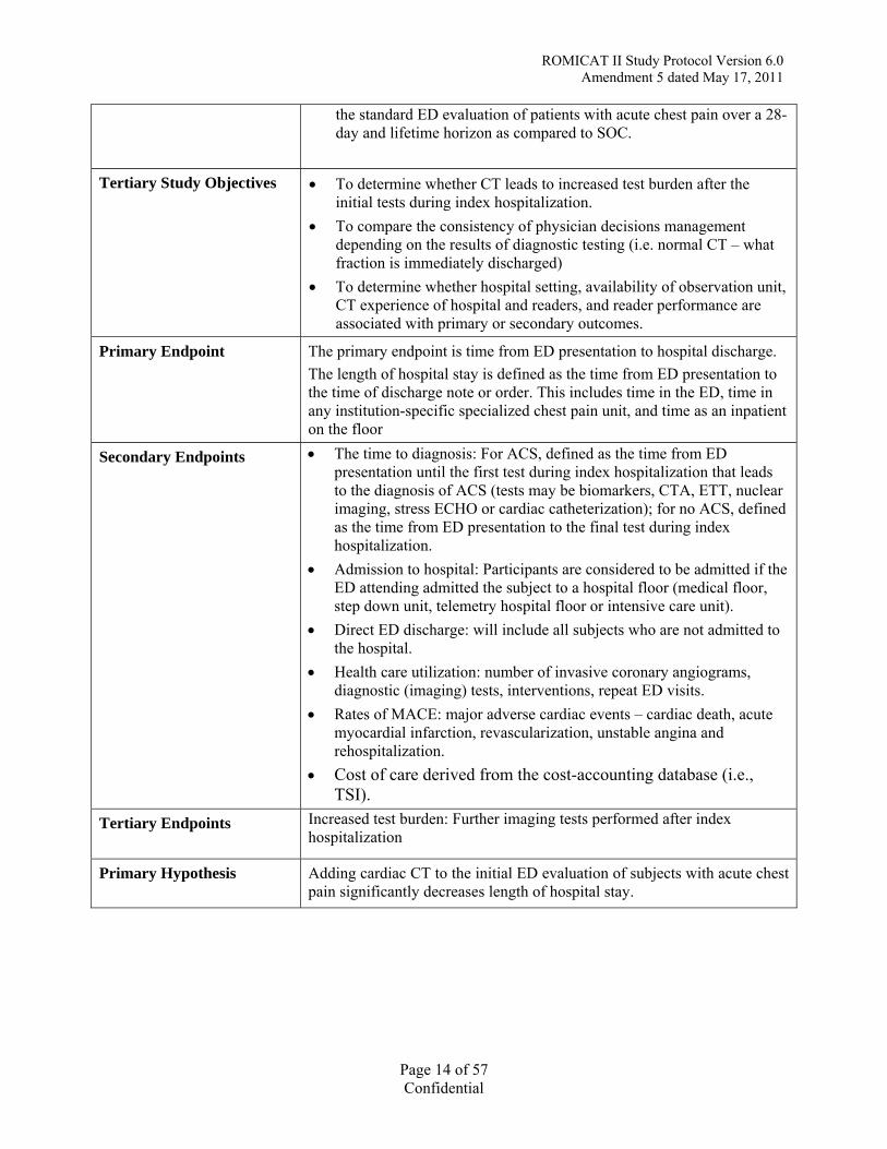
ROMICAT II Study Protocol Version 60 Amendment 5 dated May 17 2011
Abbreviations
ACS Acute Coronary Syndrome CABG Coronary artery bypass graft CAD Coronary Artery Disease CK-MB Creatinine kinase MB type CRC Clinical Research Coordinator CT Computed Tomography ECG Electrocardiogram ED Emergency Department ETT Exercise treadmill ECG testing MACE Major adverse cardiac event MI Myocardial Infarction NPV Negative predictive value PPV Positive predictive value SOC Standard of care SPECT Rest or stress myocardial perfusion imaging
Page 16 of 57 Confidential
ROMICAT II Study Protocol Version 60 Amendment 5 dated May 17 2011
Part I Study Overview The study is designed as a randomized controlled diagnostic multicenter trial and will enroll 1000 participants at nine sites over a 15 month period Participants presenting with acute chest pain in the ED will be randomized to a standard of care arm (SOC) or interventional arm patients in the interventional arm will receive standard evaluation supplemented by contrast-enhanced cardiac computed tomography (CT) imaging (Figure 1)
In both arms the initial Emergency Department (ED) evaluation (electrocardiogram [ECG] physical examination clinical presentation renal function lab and medical history) will determine eligibility of patients After randomization standard of care will be performed in both arms However for patients randomized to the interventional arm a CT will be performed The following decisions made by the caregiver team will be tracked admission or discharge after initial ED evaluation plus CTother initial diagnostic test in admitted participants the length of hospital stay the extent of further testing including serial cardiac markers and ECG and other diagnostic tests or interventions For safety reasons participants discharged in less than 24 hours of ED arrival will receive a phone call 48-72 hours after discharge All participants will receive a 28-day follow-up phone interview The efficiency of each strategy will be determined by comparing 1) the length of hospital stay (primary endpoint) 2) the time to diagnosis (secondary endpoint) 3) the rates of ED discharge (secondary endpoint) 4) utilization of other diagnostic testing ie invasive coronary angiograms and resulting interventions ie coronary revascularization procedures during index hospitalization and28 days (secondary endpoint) The safety of the two strategies will be compared by determining the occurrence of adverse events within 48-72 hours in directly discharged patients as well as determination of major adverse cardiovascular events (MACE) within 28 days after discharge from the index hospitalization We will also perform a cost and cost effectiveness analysis In subsequent analyses we will determine the patient physician and institutional factors that affect the primary endpoint or secondary endpoints
Primary Objective To determine whether length of hospital stay is significantly shortened in the interventional arm vs SOC
Hypothesis
Adding cardiac CT to the initial ED evaluation of subjects with acute chest pain decreases length of hospital stay
Page 17 of 57 Confidential
Secondary Endpoints
Primary Endpoint
Screening
Consent
Length of Hospital Stay
Tertiary Endpoints
Cost and Cost Effectiveness
Incremental Value of CTA over a CAC scan
Rates of Direct ED Discharge
Time to Diagnosis
No of invasive coronary angiograms and revascularizations
Rates of MACE after immediate ED discharge after 28 days
Health care utilization after 28 days
Institutional and Caregiver Characteristics associated with primary and secondary outcomes
Incremental Value of LV function over a CTA
Radiation Exposure during index hospitalization and follow up
ROMICAT II Study Protocol Version 60 Amendment 5 dated May 17 2011
Study Design
The study is designed as a randomized controlled diagnostic multicenter trial and will enroll 1000 participants at nine sites over a 15 month period
Patients with Acute Chest Pain at Low to Intermediate Risk for ACS Screening
Consent
StStandaandard ord off C Caarere CaCarrddiiaac Cc CTT RanRanddomomizizatatioionn
Length of Hospital Stay Primary Endpoint
Rates of Direct ED Discharge
Time to Diagnosis
No of invasive coronary angiograms and revascularizations
Rates of MACE after immediate ED discharge after 28 days Secondary Endpoints
Health care utilization after 28 days
Cost and Cost--Effectiveness
Institutional and Caregiver Characteristics associated with primary and secondary outcomes
Incremental Value of CTA over a CAC scan
Incremental Value of LV function over a CTA Tertiary Endpoints
Radiation Exposure during index hospitalization and follow--up
Page 18 of 57 Confidential
Patients with Acute Chest Pain at Low to Intermediate Risk for ACS
ROMICAT II Study Protocol Version 60 Amendment 5 dated May 17 2011
Part II Study Description
1 Background and Significance
11 Standard Evaluation of Participants with Acute Chest Pain Accurate triage of participants presenting with acute chest pain to the ED remains difficult because neither the chest pain history12 a single set of established biochemical markers for myocardial necrosis (troponin I troponin T creatine kinase MB-type [CK-MB])34 nor initial 12-lead ECG alone or in combination (acute cardiac ischemia time-insensitive predictive instrument) identifies a group of participants that can be safely discharged home without further diagnostic testing5ndash7 As a result the threshold to admit chest pain participants remains low and over six million are admitted annually to US hospitals28ndash10
The standard ldquorule outrdquo myocardial infarction (MI) protocol consists of serial ECG and cardiac biomarker measurements and usually requires a noninvasive stress test to rule out MI These tests (exercise treadmill ECG testing [ETT] stress echocardiography [Echo] and rest or stress myocardial perfusion imaging with Tc-99m [SPECT]) require the exclusion of myocardial necrosis with negative serial biomarkers and are performed to rule out the presence of a hemodynamically significant coronary stenosis While they have good sensitivities (ETT 76 SPECT 83 and Echo 85) for detecting the presence of significant coronary artery stenosis when compared to coronary angiography11ndash13 their specificities are only moderate (ETT 60 SPECT 64 and Echo 77) Moreover these tests are time consuming (ie SPECT 2ndash3 hours) and usually not available 247 Thus the standard evaluation of patients with chest pain to rule out myocardial ischemia requires hospital admission for 24 to 36 hours in over 90 of US hospitals14 Because of the moderate specificities of standard diagnostic testing 33 to 44 of patients with suspected acute coronary syndrome (ACS) who undergo cardiac catheterization have no significant coronary artery disease (CAD)1516 The remarkable inefficiency of current evaluation strategies is also documented by the fact that only lt10 of the six million admitted each year in the US ultimately receive a diagnosis of ACS at discharge1718 The inpatient care for the negative evaluations imparts a significant economic burden in excess of $8 billion annually19ndash21 for the US health care system
Thus an improvement of the ED triage of participants with acute chest pain particularly the increase in ED discharge rates in participants who ultimately do not have ACS would have enormous clinical and economic implications This research will determine if noninvasive cardiac CT that accurately visualizes the coronary arteries for plaques andor stenosis can be an effective strategy to facilitate early ED discharge in this patient population
12 Accuracy of Cardiac CT for Detection of Coronary Artery Disease and LV Function Advances in CT technology with an improvement of volume coverage (ge64 parallel detectors) and temporal (lt210 ms) and spatial (lt06 mm) resolution enables a nearly motion free contrast-enhanced imaging of the coronary artery tree and the heart chambers during a single short breath
Page 19 of 57 Confidential
ROMICAT II Study Protocol Version 60 Amendment 5 dated May 17 2011
hold (8ndash13 second scan)22 The data acquired during an ECG-gated contrast-enhanced multi-detector CT scan allows for 1) accurate assessment of the coronary arteries for plaque and stenosis and 2) the assessment of global and regional LV function in a cine mode
CAD Over the last several years we and others have demonstrated that cardiac CT has excellent diagnostic test characteristics for the detection of significant coronary artery stenosis as compared to invasive coronary angiography23ndash28 There is strong consensus that the strength of cardiac CT is its ability to reliably rule out the presence of stenosis and relevant coronary plaque (negative predictive value [NPV] 95 to 98 positive predictive value [PPV] 92 to 97)22
In addition cardiac CT also accurately detects the presence and extent of calcified and non-calcified coronary atherosclerotic plaque in good agreement with intravascular ultrasound (IVUS) especially in proximal coronary segments (sensitivity 84 to 92)29 30
Global and Regional LV Function Cardiac CT also permits the assessment of global regional LV function in a cine mode Studies have shown excellent reproducibility (κ=086) and agreement to cine magnetic resonance imaging (CMRI) for the detection of global and regional LV dysfunction (r=091)31ndash36
13 Accuracy of Cardiac CT to Detect ACS in Participants with Acute Chest Pain Preliminary data generated by the investigators suggest that cardiac CT using at least 64-slice technology can be performed safely and with excellent image quality in the acute care setting37
In addition our observational data in 368 patients demonstrated that the presence of significant coronary artery stenosis as assessed by cardiac CT can be definitively ruled out in 70 of participants presenting with acute chest pain to the ED (see Sections 31 and 32) This finding has a 100 NPV for ACS during index hospitalization and major adverse cardiac event (MACE) during follow-up (see Section 31)38 There are now several studies that support these results and suggest that cardiac CT has the potential to improve the management of participants with acute chest pain in the ED Rubinshtein et al in a study of 58 participants with acute chest pain but negative initial cardiac troponin levels and non-diagnostic ECG demonstrated a NPV of 100 and specificity of 92 (n=3538) for a diagnosis of ACS in the ED by cardiac CT39 Participants were discharged (n=3558) without stress testing if obstructive CAD was excluded by cardiac CT and if serial troponin measurements were negative There were no deaths or myocardial infarctions during a 15-month follow-up Earlier Goldstein et al40 randomized 200 participants at very low risk for ACS into a cardiac CT based triage system or a stress SPECT based triage system They found no or minimal CAD in 68 of patients which was a safe predictor for the absence of ACS and MACE over 6 months The study further shows the potential of cardiac CT to shorten length of hospital stay and decrease costs of evaluation of patients with acute chest pain However a potential drawback of CT was noted as the CT arm had more invasive angiograms and revascularization procedures (n=11 vs 3 and 4 vs 1 respectively) However this most likely represents that at this time the association between CT findings and clinical outcomes was unknown
In addition studies have suggested that the absence of coronary artery calcification (CAC) as detected in a non-contrast low radiation CT scan also has a high negative predictive value for
Page 20 of 57 Confidential
ROMICAT II Study Protocol Version 60 Amendment 5 dated May 17 2011
ACS in these participants4142 However the clinical utility of CAC alone for the assessment of ACS is inadequate because it cannot exclude the presence of high-grade non-calcified plaquestenosis
14 Cost Effectiveness of Diagnostic Testing in Subjects with Acute Chest Pain Current management of subjects with suspected ACS is managed by a sequential strategy (decision rule) involving quality and duration of chest pain risk factors biomarkers ECGrsquos and diagnostic tests such as SPECT echo or ETT However cost-effective utilization of these tests remains poor43 44 and hospitalization rates for ACS have increased while length of hospitalization has been constant over the last 10 years9 45 The introduction of chest pain observation units may have further decreased the threshold to admit patients Acute appendicitis and pulmonary embolism are excellent examples of the effects of the implementation of advanced imaging technology on patient management in the ED Because of the costs associated with unnecessary appendectomy and one day of in patient observation the implementation of the CT scan resulted in significant cost savings46 A similar approach is warranted to determine whether cardiac CT reduces cost and is cost effective in subjects with acute chest pain Moreover a formal model-based cost-effectiveness analysis is essential to project possible costs and benefits from a broader perspective (including the assessment of potential risks associated with CT) and longer time horizon Modeling can also help to answer questions and perform comparisons not possible within the confines of the clinical trial The current proposal critically investigates these issues
15 Significance of the Proposed Clinical Trial Evaluation of the appropriate clinical use of novel diagnostic imaging modalities is now more important than ever as the cost of these examinations steeply rises and now exceeds the cost spent on prescribed drugs All major professional medical societies have publicly demanded that clinical use of diagnostic imaging should be dictated by evidence-based medicine to not only control costs but also to minimize additional extraneous testing as it is currently unclear whether their usage will result in improvement in the current standard of care
Advanced cardiac CT is one of these novel diagnostic imaging technologies that may potentially enhance standard of care in patients presenting to the ED with acute chest pain but non-diagnostic ECG and negative initial biomarkers a population that remains diagnostically challenging and currently causes a significant clinical burden on each hospital every day Preliminary studies demonstrate that cardiac CT accurately detects absence of coronary atherosclerotic plaque Levels of atherosclerotic plaque as detected by cardiac CT is a powerful predictor of the absence of ACS and can be detected in a significant fraction of participants with acute chest pain Thus we anticipate the implementation of cardiac CT in the early ED triage process will (1) enable earlier and safe discharge of significant fraction of patients directly from the ED who currently would have been unnecessarily admitted as well as (2) allow for earlier detection and better treatment of CAD compared to the current standard of care
The proposed research will constitute the largest randomized multicenter clinical trial using cardiac CT to date and will provide an unbiased assessment of the above stated hypotheses It
Page 21 of 57 Confidential
ROMICAT II Study Protocol Version 60 Amendment 5 dated May 17 2011
will be conducted by a group of experienced clinical researchers consisting of CT imagers ED physicians and cardiologist with clinical expertise in the management of chest pain syndromes The results from this trial will determine the clinical utility of cardiac CT in patients with acute chest pain and may lead to a change in the management of these patients
2 Preliminary Data In support of the aim of the proposed research we provide data from a double blinded observational cohort study (ldquoCardiac CT for triage of participants with intermediate likelihood of ACSrdquo R01 HL080053-03) demonstrating the association between cardiac CT findings on plaque stenosis and LV function with clinical outcomes in patients with acute chest pain These data demonstrate 1) the absence of any CAD in up to 50 of patients with acute chest pain 2) the high NPV of absence of plaque and stenosis for ACS and thus the safety to discharge patients from the ED and the incremental diagnostic value of LV function assessment for risk stratification
21 Final Results of ROMICAT I
211 Cardiac CT Based Detection of Coronary Stenosis and Plaque in the Assessment of Participants with Acute Chest Pain Objective To determine the usefulness of cardiac CT angiography in patients with acute chest pain Methods Observational cohort study in chest pain patients with normal initial troponin and non-ischemic ECG 64-slice coronary CT was performed prior to admission to detect coronary plaque and stenosis (gt 50 luminal narrowing) Results were not disclosed Endpoints were ACS during index hospitalization and MACE during 6- month follow up Results Among 368 participants (mean age 53 plusmn 12 years 61 male) 31 had ACS (8) By cardiac CT 50 of these participants were free of CAD 31 had nonobstructive disease and 19 had inconclusive or positive CT for significant stenosis Sensitivity and NPV for ACS were 100 (n=183368 95 confidence interval [CI] 98ndash100) and 100 (95 CI 089ndash100) with the absence of CAD and 77 (95 CI 59ndash90) and 98 (n=300368 95 CI 95ndash99) with significant stenosis by cardiac CT Specificity of presence of plaque and stenosis for ACS were 54 (95 CI 049ndash060) and 87 (95 CI 083ndash090) respectively Only one ACS occurred in the absence of calcified plaque Both the extent of coronary plaque and presence of stenosis predicted ACS independently and incrementally to TIMI risk score (AUC 088 082 vs 063 respectively all plt00001) Conclusion Fifty percent of participants with acute chest pain and low to intermediate likelihood of ACS are free of CAD by CT and have no ACS Given the large number of such patients early cardiac CT may significantly improve patient management in the ED
Page 22 of 57 Confidential
ROMICAT II Study Protocol Version 60 Amendment 5 dated May 17 2011
Figure 1 A-C 67 year old female 3 hours of substernal chest pain radiating to the back negative initial troponin and CK-MB ECG sinus bradycardia admitted to rule out MI All coronary arteries are widely patent with no evidence of coronary atherosclerosis A Volume rendered 3-dimensional CT image (surface weighted volume rendering technique) of the heart depicting the left circumflex coronary artery (LCX arrowhead) obtuse marginal branch (dashed arrow) and left anterior descending (LAD arrow) two hours after presentation to the ED B CT based evaluation of segments 1 to 3 of the right coronary artery (RCA arrowheads) and conus branch (dashed arrow) using a 5mm thick maximum intensity projection (MIP) C Curved multiplaner reformatted (MPR) image of the LAD (arrowheads) and proximal LCX (dashed arrow)
Figure 2 A-C 40-year old male with hypercholesterolemia and a family history of diabetes woke with chest pressure and noticed intermittent chest pressure during his 1 mile walk to work He had similar symptoms a week prior to admission Initial troponin and CK-MB were negative and ECG showed a normal sinus rhythm without ST-segment or T-wave abnormalities Coronary CT was performed two hours after presentation to the ED and revealed a significant mid LAD stenosis Eight hours after ED presentation cardiac biomarkers became positive The mid LAD stenosis was confirmed on coronary angiography the next day and the participant was treated with a stent placement A Volume rendered 3-dimensional CT image of the heart depicting the right coronary artery (RCA arrow) and the left anterior descending coronary artery (LAD arrowhead) Discontinuation of the contrast-enhanced coronary lumen in the mid section of the LAD (dashed arrow) was detected B CT based evaluation of the right coronary artery (RCA arrowheads) demonstrates calcified and non-calcified plaque in segments 1 to 3 no significant coronary stenosis was detected C Curved MPR image of the LAD reveals good luminal contrast enhancement without any plaque in the distal LAD and calcified plaque in the proximal
Page 23 of 57 Confidential
ROMICAT II Study Protocol Version 60 Amendment 5 dated May 17 2011
segment (arrowheads) A significant luminal narrowing (arrow) was detected in the mid portion of the LAD
212 Incremental Diagnostic Value of Regional LV Function Over Coronary Assessment of Cardiac CT for the Detection of ACS in Participants with Acute Chest Pain Objective To determine incremental value of regional left ventricular function (LVF) over coronary assessment for the diagnosis and prediction of acute coronary (ACS) in patients with acute chest pain but inconclusive initial emergency department (ED) evaluation Methods We enrolled 356 consecutive patients (mean age 53plusmn12 years 62 male) with acute chest pain and inconclusive initial ED evaluation These patients underwent 64-slice contrast-enhanced cardiac CT prior to hospital admission Regional LVF and presence of coronary atherosclerotic plaque and significant stenosis (gt50) were separately assessed by two experienced readers blinded to the clinical course and other CT findings Caregivers and patients remained blinded to the CT results Incremental diagnostic accuracy and predictive value of regional LVF to predict ACS was determined in the entire cohort and in subgroups of patients with inconclusive or positive coronary assessment Results Regional LVF was impaired in 46 patients (129) and 31 patients developed ACS (87) Adding regional LVF resulted in a 10 increase in sensitivity to detect ACS (87 95CI 70shy96) when compared to detection of significant stenosis In the subgroup of 33 patients with significant stenosis impaired regional LVF correctly predicted ACS in 1719 patients (PPV 895 95CI 67-99) In the subgroup of patients with inconclusive coronary CTA (n=33) normal regional LVF correctly predicted the absence of ACS in 2426 patients (NPV 923 95CI 75-99) Impaired regional LVF independent of the extent of plaque and the presence of stenosis was associated with a 25 and 20 fold increased risk for ACS (RR 2023 95CI 747 to 5482 and RR 2534 95CI 936 to 6857 respectively) Moreover c-statistics improved significantly after the addition of regional LVF to both the extent of plaque and the presence of stenosis (c-statistic 088 vs 094 and 082 vs 090 respectively both plt003) Conclusions Regional LVF assessment at rest improves overall sensitivity of coronary CTA for ACS in patients with acute chest pain Combined assessment of coronary morphology and regional LVF is especially helpful to guide further testing and interventions if coronary assessment is inconclusive or positive
213 Initial Decision Model to Assess the Cost Effectiveness of Coronary CT in Triage of Subjects with Acute Chest Pain Objective To develop a preliminary decision-analytic Markov model to predict the long-term consequences of four competing strategies 47 in the ED evaluation of subjects with acute chest pain Methods The model consists of the six health states chest pain healthy early heart disease undiagnosed CAD diagnosed CAD and death Subjects start in the chest pain state The probability that the chest pain is caused by CAD depends on the underlying risk of CAD as
Page 24 of 57 Confidential
ROMICAT II Study Protocol Version 60 Amendment 5 dated May 17 2011
determined by age gender and cardiovascular risk factors As simulated subjects progress through the model CAD (if present) can remain stable or progress Subjects may die from cardiovascular disease or other causes at rates determined by disease stage and demographics By simulating hypothetical cohorts of subjects we determined quality-adjusted life expectancy (QALE) quality-adjusted life year (QALY) and cost-effectiveness ratios for competing strategies We considered four competing strategies for subjects presenting with chest pain to the ED 1) admission in which all subjects are admitted to the hospital for further evaluation 2) discharge in which all subjects are discharged directly home from the ED 3) cardiac CT with conservative treatment in which all subjects without any evidence of CAD are discharged and all other patients are admitted and undergo stress testing and 4) cardiac CT with aggressive treatment which is similar to strategy 3 but sends subjects with evidence of significant stenosis directly for invasive coronary angiography It is important to note that the model permits an evaluation of each strategy using a similar cohort of subjects The model also permits an evaluation of estimate life expectancy and lifetime costs Results Strategies involving cardiac CT were associated with lower ED costs and incremental improvements in QALE when compared to a strategy of admission for all subjects Relative to admitting all subjects the use of cardiac CT yielded savings in ED and hospital costs ranging from $1800-$2200 in men and women ages 30-40 and from $500-$1200 men and women ages 60-70 respectively The incremental cost-effectiveness of strategies involving cardiac CT compared with a strategy of discharging all subjects ranged from $94100-$267200 per QALY in men and $74800-$326100 per QALY in women Conclusions This preliminary decision model suggests that strategies involving cardiac CT are associated with lower ED costs and incremental improvements in QALE in both men and women across all ages The precision of the model will be greatly enhanced by input of real clinical scenarios derived from this proposal
3 Study ObjectiveSpecific Aims This primary objective of this protocol is to assess the efficiency of implementing cardiac CT into the diagnostic workup of patients with acute chest pain normal initial biomarkers and normal or non-diagnostic ECGs Study participants will be patients with acute chest pain and low to intermediate likelihood of ACS and will be randomly assigned to the SOC arm or the interventional arm patients in the interventional arm will receive the standard of care supplemented by cardiac CT
31 Primary Aim The primary aim is to determine whether length of hospital stay is significantly reduced in the interventional arm compared to standard of care The primary endpoint is time from ED presentation to hospital discharge
The length of hospital stay is defined as the time from ED presentation to the time of discharge note or order This includes time in the ED time in any institution-specific
Page 25 of 57 Confidential
ROMICAT II Study Protocol Version 60 Amendment 5 dated May 17 2011
specialized chest pain unit and time as an inpatient on the floor All time sequences must be documented and reported to comprise total length of hospital stay Hypothesis Adding cardiac CT to the initial ED evaluation of subjects with acute chest pain significantly decreases length of hospital stay
32 Secondary Aims 1 To determine whether time to diagnosis is significantly shortened and rates of direct
discharge from ED are increased in the interventional arm vs SOC
A) Time to Diagnosis For ACS Defined as the time from ED presentation until the first test during index hospitalization that leads to the diagnosis of ACS (tests may be biomarkers CTA ETT nuclear imaging stress ECHO or cardiac catheterization) For no ACS Defined as the time from ED presentation defined as the final test during index hospitalization
B) Admission to Hospital Participants are considered to be admitted if the ED attending admitted the subject to a hospital floor (medical floor step down unit telemetry hospital floor or intensive care unit)
Direct ED Discharge will include all subjects who are not included in (B)
Hypothesis ED evaluation of subjects with acute chest pain supplemented by cardiac CT significantly increases direct ED discharge rates and shortens time to diagnosis for both ACS positive and negative patients
2 To determine a) the additional number of invasive coronary angiograms both arms and b) to determine the number of coronary revascularization procedures
Hypothesis ED evaluation of subjects with acute chest pain supplemented by cardiac CT will not increase the number of invasive coronary angiograms without revascularization or revascularizations during index hospitalization
3 To determine the safety of immediate ED discharge after a normal cardiac CT defined as the occurrence of ACS within 48-72 hours after discharge
Hypothesis CTA has a high negative predictive value based on which immediate ED discharge should not compromise patient safety
4 To determine whether subsequent testing and follow up including invasive coronary angiograms diagnostic (imaging) tests interventions repeat ED visits for cardiac related problems and repeat hospitalizations for chest pain or equivalent during 28 days after hospital discharge is decreased in the interventional arm compared to SOC
Hypothesis Incorporation of CTA in the evaluation of acute chest pain patients in the ED will not lead to decreased further testing to manage chest pain when as compared to the SOC arm
5 To compare the two study arms in terms of rates of MACE (major adverse cardiac eventsmdashcardiac death AMI revascularization unstable angina and rehospitalization) within 28 days
28 Days Defined as 28 days post the day of discharge from index hospitalization
Page 26 of 57 Confidential
ROMICAT II Study Protocol Version 60 Amendment 5 dated May 17 2011
Hypothesis The rates of MACE in both arms during the 28 day follow up period will be similar
6 To estimate and compare the sensitivity specificity NPV and PPV of contrast-enhanced CT and non-contrast CT with clinical diagnosis of ACS as the reference standard
Hypothesis The test characteristics of contrast enhanced CT will be significantly better than those of a non-contrast CT for the diagnosis of ACS
7 Determine whether cost of care (per TSI) for index hospitalization and after 28 days is decreased by implementing cardiac CT into the early ED evaluation of patients with acute chest pain
Hypothesis Incorporation of cardiac CT into standard ED evaluation of patients with acute chest pain will decrease the cost of care
8 Determine the cost-effectiveness of incorporating cardiac CT into the standard ED evaluation of patients with acute chest pain over a 28-day and lifetime horizon as compared to SOC
Hypothesis Incorporating cardiac CT into the standard evaluation of patients with acute chest pain will be cost-saving and cost-effective over a 28-day and lifetime horizon
33 Tertiary Aims 1 To determine whether CT leads to increased test burden after the initial tests during index
hospitalization
Hypothesis Cardiac CT may diagnose coronary artery disease at a more incipient stage than determined by imaging tests in the SOC arm which may increase test burden during the follow up period
2 Compare the consistency of physician decisions management depending on the results of diagnostic testing (ie normal CT - what fraction is immediately discharged)
Hypothesis There will be comparable consistency between the two arms
3 Determine whether hospital setting availability of Observation Unit CT experience of hospital and readers and reader performance are associated with primary or secondary outcomes
Hypothesis Enhanced CT experience of the hospital and readers better reader performance during CT certification and hospital setting will be associated with improved primary or secondary outcomes
4 Participant SelectionEligibility Criteria
Adult ED participants presenting with a lead symptom of acute chest pain suggestive of acute cardiac ischemia - defined as chest pain lasting for at least 5 minutes and occurring within the last 24 hours but without diagnostic ECG changes of acute myocardial ischemia - will be eligible for participation
Page 27 of 57 Confidential
ROMICAT II Study Protocol Version 60 Amendment 5 dated May 17 2011
Only patients in whom the ED attending feels that further inpatient testing is required will be included Patients with knowndocumented history of CAD will not be included since such patients most often have an abnormal or non-diagnostic cardiac CT In addition men and women younger than 40 years will be excluded due to the low pre-test likelihood for CAD and the increased radiation risk Detailed inclusion and exclusion criteria are presented below
41 Inclusion Criteria 1 Participant must have at least five minutes of chest pain or equivalent (chest tightness
pain radiating to left right or both arms or shoulders back neck epigastrium jawthroat or unexplained shortness of breath syncopepresyncope generalized weakness nausea or vomiting thought to be of cardiac origin) at rest or during exercise within 24 hours of ED presentation warranting further risk stratification as determined by an ED attending
2 Participant must be able to provide a written informed consent
3 Participants must be lt75 years of age but gt40 years of age
4 Participant must be able to hold breath for at least 10 seconds
5 Participant must be in sinus rhythm
42 Exclusion Criteria 1 New diagnostic ischemic ECG changes (ST-segment elevation or depression gt 1 mm or
T-wave inversion gt 4 mm) in more than two anatomically adjacent leads or left bundle branch block
2 Documented or self-reported history of CAD (MI percutaneous coronary interventions [PCIs] coronary artery bypass graft [CABG] known significant coronary stenosis [gt50])
3 Greater than 6 hours since presentation to ED to time of consent
4 BMI gt40 kgm2
5 Impaired renal function as defined by local standard of care - for example measured serum creatinine gt15 mgdL
6 Markedly elevated troponin as defined by local standard of care
7 Hemodynamically or clinically unstable condition (BP systolic lt 80 mm Hg atrial or ventricular arrhythmias persistent chest pain despite adequate therapy)
8 Known allergy to iodinated contrast agent
9 Currently symptomatic asthma
10 Documented or self-reported cocaine use within the past 48 hours (acute)
11 On Metformin therapy and unable or unwilling to discontinue for 48 hours after the CT scan
Page 28 of 57 Confidential
ROMICAT II Study Protocol Version 60 Amendment 5 dated May 17 2011
12 Contraindication to beta blockers (taking daily antiasthmatic medication) This exclusion only applies to patients with a heart rate gt 65 bpm at sites using a non-dual source CT scanner
13 Participant with no telephone or cell phone numbers (preventing follow-up)
14 Participant with positive pregnancy test Women of childbearing potential defined as lt2 years of menopause in the absence of hysterectomy or tube ligation must have a pregnancy test performed within 24 hours before the CT scan
15 Participant unwilling to provide a written informed consent
43 Recruitment Screening and Consent
The research team at each participating site includes the ED attending physician CT technologist and research associate(s) The investigator and the research staff will be responsible for the screening review of participant medical records and investigator-designated data submission All clinical sites will perform screening Monday through Friday during daytime hours (9am-5pm) Potential subjects will be identified by research coordinatorstudy nurse staff shortly after admission to the ED usually after the initial ED evaluation consisting of ECG physical examination clinical symptoms and presentation immediate and past medical history has been concluded and screened for eligibility based on a checklist providing inclusion and exclusion criteria
Eligible subjects will be approached and if the subject considers participation the Clinical Research Coordinator (CRC) will describe the rationale of the study the study procedures with associated risks and benefits A study physicianstudy nurseCRC will speak directly with an ED physician caring for the patient to obtain an assessment of pretest probability for ACS and will verify that the ED physician feels that further observation or diagnostic testing is warranted in this patient
If the subject agrees and further testing or observation is planned as SOC key study personnel typically a study physician or the attending ED physician will review the study procedures as well as all potential risks and discomforts and the opportunity to decline or cease participation in the study at any time answer any remaining questions and consent the subject
Patients can be consented but not randomized before the first troponin test result is available Patients whose initial troponin level is markedly elevated will not be randomized and will not be included in the intent-to-treat population Patients whose subsequent troponin testing is markedly elevated will be included in the intent to treat population
Patients can be consented before a pregnancy test is available However patients with a positive pregnancy test will not be randomized
Patients who are not randomized will not be considered part of the intent to treat population
Informed Consent
Page 29 of 57 Confidential
ROMICAT II Study Protocol Version 60 Amendment 5 dated May 17 2011
bull A potential subject will then be afforded as much time as clinically feasible to review the consent form and decide upon their participation without adversely effecting their ED care which is typically 1 hour
bull If the ED staff caring for the patient determines that the potential subjectrsquos previously determined plan of care must proceed before the potential subject has had adequate time to decide upon hisher participation without undue pressure the potential subject will be excluded from enrollment
bull Subjects will be provided with a copy of the informed consent form
44 Randomization
After the patient has been consented by the study physician the study subject will be randomized into the standard care arm or the intervention arm (cardiac CT) by means randomization stratified by institution using a central computerized randomization system
5 Study Procedures
51 Index Hospitalization Standard of Care Arm Subjects will be evaluated according to each hospitalrsquos specific protocol to evaluate and manage patients with acute chest pain Typically the standard evaluation in the ED will include the past and current medical history a physical examination an ECG and a cardiac biomarker (Troponin and CK-MB) as well as other routinely obtained blood testing Patients may undergo cardiac CT as part of SOC but only as a secondary diagnostic test Patients in the CT arm may undergo further diagnostic testing as well
All admitted subjects will undergo each hospitalrsquos standard rule out myocardial ischemia protocol This protocol typically consists of observation and monitoring including serial ECGs and repeated cardiac biomarker measurements as well as a noninvasive stress test (often imaging based) to evaluate for myocardial ischemia The participating clinical sites perform routinely either nuclear perfusion imaging [SPECT] at rest and stress andor stress echocardiography andor exercise treadmill test [ETT] Depending on the results subjects may undergo additional noninvasive or invasive testing (coronary angiography) andor coronary revascularization during their hospital stay
Interventional Arm In subjects randomized to the intervention arm a contrast enhanced cardiac CT will be performed (after initial troponin level is available) in addition to the initial ED evaluation The ED physicians will resume care of the patient and make all clinical decisions for further care of the patient (eg need for further evaluation or admission) based upon their cumulative clinical assessment of the patient including findings revealed on the CT scan
Page 30 of 57 Confidential
ROMICAT II Study Protocol Version 60 Amendment 5 dated May 17 2011
In addition in patients from both study arms who consented to a blood draw we will obtain three fifteen (15) mL venous blood samples over a six-hour period less than forty-five (45) mL in total Blood samples will be centrifuged and frozen for future analysis for cardiac-related markers
All diagnostic tests will be performed per Society Guidelines Cardiac Computed Tomography is performed in accordance with best practice standards as delineated in the imaging guidelines of the Society of Cardiovascular Computed Tomography by competent63 and appropriately credentialed physicians This includes the optimization of the scan protocol to limit radiation exposure
52 48-72 Hour Follow up after ED discharge Follow-up phone call at 48-72 hours Subjects who are discharged from the ED within 24 hours of presentation (for both randomization arms) will be contacted via telephone between 48 and 72 hours after discharge to avoid ascertainment bias During this call subjects will be interviewed to determine whether they have had any recurrent symptoms suggestive of myocardial ischemia and andor whether they needed to seek further medical attention following their emergency department visit If these phone attempts at 48 and 72 hours are not successful a final attempt will be made at 96 hours to contact the subject
NOTE If a subject is discharged on a Thursday the 48-72 hour phone call may be extended to 96 hours In the rare circumstance that a subject is discharged on a Thursday and the following Monday is a holiday the follow-up call can be made during the next working business day
53 28 Day Follow up Follow up 28 day phone call All subjects will be interviewed via a telephone call 28 days post ED discharge (+4 weeks) to determine the occurrence of MACE and healthcare utilization using a standardized questionnaire All cases of recurrent chest pain hospital admissions and diagnostic testing will be verified by review of medical records
A reminder letter will be sent or phone call will be made to participants two weeks prior to the 28-day follow up phone call If unable to contact the participant by phone mortality will be assessed online using the SSDI website
Page 31 of 57 Confidential
ROMICAT II Study Protocol Version 60 Amendment 5 dated May 17 2011
6 Data Collection The following subject data will be collected during index ED visithospitalization and follow up phone interviews bull Subject demographic information bull Medical history including cardiac risk factors and medications bull Description of recent pain episodes bull Vitals signs ECG cardiac biomarker results bull Cardiac CT imaging and other cardiac-related diagnostic testing data bull Adverse events
7 Statistical Considerations A total of 1000 participants will be enrolled in this study Enrollment is planned to be completed in 15 months
71 Primary Aim To compare the two study arms in terms of the time from ED presentation to hospital discharge The primary aim of the study is to compare the two study arms in terms of a difference in mean length of hospital stay (LOS) between the two arms Based on the results of ROMICAT I we estimate the mean (plusmn standard deviation) hospital LOS (including ED) of these patients in standard evaluation to be 405 plusmn 432 hours The observed frequency distribution appeared to fit well to a log-normal distribution (Median was 77 hours and Range was 275 ndash 3815 hours) We anticipate that the observed distributions of both Group A and Group B follow such a pattern Because the independent samples t-test is robust to a departure from the data normality when applied to a large sample size we will employ the t-test for this inference
We have conducted a power evaluation using the estimated mean and standard deviation based on ROMICAT I for Group A and using the estimated the mean and standard deviation based on a simulated data for Group B The simulated data consisted of 500 LOS values resulted as a mixture of three subsets randomly drawn from log-normal distributions of which the first subgroup was to represent the patients with normal LV function and without CAD (486 Mean plusmn SD of 6 plusmn 12 hours) the second was to represent the patients with normal LV function and with non-obstructive CAD (288 Mean plusmn SD of 10 plusmn 20 hours) and the third group was to represent all other possible conditions (226 Mean plusmn SD of 579 plusmn 604 hours which was estimated from ROMICAT 1) We also incorporated in the possibility that some patients without CAD will not be correctly triaged The triage accuracy rate in the following table is the rate at which a patient without CAD would be correctly triaged The following table summarizes for each ED triage accuracy rate assumption the estimated mean LOS and the power at a Type-1 error rate of 5 for an independent samples t-test using 500 patients in each arm The proposed sample size will attain a sufficient power to detect a clinically meaningful difference in mean LOS between the two groups even in the presence some degree of ED triage inaccuracy
Page 32 of 57 Confidential
ROMICAT II Study Protocol Version 60 Amendment 5 dated May 17 2011
Powers to Detect Estimated Differences in Mean LOS values at Assumed ED Triage Accuracy Rates (Type-1 error rate = 005)
Assumed ED Triage Accuracy Rate
Estimated Mean (plusmn SD) LOS Estimated Difference in Mean (hours) Power
Group A (Standard Eval)
N=500
Group B (CT Eval)
N=500 65 405 (plusmn 432) 328 (plusmn 467) -83 83 70 405 (plusmn 432) 305 (plusmn 455) -101 95 80 405 (plusmn 432) 263 (plusmn 417) -142 99 90 405 (plusmn 432) 224 (plusmn 373) -181 gt99
100 405 (plusmn 432) 188 (plusmn 335) -217 gt99
72 Secondary Aims To compare the two study arms in terms of time to diagnosis and rates of direct discharge The time to discharge (defined as time from ED presentation until discharge to home) will be plotted using Kaplan-Meier curves because some patients will be in the hospital at 30 days post day of presentationrandomization The curves for the two groups will be compared using a test based on the difference in the average length of stay in the two groups (truncated at 30 days) divided by the standard error of this difference60 We choose this test because the average length of stay is most closely associated with cost It will be also instructive to compare the curves themselves as they will provide information on the effect of cardiac CT on different groups of patients For instance one group of patients those who are discharged due to cardiac CT may have a shorter length of stay while others may have a longer length of stay due to waiting for the results of the CT or other procedures that the CT indicates
Sample Size The planned sample size will also provide adequate power to detect the rates of direct discharge from ED Screening data from a site survey of our clinical sites as well as published data indicate that the standard ED discharge rate is approximately 15 in the target population According to our preliminary data and data from other groups we expect that 40 to 50 of participants have no detectable CAD by cardiac CT The negative predictive value of this finding for ACS has been shown to be 100 Our guidelines suggest that these participants will be discharged directly from the ED However realistically we expect that because of administrative issues and remaining individual variation of the decision to discharge depending on the clinical presentation of participants actually only 60 to 80 of these participants will be discharged Thus anywhere from 24 to 40 participants could be discharged directly from the ED in the interventional arm For the sample size analysis we assume that 32 of participants will be discharged
Based on the aforementioned key assumptions for the Standard Evaluation we anticipate that 15 of all participants will be discharged and 85 will be admitted (discharge rate 15) For the Cardiac CT Evaluation we assume a discharge rate of 32 For the objective of superiority
Page 33 of 57 Confidential
ROMICAT II Study Protocol Version 60 Amendment 5 dated May 17 2011
of discharge rates we will have a 100 chance at a significance level of 005 to detect a 17 increase in discharge rates from 15 to 32
The comparison of time to discharge among patients who are admitted to the hospital is complicated by the fact that the two groups will not be the same group of patients For instance patients who would have been discharged in the cardiac CT group but admitted in the usual care group may have a relatively shorter time post admission than patients who would have been admitted in both groups We have developed methods for analyzing these types of data using causal inference that will be applied to this analysis61 A similar analysis will be applied to other subgroups such as those with and without ACS
To compare the two study arms in terms of cardiac health care utilization during the index visit Health care utilization components will include invasive coronary angiograms and coronary revascularization
The analysis for this secondary aim will be performed from the intent to treat perspective Health care utilization will be assessed using medical record review Measures of key components of utilization will be compared across the two study groups using procedures appropriate for each measure (rate or count)
In particular the two arms will be compared in terms of the number of invasive coronary angiograms This secondary endpoint will be analyzed in terms of a non-inferiority trial In a non-inferiority trial the null hypothesis is that cardiac CT is worse than usual care in that the rate of coronary angiograms is increased and the alternative hypothesis is that it is not in worse and the rate of angiograms is the same The reason for reversing the usual null and alternative hypothesis is that the purpose of this analysis is to prove that cardiac CT is not worse than usual care this would not be accomplished by just showing that the difference between the two was not significant
Screening data from a survey of clinical sites as well as published data indicate that the standard rate of invasive coronary angiograms in the target population is approximately 10 According to our study guidelines we assume that most but not all participants with significant stenosis in CT (10 of participants) undergo invasive coronary angiography (notably those who also have a positive stress test and those who have high grade proximal stenosis or LAD stenosis) In addition we assume that a minority of participants with indeterminate stenosis (lt5) and positive stress testing undergo invasive coronary angiography Because of the moderate specificities of standard diagnostic testing up to 40 of participants who undergo cardiac catheterization have no significant CAD1516 Because the diagnostic accuracy of cardiac CT for the detection of coronary stenosis is comparable or better than standard diagnostic testing we believe that there will be no increase in the rates of invasive coronary angiograms If we define a 5 difference as an ldquoacceptablerdquo difference in cardiac catheterization rates than we will have an 84 chance of showing that the CT arm is significantly less than 5 worse than the standard arm at a one sided p=005 significance level
Page 34 of 57 Confidential
ROMICAT II Study Protocol Version 60 Amendment 5 dated May 17 2011
Another key measure of utilization is the number of coronary revascularization procedures In ROMICAT I there were 18 revascularization procedures performed in 368 patients (5) Thus we expect 5 procedures in the standard evaluation If we assume that not all participants with significant stenosis will undergo coronary revascularization (because if no MI it can be medically managed) and that cardiac CT may also avoid some angiograms by excluding disease than we can realistically assume that there is not a difference between the two study arms If we consider this in terms of a non-inferiority trial as above we will have 80 power to rule a difference of 35
To compare the two study arms in terms of cardiac health care utilization within one year after index hospitalization Health care utilization components will include invasive coronary angiograms cardiac related diagnostic and lab testing and ED visits
This analysis will be performed from the intent to treat perspective CP and CT related health care utilization will be assessed using medical record review Measures of key components of utilization will be compared across the two study groups using procedures appropriate for each measure (rate or count) We expect that the rate of additional cardiac work-up will be 29 in the SOC arm and will be 14 in the interventional arm We will have gt95 power to detect this difference We expect that the rate of hospital admissions with additional diagnostic cardiac testing will be 93 in the SOC arm and will be 4 in the interventional arm We will have gt95 power to detect this difference We expect that the rates of these measures will be associated with the CT findings at index hospitalization
To evaluate the cost and cost-effectiveness of incorporating cardiac CT into the standard ED evaluation of patients with acute chest pain These analyses will be performed by the Decision Analysis and Cost-Effectiveness Center (DACE) Therefore the sites will collect and provide the necessary data to the DACE and the costs will be derived from the cost-accounting database (TSI) As a first step incorporating cardiac CT into the standard evaluation of patients with acute chest pain will be tested for cost-saving over a 28-day and lifetime span as compared to standard care Further on cost-effectiveness of incorporating cardiac CT into the standard ED evaluation of patients with acute chest pain will be determined using first-order Monte Carlo microsimulations
To compare the two study arms in terms of rates of MACE (cardiac death AMI revascularization unstable angina and rehospitalization) within 28 days The analysis for this secondary aim will be performed from the intent to treat perspective The occurrence of MACE will be assessed via patient follow-up and medical chart review The primary analysis for this aim will be performed at the patient level by determining whether any MACE occurred or not Event rates will be estimated and compared between study groups using exact procedures In a secondary analysis the rates of each type of MACE will be examined separately
Sample Size- Safety Considerations- Missed ACS Mistaken discharges are the primary safety issue in this study All available data indicate that the absence of any detectable CAD (absence
Page 35 of 57 Confidential
ROMICAT II Study Protocol Version 60 Amendment 5 dated May 17 2011
of any plaque and stenosis) has a very high NPV for ACS (100 or close to 100)62 Thus we anticipate that the occurrence of ACS in participants discharged home directly from the ED in the interventional arm will be also minimal or even zero and similar to the standard of care If as demonstrated in ROMICAT I none of the participants without CAD will have MACE defined as ACS within 30 days after direct ED discharge this observation has 90 power to ensure that the true event rate is less than 1 These assumptions are supported by the results of the ERASE trial which reported similar and very minimal event rates in each group (only 2 pts with acute MI were mistakenly discharged 1 in each randomization group out of 1200 [event rate 00008]) In this trial we would have an 80 chance of detecting at least one occurrence of an inappropriately discharged participant if the true rate where over 1
To estimate and compare the sensitivity specificity NPV and PPV of contrast-enhanced CT and non-contrast CT with clinical diagnosis of ACS as the reference standard The analysis for this aim will utilize data on participants randomized to Group B In this arm each participant will undergo non-contrast CT and contrast-enhanced CT which will be interpreted as positive or negative for ACS Clinical determination of presence or absence of ACS will be used as the reference standard For each of the two types of CT we will estimate the sensitivity specificity and positive and negative predictive value of the modality for ACS Each measure of diagnostic or predictive performance will be compared between the two modalities using statistical techniques that account for the correlation due to the paired test design In addition to these measures we will estimate the proportion of cases with a clinical diagnosis of ACS and a negative non-contrast CT in which contrasted enhanced CT was positive for ACS This proportion provides a measure of the incremental value of contrast-enhanced CT for diagnosis ACS
73 Tertiary Aims To determine whether CT leads to increased test burden after the initial tests during index hospitalization
To compare the consistency of physician decision management depending on the results of the diagnostic testing (ie normal CT ndash what fraction is immediately discharged)
To determine whether hospital setting availability of Observation Unit CT experience of hospital and readers and reader performance are associated with primary or secondary outcomes
Analysis of Data on Women and Minorities We will analyze the effect of the intervention on women and minorities as outlined in the NIH guidelines Thus we include testing for gender-treatment and minority status-treatment interactions with the discharge rate and the angiogram rate
Page 36 of 57 Confidential
ROMICAT II Study Protocol Version 60 Amendment 5 dated May 17 2011
8 Data Entry and Site Monitoring
81 Data Collection Subject data will be collected by the research associates and paper records will be maintained at individual clinical sites The Data Coordinating and Statistical Center (DCSC) will develop a web based electronic case report form The de-identified study data will be entered into this electronic data capture system (EDC) and sent to the Data Coordinating and Statistical Center (DCSC) at Massachusetts General Hospital for further analysis
82 Site Monitoring Site visits will be performed on a regular basis by the Data Coordinating Center to ensure that all regulatory requirements are being met and to monitor the quality of the data collected Records of Institutional Review Board approvals and patientsrsquo charts will be examined on a spot check basis to evaluate the accuracy of the data entered into the database
9 Risk and Benefit Assessment
91 Risks of Nonionic Contrast Agent
Rare risks of the nonionic contrast agent bull Itchiness bull Rash bull Mild allergic reaction
Very rare risks of the nonionic contrast agent bull Kidney diseaseContrast-induced nephropathy bull Death
Regarding the risks of contrast material the rate of adverse reactions with nonionic contrast reagents is 0250 More than 90 of such adverse reactions are allergic-like very mild (itching rush) and can be very effectively treated with available drugs (ie antihistaminic) Severe reactions occur in 002 and one death occurred in 57739 injections of nonionic contrast material50
Contrast-induced nephropathy (CIN) CIN is expected to be a very rare event among this patient population because patients with already impaired renal function (the strongest predictor of CIN) will not be enrolled) This assumption is strongly supported by our preliminary experience from ROMICAT I In this study none of the participants had such an event (0368)
The very low risk associated with contrast administration has been documented in large scale trials previously In a randomized trial in 1196 participants the incidence rate of CIN defined as an increase in serum creatinine ge1mgdL from baseline within 48 to 72 hours following the injection of contrast material was 0 in 359 participants (0) who had no renal insufficiency and no diabetes melliltus51 The risk was 2 in 315 (06) in participants with no renal insufficiency but who had diabetes mellitus51 In this study the mean volume of contrast-agent administered
Page 37 of 57 Confidential
ROMICAT II Study Protocol Version 60 Amendment 5 dated May 17 2011
was 140 plusmn 57 mL which is much higher than the 80 mL of contrast that is administered for a coronary CTA The risk of CIN may increase if participants in the cardiac interventional arm will be admitted and subsequently undergo further contrast-related diagnostic testing However among 209 participants who had both a pulmonary angiogram and a contrast-enhanced spiral CT in the Prospective Investigation of Pulmonary Embolism Diagnosis II only one participant with diabetes mellitus had a transient episode of acute renal failure characterized by an increase in the serum creatinine level from 13 to 29 mgdL52 In this study the contrast-enhanced CT and pulmonary angiogram were performed within 22 hours The elevated creatinine returned to normal after administration of intravenous fluids Thus overall we expect that the occurrence of CIN will be a rare event in this study
92 Risks Associated With Radiation While the radiation dosage for CT scanning varies with the part of the body being scanned the exposure for this examination is approximately between 7 and 17 mSv This exposure is comparable or less than stress nuclear myocardial perfusion imaging (14ndash22 mSv) (personal communication with Prof Ronald Callahan Chairman of the MGH Radiation Safety Committee) In comparison the average yearly effective dose of natural background radiation is 25 mSv53 the allowed annual exposure of radiation workers is 50 mSv the allowed exposure in five years is 100 mSv (Table 3)54 Thus even if an enrolled participant would undergo cardiac CT followed by a conventional stress test and selective coronary angiography the participant would be well below the allowable annual effective dose for radiation workers The radiation dose from cardiac CT has not been shown to have any adverse effects
Radiation Exposure
Cardiac CT scanning results in a measurable radiation exposure55 In order to minimize risk for participants in this study we have implemented the following measures
bull Exclude women lt40 years of age bull Exclude pregnant and breast feeding women bull Include only women who have a negative pregnancy test bull Application of cardiac CT protocols with the goal to minimize radiation exposure (ie
use of tube modulationmdashdecrease of radiation exposure by 30 to 50 and prospective triggering in appropriate conditions)
bull Review and approval of these CT protocols by the CVCT Core Lab
General Risk Associated with Radiation Exposure
The risk of inducing a malignancy by exposure to radiation is estimated to be 5 cases of malignancy for each 100 individuals exposed to an effective dose of 1 Sv54 The risk is estimated to be higher in infants and children and lower in the elderly The data which these estimates are based on are weak and apply to relatively high levels of radiation exposure56 and it remains unclear whether there is a linear relation in lower levels of radiation Assuming a linear proportion with a low level of radiation exposure to 1 mSv would lead to 5 malignancies in 100000 individuals exposed and a dose of 8 mSV would lead to 40 malignancies in 100000 individuals56 Small amounts of radiation exposure less than 100 mSv have not been clearly
Page 38 of 57 Confidential
ROMICAT II Study Protocol Version 60 Amendment 5 dated May 17 2011
shown to have harmful effects The amount of radiation to which patients will be exposed in this study is less than the amount that is known to cause a measurable harmful effect Even so it is generally assumed that a small amount of radiation will have a small chance of producing a harmful effect These extrapolated risks of the effects of small amounts of radiation are probably overestimates because the estimates ignore the fact that the body can repair the damage done by radiation if the repair mechanisms are not overwhelmed Radiation is a well-known teratogenic agent and the developing fetus is much more sensitive to radiation than the mother
Table 3 Estimated radiation exposure for diagnostic tests background radiation and exposure limits
Examination Effective Dose (mSv) Chest PA and lateral 007 Myocardial Perfusion Imaging (Rest and 14ndash22Stress) Thorax CT for the Exclusion of Pulmonary 7-12Embolism 64-Slice CT coronary angiogram 7ndash17 2-view mammogram 30 Natural yearly background radiation 25 Max exposure in radiation workers 50
Lifetime Cancer Risk Attributable to Cardiac CT
The absolute cancer risk has been described recently and recommendations have been made57
The life time cancer risk from cardiac CT decreases with age and is higher in women than in men in particular exposure of younger women should be avoided In our study these recommendations have been incorporated In comparison the absolute cancer risk for women to develop breast cancer is 13 and for men to develop colon cancer is 7 the risk attributed to a cardiac CT scan is 1466 vs 1715 for a 60 year old women and 11241 vs 11911 for 60 year old men (for CT without and with tube modulation (female 21 and 14 mSv male 15 and 9 mSv respectively)57 Thus the incremental risk induced by cardiac CT compared to the overall risk of the population to develop cancer seems moderate
Effect of the Implementation of Cardiac CT in the ED Evaluation of Patients with Acute Chest Pain on a Population Basis
The number of CT scans performed in the US was 62 million in 200658 Imaging of the abdomen (50) and the head (33) were major single contributors Specific concern was given to the rise of pediatric CT imaging (from 6 to now 11) and screening the adult asymptomatic population (ie lung heart) Implementation of cardiac CT in the ED could lead to a small increase of the number of overall CT procedures However the effects of the implementation of cardiac CT in the ED would be minimal if 1) other procedures associated with radiation doses would be saved 2) repeated testing currently performed in 25 of patients would be minimized and 3) rates of unnecessary purely diagnostic cardiac catheterizations would
Page 39 of 57 Confidential
ROMICAT II Study Protocol Version 60 Amendment 5 dated May 17 2011
decrease Furthermore already cardiac CT scanning could be performed using single phase acquisition with prospective triggering which would decrease the dose to lt5 mSv In addition MD over-read will be performed for internal quality control assessment of findings related to CAD and significant cardiac findings such as pulmonary embolism and aortic dissection to increase awareness to sites of non-coronary findings
93 Other Risks Additional Risks of CT Scans bull Discomfort bull Claustrophobia
Risks of IV Needle Placement
bull Hemorrhage (hematoma at the injection site) bull Infection (catheter related infection) at the injection site bull Minor discomfort bull Bleeding bull Infection bull Bruising
94 Potential Benefits bull Subjects may benefit from randomization into the interventional arm of trial from detection
of pathology that may not have been otherwise detected during SOC (eg a non-significant coronary stenosis or non-obstructive coronary artery disease that would likely not have been detected by a stress test)
bull Data from prior studies indicate that patients in the interventional arm may be earlier discharged and may not have to spend the night in the hospital there is also the chance that alternative diagnosis such as pneumonia or Pulmonary embolism may be detected - patients may avoid the dual radiation exposure of a CT protocol solely to evaluate for pulmonary embolus followed by a SPECT myocardial perfusion scan
bull Subjects may also benefit in their assessment for long term prognosis and risk for adverse cardiac events and may receive preventive medication as a result of CT
10 Ethical Considerations This study is to be conducted according to US and international standards of Good Clinical Practice (International Conference of Harmonization [ICH] guidelines) applicable government regulations and NIH research policies and procedures
This protocol and any amendments will be submitted to a properly constituted independent Institutional Review Board (IRB) for formal approval of the study conduct The decision of the IRB concerning the conduct of the study will be made in writing to the investigator and a copy of this decision will be provided to the ROMICAT PI before implementation of the study
Page 40 of 57 Confidential
ROMICAT II Study Protocol Version 60 Amendment 5 dated May 17 2011
All study participants in this study will be given an IRB-approved site-specific ICF describing the study and providing sufficient information for participants to make informed decisions about their participation in this study The study participant MUST be consented with the ECIRBshyapproved ICF before the participant is subjected to any study procedures The IRB-approved ICF MUST be signed and dated by the study participant or legally acceptable representative and the investigator-designated research staff obtaining the consent before the participant is subjected to any study procedures
101 Inclusion of Women and Minorities The participating institutions will not exclude potential participants from participating in this or any study solely based on ethnic origin or socioeconomic status Every attempt will be made to enter all eligible participants into this protocol and therefore address the study objectives in a patient population representative of the entire English-speaking population at risk for ACS treated by the institution
Both men and women and members of all ethnic groups are eligible for this trial In conformance with the National Institutes of Health (NIH) Revitalization Act of 1993 with regard to inclusion of women and minorities in clinical research the projected gender and minority accruals are shown below
Table 1 Gender and Minority Accrual Estimates
Ethnic Category SexGender Females Males Total
Hispanic or Latino 66 79 145 Not Hispanic or Latino 384 471 855
Ethnic Category Total of all participants
450 550 1000
Racial Category American Indian or Alaskan Native 2 2 4 Asian 35 42 77 Black or African American 125 153 278 Native Hawaiian or other Pacific
Islander 2 2 4
White 254 312 566 OtherUnknown 32 39 71
Racial Category Total of allparticipants
450 550 1000
Page 41 of 57 Confidential
ROMICAT II Study Protocol Version 60 Amendment 5 dated May 17 2011
11 Adverse Event Reporting Mistaken discharges are the primary safety issue in this study All available data indicate that the absence of any detectable CAD (absence of any plaque and stenosis) has a very high NPV for ACS (100 or close to 100)62 Thus we anticipate that the occurrence of ACS in participants discharged home directly from the ED in the cardiac interventional arm will be also minimal or even zero and similar to the standard of care If as demonstrated in ROMICAT I none of the participants without CAD will have MACE defined as ACS within 30 days after direct ED discharge this observation has 90 power to ensure that the true event rate is less than 1 These assumptions are supported by the results of the ERASE trial which reported similar and very minimal event rates in each group (only 2 pts with acute MI were mistakenly discharged 1 in each randomization group out of 1200 [event rate 00008]) In this trial we would have an 80 chance of detecting at least one occurrence of an inappropriately discharged participant if the true rate where over 1
At the time of ED discharge investigators will determine at if any adverse events have occurred An Adverse Event (AE) is any untoward medical occurrence in a study participant that may or may not have a causal relationship with the study intervention An AE can therefore be any unfavorable and unintended sign (including an abnormal laboratory or physiological finding) symptom or disease temporally associated with the use of a medical treatment or procedure regardless of whether it is considered related to the medical treatment or procedure (attribution of unrelated unlikely possible probable or definite) Abnormal results of diagnostic procedures are considered to be AEs if the abnormality
bull results in study withdrawal bull is associated with a serious adverse event (SAE) bull is associated with clinical signs or symptoms bull leads to additional treatment or to further diagnostic tests bull is considered by the investigator to be of clinical significance
Serious Adverse Event (SAE) An SAE is defined as any untoward medical occurrence that
bull results in death or bull is life-threatening (at the time of the event) or bull requires inpatient hospitalization or prolongation of an existing hospitalization or bull results in persistent or significant disability or incapacity or bull is a congenital anomalybirth defect
Reportable Adverse Events Any untoward medical occurrence that occurs after randomization and prior to discharge from the ED should be reported to the CCC by completing the electronic adverse event case report form
Immediately Reportable Adverse Events
Page 42 of 57 Confidential
ROMICAT II Study Protocol Version 60 Amendment 5 dated May 17 2011
Any adverse event that is judged by the investigator to be serious AND unexpected AND related to study procedures should be reported within 24 hours of discovery by phone or email to the Coordinating Center The local Institutional Review Board must also be notified in a timely manner
Investigators must also report Unanticipated Problems regardless of severity associated with the study procedures within 24 hours An unanticipated problem is defined as follows
Unanticipated Problem (UP) any incident experience or outcome that meets all of the following criteria
bull Unexpected in terms of nature severity or frequency given the research procedures that are described in the protocol-related documents such as the IRB-approved research protocol and informed consent document and the characteristics of the subject population being studied
bull Related or possibly related to participation in the research in this guidance document possibly related means there is a reasonable possibility that the incident experience or outcome may have been caused by the procedures involved in the research
bull Suggests that the research places subjects or others at a greater risk of harm (including physical psychological economic or social harm) than was previously known or recognized
12 References 1 Lee TH Rouan GW Weisberg MC Brand DA et al Clinical characteristics and natural
history of patients with acute myocardial infarction sent home from the emergency room Am J Cardio 198760(4)219-224
2 Swap CJ Nagurney JT Value and limitations of chest pain history in the evaluation of patients with suspected acute coronary syndromes JAMA 2005294(20)2623-2629
3 Limkakeng A Jr Gibler WB Pollack C et al Combination of Goldman risk and initial cardiac troponin I for emergency department chest pain patient risk stratification Acad Emerg Med 20018(7)696-702
4 Zimmerman J Fromm R Meyer D Boudreaux A et al Diagnostic marker cooperative study for the diagnosis of myocardial infarction Circulation 199999(13)1671-1677
5 Fesmire FM Hughes AD Fody EP et al The Erlanger chest pain evaluation protocol a one-year experience with serial 12-lead ECG monitoring two-hour delta serum marker measurements and selective nuclear stress testing to identify and exclude acute coronary syndromes Ann Emerg Med 200240(6)584-594
6 Hedges JR Young GP Henkel GF et al Serial ECGs are less accurate than serial CKshyMB results for emergency department diagnosis of myocardial infarction Ann Emerg Med 199221(12)1445-1450
Page 43 of 57 Confidential
ROMICAT II Study Protocol Version 60 Amendment 5 dated May 17 2011
7 Selker HP Beshansky JR Griffith JL et al Use of the acute cardiac ischemia time-insensitive predictive instrument (ACI-TIPI) to assist with triage of patients with chest pain or other symptoms suggestive of acute cardiac ischemia A multicenter controlled clinical trial Ann Intern Med 1998129(11)845-855
8 Hollander JE Sease KL Sparano DM et al Effects of neural network feedback to physicians on admitdischarge decision for emergency department patients with chest pain Ann Emerg Med 200444(3)199-205
9 McCaig LF Burt CW National Hospital Ambulatory Medical Care Survey 2002 emergency department summary Adv Data Mar 18 2004(340)1-34
10 McCaig LF Nawar EW National Hospital Ambulatory Medical Care Survey 2004 emergency department summary Adv Data 20063721-29
11 Fleischmann KE Hunink MG Kuntz KM Douglas PS Exercise echocardiography or exercise SPECT imaging A meta-analysis of diagnostic test performance J Nucl Cardiol 20029(1)133-134
12 Klocke FJ Baird MG Lorell BH et al ACCAHAASNC guidelines for the clinical use of cardiac radionuclide imaging--executive summary a report of the American College of CardiologyAmerican Heart Association Task Force on Practice Guidelines (ACCAHAASNC Committee to Revise the 1995 Guidelines for the Clinical Use of Cardiac Radionuclide Imaging) Circulation 2003108(11)1404-1418
13 Cheitlin MD Armstrong WF Aurigemma GP et al ACCAHAASE 2003 guideline update for the clinical application of echocardiography summary article a report of the American College of CardiologyAmerican Heart Association Task Force on Practice Guidelines (ACCAHAASE Committee to Update the 1997 Guidelines for the Clinical Application of Echocardiography) Circulation 2003108(9)1146-1162
14 Zalenski RJ Rydman RJ Ting S et al A national survey of emergency department chest pain centers in the United States Am J Cardio 199881(11)1305-1309
15 Amsterdam EA Kirk JD Diercks DB et al Immediate exercise testing to evaluate low-risk patients presenting to the emergency department with chest pain J Am Coll Cardiol 200240(2)251-256
16 Jeetley P Burden L Greaves K Senior R Prognostic value of myocardial contrast echocardiography in patients presenting to hospital with acute chest pain and negative troponin Am J Cardiol 200799(10)1369-1373
17 Burt CW Summary statistics for acute cardiac ischemia and chest pain visits to United States EDs 1995-1996 Am J Emerg Med 199917(6)552-559
18 Lee TH Goldman L Evaluation of the patient with acute chest pain N Engl J Med 2000342(16)1187-1195
Page 44 of 57 Confidential
ROMICAT II Study Protocol Version 60 Amendment 5 dated May 17 2011
19 Tosteson AN Goldman L Udvarhelyi IS Lee TH Cost-effectiveness of a coronary care unit versus an intermediate care unit for emergency department patients with chest pain Circulation 199694(2)143-150
20 Conti A Paladini B Magazzini S Toccafondi S et al Chest pain unit management of patients at low and not low-risk for coronary artery disease in the emergency department A 5-year experience in the Florence area Eur J Emerg Med 20029(1)31-36
21 Solinas L Raucci R Terrazzino S et al Prevalence clinical characteristics resource utilization and outcome of patients with acute chest pain in the emergency department A multicenter prospective observational study in north-eastern Italy Ital Heart J 20034(5)318-324
22 Budoff MJ Achenbach S Blumenthal RS et al Assessment of coronary artery disease by cardiac computed tomography a scientific statement from the American Heart Association Committee on Cardiovascular Imaging and Intervention Council on Cardiovascular Radiology and Intervention and Committee on Cardiac Imaging Council on Clinical Cardiology Circulation 2006114(16)1761-1791
23 Hamon M Biondi-Zoccai GG Malagutti P et al Diagnostic performance of multislice spiral computed tomography of coronary arteries as compared with conventional invasive coronary angiography a meta-analysis J Am Coll Cardiol 200648(9)1896-1910
24 Stein PD Beemath A Kayali F et al Multidetector computed tomography for the diagnosis of coronary artery disease a systematic review Am J Med 2006119(3)203shy216
25 Scheffel H Alkadhi H Plass A et al Accuracy of dual-source CT coronary angiography First experience in a high pre-test probability population without heart rate control Eur Radiol 200616(12)2739-2747
26 Dewey M Teige F Schnapauff D et al Noninvasive detection of coronary artery stenoses with multislice computed tomography or magnetic resonance imaging Ann Intern Med 2006145(6)407-415
27 Martuscelli E Romagnoli A DEliseo A et al Accuracy of thin-slice computed tomography in the detection of coronary stenoses Eur Heart J 200425(12)1043-1048
28 Hoffmann MH Shi H Schmitz BL et al Noninvasive coronary angiography with multislice computed tomography JAMA 2005293(20)2471-2478
29 Achenbach S Moselewski F Ropers D et al Detection of calcified and noncalcified coronary atherosclerotic plaque by contrast-enhanced submillimeter multidetector spiral computed tomography a segment-based comparison with intravascular ultrasound Circulation 2004109(1)14-17
30 Leber AW Knez A von Ziegler F et al Quantification of Obstructive and Nonobstructive Coronary Lesions by 64-Slice Computed Tomography A Comparative
Page 45 of 57 Confidential
ROMICAT II Study Protocol Version 60 Amendment 5 dated May 17 2011
Study With Quantitative Coronary Angiography and Intravascular Ultrasound JACC 200546(1)154
31 Belge B Coche E Pasquet A et al Accurate estimation of global and regional cardiac function by retrospectively gated multidetector row computed tomography comparison with cine magnetic resonance imaging Eur Radiol 200616(7)1424-1433
32 Raman SV Shah M McCarthy B et al Multi-detector row cardiac computed tomography accurately quantifies right and left ventricular size and function compared with cardiac magnetic resonance Am Heart J 2006151(3)736-744
33 Salm LP Schuijf JD de Roos A et al Global and regional left ventricular function assessment with 16-detector row CT comparison with echocardiography and cardiovascular magnetic resonance Eur J Echocardiogr 20067(4)308-314
34 Schuijf JD Bax JJ Salm LP et al Noninvasive coronary imaging and assessment of left ventricular function using 16-slice computed tomography Am J Cardiol 200595(5)571shy574
35 Mahnken AH Koos R Katoh M et al Sixteen-slice spiral CT versus MR imaging for the assessment of left ventricular function in acute myocardial infarction Eur Radiol 200515(4)714-720
36 Dirksen MS Jukema JW Bax JJ et al Cardiac multidetector-row computed tomography in patients with unstable angina Am J Cardiol 200595(4)457-461
37 Hoffmann U Pena AJ Moselewski F ettt al MDCT in early triage of patients with acute chest pain Am J Roentgenol 2006187(5)1240-1247
38 Hoffmann U Nagurney JT Moselewski F et al Coronary multidetector computed tomography in the assessment of patients with acute chest pain Circulation 2006114(21)2251-2260
39 Rubinshtein R Halon DA Gaspar T et al Usefulness of 64-Slice Cardiac Computed Tomographic Angiography for Diagnosing Acute Coronary Syndromes and Predicting Clinical Outcome in Emergency Department Patients With Chest Pain of Uncertain Origin Circulation 2007
40 Goldstein JA Gallagher MJ ONeill WW et al A randomized controlled trial of multi-slice coronary computed tomography for evaluation of acute chest pain JACC 200749(8)863-871
41 Laudon DA Vukov LF Breen JF et al Use of electron-beam computed tomography in the evaluation of chest pain patients in the emergency department Ann Emerg Med 199933(1)15-21
Page 46 of 57 Confidential
ROMICAT II Study Protocol Version 60 Amendment 5 dated May 17 2011
42 Georgiou D Budoff MJ Kaufer E et al Screening patients with chest pain in the emergency department using electron beam tomography a follow-up study JACC 200138(1)105-110
43 Dewey M Hoffmann H Hamm B Multislice CT coronary angiography effect of sublingual nitroglycerine on the diameter of coronary arteries Rofo 2006178(6)600shy604
44 Hong C Bae KT Pilgram TK Coronary artery calcium accuracy and reproducibility of measurements with multi-detector row CT--assessment of effects of different thresholds and quantification methods Radiology 2003227(3)795-801
45 Agatston AS Janowitz WR Hildner FJ et al Quantification of coronary artery calcium using ultrafast computed tomography JACC 199015(4)827-832
46 Ropers D Baum U Pohle K et al Detection of coronary artery stenoses with thin-slice multi-detector row spiral computed tomography and multiplanar reconstruction Circulation 2003107(5)664-666
47 Nieman K Oudkerk M Rensing BJ et al Coronary angiography with multi-slice computed tomography Lancet 2001357(9256)599-603
48 Hoffmann U Moselewski F Cury RC et al Predictive value of 16-slice multidetector spiral computed tomography to detect significant obstructive coronary artery disease in patients at high risk for coronary artery disease patient-versus segment-based analysis Circulation 2004110(17)2638-2643
49 Cerqueira MD Weissman NJ Dilsizian V ettt al Standardized myocardial segmentation and nomenclature for tomographic imaging of the heart a statement for healthcare professionals from the Cardiac Imaging Committee of the Council on Clinical Cardiology of the American Heart Association Circulation 2002105(4)539-542
50 Cochran ST Bomyea K Sayre JW Trends in adverse events after IV administration of contrast media Ajr 2001176(6)1385-1388
51 Rudnick MR Goldfarb S Wexler L et al Nephrotoxicity of ionic and nonionic contrast media in 1196 patients a randomized trial The Iohexol Cooperative Study Kidney Int 199547(1)254-261
52 Stein PD Fowler SE Goodman LR et al Multidetector computed tomography for acute pulmonary embolism New Engl J Med 2006354(22)2317-2327
53 Robinson A Protection and Patient doses in diagnostic radiology New York Churchill Livingstone 1997
54 Radiological protection and safety in medicine A report of the International Commission on Radiological Protection Ann ICRP 199626(2)1-47
Page 47 of 57 Confidential
ROMICAT II Study Protocol Version 60 Amendment 5 dated May 17 2011
55 Hunold P Vogt FM Schmermund A et al Radiation exposure during cardiac CT effective doses at multi-detector row CT and electron-beam CT Radiology 2003226(1)145-152
56 Diederich S Lenzen H Radiation exposure associated with imaging of the chest comparison of different radiographic and computed tomography techniques Cancer 200089(11 Suppl)2457-2460
57 Einstein AJ Moser KW Thompson RC et al Radiation dose to patients from cardiac diagnostic imaging Circulation2007116(11)1290-1305
58 Brenner DJ Hall EJ Computed tomography--an increasing source of radiation exposure N Engl J Med 2007357(22)2277-2284
59 Udelson JE Beshansky JR Ballin DS et al Myocardial perfusion imaging for evaluation and triage of patients with suspected acute cardiac ischemia a randomized controlled trial JAMA 2002288(21)2693-2700
60 Hastie T Tibshirani R Friedman J Additive Models Trees and Related Methods The Elements of Statistical Learning New York Springer 2001266-275
61 Hayden D Pauler D Schoenfeld D An Estimator for Treatment Comparisons among Survivors in Randomized Trials Biometrics 200561(1)305-310
62 Hoffmann U Shapiro M Coronary multidetector computed tomography a new standard for preoperative risk assessment J Am Coll Cardiol 200647(10)2025-2026
63 Budoff MJ Cohen MC Garcia MJ Hodgson JMcB Hundley WG Lima AC Manning WJ Pohost GM Raggi PM Rodgers GP Rumberger JA Taylor AJ ACCAHA clinical competence statement on cardiac imaging with computed tomography and magnetic resonance a report of the American College of Cardiology FoundationAmerican Heart Association American College of Physicians Task Force on Clinical Competence (ACCAHA Committee on CV Tomography) J Am Coll Cardiol 200546383ndash402 PMID 16046290
Page 48 of 57 Confidential
ROMICAT II Study Protocol Version 60 Amendment 5 dated May 17 2011
Appendix A Cardiac CT Imaging Protocol Guidelinesdagger
Cardiac CT Imaging Diagnostic Intervention to Be Tested
Equipment Cardiac CT imaging will be performed using at least 64-slice CT scanner technology according to the vendor specific protocol for the evaluation of the coronary arteries and LV function (Table 2)
Table 2 Overview of vender-specific protocols as used in the proposed trial CT Scanner Spatial
Resolution Slice Collimation
Gantry Rotation Time Temporal Resolution
Tube Voltage Tube Current
Estimated Radiation Exposure (mSv)
Siemens Definition Dual Source 64-row MDCT 2 x 64 x 06 mm 330 ms83 ms 100-120 Kvp 850
effective mAs 7-15 mSv Toshiba Acquillion 64-row MDCT 64 x 045 mm 400 ms200 ms 100-120 Kvp 700
mAs 10-14 mSv Siemens Somatom 64-row MDCT 64 x 06 mm 330 ms165 ms 100-120 Kvp 850
mAs 7-14 mSv GE Lightspeed VCT 64-row MDCT 64 x 06 mm 350 ms175 ms 100-120 Kvp 750
mAs 11-17 mSv Philips Brilliance iCT 256-row MDCT 64 x 0624mm 270 ms135 ms 100-120 kVp 850
mAs 7-13 mSv
includes radiation exposure from CAC imaging assumes the use of tube modulation Actual radiation doses will depend on the subjectsrsquo body habitus as well as the rate and regularity of the heart during acquisition
Image Acquisition Based on preliminary data from the clinical sites the image acquisition takes approximately 20 minutes The participant will be transported to the CT suite and returned by qualified study personnel Continuous heart rate and rhythm will be monitored during the study exam with 4shylead ECG electrodes Blood pressure and heart rate will be measured before and after the study exam
General recommendations include bull Minimal flow rate 5mlsec
bull Retrospective ECG triggering with use of tube modulation techniques to permit assessment of LV function
dagger This appendix is intended as example of a cardiac CT imaging protocol Please refer to the current version of the Massachusetts General Hospital Cardiovascular Imaging Core Laboratory (CVIC) Study Imaging Procedure for ROMICAT II study imaging procedures
Page 49 of 57 Confidential
ROMICAT II Study Protocol Version 60 Amendment 5 dated May 17 2011
bull Prospective triggering in subjects with appropriate conditions to further minimize radiation exposure
bull 100 kvp if BMI lt30 kgm2
bull 120 kvp if BMI gt 30 kgm2
In general the image acquisition protocol consists of the following 3 steps
Step 1mdashParticipant Preparation Participants will be instructed and trained in breath holding ECG electrodes will be mounted on the chest to enable co-registration of image acquisition and ECG signal At sites WITHOUT a dual source computed tomography (DSCT) scanner all participants with a heart rate gt65 beats per minute will receive beta blocker (5 to 20 mg Metoprolol IV) to optimize image quality and 600 mcg sublingual nitroglycerin to maximally dilate the coronary arteries unless their systolic blood pressure is lt100 mm Hg or other contraindications are present43 At sites with a DSCT scanner due to the improved temporal resolution beta blockers are not required though may be given and participants will receive sublingual nitroglycerin if there are no contraindications
Step 2mdashImage Acquisition All image acquisitions will be performed during a breath hold on inspiration with the following acquisitions
a TopogrammdashThe cardiac CT imaging protocol will start with a topogram of the chest permitting the localization of the position of the heart
b Noncontrast calcium score scanmdashA prospective gated low dose non-contrast CT scan (100 kVp for BMIlt30 or 120 kVp for BMI gt30 320 mA) with scan initiation at 70 (Siemens scanners) or 75 (all other manufacturers) of the RR-interval will be performed44 Contiguous 25 to 3 mm images will be acquired to quantify the presence and amount of coronary artery calcification
c Contrast-enhanced CT angiographymdashImage acquisition will conclude with a contrast-enhanced CT scan to evaluate the presence of coronary atherosclerotic plaque significant coronary artery stenosis and global and regional LV dysfunction For this scan on average 90 mL of contrast will be injected as a bolus at a rate of 5-6 mLs to opacify the lumen of the coronary arteries the aorta and the left ventricle Appropriate timing of the contrast bolus will be ensured by either the determination of the transit time or the bolus trigger technique Scanning will be performed continuously throughout the cardiac cycle Using ECG gating the tube current will be reduced during systole according to each manufacturerrsquos specific protocol to minimize radiation exposure Alternatively in subjects where prospective triggering is appropriate it may be performed per the manufacturerrsquos specific protocol to further reduce radiation exposure
Step 3mdashImage Reconstruction The following data sets will be reconstructed immediately after the scanning on a dedicated offline work station 1) axial images from the non-contrast-enhanced scan for the quantification of CAC (pixel matrix 512 x 512 field of view [FOV] 25 cm) 2) two end-diastolic and one end-systolic data set (05-075 mm thick axial images 50 overlap) from the contrast-enhanced CT scan for the
Page 50 of 57 Confidential
ROMICAT II Study Protocol Version 60 Amendment 5 dated May 17 2011
detection of coronary plaque and stenosis (pixel matrix 512 x 512 FOV 25 cm) 3) one multiphase reformatted dataset of 15 mm thick axial images at 10 increments (10 phases) for single source CT scanners or 5 increments (20 phases) for dual-source CT scanners throughout the cardiac cycle from the onset of the R-wave for the assessment of global and regional LV function in a cine mode and 4) a full field of view with 3 mm thick axial images covering the portions of the thorax acquired during the cardiac CT scan for the assessment of major incidental findings All CT data sets will be transmitted to the CVIC Labrsquos picture archiving and communication system (PACS) via internet using a dedicated routing system (Code of Federal Regulatory compliant) and stored
Image Evaluation Reconstructed data sets will be evaluated for the presence of CAC coronary atherosclerotic plaque and stenosis and global and regional LV function by dedicated highly trained cardiac CT imagers using established criteria22
Step 1mdashCoronary Artery Calcification All non-contrast enhanced CT scans will be assessed for the presence of CAC If present the amount of CAC will be quantified using a dedicated semiautomatic software and expressed as an Agatston Score (AS) as described elsewhere45
Step 2mdashAssessment for Stenosis Contrast-enhanced CT images will be qualitatively evaluated for the presence of coronary atherosclerotic plaque and significant coronary artery stenosis using axial data sets thin slice (5 mm) reconstructions orthogonal and perpendicular to the vessel centerline and short axis cross-sectional reconstructions Assessment will include each of the major epicardial vessels (left main left anterior descending left circumflex and right coronary artery) and their side branches46ndash48
Step 3mdashGlobal and Regional LV Function Global and regional LV function will be assessed visually with the multiphase reformatted dataset in cine mode to correspond to those typically used in echocardiography including LV horizontal long-axis (4-chamber view) vertical long-axis (2-chamber view) LV outflow tract long-axis (parasternal long axis) and short-axis view Global LV function will be reported as normal or impaired (mild moderate or severe) Regional wall motion (RWM) of the myocardium will be assessed to be normal or abnormal (hypokinesia akinesia dyskinesia aneurysm) in short-axis segments of the LV corresponding to the AHAACCASE 17-segment model Hypokinesia will be defined as a segment with impaired thickening and motion akinesia by absent thickening and motion dyskinesia as a paradoxical outward motion of the segment during systole and an aneurysm as a segment with wall thinning and diastolic deformation The abnormality has to be present in at least two contiguous myocardial segments or in one segment visualized in two different views
Step 4mdashMajor Incidental Findings The full field of view will be assessed by a CT reader for the presence of major cardiac and non-cardiac incidental findings such as aortic dissection pericardial effusion pulmonary embolism pneumothorax or mediastinal masses
Page 51 of 57 Confidential
ROMICAT II Study Protocol Version 60 Amendment 5 dated May 17 2011
CT Reporting Cardiac CT Findings
The Cardiac CT Report will be reported and included in the hospitalrsquos electronic medical record which is accessible to all treating physicians and will be the basis for in-hospital patient management The triage and management of the patient will be at the discretion of the ED physician
Image Submissions The protocol-required images must be in DICOM format and submitted to the Cardiovascular Imaging Core Laboratory (CVIC) via the internet using secure File Transfer Protocol (FTP) along with an Imaging Transmittal Worksheet The submission procedure will be detailed in the CVIC site operations manual
Page 52 of 57 Confidential
ROMICAT II Study Protocol Version 60 Amendment 5 dated May 17 2011
Appendix B Adjudication of Events by Clinical Events Committee
Introduction
Because patient safety is one of the concerns in a trial assessing new technology clinical events and serious adverse events will be independently adjudicated by the Clinical Events Committee under the direction of Stephen Wiviott MD This committee will adjudicate events in all subjects who potentially could have a major adverse cardiovascular event (MACE) such as death unstable angina pectoris (UA) myocardial infarction (MI) or coronary revascularization that occur after symptom assessment at patientrsquos initial emergency department (ED) presentation There is a possibility that MACE will occur during index hospitalization For example a second episode of chest pain diagnosed as acute coronary syndrome (ACS) occurring after the chest pain the patient presented with has resolved and all diagnostic tests were negative or recurrence of symptoms with or without hospitalization and re-hospitalization with or without testing
Definitions of Adverse Cardiovascular Events Adverse cardiovascular events comprise cardiovascular death MI unstable angina pectoris (UAP) coronary revascularization andor re-hospitalization that are distinct from the qualifying event (after patientrsquos initial ED presentation)
Definitions of events are listed below
Cardiovascular Death Any sudden cardiac death death due to acute myocardial infarction death due to heart failure death due to stroke and death due to other cardiovascular causes In addition any death without a clear non-cardiovascular cause or a death without known cause will be considered cardiovascular death
MI In Subjects with no recent revascularization in whom normal biomarkers were never elevated or have been documented to return to normal after a qualifying (or recent) MI who meet the following criteria
1) Typical cardiac biomarker rise andor fall AND at least one of the following a) Ischemic discomfort at rest lasing ge10 minutes b) ECG changes indicative of ischemia (ST elevation ge01 mV or ST
depression ge005 mV or new T-wave inversions OR
2) Development of new abnormal Q waves (ge30 msec in duration and ge1 mm in depth) in gt2 contiguous precordial leads or ge2 adjacent limb leads or increase R amplitude in V1-V3 consistent with posterior infarction
OR
3) Pathologic findings of an acute MI
Page 53 of 57 Confidential
ROMICAT II Study Protocol Version 60 Amendment 5 dated May 17 2011
4) Sudden unexpected cardiac death including cardiac arrest often with symptoms suggestive of myocardial ischemia accompanied by presumably new ST elevation or new LBBB or evidence of fresh thrombus in a coronary artery by angiography andor at autopsy but death occurring before blood samples could be obtained or at a time before the appearance of cardiac biomarkers in the blood
In subjects with percutaneous coronary intervention within 48 hours an elevation of CK-MB gt3x ULN distinct from a prior event will be considered to be a procedural MI
In subjects with CABG within 48 hours an elevation of CK-MB gt 10x ULN distinct from a prior event will be considered to be a procedural MI
UA An event not meeting the definition of myocardial infarction and with the following characteristics Chest pain or anginal equivalent at rest or in accelerating pattern AND at least one of the following objective signs
a) New ST-segment changes
b) New TWI
c) Positive stress test with imaging showing ischemia
d) Positive stress test without imaging resulting in increased anginal medication
e) Cath ge70 stenosis or thrombus
Urgent Coronary Revascularization Ischemic discomfort or equivalent meeting the following criteria
a) Lasting ge10 minutes at rest or repeated episodes at rest lasting ge5 minutes considered to be myocardial ischemia upon final diagnosis
AND
b) Prompting coronary revascularization during an unscheduled visit to healthcare facility or during an unplanned (or prolonged) hospitalization for these symptoms
Note Attempted revascularization procedures even if not successful will be counted Potential ischemic events meeting the criteria for myocardial infarction will not be adjudicated as urgent coronary revascularization
Re-hospitalization Coronary ischemia requiring re-hospitalization is defined as an event not meeting the definitions of myocardial infarction or urgent coronary revascularization ischemic discomfort or equivalent meeting the following criteria
a) Lasting ge10 minutes at rest or repeated episodes at rest lasting ge5 minutes or an accelerating pattern of ischemic discomfort (episodes that
Page 54 of 57 Confidential
ROMICAT II Study Protocol Version 60 Amendment 5 dated May 17 2011
are more frequent severe longer in duration and precipitated by minimal exertion) considered to be myocardial ischemia upon final diagnosis
AND
b) Prompting hospitalization (including overnight stay on an inpatient unit) within 48 hours of the most recent symptoms or prolonging hospitalization if occurring during existing hospitalization
AND
c) At least one of the following additional criteria for coronary artery disease andor ischemia
New andor dynamic ST-depression ge005 mV ST-elevation ge01 mV or symmetric T wave inversion ge02 mV on a resting ECG
Definite evidence of ischemia on stress sechocardiography myocardial scintigraphy (eg an area of clear reversible ischemia) or ECG-only stress test (eg significant dynamic ST shift horizontal or downsloping)
Angiographic evidence of epicardial coronary stenosis of ge70 diameter reduction andor evidence for intraluminal arterial thrombus
Note If subjects are admitted with suspected myocardial ischemia and subsequent testing reveals non-cardiac or non-ischemic etiology this will not be adjudicated as meeting this definition Potential ischemic events meeting the criteria for myocardial infarction will not be adjudicated as ischemia requiring hospitalization
Adjudication Process Adverse cardiovascular events (both during index hospitalization and within 28 days) will be independently adjudicated by the Clinical Events Committee (CEC) Potential events will be identified by the sites during a follow-up phone interview at 48-72 hours and 28 days The information collected during the call will be recorded on an ldquoAssessment of MACErdquo Case Report Form which will be submitted to the Data Coordinating Center (DCC) The DCC will request de-identified medical records and source documents supporting the adjudication of the events including but not limited to as discharge summary cardiac consultation notes ECG reports cardiac catheterization reports biomarker laboratory values death certificates autopsy reports etc The DCC will provide copies of the medical record pertaining to the index hospitalization and the potential adverse event for adjudication to the CEC Based on the medical record review and site reported information the adjudicator will complete an ldquoAssessment of MACErdquo Case Report Form which in turn will be submitted to the DCC
Adjudicators are independent board certified or board eligible cardiologists who have received formal documented training based on a CEC Charter and will perform a composite review of the medical records to ascertain the presence or absence of MACE as defined in the CEC charter The adjudication is performed by the TIMI (Thrombolysis In Myocardial Infarction) Study
Page 55 of 57 Confidential
ROMICAT II Study Protocol Version 60 Amendment 5 dated May 17 2011
Group CEC which provides adjudication services for cardiovascular trials performed worldwide Cases will be adjudicated by two independent cardiologists with an experience in cardiovascular event adjudication In case of disagreement the adjudicators will meet discuss the case and attempt to come to consensus If consensus cannot be reached a third adjudicator will cast a tie breaking vote This is a standard accepted technique to assess the presence of clinical events in cardiovascular trials 18 55-58
Discharge Diagnosis from Index Hospitalization The CEC will adjudicate the accuracy of the discharge diagnosis during index hospitalization for the first eight patients for each site followed by a random selection of 10 of the participants Discharge diagnoses are ACS as defined above and consistent with the ACCAHA Guidelines59ndash
61 including MI and UA chest pain of cardiac origin and chest pain of non-cardiac origin with or without a clear differential diagnosis
Clinical sites use different cut-off values and tests to determine MI diagnosis The DCC will provide each sitersquos biomarker cutoffs for each individual case The CEC therefore will adjudicate events in a manner consistent application of site-specific criteria
For each MI identified by the CEC a Type of MI will be assigned using the following guidelines
Type 1 Spontaneous myocardial infarction related to ischemia due to a primary coronary event such as plaque erosion andor rupture fissuring or dissection
Type 2 Myocardial infarction secondary to ischemia due to either increased oxygen demand or decreased supply eg coronary artery spasm coronary embolism anemia arrhythmias hypertension or hypotension
Type 3 Sudden unexpected cardiac death including cardiac arrest often with symptoms suggestive of myocardial ischemia accompanied by presumably new ST elevation or new LBBB or evidence of fresh thrombus in a coronary artery by angiography andor at autopsy but death occurring before blood samples could be obtained or at a time before the appearance of cardiac biomarkers in the blood
Type 4a Myocardial infarction associated with PCI
Type 4b Myocardial infarction associated with stent thrombosis as documented by angiography or at autopsy
Type 5 Myocardial infarction associated with CABG
Page 56 of 57 Confidential
ROMICAT II Study Protocol Version 60 Amendment 5 dated May 17 2011
Appendix C Data and Safety Monitoring Board
The DSMB is responsible for safeguarding the interests of study participants assessing the safety and efficacy of study procedures and for monitoring the overall conduct of the study
The DSMB is an independent group advisory to the Director NHLBI and is required to provide recommendations about starting continuing and stopping the study In addition the DSMB is asked to make recommendations as appropriate to the NHLBI about bull Efficacy of the study intervention (DSMB only) bull Benefitrisk ratio of procedures and participant burden bull Selection recruitment and retention of participants bull Adherence to protocol requirements bull Completeness quality and analysis of measurements bull Amendments to the study protocol and consent forms bull Performance of individual centers and core labs bull Participant safety bull Notification of and referral for abnormal findings
Meetings are usually held approximately twice a year with additional meetings or conference calls scheduled as needed
Page 57 of 57 Confidential
- TABLE OF CONTENTS
- Amendment 1 Summary of Changes
- Amendment 2 Summary of Changes
- Amendment 3 Summary of Changes
- Amendment 4 Summary of Changes
- Amendment 5 Summary of Changes
- Study Synopsis
- Abbreviations
- Part I Study Overview
- Part II Study Description
-
- 1 Background and Significance
-
- 11 Standard Evaluation of Participants with Acute Chest Pain
- 12 Accuracy of Cardiac CT for Detection of Coronary Artery Disease and LV Function
- 13 Accuracy of Cardiac CT to Detect ACS in Participants with Acute Chest Pain
- 14 Cost Effectiveness of Diagnostic Testing in Subjects with Acute Chest Pain
- 15 Significance of the Proposed Clinical Trial
-
- 2 Preliminary Data
-
- 21 Final Results of ROMICAT I
-
- 3 Study ObjectiveSpecific Aims
-
- 31 Primary Aim
- 32 Secondary Aims
- 33 Tertiary Aims
-
- 4 Participant SelectionEligibility Criteria
-
- 41 Inclusion Criteria
- 42 Exclusion Criteria
- 43 Recruitment Screening and Consent
- 44 Randomization
-
- 5 Study Procedures
-
- 51 Index Hospitalization
- 52 48-72 Hour Follow up after ED discharge
- 53 28 Day Follow up
-
- 6 Data Collection
- 7 Statistical Considerations
-
- 71 Primary Aim
- 72 Secondary Aims
- 73 Tertiary Aims
-
- 8 Data Entry and Site Monitoring
-
- 81 Data Collection
- 82 Site Monitoring
-
- 9 Risk and Benefit Assessment
-
- 91 Risks of Nonionic Contrast Agent
- 92 Risks Associated With Radiation
- 93 Other Risks Additional Risks of CT Scans
- 94 Potential Benefits
-
- 10 Ethical Considerations
-
- 101 Inclusion of Women and Minorities
-
- 11 Adverse Event Reporting
- 12 References
-
- Appendix A Cardiac CT Imaging Protocol Guidelinesdagger
- Appendix B Adjudication of Events by Clinical Events Committee
- Appendix C Data and Safety Monitoring Board
-
ROMICAT II Study Protocol Version 60 Amendment 5 dated May 17 2011
Part I Study Overview The study is designed as a randomized controlled diagnostic multicenter trial and will enroll 1000 participants at nine sites over a 15 month period Participants presenting with acute chest pain in the ED will be randomized to a standard of care arm (SOC) or interventional arm patients in the interventional arm will receive standard evaluation supplemented by contrast-enhanced cardiac computed tomography (CT) imaging (Figure 1)
In both arms the initial Emergency Department (ED) evaluation (electrocardiogram [ECG] physical examination clinical presentation renal function lab and medical history) will determine eligibility of patients After randomization standard of care will be performed in both arms However for patients randomized to the interventional arm a CT will be performed The following decisions made by the caregiver team will be tracked admission or discharge after initial ED evaluation plus CTother initial diagnostic test in admitted participants the length of hospital stay the extent of further testing including serial cardiac markers and ECG and other diagnostic tests or interventions For safety reasons participants discharged in less than 24 hours of ED arrival will receive a phone call 48-72 hours after discharge All participants will receive a 28-day follow-up phone interview The efficiency of each strategy will be determined by comparing 1) the length of hospital stay (primary endpoint) 2) the time to diagnosis (secondary endpoint) 3) the rates of ED discharge (secondary endpoint) 4) utilization of other diagnostic testing ie invasive coronary angiograms and resulting interventions ie coronary revascularization procedures during index hospitalization and28 days (secondary endpoint) The safety of the two strategies will be compared by determining the occurrence of adverse events within 48-72 hours in directly discharged patients as well as determination of major adverse cardiovascular events (MACE) within 28 days after discharge from the index hospitalization We will also perform a cost and cost effectiveness analysis In subsequent analyses we will determine the patient physician and institutional factors that affect the primary endpoint or secondary endpoints
Primary Objective To determine whether length of hospital stay is significantly shortened in the interventional arm vs SOC
Hypothesis
Adding cardiac CT to the initial ED evaluation of subjects with acute chest pain decreases length of hospital stay
Page 17 of 57 Confidential
Secondary Endpoints
Primary Endpoint
Screening
Consent
Length of Hospital Stay
Tertiary Endpoints
Cost and Cost Effectiveness
Incremental Value of CTA over a CAC scan
Rates of Direct ED Discharge
Time to Diagnosis
No of invasive coronary angiograms and revascularizations
Rates of MACE after immediate ED discharge after 28 days
Health care utilization after 28 days
Institutional and Caregiver Characteristics associated with primary and secondary outcomes
Incremental Value of LV function over a CTA
Radiation Exposure during index hospitalization and follow up
ROMICAT II Study Protocol Version 60 Amendment 5 dated May 17 2011
Study Design
The study is designed as a randomized controlled diagnostic multicenter trial and will enroll 1000 participants at nine sites over a 15 month period
Patients with Acute Chest Pain at Low to Intermediate Risk for ACS Screening
Consent
StStandaandard ord off C Caarere CaCarrddiiaac Cc CTT RanRanddomomizizatatioionn
Length of Hospital Stay Primary Endpoint
Rates of Direct ED Discharge
Time to Diagnosis
No of invasive coronary angiograms and revascularizations
Rates of MACE after immediate ED discharge after 28 days Secondary Endpoints
Health care utilization after 28 days
Cost and Cost--Effectiveness
Institutional and Caregiver Characteristics associated with primary and secondary outcomes
Incremental Value of CTA over a CAC scan
Incremental Value of LV function over a CTA Tertiary Endpoints
Radiation Exposure during index hospitalization and follow--up
Page 18 of 57 Confidential
Patients with Acute Chest Pain at Low to Intermediate Risk for ACS
ROMICAT II Study Protocol Version 60 Amendment 5 dated May 17 2011
Part II Study Description
1 Background and Significance
11 Standard Evaluation of Participants with Acute Chest Pain Accurate triage of participants presenting with acute chest pain to the ED remains difficult because neither the chest pain history12 a single set of established biochemical markers for myocardial necrosis (troponin I troponin T creatine kinase MB-type [CK-MB])34 nor initial 12-lead ECG alone or in combination (acute cardiac ischemia time-insensitive predictive instrument) identifies a group of participants that can be safely discharged home without further diagnostic testing5ndash7 As a result the threshold to admit chest pain participants remains low and over six million are admitted annually to US hospitals28ndash10
The standard ldquorule outrdquo myocardial infarction (MI) protocol consists of serial ECG and cardiac biomarker measurements and usually requires a noninvasive stress test to rule out MI These tests (exercise treadmill ECG testing [ETT] stress echocardiography [Echo] and rest or stress myocardial perfusion imaging with Tc-99m [SPECT]) require the exclusion of myocardial necrosis with negative serial biomarkers and are performed to rule out the presence of a hemodynamically significant coronary stenosis While they have good sensitivities (ETT 76 SPECT 83 and Echo 85) for detecting the presence of significant coronary artery stenosis when compared to coronary angiography11ndash13 their specificities are only moderate (ETT 60 SPECT 64 and Echo 77) Moreover these tests are time consuming (ie SPECT 2ndash3 hours) and usually not available 247 Thus the standard evaluation of patients with chest pain to rule out myocardial ischemia requires hospital admission for 24 to 36 hours in over 90 of US hospitals14 Because of the moderate specificities of standard diagnostic testing 33 to 44 of patients with suspected acute coronary syndrome (ACS) who undergo cardiac catheterization have no significant coronary artery disease (CAD)1516 The remarkable inefficiency of current evaluation strategies is also documented by the fact that only lt10 of the six million admitted each year in the US ultimately receive a diagnosis of ACS at discharge1718 The inpatient care for the negative evaluations imparts a significant economic burden in excess of $8 billion annually19ndash21 for the US health care system
Thus an improvement of the ED triage of participants with acute chest pain particularly the increase in ED discharge rates in participants who ultimately do not have ACS would have enormous clinical and economic implications This research will determine if noninvasive cardiac CT that accurately visualizes the coronary arteries for plaques andor stenosis can be an effective strategy to facilitate early ED discharge in this patient population
12 Accuracy of Cardiac CT for Detection of Coronary Artery Disease and LV Function Advances in CT technology with an improvement of volume coverage (ge64 parallel detectors) and temporal (lt210 ms) and spatial (lt06 mm) resolution enables a nearly motion free contrast-enhanced imaging of the coronary artery tree and the heart chambers during a single short breath
Page 19 of 57 Confidential
ROMICAT II Study Protocol Version 60 Amendment 5 dated May 17 2011
hold (8ndash13 second scan)22 The data acquired during an ECG-gated contrast-enhanced multi-detector CT scan allows for 1) accurate assessment of the coronary arteries for plaque and stenosis and 2) the assessment of global and regional LV function in a cine mode
CAD Over the last several years we and others have demonstrated that cardiac CT has excellent diagnostic test characteristics for the detection of significant coronary artery stenosis as compared to invasive coronary angiography23ndash28 There is strong consensus that the strength of cardiac CT is its ability to reliably rule out the presence of stenosis and relevant coronary plaque (negative predictive value [NPV] 95 to 98 positive predictive value [PPV] 92 to 97)22
In addition cardiac CT also accurately detects the presence and extent of calcified and non-calcified coronary atherosclerotic plaque in good agreement with intravascular ultrasound (IVUS) especially in proximal coronary segments (sensitivity 84 to 92)29 30
Global and Regional LV Function Cardiac CT also permits the assessment of global regional LV function in a cine mode Studies have shown excellent reproducibility (κ=086) and agreement to cine magnetic resonance imaging (CMRI) for the detection of global and regional LV dysfunction (r=091)31ndash36
13 Accuracy of Cardiac CT to Detect ACS in Participants with Acute Chest Pain Preliminary data generated by the investigators suggest that cardiac CT using at least 64-slice technology can be performed safely and with excellent image quality in the acute care setting37
In addition our observational data in 368 patients demonstrated that the presence of significant coronary artery stenosis as assessed by cardiac CT can be definitively ruled out in 70 of participants presenting with acute chest pain to the ED (see Sections 31 and 32) This finding has a 100 NPV for ACS during index hospitalization and major adverse cardiac event (MACE) during follow-up (see Section 31)38 There are now several studies that support these results and suggest that cardiac CT has the potential to improve the management of participants with acute chest pain in the ED Rubinshtein et al in a study of 58 participants with acute chest pain but negative initial cardiac troponin levels and non-diagnostic ECG demonstrated a NPV of 100 and specificity of 92 (n=3538) for a diagnosis of ACS in the ED by cardiac CT39 Participants were discharged (n=3558) without stress testing if obstructive CAD was excluded by cardiac CT and if serial troponin measurements were negative There were no deaths or myocardial infarctions during a 15-month follow-up Earlier Goldstein et al40 randomized 200 participants at very low risk for ACS into a cardiac CT based triage system or a stress SPECT based triage system They found no or minimal CAD in 68 of patients which was a safe predictor for the absence of ACS and MACE over 6 months The study further shows the potential of cardiac CT to shorten length of hospital stay and decrease costs of evaluation of patients with acute chest pain However a potential drawback of CT was noted as the CT arm had more invasive angiograms and revascularization procedures (n=11 vs 3 and 4 vs 1 respectively) However this most likely represents that at this time the association between CT findings and clinical outcomes was unknown
In addition studies have suggested that the absence of coronary artery calcification (CAC) as detected in a non-contrast low radiation CT scan also has a high negative predictive value for
Page 20 of 57 Confidential
ROMICAT II Study Protocol Version 60 Amendment 5 dated May 17 2011
ACS in these participants4142 However the clinical utility of CAC alone for the assessment of ACS is inadequate because it cannot exclude the presence of high-grade non-calcified plaquestenosis
14 Cost Effectiveness of Diagnostic Testing in Subjects with Acute Chest Pain Current management of subjects with suspected ACS is managed by a sequential strategy (decision rule) involving quality and duration of chest pain risk factors biomarkers ECGrsquos and diagnostic tests such as SPECT echo or ETT However cost-effective utilization of these tests remains poor43 44 and hospitalization rates for ACS have increased while length of hospitalization has been constant over the last 10 years9 45 The introduction of chest pain observation units may have further decreased the threshold to admit patients Acute appendicitis and pulmonary embolism are excellent examples of the effects of the implementation of advanced imaging technology on patient management in the ED Because of the costs associated with unnecessary appendectomy and one day of in patient observation the implementation of the CT scan resulted in significant cost savings46 A similar approach is warranted to determine whether cardiac CT reduces cost and is cost effective in subjects with acute chest pain Moreover a formal model-based cost-effectiveness analysis is essential to project possible costs and benefits from a broader perspective (including the assessment of potential risks associated with CT) and longer time horizon Modeling can also help to answer questions and perform comparisons not possible within the confines of the clinical trial The current proposal critically investigates these issues
15 Significance of the Proposed Clinical Trial Evaluation of the appropriate clinical use of novel diagnostic imaging modalities is now more important than ever as the cost of these examinations steeply rises and now exceeds the cost spent on prescribed drugs All major professional medical societies have publicly demanded that clinical use of diagnostic imaging should be dictated by evidence-based medicine to not only control costs but also to minimize additional extraneous testing as it is currently unclear whether their usage will result in improvement in the current standard of care
Advanced cardiac CT is one of these novel diagnostic imaging technologies that may potentially enhance standard of care in patients presenting to the ED with acute chest pain but non-diagnostic ECG and negative initial biomarkers a population that remains diagnostically challenging and currently causes a significant clinical burden on each hospital every day Preliminary studies demonstrate that cardiac CT accurately detects absence of coronary atherosclerotic plaque Levels of atherosclerotic plaque as detected by cardiac CT is a powerful predictor of the absence of ACS and can be detected in a significant fraction of participants with acute chest pain Thus we anticipate the implementation of cardiac CT in the early ED triage process will (1) enable earlier and safe discharge of significant fraction of patients directly from the ED who currently would have been unnecessarily admitted as well as (2) allow for earlier detection and better treatment of CAD compared to the current standard of care
The proposed research will constitute the largest randomized multicenter clinical trial using cardiac CT to date and will provide an unbiased assessment of the above stated hypotheses It
Page 21 of 57 Confidential
ROMICAT II Study Protocol Version 60 Amendment 5 dated May 17 2011
will be conducted by a group of experienced clinical researchers consisting of CT imagers ED physicians and cardiologist with clinical expertise in the management of chest pain syndromes The results from this trial will determine the clinical utility of cardiac CT in patients with acute chest pain and may lead to a change in the management of these patients
2 Preliminary Data In support of the aim of the proposed research we provide data from a double blinded observational cohort study (ldquoCardiac CT for triage of participants with intermediate likelihood of ACSrdquo R01 HL080053-03) demonstrating the association between cardiac CT findings on plaque stenosis and LV function with clinical outcomes in patients with acute chest pain These data demonstrate 1) the absence of any CAD in up to 50 of patients with acute chest pain 2) the high NPV of absence of plaque and stenosis for ACS and thus the safety to discharge patients from the ED and the incremental diagnostic value of LV function assessment for risk stratification
21 Final Results of ROMICAT I
211 Cardiac CT Based Detection of Coronary Stenosis and Plaque in the Assessment of Participants with Acute Chest Pain Objective To determine the usefulness of cardiac CT angiography in patients with acute chest pain Methods Observational cohort study in chest pain patients with normal initial troponin and non-ischemic ECG 64-slice coronary CT was performed prior to admission to detect coronary plaque and stenosis (gt 50 luminal narrowing) Results were not disclosed Endpoints were ACS during index hospitalization and MACE during 6- month follow up Results Among 368 participants (mean age 53 plusmn 12 years 61 male) 31 had ACS (8) By cardiac CT 50 of these participants were free of CAD 31 had nonobstructive disease and 19 had inconclusive or positive CT for significant stenosis Sensitivity and NPV for ACS were 100 (n=183368 95 confidence interval [CI] 98ndash100) and 100 (95 CI 089ndash100) with the absence of CAD and 77 (95 CI 59ndash90) and 98 (n=300368 95 CI 95ndash99) with significant stenosis by cardiac CT Specificity of presence of plaque and stenosis for ACS were 54 (95 CI 049ndash060) and 87 (95 CI 083ndash090) respectively Only one ACS occurred in the absence of calcified plaque Both the extent of coronary plaque and presence of stenosis predicted ACS independently and incrementally to TIMI risk score (AUC 088 082 vs 063 respectively all plt00001) Conclusion Fifty percent of participants with acute chest pain and low to intermediate likelihood of ACS are free of CAD by CT and have no ACS Given the large number of such patients early cardiac CT may significantly improve patient management in the ED
Page 22 of 57 Confidential
ROMICAT II Study Protocol Version 60 Amendment 5 dated May 17 2011
Figure 1 A-C 67 year old female 3 hours of substernal chest pain radiating to the back negative initial troponin and CK-MB ECG sinus bradycardia admitted to rule out MI All coronary arteries are widely patent with no evidence of coronary atherosclerosis A Volume rendered 3-dimensional CT image (surface weighted volume rendering technique) of the heart depicting the left circumflex coronary artery (LCX arrowhead) obtuse marginal branch (dashed arrow) and left anterior descending (LAD arrow) two hours after presentation to the ED B CT based evaluation of segments 1 to 3 of the right coronary artery (RCA arrowheads) and conus branch (dashed arrow) using a 5mm thick maximum intensity projection (MIP) C Curved multiplaner reformatted (MPR) image of the LAD (arrowheads) and proximal LCX (dashed arrow)
Figure 2 A-C 40-year old male with hypercholesterolemia and a family history of diabetes woke with chest pressure and noticed intermittent chest pressure during his 1 mile walk to work He had similar symptoms a week prior to admission Initial troponin and CK-MB were negative and ECG showed a normal sinus rhythm without ST-segment or T-wave abnormalities Coronary CT was performed two hours after presentation to the ED and revealed a significant mid LAD stenosis Eight hours after ED presentation cardiac biomarkers became positive The mid LAD stenosis was confirmed on coronary angiography the next day and the participant was treated with a stent placement A Volume rendered 3-dimensional CT image of the heart depicting the right coronary artery (RCA arrow) and the left anterior descending coronary artery (LAD arrowhead) Discontinuation of the contrast-enhanced coronary lumen in the mid section of the LAD (dashed arrow) was detected B CT based evaluation of the right coronary artery (RCA arrowheads) demonstrates calcified and non-calcified plaque in segments 1 to 3 no significant coronary stenosis was detected C Curved MPR image of the LAD reveals good luminal contrast enhancement without any plaque in the distal LAD and calcified plaque in the proximal
Page 23 of 57 Confidential
ROMICAT II Study Protocol Version 60 Amendment 5 dated May 17 2011
segment (arrowheads) A significant luminal narrowing (arrow) was detected in the mid portion of the LAD
212 Incremental Diagnostic Value of Regional LV Function Over Coronary Assessment of Cardiac CT for the Detection of ACS in Participants with Acute Chest Pain Objective To determine incremental value of regional left ventricular function (LVF) over coronary assessment for the diagnosis and prediction of acute coronary (ACS) in patients with acute chest pain but inconclusive initial emergency department (ED) evaluation Methods We enrolled 356 consecutive patients (mean age 53plusmn12 years 62 male) with acute chest pain and inconclusive initial ED evaluation These patients underwent 64-slice contrast-enhanced cardiac CT prior to hospital admission Regional LVF and presence of coronary atherosclerotic plaque and significant stenosis (gt50) were separately assessed by two experienced readers blinded to the clinical course and other CT findings Caregivers and patients remained blinded to the CT results Incremental diagnostic accuracy and predictive value of regional LVF to predict ACS was determined in the entire cohort and in subgroups of patients with inconclusive or positive coronary assessment Results Regional LVF was impaired in 46 patients (129) and 31 patients developed ACS (87) Adding regional LVF resulted in a 10 increase in sensitivity to detect ACS (87 95CI 70shy96) when compared to detection of significant stenosis In the subgroup of 33 patients with significant stenosis impaired regional LVF correctly predicted ACS in 1719 patients (PPV 895 95CI 67-99) In the subgroup of patients with inconclusive coronary CTA (n=33) normal regional LVF correctly predicted the absence of ACS in 2426 patients (NPV 923 95CI 75-99) Impaired regional LVF independent of the extent of plaque and the presence of stenosis was associated with a 25 and 20 fold increased risk for ACS (RR 2023 95CI 747 to 5482 and RR 2534 95CI 936 to 6857 respectively) Moreover c-statistics improved significantly after the addition of regional LVF to both the extent of plaque and the presence of stenosis (c-statistic 088 vs 094 and 082 vs 090 respectively both plt003) Conclusions Regional LVF assessment at rest improves overall sensitivity of coronary CTA for ACS in patients with acute chest pain Combined assessment of coronary morphology and regional LVF is especially helpful to guide further testing and interventions if coronary assessment is inconclusive or positive
213 Initial Decision Model to Assess the Cost Effectiveness of Coronary CT in Triage of Subjects with Acute Chest Pain Objective To develop a preliminary decision-analytic Markov model to predict the long-term consequences of four competing strategies 47 in the ED evaluation of subjects with acute chest pain Methods The model consists of the six health states chest pain healthy early heart disease undiagnosed CAD diagnosed CAD and death Subjects start in the chest pain state The probability that the chest pain is caused by CAD depends on the underlying risk of CAD as
Page 24 of 57 Confidential
ROMICAT II Study Protocol Version 60 Amendment 5 dated May 17 2011
determined by age gender and cardiovascular risk factors As simulated subjects progress through the model CAD (if present) can remain stable or progress Subjects may die from cardiovascular disease or other causes at rates determined by disease stage and demographics By simulating hypothetical cohorts of subjects we determined quality-adjusted life expectancy (QALE) quality-adjusted life year (QALY) and cost-effectiveness ratios for competing strategies We considered four competing strategies for subjects presenting with chest pain to the ED 1) admission in which all subjects are admitted to the hospital for further evaluation 2) discharge in which all subjects are discharged directly home from the ED 3) cardiac CT with conservative treatment in which all subjects without any evidence of CAD are discharged and all other patients are admitted and undergo stress testing and 4) cardiac CT with aggressive treatment which is similar to strategy 3 but sends subjects with evidence of significant stenosis directly for invasive coronary angiography It is important to note that the model permits an evaluation of each strategy using a similar cohort of subjects The model also permits an evaluation of estimate life expectancy and lifetime costs Results Strategies involving cardiac CT were associated with lower ED costs and incremental improvements in QALE when compared to a strategy of admission for all subjects Relative to admitting all subjects the use of cardiac CT yielded savings in ED and hospital costs ranging from $1800-$2200 in men and women ages 30-40 and from $500-$1200 men and women ages 60-70 respectively The incremental cost-effectiveness of strategies involving cardiac CT compared with a strategy of discharging all subjects ranged from $94100-$267200 per QALY in men and $74800-$326100 per QALY in women Conclusions This preliminary decision model suggests that strategies involving cardiac CT are associated with lower ED costs and incremental improvements in QALE in both men and women across all ages The precision of the model will be greatly enhanced by input of real clinical scenarios derived from this proposal
3 Study ObjectiveSpecific Aims This primary objective of this protocol is to assess the efficiency of implementing cardiac CT into the diagnostic workup of patients with acute chest pain normal initial biomarkers and normal or non-diagnostic ECGs Study participants will be patients with acute chest pain and low to intermediate likelihood of ACS and will be randomly assigned to the SOC arm or the interventional arm patients in the interventional arm will receive the standard of care supplemented by cardiac CT
31 Primary Aim The primary aim is to determine whether length of hospital stay is significantly reduced in the interventional arm compared to standard of care The primary endpoint is time from ED presentation to hospital discharge
The length of hospital stay is defined as the time from ED presentation to the time of discharge note or order This includes time in the ED time in any institution-specific
Page 25 of 57 Confidential
ROMICAT II Study Protocol Version 60 Amendment 5 dated May 17 2011
specialized chest pain unit and time as an inpatient on the floor All time sequences must be documented and reported to comprise total length of hospital stay Hypothesis Adding cardiac CT to the initial ED evaluation of subjects with acute chest pain significantly decreases length of hospital stay
32 Secondary Aims 1 To determine whether time to diagnosis is significantly shortened and rates of direct
discharge from ED are increased in the interventional arm vs SOC
A) Time to Diagnosis For ACS Defined as the time from ED presentation until the first test during index hospitalization that leads to the diagnosis of ACS (tests may be biomarkers CTA ETT nuclear imaging stress ECHO or cardiac catheterization) For no ACS Defined as the time from ED presentation defined as the final test during index hospitalization
B) Admission to Hospital Participants are considered to be admitted if the ED attending admitted the subject to a hospital floor (medical floor step down unit telemetry hospital floor or intensive care unit)
Direct ED Discharge will include all subjects who are not included in (B)
Hypothesis ED evaluation of subjects with acute chest pain supplemented by cardiac CT significantly increases direct ED discharge rates and shortens time to diagnosis for both ACS positive and negative patients
2 To determine a) the additional number of invasive coronary angiograms both arms and b) to determine the number of coronary revascularization procedures
Hypothesis ED evaluation of subjects with acute chest pain supplemented by cardiac CT will not increase the number of invasive coronary angiograms without revascularization or revascularizations during index hospitalization
3 To determine the safety of immediate ED discharge after a normal cardiac CT defined as the occurrence of ACS within 48-72 hours after discharge
Hypothesis CTA has a high negative predictive value based on which immediate ED discharge should not compromise patient safety
4 To determine whether subsequent testing and follow up including invasive coronary angiograms diagnostic (imaging) tests interventions repeat ED visits for cardiac related problems and repeat hospitalizations for chest pain or equivalent during 28 days after hospital discharge is decreased in the interventional arm compared to SOC
Hypothesis Incorporation of CTA in the evaluation of acute chest pain patients in the ED will not lead to decreased further testing to manage chest pain when as compared to the SOC arm
5 To compare the two study arms in terms of rates of MACE (major adverse cardiac eventsmdashcardiac death AMI revascularization unstable angina and rehospitalization) within 28 days
28 Days Defined as 28 days post the day of discharge from index hospitalization
Page 26 of 57 Confidential
ROMICAT II Study Protocol Version 60 Amendment 5 dated May 17 2011
Hypothesis The rates of MACE in both arms during the 28 day follow up period will be similar
6 To estimate and compare the sensitivity specificity NPV and PPV of contrast-enhanced CT and non-contrast CT with clinical diagnosis of ACS as the reference standard
Hypothesis The test characteristics of contrast enhanced CT will be significantly better than those of a non-contrast CT for the diagnosis of ACS
7 Determine whether cost of care (per TSI) for index hospitalization and after 28 days is decreased by implementing cardiac CT into the early ED evaluation of patients with acute chest pain
Hypothesis Incorporation of cardiac CT into standard ED evaluation of patients with acute chest pain will decrease the cost of care
8 Determine the cost-effectiveness of incorporating cardiac CT into the standard ED evaluation of patients with acute chest pain over a 28-day and lifetime horizon as compared to SOC
Hypothesis Incorporating cardiac CT into the standard evaluation of patients with acute chest pain will be cost-saving and cost-effective over a 28-day and lifetime horizon
33 Tertiary Aims 1 To determine whether CT leads to increased test burden after the initial tests during index
hospitalization
Hypothesis Cardiac CT may diagnose coronary artery disease at a more incipient stage than determined by imaging tests in the SOC arm which may increase test burden during the follow up period
2 Compare the consistency of physician decisions management depending on the results of diagnostic testing (ie normal CT - what fraction is immediately discharged)
Hypothesis There will be comparable consistency between the two arms
3 Determine whether hospital setting availability of Observation Unit CT experience of hospital and readers and reader performance are associated with primary or secondary outcomes
Hypothesis Enhanced CT experience of the hospital and readers better reader performance during CT certification and hospital setting will be associated with improved primary or secondary outcomes
4 Participant SelectionEligibility Criteria
Adult ED participants presenting with a lead symptom of acute chest pain suggestive of acute cardiac ischemia - defined as chest pain lasting for at least 5 minutes and occurring within the last 24 hours but without diagnostic ECG changes of acute myocardial ischemia - will be eligible for participation
Page 27 of 57 Confidential
ROMICAT II Study Protocol Version 60 Amendment 5 dated May 17 2011
Only patients in whom the ED attending feels that further inpatient testing is required will be included Patients with knowndocumented history of CAD will not be included since such patients most often have an abnormal or non-diagnostic cardiac CT In addition men and women younger than 40 years will be excluded due to the low pre-test likelihood for CAD and the increased radiation risk Detailed inclusion and exclusion criteria are presented below
41 Inclusion Criteria 1 Participant must have at least five minutes of chest pain or equivalent (chest tightness
pain radiating to left right or both arms or shoulders back neck epigastrium jawthroat or unexplained shortness of breath syncopepresyncope generalized weakness nausea or vomiting thought to be of cardiac origin) at rest or during exercise within 24 hours of ED presentation warranting further risk stratification as determined by an ED attending
2 Participant must be able to provide a written informed consent
3 Participants must be lt75 years of age but gt40 years of age
4 Participant must be able to hold breath for at least 10 seconds
5 Participant must be in sinus rhythm
42 Exclusion Criteria 1 New diagnostic ischemic ECG changes (ST-segment elevation or depression gt 1 mm or
T-wave inversion gt 4 mm) in more than two anatomically adjacent leads or left bundle branch block
2 Documented or self-reported history of CAD (MI percutaneous coronary interventions [PCIs] coronary artery bypass graft [CABG] known significant coronary stenosis [gt50])
3 Greater than 6 hours since presentation to ED to time of consent
4 BMI gt40 kgm2
5 Impaired renal function as defined by local standard of care - for example measured serum creatinine gt15 mgdL
6 Markedly elevated troponin as defined by local standard of care
7 Hemodynamically or clinically unstable condition (BP systolic lt 80 mm Hg atrial or ventricular arrhythmias persistent chest pain despite adequate therapy)
8 Known allergy to iodinated contrast agent
9 Currently symptomatic asthma
10 Documented or self-reported cocaine use within the past 48 hours (acute)
11 On Metformin therapy and unable or unwilling to discontinue for 48 hours after the CT scan
Page 28 of 57 Confidential
ROMICAT II Study Protocol Version 60 Amendment 5 dated May 17 2011
12 Contraindication to beta blockers (taking daily antiasthmatic medication) This exclusion only applies to patients with a heart rate gt 65 bpm at sites using a non-dual source CT scanner
13 Participant with no telephone or cell phone numbers (preventing follow-up)
14 Participant with positive pregnancy test Women of childbearing potential defined as lt2 years of menopause in the absence of hysterectomy or tube ligation must have a pregnancy test performed within 24 hours before the CT scan
15 Participant unwilling to provide a written informed consent
43 Recruitment Screening and Consent
The research team at each participating site includes the ED attending physician CT technologist and research associate(s) The investigator and the research staff will be responsible for the screening review of participant medical records and investigator-designated data submission All clinical sites will perform screening Monday through Friday during daytime hours (9am-5pm) Potential subjects will be identified by research coordinatorstudy nurse staff shortly after admission to the ED usually after the initial ED evaluation consisting of ECG physical examination clinical symptoms and presentation immediate and past medical history has been concluded and screened for eligibility based on a checklist providing inclusion and exclusion criteria
Eligible subjects will be approached and if the subject considers participation the Clinical Research Coordinator (CRC) will describe the rationale of the study the study procedures with associated risks and benefits A study physicianstudy nurseCRC will speak directly with an ED physician caring for the patient to obtain an assessment of pretest probability for ACS and will verify that the ED physician feels that further observation or diagnostic testing is warranted in this patient
If the subject agrees and further testing or observation is planned as SOC key study personnel typically a study physician or the attending ED physician will review the study procedures as well as all potential risks and discomforts and the opportunity to decline or cease participation in the study at any time answer any remaining questions and consent the subject
Patients can be consented but not randomized before the first troponin test result is available Patients whose initial troponin level is markedly elevated will not be randomized and will not be included in the intent-to-treat population Patients whose subsequent troponin testing is markedly elevated will be included in the intent to treat population
Patients can be consented before a pregnancy test is available However patients with a positive pregnancy test will not be randomized
Patients who are not randomized will not be considered part of the intent to treat population
Informed Consent
Page 29 of 57 Confidential
ROMICAT II Study Protocol Version 60 Amendment 5 dated May 17 2011
bull A potential subject will then be afforded as much time as clinically feasible to review the consent form and decide upon their participation without adversely effecting their ED care which is typically 1 hour
bull If the ED staff caring for the patient determines that the potential subjectrsquos previously determined plan of care must proceed before the potential subject has had adequate time to decide upon hisher participation without undue pressure the potential subject will be excluded from enrollment
bull Subjects will be provided with a copy of the informed consent form
44 Randomization
After the patient has been consented by the study physician the study subject will be randomized into the standard care arm or the intervention arm (cardiac CT) by means randomization stratified by institution using a central computerized randomization system
5 Study Procedures
51 Index Hospitalization Standard of Care Arm Subjects will be evaluated according to each hospitalrsquos specific protocol to evaluate and manage patients with acute chest pain Typically the standard evaluation in the ED will include the past and current medical history a physical examination an ECG and a cardiac biomarker (Troponin and CK-MB) as well as other routinely obtained blood testing Patients may undergo cardiac CT as part of SOC but only as a secondary diagnostic test Patients in the CT arm may undergo further diagnostic testing as well
All admitted subjects will undergo each hospitalrsquos standard rule out myocardial ischemia protocol This protocol typically consists of observation and monitoring including serial ECGs and repeated cardiac biomarker measurements as well as a noninvasive stress test (often imaging based) to evaluate for myocardial ischemia The participating clinical sites perform routinely either nuclear perfusion imaging [SPECT] at rest and stress andor stress echocardiography andor exercise treadmill test [ETT] Depending on the results subjects may undergo additional noninvasive or invasive testing (coronary angiography) andor coronary revascularization during their hospital stay
Interventional Arm In subjects randomized to the intervention arm a contrast enhanced cardiac CT will be performed (after initial troponin level is available) in addition to the initial ED evaluation The ED physicians will resume care of the patient and make all clinical decisions for further care of the patient (eg need for further evaluation or admission) based upon their cumulative clinical assessment of the patient including findings revealed on the CT scan
Page 30 of 57 Confidential
ROMICAT II Study Protocol Version 60 Amendment 5 dated May 17 2011
In addition in patients from both study arms who consented to a blood draw we will obtain three fifteen (15) mL venous blood samples over a six-hour period less than forty-five (45) mL in total Blood samples will be centrifuged and frozen for future analysis for cardiac-related markers
All diagnostic tests will be performed per Society Guidelines Cardiac Computed Tomography is performed in accordance with best practice standards as delineated in the imaging guidelines of the Society of Cardiovascular Computed Tomography by competent63 and appropriately credentialed physicians This includes the optimization of the scan protocol to limit radiation exposure
52 48-72 Hour Follow up after ED discharge Follow-up phone call at 48-72 hours Subjects who are discharged from the ED within 24 hours of presentation (for both randomization arms) will be contacted via telephone between 48 and 72 hours after discharge to avoid ascertainment bias During this call subjects will be interviewed to determine whether they have had any recurrent symptoms suggestive of myocardial ischemia and andor whether they needed to seek further medical attention following their emergency department visit If these phone attempts at 48 and 72 hours are not successful a final attempt will be made at 96 hours to contact the subject
NOTE If a subject is discharged on a Thursday the 48-72 hour phone call may be extended to 96 hours In the rare circumstance that a subject is discharged on a Thursday and the following Monday is a holiday the follow-up call can be made during the next working business day
53 28 Day Follow up Follow up 28 day phone call All subjects will be interviewed via a telephone call 28 days post ED discharge (+4 weeks) to determine the occurrence of MACE and healthcare utilization using a standardized questionnaire All cases of recurrent chest pain hospital admissions and diagnostic testing will be verified by review of medical records
A reminder letter will be sent or phone call will be made to participants two weeks prior to the 28-day follow up phone call If unable to contact the participant by phone mortality will be assessed online using the SSDI website
Page 31 of 57 Confidential
ROMICAT II Study Protocol Version 60 Amendment 5 dated May 17 2011
6 Data Collection The following subject data will be collected during index ED visithospitalization and follow up phone interviews bull Subject demographic information bull Medical history including cardiac risk factors and medications bull Description of recent pain episodes bull Vitals signs ECG cardiac biomarker results bull Cardiac CT imaging and other cardiac-related diagnostic testing data bull Adverse events
7 Statistical Considerations A total of 1000 participants will be enrolled in this study Enrollment is planned to be completed in 15 months
71 Primary Aim To compare the two study arms in terms of the time from ED presentation to hospital discharge The primary aim of the study is to compare the two study arms in terms of a difference in mean length of hospital stay (LOS) between the two arms Based on the results of ROMICAT I we estimate the mean (plusmn standard deviation) hospital LOS (including ED) of these patients in standard evaluation to be 405 plusmn 432 hours The observed frequency distribution appeared to fit well to a log-normal distribution (Median was 77 hours and Range was 275 ndash 3815 hours) We anticipate that the observed distributions of both Group A and Group B follow such a pattern Because the independent samples t-test is robust to a departure from the data normality when applied to a large sample size we will employ the t-test for this inference
We have conducted a power evaluation using the estimated mean and standard deviation based on ROMICAT I for Group A and using the estimated the mean and standard deviation based on a simulated data for Group B The simulated data consisted of 500 LOS values resulted as a mixture of three subsets randomly drawn from log-normal distributions of which the first subgroup was to represent the patients with normal LV function and without CAD (486 Mean plusmn SD of 6 plusmn 12 hours) the second was to represent the patients with normal LV function and with non-obstructive CAD (288 Mean plusmn SD of 10 plusmn 20 hours) and the third group was to represent all other possible conditions (226 Mean plusmn SD of 579 plusmn 604 hours which was estimated from ROMICAT 1) We also incorporated in the possibility that some patients without CAD will not be correctly triaged The triage accuracy rate in the following table is the rate at which a patient without CAD would be correctly triaged The following table summarizes for each ED triage accuracy rate assumption the estimated mean LOS and the power at a Type-1 error rate of 5 for an independent samples t-test using 500 patients in each arm The proposed sample size will attain a sufficient power to detect a clinically meaningful difference in mean LOS between the two groups even in the presence some degree of ED triage inaccuracy
Page 32 of 57 Confidential
ROMICAT II Study Protocol Version 60 Amendment 5 dated May 17 2011
Powers to Detect Estimated Differences in Mean LOS values at Assumed ED Triage Accuracy Rates (Type-1 error rate = 005)
Assumed ED Triage Accuracy Rate
Estimated Mean (plusmn SD) LOS Estimated Difference in Mean (hours) Power
Group A (Standard Eval)
N=500
Group B (CT Eval)
N=500 65 405 (plusmn 432) 328 (plusmn 467) -83 83 70 405 (plusmn 432) 305 (plusmn 455) -101 95 80 405 (plusmn 432) 263 (plusmn 417) -142 99 90 405 (plusmn 432) 224 (plusmn 373) -181 gt99
100 405 (plusmn 432) 188 (plusmn 335) -217 gt99
72 Secondary Aims To compare the two study arms in terms of time to diagnosis and rates of direct discharge The time to discharge (defined as time from ED presentation until discharge to home) will be plotted using Kaplan-Meier curves because some patients will be in the hospital at 30 days post day of presentationrandomization The curves for the two groups will be compared using a test based on the difference in the average length of stay in the two groups (truncated at 30 days) divided by the standard error of this difference60 We choose this test because the average length of stay is most closely associated with cost It will be also instructive to compare the curves themselves as they will provide information on the effect of cardiac CT on different groups of patients For instance one group of patients those who are discharged due to cardiac CT may have a shorter length of stay while others may have a longer length of stay due to waiting for the results of the CT or other procedures that the CT indicates
Sample Size The planned sample size will also provide adequate power to detect the rates of direct discharge from ED Screening data from a site survey of our clinical sites as well as published data indicate that the standard ED discharge rate is approximately 15 in the target population According to our preliminary data and data from other groups we expect that 40 to 50 of participants have no detectable CAD by cardiac CT The negative predictive value of this finding for ACS has been shown to be 100 Our guidelines suggest that these participants will be discharged directly from the ED However realistically we expect that because of administrative issues and remaining individual variation of the decision to discharge depending on the clinical presentation of participants actually only 60 to 80 of these participants will be discharged Thus anywhere from 24 to 40 participants could be discharged directly from the ED in the interventional arm For the sample size analysis we assume that 32 of participants will be discharged
Based on the aforementioned key assumptions for the Standard Evaluation we anticipate that 15 of all participants will be discharged and 85 will be admitted (discharge rate 15) For the Cardiac CT Evaluation we assume a discharge rate of 32 For the objective of superiority
Page 33 of 57 Confidential
ROMICAT II Study Protocol Version 60 Amendment 5 dated May 17 2011
of discharge rates we will have a 100 chance at a significance level of 005 to detect a 17 increase in discharge rates from 15 to 32
The comparison of time to discharge among patients who are admitted to the hospital is complicated by the fact that the two groups will not be the same group of patients For instance patients who would have been discharged in the cardiac CT group but admitted in the usual care group may have a relatively shorter time post admission than patients who would have been admitted in both groups We have developed methods for analyzing these types of data using causal inference that will be applied to this analysis61 A similar analysis will be applied to other subgroups such as those with and without ACS
To compare the two study arms in terms of cardiac health care utilization during the index visit Health care utilization components will include invasive coronary angiograms and coronary revascularization
The analysis for this secondary aim will be performed from the intent to treat perspective Health care utilization will be assessed using medical record review Measures of key components of utilization will be compared across the two study groups using procedures appropriate for each measure (rate or count)
In particular the two arms will be compared in terms of the number of invasive coronary angiograms This secondary endpoint will be analyzed in terms of a non-inferiority trial In a non-inferiority trial the null hypothesis is that cardiac CT is worse than usual care in that the rate of coronary angiograms is increased and the alternative hypothesis is that it is not in worse and the rate of angiograms is the same The reason for reversing the usual null and alternative hypothesis is that the purpose of this analysis is to prove that cardiac CT is not worse than usual care this would not be accomplished by just showing that the difference between the two was not significant
Screening data from a survey of clinical sites as well as published data indicate that the standard rate of invasive coronary angiograms in the target population is approximately 10 According to our study guidelines we assume that most but not all participants with significant stenosis in CT (10 of participants) undergo invasive coronary angiography (notably those who also have a positive stress test and those who have high grade proximal stenosis or LAD stenosis) In addition we assume that a minority of participants with indeterminate stenosis (lt5) and positive stress testing undergo invasive coronary angiography Because of the moderate specificities of standard diagnostic testing up to 40 of participants who undergo cardiac catheterization have no significant CAD1516 Because the diagnostic accuracy of cardiac CT for the detection of coronary stenosis is comparable or better than standard diagnostic testing we believe that there will be no increase in the rates of invasive coronary angiograms If we define a 5 difference as an ldquoacceptablerdquo difference in cardiac catheterization rates than we will have an 84 chance of showing that the CT arm is significantly less than 5 worse than the standard arm at a one sided p=005 significance level
Page 34 of 57 Confidential
ROMICAT II Study Protocol Version 60 Amendment 5 dated May 17 2011
Another key measure of utilization is the number of coronary revascularization procedures In ROMICAT I there were 18 revascularization procedures performed in 368 patients (5) Thus we expect 5 procedures in the standard evaluation If we assume that not all participants with significant stenosis will undergo coronary revascularization (because if no MI it can be medically managed) and that cardiac CT may also avoid some angiograms by excluding disease than we can realistically assume that there is not a difference between the two study arms If we consider this in terms of a non-inferiority trial as above we will have 80 power to rule a difference of 35
To compare the two study arms in terms of cardiac health care utilization within one year after index hospitalization Health care utilization components will include invasive coronary angiograms cardiac related diagnostic and lab testing and ED visits
This analysis will be performed from the intent to treat perspective CP and CT related health care utilization will be assessed using medical record review Measures of key components of utilization will be compared across the two study groups using procedures appropriate for each measure (rate or count) We expect that the rate of additional cardiac work-up will be 29 in the SOC arm and will be 14 in the interventional arm We will have gt95 power to detect this difference We expect that the rate of hospital admissions with additional diagnostic cardiac testing will be 93 in the SOC arm and will be 4 in the interventional arm We will have gt95 power to detect this difference We expect that the rates of these measures will be associated with the CT findings at index hospitalization
To evaluate the cost and cost-effectiveness of incorporating cardiac CT into the standard ED evaluation of patients with acute chest pain These analyses will be performed by the Decision Analysis and Cost-Effectiveness Center (DACE) Therefore the sites will collect and provide the necessary data to the DACE and the costs will be derived from the cost-accounting database (TSI) As a first step incorporating cardiac CT into the standard evaluation of patients with acute chest pain will be tested for cost-saving over a 28-day and lifetime span as compared to standard care Further on cost-effectiveness of incorporating cardiac CT into the standard ED evaluation of patients with acute chest pain will be determined using first-order Monte Carlo microsimulations
To compare the two study arms in terms of rates of MACE (cardiac death AMI revascularization unstable angina and rehospitalization) within 28 days The analysis for this secondary aim will be performed from the intent to treat perspective The occurrence of MACE will be assessed via patient follow-up and medical chart review The primary analysis for this aim will be performed at the patient level by determining whether any MACE occurred or not Event rates will be estimated and compared between study groups using exact procedures In a secondary analysis the rates of each type of MACE will be examined separately
Sample Size- Safety Considerations- Missed ACS Mistaken discharges are the primary safety issue in this study All available data indicate that the absence of any detectable CAD (absence
Page 35 of 57 Confidential
ROMICAT II Study Protocol Version 60 Amendment 5 dated May 17 2011
of any plaque and stenosis) has a very high NPV for ACS (100 or close to 100)62 Thus we anticipate that the occurrence of ACS in participants discharged home directly from the ED in the interventional arm will be also minimal or even zero and similar to the standard of care If as demonstrated in ROMICAT I none of the participants without CAD will have MACE defined as ACS within 30 days after direct ED discharge this observation has 90 power to ensure that the true event rate is less than 1 These assumptions are supported by the results of the ERASE trial which reported similar and very minimal event rates in each group (only 2 pts with acute MI were mistakenly discharged 1 in each randomization group out of 1200 [event rate 00008]) In this trial we would have an 80 chance of detecting at least one occurrence of an inappropriately discharged participant if the true rate where over 1
To estimate and compare the sensitivity specificity NPV and PPV of contrast-enhanced CT and non-contrast CT with clinical diagnosis of ACS as the reference standard The analysis for this aim will utilize data on participants randomized to Group B In this arm each participant will undergo non-contrast CT and contrast-enhanced CT which will be interpreted as positive or negative for ACS Clinical determination of presence or absence of ACS will be used as the reference standard For each of the two types of CT we will estimate the sensitivity specificity and positive and negative predictive value of the modality for ACS Each measure of diagnostic or predictive performance will be compared between the two modalities using statistical techniques that account for the correlation due to the paired test design In addition to these measures we will estimate the proportion of cases with a clinical diagnosis of ACS and a negative non-contrast CT in which contrasted enhanced CT was positive for ACS This proportion provides a measure of the incremental value of contrast-enhanced CT for diagnosis ACS
73 Tertiary Aims To determine whether CT leads to increased test burden after the initial tests during index hospitalization
To compare the consistency of physician decision management depending on the results of the diagnostic testing (ie normal CT ndash what fraction is immediately discharged)
To determine whether hospital setting availability of Observation Unit CT experience of hospital and readers and reader performance are associated with primary or secondary outcomes
Analysis of Data on Women and Minorities We will analyze the effect of the intervention on women and minorities as outlined in the NIH guidelines Thus we include testing for gender-treatment and minority status-treatment interactions with the discharge rate and the angiogram rate
Page 36 of 57 Confidential
ROMICAT II Study Protocol Version 60 Amendment 5 dated May 17 2011
8 Data Entry and Site Monitoring
81 Data Collection Subject data will be collected by the research associates and paper records will be maintained at individual clinical sites The Data Coordinating and Statistical Center (DCSC) will develop a web based electronic case report form The de-identified study data will be entered into this electronic data capture system (EDC) and sent to the Data Coordinating and Statistical Center (DCSC) at Massachusetts General Hospital for further analysis
82 Site Monitoring Site visits will be performed on a regular basis by the Data Coordinating Center to ensure that all regulatory requirements are being met and to monitor the quality of the data collected Records of Institutional Review Board approvals and patientsrsquo charts will be examined on a spot check basis to evaluate the accuracy of the data entered into the database
9 Risk and Benefit Assessment
91 Risks of Nonionic Contrast Agent
Rare risks of the nonionic contrast agent bull Itchiness bull Rash bull Mild allergic reaction
Very rare risks of the nonionic contrast agent bull Kidney diseaseContrast-induced nephropathy bull Death
Regarding the risks of contrast material the rate of adverse reactions with nonionic contrast reagents is 0250 More than 90 of such adverse reactions are allergic-like very mild (itching rush) and can be very effectively treated with available drugs (ie antihistaminic) Severe reactions occur in 002 and one death occurred in 57739 injections of nonionic contrast material50
Contrast-induced nephropathy (CIN) CIN is expected to be a very rare event among this patient population because patients with already impaired renal function (the strongest predictor of CIN) will not be enrolled) This assumption is strongly supported by our preliminary experience from ROMICAT I In this study none of the participants had such an event (0368)
The very low risk associated with contrast administration has been documented in large scale trials previously In a randomized trial in 1196 participants the incidence rate of CIN defined as an increase in serum creatinine ge1mgdL from baseline within 48 to 72 hours following the injection of contrast material was 0 in 359 participants (0) who had no renal insufficiency and no diabetes melliltus51 The risk was 2 in 315 (06) in participants with no renal insufficiency but who had diabetes mellitus51 In this study the mean volume of contrast-agent administered
Page 37 of 57 Confidential
ROMICAT II Study Protocol Version 60 Amendment 5 dated May 17 2011
was 140 plusmn 57 mL which is much higher than the 80 mL of contrast that is administered for a coronary CTA The risk of CIN may increase if participants in the cardiac interventional arm will be admitted and subsequently undergo further contrast-related diagnostic testing However among 209 participants who had both a pulmonary angiogram and a contrast-enhanced spiral CT in the Prospective Investigation of Pulmonary Embolism Diagnosis II only one participant with diabetes mellitus had a transient episode of acute renal failure characterized by an increase in the serum creatinine level from 13 to 29 mgdL52 In this study the contrast-enhanced CT and pulmonary angiogram were performed within 22 hours The elevated creatinine returned to normal after administration of intravenous fluids Thus overall we expect that the occurrence of CIN will be a rare event in this study
92 Risks Associated With Radiation While the radiation dosage for CT scanning varies with the part of the body being scanned the exposure for this examination is approximately between 7 and 17 mSv This exposure is comparable or less than stress nuclear myocardial perfusion imaging (14ndash22 mSv) (personal communication with Prof Ronald Callahan Chairman of the MGH Radiation Safety Committee) In comparison the average yearly effective dose of natural background radiation is 25 mSv53 the allowed annual exposure of radiation workers is 50 mSv the allowed exposure in five years is 100 mSv (Table 3)54 Thus even if an enrolled participant would undergo cardiac CT followed by a conventional stress test and selective coronary angiography the participant would be well below the allowable annual effective dose for radiation workers The radiation dose from cardiac CT has not been shown to have any adverse effects
Radiation Exposure
Cardiac CT scanning results in a measurable radiation exposure55 In order to minimize risk for participants in this study we have implemented the following measures
bull Exclude women lt40 years of age bull Exclude pregnant and breast feeding women bull Include only women who have a negative pregnancy test bull Application of cardiac CT protocols with the goal to minimize radiation exposure (ie
use of tube modulationmdashdecrease of radiation exposure by 30 to 50 and prospective triggering in appropriate conditions)
bull Review and approval of these CT protocols by the CVCT Core Lab
General Risk Associated with Radiation Exposure
The risk of inducing a malignancy by exposure to radiation is estimated to be 5 cases of malignancy for each 100 individuals exposed to an effective dose of 1 Sv54 The risk is estimated to be higher in infants and children and lower in the elderly The data which these estimates are based on are weak and apply to relatively high levels of radiation exposure56 and it remains unclear whether there is a linear relation in lower levels of radiation Assuming a linear proportion with a low level of radiation exposure to 1 mSv would lead to 5 malignancies in 100000 individuals exposed and a dose of 8 mSV would lead to 40 malignancies in 100000 individuals56 Small amounts of radiation exposure less than 100 mSv have not been clearly
Page 38 of 57 Confidential
ROMICAT II Study Protocol Version 60 Amendment 5 dated May 17 2011
shown to have harmful effects The amount of radiation to which patients will be exposed in this study is less than the amount that is known to cause a measurable harmful effect Even so it is generally assumed that a small amount of radiation will have a small chance of producing a harmful effect These extrapolated risks of the effects of small amounts of radiation are probably overestimates because the estimates ignore the fact that the body can repair the damage done by radiation if the repair mechanisms are not overwhelmed Radiation is a well-known teratogenic agent and the developing fetus is much more sensitive to radiation than the mother
Table 3 Estimated radiation exposure for diagnostic tests background radiation and exposure limits
Examination Effective Dose (mSv) Chest PA and lateral 007 Myocardial Perfusion Imaging (Rest and 14ndash22Stress) Thorax CT for the Exclusion of Pulmonary 7-12Embolism 64-Slice CT coronary angiogram 7ndash17 2-view mammogram 30 Natural yearly background radiation 25 Max exposure in radiation workers 50
Lifetime Cancer Risk Attributable to Cardiac CT
The absolute cancer risk has been described recently and recommendations have been made57
The life time cancer risk from cardiac CT decreases with age and is higher in women than in men in particular exposure of younger women should be avoided In our study these recommendations have been incorporated In comparison the absolute cancer risk for women to develop breast cancer is 13 and for men to develop colon cancer is 7 the risk attributed to a cardiac CT scan is 1466 vs 1715 for a 60 year old women and 11241 vs 11911 for 60 year old men (for CT without and with tube modulation (female 21 and 14 mSv male 15 and 9 mSv respectively)57 Thus the incremental risk induced by cardiac CT compared to the overall risk of the population to develop cancer seems moderate
Effect of the Implementation of Cardiac CT in the ED Evaluation of Patients with Acute Chest Pain on a Population Basis
The number of CT scans performed in the US was 62 million in 200658 Imaging of the abdomen (50) and the head (33) were major single contributors Specific concern was given to the rise of pediatric CT imaging (from 6 to now 11) and screening the adult asymptomatic population (ie lung heart) Implementation of cardiac CT in the ED could lead to a small increase of the number of overall CT procedures However the effects of the implementation of cardiac CT in the ED would be minimal if 1) other procedures associated with radiation doses would be saved 2) repeated testing currently performed in 25 of patients would be minimized and 3) rates of unnecessary purely diagnostic cardiac catheterizations would
Page 39 of 57 Confidential
ROMICAT II Study Protocol Version 60 Amendment 5 dated May 17 2011
decrease Furthermore already cardiac CT scanning could be performed using single phase acquisition with prospective triggering which would decrease the dose to lt5 mSv In addition MD over-read will be performed for internal quality control assessment of findings related to CAD and significant cardiac findings such as pulmonary embolism and aortic dissection to increase awareness to sites of non-coronary findings
93 Other Risks Additional Risks of CT Scans bull Discomfort bull Claustrophobia
Risks of IV Needle Placement
bull Hemorrhage (hematoma at the injection site) bull Infection (catheter related infection) at the injection site bull Minor discomfort bull Bleeding bull Infection bull Bruising
94 Potential Benefits bull Subjects may benefit from randomization into the interventional arm of trial from detection
of pathology that may not have been otherwise detected during SOC (eg a non-significant coronary stenosis or non-obstructive coronary artery disease that would likely not have been detected by a stress test)
bull Data from prior studies indicate that patients in the interventional arm may be earlier discharged and may not have to spend the night in the hospital there is also the chance that alternative diagnosis such as pneumonia or Pulmonary embolism may be detected - patients may avoid the dual radiation exposure of a CT protocol solely to evaluate for pulmonary embolus followed by a SPECT myocardial perfusion scan
bull Subjects may also benefit in their assessment for long term prognosis and risk for adverse cardiac events and may receive preventive medication as a result of CT
10 Ethical Considerations This study is to be conducted according to US and international standards of Good Clinical Practice (International Conference of Harmonization [ICH] guidelines) applicable government regulations and NIH research policies and procedures
This protocol and any amendments will be submitted to a properly constituted independent Institutional Review Board (IRB) for formal approval of the study conduct The decision of the IRB concerning the conduct of the study will be made in writing to the investigator and a copy of this decision will be provided to the ROMICAT PI before implementation of the study
Page 40 of 57 Confidential
ROMICAT II Study Protocol Version 60 Amendment 5 dated May 17 2011
All study participants in this study will be given an IRB-approved site-specific ICF describing the study and providing sufficient information for participants to make informed decisions about their participation in this study The study participant MUST be consented with the ECIRBshyapproved ICF before the participant is subjected to any study procedures The IRB-approved ICF MUST be signed and dated by the study participant or legally acceptable representative and the investigator-designated research staff obtaining the consent before the participant is subjected to any study procedures
101 Inclusion of Women and Minorities The participating institutions will not exclude potential participants from participating in this or any study solely based on ethnic origin or socioeconomic status Every attempt will be made to enter all eligible participants into this protocol and therefore address the study objectives in a patient population representative of the entire English-speaking population at risk for ACS treated by the institution
Both men and women and members of all ethnic groups are eligible for this trial In conformance with the National Institutes of Health (NIH) Revitalization Act of 1993 with regard to inclusion of women and minorities in clinical research the projected gender and minority accruals are shown below
Table 1 Gender and Minority Accrual Estimates
Ethnic Category SexGender Females Males Total
Hispanic or Latino 66 79 145 Not Hispanic or Latino 384 471 855
Ethnic Category Total of all participants
450 550 1000
Racial Category American Indian or Alaskan Native 2 2 4 Asian 35 42 77 Black or African American 125 153 278 Native Hawaiian or other Pacific
Islander 2 2 4
White 254 312 566 OtherUnknown 32 39 71
Racial Category Total of allparticipants
450 550 1000
Page 41 of 57 Confidential
ROMICAT II Study Protocol Version 60 Amendment 5 dated May 17 2011
11 Adverse Event Reporting Mistaken discharges are the primary safety issue in this study All available data indicate that the absence of any detectable CAD (absence of any plaque and stenosis) has a very high NPV for ACS (100 or close to 100)62 Thus we anticipate that the occurrence of ACS in participants discharged home directly from the ED in the cardiac interventional arm will be also minimal or even zero and similar to the standard of care If as demonstrated in ROMICAT I none of the participants without CAD will have MACE defined as ACS within 30 days after direct ED discharge this observation has 90 power to ensure that the true event rate is less than 1 These assumptions are supported by the results of the ERASE trial which reported similar and very minimal event rates in each group (only 2 pts with acute MI were mistakenly discharged 1 in each randomization group out of 1200 [event rate 00008]) In this trial we would have an 80 chance of detecting at least one occurrence of an inappropriately discharged participant if the true rate where over 1
At the time of ED discharge investigators will determine at if any adverse events have occurred An Adverse Event (AE) is any untoward medical occurrence in a study participant that may or may not have a causal relationship with the study intervention An AE can therefore be any unfavorable and unintended sign (including an abnormal laboratory or physiological finding) symptom or disease temporally associated with the use of a medical treatment or procedure regardless of whether it is considered related to the medical treatment or procedure (attribution of unrelated unlikely possible probable or definite) Abnormal results of diagnostic procedures are considered to be AEs if the abnormality
bull results in study withdrawal bull is associated with a serious adverse event (SAE) bull is associated with clinical signs or symptoms bull leads to additional treatment or to further diagnostic tests bull is considered by the investigator to be of clinical significance
Serious Adverse Event (SAE) An SAE is defined as any untoward medical occurrence that
bull results in death or bull is life-threatening (at the time of the event) or bull requires inpatient hospitalization or prolongation of an existing hospitalization or bull results in persistent or significant disability or incapacity or bull is a congenital anomalybirth defect
Reportable Adverse Events Any untoward medical occurrence that occurs after randomization and prior to discharge from the ED should be reported to the CCC by completing the electronic adverse event case report form
Immediately Reportable Adverse Events
Page 42 of 57 Confidential
ROMICAT II Study Protocol Version 60 Amendment 5 dated May 17 2011
Any adverse event that is judged by the investigator to be serious AND unexpected AND related to study procedures should be reported within 24 hours of discovery by phone or email to the Coordinating Center The local Institutional Review Board must also be notified in a timely manner
Investigators must also report Unanticipated Problems regardless of severity associated with the study procedures within 24 hours An unanticipated problem is defined as follows
Unanticipated Problem (UP) any incident experience or outcome that meets all of the following criteria
bull Unexpected in terms of nature severity or frequency given the research procedures that are described in the protocol-related documents such as the IRB-approved research protocol and informed consent document and the characteristics of the subject population being studied
bull Related or possibly related to participation in the research in this guidance document possibly related means there is a reasonable possibility that the incident experience or outcome may have been caused by the procedures involved in the research
bull Suggests that the research places subjects or others at a greater risk of harm (including physical psychological economic or social harm) than was previously known or recognized
12 References 1 Lee TH Rouan GW Weisberg MC Brand DA et al Clinical characteristics and natural
history of patients with acute myocardial infarction sent home from the emergency room Am J Cardio 198760(4)219-224
2 Swap CJ Nagurney JT Value and limitations of chest pain history in the evaluation of patients with suspected acute coronary syndromes JAMA 2005294(20)2623-2629
3 Limkakeng A Jr Gibler WB Pollack C et al Combination of Goldman risk and initial cardiac troponin I for emergency department chest pain patient risk stratification Acad Emerg Med 20018(7)696-702
4 Zimmerman J Fromm R Meyer D Boudreaux A et al Diagnostic marker cooperative study for the diagnosis of myocardial infarction Circulation 199999(13)1671-1677
5 Fesmire FM Hughes AD Fody EP et al The Erlanger chest pain evaluation protocol a one-year experience with serial 12-lead ECG monitoring two-hour delta serum marker measurements and selective nuclear stress testing to identify and exclude acute coronary syndromes Ann Emerg Med 200240(6)584-594
6 Hedges JR Young GP Henkel GF et al Serial ECGs are less accurate than serial CKshyMB results for emergency department diagnosis of myocardial infarction Ann Emerg Med 199221(12)1445-1450
Page 43 of 57 Confidential
ROMICAT II Study Protocol Version 60 Amendment 5 dated May 17 2011
7 Selker HP Beshansky JR Griffith JL et al Use of the acute cardiac ischemia time-insensitive predictive instrument (ACI-TIPI) to assist with triage of patients with chest pain or other symptoms suggestive of acute cardiac ischemia A multicenter controlled clinical trial Ann Intern Med 1998129(11)845-855
8 Hollander JE Sease KL Sparano DM et al Effects of neural network feedback to physicians on admitdischarge decision for emergency department patients with chest pain Ann Emerg Med 200444(3)199-205
9 McCaig LF Burt CW National Hospital Ambulatory Medical Care Survey 2002 emergency department summary Adv Data Mar 18 2004(340)1-34
10 McCaig LF Nawar EW National Hospital Ambulatory Medical Care Survey 2004 emergency department summary Adv Data 20063721-29
11 Fleischmann KE Hunink MG Kuntz KM Douglas PS Exercise echocardiography or exercise SPECT imaging A meta-analysis of diagnostic test performance J Nucl Cardiol 20029(1)133-134
12 Klocke FJ Baird MG Lorell BH et al ACCAHAASNC guidelines for the clinical use of cardiac radionuclide imaging--executive summary a report of the American College of CardiologyAmerican Heart Association Task Force on Practice Guidelines (ACCAHAASNC Committee to Revise the 1995 Guidelines for the Clinical Use of Cardiac Radionuclide Imaging) Circulation 2003108(11)1404-1418
13 Cheitlin MD Armstrong WF Aurigemma GP et al ACCAHAASE 2003 guideline update for the clinical application of echocardiography summary article a report of the American College of CardiologyAmerican Heart Association Task Force on Practice Guidelines (ACCAHAASE Committee to Update the 1997 Guidelines for the Clinical Application of Echocardiography) Circulation 2003108(9)1146-1162
14 Zalenski RJ Rydman RJ Ting S et al A national survey of emergency department chest pain centers in the United States Am J Cardio 199881(11)1305-1309
15 Amsterdam EA Kirk JD Diercks DB et al Immediate exercise testing to evaluate low-risk patients presenting to the emergency department with chest pain J Am Coll Cardiol 200240(2)251-256
16 Jeetley P Burden L Greaves K Senior R Prognostic value of myocardial contrast echocardiography in patients presenting to hospital with acute chest pain and negative troponin Am J Cardiol 200799(10)1369-1373
17 Burt CW Summary statistics for acute cardiac ischemia and chest pain visits to United States EDs 1995-1996 Am J Emerg Med 199917(6)552-559
18 Lee TH Goldman L Evaluation of the patient with acute chest pain N Engl J Med 2000342(16)1187-1195
Page 44 of 57 Confidential
ROMICAT II Study Protocol Version 60 Amendment 5 dated May 17 2011
19 Tosteson AN Goldman L Udvarhelyi IS Lee TH Cost-effectiveness of a coronary care unit versus an intermediate care unit for emergency department patients with chest pain Circulation 199694(2)143-150
20 Conti A Paladini B Magazzini S Toccafondi S et al Chest pain unit management of patients at low and not low-risk for coronary artery disease in the emergency department A 5-year experience in the Florence area Eur J Emerg Med 20029(1)31-36
21 Solinas L Raucci R Terrazzino S et al Prevalence clinical characteristics resource utilization and outcome of patients with acute chest pain in the emergency department A multicenter prospective observational study in north-eastern Italy Ital Heart J 20034(5)318-324
22 Budoff MJ Achenbach S Blumenthal RS et al Assessment of coronary artery disease by cardiac computed tomography a scientific statement from the American Heart Association Committee on Cardiovascular Imaging and Intervention Council on Cardiovascular Radiology and Intervention and Committee on Cardiac Imaging Council on Clinical Cardiology Circulation 2006114(16)1761-1791
23 Hamon M Biondi-Zoccai GG Malagutti P et al Diagnostic performance of multislice spiral computed tomography of coronary arteries as compared with conventional invasive coronary angiography a meta-analysis J Am Coll Cardiol 200648(9)1896-1910
24 Stein PD Beemath A Kayali F et al Multidetector computed tomography for the diagnosis of coronary artery disease a systematic review Am J Med 2006119(3)203shy216
25 Scheffel H Alkadhi H Plass A et al Accuracy of dual-source CT coronary angiography First experience in a high pre-test probability population without heart rate control Eur Radiol 200616(12)2739-2747
26 Dewey M Teige F Schnapauff D et al Noninvasive detection of coronary artery stenoses with multislice computed tomography or magnetic resonance imaging Ann Intern Med 2006145(6)407-415
27 Martuscelli E Romagnoli A DEliseo A et al Accuracy of thin-slice computed tomography in the detection of coronary stenoses Eur Heart J 200425(12)1043-1048
28 Hoffmann MH Shi H Schmitz BL et al Noninvasive coronary angiography with multislice computed tomography JAMA 2005293(20)2471-2478
29 Achenbach S Moselewski F Ropers D et al Detection of calcified and noncalcified coronary atherosclerotic plaque by contrast-enhanced submillimeter multidetector spiral computed tomography a segment-based comparison with intravascular ultrasound Circulation 2004109(1)14-17
30 Leber AW Knez A von Ziegler F et al Quantification of Obstructive and Nonobstructive Coronary Lesions by 64-Slice Computed Tomography A Comparative
Page 45 of 57 Confidential
ROMICAT II Study Protocol Version 60 Amendment 5 dated May 17 2011
Study With Quantitative Coronary Angiography and Intravascular Ultrasound JACC 200546(1)154
31 Belge B Coche E Pasquet A et al Accurate estimation of global and regional cardiac function by retrospectively gated multidetector row computed tomography comparison with cine magnetic resonance imaging Eur Radiol 200616(7)1424-1433
32 Raman SV Shah M McCarthy B et al Multi-detector row cardiac computed tomography accurately quantifies right and left ventricular size and function compared with cardiac magnetic resonance Am Heart J 2006151(3)736-744
33 Salm LP Schuijf JD de Roos A et al Global and regional left ventricular function assessment with 16-detector row CT comparison with echocardiography and cardiovascular magnetic resonance Eur J Echocardiogr 20067(4)308-314
34 Schuijf JD Bax JJ Salm LP et al Noninvasive coronary imaging and assessment of left ventricular function using 16-slice computed tomography Am J Cardiol 200595(5)571shy574
35 Mahnken AH Koos R Katoh M et al Sixteen-slice spiral CT versus MR imaging for the assessment of left ventricular function in acute myocardial infarction Eur Radiol 200515(4)714-720
36 Dirksen MS Jukema JW Bax JJ et al Cardiac multidetector-row computed tomography in patients with unstable angina Am J Cardiol 200595(4)457-461
37 Hoffmann U Pena AJ Moselewski F ettt al MDCT in early triage of patients with acute chest pain Am J Roentgenol 2006187(5)1240-1247
38 Hoffmann U Nagurney JT Moselewski F et al Coronary multidetector computed tomography in the assessment of patients with acute chest pain Circulation 2006114(21)2251-2260
39 Rubinshtein R Halon DA Gaspar T et al Usefulness of 64-Slice Cardiac Computed Tomographic Angiography for Diagnosing Acute Coronary Syndromes and Predicting Clinical Outcome in Emergency Department Patients With Chest Pain of Uncertain Origin Circulation 2007
40 Goldstein JA Gallagher MJ ONeill WW et al A randomized controlled trial of multi-slice coronary computed tomography for evaluation of acute chest pain JACC 200749(8)863-871
41 Laudon DA Vukov LF Breen JF et al Use of electron-beam computed tomography in the evaluation of chest pain patients in the emergency department Ann Emerg Med 199933(1)15-21
Page 46 of 57 Confidential
ROMICAT II Study Protocol Version 60 Amendment 5 dated May 17 2011
42 Georgiou D Budoff MJ Kaufer E et al Screening patients with chest pain in the emergency department using electron beam tomography a follow-up study JACC 200138(1)105-110
43 Dewey M Hoffmann H Hamm B Multislice CT coronary angiography effect of sublingual nitroglycerine on the diameter of coronary arteries Rofo 2006178(6)600shy604
44 Hong C Bae KT Pilgram TK Coronary artery calcium accuracy and reproducibility of measurements with multi-detector row CT--assessment of effects of different thresholds and quantification methods Radiology 2003227(3)795-801
45 Agatston AS Janowitz WR Hildner FJ et al Quantification of coronary artery calcium using ultrafast computed tomography JACC 199015(4)827-832
46 Ropers D Baum U Pohle K et al Detection of coronary artery stenoses with thin-slice multi-detector row spiral computed tomography and multiplanar reconstruction Circulation 2003107(5)664-666
47 Nieman K Oudkerk M Rensing BJ et al Coronary angiography with multi-slice computed tomography Lancet 2001357(9256)599-603
48 Hoffmann U Moselewski F Cury RC et al Predictive value of 16-slice multidetector spiral computed tomography to detect significant obstructive coronary artery disease in patients at high risk for coronary artery disease patient-versus segment-based analysis Circulation 2004110(17)2638-2643
49 Cerqueira MD Weissman NJ Dilsizian V ettt al Standardized myocardial segmentation and nomenclature for tomographic imaging of the heart a statement for healthcare professionals from the Cardiac Imaging Committee of the Council on Clinical Cardiology of the American Heart Association Circulation 2002105(4)539-542
50 Cochran ST Bomyea K Sayre JW Trends in adverse events after IV administration of contrast media Ajr 2001176(6)1385-1388
51 Rudnick MR Goldfarb S Wexler L et al Nephrotoxicity of ionic and nonionic contrast media in 1196 patients a randomized trial The Iohexol Cooperative Study Kidney Int 199547(1)254-261
52 Stein PD Fowler SE Goodman LR et al Multidetector computed tomography for acute pulmonary embolism New Engl J Med 2006354(22)2317-2327
53 Robinson A Protection and Patient doses in diagnostic radiology New York Churchill Livingstone 1997
54 Radiological protection and safety in medicine A report of the International Commission on Radiological Protection Ann ICRP 199626(2)1-47
Page 47 of 57 Confidential
ROMICAT II Study Protocol Version 60 Amendment 5 dated May 17 2011
55 Hunold P Vogt FM Schmermund A et al Radiation exposure during cardiac CT effective doses at multi-detector row CT and electron-beam CT Radiology 2003226(1)145-152
56 Diederich S Lenzen H Radiation exposure associated with imaging of the chest comparison of different radiographic and computed tomography techniques Cancer 200089(11 Suppl)2457-2460
57 Einstein AJ Moser KW Thompson RC et al Radiation dose to patients from cardiac diagnostic imaging Circulation2007116(11)1290-1305
58 Brenner DJ Hall EJ Computed tomography--an increasing source of radiation exposure N Engl J Med 2007357(22)2277-2284
59 Udelson JE Beshansky JR Ballin DS et al Myocardial perfusion imaging for evaluation and triage of patients with suspected acute cardiac ischemia a randomized controlled trial JAMA 2002288(21)2693-2700
60 Hastie T Tibshirani R Friedman J Additive Models Trees and Related Methods The Elements of Statistical Learning New York Springer 2001266-275
61 Hayden D Pauler D Schoenfeld D An Estimator for Treatment Comparisons among Survivors in Randomized Trials Biometrics 200561(1)305-310
62 Hoffmann U Shapiro M Coronary multidetector computed tomography a new standard for preoperative risk assessment J Am Coll Cardiol 200647(10)2025-2026
63 Budoff MJ Cohen MC Garcia MJ Hodgson JMcB Hundley WG Lima AC Manning WJ Pohost GM Raggi PM Rodgers GP Rumberger JA Taylor AJ ACCAHA clinical competence statement on cardiac imaging with computed tomography and magnetic resonance a report of the American College of Cardiology FoundationAmerican Heart Association American College of Physicians Task Force on Clinical Competence (ACCAHA Committee on CV Tomography) J Am Coll Cardiol 200546383ndash402 PMID 16046290
Page 48 of 57 Confidential
ROMICAT II Study Protocol Version 60 Amendment 5 dated May 17 2011
Appendix A Cardiac CT Imaging Protocol Guidelinesdagger
Cardiac CT Imaging Diagnostic Intervention to Be Tested
Equipment Cardiac CT imaging will be performed using at least 64-slice CT scanner technology according to the vendor specific protocol for the evaluation of the coronary arteries and LV function (Table 2)
Table 2 Overview of vender-specific protocols as used in the proposed trial CT Scanner Spatial
Resolution Slice Collimation
Gantry Rotation Time Temporal Resolution
Tube Voltage Tube Current
Estimated Radiation Exposure (mSv)
Siemens Definition Dual Source 64-row MDCT 2 x 64 x 06 mm 330 ms83 ms 100-120 Kvp 850
effective mAs 7-15 mSv Toshiba Acquillion 64-row MDCT 64 x 045 mm 400 ms200 ms 100-120 Kvp 700
mAs 10-14 mSv Siemens Somatom 64-row MDCT 64 x 06 mm 330 ms165 ms 100-120 Kvp 850
mAs 7-14 mSv GE Lightspeed VCT 64-row MDCT 64 x 06 mm 350 ms175 ms 100-120 Kvp 750
mAs 11-17 mSv Philips Brilliance iCT 256-row MDCT 64 x 0624mm 270 ms135 ms 100-120 kVp 850
mAs 7-13 mSv
includes radiation exposure from CAC imaging assumes the use of tube modulation Actual radiation doses will depend on the subjectsrsquo body habitus as well as the rate and regularity of the heart during acquisition
Image Acquisition Based on preliminary data from the clinical sites the image acquisition takes approximately 20 minutes The participant will be transported to the CT suite and returned by qualified study personnel Continuous heart rate and rhythm will be monitored during the study exam with 4shylead ECG electrodes Blood pressure and heart rate will be measured before and after the study exam
General recommendations include bull Minimal flow rate 5mlsec
bull Retrospective ECG triggering with use of tube modulation techniques to permit assessment of LV function
dagger This appendix is intended as example of a cardiac CT imaging protocol Please refer to the current version of the Massachusetts General Hospital Cardiovascular Imaging Core Laboratory (CVIC) Study Imaging Procedure for ROMICAT II study imaging procedures
Page 49 of 57 Confidential
ROMICAT II Study Protocol Version 60 Amendment 5 dated May 17 2011
bull Prospective triggering in subjects with appropriate conditions to further minimize radiation exposure
bull 100 kvp if BMI lt30 kgm2
bull 120 kvp if BMI gt 30 kgm2
In general the image acquisition protocol consists of the following 3 steps
Step 1mdashParticipant Preparation Participants will be instructed and trained in breath holding ECG electrodes will be mounted on the chest to enable co-registration of image acquisition and ECG signal At sites WITHOUT a dual source computed tomography (DSCT) scanner all participants with a heart rate gt65 beats per minute will receive beta blocker (5 to 20 mg Metoprolol IV) to optimize image quality and 600 mcg sublingual nitroglycerin to maximally dilate the coronary arteries unless their systolic blood pressure is lt100 mm Hg or other contraindications are present43 At sites with a DSCT scanner due to the improved temporal resolution beta blockers are not required though may be given and participants will receive sublingual nitroglycerin if there are no contraindications
Step 2mdashImage Acquisition All image acquisitions will be performed during a breath hold on inspiration with the following acquisitions
a TopogrammdashThe cardiac CT imaging protocol will start with a topogram of the chest permitting the localization of the position of the heart
b Noncontrast calcium score scanmdashA prospective gated low dose non-contrast CT scan (100 kVp for BMIlt30 or 120 kVp for BMI gt30 320 mA) with scan initiation at 70 (Siemens scanners) or 75 (all other manufacturers) of the RR-interval will be performed44 Contiguous 25 to 3 mm images will be acquired to quantify the presence and amount of coronary artery calcification
c Contrast-enhanced CT angiographymdashImage acquisition will conclude with a contrast-enhanced CT scan to evaluate the presence of coronary atherosclerotic plaque significant coronary artery stenosis and global and regional LV dysfunction For this scan on average 90 mL of contrast will be injected as a bolus at a rate of 5-6 mLs to opacify the lumen of the coronary arteries the aorta and the left ventricle Appropriate timing of the contrast bolus will be ensured by either the determination of the transit time or the bolus trigger technique Scanning will be performed continuously throughout the cardiac cycle Using ECG gating the tube current will be reduced during systole according to each manufacturerrsquos specific protocol to minimize radiation exposure Alternatively in subjects where prospective triggering is appropriate it may be performed per the manufacturerrsquos specific protocol to further reduce radiation exposure
Step 3mdashImage Reconstruction The following data sets will be reconstructed immediately after the scanning on a dedicated offline work station 1) axial images from the non-contrast-enhanced scan for the quantification of CAC (pixel matrix 512 x 512 field of view [FOV] 25 cm) 2) two end-diastolic and one end-systolic data set (05-075 mm thick axial images 50 overlap) from the contrast-enhanced CT scan for the
Page 50 of 57 Confidential
ROMICAT II Study Protocol Version 60 Amendment 5 dated May 17 2011
detection of coronary plaque and stenosis (pixel matrix 512 x 512 FOV 25 cm) 3) one multiphase reformatted dataset of 15 mm thick axial images at 10 increments (10 phases) for single source CT scanners or 5 increments (20 phases) for dual-source CT scanners throughout the cardiac cycle from the onset of the R-wave for the assessment of global and regional LV function in a cine mode and 4) a full field of view with 3 mm thick axial images covering the portions of the thorax acquired during the cardiac CT scan for the assessment of major incidental findings All CT data sets will be transmitted to the CVIC Labrsquos picture archiving and communication system (PACS) via internet using a dedicated routing system (Code of Federal Regulatory compliant) and stored
Image Evaluation Reconstructed data sets will be evaluated for the presence of CAC coronary atherosclerotic plaque and stenosis and global and regional LV function by dedicated highly trained cardiac CT imagers using established criteria22
Step 1mdashCoronary Artery Calcification All non-contrast enhanced CT scans will be assessed for the presence of CAC If present the amount of CAC will be quantified using a dedicated semiautomatic software and expressed as an Agatston Score (AS) as described elsewhere45
Step 2mdashAssessment for Stenosis Contrast-enhanced CT images will be qualitatively evaluated for the presence of coronary atherosclerotic plaque and significant coronary artery stenosis using axial data sets thin slice (5 mm) reconstructions orthogonal and perpendicular to the vessel centerline and short axis cross-sectional reconstructions Assessment will include each of the major epicardial vessels (left main left anterior descending left circumflex and right coronary artery) and their side branches46ndash48
Step 3mdashGlobal and Regional LV Function Global and regional LV function will be assessed visually with the multiphase reformatted dataset in cine mode to correspond to those typically used in echocardiography including LV horizontal long-axis (4-chamber view) vertical long-axis (2-chamber view) LV outflow tract long-axis (parasternal long axis) and short-axis view Global LV function will be reported as normal or impaired (mild moderate or severe) Regional wall motion (RWM) of the myocardium will be assessed to be normal or abnormal (hypokinesia akinesia dyskinesia aneurysm) in short-axis segments of the LV corresponding to the AHAACCASE 17-segment model Hypokinesia will be defined as a segment with impaired thickening and motion akinesia by absent thickening and motion dyskinesia as a paradoxical outward motion of the segment during systole and an aneurysm as a segment with wall thinning and diastolic deformation The abnormality has to be present in at least two contiguous myocardial segments or in one segment visualized in two different views
Step 4mdashMajor Incidental Findings The full field of view will be assessed by a CT reader for the presence of major cardiac and non-cardiac incidental findings such as aortic dissection pericardial effusion pulmonary embolism pneumothorax or mediastinal masses
Page 51 of 57 Confidential
ROMICAT II Study Protocol Version 60 Amendment 5 dated May 17 2011
CT Reporting Cardiac CT Findings
The Cardiac CT Report will be reported and included in the hospitalrsquos electronic medical record which is accessible to all treating physicians and will be the basis for in-hospital patient management The triage and management of the patient will be at the discretion of the ED physician
Image Submissions The protocol-required images must be in DICOM format and submitted to the Cardiovascular Imaging Core Laboratory (CVIC) via the internet using secure File Transfer Protocol (FTP) along with an Imaging Transmittal Worksheet The submission procedure will be detailed in the CVIC site operations manual
Page 52 of 57 Confidential
ROMICAT II Study Protocol Version 60 Amendment 5 dated May 17 2011
Appendix B Adjudication of Events by Clinical Events Committee
Introduction
Because patient safety is one of the concerns in a trial assessing new technology clinical events and serious adverse events will be independently adjudicated by the Clinical Events Committee under the direction of Stephen Wiviott MD This committee will adjudicate events in all subjects who potentially could have a major adverse cardiovascular event (MACE) such as death unstable angina pectoris (UA) myocardial infarction (MI) or coronary revascularization that occur after symptom assessment at patientrsquos initial emergency department (ED) presentation There is a possibility that MACE will occur during index hospitalization For example a second episode of chest pain diagnosed as acute coronary syndrome (ACS) occurring after the chest pain the patient presented with has resolved and all diagnostic tests were negative or recurrence of symptoms with or without hospitalization and re-hospitalization with or without testing
Definitions of Adverse Cardiovascular Events Adverse cardiovascular events comprise cardiovascular death MI unstable angina pectoris (UAP) coronary revascularization andor re-hospitalization that are distinct from the qualifying event (after patientrsquos initial ED presentation)
Definitions of events are listed below
Cardiovascular Death Any sudden cardiac death death due to acute myocardial infarction death due to heart failure death due to stroke and death due to other cardiovascular causes In addition any death without a clear non-cardiovascular cause or a death without known cause will be considered cardiovascular death
MI In Subjects with no recent revascularization in whom normal biomarkers were never elevated or have been documented to return to normal after a qualifying (or recent) MI who meet the following criteria
1) Typical cardiac biomarker rise andor fall AND at least one of the following a) Ischemic discomfort at rest lasing ge10 minutes b) ECG changes indicative of ischemia (ST elevation ge01 mV or ST
depression ge005 mV or new T-wave inversions OR
2) Development of new abnormal Q waves (ge30 msec in duration and ge1 mm in depth) in gt2 contiguous precordial leads or ge2 adjacent limb leads or increase R amplitude in V1-V3 consistent with posterior infarction
OR
3) Pathologic findings of an acute MI
Page 53 of 57 Confidential
ROMICAT II Study Protocol Version 60 Amendment 5 dated May 17 2011
4) Sudden unexpected cardiac death including cardiac arrest often with symptoms suggestive of myocardial ischemia accompanied by presumably new ST elevation or new LBBB or evidence of fresh thrombus in a coronary artery by angiography andor at autopsy but death occurring before blood samples could be obtained or at a time before the appearance of cardiac biomarkers in the blood
In subjects with percutaneous coronary intervention within 48 hours an elevation of CK-MB gt3x ULN distinct from a prior event will be considered to be a procedural MI
In subjects with CABG within 48 hours an elevation of CK-MB gt 10x ULN distinct from a prior event will be considered to be a procedural MI
UA An event not meeting the definition of myocardial infarction and with the following characteristics Chest pain or anginal equivalent at rest or in accelerating pattern AND at least one of the following objective signs
a) New ST-segment changes
b) New TWI
c) Positive stress test with imaging showing ischemia
d) Positive stress test without imaging resulting in increased anginal medication
e) Cath ge70 stenosis or thrombus
Urgent Coronary Revascularization Ischemic discomfort or equivalent meeting the following criteria
a) Lasting ge10 minutes at rest or repeated episodes at rest lasting ge5 minutes considered to be myocardial ischemia upon final diagnosis
AND
b) Prompting coronary revascularization during an unscheduled visit to healthcare facility or during an unplanned (or prolonged) hospitalization for these symptoms
Note Attempted revascularization procedures even if not successful will be counted Potential ischemic events meeting the criteria for myocardial infarction will not be adjudicated as urgent coronary revascularization
Re-hospitalization Coronary ischemia requiring re-hospitalization is defined as an event not meeting the definitions of myocardial infarction or urgent coronary revascularization ischemic discomfort or equivalent meeting the following criteria
a) Lasting ge10 minutes at rest or repeated episodes at rest lasting ge5 minutes or an accelerating pattern of ischemic discomfort (episodes that
Page 54 of 57 Confidential
ROMICAT II Study Protocol Version 60 Amendment 5 dated May 17 2011
are more frequent severe longer in duration and precipitated by minimal exertion) considered to be myocardial ischemia upon final diagnosis
AND
b) Prompting hospitalization (including overnight stay on an inpatient unit) within 48 hours of the most recent symptoms or prolonging hospitalization if occurring during existing hospitalization
AND
c) At least one of the following additional criteria for coronary artery disease andor ischemia
New andor dynamic ST-depression ge005 mV ST-elevation ge01 mV or symmetric T wave inversion ge02 mV on a resting ECG
Definite evidence of ischemia on stress sechocardiography myocardial scintigraphy (eg an area of clear reversible ischemia) or ECG-only stress test (eg significant dynamic ST shift horizontal or downsloping)
Angiographic evidence of epicardial coronary stenosis of ge70 diameter reduction andor evidence for intraluminal arterial thrombus
Note If subjects are admitted with suspected myocardial ischemia and subsequent testing reveals non-cardiac or non-ischemic etiology this will not be adjudicated as meeting this definition Potential ischemic events meeting the criteria for myocardial infarction will not be adjudicated as ischemia requiring hospitalization
Adjudication Process Adverse cardiovascular events (both during index hospitalization and within 28 days) will be independently adjudicated by the Clinical Events Committee (CEC) Potential events will be identified by the sites during a follow-up phone interview at 48-72 hours and 28 days The information collected during the call will be recorded on an ldquoAssessment of MACErdquo Case Report Form which will be submitted to the Data Coordinating Center (DCC) The DCC will request de-identified medical records and source documents supporting the adjudication of the events including but not limited to as discharge summary cardiac consultation notes ECG reports cardiac catheterization reports biomarker laboratory values death certificates autopsy reports etc The DCC will provide copies of the medical record pertaining to the index hospitalization and the potential adverse event for adjudication to the CEC Based on the medical record review and site reported information the adjudicator will complete an ldquoAssessment of MACErdquo Case Report Form which in turn will be submitted to the DCC
Adjudicators are independent board certified or board eligible cardiologists who have received formal documented training based on a CEC Charter and will perform a composite review of the medical records to ascertain the presence or absence of MACE as defined in the CEC charter The adjudication is performed by the TIMI (Thrombolysis In Myocardial Infarction) Study
Page 55 of 57 Confidential
ROMICAT II Study Protocol Version 60 Amendment 5 dated May 17 2011
Group CEC which provides adjudication services for cardiovascular trials performed worldwide Cases will be adjudicated by two independent cardiologists with an experience in cardiovascular event adjudication In case of disagreement the adjudicators will meet discuss the case and attempt to come to consensus If consensus cannot be reached a third adjudicator will cast a tie breaking vote This is a standard accepted technique to assess the presence of clinical events in cardiovascular trials 18 55-58
Discharge Diagnosis from Index Hospitalization The CEC will adjudicate the accuracy of the discharge diagnosis during index hospitalization for the first eight patients for each site followed by a random selection of 10 of the participants Discharge diagnoses are ACS as defined above and consistent with the ACCAHA Guidelines59ndash
61 including MI and UA chest pain of cardiac origin and chest pain of non-cardiac origin with or without a clear differential diagnosis
Clinical sites use different cut-off values and tests to determine MI diagnosis The DCC will provide each sitersquos biomarker cutoffs for each individual case The CEC therefore will adjudicate events in a manner consistent application of site-specific criteria
For each MI identified by the CEC a Type of MI will be assigned using the following guidelines
Type 1 Spontaneous myocardial infarction related to ischemia due to a primary coronary event such as plaque erosion andor rupture fissuring or dissection
Type 2 Myocardial infarction secondary to ischemia due to either increased oxygen demand or decreased supply eg coronary artery spasm coronary embolism anemia arrhythmias hypertension or hypotension
Type 3 Sudden unexpected cardiac death including cardiac arrest often with symptoms suggestive of myocardial ischemia accompanied by presumably new ST elevation or new LBBB or evidence of fresh thrombus in a coronary artery by angiography andor at autopsy but death occurring before blood samples could be obtained or at a time before the appearance of cardiac biomarkers in the blood
Type 4a Myocardial infarction associated with PCI
Type 4b Myocardial infarction associated with stent thrombosis as documented by angiography or at autopsy
Type 5 Myocardial infarction associated with CABG
Page 56 of 57 Confidential
ROMICAT II Study Protocol Version 60 Amendment 5 dated May 17 2011
Appendix C Data and Safety Monitoring Board
The DSMB is responsible for safeguarding the interests of study participants assessing the safety and efficacy of study procedures and for monitoring the overall conduct of the study
The DSMB is an independent group advisory to the Director NHLBI and is required to provide recommendations about starting continuing and stopping the study In addition the DSMB is asked to make recommendations as appropriate to the NHLBI about bull Efficacy of the study intervention (DSMB only) bull Benefitrisk ratio of procedures and participant burden bull Selection recruitment and retention of participants bull Adherence to protocol requirements bull Completeness quality and analysis of measurements bull Amendments to the study protocol and consent forms bull Performance of individual centers and core labs bull Participant safety bull Notification of and referral for abnormal findings
Meetings are usually held approximately twice a year with additional meetings or conference calls scheduled as needed
Page 57 of 57 Confidential
- TABLE OF CONTENTS
- Amendment 1 Summary of Changes
- Amendment 2 Summary of Changes
- Amendment 3 Summary of Changes
- Amendment 4 Summary of Changes
- Amendment 5 Summary of Changes
- Study Synopsis
- Abbreviations
- Part I Study Overview
- Part II Study Description
-
- 1 Background and Significance
-
- 11 Standard Evaluation of Participants with Acute Chest Pain
- 12 Accuracy of Cardiac CT for Detection of Coronary Artery Disease and LV Function
- 13 Accuracy of Cardiac CT to Detect ACS in Participants with Acute Chest Pain
- 14 Cost Effectiveness of Diagnostic Testing in Subjects with Acute Chest Pain
- 15 Significance of the Proposed Clinical Trial
-
- 2 Preliminary Data
-
- 21 Final Results of ROMICAT I
-
- 3 Study ObjectiveSpecific Aims
-
- 31 Primary Aim
- 32 Secondary Aims
- 33 Tertiary Aims
-
- 4 Participant SelectionEligibility Criteria
-
- 41 Inclusion Criteria
- 42 Exclusion Criteria
- 43 Recruitment Screening and Consent
- 44 Randomization
-
- 5 Study Procedures
-
- 51 Index Hospitalization
- 52 48-72 Hour Follow up after ED discharge
- 53 28 Day Follow up
-
- 6 Data Collection
- 7 Statistical Considerations
-
- 71 Primary Aim
- 72 Secondary Aims
- 73 Tertiary Aims
-
- 8 Data Entry and Site Monitoring
-
- 81 Data Collection
- 82 Site Monitoring
-
- 9 Risk and Benefit Assessment
-
- 91 Risks of Nonionic Contrast Agent
- 92 Risks Associated With Radiation
- 93 Other Risks Additional Risks of CT Scans
- 94 Potential Benefits
-
- 10 Ethical Considerations
-
- 101 Inclusion of Women and Minorities
-
- 11 Adverse Event Reporting
- 12 References
-
- Appendix A Cardiac CT Imaging Protocol Guidelinesdagger
- Appendix B Adjudication of Events by Clinical Events Committee
- Appendix C Data and Safety Monitoring Board
-

ROMICAT II Study Protocol Version 60 Amendment 5 dated May 17 2011
6 Data Collection The following subject data will be collected during index ED visithospitalization and follow up phone interviews bull Subject demographic information bull Medical history including cardiac risk factors and medications bull Description of recent pain episodes bull Vitals signs ECG cardiac biomarker results bull Cardiac CT imaging and other cardiac-related diagnostic testing data bull Adverse events
7 Statistical Considerations A total of 1000 participants will be enrolled in this study Enrollment is planned to be completed in 15 months
71 Primary Aim To compare the two study arms in terms of the time from ED presentation to hospital discharge The primary aim of the study is to compare the two study arms in terms of a difference in mean length of hospital stay (LOS) between the two arms Based on the results of ROMICAT I we estimate the mean (plusmn standard deviation) hospital LOS (including ED) of these patients in standard evaluation to be 405 plusmn 432 hours The observed frequency distribution appeared to fit well to a log-normal distribution (Median was 77 hours and Range was 275 ndash 3815 hours) We anticipate that the observed distributions of both Group A and Group B follow such a pattern Because the independent samples t-test is robust to a departure from the data normality when applied to a large sample size we will employ the t-test for this inference
We have conducted a power evaluation using the estimated mean and standard deviation based on ROMICAT I for Group A and using the estimated the mean and standard deviation based on a simulated data for Group B The simulated data consisted of 500 LOS values resulted as a mixture of three subsets randomly drawn from log-normal distributions of which the first subgroup was to represent the patients with normal LV function and without CAD (486 Mean plusmn SD of 6 plusmn 12 hours) the second was to represent the patients with normal LV function and with non-obstructive CAD (288 Mean plusmn SD of 10 plusmn 20 hours) and the third group was to represent all other possible conditions (226 Mean plusmn SD of 579 plusmn 604 hours which was estimated from ROMICAT 1) We also incorporated in the possibility that some patients without CAD will not be correctly triaged The triage accuracy rate in the following table is the rate at which a patient without CAD would be correctly triaged The following table summarizes for each ED triage accuracy rate assumption the estimated mean LOS and the power at a Type-1 error rate of 5 for an independent samples t-test using 500 patients in each arm The proposed sample size will attain a sufficient power to detect a clinically meaningful difference in mean LOS between the two groups even in the presence some degree of ED triage inaccuracy
Page 32 of 57 Confidential
ROMICAT II Study Protocol Version 60 Amendment 5 dated May 17 2011
Powers to Detect Estimated Differences in Mean LOS values at Assumed ED Triage Accuracy Rates (Type-1 error rate = 005)
Assumed ED Triage Accuracy Rate
Estimated Mean (plusmn SD) LOS Estimated Difference in Mean (hours) Power
Group A (Standard Eval)
N=500
Group B (CT Eval)
N=500 65 405 (plusmn 432) 328 (plusmn 467) -83 83 70 405 (plusmn 432) 305 (plusmn 455) -101 95 80 405 (plusmn 432) 263 (plusmn 417) -142 99 90 405 (plusmn 432) 224 (plusmn 373) -181 gt99
100 405 (plusmn 432) 188 (plusmn 335) -217 gt99
72 Secondary Aims To compare the two study arms in terms of time to diagnosis and rates of direct discharge The time to discharge (defined as time from ED presentation until discharge to home) will be plotted using Kaplan-Meier curves because some patients will be in the hospital at 30 days post day of presentationrandomization The curves for the two groups will be compared using a test based on the difference in the average length of stay in the two groups (truncated at 30 days) divided by the standard error of this difference60 We choose this test because the average length of stay is most closely associated with cost It will be also instructive to compare the curves themselves as they will provide information on the effect of cardiac CT on different groups of patients For instance one group of patients those who are discharged due to cardiac CT may have a shorter length of stay while others may have a longer length of stay due to waiting for the results of the CT or other procedures that the CT indicates
Sample Size The planned sample size will also provide adequate power to detect the rates of direct discharge from ED Screening data from a site survey of our clinical sites as well as published data indicate that the standard ED discharge rate is approximately 15 in the target population According to our preliminary data and data from other groups we expect that 40 to 50 of participants have no detectable CAD by cardiac CT The negative predictive value of this finding for ACS has been shown to be 100 Our guidelines suggest that these participants will be discharged directly from the ED However realistically we expect that because of administrative issues and remaining individual variation of the decision to discharge depending on the clinical presentation of participants actually only 60 to 80 of these participants will be discharged Thus anywhere from 24 to 40 participants could be discharged directly from the ED in the interventional arm For the sample size analysis we assume that 32 of participants will be discharged
Based on the aforementioned key assumptions for the Standard Evaluation we anticipate that 15 of all participants will be discharged and 85 will be admitted (discharge rate 15) For the Cardiac CT Evaluation we assume a discharge rate of 32 For the objective of superiority
Page 33 of 57 Confidential
ROMICAT II Study Protocol Version 60 Amendment 5 dated May 17 2011
of discharge rates we will have a 100 chance at a significance level of 005 to detect a 17 increase in discharge rates from 15 to 32
The comparison of time to discharge among patients who are admitted to the hospital is complicated by the fact that the two groups will not be the same group of patients For instance patients who would have been discharged in the cardiac CT group but admitted in the usual care group may have a relatively shorter time post admission than patients who would have been admitted in both groups We have developed methods for analyzing these types of data using causal inference that will be applied to this analysis61 A similar analysis will be applied to other subgroups such as those with and without ACS
To compare the two study arms in terms of cardiac health care utilization during the index visit Health care utilization components will include invasive coronary angiograms and coronary revascularization
The analysis for this secondary aim will be performed from the intent to treat perspective Health care utilization will be assessed using medical record review Measures of key components of utilization will be compared across the two study groups using procedures appropriate for each measure (rate or count)
In particular the two arms will be compared in terms of the number of invasive coronary angiograms This secondary endpoint will be analyzed in terms of a non-inferiority trial In a non-inferiority trial the null hypothesis is that cardiac CT is worse than usual care in that the rate of coronary angiograms is increased and the alternative hypothesis is that it is not in worse and the rate of angiograms is the same The reason for reversing the usual null and alternative hypothesis is that the purpose of this analysis is to prove that cardiac CT is not worse than usual care this would not be accomplished by just showing that the difference between the two was not significant
Screening data from a survey of clinical sites as well as published data indicate that the standard rate of invasive coronary angiograms in the target population is approximately 10 According to our study guidelines we assume that most but not all participants with significant stenosis in CT (10 of participants) undergo invasive coronary angiography (notably those who also have a positive stress test and those who have high grade proximal stenosis or LAD stenosis) In addition we assume that a minority of participants with indeterminate stenosis (lt5) and positive stress testing undergo invasive coronary angiography Because of the moderate specificities of standard diagnostic testing up to 40 of participants who undergo cardiac catheterization have no significant CAD1516 Because the diagnostic accuracy of cardiac CT for the detection of coronary stenosis is comparable or better than standard diagnostic testing we believe that there will be no increase in the rates of invasive coronary angiograms If we define a 5 difference as an ldquoacceptablerdquo difference in cardiac catheterization rates than we will have an 84 chance of showing that the CT arm is significantly less than 5 worse than the standard arm at a one sided p=005 significance level
Page 34 of 57 Confidential
ROMICAT II Study Protocol Version 60 Amendment 5 dated May 17 2011
Another key measure of utilization is the number of coronary revascularization procedures In ROMICAT I there were 18 revascularization procedures performed in 368 patients (5) Thus we expect 5 procedures in the standard evaluation If we assume that not all participants with significant stenosis will undergo coronary revascularization (because if no MI it can be medically managed) and that cardiac CT may also avoid some angiograms by excluding disease than we can realistically assume that there is not a difference between the two study arms If we consider this in terms of a non-inferiority trial as above we will have 80 power to rule a difference of 35
To compare the two study arms in terms of cardiac health care utilization within one year after index hospitalization Health care utilization components will include invasive coronary angiograms cardiac related diagnostic and lab testing and ED visits
This analysis will be performed from the intent to treat perspective CP and CT related health care utilization will be assessed using medical record review Measures of key components of utilization will be compared across the two study groups using procedures appropriate for each measure (rate or count) We expect that the rate of additional cardiac work-up will be 29 in the SOC arm and will be 14 in the interventional arm We will have gt95 power to detect this difference We expect that the rate of hospital admissions with additional diagnostic cardiac testing will be 93 in the SOC arm and will be 4 in the interventional arm We will have gt95 power to detect this difference We expect that the rates of these measures will be associated with the CT findings at index hospitalization
To evaluate the cost and cost-effectiveness of incorporating cardiac CT into the standard ED evaluation of patients with acute chest pain These analyses will be performed by the Decision Analysis and Cost-Effectiveness Center (DACE) Therefore the sites will collect and provide the necessary data to the DACE and the costs will be derived from the cost-accounting database (TSI) As a first step incorporating cardiac CT into the standard evaluation of patients with acute chest pain will be tested for cost-saving over a 28-day and lifetime span as compared to standard care Further on cost-effectiveness of incorporating cardiac CT into the standard ED evaluation of patients with acute chest pain will be determined using first-order Monte Carlo microsimulations
To compare the two study arms in terms of rates of MACE (cardiac death AMI revascularization unstable angina and rehospitalization) within 28 days The analysis for this secondary aim will be performed from the intent to treat perspective The occurrence of MACE will be assessed via patient follow-up and medical chart review The primary analysis for this aim will be performed at the patient level by determining whether any MACE occurred or not Event rates will be estimated and compared between study groups using exact procedures In a secondary analysis the rates of each type of MACE will be examined separately
Sample Size- Safety Considerations- Missed ACS Mistaken discharges are the primary safety issue in this study All available data indicate that the absence of any detectable CAD (absence
Page 35 of 57 Confidential
ROMICAT II Study Protocol Version 60 Amendment 5 dated May 17 2011
of any plaque and stenosis) has a very high NPV for ACS (100 or close to 100)62 Thus we anticipate that the occurrence of ACS in participants discharged home directly from the ED in the interventional arm will be also minimal or even zero and similar to the standard of care If as demonstrated in ROMICAT I none of the participants without CAD will have MACE defined as ACS within 30 days after direct ED discharge this observation has 90 power to ensure that the true event rate is less than 1 These assumptions are supported by the results of the ERASE trial which reported similar and very minimal event rates in each group (only 2 pts with acute MI were mistakenly discharged 1 in each randomization group out of 1200 [event rate 00008]) In this trial we would have an 80 chance of detecting at least one occurrence of an inappropriately discharged participant if the true rate where over 1
To estimate and compare the sensitivity specificity NPV and PPV of contrast-enhanced CT and non-contrast CT with clinical diagnosis of ACS as the reference standard The analysis for this aim will utilize data on participants randomized to Group B In this arm each participant will undergo non-contrast CT and contrast-enhanced CT which will be interpreted as positive or negative for ACS Clinical determination of presence or absence of ACS will be used as the reference standard For each of the two types of CT we will estimate the sensitivity specificity and positive and negative predictive value of the modality for ACS Each measure of diagnostic or predictive performance will be compared between the two modalities using statistical techniques that account for the correlation due to the paired test design In addition to these measures we will estimate the proportion of cases with a clinical diagnosis of ACS and a negative non-contrast CT in which contrasted enhanced CT was positive for ACS This proportion provides a measure of the incremental value of contrast-enhanced CT for diagnosis ACS
73 Tertiary Aims To determine whether CT leads to increased test burden after the initial tests during index hospitalization
To compare the consistency of physician decision management depending on the results of the diagnostic testing (ie normal CT ndash what fraction is immediately discharged)
To determine whether hospital setting availability of Observation Unit CT experience of hospital and readers and reader performance are associated with primary or secondary outcomes
Analysis of Data on Women and Minorities We will analyze the effect of the intervention on women and minorities as outlined in the NIH guidelines Thus we include testing for gender-treatment and minority status-treatment interactions with the discharge rate and the angiogram rate
Page 36 of 57 Confidential
ROMICAT II Study Protocol Version 60 Amendment 5 dated May 17 2011
8 Data Entry and Site Monitoring
81 Data Collection Subject data will be collected by the research associates and paper records will be maintained at individual clinical sites The Data Coordinating and Statistical Center (DCSC) will develop a web based electronic case report form The de-identified study data will be entered into this electronic data capture system (EDC) and sent to the Data Coordinating and Statistical Center (DCSC) at Massachusetts General Hospital for further analysis
82 Site Monitoring Site visits will be performed on a regular basis by the Data Coordinating Center to ensure that all regulatory requirements are being met and to monitor the quality of the data collected Records of Institutional Review Board approvals and patientsrsquo charts will be examined on a spot check basis to evaluate the accuracy of the data entered into the database
9 Risk and Benefit Assessment
91 Risks of Nonionic Contrast Agent
Rare risks of the nonionic contrast agent bull Itchiness bull Rash bull Mild allergic reaction
Very rare risks of the nonionic contrast agent bull Kidney diseaseContrast-induced nephropathy bull Death
Regarding the risks of contrast material the rate of adverse reactions with nonionic contrast reagents is 0250 More than 90 of such adverse reactions are allergic-like very mild (itching rush) and can be very effectively treated with available drugs (ie antihistaminic) Severe reactions occur in 002 and one death occurred in 57739 injections of nonionic contrast material50
Contrast-induced nephropathy (CIN) CIN is expected to be a very rare event among this patient population because patients with already impaired renal function (the strongest predictor of CIN) will not be enrolled) This assumption is strongly supported by our preliminary experience from ROMICAT I In this study none of the participants had such an event (0368)
The very low risk associated with contrast administration has been documented in large scale trials previously In a randomized trial in 1196 participants the incidence rate of CIN defined as an increase in serum creatinine ge1mgdL from baseline within 48 to 72 hours following the injection of contrast material was 0 in 359 participants (0) who had no renal insufficiency and no diabetes melliltus51 The risk was 2 in 315 (06) in participants with no renal insufficiency but who had diabetes mellitus51 In this study the mean volume of contrast-agent administered
Page 37 of 57 Confidential
ROMICAT II Study Protocol Version 60 Amendment 5 dated May 17 2011
was 140 plusmn 57 mL which is much higher than the 80 mL of contrast that is administered for a coronary CTA The risk of CIN may increase if participants in the cardiac interventional arm will be admitted and subsequently undergo further contrast-related diagnostic testing However among 209 participants who had both a pulmonary angiogram and a contrast-enhanced spiral CT in the Prospective Investigation of Pulmonary Embolism Diagnosis II only one participant with diabetes mellitus had a transient episode of acute renal failure characterized by an increase in the serum creatinine level from 13 to 29 mgdL52 In this study the contrast-enhanced CT and pulmonary angiogram were performed within 22 hours The elevated creatinine returned to normal after administration of intravenous fluids Thus overall we expect that the occurrence of CIN will be a rare event in this study
92 Risks Associated With Radiation While the radiation dosage for CT scanning varies with the part of the body being scanned the exposure for this examination is approximately between 7 and 17 mSv This exposure is comparable or less than stress nuclear myocardial perfusion imaging (14ndash22 mSv) (personal communication with Prof Ronald Callahan Chairman of the MGH Radiation Safety Committee) In comparison the average yearly effective dose of natural background radiation is 25 mSv53 the allowed annual exposure of radiation workers is 50 mSv the allowed exposure in five years is 100 mSv (Table 3)54 Thus even if an enrolled participant would undergo cardiac CT followed by a conventional stress test and selective coronary angiography the participant would be well below the allowable annual effective dose for radiation workers The radiation dose from cardiac CT has not been shown to have any adverse effects
Radiation Exposure
Cardiac CT scanning results in a measurable radiation exposure55 In order to minimize risk for participants in this study we have implemented the following measures
bull Exclude women lt40 years of age bull Exclude pregnant and breast feeding women bull Include only women who have a negative pregnancy test bull Application of cardiac CT protocols with the goal to minimize radiation exposure (ie
use of tube modulationmdashdecrease of radiation exposure by 30 to 50 and prospective triggering in appropriate conditions)
bull Review and approval of these CT protocols by the CVCT Core Lab
General Risk Associated with Radiation Exposure
The risk of inducing a malignancy by exposure to radiation is estimated to be 5 cases of malignancy for each 100 individuals exposed to an effective dose of 1 Sv54 The risk is estimated to be higher in infants and children and lower in the elderly The data which these estimates are based on are weak and apply to relatively high levels of radiation exposure56 and it remains unclear whether there is a linear relation in lower levels of radiation Assuming a linear proportion with a low level of radiation exposure to 1 mSv would lead to 5 malignancies in 100000 individuals exposed and a dose of 8 mSV would lead to 40 malignancies in 100000 individuals56 Small amounts of radiation exposure less than 100 mSv have not been clearly
Page 38 of 57 Confidential
ROMICAT II Study Protocol Version 60 Amendment 5 dated May 17 2011
shown to have harmful effects The amount of radiation to which patients will be exposed in this study is less than the amount that is known to cause a measurable harmful effect Even so it is generally assumed that a small amount of radiation will have a small chance of producing a harmful effect These extrapolated risks of the effects of small amounts of radiation are probably overestimates because the estimates ignore the fact that the body can repair the damage done by radiation if the repair mechanisms are not overwhelmed Radiation is a well-known teratogenic agent and the developing fetus is much more sensitive to radiation than the mother
Table 3 Estimated radiation exposure for diagnostic tests background radiation and exposure limits
Examination Effective Dose (mSv) Chest PA and lateral 007 Myocardial Perfusion Imaging (Rest and 14ndash22Stress) Thorax CT for the Exclusion of Pulmonary 7-12Embolism 64-Slice CT coronary angiogram 7ndash17 2-view mammogram 30 Natural yearly background radiation 25 Max exposure in radiation workers 50
Lifetime Cancer Risk Attributable to Cardiac CT
The absolute cancer risk has been described recently and recommendations have been made57
The life time cancer risk from cardiac CT decreases with age and is higher in women than in men in particular exposure of younger women should be avoided In our study these recommendations have been incorporated In comparison the absolute cancer risk for women to develop breast cancer is 13 and for men to develop colon cancer is 7 the risk attributed to a cardiac CT scan is 1466 vs 1715 for a 60 year old women and 11241 vs 11911 for 60 year old men (for CT without and with tube modulation (female 21 and 14 mSv male 15 and 9 mSv respectively)57 Thus the incremental risk induced by cardiac CT compared to the overall risk of the population to develop cancer seems moderate
Effect of the Implementation of Cardiac CT in the ED Evaluation of Patients with Acute Chest Pain on a Population Basis
The number of CT scans performed in the US was 62 million in 200658 Imaging of the abdomen (50) and the head (33) were major single contributors Specific concern was given to the rise of pediatric CT imaging (from 6 to now 11) and screening the adult asymptomatic population (ie lung heart) Implementation of cardiac CT in the ED could lead to a small increase of the number of overall CT procedures However the effects of the implementation of cardiac CT in the ED would be minimal if 1) other procedures associated with radiation doses would be saved 2) repeated testing currently performed in 25 of patients would be minimized and 3) rates of unnecessary purely diagnostic cardiac catheterizations would
Page 39 of 57 Confidential
ROMICAT II Study Protocol Version 60 Amendment 5 dated May 17 2011
decrease Furthermore already cardiac CT scanning could be performed using single phase acquisition with prospective triggering which would decrease the dose to lt5 mSv In addition MD over-read will be performed for internal quality control assessment of findings related to CAD and significant cardiac findings such as pulmonary embolism and aortic dissection to increase awareness to sites of non-coronary findings
93 Other Risks Additional Risks of CT Scans bull Discomfort bull Claustrophobia
Risks of IV Needle Placement
bull Hemorrhage (hematoma at the injection site) bull Infection (catheter related infection) at the injection site bull Minor discomfort bull Bleeding bull Infection bull Bruising
94 Potential Benefits bull Subjects may benefit from randomization into the interventional arm of trial from detection
of pathology that may not have been otherwise detected during SOC (eg a non-significant coronary stenosis or non-obstructive coronary artery disease that would likely not have been detected by a stress test)
bull Data from prior studies indicate that patients in the interventional arm may be earlier discharged and may not have to spend the night in the hospital there is also the chance that alternative diagnosis such as pneumonia or Pulmonary embolism may be detected - patients may avoid the dual radiation exposure of a CT protocol solely to evaluate for pulmonary embolus followed by a SPECT myocardial perfusion scan
bull Subjects may also benefit in their assessment for long term prognosis and risk for adverse cardiac events and may receive preventive medication as a result of CT
10 Ethical Considerations This study is to be conducted according to US and international standards of Good Clinical Practice (International Conference of Harmonization [ICH] guidelines) applicable government regulations and NIH research policies and procedures
This protocol and any amendments will be submitted to a properly constituted independent Institutional Review Board (IRB) for formal approval of the study conduct The decision of the IRB concerning the conduct of the study will be made in writing to the investigator and a copy of this decision will be provided to the ROMICAT PI before implementation of the study
Page 40 of 57 Confidential
ROMICAT II Study Protocol Version 60 Amendment 5 dated May 17 2011
All study participants in this study will be given an IRB-approved site-specific ICF describing the study and providing sufficient information for participants to make informed decisions about their participation in this study The study participant MUST be consented with the ECIRBshyapproved ICF before the participant is subjected to any study procedures The IRB-approved ICF MUST be signed and dated by the study participant or legally acceptable representative and the investigator-designated research staff obtaining the consent before the participant is subjected to any study procedures
101 Inclusion of Women and Minorities The participating institutions will not exclude potential participants from participating in this or any study solely based on ethnic origin or socioeconomic status Every attempt will be made to enter all eligible participants into this protocol and therefore address the study objectives in a patient population representative of the entire English-speaking population at risk for ACS treated by the institution
Both men and women and members of all ethnic groups are eligible for this trial In conformance with the National Institutes of Health (NIH) Revitalization Act of 1993 with regard to inclusion of women and minorities in clinical research the projected gender and minority accruals are shown below
Table 1 Gender and Minority Accrual Estimates
Ethnic Category SexGender Females Males Total
Hispanic or Latino 66 79 145 Not Hispanic or Latino 384 471 855
Ethnic Category Total of all participants
450 550 1000
Racial Category American Indian or Alaskan Native 2 2 4 Asian 35 42 77 Black or African American 125 153 278 Native Hawaiian or other Pacific
Islander 2 2 4
White 254 312 566 OtherUnknown 32 39 71
Racial Category Total of allparticipants
450 550 1000
Page 41 of 57 Confidential
ROMICAT II Study Protocol Version 60 Amendment 5 dated May 17 2011
11 Adverse Event Reporting Mistaken discharges are the primary safety issue in this study All available data indicate that the absence of any detectable CAD (absence of any plaque and stenosis) has a very high NPV for ACS (100 or close to 100)62 Thus we anticipate that the occurrence of ACS in participants discharged home directly from the ED in the cardiac interventional arm will be also minimal or even zero and similar to the standard of care If as demonstrated in ROMICAT I none of the participants without CAD will have MACE defined as ACS within 30 days after direct ED discharge this observation has 90 power to ensure that the true event rate is less than 1 These assumptions are supported by the results of the ERASE trial which reported similar and very minimal event rates in each group (only 2 pts with acute MI were mistakenly discharged 1 in each randomization group out of 1200 [event rate 00008]) In this trial we would have an 80 chance of detecting at least one occurrence of an inappropriately discharged participant if the true rate where over 1
At the time of ED discharge investigators will determine at if any adverse events have occurred An Adverse Event (AE) is any untoward medical occurrence in a study participant that may or may not have a causal relationship with the study intervention An AE can therefore be any unfavorable and unintended sign (including an abnormal laboratory or physiological finding) symptom or disease temporally associated with the use of a medical treatment or procedure regardless of whether it is considered related to the medical treatment or procedure (attribution of unrelated unlikely possible probable or definite) Abnormal results of diagnostic procedures are considered to be AEs if the abnormality
bull results in study withdrawal bull is associated with a serious adverse event (SAE) bull is associated with clinical signs or symptoms bull leads to additional treatment or to further diagnostic tests bull is considered by the investigator to be of clinical significance
Serious Adverse Event (SAE) An SAE is defined as any untoward medical occurrence that
bull results in death or bull is life-threatening (at the time of the event) or bull requires inpatient hospitalization or prolongation of an existing hospitalization or bull results in persistent or significant disability or incapacity or bull is a congenital anomalybirth defect
Reportable Adverse Events Any untoward medical occurrence that occurs after randomization and prior to discharge from the ED should be reported to the CCC by completing the electronic adverse event case report form
Immediately Reportable Adverse Events
Page 42 of 57 Confidential
ROMICAT II Study Protocol Version 60 Amendment 5 dated May 17 2011
Any adverse event that is judged by the investigator to be serious AND unexpected AND related to study procedures should be reported within 24 hours of discovery by phone or email to the Coordinating Center The local Institutional Review Board must also be notified in a timely manner
Investigators must also report Unanticipated Problems regardless of severity associated with the study procedures within 24 hours An unanticipated problem is defined as follows
Unanticipated Problem (UP) any incident experience or outcome that meets all of the following criteria
bull Unexpected in terms of nature severity or frequency given the research procedures that are described in the protocol-related documents such as the IRB-approved research protocol and informed consent document and the characteristics of the subject population being studied
bull Related or possibly related to participation in the research in this guidance document possibly related means there is a reasonable possibility that the incident experience or outcome may have been caused by the procedures involved in the research
bull Suggests that the research places subjects or others at a greater risk of harm (including physical psychological economic or social harm) than was previously known or recognized
12 References 1 Lee TH Rouan GW Weisberg MC Brand DA et al Clinical characteristics and natural
history of patients with acute myocardial infarction sent home from the emergency room Am J Cardio 198760(4)219-224
2 Swap CJ Nagurney JT Value and limitations of chest pain history in the evaluation of patients with suspected acute coronary syndromes JAMA 2005294(20)2623-2629
3 Limkakeng A Jr Gibler WB Pollack C et al Combination of Goldman risk and initial cardiac troponin I for emergency department chest pain patient risk stratification Acad Emerg Med 20018(7)696-702
4 Zimmerman J Fromm R Meyer D Boudreaux A et al Diagnostic marker cooperative study for the diagnosis of myocardial infarction Circulation 199999(13)1671-1677
5 Fesmire FM Hughes AD Fody EP et al The Erlanger chest pain evaluation protocol a one-year experience with serial 12-lead ECG monitoring two-hour delta serum marker measurements and selective nuclear stress testing to identify and exclude acute coronary syndromes Ann Emerg Med 200240(6)584-594
6 Hedges JR Young GP Henkel GF et al Serial ECGs are less accurate than serial CKshyMB results for emergency department diagnosis of myocardial infarction Ann Emerg Med 199221(12)1445-1450
Page 43 of 57 Confidential
ROMICAT II Study Protocol Version 60 Amendment 5 dated May 17 2011
7 Selker HP Beshansky JR Griffith JL et al Use of the acute cardiac ischemia time-insensitive predictive instrument (ACI-TIPI) to assist with triage of patients with chest pain or other symptoms suggestive of acute cardiac ischemia A multicenter controlled clinical trial Ann Intern Med 1998129(11)845-855
8 Hollander JE Sease KL Sparano DM et al Effects of neural network feedback to physicians on admitdischarge decision for emergency department patients with chest pain Ann Emerg Med 200444(3)199-205
9 McCaig LF Burt CW National Hospital Ambulatory Medical Care Survey 2002 emergency department summary Adv Data Mar 18 2004(340)1-34
10 McCaig LF Nawar EW National Hospital Ambulatory Medical Care Survey 2004 emergency department summary Adv Data 20063721-29
11 Fleischmann KE Hunink MG Kuntz KM Douglas PS Exercise echocardiography or exercise SPECT imaging A meta-analysis of diagnostic test performance J Nucl Cardiol 20029(1)133-134
12 Klocke FJ Baird MG Lorell BH et al ACCAHAASNC guidelines for the clinical use of cardiac radionuclide imaging--executive summary a report of the American College of CardiologyAmerican Heart Association Task Force on Practice Guidelines (ACCAHAASNC Committee to Revise the 1995 Guidelines for the Clinical Use of Cardiac Radionuclide Imaging) Circulation 2003108(11)1404-1418
13 Cheitlin MD Armstrong WF Aurigemma GP et al ACCAHAASE 2003 guideline update for the clinical application of echocardiography summary article a report of the American College of CardiologyAmerican Heart Association Task Force on Practice Guidelines (ACCAHAASE Committee to Update the 1997 Guidelines for the Clinical Application of Echocardiography) Circulation 2003108(9)1146-1162
14 Zalenski RJ Rydman RJ Ting S et al A national survey of emergency department chest pain centers in the United States Am J Cardio 199881(11)1305-1309
15 Amsterdam EA Kirk JD Diercks DB et al Immediate exercise testing to evaluate low-risk patients presenting to the emergency department with chest pain J Am Coll Cardiol 200240(2)251-256
16 Jeetley P Burden L Greaves K Senior R Prognostic value of myocardial contrast echocardiography in patients presenting to hospital with acute chest pain and negative troponin Am J Cardiol 200799(10)1369-1373
17 Burt CW Summary statistics for acute cardiac ischemia and chest pain visits to United States EDs 1995-1996 Am J Emerg Med 199917(6)552-559
18 Lee TH Goldman L Evaluation of the patient with acute chest pain N Engl J Med 2000342(16)1187-1195
Page 44 of 57 Confidential
ROMICAT II Study Protocol Version 60 Amendment 5 dated May 17 2011
19 Tosteson AN Goldman L Udvarhelyi IS Lee TH Cost-effectiveness of a coronary care unit versus an intermediate care unit for emergency department patients with chest pain Circulation 199694(2)143-150
20 Conti A Paladini B Magazzini S Toccafondi S et al Chest pain unit management of patients at low and not low-risk for coronary artery disease in the emergency department A 5-year experience in the Florence area Eur J Emerg Med 20029(1)31-36
21 Solinas L Raucci R Terrazzino S et al Prevalence clinical characteristics resource utilization and outcome of patients with acute chest pain in the emergency department A multicenter prospective observational study in north-eastern Italy Ital Heart J 20034(5)318-324
22 Budoff MJ Achenbach S Blumenthal RS et al Assessment of coronary artery disease by cardiac computed tomography a scientific statement from the American Heart Association Committee on Cardiovascular Imaging and Intervention Council on Cardiovascular Radiology and Intervention and Committee on Cardiac Imaging Council on Clinical Cardiology Circulation 2006114(16)1761-1791
23 Hamon M Biondi-Zoccai GG Malagutti P et al Diagnostic performance of multislice spiral computed tomography of coronary arteries as compared with conventional invasive coronary angiography a meta-analysis J Am Coll Cardiol 200648(9)1896-1910
24 Stein PD Beemath A Kayali F et al Multidetector computed tomography for the diagnosis of coronary artery disease a systematic review Am J Med 2006119(3)203shy216
25 Scheffel H Alkadhi H Plass A et al Accuracy of dual-source CT coronary angiography First experience in a high pre-test probability population without heart rate control Eur Radiol 200616(12)2739-2747
26 Dewey M Teige F Schnapauff D et al Noninvasive detection of coronary artery stenoses with multislice computed tomography or magnetic resonance imaging Ann Intern Med 2006145(6)407-415
27 Martuscelli E Romagnoli A DEliseo A et al Accuracy of thin-slice computed tomography in the detection of coronary stenoses Eur Heart J 200425(12)1043-1048
28 Hoffmann MH Shi H Schmitz BL et al Noninvasive coronary angiography with multislice computed tomography JAMA 2005293(20)2471-2478
29 Achenbach S Moselewski F Ropers D et al Detection of calcified and noncalcified coronary atherosclerotic plaque by contrast-enhanced submillimeter multidetector spiral computed tomography a segment-based comparison with intravascular ultrasound Circulation 2004109(1)14-17
30 Leber AW Knez A von Ziegler F et al Quantification of Obstructive and Nonobstructive Coronary Lesions by 64-Slice Computed Tomography A Comparative
Page 45 of 57 Confidential
ROMICAT II Study Protocol Version 60 Amendment 5 dated May 17 2011
Study With Quantitative Coronary Angiography and Intravascular Ultrasound JACC 200546(1)154
31 Belge B Coche E Pasquet A et al Accurate estimation of global and regional cardiac function by retrospectively gated multidetector row computed tomography comparison with cine magnetic resonance imaging Eur Radiol 200616(7)1424-1433
32 Raman SV Shah M McCarthy B et al Multi-detector row cardiac computed tomography accurately quantifies right and left ventricular size and function compared with cardiac magnetic resonance Am Heart J 2006151(3)736-744
33 Salm LP Schuijf JD de Roos A et al Global and regional left ventricular function assessment with 16-detector row CT comparison with echocardiography and cardiovascular magnetic resonance Eur J Echocardiogr 20067(4)308-314
34 Schuijf JD Bax JJ Salm LP et al Noninvasive coronary imaging and assessment of left ventricular function using 16-slice computed tomography Am J Cardiol 200595(5)571shy574
35 Mahnken AH Koos R Katoh M et al Sixteen-slice spiral CT versus MR imaging for the assessment of left ventricular function in acute myocardial infarction Eur Radiol 200515(4)714-720
36 Dirksen MS Jukema JW Bax JJ et al Cardiac multidetector-row computed tomography in patients with unstable angina Am J Cardiol 200595(4)457-461
37 Hoffmann U Pena AJ Moselewski F ettt al MDCT in early triage of patients with acute chest pain Am J Roentgenol 2006187(5)1240-1247
38 Hoffmann U Nagurney JT Moselewski F et al Coronary multidetector computed tomography in the assessment of patients with acute chest pain Circulation 2006114(21)2251-2260
39 Rubinshtein R Halon DA Gaspar T et al Usefulness of 64-Slice Cardiac Computed Tomographic Angiography for Diagnosing Acute Coronary Syndromes and Predicting Clinical Outcome in Emergency Department Patients With Chest Pain of Uncertain Origin Circulation 2007
40 Goldstein JA Gallagher MJ ONeill WW et al A randomized controlled trial of multi-slice coronary computed tomography for evaluation of acute chest pain JACC 200749(8)863-871
41 Laudon DA Vukov LF Breen JF et al Use of electron-beam computed tomography in the evaluation of chest pain patients in the emergency department Ann Emerg Med 199933(1)15-21
Page 46 of 57 Confidential
ROMICAT II Study Protocol Version 60 Amendment 5 dated May 17 2011
42 Georgiou D Budoff MJ Kaufer E et al Screening patients with chest pain in the emergency department using electron beam tomography a follow-up study JACC 200138(1)105-110
43 Dewey M Hoffmann H Hamm B Multislice CT coronary angiography effect of sublingual nitroglycerine on the diameter of coronary arteries Rofo 2006178(6)600shy604
44 Hong C Bae KT Pilgram TK Coronary artery calcium accuracy and reproducibility of measurements with multi-detector row CT--assessment of effects of different thresholds and quantification methods Radiology 2003227(3)795-801
45 Agatston AS Janowitz WR Hildner FJ et al Quantification of coronary artery calcium using ultrafast computed tomography JACC 199015(4)827-832
46 Ropers D Baum U Pohle K et al Detection of coronary artery stenoses with thin-slice multi-detector row spiral computed tomography and multiplanar reconstruction Circulation 2003107(5)664-666
47 Nieman K Oudkerk M Rensing BJ et al Coronary angiography with multi-slice computed tomography Lancet 2001357(9256)599-603
48 Hoffmann U Moselewski F Cury RC et al Predictive value of 16-slice multidetector spiral computed tomography to detect significant obstructive coronary artery disease in patients at high risk for coronary artery disease patient-versus segment-based analysis Circulation 2004110(17)2638-2643
49 Cerqueira MD Weissman NJ Dilsizian V ettt al Standardized myocardial segmentation and nomenclature for tomographic imaging of the heart a statement for healthcare professionals from the Cardiac Imaging Committee of the Council on Clinical Cardiology of the American Heart Association Circulation 2002105(4)539-542
50 Cochran ST Bomyea K Sayre JW Trends in adverse events after IV administration of contrast media Ajr 2001176(6)1385-1388
51 Rudnick MR Goldfarb S Wexler L et al Nephrotoxicity of ionic and nonionic contrast media in 1196 patients a randomized trial The Iohexol Cooperative Study Kidney Int 199547(1)254-261
52 Stein PD Fowler SE Goodman LR et al Multidetector computed tomography for acute pulmonary embolism New Engl J Med 2006354(22)2317-2327
53 Robinson A Protection and Patient doses in diagnostic radiology New York Churchill Livingstone 1997
54 Radiological protection and safety in medicine A report of the International Commission on Radiological Protection Ann ICRP 199626(2)1-47
Page 47 of 57 Confidential
ROMICAT II Study Protocol Version 60 Amendment 5 dated May 17 2011
55 Hunold P Vogt FM Schmermund A et al Radiation exposure during cardiac CT effective doses at multi-detector row CT and electron-beam CT Radiology 2003226(1)145-152
56 Diederich S Lenzen H Radiation exposure associated with imaging of the chest comparison of different radiographic and computed tomography techniques Cancer 200089(11 Suppl)2457-2460
57 Einstein AJ Moser KW Thompson RC et al Radiation dose to patients from cardiac diagnostic imaging Circulation2007116(11)1290-1305
58 Brenner DJ Hall EJ Computed tomography--an increasing source of radiation exposure N Engl J Med 2007357(22)2277-2284
59 Udelson JE Beshansky JR Ballin DS et al Myocardial perfusion imaging for evaluation and triage of patients with suspected acute cardiac ischemia a randomized controlled trial JAMA 2002288(21)2693-2700
60 Hastie T Tibshirani R Friedman J Additive Models Trees and Related Methods The Elements of Statistical Learning New York Springer 2001266-275
61 Hayden D Pauler D Schoenfeld D An Estimator for Treatment Comparisons among Survivors in Randomized Trials Biometrics 200561(1)305-310
62 Hoffmann U Shapiro M Coronary multidetector computed tomography a new standard for preoperative risk assessment J Am Coll Cardiol 200647(10)2025-2026
63 Budoff MJ Cohen MC Garcia MJ Hodgson JMcB Hundley WG Lima AC Manning WJ Pohost GM Raggi PM Rodgers GP Rumberger JA Taylor AJ ACCAHA clinical competence statement on cardiac imaging with computed tomography and magnetic resonance a report of the American College of Cardiology FoundationAmerican Heart Association American College of Physicians Task Force on Clinical Competence (ACCAHA Committee on CV Tomography) J Am Coll Cardiol 200546383ndash402 PMID 16046290
Page 48 of 57 Confidential
ROMICAT II Study Protocol Version 60 Amendment 5 dated May 17 2011
Appendix A Cardiac CT Imaging Protocol Guidelinesdagger
Cardiac CT Imaging Diagnostic Intervention to Be Tested
Equipment Cardiac CT imaging will be performed using at least 64-slice CT scanner technology according to the vendor specific protocol for the evaluation of the coronary arteries and LV function (Table 2)
Table 2 Overview of vender-specific protocols as used in the proposed trial CT Scanner Spatial
Resolution Slice Collimation
Gantry Rotation Time Temporal Resolution
Tube Voltage Tube Current
Estimated Radiation Exposure (mSv)
Siemens Definition Dual Source 64-row MDCT 2 x 64 x 06 mm 330 ms83 ms 100-120 Kvp 850
effective mAs 7-15 mSv Toshiba Acquillion 64-row MDCT 64 x 045 mm 400 ms200 ms 100-120 Kvp 700
mAs 10-14 mSv Siemens Somatom 64-row MDCT 64 x 06 mm 330 ms165 ms 100-120 Kvp 850
mAs 7-14 mSv GE Lightspeed VCT 64-row MDCT 64 x 06 mm 350 ms175 ms 100-120 Kvp 750
mAs 11-17 mSv Philips Brilliance iCT 256-row MDCT 64 x 0624mm 270 ms135 ms 100-120 kVp 850
mAs 7-13 mSv
includes radiation exposure from CAC imaging assumes the use of tube modulation Actual radiation doses will depend on the subjectsrsquo body habitus as well as the rate and regularity of the heart during acquisition
Image Acquisition Based on preliminary data from the clinical sites the image acquisition takes approximately 20 minutes The participant will be transported to the CT suite and returned by qualified study personnel Continuous heart rate and rhythm will be monitored during the study exam with 4shylead ECG electrodes Blood pressure and heart rate will be measured before and after the study exam
General recommendations include bull Minimal flow rate 5mlsec
bull Retrospective ECG triggering with use of tube modulation techniques to permit assessment of LV function
dagger This appendix is intended as example of a cardiac CT imaging protocol Please refer to the current version of the Massachusetts General Hospital Cardiovascular Imaging Core Laboratory (CVIC) Study Imaging Procedure for ROMICAT II study imaging procedures
Page 49 of 57 Confidential
ROMICAT II Study Protocol Version 60 Amendment 5 dated May 17 2011
bull Prospective triggering in subjects with appropriate conditions to further minimize radiation exposure
bull 100 kvp if BMI lt30 kgm2
bull 120 kvp if BMI gt 30 kgm2
In general the image acquisition protocol consists of the following 3 steps
Step 1mdashParticipant Preparation Participants will be instructed and trained in breath holding ECG electrodes will be mounted on the chest to enable co-registration of image acquisition and ECG signal At sites WITHOUT a dual source computed tomography (DSCT) scanner all participants with a heart rate gt65 beats per minute will receive beta blocker (5 to 20 mg Metoprolol IV) to optimize image quality and 600 mcg sublingual nitroglycerin to maximally dilate the coronary arteries unless their systolic blood pressure is lt100 mm Hg or other contraindications are present43 At sites with a DSCT scanner due to the improved temporal resolution beta blockers are not required though may be given and participants will receive sublingual nitroglycerin if there are no contraindications
Step 2mdashImage Acquisition All image acquisitions will be performed during a breath hold on inspiration with the following acquisitions
a TopogrammdashThe cardiac CT imaging protocol will start with a topogram of the chest permitting the localization of the position of the heart
b Noncontrast calcium score scanmdashA prospective gated low dose non-contrast CT scan (100 kVp for BMIlt30 or 120 kVp for BMI gt30 320 mA) with scan initiation at 70 (Siemens scanners) or 75 (all other manufacturers) of the RR-interval will be performed44 Contiguous 25 to 3 mm images will be acquired to quantify the presence and amount of coronary artery calcification
c Contrast-enhanced CT angiographymdashImage acquisition will conclude with a contrast-enhanced CT scan to evaluate the presence of coronary atherosclerotic plaque significant coronary artery stenosis and global and regional LV dysfunction For this scan on average 90 mL of contrast will be injected as a bolus at a rate of 5-6 mLs to opacify the lumen of the coronary arteries the aorta and the left ventricle Appropriate timing of the contrast bolus will be ensured by either the determination of the transit time or the bolus trigger technique Scanning will be performed continuously throughout the cardiac cycle Using ECG gating the tube current will be reduced during systole according to each manufacturerrsquos specific protocol to minimize radiation exposure Alternatively in subjects where prospective triggering is appropriate it may be performed per the manufacturerrsquos specific protocol to further reduce radiation exposure
Step 3mdashImage Reconstruction The following data sets will be reconstructed immediately after the scanning on a dedicated offline work station 1) axial images from the non-contrast-enhanced scan for the quantification of CAC (pixel matrix 512 x 512 field of view [FOV] 25 cm) 2) two end-diastolic and one end-systolic data set (05-075 mm thick axial images 50 overlap) from the contrast-enhanced CT scan for the
Page 50 of 57 Confidential
ROMICAT II Study Protocol Version 60 Amendment 5 dated May 17 2011
detection of coronary plaque and stenosis (pixel matrix 512 x 512 FOV 25 cm) 3) one multiphase reformatted dataset of 15 mm thick axial images at 10 increments (10 phases) for single source CT scanners or 5 increments (20 phases) for dual-source CT scanners throughout the cardiac cycle from the onset of the R-wave for the assessment of global and regional LV function in a cine mode and 4) a full field of view with 3 mm thick axial images covering the portions of the thorax acquired during the cardiac CT scan for the assessment of major incidental findings All CT data sets will be transmitted to the CVIC Labrsquos picture archiving and communication system (PACS) via internet using a dedicated routing system (Code of Federal Regulatory compliant) and stored
Image Evaluation Reconstructed data sets will be evaluated for the presence of CAC coronary atherosclerotic plaque and stenosis and global and regional LV function by dedicated highly trained cardiac CT imagers using established criteria22
Step 1mdashCoronary Artery Calcification All non-contrast enhanced CT scans will be assessed for the presence of CAC If present the amount of CAC will be quantified using a dedicated semiautomatic software and expressed as an Agatston Score (AS) as described elsewhere45
Step 2mdashAssessment for Stenosis Contrast-enhanced CT images will be qualitatively evaluated for the presence of coronary atherosclerotic plaque and significant coronary artery stenosis using axial data sets thin slice (5 mm) reconstructions orthogonal and perpendicular to the vessel centerline and short axis cross-sectional reconstructions Assessment will include each of the major epicardial vessels (left main left anterior descending left circumflex and right coronary artery) and their side branches46ndash48
Step 3mdashGlobal and Regional LV Function Global and regional LV function will be assessed visually with the multiphase reformatted dataset in cine mode to correspond to those typically used in echocardiography including LV horizontal long-axis (4-chamber view) vertical long-axis (2-chamber view) LV outflow tract long-axis (parasternal long axis) and short-axis view Global LV function will be reported as normal or impaired (mild moderate or severe) Regional wall motion (RWM) of the myocardium will be assessed to be normal or abnormal (hypokinesia akinesia dyskinesia aneurysm) in short-axis segments of the LV corresponding to the AHAACCASE 17-segment model Hypokinesia will be defined as a segment with impaired thickening and motion akinesia by absent thickening and motion dyskinesia as a paradoxical outward motion of the segment during systole and an aneurysm as a segment with wall thinning and diastolic deformation The abnormality has to be present in at least two contiguous myocardial segments or in one segment visualized in two different views
Step 4mdashMajor Incidental Findings The full field of view will be assessed by a CT reader for the presence of major cardiac and non-cardiac incidental findings such as aortic dissection pericardial effusion pulmonary embolism pneumothorax or mediastinal masses
Page 51 of 57 Confidential
ROMICAT II Study Protocol Version 60 Amendment 5 dated May 17 2011
CT Reporting Cardiac CT Findings
The Cardiac CT Report will be reported and included in the hospitalrsquos electronic medical record which is accessible to all treating physicians and will be the basis for in-hospital patient management The triage and management of the patient will be at the discretion of the ED physician
Image Submissions The protocol-required images must be in DICOM format and submitted to the Cardiovascular Imaging Core Laboratory (CVIC) via the internet using secure File Transfer Protocol (FTP) along with an Imaging Transmittal Worksheet The submission procedure will be detailed in the CVIC site operations manual
Page 52 of 57 Confidential
ROMICAT II Study Protocol Version 60 Amendment 5 dated May 17 2011
Appendix B Adjudication of Events by Clinical Events Committee
Introduction
Because patient safety is one of the concerns in a trial assessing new technology clinical events and serious adverse events will be independently adjudicated by the Clinical Events Committee under the direction of Stephen Wiviott MD This committee will adjudicate events in all subjects who potentially could have a major adverse cardiovascular event (MACE) such as death unstable angina pectoris (UA) myocardial infarction (MI) or coronary revascularization that occur after symptom assessment at patientrsquos initial emergency department (ED) presentation There is a possibility that MACE will occur during index hospitalization For example a second episode of chest pain diagnosed as acute coronary syndrome (ACS) occurring after the chest pain the patient presented with has resolved and all diagnostic tests were negative or recurrence of symptoms with or without hospitalization and re-hospitalization with or without testing
Definitions of Adverse Cardiovascular Events Adverse cardiovascular events comprise cardiovascular death MI unstable angina pectoris (UAP) coronary revascularization andor re-hospitalization that are distinct from the qualifying event (after patientrsquos initial ED presentation)
Definitions of events are listed below
Cardiovascular Death Any sudden cardiac death death due to acute myocardial infarction death due to heart failure death due to stroke and death due to other cardiovascular causes In addition any death without a clear non-cardiovascular cause or a death without known cause will be considered cardiovascular death
MI In Subjects with no recent revascularization in whom normal biomarkers were never elevated or have been documented to return to normal after a qualifying (or recent) MI who meet the following criteria
1) Typical cardiac biomarker rise andor fall AND at least one of the following a) Ischemic discomfort at rest lasing ge10 minutes b) ECG changes indicative of ischemia (ST elevation ge01 mV or ST
depression ge005 mV or new T-wave inversions OR
2) Development of new abnormal Q waves (ge30 msec in duration and ge1 mm in depth) in gt2 contiguous precordial leads or ge2 adjacent limb leads or increase R amplitude in V1-V3 consistent with posterior infarction
OR
3) Pathologic findings of an acute MI
Page 53 of 57 Confidential
ROMICAT II Study Protocol Version 60 Amendment 5 dated May 17 2011
4) Sudden unexpected cardiac death including cardiac arrest often with symptoms suggestive of myocardial ischemia accompanied by presumably new ST elevation or new LBBB or evidence of fresh thrombus in a coronary artery by angiography andor at autopsy but death occurring before blood samples could be obtained or at a time before the appearance of cardiac biomarkers in the blood
In subjects with percutaneous coronary intervention within 48 hours an elevation of CK-MB gt3x ULN distinct from a prior event will be considered to be a procedural MI
In subjects with CABG within 48 hours an elevation of CK-MB gt 10x ULN distinct from a prior event will be considered to be a procedural MI
UA An event not meeting the definition of myocardial infarction and with the following characteristics Chest pain or anginal equivalent at rest or in accelerating pattern AND at least one of the following objective signs
a) New ST-segment changes
b) New TWI
c) Positive stress test with imaging showing ischemia
d) Positive stress test without imaging resulting in increased anginal medication
e) Cath ge70 stenosis or thrombus
Urgent Coronary Revascularization Ischemic discomfort or equivalent meeting the following criteria
a) Lasting ge10 minutes at rest or repeated episodes at rest lasting ge5 minutes considered to be myocardial ischemia upon final diagnosis
AND
b) Prompting coronary revascularization during an unscheduled visit to healthcare facility or during an unplanned (or prolonged) hospitalization for these symptoms
Note Attempted revascularization procedures even if not successful will be counted Potential ischemic events meeting the criteria for myocardial infarction will not be adjudicated as urgent coronary revascularization
Re-hospitalization Coronary ischemia requiring re-hospitalization is defined as an event not meeting the definitions of myocardial infarction or urgent coronary revascularization ischemic discomfort or equivalent meeting the following criteria
a) Lasting ge10 minutes at rest or repeated episodes at rest lasting ge5 minutes or an accelerating pattern of ischemic discomfort (episodes that
Page 54 of 57 Confidential
ROMICAT II Study Protocol Version 60 Amendment 5 dated May 17 2011
are more frequent severe longer in duration and precipitated by minimal exertion) considered to be myocardial ischemia upon final diagnosis
AND
b) Prompting hospitalization (including overnight stay on an inpatient unit) within 48 hours of the most recent symptoms or prolonging hospitalization if occurring during existing hospitalization
AND
c) At least one of the following additional criteria for coronary artery disease andor ischemia
New andor dynamic ST-depression ge005 mV ST-elevation ge01 mV or symmetric T wave inversion ge02 mV on a resting ECG
Definite evidence of ischemia on stress sechocardiography myocardial scintigraphy (eg an area of clear reversible ischemia) or ECG-only stress test (eg significant dynamic ST shift horizontal or downsloping)
Angiographic evidence of epicardial coronary stenosis of ge70 diameter reduction andor evidence for intraluminal arterial thrombus
Note If subjects are admitted with suspected myocardial ischemia and subsequent testing reveals non-cardiac or non-ischemic etiology this will not be adjudicated as meeting this definition Potential ischemic events meeting the criteria for myocardial infarction will not be adjudicated as ischemia requiring hospitalization
Adjudication Process Adverse cardiovascular events (both during index hospitalization and within 28 days) will be independently adjudicated by the Clinical Events Committee (CEC) Potential events will be identified by the sites during a follow-up phone interview at 48-72 hours and 28 days The information collected during the call will be recorded on an ldquoAssessment of MACErdquo Case Report Form which will be submitted to the Data Coordinating Center (DCC) The DCC will request de-identified medical records and source documents supporting the adjudication of the events including but not limited to as discharge summary cardiac consultation notes ECG reports cardiac catheterization reports biomarker laboratory values death certificates autopsy reports etc The DCC will provide copies of the medical record pertaining to the index hospitalization and the potential adverse event for adjudication to the CEC Based on the medical record review and site reported information the adjudicator will complete an ldquoAssessment of MACErdquo Case Report Form which in turn will be submitted to the DCC
Adjudicators are independent board certified or board eligible cardiologists who have received formal documented training based on a CEC Charter and will perform a composite review of the medical records to ascertain the presence or absence of MACE as defined in the CEC charter The adjudication is performed by the TIMI (Thrombolysis In Myocardial Infarction) Study
Page 55 of 57 Confidential
ROMICAT II Study Protocol Version 60 Amendment 5 dated May 17 2011
Group CEC which provides adjudication services for cardiovascular trials performed worldwide Cases will be adjudicated by two independent cardiologists with an experience in cardiovascular event adjudication In case of disagreement the adjudicators will meet discuss the case and attempt to come to consensus If consensus cannot be reached a third adjudicator will cast a tie breaking vote This is a standard accepted technique to assess the presence of clinical events in cardiovascular trials 18 55-58
Discharge Diagnosis from Index Hospitalization The CEC will adjudicate the accuracy of the discharge diagnosis during index hospitalization for the first eight patients for each site followed by a random selection of 10 of the participants Discharge diagnoses are ACS as defined above and consistent with the ACCAHA Guidelines59ndash
61 including MI and UA chest pain of cardiac origin and chest pain of non-cardiac origin with or without a clear differential diagnosis
Clinical sites use different cut-off values and tests to determine MI diagnosis The DCC will provide each sitersquos biomarker cutoffs for each individual case The CEC therefore will adjudicate events in a manner consistent application of site-specific criteria
For each MI identified by the CEC a Type of MI will be assigned using the following guidelines
Type 1 Spontaneous myocardial infarction related to ischemia due to a primary coronary event such as plaque erosion andor rupture fissuring or dissection
Type 2 Myocardial infarction secondary to ischemia due to either increased oxygen demand or decreased supply eg coronary artery spasm coronary embolism anemia arrhythmias hypertension or hypotension
Type 3 Sudden unexpected cardiac death including cardiac arrest often with symptoms suggestive of myocardial ischemia accompanied by presumably new ST elevation or new LBBB or evidence of fresh thrombus in a coronary artery by angiography andor at autopsy but death occurring before blood samples could be obtained or at a time before the appearance of cardiac biomarkers in the blood
Type 4a Myocardial infarction associated with PCI
Type 4b Myocardial infarction associated with stent thrombosis as documented by angiography or at autopsy
Type 5 Myocardial infarction associated with CABG
Page 56 of 57 Confidential
ROMICAT II Study Protocol Version 60 Amendment 5 dated May 17 2011
Appendix C Data and Safety Monitoring Board
The DSMB is responsible for safeguarding the interests of study participants assessing the safety and efficacy of study procedures and for monitoring the overall conduct of the study
The DSMB is an independent group advisory to the Director NHLBI and is required to provide recommendations about starting continuing and stopping the study In addition the DSMB is asked to make recommendations as appropriate to the NHLBI about bull Efficacy of the study intervention (DSMB only) bull Benefitrisk ratio of procedures and participant burden bull Selection recruitment and retention of participants bull Adherence to protocol requirements bull Completeness quality and analysis of measurements bull Amendments to the study protocol and consent forms bull Performance of individual centers and core labs bull Participant safety bull Notification of and referral for abnormal findings
Meetings are usually held approximately twice a year with additional meetings or conference calls scheduled as needed
Page 57 of 57 Confidential
- TABLE OF CONTENTS
- Amendment 1 Summary of Changes
- Amendment 2 Summary of Changes
- Amendment 3 Summary of Changes
- Amendment 4 Summary of Changes
- Amendment 5 Summary of Changes
- Study Synopsis
- Abbreviations
- Part I Study Overview
- Part II Study Description
-
- 1 Background and Significance
-
- 11 Standard Evaluation of Participants with Acute Chest Pain
- 12 Accuracy of Cardiac CT for Detection of Coronary Artery Disease and LV Function
- 13 Accuracy of Cardiac CT to Detect ACS in Participants with Acute Chest Pain
- 14 Cost Effectiveness of Diagnostic Testing in Subjects with Acute Chest Pain
- 15 Significance of the Proposed Clinical Trial
-
- 2 Preliminary Data
-
- 21 Final Results of ROMICAT I
-
- 3 Study ObjectiveSpecific Aims
-
- 31 Primary Aim
- 32 Secondary Aims
- 33 Tertiary Aims
-
- 4 Participant SelectionEligibility Criteria
-
- 41 Inclusion Criteria
- 42 Exclusion Criteria
- 43 Recruitment Screening and Consent
- 44 Randomization
-
- 5 Study Procedures
-
- 51 Index Hospitalization
- 52 48-72 Hour Follow up after ED discharge
- 53 28 Day Follow up
-
- 6 Data Collection
- 7 Statistical Considerations
-
- 71 Primary Aim
- 72 Secondary Aims
- 73 Tertiary Aims
-
- 8 Data Entry and Site Monitoring
-
- 81 Data Collection
- 82 Site Monitoring
-
- 9 Risk and Benefit Assessment
-
- 91 Risks of Nonionic Contrast Agent
- 92 Risks Associated With Radiation
- 93 Other Risks Additional Risks of CT Scans
- 94 Potential Benefits
-
- 10 Ethical Considerations
-
- 101 Inclusion of Women and Minorities
-
- 11 Adverse Event Reporting
- 12 References
-
- Appendix A Cardiac CT Imaging Protocol Guidelinesdagger
- Appendix B Adjudication of Events by Clinical Events Committee
- Appendix C Data and Safety Monitoring Board
-
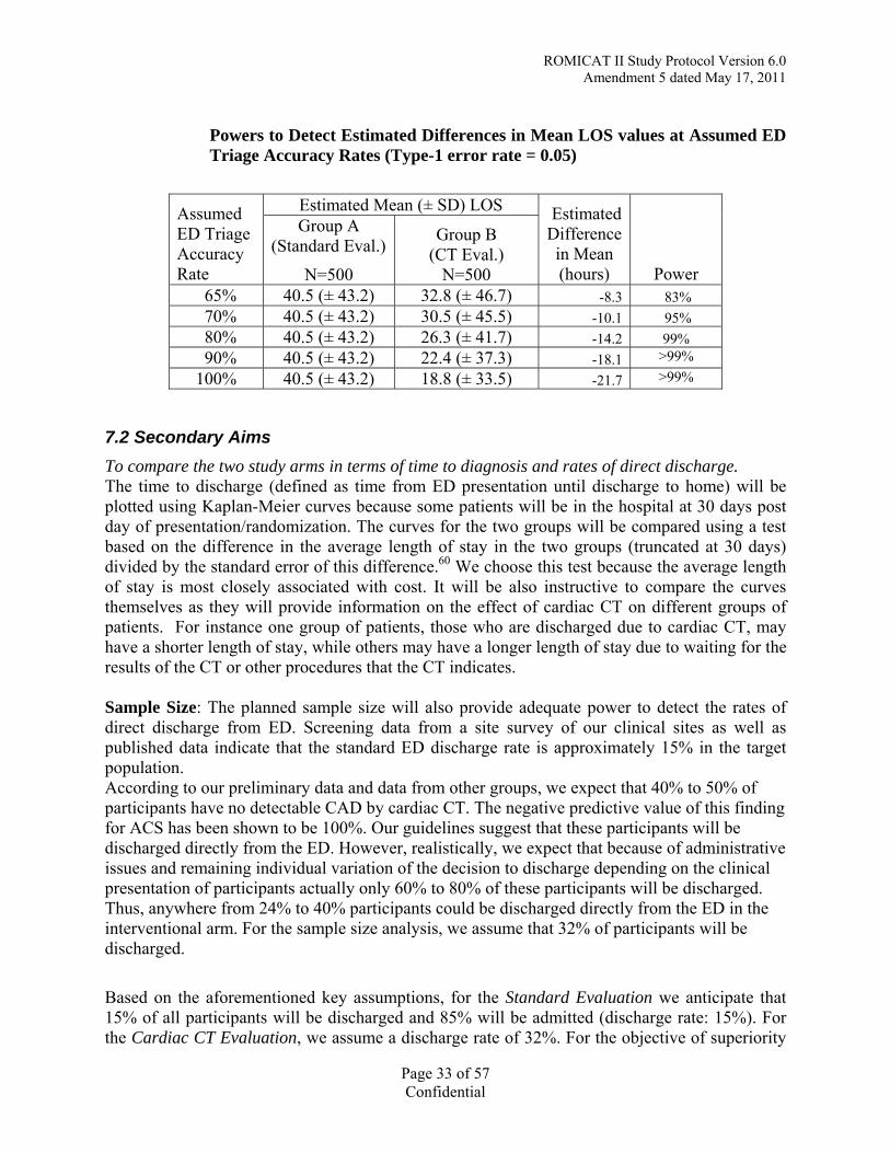
ROMICAT II Study Protocol Version 60 Amendment 5 dated May 17 2011
of discharge rates we will have a 100 chance at a significance level of 005 to detect a 17 increase in discharge rates from 15 to 32
The comparison of time to discharge among patients who are admitted to the hospital is complicated by the fact that the two groups will not be the same group of patients For instance patients who would have been discharged in the cardiac CT group but admitted in the usual care group may have a relatively shorter time post admission than patients who would have been admitted in both groups We have developed methods for analyzing these types of data using causal inference that will be applied to this analysis61 A similar analysis will be applied to other subgroups such as those with and without ACS
To compare the two study arms in terms of cardiac health care utilization during the index visit Health care utilization components will include invasive coronary angiograms and coronary revascularization
The analysis for this secondary aim will be performed from the intent to treat perspective Health care utilization will be assessed using medical record review Measures of key components of utilization will be compared across the two study groups using procedures appropriate for each measure (rate or count)
In particular the two arms will be compared in terms of the number of invasive coronary angiograms This secondary endpoint will be analyzed in terms of a non-inferiority trial In a non-inferiority trial the null hypothesis is that cardiac CT is worse than usual care in that the rate of coronary angiograms is increased and the alternative hypothesis is that it is not in worse and the rate of angiograms is the same The reason for reversing the usual null and alternative hypothesis is that the purpose of this analysis is to prove that cardiac CT is not worse than usual care this would not be accomplished by just showing that the difference between the two was not significant
Screening data from a survey of clinical sites as well as published data indicate that the standard rate of invasive coronary angiograms in the target population is approximately 10 According to our study guidelines we assume that most but not all participants with significant stenosis in CT (10 of participants) undergo invasive coronary angiography (notably those who also have a positive stress test and those who have high grade proximal stenosis or LAD stenosis) In addition we assume that a minority of participants with indeterminate stenosis (lt5) and positive stress testing undergo invasive coronary angiography Because of the moderate specificities of standard diagnostic testing up to 40 of participants who undergo cardiac catheterization have no significant CAD1516 Because the diagnostic accuracy of cardiac CT for the detection of coronary stenosis is comparable or better than standard diagnostic testing we believe that there will be no increase in the rates of invasive coronary angiograms If we define a 5 difference as an ldquoacceptablerdquo difference in cardiac catheterization rates than we will have an 84 chance of showing that the CT arm is significantly less than 5 worse than the standard arm at a one sided p=005 significance level
Page 34 of 57 Confidential
ROMICAT II Study Protocol Version 60 Amendment 5 dated May 17 2011
Another key measure of utilization is the number of coronary revascularization procedures In ROMICAT I there were 18 revascularization procedures performed in 368 patients (5) Thus we expect 5 procedures in the standard evaluation If we assume that not all participants with significant stenosis will undergo coronary revascularization (because if no MI it can be medically managed) and that cardiac CT may also avoid some angiograms by excluding disease than we can realistically assume that there is not a difference between the two study arms If we consider this in terms of a non-inferiority trial as above we will have 80 power to rule a difference of 35
To compare the two study arms in terms of cardiac health care utilization within one year after index hospitalization Health care utilization components will include invasive coronary angiograms cardiac related diagnostic and lab testing and ED visits
This analysis will be performed from the intent to treat perspective CP and CT related health care utilization will be assessed using medical record review Measures of key components of utilization will be compared across the two study groups using procedures appropriate for each measure (rate or count) We expect that the rate of additional cardiac work-up will be 29 in the SOC arm and will be 14 in the interventional arm We will have gt95 power to detect this difference We expect that the rate of hospital admissions with additional diagnostic cardiac testing will be 93 in the SOC arm and will be 4 in the interventional arm We will have gt95 power to detect this difference We expect that the rates of these measures will be associated with the CT findings at index hospitalization
To evaluate the cost and cost-effectiveness of incorporating cardiac CT into the standard ED evaluation of patients with acute chest pain These analyses will be performed by the Decision Analysis and Cost-Effectiveness Center (DACE) Therefore the sites will collect and provide the necessary data to the DACE and the costs will be derived from the cost-accounting database (TSI) As a first step incorporating cardiac CT into the standard evaluation of patients with acute chest pain will be tested for cost-saving over a 28-day and lifetime span as compared to standard care Further on cost-effectiveness of incorporating cardiac CT into the standard ED evaluation of patients with acute chest pain will be determined using first-order Monte Carlo microsimulations
To compare the two study arms in terms of rates of MACE (cardiac death AMI revascularization unstable angina and rehospitalization) within 28 days The analysis for this secondary aim will be performed from the intent to treat perspective The occurrence of MACE will be assessed via patient follow-up and medical chart review The primary analysis for this aim will be performed at the patient level by determining whether any MACE occurred or not Event rates will be estimated and compared between study groups using exact procedures In a secondary analysis the rates of each type of MACE will be examined separately
Sample Size- Safety Considerations- Missed ACS Mistaken discharges are the primary safety issue in this study All available data indicate that the absence of any detectable CAD (absence
Page 35 of 57 Confidential
ROMICAT II Study Protocol Version 60 Amendment 5 dated May 17 2011
of any plaque and stenosis) has a very high NPV for ACS (100 or close to 100)62 Thus we anticipate that the occurrence of ACS in participants discharged home directly from the ED in the interventional arm will be also minimal or even zero and similar to the standard of care If as demonstrated in ROMICAT I none of the participants without CAD will have MACE defined as ACS within 30 days after direct ED discharge this observation has 90 power to ensure that the true event rate is less than 1 These assumptions are supported by the results of the ERASE trial which reported similar and very minimal event rates in each group (only 2 pts with acute MI were mistakenly discharged 1 in each randomization group out of 1200 [event rate 00008]) In this trial we would have an 80 chance of detecting at least one occurrence of an inappropriately discharged participant if the true rate where over 1
To estimate and compare the sensitivity specificity NPV and PPV of contrast-enhanced CT and non-contrast CT with clinical diagnosis of ACS as the reference standard The analysis for this aim will utilize data on participants randomized to Group B In this arm each participant will undergo non-contrast CT and contrast-enhanced CT which will be interpreted as positive or negative for ACS Clinical determination of presence or absence of ACS will be used as the reference standard For each of the two types of CT we will estimate the sensitivity specificity and positive and negative predictive value of the modality for ACS Each measure of diagnostic or predictive performance will be compared between the two modalities using statistical techniques that account for the correlation due to the paired test design In addition to these measures we will estimate the proportion of cases with a clinical diagnosis of ACS and a negative non-contrast CT in which contrasted enhanced CT was positive for ACS This proportion provides a measure of the incremental value of contrast-enhanced CT for diagnosis ACS
73 Tertiary Aims To determine whether CT leads to increased test burden after the initial tests during index hospitalization
To compare the consistency of physician decision management depending on the results of the diagnostic testing (ie normal CT ndash what fraction is immediately discharged)
To determine whether hospital setting availability of Observation Unit CT experience of hospital and readers and reader performance are associated with primary or secondary outcomes
Analysis of Data on Women and Minorities We will analyze the effect of the intervention on women and minorities as outlined in the NIH guidelines Thus we include testing for gender-treatment and minority status-treatment interactions with the discharge rate and the angiogram rate
Page 36 of 57 Confidential
ROMICAT II Study Protocol Version 60 Amendment 5 dated May 17 2011
8 Data Entry and Site Monitoring
81 Data Collection Subject data will be collected by the research associates and paper records will be maintained at individual clinical sites The Data Coordinating and Statistical Center (DCSC) will develop a web based electronic case report form The de-identified study data will be entered into this electronic data capture system (EDC) and sent to the Data Coordinating and Statistical Center (DCSC) at Massachusetts General Hospital for further analysis
82 Site Monitoring Site visits will be performed on a regular basis by the Data Coordinating Center to ensure that all regulatory requirements are being met and to monitor the quality of the data collected Records of Institutional Review Board approvals and patientsrsquo charts will be examined on a spot check basis to evaluate the accuracy of the data entered into the database
9 Risk and Benefit Assessment
91 Risks of Nonionic Contrast Agent
Rare risks of the nonionic contrast agent bull Itchiness bull Rash bull Mild allergic reaction
Very rare risks of the nonionic contrast agent bull Kidney diseaseContrast-induced nephropathy bull Death
Regarding the risks of contrast material the rate of adverse reactions with nonionic contrast reagents is 0250 More than 90 of such adverse reactions are allergic-like very mild (itching rush) and can be very effectively treated with available drugs (ie antihistaminic) Severe reactions occur in 002 and one death occurred in 57739 injections of nonionic contrast material50
Contrast-induced nephropathy (CIN) CIN is expected to be a very rare event among this patient population because patients with already impaired renal function (the strongest predictor of CIN) will not be enrolled) This assumption is strongly supported by our preliminary experience from ROMICAT I In this study none of the participants had such an event (0368)
The very low risk associated with contrast administration has been documented in large scale trials previously In a randomized trial in 1196 participants the incidence rate of CIN defined as an increase in serum creatinine ge1mgdL from baseline within 48 to 72 hours following the injection of contrast material was 0 in 359 participants (0) who had no renal insufficiency and no diabetes melliltus51 The risk was 2 in 315 (06) in participants with no renal insufficiency but who had diabetes mellitus51 In this study the mean volume of contrast-agent administered
Page 37 of 57 Confidential
ROMICAT II Study Protocol Version 60 Amendment 5 dated May 17 2011
was 140 plusmn 57 mL which is much higher than the 80 mL of contrast that is administered for a coronary CTA The risk of CIN may increase if participants in the cardiac interventional arm will be admitted and subsequently undergo further contrast-related diagnostic testing However among 209 participants who had both a pulmonary angiogram and a contrast-enhanced spiral CT in the Prospective Investigation of Pulmonary Embolism Diagnosis II only one participant with diabetes mellitus had a transient episode of acute renal failure characterized by an increase in the serum creatinine level from 13 to 29 mgdL52 In this study the contrast-enhanced CT and pulmonary angiogram were performed within 22 hours The elevated creatinine returned to normal after administration of intravenous fluids Thus overall we expect that the occurrence of CIN will be a rare event in this study
92 Risks Associated With Radiation While the radiation dosage for CT scanning varies with the part of the body being scanned the exposure for this examination is approximately between 7 and 17 mSv This exposure is comparable or less than stress nuclear myocardial perfusion imaging (14ndash22 mSv) (personal communication with Prof Ronald Callahan Chairman of the MGH Radiation Safety Committee) In comparison the average yearly effective dose of natural background radiation is 25 mSv53 the allowed annual exposure of radiation workers is 50 mSv the allowed exposure in five years is 100 mSv (Table 3)54 Thus even if an enrolled participant would undergo cardiac CT followed by a conventional stress test and selective coronary angiography the participant would be well below the allowable annual effective dose for radiation workers The radiation dose from cardiac CT has not been shown to have any adverse effects
Radiation Exposure
Cardiac CT scanning results in a measurable radiation exposure55 In order to minimize risk for participants in this study we have implemented the following measures
bull Exclude women lt40 years of age bull Exclude pregnant and breast feeding women bull Include only women who have a negative pregnancy test bull Application of cardiac CT protocols with the goal to minimize radiation exposure (ie
use of tube modulationmdashdecrease of radiation exposure by 30 to 50 and prospective triggering in appropriate conditions)
bull Review and approval of these CT protocols by the CVCT Core Lab
General Risk Associated with Radiation Exposure
The risk of inducing a malignancy by exposure to radiation is estimated to be 5 cases of malignancy for each 100 individuals exposed to an effective dose of 1 Sv54 The risk is estimated to be higher in infants and children and lower in the elderly The data which these estimates are based on are weak and apply to relatively high levels of radiation exposure56 and it remains unclear whether there is a linear relation in lower levels of radiation Assuming a linear proportion with a low level of radiation exposure to 1 mSv would lead to 5 malignancies in 100000 individuals exposed and a dose of 8 mSV would lead to 40 malignancies in 100000 individuals56 Small amounts of radiation exposure less than 100 mSv have not been clearly
Page 38 of 57 Confidential
ROMICAT II Study Protocol Version 60 Amendment 5 dated May 17 2011
shown to have harmful effects The amount of radiation to which patients will be exposed in this study is less than the amount that is known to cause a measurable harmful effect Even so it is generally assumed that a small amount of radiation will have a small chance of producing a harmful effect These extrapolated risks of the effects of small amounts of radiation are probably overestimates because the estimates ignore the fact that the body can repair the damage done by radiation if the repair mechanisms are not overwhelmed Radiation is a well-known teratogenic agent and the developing fetus is much more sensitive to radiation than the mother
Table 3 Estimated radiation exposure for diagnostic tests background radiation and exposure limits
Examination Effective Dose (mSv) Chest PA and lateral 007 Myocardial Perfusion Imaging (Rest and 14ndash22Stress) Thorax CT for the Exclusion of Pulmonary 7-12Embolism 64-Slice CT coronary angiogram 7ndash17 2-view mammogram 30 Natural yearly background radiation 25 Max exposure in radiation workers 50
Lifetime Cancer Risk Attributable to Cardiac CT
The absolute cancer risk has been described recently and recommendations have been made57
The life time cancer risk from cardiac CT decreases with age and is higher in women than in men in particular exposure of younger women should be avoided In our study these recommendations have been incorporated In comparison the absolute cancer risk for women to develop breast cancer is 13 and for men to develop colon cancer is 7 the risk attributed to a cardiac CT scan is 1466 vs 1715 for a 60 year old women and 11241 vs 11911 for 60 year old men (for CT without and with tube modulation (female 21 and 14 mSv male 15 and 9 mSv respectively)57 Thus the incremental risk induced by cardiac CT compared to the overall risk of the population to develop cancer seems moderate
Effect of the Implementation of Cardiac CT in the ED Evaluation of Patients with Acute Chest Pain on a Population Basis
The number of CT scans performed in the US was 62 million in 200658 Imaging of the abdomen (50) and the head (33) were major single contributors Specific concern was given to the rise of pediatric CT imaging (from 6 to now 11) and screening the adult asymptomatic population (ie lung heart) Implementation of cardiac CT in the ED could lead to a small increase of the number of overall CT procedures However the effects of the implementation of cardiac CT in the ED would be minimal if 1) other procedures associated with radiation doses would be saved 2) repeated testing currently performed in 25 of patients would be minimized and 3) rates of unnecessary purely diagnostic cardiac catheterizations would
Page 39 of 57 Confidential
ROMICAT II Study Protocol Version 60 Amendment 5 dated May 17 2011
decrease Furthermore already cardiac CT scanning could be performed using single phase acquisition with prospective triggering which would decrease the dose to lt5 mSv In addition MD over-read will be performed for internal quality control assessment of findings related to CAD and significant cardiac findings such as pulmonary embolism and aortic dissection to increase awareness to sites of non-coronary findings
93 Other Risks Additional Risks of CT Scans bull Discomfort bull Claustrophobia
Risks of IV Needle Placement
bull Hemorrhage (hematoma at the injection site) bull Infection (catheter related infection) at the injection site bull Minor discomfort bull Bleeding bull Infection bull Bruising
94 Potential Benefits bull Subjects may benefit from randomization into the interventional arm of trial from detection
of pathology that may not have been otherwise detected during SOC (eg a non-significant coronary stenosis or non-obstructive coronary artery disease that would likely not have been detected by a stress test)
bull Data from prior studies indicate that patients in the interventional arm may be earlier discharged and may not have to spend the night in the hospital there is also the chance that alternative diagnosis such as pneumonia or Pulmonary embolism may be detected - patients may avoid the dual radiation exposure of a CT protocol solely to evaluate for pulmonary embolus followed by a SPECT myocardial perfusion scan
bull Subjects may also benefit in their assessment for long term prognosis and risk for adverse cardiac events and may receive preventive medication as a result of CT
10 Ethical Considerations This study is to be conducted according to US and international standards of Good Clinical Practice (International Conference of Harmonization [ICH] guidelines) applicable government regulations and NIH research policies and procedures
This protocol and any amendments will be submitted to a properly constituted independent Institutional Review Board (IRB) for formal approval of the study conduct The decision of the IRB concerning the conduct of the study will be made in writing to the investigator and a copy of this decision will be provided to the ROMICAT PI before implementation of the study
Page 40 of 57 Confidential
ROMICAT II Study Protocol Version 60 Amendment 5 dated May 17 2011
All study participants in this study will be given an IRB-approved site-specific ICF describing the study and providing sufficient information for participants to make informed decisions about their participation in this study The study participant MUST be consented with the ECIRBshyapproved ICF before the participant is subjected to any study procedures The IRB-approved ICF MUST be signed and dated by the study participant or legally acceptable representative and the investigator-designated research staff obtaining the consent before the participant is subjected to any study procedures
101 Inclusion of Women and Minorities The participating institutions will not exclude potential participants from participating in this or any study solely based on ethnic origin or socioeconomic status Every attempt will be made to enter all eligible participants into this protocol and therefore address the study objectives in a patient population representative of the entire English-speaking population at risk for ACS treated by the institution
Both men and women and members of all ethnic groups are eligible for this trial In conformance with the National Institutes of Health (NIH) Revitalization Act of 1993 with regard to inclusion of women and minorities in clinical research the projected gender and minority accruals are shown below
Table 1 Gender and Minority Accrual Estimates
Ethnic Category SexGender Females Males Total
Hispanic or Latino 66 79 145 Not Hispanic or Latino 384 471 855
Ethnic Category Total of all participants
450 550 1000
Racial Category American Indian or Alaskan Native 2 2 4 Asian 35 42 77 Black or African American 125 153 278 Native Hawaiian or other Pacific
Islander 2 2 4
White 254 312 566 OtherUnknown 32 39 71
Racial Category Total of allparticipants
450 550 1000
Page 41 of 57 Confidential
ROMICAT II Study Protocol Version 60 Amendment 5 dated May 17 2011
11 Adverse Event Reporting Mistaken discharges are the primary safety issue in this study All available data indicate that the absence of any detectable CAD (absence of any plaque and stenosis) has a very high NPV for ACS (100 or close to 100)62 Thus we anticipate that the occurrence of ACS in participants discharged home directly from the ED in the cardiac interventional arm will be also minimal or even zero and similar to the standard of care If as demonstrated in ROMICAT I none of the participants without CAD will have MACE defined as ACS within 30 days after direct ED discharge this observation has 90 power to ensure that the true event rate is less than 1 These assumptions are supported by the results of the ERASE trial which reported similar and very minimal event rates in each group (only 2 pts with acute MI were mistakenly discharged 1 in each randomization group out of 1200 [event rate 00008]) In this trial we would have an 80 chance of detecting at least one occurrence of an inappropriately discharged participant if the true rate where over 1
At the time of ED discharge investigators will determine at if any adverse events have occurred An Adverse Event (AE) is any untoward medical occurrence in a study participant that may or may not have a causal relationship with the study intervention An AE can therefore be any unfavorable and unintended sign (including an abnormal laboratory or physiological finding) symptom or disease temporally associated with the use of a medical treatment or procedure regardless of whether it is considered related to the medical treatment or procedure (attribution of unrelated unlikely possible probable or definite) Abnormal results of diagnostic procedures are considered to be AEs if the abnormality
bull results in study withdrawal bull is associated with a serious adverse event (SAE) bull is associated with clinical signs or symptoms bull leads to additional treatment or to further diagnostic tests bull is considered by the investigator to be of clinical significance
Serious Adverse Event (SAE) An SAE is defined as any untoward medical occurrence that
bull results in death or bull is life-threatening (at the time of the event) or bull requires inpatient hospitalization or prolongation of an existing hospitalization or bull results in persistent or significant disability or incapacity or bull is a congenital anomalybirth defect
Reportable Adverse Events Any untoward medical occurrence that occurs after randomization and prior to discharge from the ED should be reported to the CCC by completing the electronic adverse event case report form
Immediately Reportable Adverse Events
Page 42 of 57 Confidential
ROMICAT II Study Protocol Version 60 Amendment 5 dated May 17 2011
Any adverse event that is judged by the investigator to be serious AND unexpected AND related to study procedures should be reported within 24 hours of discovery by phone or email to the Coordinating Center The local Institutional Review Board must also be notified in a timely manner
Investigators must also report Unanticipated Problems regardless of severity associated with the study procedures within 24 hours An unanticipated problem is defined as follows
Unanticipated Problem (UP) any incident experience or outcome that meets all of the following criteria
bull Unexpected in terms of nature severity or frequency given the research procedures that are described in the protocol-related documents such as the IRB-approved research protocol and informed consent document and the characteristics of the subject population being studied
bull Related or possibly related to participation in the research in this guidance document possibly related means there is a reasonable possibility that the incident experience or outcome may have been caused by the procedures involved in the research
bull Suggests that the research places subjects or others at a greater risk of harm (including physical psychological economic or social harm) than was previously known or recognized
12 References 1 Lee TH Rouan GW Weisberg MC Brand DA et al Clinical characteristics and natural
history of patients with acute myocardial infarction sent home from the emergency room Am J Cardio 198760(4)219-224
2 Swap CJ Nagurney JT Value and limitations of chest pain history in the evaluation of patients with suspected acute coronary syndromes JAMA 2005294(20)2623-2629
3 Limkakeng A Jr Gibler WB Pollack C et al Combination of Goldman risk and initial cardiac troponin I for emergency department chest pain patient risk stratification Acad Emerg Med 20018(7)696-702
4 Zimmerman J Fromm R Meyer D Boudreaux A et al Diagnostic marker cooperative study for the diagnosis of myocardial infarction Circulation 199999(13)1671-1677
5 Fesmire FM Hughes AD Fody EP et al The Erlanger chest pain evaluation protocol a one-year experience with serial 12-lead ECG monitoring two-hour delta serum marker measurements and selective nuclear stress testing to identify and exclude acute coronary syndromes Ann Emerg Med 200240(6)584-594
6 Hedges JR Young GP Henkel GF et al Serial ECGs are less accurate than serial CKshyMB results for emergency department diagnosis of myocardial infarction Ann Emerg Med 199221(12)1445-1450
Page 43 of 57 Confidential
ROMICAT II Study Protocol Version 60 Amendment 5 dated May 17 2011
7 Selker HP Beshansky JR Griffith JL et al Use of the acute cardiac ischemia time-insensitive predictive instrument (ACI-TIPI) to assist with triage of patients with chest pain or other symptoms suggestive of acute cardiac ischemia A multicenter controlled clinical trial Ann Intern Med 1998129(11)845-855
8 Hollander JE Sease KL Sparano DM et al Effects of neural network feedback to physicians on admitdischarge decision for emergency department patients with chest pain Ann Emerg Med 200444(3)199-205
9 McCaig LF Burt CW National Hospital Ambulatory Medical Care Survey 2002 emergency department summary Adv Data Mar 18 2004(340)1-34
10 McCaig LF Nawar EW National Hospital Ambulatory Medical Care Survey 2004 emergency department summary Adv Data 20063721-29
11 Fleischmann KE Hunink MG Kuntz KM Douglas PS Exercise echocardiography or exercise SPECT imaging A meta-analysis of diagnostic test performance J Nucl Cardiol 20029(1)133-134
12 Klocke FJ Baird MG Lorell BH et al ACCAHAASNC guidelines for the clinical use of cardiac radionuclide imaging--executive summary a report of the American College of CardiologyAmerican Heart Association Task Force on Practice Guidelines (ACCAHAASNC Committee to Revise the 1995 Guidelines for the Clinical Use of Cardiac Radionuclide Imaging) Circulation 2003108(11)1404-1418
13 Cheitlin MD Armstrong WF Aurigemma GP et al ACCAHAASE 2003 guideline update for the clinical application of echocardiography summary article a report of the American College of CardiologyAmerican Heart Association Task Force on Practice Guidelines (ACCAHAASE Committee to Update the 1997 Guidelines for the Clinical Application of Echocardiography) Circulation 2003108(9)1146-1162
14 Zalenski RJ Rydman RJ Ting S et al A national survey of emergency department chest pain centers in the United States Am J Cardio 199881(11)1305-1309
15 Amsterdam EA Kirk JD Diercks DB et al Immediate exercise testing to evaluate low-risk patients presenting to the emergency department with chest pain J Am Coll Cardiol 200240(2)251-256
16 Jeetley P Burden L Greaves K Senior R Prognostic value of myocardial contrast echocardiography in patients presenting to hospital with acute chest pain and negative troponin Am J Cardiol 200799(10)1369-1373
17 Burt CW Summary statistics for acute cardiac ischemia and chest pain visits to United States EDs 1995-1996 Am J Emerg Med 199917(6)552-559
18 Lee TH Goldman L Evaluation of the patient with acute chest pain N Engl J Med 2000342(16)1187-1195
Page 44 of 57 Confidential
ROMICAT II Study Protocol Version 60 Amendment 5 dated May 17 2011
19 Tosteson AN Goldman L Udvarhelyi IS Lee TH Cost-effectiveness of a coronary care unit versus an intermediate care unit for emergency department patients with chest pain Circulation 199694(2)143-150
20 Conti A Paladini B Magazzini S Toccafondi S et al Chest pain unit management of patients at low and not low-risk for coronary artery disease in the emergency department A 5-year experience in the Florence area Eur J Emerg Med 20029(1)31-36
21 Solinas L Raucci R Terrazzino S et al Prevalence clinical characteristics resource utilization and outcome of patients with acute chest pain in the emergency department A multicenter prospective observational study in north-eastern Italy Ital Heart J 20034(5)318-324
22 Budoff MJ Achenbach S Blumenthal RS et al Assessment of coronary artery disease by cardiac computed tomography a scientific statement from the American Heart Association Committee on Cardiovascular Imaging and Intervention Council on Cardiovascular Radiology and Intervention and Committee on Cardiac Imaging Council on Clinical Cardiology Circulation 2006114(16)1761-1791
23 Hamon M Biondi-Zoccai GG Malagutti P et al Diagnostic performance of multislice spiral computed tomography of coronary arteries as compared with conventional invasive coronary angiography a meta-analysis J Am Coll Cardiol 200648(9)1896-1910
24 Stein PD Beemath A Kayali F et al Multidetector computed tomography for the diagnosis of coronary artery disease a systematic review Am J Med 2006119(3)203shy216
25 Scheffel H Alkadhi H Plass A et al Accuracy of dual-source CT coronary angiography First experience in a high pre-test probability population without heart rate control Eur Radiol 200616(12)2739-2747
26 Dewey M Teige F Schnapauff D et al Noninvasive detection of coronary artery stenoses with multislice computed tomography or magnetic resonance imaging Ann Intern Med 2006145(6)407-415
27 Martuscelli E Romagnoli A DEliseo A et al Accuracy of thin-slice computed tomography in the detection of coronary stenoses Eur Heart J 200425(12)1043-1048
28 Hoffmann MH Shi H Schmitz BL et al Noninvasive coronary angiography with multislice computed tomography JAMA 2005293(20)2471-2478
29 Achenbach S Moselewski F Ropers D et al Detection of calcified and noncalcified coronary atherosclerotic plaque by contrast-enhanced submillimeter multidetector spiral computed tomography a segment-based comparison with intravascular ultrasound Circulation 2004109(1)14-17
30 Leber AW Knez A von Ziegler F et al Quantification of Obstructive and Nonobstructive Coronary Lesions by 64-Slice Computed Tomography A Comparative
Page 45 of 57 Confidential
ROMICAT II Study Protocol Version 60 Amendment 5 dated May 17 2011
Study With Quantitative Coronary Angiography and Intravascular Ultrasound JACC 200546(1)154
31 Belge B Coche E Pasquet A et al Accurate estimation of global and regional cardiac function by retrospectively gated multidetector row computed tomography comparison with cine magnetic resonance imaging Eur Radiol 200616(7)1424-1433
32 Raman SV Shah M McCarthy B et al Multi-detector row cardiac computed tomography accurately quantifies right and left ventricular size and function compared with cardiac magnetic resonance Am Heart J 2006151(3)736-744
33 Salm LP Schuijf JD de Roos A et al Global and regional left ventricular function assessment with 16-detector row CT comparison with echocardiography and cardiovascular magnetic resonance Eur J Echocardiogr 20067(4)308-314
34 Schuijf JD Bax JJ Salm LP et al Noninvasive coronary imaging and assessment of left ventricular function using 16-slice computed tomography Am J Cardiol 200595(5)571shy574
35 Mahnken AH Koos R Katoh M et al Sixteen-slice spiral CT versus MR imaging for the assessment of left ventricular function in acute myocardial infarction Eur Radiol 200515(4)714-720
36 Dirksen MS Jukema JW Bax JJ et al Cardiac multidetector-row computed tomography in patients with unstable angina Am J Cardiol 200595(4)457-461
37 Hoffmann U Pena AJ Moselewski F ettt al MDCT in early triage of patients with acute chest pain Am J Roentgenol 2006187(5)1240-1247
38 Hoffmann U Nagurney JT Moselewski F et al Coronary multidetector computed tomography in the assessment of patients with acute chest pain Circulation 2006114(21)2251-2260
39 Rubinshtein R Halon DA Gaspar T et al Usefulness of 64-Slice Cardiac Computed Tomographic Angiography for Diagnosing Acute Coronary Syndromes and Predicting Clinical Outcome in Emergency Department Patients With Chest Pain of Uncertain Origin Circulation 2007
40 Goldstein JA Gallagher MJ ONeill WW et al A randomized controlled trial of multi-slice coronary computed tomography for evaluation of acute chest pain JACC 200749(8)863-871
41 Laudon DA Vukov LF Breen JF et al Use of electron-beam computed tomography in the evaluation of chest pain patients in the emergency department Ann Emerg Med 199933(1)15-21
Page 46 of 57 Confidential
ROMICAT II Study Protocol Version 60 Amendment 5 dated May 17 2011
42 Georgiou D Budoff MJ Kaufer E et al Screening patients with chest pain in the emergency department using electron beam tomography a follow-up study JACC 200138(1)105-110
43 Dewey M Hoffmann H Hamm B Multislice CT coronary angiography effect of sublingual nitroglycerine on the diameter of coronary arteries Rofo 2006178(6)600shy604
44 Hong C Bae KT Pilgram TK Coronary artery calcium accuracy and reproducibility of measurements with multi-detector row CT--assessment of effects of different thresholds and quantification methods Radiology 2003227(3)795-801
45 Agatston AS Janowitz WR Hildner FJ et al Quantification of coronary artery calcium using ultrafast computed tomography JACC 199015(4)827-832
46 Ropers D Baum U Pohle K et al Detection of coronary artery stenoses with thin-slice multi-detector row spiral computed tomography and multiplanar reconstruction Circulation 2003107(5)664-666
47 Nieman K Oudkerk M Rensing BJ et al Coronary angiography with multi-slice computed tomography Lancet 2001357(9256)599-603
48 Hoffmann U Moselewski F Cury RC et al Predictive value of 16-slice multidetector spiral computed tomography to detect significant obstructive coronary artery disease in patients at high risk for coronary artery disease patient-versus segment-based analysis Circulation 2004110(17)2638-2643
49 Cerqueira MD Weissman NJ Dilsizian V ettt al Standardized myocardial segmentation and nomenclature for tomographic imaging of the heart a statement for healthcare professionals from the Cardiac Imaging Committee of the Council on Clinical Cardiology of the American Heart Association Circulation 2002105(4)539-542
50 Cochran ST Bomyea K Sayre JW Trends in adverse events after IV administration of contrast media Ajr 2001176(6)1385-1388
51 Rudnick MR Goldfarb S Wexler L et al Nephrotoxicity of ionic and nonionic contrast media in 1196 patients a randomized trial The Iohexol Cooperative Study Kidney Int 199547(1)254-261
52 Stein PD Fowler SE Goodman LR et al Multidetector computed tomography for acute pulmonary embolism New Engl J Med 2006354(22)2317-2327
53 Robinson A Protection and Patient doses in diagnostic radiology New York Churchill Livingstone 1997
54 Radiological protection and safety in medicine A report of the International Commission on Radiological Protection Ann ICRP 199626(2)1-47
Page 47 of 57 Confidential
ROMICAT II Study Protocol Version 60 Amendment 5 dated May 17 2011
55 Hunold P Vogt FM Schmermund A et al Radiation exposure during cardiac CT effective doses at multi-detector row CT and electron-beam CT Radiology 2003226(1)145-152
56 Diederich S Lenzen H Radiation exposure associated with imaging of the chest comparison of different radiographic and computed tomography techniques Cancer 200089(11 Suppl)2457-2460
57 Einstein AJ Moser KW Thompson RC et al Radiation dose to patients from cardiac diagnostic imaging Circulation2007116(11)1290-1305
58 Brenner DJ Hall EJ Computed tomography--an increasing source of radiation exposure N Engl J Med 2007357(22)2277-2284
59 Udelson JE Beshansky JR Ballin DS et al Myocardial perfusion imaging for evaluation and triage of patients with suspected acute cardiac ischemia a randomized controlled trial JAMA 2002288(21)2693-2700
60 Hastie T Tibshirani R Friedman J Additive Models Trees and Related Methods The Elements of Statistical Learning New York Springer 2001266-275
61 Hayden D Pauler D Schoenfeld D An Estimator for Treatment Comparisons among Survivors in Randomized Trials Biometrics 200561(1)305-310
62 Hoffmann U Shapiro M Coronary multidetector computed tomography a new standard for preoperative risk assessment J Am Coll Cardiol 200647(10)2025-2026
63 Budoff MJ Cohen MC Garcia MJ Hodgson JMcB Hundley WG Lima AC Manning WJ Pohost GM Raggi PM Rodgers GP Rumberger JA Taylor AJ ACCAHA clinical competence statement on cardiac imaging with computed tomography and magnetic resonance a report of the American College of Cardiology FoundationAmerican Heart Association American College of Physicians Task Force on Clinical Competence (ACCAHA Committee on CV Tomography) J Am Coll Cardiol 200546383ndash402 PMID 16046290
Page 48 of 57 Confidential
ROMICAT II Study Protocol Version 60 Amendment 5 dated May 17 2011
Appendix A Cardiac CT Imaging Protocol Guidelinesdagger
Cardiac CT Imaging Diagnostic Intervention to Be Tested
Equipment Cardiac CT imaging will be performed using at least 64-slice CT scanner technology according to the vendor specific protocol for the evaluation of the coronary arteries and LV function (Table 2)
Table 2 Overview of vender-specific protocols as used in the proposed trial CT Scanner Spatial
Resolution Slice Collimation
Gantry Rotation Time Temporal Resolution
Tube Voltage Tube Current
Estimated Radiation Exposure (mSv)
Siemens Definition Dual Source 64-row MDCT 2 x 64 x 06 mm 330 ms83 ms 100-120 Kvp 850
effective mAs 7-15 mSv Toshiba Acquillion 64-row MDCT 64 x 045 mm 400 ms200 ms 100-120 Kvp 700
mAs 10-14 mSv Siemens Somatom 64-row MDCT 64 x 06 mm 330 ms165 ms 100-120 Kvp 850
mAs 7-14 mSv GE Lightspeed VCT 64-row MDCT 64 x 06 mm 350 ms175 ms 100-120 Kvp 750
mAs 11-17 mSv Philips Brilliance iCT 256-row MDCT 64 x 0624mm 270 ms135 ms 100-120 kVp 850
mAs 7-13 mSv
includes radiation exposure from CAC imaging assumes the use of tube modulation Actual radiation doses will depend on the subjectsrsquo body habitus as well as the rate and regularity of the heart during acquisition
Image Acquisition Based on preliminary data from the clinical sites the image acquisition takes approximately 20 minutes The participant will be transported to the CT suite and returned by qualified study personnel Continuous heart rate and rhythm will be monitored during the study exam with 4shylead ECG electrodes Blood pressure and heart rate will be measured before and after the study exam
General recommendations include bull Minimal flow rate 5mlsec
bull Retrospective ECG triggering with use of tube modulation techniques to permit assessment of LV function
dagger This appendix is intended as example of a cardiac CT imaging protocol Please refer to the current version of the Massachusetts General Hospital Cardiovascular Imaging Core Laboratory (CVIC) Study Imaging Procedure for ROMICAT II study imaging procedures
Page 49 of 57 Confidential
ROMICAT II Study Protocol Version 60 Amendment 5 dated May 17 2011
bull Prospective triggering in subjects with appropriate conditions to further minimize radiation exposure
bull 100 kvp if BMI lt30 kgm2
bull 120 kvp if BMI gt 30 kgm2
In general the image acquisition protocol consists of the following 3 steps
Step 1mdashParticipant Preparation Participants will be instructed and trained in breath holding ECG electrodes will be mounted on the chest to enable co-registration of image acquisition and ECG signal At sites WITHOUT a dual source computed tomography (DSCT) scanner all participants with a heart rate gt65 beats per minute will receive beta blocker (5 to 20 mg Metoprolol IV) to optimize image quality and 600 mcg sublingual nitroglycerin to maximally dilate the coronary arteries unless their systolic blood pressure is lt100 mm Hg or other contraindications are present43 At sites with a DSCT scanner due to the improved temporal resolution beta blockers are not required though may be given and participants will receive sublingual nitroglycerin if there are no contraindications
Step 2mdashImage Acquisition All image acquisitions will be performed during a breath hold on inspiration with the following acquisitions
a TopogrammdashThe cardiac CT imaging protocol will start with a topogram of the chest permitting the localization of the position of the heart
b Noncontrast calcium score scanmdashA prospective gated low dose non-contrast CT scan (100 kVp for BMIlt30 or 120 kVp for BMI gt30 320 mA) with scan initiation at 70 (Siemens scanners) or 75 (all other manufacturers) of the RR-interval will be performed44 Contiguous 25 to 3 mm images will be acquired to quantify the presence and amount of coronary artery calcification
c Contrast-enhanced CT angiographymdashImage acquisition will conclude with a contrast-enhanced CT scan to evaluate the presence of coronary atherosclerotic plaque significant coronary artery stenosis and global and regional LV dysfunction For this scan on average 90 mL of contrast will be injected as a bolus at a rate of 5-6 mLs to opacify the lumen of the coronary arteries the aorta and the left ventricle Appropriate timing of the contrast bolus will be ensured by either the determination of the transit time or the bolus trigger technique Scanning will be performed continuously throughout the cardiac cycle Using ECG gating the tube current will be reduced during systole according to each manufacturerrsquos specific protocol to minimize radiation exposure Alternatively in subjects where prospective triggering is appropriate it may be performed per the manufacturerrsquos specific protocol to further reduce radiation exposure
Step 3mdashImage Reconstruction The following data sets will be reconstructed immediately after the scanning on a dedicated offline work station 1) axial images from the non-contrast-enhanced scan for the quantification of CAC (pixel matrix 512 x 512 field of view [FOV] 25 cm) 2) two end-diastolic and one end-systolic data set (05-075 mm thick axial images 50 overlap) from the contrast-enhanced CT scan for the
Page 50 of 57 Confidential
ROMICAT II Study Protocol Version 60 Amendment 5 dated May 17 2011
detection of coronary plaque and stenosis (pixel matrix 512 x 512 FOV 25 cm) 3) one multiphase reformatted dataset of 15 mm thick axial images at 10 increments (10 phases) for single source CT scanners or 5 increments (20 phases) for dual-source CT scanners throughout the cardiac cycle from the onset of the R-wave for the assessment of global and regional LV function in a cine mode and 4) a full field of view with 3 mm thick axial images covering the portions of the thorax acquired during the cardiac CT scan for the assessment of major incidental findings All CT data sets will be transmitted to the CVIC Labrsquos picture archiving and communication system (PACS) via internet using a dedicated routing system (Code of Federal Regulatory compliant) and stored
Image Evaluation Reconstructed data sets will be evaluated for the presence of CAC coronary atherosclerotic plaque and stenosis and global and regional LV function by dedicated highly trained cardiac CT imagers using established criteria22
Step 1mdashCoronary Artery Calcification All non-contrast enhanced CT scans will be assessed for the presence of CAC If present the amount of CAC will be quantified using a dedicated semiautomatic software and expressed as an Agatston Score (AS) as described elsewhere45
Step 2mdashAssessment for Stenosis Contrast-enhanced CT images will be qualitatively evaluated for the presence of coronary atherosclerotic plaque and significant coronary artery stenosis using axial data sets thin slice (5 mm) reconstructions orthogonal and perpendicular to the vessel centerline and short axis cross-sectional reconstructions Assessment will include each of the major epicardial vessels (left main left anterior descending left circumflex and right coronary artery) and their side branches46ndash48
Step 3mdashGlobal and Regional LV Function Global and regional LV function will be assessed visually with the multiphase reformatted dataset in cine mode to correspond to those typically used in echocardiography including LV horizontal long-axis (4-chamber view) vertical long-axis (2-chamber view) LV outflow tract long-axis (parasternal long axis) and short-axis view Global LV function will be reported as normal or impaired (mild moderate or severe) Regional wall motion (RWM) of the myocardium will be assessed to be normal or abnormal (hypokinesia akinesia dyskinesia aneurysm) in short-axis segments of the LV corresponding to the AHAACCASE 17-segment model Hypokinesia will be defined as a segment with impaired thickening and motion akinesia by absent thickening and motion dyskinesia as a paradoxical outward motion of the segment during systole and an aneurysm as a segment with wall thinning and diastolic deformation The abnormality has to be present in at least two contiguous myocardial segments or in one segment visualized in two different views
Step 4mdashMajor Incidental Findings The full field of view will be assessed by a CT reader for the presence of major cardiac and non-cardiac incidental findings such as aortic dissection pericardial effusion pulmonary embolism pneumothorax or mediastinal masses
Page 51 of 57 Confidential
ROMICAT II Study Protocol Version 60 Amendment 5 dated May 17 2011
CT Reporting Cardiac CT Findings
The Cardiac CT Report will be reported and included in the hospitalrsquos electronic medical record which is accessible to all treating physicians and will be the basis for in-hospital patient management The triage and management of the patient will be at the discretion of the ED physician
Image Submissions The protocol-required images must be in DICOM format and submitted to the Cardiovascular Imaging Core Laboratory (CVIC) via the internet using secure File Transfer Protocol (FTP) along with an Imaging Transmittal Worksheet The submission procedure will be detailed in the CVIC site operations manual
Page 52 of 57 Confidential
ROMICAT II Study Protocol Version 60 Amendment 5 dated May 17 2011
Appendix B Adjudication of Events by Clinical Events Committee
Introduction
Because patient safety is one of the concerns in a trial assessing new technology clinical events and serious adverse events will be independently adjudicated by the Clinical Events Committee under the direction of Stephen Wiviott MD This committee will adjudicate events in all subjects who potentially could have a major adverse cardiovascular event (MACE) such as death unstable angina pectoris (UA) myocardial infarction (MI) or coronary revascularization that occur after symptom assessment at patientrsquos initial emergency department (ED) presentation There is a possibility that MACE will occur during index hospitalization For example a second episode of chest pain diagnosed as acute coronary syndrome (ACS) occurring after the chest pain the patient presented with has resolved and all diagnostic tests were negative or recurrence of symptoms with or without hospitalization and re-hospitalization with or without testing
Definitions of Adverse Cardiovascular Events Adverse cardiovascular events comprise cardiovascular death MI unstable angina pectoris (UAP) coronary revascularization andor re-hospitalization that are distinct from the qualifying event (after patientrsquos initial ED presentation)
Definitions of events are listed below
Cardiovascular Death Any sudden cardiac death death due to acute myocardial infarction death due to heart failure death due to stroke and death due to other cardiovascular causes In addition any death without a clear non-cardiovascular cause or a death without known cause will be considered cardiovascular death
MI In Subjects with no recent revascularization in whom normal biomarkers were never elevated or have been documented to return to normal after a qualifying (or recent) MI who meet the following criteria
1) Typical cardiac biomarker rise andor fall AND at least one of the following a) Ischemic discomfort at rest lasing ge10 minutes b) ECG changes indicative of ischemia (ST elevation ge01 mV or ST
depression ge005 mV or new T-wave inversions OR
2) Development of new abnormal Q waves (ge30 msec in duration and ge1 mm in depth) in gt2 contiguous precordial leads or ge2 adjacent limb leads or increase R amplitude in V1-V3 consistent with posterior infarction
OR
3) Pathologic findings of an acute MI
Page 53 of 57 Confidential
ROMICAT II Study Protocol Version 60 Amendment 5 dated May 17 2011
4) Sudden unexpected cardiac death including cardiac arrest often with symptoms suggestive of myocardial ischemia accompanied by presumably new ST elevation or new LBBB or evidence of fresh thrombus in a coronary artery by angiography andor at autopsy but death occurring before blood samples could be obtained or at a time before the appearance of cardiac biomarkers in the blood
In subjects with percutaneous coronary intervention within 48 hours an elevation of CK-MB gt3x ULN distinct from a prior event will be considered to be a procedural MI
In subjects with CABG within 48 hours an elevation of CK-MB gt 10x ULN distinct from a prior event will be considered to be a procedural MI
UA An event not meeting the definition of myocardial infarction and with the following characteristics Chest pain or anginal equivalent at rest or in accelerating pattern AND at least one of the following objective signs
a) New ST-segment changes
b) New TWI
c) Positive stress test with imaging showing ischemia
d) Positive stress test without imaging resulting in increased anginal medication
e) Cath ge70 stenosis or thrombus
Urgent Coronary Revascularization Ischemic discomfort or equivalent meeting the following criteria
a) Lasting ge10 minutes at rest or repeated episodes at rest lasting ge5 minutes considered to be myocardial ischemia upon final diagnosis
AND
b) Prompting coronary revascularization during an unscheduled visit to healthcare facility or during an unplanned (or prolonged) hospitalization for these symptoms
Note Attempted revascularization procedures even if not successful will be counted Potential ischemic events meeting the criteria for myocardial infarction will not be adjudicated as urgent coronary revascularization
Re-hospitalization Coronary ischemia requiring re-hospitalization is defined as an event not meeting the definitions of myocardial infarction or urgent coronary revascularization ischemic discomfort or equivalent meeting the following criteria
a) Lasting ge10 minutes at rest or repeated episodes at rest lasting ge5 minutes or an accelerating pattern of ischemic discomfort (episodes that
Page 54 of 57 Confidential
ROMICAT II Study Protocol Version 60 Amendment 5 dated May 17 2011
are more frequent severe longer in duration and precipitated by minimal exertion) considered to be myocardial ischemia upon final diagnosis
AND
b) Prompting hospitalization (including overnight stay on an inpatient unit) within 48 hours of the most recent symptoms or prolonging hospitalization if occurring during existing hospitalization
AND
c) At least one of the following additional criteria for coronary artery disease andor ischemia
New andor dynamic ST-depression ge005 mV ST-elevation ge01 mV or symmetric T wave inversion ge02 mV on a resting ECG
Definite evidence of ischemia on stress sechocardiography myocardial scintigraphy (eg an area of clear reversible ischemia) or ECG-only stress test (eg significant dynamic ST shift horizontal or downsloping)
Angiographic evidence of epicardial coronary stenosis of ge70 diameter reduction andor evidence for intraluminal arterial thrombus
Note If subjects are admitted with suspected myocardial ischemia and subsequent testing reveals non-cardiac or non-ischemic etiology this will not be adjudicated as meeting this definition Potential ischemic events meeting the criteria for myocardial infarction will not be adjudicated as ischemia requiring hospitalization
Adjudication Process Adverse cardiovascular events (both during index hospitalization and within 28 days) will be independently adjudicated by the Clinical Events Committee (CEC) Potential events will be identified by the sites during a follow-up phone interview at 48-72 hours and 28 days The information collected during the call will be recorded on an ldquoAssessment of MACErdquo Case Report Form which will be submitted to the Data Coordinating Center (DCC) The DCC will request de-identified medical records and source documents supporting the adjudication of the events including but not limited to as discharge summary cardiac consultation notes ECG reports cardiac catheterization reports biomarker laboratory values death certificates autopsy reports etc The DCC will provide copies of the medical record pertaining to the index hospitalization and the potential adverse event for adjudication to the CEC Based on the medical record review and site reported information the adjudicator will complete an ldquoAssessment of MACErdquo Case Report Form which in turn will be submitted to the DCC
Adjudicators are independent board certified or board eligible cardiologists who have received formal documented training based on a CEC Charter and will perform a composite review of the medical records to ascertain the presence or absence of MACE as defined in the CEC charter The adjudication is performed by the TIMI (Thrombolysis In Myocardial Infarction) Study
Page 55 of 57 Confidential
ROMICAT II Study Protocol Version 60 Amendment 5 dated May 17 2011
Group CEC which provides adjudication services for cardiovascular trials performed worldwide Cases will be adjudicated by two independent cardiologists with an experience in cardiovascular event adjudication In case of disagreement the adjudicators will meet discuss the case and attempt to come to consensus If consensus cannot be reached a third adjudicator will cast a tie breaking vote This is a standard accepted technique to assess the presence of clinical events in cardiovascular trials 18 55-58
Discharge Diagnosis from Index Hospitalization The CEC will adjudicate the accuracy of the discharge diagnosis during index hospitalization for the first eight patients for each site followed by a random selection of 10 of the participants Discharge diagnoses are ACS as defined above and consistent with the ACCAHA Guidelines59ndash
61 including MI and UA chest pain of cardiac origin and chest pain of non-cardiac origin with or without a clear differential diagnosis
Clinical sites use different cut-off values and tests to determine MI diagnosis The DCC will provide each sitersquos biomarker cutoffs for each individual case The CEC therefore will adjudicate events in a manner consistent application of site-specific criteria
For each MI identified by the CEC a Type of MI will be assigned using the following guidelines
Type 1 Spontaneous myocardial infarction related to ischemia due to a primary coronary event such as plaque erosion andor rupture fissuring or dissection
Type 2 Myocardial infarction secondary to ischemia due to either increased oxygen demand or decreased supply eg coronary artery spasm coronary embolism anemia arrhythmias hypertension or hypotension
Type 3 Sudden unexpected cardiac death including cardiac arrest often with symptoms suggestive of myocardial ischemia accompanied by presumably new ST elevation or new LBBB or evidence of fresh thrombus in a coronary artery by angiography andor at autopsy but death occurring before blood samples could be obtained or at a time before the appearance of cardiac biomarkers in the blood
Type 4a Myocardial infarction associated with PCI
Type 4b Myocardial infarction associated with stent thrombosis as documented by angiography or at autopsy
Type 5 Myocardial infarction associated with CABG
Page 56 of 57 Confidential
ROMICAT II Study Protocol Version 60 Amendment 5 dated May 17 2011
Appendix C Data and Safety Monitoring Board
The DSMB is responsible for safeguarding the interests of study participants assessing the safety and efficacy of study procedures and for monitoring the overall conduct of the study
The DSMB is an independent group advisory to the Director NHLBI and is required to provide recommendations about starting continuing and stopping the study In addition the DSMB is asked to make recommendations as appropriate to the NHLBI about bull Efficacy of the study intervention (DSMB only) bull Benefitrisk ratio of procedures and participant burden bull Selection recruitment and retention of participants bull Adherence to protocol requirements bull Completeness quality and analysis of measurements bull Amendments to the study protocol and consent forms bull Performance of individual centers and core labs bull Participant safety bull Notification of and referral for abnormal findings
Meetings are usually held approximately twice a year with additional meetings or conference calls scheduled as needed
Page 57 of 57 Confidential
- TABLE OF CONTENTS
- Amendment 1 Summary of Changes
- Amendment 2 Summary of Changes
- Amendment 3 Summary of Changes
- Amendment 4 Summary of Changes
- Amendment 5 Summary of Changes
- Study Synopsis
- Abbreviations
- Part I Study Overview
- Part II Study Description
-
- 1 Background and Significance
-
- 11 Standard Evaluation of Participants with Acute Chest Pain
- 12 Accuracy of Cardiac CT for Detection of Coronary Artery Disease and LV Function
- 13 Accuracy of Cardiac CT to Detect ACS in Participants with Acute Chest Pain
- 14 Cost Effectiveness of Diagnostic Testing in Subjects with Acute Chest Pain
- 15 Significance of the Proposed Clinical Trial
-
- 2 Preliminary Data
-
- 21 Final Results of ROMICAT I
-
- 3 Study ObjectiveSpecific Aims
-
- 31 Primary Aim
- 32 Secondary Aims
- 33 Tertiary Aims
-
- 4 Participant SelectionEligibility Criteria
-
- 41 Inclusion Criteria
- 42 Exclusion Criteria
- 43 Recruitment Screening and Consent
- 44 Randomization
-
- 5 Study Procedures
-
- 51 Index Hospitalization
- 52 48-72 Hour Follow up after ED discharge
- 53 28 Day Follow up
-
- 6 Data Collection
- 7 Statistical Considerations
-
- 71 Primary Aim
- 72 Secondary Aims
- 73 Tertiary Aims
-
- 8 Data Entry and Site Monitoring
-
- 81 Data Collection
- 82 Site Monitoring
-
- 9 Risk and Benefit Assessment
-
- 91 Risks of Nonionic Contrast Agent
- 92 Risks Associated With Radiation
- 93 Other Risks Additional Risks of CT Scans
- 94 Potential Benefits
-
- 10 Ethical Considerations
-
- 101 Inclusion of Women and Minorities
-
- 11 Adverse Event Reporting
- 12 References
-
- Appendix A Cardiac CT Imaging Protocol Guidelinesdagger
- Appendix B Adjudication of Events by Clinical Events Committee
- Appendix C Data and Safety Monitoring Board
-
ROMICAT II Study Protocol Version 60 Amendment 5 dated May 17 2011
Another key measure of utilization is the number of coronary revascularization procedures In ROMICAT I there were 18 revascularization procedures performed in 368 patients (5) Thus we expect 5 procedures in the standard evaluation If we assume that not all participants with significant stenosis will undergo coronary revascularization (because if no MI it can be medically managed) and that cardiac CT may also avoid some angiograms by excluding disease than we can realistically assume that there is not a difference between the two study arms If we consider this in terms of a non-inferiority trial as above we will have 80 power to rule a difference of 35
To compare the two study arms in terms of cardiac health care utilization within one year after index hospitalization Health care utilization components will include invasive coronary angiograms cardiac related diagnostic and lab testing and ED visits
This analysis will be performed from the intent to treat perspective CP and CT related health care utilization will be assessed using medical record review Measures of key components of utilization will be compared across the two study groups using procedures appropriate for each measure (rate or count) We expect that the rate of additional cardiac work-up will be 29 in the SOC arm and will be 14 in the interventional arm We will have gt95 power to detect this difference We expect that the rate of hospital admissions with additional diagnostic cardiac testing will be 93 in the SOC arm and will be 4 in the interventional arm We will have gt95 power to detect this difference We expect that the rates of these measures will be associated with the CT findings at index hospitalization
To evaluate the cost and cost-effectiveness of incorporating cardiac CT into the standard ED evaluation of patients with acute chest pain These analyses will be performed by the Decision Analysis and Cost-Effectiveness Center (DACE) Therefore the sites will collect and provide the necessary data to the DACE and the costs will be derived from the cost-accounting database (TSI) As a first step incorporating cardiac CT into the standard evaluation of patients with acute chest pain will be tested for cost-saving over a 28-day and lifetime span as compared to standard care Further on cost-effectiveness of incorporating cardiac CT into the standard ED evaluation of patients with acute chest pain will be determined using first-order Monte Carlo microsimulations
To compare the two study arms in terms of rates of MACE (cardiac death AMI revascularization unstable angina and rehospitalization) within 28 days The analysis for this secondary aim will be performed from the intent to treat perspective The occurrence of MACE will be assessed via patient follow-up and medical chart review The primary analysis for this aim will be performed at the patient level by determining whether any MACE occurred or not Event rates will be estimated and compared between study groups using exact procedures In a secondary analysis the rates of each type of MACE will be examined separately
Sample Size- Safety Considerations- Missed ACS Mistaken discharges are the primary safety issue in this study All available data indicate that the absence of any detectable CAD (absence
Page 35 of 57 Confidential
ROMICAT II Study Protocol Version 60 Amendment 5 dated May 17 2011
of any plaque and stenosis) has a very high NPV for ACS (100 or close to 100)62 Thus we anticipate that the occurrence of ACS in participants discharged home directly from the ED in the interventional arm will be also minimal or even zero and similar to the standard of care If as demonstrated in ROMICAT I none of the participants without CAD will have MACE defined as ACS within 30 days after direct ED discharge this observation has 90 power to ensure that the true event rate is less than 1 These assumptions are supported by the results of the ERASE trial which reported similar and very minimal event rates in each group (only 2 pts with acute MI were mistakenly discharged 1 in each randomization group out of 1200 [event rate 00008]) In this trial we would have an 80 chance of detecting at least one occurrence of an inappropriately discharged participant if the true rate where over 1
To estimate and compare the sensitivity specificity NPV and PPV of contrast-enhanced CT and non-contrast CT with clinical diagnosis of ACS as the reference standard The analysis for this aim will utilize data on participants randomized to Group B In this arm each participant will undergo non-contrast CT and contrast-enhanced CT which will be interpreted as positive or negative for ACS Clinical determination of presence or absence of ACS will be used as the reference standard For each of the two types of CT we will estimate the sensitivity specificity and positive and negative predictive value of the modality for ACS Each measure of diagnostic or predictive performance will be compared between the two modalities using statistical techniques that account for the correlation due to the paired test design In addition to these measures we will estimate the proportion of cases with a clinical diagnosis of ACS and a negative non-contrast CT in which contrasted enhanced CT was positive for ACS This proportion provides a measure of the incremental value of contrast-enhanced CT for diagnosis ACS
73 Tertiary Aims To determine whether CT leads to increased test burden after the initial tests during index hospitalization
To compare the consistency of physician decision management depending on the results of the diagnostic testing (ie normal CT ndash what fraction is immediately discharged)
To determine whether hospital setting availability of Observation Unit CT experience of hospital and readers and reader performance are associated with primary or secondary outcomes
Analysis of Data on Women and Minorities We will analyze the effect of the intervention on women and minorities as outlined in the NIH guidelines Thus we include testing for gender-treatment and minority status-treatment interactions with the discharge rate and the angiogram rate
Page 36 of 57 Confidential
ROMICAT II Study Protocol Version 60 Amendment 5 dated May 17 2011
8 Data Entry and Site Monitoring
81 Data Collection Subject data will be collected by the research associates and paper records will be maintained at individual clinical sites The Data Coordinating and Statistical Center (DCSC) will develop a web based electronic case report form The de-identified study data will be entered into this electronic data capture system (EDC) and sent to the Data Coordinating and Statistical Center (DCSC) at Massachusetts General Hospital for further analysis
82 Site Monitoring Site visits will be performed on a regular basis by the Data Coordinating Center to ensure that all regulatory requirements are being met and to monitor the quality of the data collected Records of Institutional Review Board approvals and patientsrsquo charts will be examined on a spot check basis to evaluate the accuracy of the data entered into the database
9 Risk and Benefit Assessment
91 Risks of Nonionic Contrast Agent
Rare risks of the nonionic contrast agent bull Itchiness bull Rash bull Mild allergic reaction
Very rare risks of the nonionic contrast agent bull Kidney diseaseContrast-induced nephropathy bull Death
Regarding the risks of contrast material the rate of adverse reactions with nonionic contrast reagents is 0250 More than 90 of such adverse reactions are allergic-like very mild (itching rush) and can be very effectively treated with available drugs (ie antihistaminic) Severe reactions occur in 002 and one death occurred in 57739 injections of nonionic contrast material50
Contrast-induced nephropathy (CIN) CIN is expected to be a very rare event among this patient population because patients with already impaired renal function (the strongest predictor of CIN) will not be enrolled) This assumption is strongly supported by our preliminary experience from ROMICAT I In this study none of the participants had such an event (0368)
The very low risk associated with contrast administration has been documented in large scale trials previously In a randomized trial in 1196 participants the incidence rate of CIN defined as an increase in serum creatinine ge1mgdL from baseline within 48 to 72 hours following the injection of contrast material was 0 in 359 participants (0) who had no renal insufficiency and no diabetes melliltus51 The risk was 2 in 315 (06) in participants with no renal insufficiency but who had diabetes mellitus51 In this study the mean volume of contrast-agent administered
Page 37 of 57 Confidential
ROMICAT II Study Protocol Version 60 Amendment 5 dated May 17 2011
was 140 plusmn 57 mL which is much higher than the 80 mL of contrast that is administered for a coronary CTA The risk of CIN may increase if participants in the cardiac interventional arm will be admitted and subsequently undergo further contrast-related diagnostic testing However among 209 participants who had both a pulmonary angiogram and a contrast-enhanced spiral CT in the Prospective Investigation of Pulmonary Embolism Diagnosis II only one participant with diabetes mellitus had a transient episode of acute renal failure characterized by an increase in the serum creatinine level from 13 to 29 mgdL52 In this study the contrast-enhanced CT and pulmonary angiogram were performed within 22 hours The elevated creatinine returned to normal after administration of intravenous fluids Thus overall we expect that the occurrence of CIN will be a rare event in this study
92 Risks Associated With Radiation While the radiation dosage for CT scanning varies with the part of the body being scanned the exposure for this examination is approximately between 7 and 17 mSv This exposure is comparable or less than stress nuclear myocardial perfusion imaging (14ndash22 mSv) (personal communication with Prof Ronald Callahan Chairman of the MGH Radiation Safety Committee) In comparison the average yearly effective dose of natural background radiation is 25 mSv53 the allowed annual exposure of radiation workers is 50 mSv the allowed exposure in five years is 100 mSv (Table 3)54 Thus even if an enrolled participant would undergo cardiac CT followed by a conventional stress test and selective coronary angiography the participant would be well below the allowable annual effective dose for radiation workers The radiation dose from cardiac CT has not been shown to have any adverse effects
Radiation Exposure
Cardiac CT scanning results in a measurable radiation exposure55 In order to minimize risk for participants in this study we have implemented the following measures
bull Exclude women lt40 years of age bull Exclude pregnant and breast feeding women bull Include only women who have a negative pregnancy test bull Application of cardiac CT protocols with the goal to minimize radiation exposure (ie
use of tube modulationmdashdecrease of radiation exposure by 30 to 50 and prospective triggering in appropriate conditions)
bull Review and approval of these CT protocols by the CVCT Core Lab
General Risk Associated with Radiation Exposure
The risk of inducing a malignancy by exposure to radiation is estimated to be 5 cases of malignancy for each 100 individuals exposed to an effective dose of 1 Sv54 The risk is estimated to be higher in infants and children and lower in the elderly The data which these estimates are based on are weak and apply to relatively high levels of radiation exposure56 and it remains unclear whether there is a linear relation in lower levels of radiation Assuming a linear proportion with a low level of radiation exposure to 1 mSv would lead to 5 malignancies in 100000 individuals exposed and a dose of 8 mSV would lead to 40 malignancies in 100000 individuals56 Small amounts of radiation exposure less than 100 mSv have not been clearly
Page 38 of 57 Confidential
ROMICAT II Study Protocol Version 60 Amendment 5 dated May 17 2011
shown to have harmful effects The amount of radiation to which patients will be exposed in this study is less than the amount that is known to cause a measurable harmful effect Even so it is generally assumed that a small amount of radiation will have a small chance of producing a harmful effect These extrapolated risks of the effects of small amounts of radiation are probably overestimates because the estimates ignore the fact that the body can repair the damage done by radiation if the repair mechanisms are not overwhelmed Radiation is a well-known teratogenic agent and the developing fetus is much more sensitive to radiation than the mother
Table 3 Estimated radiation exposure for diagnostic tests background radiation and exposure limits
Examination Effective Dose (mSv) Chest PA and lateral 007 Myocardial Perfusion Imaging (Rest and 14ndash22Stress) Thorax CT for the Exclusion of Pulmonary 7-12Embolism 64-Slice CT coronary angiogram 7ndash17 2-view mammogram 30 Natural yearly background radiation 25 Max exposure in radiation workers 50
Lifetime Cancer Risk Attributable to Cardiac CT
The absolute cancer risk has been described recently and recommendations have been made57
The life time cancer risk from cardiac CT decreases with age and is higher in women than in men in particular exposure of younger women should be avoided In our study these recommendations have been incorporated In comparison the absolute cancer risk for women to develop breast cancer is 13 and for men to develop colon cancer is 7 the risk attributed to a cardiac CT scan is 1466 vs 1715 for a 60 year old women and 11241 vs 11911 for 60 year old men (for CT without and with tube modulation (female 21 and 14 mSv male 15 and 9 mSv respectively)57 Thus the incremental risk induced by cardiac CT compared to the overall risk of the population to develop cancer seems moderate
Effect of the Implementation of Cardiac CT in the ED Evaluation of Patients with Acute Chest Pain on a Population Basis
The number of CT scans performed in the US was 62 million in 200658 Imaging of the abdomen (50) and the head (33) were major single contributors Specific concern was given to the rise of pediatric CT imaging (from 6 to now 11) and screening the adult asymptomatic population (ie lung heart) Implementation of cardiac CT in the ED could lead to a small increase of the number of overall CT procedures However the effects of the implementation of cardiac CT in the ED would be minimal if 1) other procedures associated with radiation doses would be saved 2) repeated testing currently performed in 25 of patients would be minimized and 3) rates of unnecessary purely diagnostic cardiac catheterizations would
Page 39 of 57 Confidential
ROMICAT II Study Protocol Version 60 Amendment 5 dated May 17 2011
decrease Furthermore already cardiac CT scanning could be performed using single phase acquisition with prospective triggering which would decrease the dose to lt5 mSv In addition MD over-read will be performed for internal quality control assessment of findings related to CAD and significant cardiac findings such as pulmonary embolism and aortic dissection to increase awareness to sites of non-coronary findings
93 Other Risks Additional Risks of CT Scans bull Discomfort bull Claustrophobia
Risks of IV Needle Placement
bull Hemorrhage (hematoma at the injection site) bull Infection (catheter related infection) at the injection site bull Minor discomfort bull Bleeding bull Infection bull Bruising
94 Potential Benefits bull Subjects may benefit from randomization into the interventional arm of trial from detection
of pathology that may not have been otherwise detected during SOC (eg a non-significant coronary stenosis or non-obstructive coronary artery disease that would likely not have been detected by a stress test)
bull Data from prior studies indicate that patients in the interventional arm may be earlier discharged and may not have to spend the night in the hospital there is also the chance that alternative diagnosis such as pneumonia or Pulmonary embolism may be detected - patients may avoid the dual radiation exposure of a CT protocol solely to evaluate for pulmonary embolus followed by a SPECT myocardial perfusion scan
bull Subjects may also benefit in their assessment for long term prognosis and risk for adverse cardiac events and may receive preventive medication as a result of CT
10 Ethical Considerations This study is to be conducted according to US and international standards of Good Clinical Practice (International Conference of Harmonization [ICH] guidelines) applicable government regulations and NIH research policies and procedures
This protocol and any amendments will be submitted to a properly constituted independent Institutional Review Board (IRB) for formal approval of the study conduct The decision of the IRB concerning the conduct of the study will be made in writing to the investigator and a copy of this decision will be provided to the ROMICAT PI before implementation of the study
Page 40 of 57 Confidential
ROMICAT II Study Protocol Version 60 Amendment 5 dated May 17 2011
All study participants in this study will be given an IRB-approved site-specific ICF describing the study and providing sufficient information for participants to make informed decisions about their participation in this study The study participant MUST be consented with the ECIRBshyapproved ICF before the participant is subjected to any study procedures The IRB-approved ICF MUST be signed and dated by the study participant or legally acceptable representative and the investigator-designated research staff obtaining the consent before the participant is subjected to any study procedures
101 Inclusion of Women and Minorities The participating institutions will not exclude potential participants from participating in this or any study solely based on ethnic origin or socioeconomic status Every attempt will be made to enter all eligible participants into this protocol and therefore address the study objectives in a patient population representative of the entire English-speaking population at risk for ACS treated by the institution
Both men and women and members of all ethnic groups are eligible for this trial In conformance with the National Institutes of Health (NIH) Revitalization Act of 1993 with regard to inclusion of women and minorities in clinical research the projected gender and minority accruals are shown below
Table 1 Gender and Minority Accrual Estimates
Ethnic Category SexGender Females Males Total
Hispanic or Latino 66 79 145 Not Hispanic or Latino 384 471 855
Ethnic Category Total of all participants
450 550 1000
Racial Category American Indian or Alaskan Native 2 2 4 Asian 35 42 77 Black or African American 125 153 278 Native Hawaiian or other Pacific
Islander 2 2 4
White 254 312 566 OtherUnknown 32 39 71
Racial Category Total of allparticipants
450 550 1000
Page 41 of 57 Confidential
ROMICAT II Study Protocol Version 60 Amendment 5 dated May 17 2011
11 Adverse Event Reporting Mistaken discharges are the primary safety issue in this study All available data indicate that the absence of any detectable CAD (absence of any plaque and stenosis) has a very high NPV for ACS (100 or close to 100)62 Thus we anticipate that the occurrence of ACS in participants discharged home directly from the ED in the cardiac interventional arm will be also minimal or even zero and similar to the standard of care If as demonstrated in ROMICAT I none of the participants without CAD will have MACE defined as ACS within 30 days after direct ED discharge this observation has 90 power to ensure that the true event rate is less than 1 These assumptions are supported by the results of the ERASE trial which reported similar and very minimal event rates in each group (only 2 pts with acute MI were mistakenly discharged 1 in each randomization group out of 1200 [event rate 00008]) In this trial we would have an 80 chance of detecting at least one occurrence of an inappropriately discharged participant if the true rate where over 1
At the time of ED discharge investigators will determine at if any adverse events have occurred An Adverse Event (AE) is any untoward medical occurrence in a study participant that may or may not have a causal relationship with the study intervention An AE can therefore be any unfavorable and unintended sign (including an abnormal laboratory or physiological finding) symptom or disease temporally associated with the use of a medical treatment or procedure regardless of whether it is considered related to the medical treatment or procedure (attribution of unrelated unlikely possible probable or definite) Abnormal results of diagnostic procedures are considered to be AEs if the abnormality
bull results in study withdrawal bull is associated with a serious adverse event (SAE) bull is associated with clinical signs or symptoms bull leads to additional treatment or to further diagnostic tests bull is considered by the investigator to be of clinical significance
Serious Adverse Event (SAE) An SAE is defined as any untoward medical occurrence that
bull results in death or bull is life-threatening (at the time of the event) or bull requires inpatient hospitalization or prolongation of an existing hospitalization or bull results in persistent or significant disability or incapacity or bull is a congenital anomalybirth defect
Reportable Adverse Events Any untoward medical occurrence that occurs after randomization and prior to discharge from the ED should be reported to the CCC by completing the electronic adverse event case report form
Immediately Reportable Adverse Events
Page 42 of 57 Confidential
ROMICAT II Study Protocol Version 60 Amendment 5 dated May 17 2011
Any adverse event that is judged by the investigator to be serious AND unexpected AND related to study procedures should be reported within 24 hours of discovery by phone or email to the Coordinating Center The local Institutional Review Board must also be notified in a timely manner
Investigators must also report Unanticipated Problems regardless of severity associated with the study procedures within 24 hours An unanticipated problem is defined as follows
Unanticipated Problem (UP) any incident experience or outcome that meets all of the following criteria
bull Unexpected in terms of nature severity or frequency given the research procedures that are described in the protocol-related documents such as the IRB-approved research protocol and informed consent document and the characteristics of the subject population being studied
bull Related or possibly related to participation in the research in this guidance document possibly related means there is a reasonable possibility that the incident experience or outcome may have been caused by the procedures involved in the research
bull Suggests that the research places subjects or others at a greater risk of harm (including physical psychological economic or social harm) than was previously known or recognized
12 References 1 Lee TH Rouan GW Weisberg MC Brand DA et al Clinical characteristics and natural
history of patients with acute myocardial infarction sent home from the emergency room Am J Cardio 198760(4)219-224
2 Swap CJ Nagurney JT Value and limitations of chest pain history in the evaluation of patients with suspected acute coronary syndromes JAMA 2005294(20)2623-2629
3 Limkakeng A Jr Gibler WB Pollack C et al Combination of Goldman risk and initial cardiac troponin I for emergency department chest pain patient risk stratification Acad Emerg Med 20018(7)696-702
4 Zimmerman J Fromm R Meyer D Boudreaux A et al Diagnostic marker cooperative study for the diagnosis of myocardial infarction Circulation 199999(13)1671-1677
5 Fesmire FM Hughes AD Fody EP et al The Erlanger chest pain evaluation protocol a one-year experience with serial 12-lead ECG monitoring two-hour delta serum marker measurements and selective nuclear stress testing to identify and exclude acute coronary syndromes Ann Emerg Med 200240(6)584-594
6 Hedges JR Young GP Henkel GF et al Serial ECGs are less accurate than serial CKshyMB results for emergency department diagnosis of myocardial infarction Ann Emerg Med 199221(12)1445-1450
Page 43 of 57 Confidential
ROMICAT II Study Protocol Version 60 Amendment 5 dated May 17 2011
7 Selker HP Beshansky JR Griffith JL et al Use of the acute cardiac ischemia time-insensitive predictive instrument (ACI-TIPI) to assist with triage of patients with chest pain or other symptoms suggestive of acute cardiac ischemia A multicenter controlled clinical trial Ann Intern Med 1998129(11)845-855
8 Hollander JE Sease KL Sparano DM et al Effects of neural network feedback to physicians on admitdischarge decision for emergency department patients with chest pain Ann Emerg Med 200444(3)199-205
9 McCaig LF Burt CW National Hospital Ambulatory Medical Care Survey 2002 emergency department summary Adv Data Mar 18 2004(340)1-34
10 McCaig LF Nawar EW National Hospital Ambulatory Medical Care Survey 2004 emergency department summary Adv Data 20063721-29
11 Fleischmann KE Hunink MG Kuntz KM Douglas PS Exercise echocardiography or exercise SPECT imaging A meta-analysis of diagnostic test performance J Nucl Cardiol 20029(1)133-134
12 Klocke FJ Baird MG Lorell BH et al ACCAHAASNC guidelines for the clinical use of cardiac radionuclide imaging--executive summary a report of the American College of CardiologyAmerican Heart Association Task Force on Practice Guidelines (ACCAHAASNC Committee to Revise the 1995 Guidelines for the Clinical Use of Cardiac Radionuclide Imaging) Circulation 2003108(11)1404-1418
13 Cheitlin MD Armstrong WF Aurigemma GP et al ACCAHAASE 2003 guideline update for the clinical application of echocardiography summary article a report of the American College of CardiologyAmerican Heart Association Task Force on Practice Guidelines (ACCAHAASE Committee to Update the 1997 Guidelines for the Clinical Application of Echocardiography) Circulation 2003108(9)1146-1162
14 Zalenski RJ Rydman RJ Ting S et al A national survey of emergency department chest pain centers in the United States Am J Cardio 199881(11)1305-1309
15 Amsterdam EA Kirk JD Diercks DB et al Immediate exercise testing to evaluate low-risk patients presenting to the emergency department with chest pain J Am Coll Cardiol 200240(2)251-256
16 Jeetley P Burden L Greaves K Senior R Prognostic value of myocardial contrast echocardiography in patients presenting to hospital with acute chest pain and negative troponin Am J Cardiol 200799(10)1369-1373
17 Burt CW Summary statistics for acute cardiac ischemia and chest pain visits to United States EDs 1995-1996 Am J Emerg Med 199917(6)552-559
18 Lee TH Goldman L Evaluation of the patient with acute chest pain N Engl J Med 2000342(16)1187-1195
Page 44 of 57 Confidential
ROMICAT II Study Protocol Version 60 Amendment 5 dated May 17 2011
19 Tosteson AN Goldman L Udvarhelyi IS Lee TH Cost-effectiveness of a coronary care unit versus an intermediate care unit for emergency department patients with chest pain Circulation 199694(2)143-150
20 Conti A Paladini B Magazzini S Toccafondi S et al Chest pain unit management of patients at low and not low-risk for coronary artery disease in the emergency department A 5-year experience in the Florence area Eur J Emerg Med 20029(1)31-36
21 Solinas L Raucci R Terrazzino S et al Prevalence clinical characteristics resource utilization and outcome of patients with acute chest pain in the emergency department A multicenter prospective observational study in north-eastern Italy Ital Heart J 20034(5)318-324
22 Budoff MJ Achenbach S Blumenthal RS et al Assessment of coronary artery disease by cardiac computed tomography a scientific statement from the American Heart Association Committee on Cardiovascular Imaging and Intervention Council on Cardiovascular Radiology and Intervention and Committee on Cardiac Imaging Council on Clinical Cardiology Circulation 2006114(16)1761-1791
23 Hamon M Biondi-Zoccai GG Malagutti P et al Diagnostic performance of multislice spiral computed tomography of coronary arteries as compared with conventional invasive coronary angiography a meta-analysis J Am Coll Cardiol 200648(9)1896-1910
24 Stein PD Beemath A Kayali F et al Multidetector computed tomography for the diagnosis of coronary artery disease a systematic review Am J Med 2006119(3)203shy216
25 Scheffel H Alkadhi H Plass A et al Accuracy of dual-source CT coronary angiography First experience in a high pre-test probability population without heart rate control Eur Radiol 200616(12)2739-2747
26 Dewey M Teige F Schnapauff D et al Noninvasive detection of coronary artery stenoses with multislice computed tomography or magnetic resonance imaging Ann Intern Med 2006145(6)407-415
27 Martuscelli E Romagnoli A DEliseo A et al Accuracy of thin-slice computed tomography in the detection of coronary stenoses Eur Heart J 200425(12)1043-1048
28 Hoffmann MH Shi H Schmitz BL et al Noninvasive coronary angiography with multislice computed tomography JAMA 2005293(20)2471-2478
29 Achenbach S Moselewski F Ropers D et al Detection of calcified and noncalcified coronary atherosclerotic plaque by contrast-enhanced submillimeter multidetector spiral computed tomography a segment-based comparison with intravascular ultrasound Circulation 2004109(1)14-17
30 Leber AW Knez A von Ziegler F et al Quantification of Obstructive and Nonobstructive Coronary Lesions by 64-Slice Computed Tomography A Comparative
Page 45 of 57 Confidential
ROMICAT II Study Protocol Version 60 Amendment 5 dated May 17 2011
Study With Quantitative Coronary Angiography and Intravascular Ultrasound JACC 200546(1)154
31 Belge B Coche E Pasquet A et al Accurate estimation of global and regional cardiac function by retrospectively gated multidetector row computed tomography comparison with cine magnetic resonance imaging Eur Radiol 200616(7)1424-1433
32 Raman SV Shah M McCarthy B et al Multi-detector row cardiac computed tomography accurately quantifies right and left ventricular size and function compared with cardiac magnetic resonance Am Heart J 2006151(3)736-744
33 Salm LP Schuijf JD de Roos A et al Global and regional left ventricular function assessment with 16-detector row CT comparison with echocardiography and cardiovascular magnetic resonance Eur J Echocardiogr 20067(4)308-314
34 Schuijf JD Bax JJ Salm LP et al Noninvasive coronary imaging and assessment of left ventricular function using 16-slice computed tomography Am J Cardiol 200595(5)571shy574
35 Mahnken AH Koos R Katoh M et al Sixteen-slice spiral CT versus MR imaging for the assessment of left ventricular function in acute myocardial infarction Eur Radiol 200515(4)714-720
36 Dirksen MS Jukema JW Bax JJ et al Cardiac multidetector-row computed tomography in patients with unstable angina Am J Cardiol 200595(4)457-461
37 Hoffmann U Pena AJ Moselewski F ettt al MDCT in early triage of patients with acute chest pain Am J Roentgenol 2006187(5)1240-1247
38 Hoffmann U Nagurney JT Moselewski F et al Coronary multidetector computed tomography in the assessment of patients with acute chest pain Circulation 2006114(21)2251-2260
39 Rubinshtein R Halon DA Gaspar T et al Usefulness of 64-Slice Cardiac Computed Tomographic Angiography for Diagnosing Acute Coronary Syndromes and Predicting Clinical Outcome in Emergency Department Patients With Chest Pain of Uncertain Origin Circulation 2007
40 Goldstein JA Gallagher MJ ONeill WW et al A randomized controlled trial of multi-slice coronary computed tomography for evaluation of acute chest pain JACC 200749(8)863-871
41 Laudon DA Vukov LF Breen JF et al Use of electron-beam computed tomography in the evaluation of chest pain patients in the emergency department Ann Emerg Med 199933(1)15-21
Page 46 of 57 Confidential
ROMICAT II Study Protocol Version 60 Amendment 5 dated May 17 2011
42 Georgiou D Budoff MJ Kaufer E et al Screening patients with chest pain in the emergency department using electron beam tomography a follow-up study JACC 200138(1)105-110
43 Dewey M Hoffmann H Hamm B Multislice CT coronary angiography effect of sublingual nitroglycerine on the diameter of coronary arteries Rofo 2006178(6)600shy604
44 Hong C Bae KT Pilgram TK Coronary artery calcium accuracy and reproducibility of measurements with multi-detector row CT--assessment of effects of different thresholds and quantification methods Radiology 2003227(3)795-801
45 Agatston AS Janowitz WR Hildner FJ et al Quantification of coronary artery calcium using ultrafast computed tomography JACC 199015(4)827-832
46 Ropers D Baum U Pohle K et al Detection of coronary artery stenoses with thin-slice multi-detector row spiral computed tomography and multiplanar reconstruction Circulation 2003107(5)664-666
47 Nieman K Oudkerk M Rensing BJ et al Coronary angiography with multi-slice computed tomography Lancet 2001357(9256)599-603
48 Hoffmann U Moselewski F Cury RC et al Predictive value of 16-slice multidetector spiral computed tomography to detect significant obstructive coronary artery disease in patients at high risk for coronary artery disease patient-versus segment-based analysis Circulation 2004110(17)2638-2643
49 Cerqueira MD Weissman NJ Dilsizian V ettt al Standardized myocardial segmentation and nomenclature for tomographic imaging of the heart a statement for healthcare professionals from the Cardiac Imaging Committee of the Council on Clinical Cardiology of the American Heart Association Circulation 2002105(4)539-542
50 Cochran ST Bomyea K Sayre JW Trends in adverse events after IV administration of contrast media Ajr 2001176(6)1385-1388
51 Rudnick MR Goldfarb S Wexler L et al Nephrotoxicity of ionic and nonionic contrast media in 1196 patients a randomized trial The Iohexol Cooperative Study Kidney Int 199547(1)254-261
52 Stein PD Fowler SE Goodman LR et al Multidetector computed tomography for acute pulmonary embolism New Engl J Med 2006354(22)2317-2327
53 Robinson A Protection and Patient doses in diagnostic radiology New York Churchill Livingstone 1997
54 Radiological protection and safety in medicine A report of the International Commission on Radiological Protection Ann ICRP 199626(2)1-47
Page 47 of 57 Confidential
ROMICAT II Study Protocol Version 60 Amendment 5 dated May 17 2011
55 Hunold P Vogt FM Schmermund A et al Radiation exposure during cardiac CT effective doses at multi-detector row CT and electron-beam CT Radiology 2003226(1)145-152
56 Diederich S Lenzen H Radiation exposure associated with imaging of the chest comparison of different radiographic and computed tomography techniques Cancer 200089(11 Suppl)2457-2460
57 Einstein AJ Moser KW Thompson RC et al Radiation dose to patients from cardiac diagnostic imaging Circulation2007116(11)1290-1305
58 Brenner DJ Hall EJ Computed tomography--an increasing source of radiation exposure N Engl J Med 2007357(22)2277-2284
59 Udelson JE Beshansky JR Ballin DS et al Myocardial perfusion imaging for evaluation and triage of patients with suspected acute cardiac ischemia a randomized controlled trial JAMA 2002288(21)2693-2700
60 Hastie T Tibshirani R Friedman J Additive Models Trees and Related Methods The Elements of Statistical Learning New York Springer 2001266-275
61 Hayden D Pauler D Schoenfeld D An Estimator for Treatment Comparisons among Survivors in Randomized Trials Biometrics 200561(1)305-310
62 Hoffmann U Shapiro M Coronary multidetector computed tomography a new standard for preoperative risk assessment J Am Coll Cardiol 200647(10)2025-2026
63 Budoff MJ Cohen MC Garcia MJ Hodgson JMcB Hundley WG Lima AC Manning WJ Pohost GM Raggi PM Rodgers GP Rumberger JA Taylor AJ ACCAHA clinical competence statement on cardiac imaging with computed tomography and magnetic resonance a report of the American College of Cardiology FoundationAmerican Heart Association American College of Physicians Task Force on Clinical Competence (ACCAHA Committee on CV Tomography) J Am Coll Cardiol 200546383ndash402 PMID 16046290
Page 48 of 57 Confidential
ROMICAT II Study Protocol Version 60 Amendment 5 dated May 17 2011
Appendix A Cardiac CT Imaging Protocol Guidelinesdagger
Cardiac CT Imaging Diagnostic Intervention to Be Tested
Equipment Cardiac CT imaging will be performed using at least 64-slice CT scanner technology according to the vendor specific protocol for the evaluation of the coronary arteries and LV function (Table 2)
Table 2 Overview of vender-specific protocols as used in the proposed trial CT Scanner Spatial
Resolution Slice Collimation
Gantry Rotation Time Temporal Resolution
Tube Voltage Tube Current
Estimated Radiation Exposure (mSv)
Siemens Definition Dual Source 64-row MDCT 2 x 64 x 06 mm 330 ms83 ms 100-120 Kvp 850
effective mAs 7-15 mSv Toshiba Acquillion 64-row MDCT 64 x 045 mm 400 ms200 ms 100-120 Kvp 700
mAs 10-14 mSv Siemens Somatom 64-row MDCT 64 x 06 mm 330 ms165 ms 100-120 Kvp 850
mAs 7-14 mSv GE Lightspeed VCT 64-row MDCT 64 x 06 mm 350 ms175 ms 100-120 Kvp 750
mAs 11-17 mSv Philips Brilliance iCT 256-row MDCT 64 x 0624mm 270 ms135 ms 100-120 kVp 850
mAs 7-13 mSv
includes radiation exposure from CAC imaging assumes the use of tube modulation Actual radiation doses will depend on the subjectsrsquo body habitus as well as the rate and regularity of the heart during acquisition
Image Acquisition Based on preliminary data from the clinical sites the image acquisition takes approximately 20 minutes The participant will be transported to the CT suite and returned by qualified study personnel Continuous heart rate and rhythm will be monitored during the study exam with 4shylead ECG electrodes Blood pressure and heart rate will be measured before and after the study exam
General recommendations include bull Minimal flow rate 5mlsec
bull Retrospective ECG triggering with use of tube modulation techniques to permit assessment of LV function
dagger This appendix is intended as example of a cardiac CT imaging protocol Please refer to the current version of the Massachusetts General Hospital Cardiovascular Imaging Core Laboratory (CVIC) Study Imaging Procedure for ROMICAT II study imaging procedures
Page 49 of 57 Confidential
ROMICAT II Study Protocol Version 60 Amendment 5 dated May 17 2011
bull Prospective triggering in subjects with appropriate conditions to further minimize radiation exposure
bull 100 kvp if BMI lt30 kgm2
bull 120 kvp if BMI gt 30 kgm2
In general the image acquisition protocol consists of the following 3 steps
Step 1mdashParticipant Preparation Participants will be instructed and trained in breath holding ECG electrodes will be mounted on the chest to enable co-registration of image acquisition and ECG signal At sites WITHOUT a dual source computed tomography (DSCT) scanner all participants with a heart rate gt65 beats per minute will receive beta blocker (5 to 20 mg Metoprolol IV) to optimize image quality and 600 mcg sublingual nitroglycerin to maximally dilate the coronary arteries unless their systolic blood pressure is lt100 mm Hg or other contraindications are present43 At sites with a DSCT scanner due to the improved temporal resolution beta blockers are not required though may be given and participants will receive sublingual nitroglycerin if there are no contraindications
Step 2mdashImage Acquisition All image acquisitions will be performed during a breath hold on inspiration with the following acquisitions
a TopogrammdashThe cardiac CT imaging protocol will start with a topogram of the chest permitting the localization of the position of the heart
b Noncontrast calcium score scanmdashA prospective gated low dose non-contrast CT scan (100 kVp for BMIlt30 or 120 kVp for BMI gt30 320 mA) with scan initiation at 70 (Siemens scanners) or 75 (all other manufacturers) of the RR-interval will be performed44 Contiguous 25 to 3 mm images will be acquired to quantify the presence and amount of coronary artery calcification
c Contrast-enhanced CT angiographymdashImage acquisition will conclude with a contrast-enhanced CT scan to evaluate the presence of coronary atherosclerotic plaque significant coronary artery stenosis and global and regional LV dysfunction For this scan on average 90 mL of contrast will be injected as a bolus at a rate of 5-6 mLs to opacify the lumen of the coronary arteries the aorta and the left ventricle Appropriate timing of the contrast bolus will be ensured by either the determination of the transit time or the bolus trigger technique Scanning will be performed continuously throughout the cardiac cycle Using ECG gating the tube current will be reduced during systole according to each manufacturerrsquos specific protocol to minimize radiation exposure Alternatively in subjects where prospective triggering is appropriate it may be performed per the manufacturerrsquos specific protocol to further reduce radiation exposure
Step 3mdashImage Reconstruction The following data sets will be reconstructed immediately after the scanning on a dedicated offline work station 1) axial images from the non-contrast-enhanced scan for the quantification of CAC (pixel matrix 512 x 512 field of view [FOV] 25 cm) 2) two end-diastolic and one end-systolic data set (05-075 mm thick axial images 50 overlap) from the contrast-enhanced CT scan for the
Page 50 of 57 Confidential
ROMICAT II Study Protocol Version 60 Amendment 5 dated May 17 2011
detection of coronary plaque and stenosis (pixel matrix 512 x 512 FOV 25 cm) 3) one multiphase reformatted dataset of 15 mm thick axial images at 10 increments (10 phases) for single source CT scanners or 5 increments (20 phases) for dual-source CT scanners throughout the cardiac cycle from the onset of the R-wave for the assessment of global and regional LV function in a cine mode and 4) a full field of view with 3 mm thick axial images covering the portions of the thorax acquired during the cardiac CT scan for the assessment of major incidental findings All CT data sets will be transmitted to the CVIC Labrsquos picture archiving and communication system (PACS) via internet using a dedicated routing system (Code of Federal Regulatory compliant) and stored
Image Evaluation Reconstructed data sets will be evaluated for the presence of CAC coronary atherosclerotic plaque and stenosis and global and regional LV function by dedicated highly trained cardiac CT imagers using established criteria22
Step 1mdashCoronary Artery Calcification All non-contrast enhanced CT scans will be assessed for the presence of CAC If present the amount of CAC will be quantified using a dedicated semiautomatic software and expressed as an Agatston Score (AS) as described elsewhere45
Step 2mdashAssessment for Stenosis Contrast-enhanced CT images will be qualitatively evaluated for the presence of coronary atherosclerotic plaque and significant coronary artery stenosis using axial data sets thin slice (5 mm) reconstructions orthogonal and perpendicular to the vessel centerline and short axis cross-sectional reconstructions Assessment will include each of the major epicardial vessels (left main left anterior descending left circumflex and right coronary artery) and their side branches46ndash48
Step 3mdashGlobal and Regional LV Function Global and regional LV function will be assessed visually with the multiphase reformatted dataset in cine mode to correspond to those typically used in echocardiography including LV horizontal long-axis (4-chamber view) vertical long-axis (2-chamber view) LV outflow tract long-axis (parasternal long axis) and short-axis view Global LV function will be reported as normal or impaired (mild moderate or severe) Regional wall motion (RWM) of the myocardium will be assessed to be normal or abnormal (hypokinesia akinesia dyskinesia aneurysm) in short-axis segments of the LV corresponding to the AHAACCASE 17-segment model Hypokinesia will be defined as a segment with impaired thickening and motion akinesia by absent thickening and motion dyskinesia as a paradoxical outward motion of the segment during systole and an aneurysm as a segment with wall thinning and diastolic deformation The abnormality has to be present in at least two contiguous myocardial segments or in one segment visualized in two different views
Step 4mdashMajor Incidental Findings The full field of view will be assessed by a CT reader for the presence of major cardiac and non-cardiac incidental findings such as aortic dissection pericardial effusion pulmonary embolism pneumothorax or mediastinal masses
Page 51 of 57 Confidential
ROMICAT II Study Protocol Version 60 Amendment 5 dated May 17 2011
CT Reporting Cardiac CT Findings
The Cardiac CT Report will be reported and included in the hospitalrsquos electronic medical record which is accessible to all treating physicians and will be the basis for in-hospital patient management The triage and management of the patient will be at the discretion of the ED physician
Image Submissions The protocol-required images must be in DICOM format and submitted to the Cardiovascular Imaging Core Laboratory (CVIC) via the internet using secure File Transfer Protocol (FTP) along with an Imaging Transmittal Worksheet The submission procedure will be detailed in the CVIC site operations manual
Page 52 of 57 Confidential
ROMICAT II Study Protocol Version 60 Amendment 5 dated May 17 2011
Appendix B Adjudication of Events by Clinical Events Committee
Introduction
Because patient safety is one of the concerns in a trial assessing new technology clinical events and serious adverse events will be independently adjudicated by the Clinical Events Committee under the direction of Stephen Wiviott MD This committee will adjudicate events in all subjects who potentially could have a major adverse cardiovascular event (MACE) such as death unstable angina pectoris (UA) myocardial infarction (MI) or coronary revascularization that occur after symptom assessment at patientrsquos initial emergency department (ED) presentation There is a possibility that MACE will occur during index hospitalization For example a second episode of chest pain diagnosed as acute coronary syndrome (ACS) occurring after the chest pain the patient presented with has resolved and all diagnostic tests were negative or recurrence of symptoms with or without hospitalization and re-hospitalization with or without testing
Definitions of Adverse Cardiovascular Events Adverse cardiovascular events comprise cardiovascular death MI unstable angina pectoris (UAP) coronary revascularization andor re-hospitalization that are distinct from the qualifying event (after patientrsquos initial ED presentation)
Definitions of events are listed below
Cardiovascular Death Any sudden cardiac death death due to acute myocardial infarction death due to heart failure death due to stroke and death due to other cardiovascular causes In addition any death without a clear non-cardiovascular cause or a death without known cause will be considered cardiovascular death
MI In Subjects with no recent revascularization in whom normal biomarkers were never elevated or have been documented to return to normal after a qualifying (or recent) MI who meet the following criteria
1) Typical cardiac biomarker rise andor fall AND at least one of the following a) Ischemic discomfort at rest lasing ge10 minutes b) ECG changes indicative of ischemia (ST elevation ge01 mV or ST
depression ge005 mV or new T-wave inversions OR
2) Development of new abnormal Q waves (ge30 msec in duration and ge1 mm in depth) in gt2 contiguous precordial leads or ge2 adjacent limb leads or increase R amplitude in V1-V3 consistent with posterior infarction
OR
3) Pathologic findings of an acute MI
Page 53 of 57 Confidential
ROMICAT II Study Protocol Version 60 Amendment 5 dated May 17 2011
4) Sudden unexpected cardiac death including cardiac arrest often with symptoms suggestive of myocardial ischemia accompanied by presumably new ST elevation or new LBBB or evidence of fresh thrombus in a coronary artery by angiography andor at autopsy but death occurring before blood samples could be obtained or at a time before the appearance of cardiac biomarkers in the blood
In subjects with percutaneous coronary intervention within 48 hours an elevation of CK-MB gt3x ULN distinct from a prior event will be considered to be a procedural MI
In subjects with CABG within 48 hours an elevation of CK-MB gt 10x ULN distinct from a prior event will be considered to be a procedural MI
UA An event not meeting the definition of myocardial infarction and with the following characteristics Chest pain or anginal equivalent at rest or in accelerating pattern AND at least one of the following objective signs
a) New ST-segment changes
b) New TWI
c) Positive stress test with imaging showing ischemia
d) Positive stress test without imaging resulting in increased anginal medication
e) Cath ge70 stenosis or thrombus
Urgent Coronary Revascularization Ischemic discomfort or equivalent meeting the following criteria
a) Lasting ge10 minutes at rest or repeated episodes at rest lasting ge5 minutes considered to be myocardial ischemia upon final diagnosis
AND
b) Prompting coronary revascularization during an unscheduled visit to healthcare facility or during an unplanned (or prolonged) hospitalization for these symptoms
Note Attempted revascularization procedures even if not successful will be counted Potential ischemic events meeting the criteria for myocardial infarction will not be adjudicated as urgent coronary revascularization
Re-hospitalization Coronary ischemia requiring re-hospitalization is defined as an event not meeting the definitions of myocardial infarction or urgent coronary revascularization ischemic discomfort or equivalent meeting the following criteria
a) Lasting ge10 minutes at rest or repeated episodes at rest lasting ge5 minutes or an accelerating pattern of ischemic discomfort (episodes that
Page 54 of 57 Confidential
ROMICAT II Study Protocol Version 60 Amendment 5 dated May 17 2011
are more frequent severe longer in duration and precipitated by minimal exertion) considered to be myocardial ischemia upon final diagnosis
AND
b) Prompting hospitalization (including overnight stay on an inpatient unit) within 48 hours of the most recent symptoms or prolonging hospitalization if occurring during existing hospitalization
AND
c) At least one of the following additional criteria for coronary artery disease andor ischemia
New andor dynamic ST-depression ge005 mV ST-elevation ge01 mV or symmetric T wave inversion ge02 mV on a resting ECG
Definite evidence of ischemia on stress sechocardiography myocardial scintigraphy (eg an area of clear reversible ischemia) or ECG-only stress test (eg significant dynamic ST shift horizontal or downsloping)
Angiographic evidence of epicardial coronary stenosis of ge70 diameter reduction andor evidence for intraluminal arterial thrombus
Note If subjects are admitted with suspected myocardial ischemia and subsequent testing reveals non-cardiac or non-ischemic etiology this will not be adjudicated as meeting this definition Potential ischemic events meeting the criteria for myocardial infarction will not be adjudicated as ischemia requiring hospitalization
Adjudication Process Adverse cardiovascular events (both during index hospitalization and within 28 days) will be independently adjudicated by the Clinical Events Committee (CEC) Potential events will be identified by the sites during a follow-up phone interview at 48-72 hours and 28 days The information collected during the call will be recorded on an ldquoAssessment of MACErdquo Case Report Form which will be submitted to the Data Coordinating Center (DCC) The DCC will request de-identified medical records and source documents supporting the adjudication of the events including but not limited to as discharge summary cardiac consultation notes ECG reports cardiac catheterization reports biomarker laboratory values death certificates autopsy reports etc The DCC will provide copies of the medical record pertaining to the index hospitalization and the potential adverse event for adjudication to the CEC Based on the medical record review and site reported information the adjudicator will complete an ldquoAssessment of MACErdquo Case Report Form which in turn will be submitted to the DCC
Adjudicators are independent board certified or board eligible cardiologists who have received formal documented training based on a CEC Charter and will perform a composite review of the medical records to ascertain the presence or absence of MACE as defined in the CEC charter The adjudication is performed by the TIMI (Thrombolysis In Myocardial Infarction) Study
Page 55 of 57 Confidential
ROMICAT II Study Protocol Version 60 Amendment 5 dated May 17 2011
Group CEC which provides adjudication services for cardiovascular trials performed worldwide Cases will be adjudicated by two independent cardiologists with an experience in cardiovascular event adjudication In case of disagreement the adjudicators will meet discuss the case and attempt to come to consensus If consensus cannot be reached a third adjudicator will cast a tie breaking vote This is a standard accepted technique to assess the presence of clinical events in cardiovascular trials 18 55-58
Discharge Diagnosis from Index Hospitalization The CEC will adjudicate the accuracy of the discharge diagnosis during index hospitalization for the first eight patients for each site followed by a random selection of 10 of the participants Discharge diagnoses are ACS as defined above and consistent with the ACCAHA Guidelines59ndash
61 including MI and UA chest pain of cardiac origin and chest pain of non-cardiac origin with or without a clear differential diagnosis
Clinical sites use different cut-off values and tests to determine MI diagnosis The DCC will provide each sitersquos biomarker cutoffs for each individual case The CEC therefore will adjudicate events in a manner consistent application of site-specific criteria
For each MI identified by the CEC a Type of MI will be assigned using the following guidelines
Type 1 Spontaneous myocardial infarction related to ischemia due to a primary coronary event such as plaque erosion andor rupture fissuring or dissection
Type 2 Myocardial infarction secondary to ischemia due to either increased oxygen demand or decreased supply eg coronary artery spasm coronary embolism anemia arrhythmias hypertension or hypotension
Type 3 Sudden unexpected cardiac death including cardiac arrest often with symptoms suggestive of myocardial ischemia accompanied by presumably new ST elevation or new LBBB or evidence of fresh thrombus in a coronary artery by angiography andor at autopsy but death occurring before blood samples could be obtained or at a time before the appearance of cardiac biomarkers in the blood
Type 4a Myocardial infarction associated with PCI
Type 4b Myocardial infarction associated with stent thrombosis as documented by angiography or at autopsy
Type 5 Myocardial infarction associated with CABG
Page 56 of 57 Confidential
ROMICAT II Study Protocol Version 60 Amendment 5 dated May 17 2011
Appendix C Data and Safety Monitoring Board
The DSMB is responsible for safeguarding the interests of study participants assessing the safety and efficacy of study procedures and for monitoring the overall conduct of the study
The DSMB is an independent group advisory to the Director NHLBI and is required to provide recommendations about starting continuing and stopping the study In addition the DSMB is asked to make recommendations as appropriate to the NHLBI about bull Efficacy of the study intervention (DSMB only) bull Benefitrisk ratio of procedures and participant burden bull Selection recruitment and retention of participants bull Adherence to protocol requirements bull Completeness quality and analysis of measurements bull Amendments to the study protocol and consent forms bull Performance of individual centers and core labs bull Participant safety bull Notification of and referral for abnormal findings
Meetings are usually held approximately twice a year with additional meetings or conference calls scheduled as needed
Page 57 of 57 Confidential
- TABLE OF CONTENTS
- Amendment 1 Summary of Changes
- Amendment 2 Summary of Changes
- Amendment 3 Summary of Changes
- Amendment 4 Summary of Changes
- Amendment 5 Summary of Changes
- Study Synopsis
- Abbreviations
- Part I Study Overview
- Part II Study Description
-
- 1 Background and Significance
-
- 11 Standard Evaluation of Participants with Acute Chest Pain
- 12 Accuracy of Cardiac CT for Detection of Coronary Artery Disease and LV Function
- 13 Accuracy of Cardiac CT to Detect ACS in Participants with Acute Chest Pain
- 14 Cost Effectiveness of Diagnostic Testing in Subjects with Acute Chest Pain
- 15 Significance of the Proposed Clinical Trial
-
- 2 Preliminary Data
-
- 21 Final Results of ROMICAT I
-
- 3 Study ObjectiveSpecific Aims
-
- 31 Primary Aim
- 32 Secondary Aims
- 33 Tertiary Aims
-
- 4 Participant SelectionEligibility Criteria
-
- 41 Inclusion Criteria
- 42 Exclusion Criteria
- 43 Recruitment Screening and Consent
- 44 Randomization
-
- 5 Study Procedures
-
- 51 Index Hospitalization
- 52 48-72 Hour Follow up after ED discharge
- 53 28 Day Follow up
-
- 6 Data Collection
- 7 Statistical Considerations
-
- 71 Primary Aim
- 72 Secondary Aims
- 73 Tertiary Aims
-
- 8 Data Entry and Site Monitoring
-
- 81 Data Collection
- 82 Site Monitoring
-
- 9 Risk and Benefit Assessment
-
- 91 Risks of Nonionic Contrast Agent
- 92 Risks Associated With Radiation
- 93 Other Risks Additional Risks of CT Scans
- 94 Potential Benefits
-
- 10 Ethical Considerations
-
- 101 Inclusion of Women and Minorities
-
- 11 Adverse Event Reporting
- 12 References
-
- Appendix A Cardiac CT Imaging Protocol Guidelinesdagger
- Appendix B Adjudication of Events by Clinical Events Committee
- Appendix C Data and Safety Monitoring Board
-

ROMICAT II Study Protocol Version 60 Amendment 5 dated May 17 2011
8 Data Entry and Site Monitoring
81 Data Collection Subject data will be collected by the research associates and paper records will be maintained at individual clinical sites The Data Coordinating and Statistical Center (DCSC) will develop a web based electronic case report form The de-identified study data will be entered into this electronic data capture system (EDC) and sent to the Data Coordinating and Statistical Center (DCSC) at Massachusetts General Hospital for further analysis
82 Site Monitoring Site visits will be performed on a regular basis by the Data Coordinating Center to ensure that all regulatory requirements are being met and to monitor the quality of the data collected Records of Institutional Review Board approvals and patientsrsquo charts will be examined on a spot check basis to evaluate the accuracy of the data entered into the database
9 Risk and Benefit Assessment
91 Risks of Nonionic Contrast Agent
Rare risks of the nonionic contrast agent bull Itchiness bull Rash bull Mild allergic reaction
Very rare risks of the nonionic contrast agent bull Kidney diseaseContrast-induced nephropathy bull Death
Regarding the risks of contrast material the rate of adverse reactions with nonionic contrast reagents is 0250 More than 90 of such adverse reactions are allergic-like very mild (itching rush) and can be very effectively treated with available drugs (ie antihistaminic) Severe reactions occur in 002 and one death occurred in 57739 injections of nonionic contrast material50
Contrast-induced nephropathy (CIN) CIN is expected to be a very rare event among this patient population because patients with already impaired renal function (the strongest predictor of CIN) will not be enrolled) This assumption is strongly supported by our preliminary experience from ROMICAT I In this study none of the participants had such an event (0368)
The very low risk associated with contrast administration has been documented in large scale trials previously In a randomized trial in 1196 participants the incidence rate of CIN defined as an increase in serum creatinine ge1mgdL from baseline within 48 to 72 hours following the injection of contrast material was 0 in 359 participants (0) who had no renal insufficiency and no diabetes melliltus51 The risk was 2 in 315 (06) in participants with no renal insufficiency but who had diabetes mellitus51 In this study the mean volume of contrast-agent administered
Page 37 of 57 Confidential
ROMICAT II Study Protocol Version 60 Amendment 5 dated May 17 2011
was 140 plusmn 57 mL which is much higher than the 80 mL of contrast that is administered for a coronary CTA The risk of CIN may increase if participants in the cardiac interventional arm will be admitted and subsequently undergo further contrast-related diagnostic testing However among 209 participants who had both a pulmonary angiogram and a contrast-enhanced spiral CT in the Prospective Investigation of Pulmonary Embolism Diagnosis II only one participant with diabetes mellitus had a transient episode of acute renal failure characterized by an increase in the serum creatinine level from 13 to 29 mgdL52 In this study the contrast-enhanced CT and pulmonary angiogram were performed within 22 hours The elevated creatinine returned to normal after administration of intravenous fluids Thus overall we expect that the occurrence of CIN will be a rare event in this study
92 Risks Associated With Radiation While the radiation dosage for CT scanning varies with the part of the body being scanned the exposure for this examination is approximately between 7 and 17 mSv This exposure is comparable or less than stress nuclear myocardial perfusion imaging (14ndash22 mSv) (personal communication with Prof Ronald Callahan Chairman of the MGH Radiation Safety Committee) In comparison the average yearly effective dose of natural background radiation is 25 mSv53 the allowed annual exposure of radiation workers is 50 mSv the allowed exposure in five years is 100 mSv (Table 3)54 Thus even if an enrolled participant would undergo cardiac CT followed by a conventional stress test and selective coronary angiography the participant would be well below the allowable annual effective dose for radiation workers The radiation dose from cardiac CT has not been shown to have any adverse effects
Radiation Exposure
Cardiac CT scanning results in a measurable radiation exposure55 In order to minimize risk for participants in this study we have implemented the following measures
bull Exclude women lt40 years of age bull Exclude pregnant and breast feeding women bull Include only women who have a negative pregnancy test bull Application of cardiac CT protocols with the goal to minimize radiation exposure (ie
use of tube modulationmdashdecrease of radiation exposure by 30 to 50 and prospective triggering in appropriate conditions)
bull Review and approval of these CT protocols by the CVCT Core Lab
General Risk Associated with Radiation Exposure
The risk of inducing a malignancy by exposure to radiation is estimated to be 5 cases of malignancy for each 100 individuals exposed to an effective dose of 1 Sv54 The risk is estimated to be higher in infants and children and lower in the elderly The data which these estimates are based on are weak and apply to relatively high levels of radiation exposure56 and it remains unclear whether there is a linear relation in lower levels of radiation Assuming a linear proportion with a low level of radiation exposure to 1 mSv would lead to 5 malignancies in 100000 individuals exposed and a dose of 8 mSV would lead to 40 malignancies in 100000 individuals56 Small amounts of radiation exposure less than 100 mSv have not been clearly
Page 38 of 57 Confidential
ROMICAT II Study Protocol Version 60 Amendment 5 dated May 17 2011
shown to have harmful effects The amount of radiation to which patients will be exposed in this study is less than the amount that is known to cause a measurable harmful effect Even so it is generally assumed that a small amount of radiation will have a small chance of producing a harmful effect These extrapolated risks of the effects of small amounts of radiation are probably overestimates because the estimates ignore the fact that the body can repair the damage done by radiation if the repair mechanisms are not overwhelmed Radiation is a well-known teratogenic agent and the developing fetus is much more sensitive to radiation than the mother
Table 3 Estimated radiation exposure for diagnostic tests background radiation and exposure limits
Examination Effective Dose (mSv) Chest PA and lateral 007 Myocardial Perfusion Imaging (Rest and 14ndash22Stress) Thorax CT for the Exclusion of Pulmonary 7-12Embolism 64-Slice CT coronary angiogram 7ndash17 2-view mammogram 30 Natural yearly background radiation 25 Max exposure in radiation workers 50
Lifetime Cancer Risk Attributable to Cardiac CT
The absolute cancer risk has been described recently and recommendations have been made57
The life time cancer risk from cardiac CT decreases with age and is higher in women than in men in particular exposure of younger women should be avoided In our study these recommendations have been incorporated In comparison the absolute cancer risk for women to develop breast cancer is 13 and for men to develop colon cancer is 7 the risk attributed to a cardiac CT scan is 1466 vs 1715 for a 60 year old women and 11241 vs 11911 for 60 year old men (for CT without and with tube modulation (female 21 and 14 mSv male 15 and 9 mSv respectively)57 Thus the incremental risk induced by cardiac CT compared to the overall risk of the population to develop cancer seems moderate
Effect of the Implementation of Cardiac CT in the ED Evaluation of Patients with Acute Chest Pain on a Population Basis
The number of CT scans performed in the US was 62 million in 200658 Imaging of the abdomen (50) and the head (33) were major single contributors Specific concern was given to the rise of pediatric CT imaging (from 6 to now 11) and screening the adult asymptomatic population (ie lung heart) Implementation of cardiac CT in the ED could lead to a small increase of the number of overall CT procedures However the effects of the implementation of cardiac CT in the ED would be minimal if 1) other procedures associated with radiation doses would be saved 2) repeated testing currently performed in 25 of patients would be minimized and 3) rates of unnecessary purely diagnostic cardiac catheterizations would
Page 39 of 57 Confidential
ROMICAT II Study Protocol Version 60 Amendment 5 dated May 17 2011
decrease Furthermore already cardiac CT scanning could be performed using single phase acquisition with prospective triggering which would decrease the dose to lt5 mSv In addition MD over-read will be performed for internal quality control assessment of findings related to CAD and significant cardiac findings such as pulmonary embolism and aortic dissection to increase awareness to sites of non-coronary findings
93 Other Risks Additional Risks of CT Scans bull Discomfort bull Claustrophobia
Risks of IV Needle Placement
bull Hemorrhage (hematoma at the injection site) bull Infection (catheter related infection) at the injection site bull Minor discomfort bull Bleeding bull Infection bull Bruising
94 Potential Benefits bull Subjects may benefit from randomization into the interventional arm of trial from detection
of pathology that may not have been otherwise detected during SOC (eg a non-significant coronary stenosis or non-obstructive coronary artery disease that would likely not have been detected by a stress test)
bull Data from prior studies indicate that patients in the interventional arm may be earlier discharged and may not have to spend the night in the hospital there is also the chance that alternative diagnosis such as pneumonia or Pulmonary embolism may be detected - patients may avoid the dual radiation exposure of a CT protocol solely to evaluate for pulmonary embolus followed by a SPECT myocardial perfusion scan
bull Subjects may also benefit in their assessment for long term prognosis and risk for adverse cardiac events and may receive preventive medication as a result of CT
10 Ethical Considerations This study is to be conducted according to US and international standards of Good Clinical Practice (International Conference of Harmonization [ICH] guidelines) applicable government regulations and NIH research policies and procedures
This protocol and any amendments will be submitted to a properly constituted independent Institutional Review Board (IRB) for formal approval of the study conduct The decision of the IRB concerning the conduct of the study will be made in writing to the investigator and a copy of this decision will be provided to the ROMICAT PI before implementation of the study
Page 40 of 57 Confidential
ROMICAT II Study Protocol Version 60 Amendment 5 dated May 17 2011
All study participants in this study will be given an IRB-approved site-specific ICF describing the study and providing sufficient information for participants to make informed decisions about their participation in this study The study participant MUST be consented with the ECIRBshyapproved ICF before the participant is subjected to any study procedures The IRB-approved ICF MUST be signed and dated by the study participant or legally acceptable representative and the investigator-designated research staff obtaining the consent before the participant is subjected to any study procedures
101 Inclusion of Women and Minorities The participating institutions will not exclude potential participants from participating in this or any study solely based on ethnic origin or socioeconomic status Every attempt will be made to enter all eligible participants into this protocol and therefore address the study objectives in a patient population representative of the entire English-speaking population at risk for ACS treated by the institution
Both men and women and members of all ethnic groups are eligible for this trial In conformance with the National Institutes of Health (NIH) Revitalization Act of 1993 with regard to inclusion of women and minorities in clinical research the projected gender and minority accruals are shown below
Table 1 Gender and Minority Accrual Estimates
Ethnic Category SexGender Females Males Total
Hispanic or Latino 66 79 145 Not Hispanic or Latino 384 471 855
Ethnic Category Total of all participants
450 550 1000
Racial Category American Indian or Alaskan Native 2 2 4 Asian 35 42 77 Black or African American 125 153 278 Native Hawaiian or other Pacific
Islander 2 2 4
White 254 312 566 OtherUnknown 32 39 71
Racial Category Total of allparticipants
450 550 1000
Page 41 of 57 Confidential
ROMICAT II Study Protocol Version 60 Amendment 5 dated May 17 2011
11 Adverse Event Reporting Mistaken discharges are the primary safety issue in this study All available data indicate that the absence of any detectable CAD (absence of any plaque and stenosis) has a very high NPV for ACS (100 or close to 100)62 Thus we anticipate that the occurrence of ACS in participants discharged home directly from the ED in the cardiac interventional arm will be also minimal or even zero and similar to the standard of care If as demonstrated in ROMICAT I none of the participants without CAD will have MACE defined as ACS within 30 days after direct ED discharge this observation has 90 power to ensure that the true event rate is less than 1 These assumptions are supported by the results of the ERASE trial which reported similar and very minimal event rates in each group (only 2 pts with acute MI were mistakenly discharged 1 in each randomization group out of 1200 [event rate 00008]) In this trial we would have an 80 chance of detecting at least one occurrence of an inappropriately discharged participant if the true rate where over 1
At the time of ED discharge investigators will determine at if any adverse events have occurred An Adverse Event (AE) is any untoward medical occurrence in a study participant that may or may not have a causal relationship with the study intervention An AE can therefore be any unfavorable and unintended sign (including an abnormal laboratory or physiological finding) symptom or disease temporally associated with the use of a medical treatment or procedure regardless of whether it is considered related to the medical treatment or procedure (attribution of unrelated unlikely possible probable or definite) Abnormal results of diagnostic procedures are considered to be AEs if the abnormality
bull results in study withdrawal bull is associated with a serious adverse event (SAE) bull is associated with clinical signs or symptoms bull leads to additional treatment or to further diagnostic tests bull is considered by the investigator to be of clinical significance
Serious Adverse Event (SAE) An SAE is defined as any untoward medical occurrence that
bull results in death or bull is life-threatening (at the time of the event) or bull requires inpatient hospitalization or prolongation of an existing hospitalization or bull results in persistent or significant disability or incapacity or bull is a congenital anomalybirth defect
Reportable Adverse Events Any untoward medical occurrence that occurs after randomization and prior to discharge from the ED should be reported to the CCC by completing the electronic adverse event case report form
Immediately Reportable Adverse Events
Page 42 of 57 Confidential
ROMICAT II Study Protocol Version 60 Amendment 5 dated May 17 2011
Any adverse event that is judged by the investigator to be serious AND unexpected AND related to study procedures should be reported within 24 hours of discovery by phone or email to the Coordinating Center The local Institutional Review Board must also be notified in a timely manner
Investigators must also report Unanticipated Problems regardless of severity associated with the study procedures within 24 hours An unanticipated problem is defined as follows
Unanticipated Problem (UP) any incident experience or outcome that meets all of the following criteria
bull Unexpected in terms of nature severity or frequency given the research procedures that are described in the protocol-related documents such as the IRB-approved research protocol and informed consent document and the characteristics of the subject population being studied
bull Related or possibly related to participation in the research in this guidance document possibly related means there is a reasonable possibility that the incident experience or outcome may have been caused by the procedures involved in the research
bull Suggests that the research places subjects or others at a greater risk of harm (including physical psychological economic or social harm) than was previously known or recognized
12 References 1 Lee TH Rouan GW Weisberg MC Brand DA et al Clinical characteristics and natural
history of patients with acute myocardial infarction sent home from the emergency room Am J Cardio 198760(4)219-224
2 Swap CJ Nagurney JT Value and limitations of chest pain history in the evaluation of patients with suspected acute coronary syndromes JAMA 2005294(20)2623-2629
3 Limkakeng A Jr Gibler WB Pollack C et al Combination of Goldman risk and initial cardiac troponin I for emergency department chest pain patient risk stratification Acad Emerg Med 20018(7)696-702
4 Zimmerman J Fromm R Meyer D Boudreaux A et al Diagnostic marker cooperative study for the diagnosis of myocardial infarction Circulation 199999(13)1671-1677
5 Fesmire FM Hughes AD Fody EP et al The Erlanger chest pain evaluation protocol a one-year experience with serial 12-lead ECG monitoring two-hour delta serum marker measurements and selective nuclear stress testing to identify and exclude acute coronary syndromes Ann Emerg Med 200240(6)584-594
6 Hedges JR Young GP Henkel GF et al Serial ECGs are less accurate than serial CKshyMB results for emergency department diagnosis of myocardial infarction Ann Emerg Med 199221(12)1445-1450
Page 43 of 57 Confidential
ROMICAT II Study Protocol Version 60 Amendment 5 dated May 17 2011
7 Selker HP Beshansky JR Griffith JL et al Use of the acute cardiac ischemia time-insensitive predictive instrument (ACI-TIPI) to assist with triage of patients with chest pain or other symptoms suggestive of acute cardiac ischemia A multicenter controlled clinical trial Ann Intern Med 1998129(11)845-855
8 Hollander JE Sease KL Sparano DM et al Effects of neural network feedback to physicians on admitdischarge decision for emergency department patients with chest pain Ann Emerg Med 200444(3)199-205
9 McCaig LF Burt CW National Hospital Ambulatory Medical Care Survey 2002 emergency department summary Adv Data Mar 18 2004(340)1-34
10 McCaig LF Nawar EW National Hospital Ambulatory Medical Care Survey 2004 emergency department summary Adv Data 20063721-29
11 Fleischmann KE Hunink MG Kuntz KM Douglas PS Exercise echocardiography or exercise SPECT imaging A meta-analysis of diagnostic test performance J Nucl Cardiol 20029(1)133-134
12 Klocke FJ Baird MG Lorell BH et al ACCAHAASNC guidelines for the clinical use of cardiac radionuclide imaging--executive summary a report of the American College of CardiologyAmerican Heart Association Task Force on Practice Guidelines (ACCAHAASNC Committee to Revise the 1995 Guidelines for the Clinical Use of Cardiac Radionuclide Imaging) Circulation 2003108(11)1404-1418
13 Cheitlin MD Armstrong WF Aurigemma GP et al ACCAHAASE 2003 guideline update for the clinical application of echocardiography summary article a report of the American College of CardiologyAmerican Heart Association Task Force on Practice Guidelines (ACCAHAASE Committee to Update the 1997 Guidelines for the Clinical Application of Echocardiography) Circulation 2003108(9)1146-1162
14 Zalenski RJ Rydman RJ Ting S et al A national survey of emergency department chest pain centers in the United States Am J Cardio 199881(11)1305-1309
15 Amsterdam EA Kirk JD Diercks DB et al Immediate exercise testing to evaluate low-risk patients presenting to the emergency department with chest pain J Am Coll Cardiol 200240(2)251-256
16 Jeetley P Burden L Greaves K Senior R Prognostic value of myocardial contrast echocardiography in patients presenting to hospital with acute chest pain and negative troponin Am J Cardiol 200799(10)1369-1373
17 Burt CW Summary statistics for acute cardiac ischemia and chest pain visits to United States EDs 1995-1996 Am J Emerg Med 199917(6)552-559
18 Lee TH Goldman L Evaluation of the patient with acute chest pain N Engl J Med 2000342(16)1187-1195
Page 44 of 57 Confidential
ROMICAT II Study Protocol Version 60 Amendment 5 dated May 17 2011
19 Tosteson AN Goldman L Udvarhelyi IS Lee TH Cost-effectiveness of a coronary care unit versus an intermediate care unit for emergency department patients with chest pain Circulation 199694(2)143-150
20 Conti A Paladini B Magazzini S Toccafondi S et al Chest pain unit management of patients at low and not low-risk for coronary artery disease in the emergency department A 5-year experience in the Florence area Eur J Emerg Med 20029(1)31-36
21 Solinas L Raucci R Terrazzino S et al Prevalence clinical characteristics resource utilization and outcome of patients with acute chest pain in the emergency department A multicenter prospective observational study in north-eastern Italy Ital Heart J 20034(5)318-324
22 Budoff MJ Achenbach S Blumenthal RS et al Assessment of coronary artery disease by cardiac computed tomography a scientific statement from the American Heart Association Committee on Cardiovascular Imaging and Intervention Council on Cardiovascular Radiology and Intervention and Committee on Cardiac Imaging Council on Clinical Cardiology Circulation 2006114(16)1761-1791
23 Hamon M Biondi-Zoccai GG Malagutti P et al Diagnostic performance of multislice spiral computed tomography of coronary arteries as compared with conventional invasive coronary angiography a meta-analysis J Am Coll Cardiol 200648(9)1896-1910
24 Stein PD Beemath A Kayali F et al Multidetector computed tomography for the diagnosis of coronary artery disease a systematic review Am J Med 2006119(3)203shy216
25 Scheffel H Alkadhi H Plass A et al Accuracy of dual-source CT coronary angiography First experience in a high pre-test probability population without heart rate control Eur Radiol 200616(12)2739-2747
26 Dewey M Teige F Schnapauff D et al Noninvasive detection of coronary artery stenoses with multislice computed tomography or magnetic resonance imaging Ann Intern Med 2006145(6)407-415
27 Martuscelli E Romagnoli A DEliseo A et al Accuracy of thin-slice computed tomography in the detection of coronary stenoses Eur Heart J 200425(12)1043-1048
28 Hoffmann MH Shi H Schmitz BL et al Noninvasive coronary angiography with multislice computed tomography JAMA 2005293(20)2471-2478
29 Achenbach S Moselewski F Ropers D et al Detection of calcified and noncalcified coronary atherosclerotic plaque by contrast-enhanced submillimeter multidetector spiral computed tomography a segment-based comparison with intravascular ultrasound Circulation 2004109(1)14-17
30 Leber AW Knez A von Ziegler F et al Quantification of Obstructive and Nonobstructive Coronary Lesions by 64-Slice Computed Tomography A Comparative
Page 45 of 57 Confidential
ROMICAT II Study Protocol Version 60 Amendment 5 dated May 17 2011
Study With Quantitative Coronary Angiography and Intravascular Ultrasound JACC 200546(1)154
31 Belge B Coche E Pasquet A et al Accurate estimation of global and regional cardiac function by retrospectively gated multidetector row computed tomography comparison with cine magnetic resonance imaging Eur Radiol 200616(7)1424-1433
32 Raman SV Shah M McCarthy B et al Multi-detector row cardiac computed tomography accurately quantifies right and left ventricular size and function compared with cardiac magnetic resonance Am Heart J 2006151(3)736-744
33 Salm LP Schuijf JD de Roos A et al Global and regional left ventricular function assessment with 16-detector row CT comparison with echocardiography and cardiovascular magnetic resonance Eur J Echocardiogr 20067(4)308-314
34 Schuijf JD Bax JJ Salm LP et al Noninvasive coronary imaging and assessment of left ventricular function using 16-slice computed tomography Am J Cardiol 200595(5)571shy574
35 Mahnken AH Koos R Katoh M et al Sixteen-slice spiral CT versus MR imaging for the assessment of left ventricular function in acute myocardial infarction Eur Radiol 200515(4)714-720
36 Dirksen MS Jukema JW Bax JJ et al Cardiac multidetector-row computed tomography in patients with unstable angina Am J Cardiol 200595(4)457-461
37 Hoffmann U Pena AJ Moselewski F ettt al MDCT in early triage of patients with acute chest pain Am J Roentgenol 2006187(5)1240-1247
38 Hoffmann U Nagurney JT Moselewski F et al Coronary multidetector computed tomography in the assessment of patients with acute chest pain Circulation 2006114(21)2251-2260
39 Rubinshtein R Halon DA Gaspar T et al Usefulness of 64-Slice Cardiac Computed Tomographic Angiography for Diagnosing Acute Coronary Syndromes and Predicting Clinical Outcome in Emergency Department Patients With Chest Pain of Uncertain Origin Circulation 2007
40 Goldstein JA Gallagher MJ ONeill WW et al A randomized controlled trial of multi-slice coronary computed tomography for evaluation of acute chest pain JACC 200749(8)863-871
41 Laudon DA Vukov LF Breen JF et al Use of electron-beam computed tomography in the evaluation of chest pain patients in the emergency department Ann Emerg Med 199933(1)15-21
Page 46 of 57 Confidential
ROMICAT II Study Protocol Version 60 Amendment 5 dated May 17 2011
42 Georgiou D Budoff MJ Kaufer E et al Screening patients with chest pain in the emergency department using electron beam tomography a follow-up study JACC 200138(1)105-110
43 Dewey M Hoffmann H Hamm B Multislice CT coronary angiography effect of sublingual nitroglycerine on the diameter of coronary arteries Rofo 2006178(6)600shy604
44 Hong C Bae KT Pilgram TK Coronary artery calcium accuracy and reproducibility of measurements with multi-detector row CT--assessment of effects of different thresholds and quantification methods Radiology 2003227(3)795-801
45 Agatston AS Janowitz WR Hildner FJ et al Quantification of coronary artery calcium using ultrafast computed tomography JACC 199015(4)827-832
46 Ropers D Baum U Pohle K et al Detection of coronary artery stenoses with thin-slice multi-detector row spiral computed tomography and multiplanar reconstruction Circulation 2003107(5)664-666
47 Nieman K Oudkerk M Rensing BJ et al Coronary angiography with multi-slice computed tomography Lancet 2001357(9256)599-603
48 Hoffmann U Moselewski F Cury RC et al Predictive value of 16-slice multidetector spiral computed tomography to detect significant obstructive coronary artery disease in patients at high risk for coronary artery disease patient-versus segment-based analysis Circulation 2004110(17)2638-2643
49 Cerqueira MD Weissman NJ Dilsizian V ettt al Standardized myocardial segmentation and nomenclature for tomographic imaging of the heart a statement for healthcare professionals from the Cardiac Imaging Committee of the Council on Clinical Cardiology of the American Heart Association Circulation 2002105(4)539-542
50 Cochran ST Bomyea K Sayre JW Trends in adverse events after IV administration of contrast media Ajr 2001176(6)1385-1388
51 Rudnick MR Goldfarb S Wexler L et al Nephrotoxicity of ionic and nonionic contrast media in 1196 patients a randomized trial The Iohexol Cooperative Study Kidney Int 199547(1)254-261
52 Stein PD Fowler SE Goodman LR et al Multidetector computed tomography for acute pulmonary embolism New Engl J Med 2006354(22)2317-2327
53 Robinson A Protection and Patient doses in diagnostic radiology New York Churchill Livingstone 1997
54 Radiological protection and safety in medicine A report of the International Commission on Radiological Protection Ann ICRP 199626(2)1-47
Page 47 of 57 Confidential
ROMICAT II Study Protocol Version 60 Amendment 5 dated May 17 2011
55 Hunold P Vogt FM Schmermund A et al Radiation exposure during cardiac CT effective doses at multi-detector row CT and electron-beam CT Radiology 2003226(1)145-152
56 Diederich S Lenzen H Radiation exposure associated with imaging of the chest comparison of different radiographic and computed tomography techniques Cancer 200089(11 Suppl)2457-2460
57 Einstein AJ Moser KW Thompson RC et al Radiation dose to patients from cardiac diagnostic imaging Circulation2007116(11)1290-1305
58 Brenner DJ Hall EJ Computed tomography--an increasing source of radiation exposure N Engl J Med 2007357(22)2277-2284
59 Udelson JE Beshansky JR Ballin DS et al Myocardial perfusion imaging for evaluation and triage of patients with suspected acute cardiac ischemia a randomized controlled trial JAMA 2002288(21)2693-2700
60 Hastie T Tibshirani R Friedman J Additive Models Trees and Related Methods The Elements of Statistical Learning New York Springer 2001266-275
61 Hayden D Pauler D Schoenfeld D An Estimator for Treatment Comparisons among Survivors in Randomized Trials Biometrics 200561(1)305-310
62 Hoffmann U Shapiro M Coronary multidetector computed tomography a new standard for preoperative risk assessment J Am Coll Cardiol 200647(10)2025-2026
63 Budoff MJ Cohen MC Garcia MJ Hodgson JMcB Hundley WG Lima AC Manning WJ Pohost GM Raggi PM Rodgers GP Rumberger JA Taylor AJ ACCAHA clinical competence statement on cardiac imaging with computed tomography and magnetic resonance a report of the American College of Cardiology FoundationAmerican Heart Association American College of Physicians Task Force on Clinical Competence (ACCAHA Committee on CV Tomography) J Am Coll Cardiol 200546383ndash402 PMID 16046290
Page 48 of 57 Confidential
ROMICAT II Study Protocol Version 60 Amendment 5 dated May 17 2011
Appendix A Cardiac CT Imaging Protocol Guidelinesdagger
Cardiac CT Imaging Diagnostic Intervention to Be Tested
Equipment Cardiac CT imaging will be performed using at least 64-slice CT scanner technology according to the vendor specific protocol for the evaluation of the coronary arteries and LV function (Table 2)
Table 2 Overview of vender-specific protocols as used in the proposed trial CT Scanner Spatial
Resolution Slice Collimation
Gantry Rotation Time Temporal Resolution
Tube Voltage Tube Current
Estimated Radiation Exposure (mSv)
Siemens Definition Dual Source 64-row MDCT 2 x 64 x 06 mm 330 ms83 ms 100-120 Kvp 850
effective mAs 7-15 mSv Toshiba Acquillion 64-row MDCT 64 x 045 mm 400 ms200 ms 100-120 Kvp 700
mAs 10-14 mSv Siemens Somatom 64-row MDCT 64 x 06 mm 330 ms165 ms 100-120 Kvp 850
mAs 7-14 mSv GE Lightspeed VCT 64-row MDCT 64 x 06 mm 350 ms175 ms 100-120 Kvp 750
mAs 11-17 mSv Philips Brilliance iCT 256-row MDCT 64 x 0624mm 270 ms135 ms 100-120 kVp 850
mAs 7-13 mSv
includes radiation exposure from CAC imaging assumes the use of tube modulation Actual radiation doses will depend on the subjectsrsquo body habitus as well as the rate and regularity of the heart during acquisition
Image Acquisition Based on preliminary data from the clinical sites the image acquisition takes approximately 20 minutes The participant will be transported to the CT suite and returned by qualified study personnel Continuous heart rate and rhythm will be monitored during the study exam with 4shylead ECG electrodes Blood pressure and heart rate will be measured before and after the study exam
General recommendations include bull Minimal flow rate 5mlsec
bull Retrospective ECG triggering with use of tube modulation techniques to permit assessment of LV function
dagger This appendix is intended as example of a cardiac CT imaging protocol Please refer to the current version of the Massachusetts General Hospital Cardiovascular Imaging Core Laboratory (CVIC) Study Imaging Procedure for ROMICAT II study imaging procedures
Page 49 of 57 Confidential
ROMICAT II Study Protocol Version 60 Amendment 5 dated May 17 2011
bull Prospective triggering in subjects with appropriate conditions to further minimize radiation exposure
bull 100 kvp if BMI lt30 kgm2
bull 120 kvp if BMI gt 30 kgm2
In general the image acquisition protocol consists of the following 3 steps
Step 1mdashParticipant Preparation Participants will be instructed and trained in breath holding ECG electrodes will be mounted on the chest to enable co-registration of image acquisition and ECG signal At sites WITHOUT a dual source computed tomography (DSCT) scanner all participants with a heart rate gt65 beats per minute will receive beta blocker (5 to 20 mg Metoprolol IV) to optimize image quality and 600 mcg sublingual nitroglycerin to maximally dilate the coronary arteries unless their systolic blood pressure is lt100 mm Hg or other contraindications are present43 At sites with a DSCT scanner due to the improved temporal resolution beta blockers are not required though may be given and participants will receive sublingual nitroglycerin if there are no contraindications
Step 2mdashImage Acquisition All image acquisitions will be performed during a breath hold on inspiration with the following acquisitions
a TopogrammdashThe cardiac CT imaging protocol will start with a topogram of the chest permitting the localization of the position of the heart
b Noncontrast calcium score scanmdashA prospective gated low dose non-contrast CT scan (100 kVp for BMIlt30 or 120 kVp for BMI gt30 320 mA) with scan initiation at 70 (Siemens scanners) or 75 (all other manufacturers) of the RR-interval will be performed44 Contiguous 25 to 3 mm images will be acquired to quantify the presence and amount of coronary artery calcification
c Contrast-enhanced CT angiographymdashImage acquisition will conclude with a contrast-enhanced CT scan to evaluate the presence of coronary atherosclerotic plaque significant coronary artery stenosis and global and regional LV dysfunction For this scan on average 90 mL of contrast will be injected as a bolus at a rate of 5-6 mLs to opacify the lumen of the coronary arteries the aorta and the left ventricle Appropriate timing of the contrast bolus will be ensured by either the determination of the transit time or the bolus trigger technique Scanning will be performed continuously throughout the cardiac cycle Using ECG gating the tube current will be reduced during systole according to each manufacturerrsquos specific protocol to minimize radiation exposure Alternatively in subjects where prospective triggering is appropriate it may be performed per the manufacturerrsquos specific protocol to further reduce radiation exposure
Step 3mdashImage Reconstruction The following data sets will be reconstructed immediately after the scanning on a dedicated offline work station 1) axial images from the non-contrast-enhanced scan for the quantification of CAC (pixel matrix 512 x 512 field of view [FOV] 25 cm) 2) two end-diastolic and one end-systolic data set (05-075 mm thick axial images 50 overlap) from the contrast-enhanced CT scan for the
Page 50 of 57 Confidential
ROMICAT II Study Protocol Version 60 Amendment 5 dated May 17 2011
detection of coronary plaque and stenosis (pixel matrix 512 x 512 FOV 25 cm) 3) one multiphase reformatted dataset of 15 mm thick axial images at 10 increments (10 phases) for single source CT scanners or 5 increments (20 phases) for dual-source CT scanners throughout the cardiac cycle from the onset of the R-wave for the assessment of global and regional LV function in a cine mode and 4) a full field of view with 3 mm thick axial images covering the portions of the thorax acquired during the cardiac CT scan for the assessment of major incidental findings All CT data sets will be transmitted to the CVIC Labrsquos picture archiving and communication system (PACS) via internet using a dedicated routing system (Code of Federal Regulatory compliant) and stored
Image Evaluation Reconstructed data sets will be evaluated for the presence of CAC coronary atherosclerotic plaque and stenosis and global and regional LV function by dedicated highly trained cardiac CT imagers using established criteria22
Step 1mdashCoronary Artery Calcification All non-contrast enhanced CT scans will be assessed for the presence of CAC If present the amount of CAC will be quantified using a dedicated semiautomatic software and expressed as an Agatston Score (AS) as described elsewhere45
Step 2mdashAssessment for Stenosis Contrast-enhanced CT images will be qualitatively evaluated for the presence of coronary atherosclerotic plaque and significant coronary artery stenosis using axial data sets thin slice (5 mm) reconstructions orthogonal and perpendicular to the vessel centerline and short axis cross-sectional reconstructions Assessment will include each of the major epicardial vessels (left main left anterior descending left circumflex and right coronary artery) and their side branches46ndash48
Step 3mdashGlobal and Regional LV Function Global and regional LV function will be assessed visually with the multiphase reformatted dataset in cine mode to correspond to those typically used in echocardiography including LV horizontal long-axis (4-chamber view) vertical long-axis (2-chamber view) LV outflow tract long-axis (parasternal long axis) and short-axis view Global LV function will be reported as normal or impaired (mild moderate or severe) Regional wall motion (RWM) of the myocardium will be assessed to be normal or abnormal (hypokinesia akinesia dyskinesia aneurysm) in short-axis segments of the LV corresponding to the AHAACCASE 17-segment model Hypokinesia will be defined as a segment with impaired thickening and motion akinesia by absent thickening and motion dyskinesia as a paradoxical outward motion of the segment during systole and an aneurysm as a segment with wall thinning and diastolic deformation The abnormality has to be present in at least two contiguous myocardial segments or in one segment visualized in two different views
Step 4mdashMajor Incidental Findings The full field of view will be assessed by a CT reader for the presence of major cardiac and non-cardiac incidental findings such as aortic dissection pericardial effusion pulmonary embolism pneumothorax or mediastinal masses
Page 51 of 57 Confidential
ROMICAT II Study Protocol Version 60 Amendment 5 dated May 17 2011
CT Reporting Cardiac CT Findings
The Cardiac CT Report will be reported and included in the hospitalrsquos electronic medical record which is accessible to all treating physicians and will be the basis for in-hospital patient management The triage and management of the patient will be at the discretion of the ED physician
Image Submissions The protocol-required images must be in DICOM format and submitted to the Cardiovascular Imaging Core Laboratory (CVIC) via the internet using secure File Transfer Protocol (FTP) along with an Imaging Transmittal Worksheet The submission procedure will be detailed in the CVIC site operations manual
Page 52 of 57 Confidential
ROMICAT II Study Protocol Version 60 Amendment 5 dated May 17 2011
Appendix B Adjudication of Events by Clinical Events Committee
Introduction
Because patient safety is one of the concerns in a trial assessing new technology clinical events and serious adverse events will be independently adjudicated by the Clinical Events Committee under the direction of Stephen Wiviott MD This committee will adjudicate events in all subjects who potentially could have a major adverse cardiovascular event (MACE) such as death unstable angina pectoris (UA) myocardial infarction (MI) or coronary revascularization that occur after symptom assessment at patientrsquos initial emergency department (ED) presentation There is a possibility that MACE will occur during index hospitalization For example a second episode of chest pain diagnosed as acute coronary syndrome (ACS) occurring after the chest pain the patient presented with has resolved and all diagnostic tests were negative or recurrence of symptoms with or without hospitalization and re-hospitalization with or without testing
Definitions of Adverse Cardiovascular Events Adverse cardiovascular events comprise cardiovascular death MI unstable angina pectoris (UAP) coronary revascularization andor re-hospitalization that are distinct from the qualifying event (after patientrsquos initial ED presentation)
Definitions of events are listed below
Cardiovascular Death Any sudden cardiac death death due to acute myocardial infarction death due to heart failure death due to stroke and death due to other cardiovascular causes In addition any death without a clear non-cardiovascular cause or a death without known cause will be considered cardiovascular death
MI In Subjects with no recent revascularization in whom normal biomarkers were never elevated or have been documented to return to normal after a qualifying (or recent) MI who meet the following criteria
1) Typical cardiac biomarker rise andor fall AND at least one of the following a) Ischemic discomfort at rest lasing ge10 minutes b) ECG changes indicative of ischemia (ST elevation ge01 mV or ST
depression ge005 mV or new T-wave inversions OR
2) Development of new abnormal Q waves (ge30 msec in duration and ge1 mm in depth) in gt2 contiguous precordial leads or ge2 adjacent limb leads or increase R amplitude in V1-V3 consistent with posterior infarction
OR
3) Pathologic findings of an acute MI
Page 53 of 57 Confidential
ROMICAT II Study Protocol Version 60 Amendment 5 dated May 17 2011
4) Sudden unexpected cardiac death including cardiac arrest often with symptoms suggestive of myocardial ischemia accompanied by presumably new ST elevation or new LBBB or evidence of fresh thrombus in a coronary artery by angiography andor at autopsy but death occurring before blood samples could be obtained or at a time before the appearance of cardiac biomarkers in the blood
In subjects with percutaneous coronary intervention within 48 hours an elevation of CK-MB gt3x ULN distinct from a prior event will be considered to be a procedural MI
In subjects with CABG within 48 hours an elevation of CK-MB gt 10x ULN distinct from a prior event will be considered to be a procedural MI
UA An event not meeting the definition of myocardial infarction and with the following characteristics Chest pain or anginal equivalent at rest or in accelerating pattern AND at least one of the following objective signs
a) New ST-segment changes
b) New TWI
c) Positive stress test with imaging showing ischemia
d) Positive stress test without imaging resulting in increased anginal medication
e) Cath ge70 stenosis or thrombus
Urgent Coronary Revascularization Ischemic discomfort or equivalent meeting the following criteria
a) Lasting ge10 minutes at rest or repeated episodes at rest lasting ge5 minutes considered to be myocardial ischemia upon final diagnosis
AND
b) Prompting coronary revascularization during an unscheduled visit to healthcare facility or during an unplanned (or prolonged) hospitalization for these symptoms
Note Attempted revascularization procedures even if not successful will be counted Potential ischemic events meeting the criteria for myocardial infarction will not be adjudicated as urgent coronary revascularization
Re-hospitalization Coronary ischemia requiring re-hospitalization is defined as an event not meeting the definitions of myocardial infarction or urgent coronary revascularization ischemic discomfort or equivalent meeting the following criteria
a) Lasting ge10 minutes at rest or repeated episodes at rest lasting ge5 minutes or an accelerating pattern of ischemic discomfort (episodes that
Page 54 of 57 Confidential
ROMICAT II Study Protocol Version 60 Amendment 5 dated May 17 2011
are more frequent severe longer in duration and precipitated by minimal exertion) considered to be myocardial ischemia upon final diagnosis
AND
b) Prompting hospitalization (including overnight stay on an inpatient unit) within 48 hours of the most recent symptoms or prolonging hospitalization if occurring during existing hospitalization
AND
c) At least one of the following additional criteria for coronary artery disease andor ischemia
New andor dynamic ST-depression ge005 mV ST-elevation ge01 mV or symmetric T wave inversion ge02 mV on a resting ECG
Definite evidence of ischemia on stress sechocardiography myocardial scintigraphy (eg an area of clear reversible ischemia) or ECG-only stress test (eg significant dynamic ST shift horizontal or downsloping)
Angiographic evidence of epicardial coronary stenosis of ge70 diameter reduction andor evidence for intraluminal arterial thrombus
Note If subjects are admitted with suspected myocardial ischemia and subsequent testing reveals non-cardiac or non-ischemic etiology this will not be adjudicated as meeting this definition Potential ischemic events meeting the criteria for myocardial infarction will not be adjudicated as ischemia requiring hospitalization
Adjudication Process Adverse cardiovascular events (both during index hospitalization and within 28 days) will be independently adjudicated by the Clinical Events Committee (CEC) Potential events will be identified by the sites during a follow-up phone interview at 48-72 hours and 28 days The information collected during the call will be recorded on an ldquoAssessment of MACErdquo Case Report Form which will be submitted to the Data Coordinating Center (DCC) The DCC will request de-identified medical records and source documents supporting the adjudication of the events including but not limited to as discharge summary cardiac consultation notes ECG reports cardiac catheterization reports biomarker laboratory values death certificates autopsy reports etc The DCC will provide copies of the medical record pertaining to the index hospitalization and the potential adverse event for adjudication to the CEC Based on the medical record review and site reported information the adjudicator will complete an ldquoAssessment of MACErdquo Case Report Form which in turn will be submitted to the DCC
Adjudicators are independent board certified or board eligible cardiologists who have received formal documented training based on a CEC Charter and will perform a composite review of the medical records to ascertain the presence or absence of MACE as defined in the CEC charter The adjudication is performed by the TIMI (Thrombolysis In Myocardial Infarction) Study
Page 55 of 57 Confidential
ROMICAT II Study Protocol Version 60 Amendment 5 dated May 17 2011
Group CEC which provides adjudication services for cardiovascular trials performed worldwide Cases will be adjudicated by two independent cardiologists with an experience in cardiovascular event adjudication In case of disagreement the adjudicators will meet discuss the case and attempt to come to consensus If consensus cannot be reached a third adjudicator will cast a tie breaking vote This is a standard accepted technique to assess the presence of clinical events in cardiovascular trials 18 55-58
Discharge Diagnosis from Index Hospitalization The CEC will adjudicate the accuracy of the discharge diagnosis during index hospitalization for the first eight patients for each site followed by a random selection of 10 of the participants Discharge diagnoses are ACS as defined above and consistent with the ACCAHA Guidelines59ndash
61 including MI and UA chest pain of cardiac origin and chest pain of non-cardiac origin with or without a clear differential diagnosis
Clinical sites use different cut-off values and tests to determine MI diagnosis The DCC will provide each sitersquos biomarker cutoffs for each individual case The CEC therefore will adjudicate events in a manner consistent application of site-specific criteria
For each MI identified by the CEC a Type of MI will be assigned using the following guidelines
Type 1 Spontaneous myocardial infarction related to ischemia due to a primary coronary event such as plaque erosion andor rupture fissuring or dissection
Type 2 Myocardial infarction secondary to ischemia due to either increased oxygen demand or decreased supply eg coronary artery spasm coronary embolism anemia arrhythmias hypertension or hypotension
Type 3 Sudden unexpected cardiac death including cardiac arrest often with symptoms suggestive of myocardial ischemia accompanied by presumably new ST elevation or new LBBB or evidence of fresh thrombus in a coronary artery by angiography andor at autopsy but death occurring before blood samples could be obtained or at a time before the appearance of cardiac biomarkers in the blood
Type 4a Myocardial infarction associated with PCI
Type 4b Myocardial infarction associated with stent thrombosis as documented by angiography or at autopsy
Type 5 Myocardial infarction associated with CABG
Page 56 of 57 Confidential
ROMICAT II Study Protocol Version 60 Amendment 5 dated May 17 2011
Appendix C Data and Safety Monitoring Board
The DSMB is responsible for safeguarding the interests of study participants assessing the safety and efficacy of study procedures and for monitoring the overall conduct of the study
The DSMB is an independent group advisory to the Director NHLBI and is required to provide recommendations about starting continuing and stopping the study In addition the DSMB is asked to make recommendations as appropriate to the NHLBI about bull Efficacy of the study intervention (DSMB only) bull Benefitrisk ratio of procedures and participant burden bull Selection recruitment and retention of participants bull Adherence to protocol requirements bull Completeness quality and analysis of measurements bull Amendments to the study protocol and consent forms bull Performance of individual centers and core labs bull Participant safety bull Notification of and referral for abnormal findings
Meetings are usually held approximately twice a year with additional meetings or conference calls scheduled as needed
Page 57 of 57 Confidential
- TABLE OF CONTENTS
- Amendment 1 Summary of Changes
- Amendment 2 Summary of Changes
- Amendment 3 Summary of Changes
- Amendment 4 Summary of Changes
- Amendment 5 Summary of Changes
- Study Synopsis
- Abbreviations
- Part I Study Overview
- Part II Study Description
-
- 1 Background and Significance
-
- 11 Standard Evaluation of Participants with Acute Chest Pain
- 12 Accuracy of Cardiac CT for Detection of Coronary Artery Disease and LV Function
- 13 Accuracy of Cardiac CT to Detect ACS in Participants with Acute Chest Pain
- 14 Cost Effectiveness of Diagnostic Testing in Subjects with Acute Chest Pain
- 15 Significance of the Proposed Clinical Trial
-
- 2 Preliminary Data
-
- 21 Final Results of ROMICAT I
-
- 3 Study ObjectiveSpecific Aims
-
- 31 Primary Aim
- 32 Secondary Aims
- 33 Tertiary Aims
-
- 4 Participant SelectionEligibility Criteria
-
- 41 Inclusion Criteria
- 42 Exclusion Criteria
- 43 Recruitment Screening and Consent
- 44 Randomization
-
- 5 Study Procedures
-
- 51 Index Hospitalization
- 52 48-72 Hour Follow up after ED discharge
- 53 28 Day Follow up
-
- 6 Data Collection
- 7 Statistical Considerations
-
- 71 Primary Aim
- 72 Secondary Aims
- 73 Tertiary Aims
-
- 8 Data Entry and Site Monitoring
-
- 81 Data Collection
- 82 Site Monitoring
-
- 9 Risk and Benefit Assessment
-
- 91 Risks of Nonionic Contrast Agent
- 92 Risks Associated With Radiation
- 93 Other Risks Additional Risks of CT Scans
- 94 Potential Benefits
-
- 10 Ethical Considerations
-
- 101 Inclusion of Women and Minorities
-
- 11 Adverse Event Reporting
- 12 References
-
- Appendix A Cardiac CT Imaging Protocol Guidelinesdagger
- Appendix B Adjudication of Events by Clinical Events Committee
- Appendix C Data and Safety Monitoring Board
-
ROMICAT II Study Protocol Version 60 Amendment 5 dated May 17 2011
was 140 plusmn 57 mL which is much higher than the 80 mL of contrast that is administered for a coronary CTA The risk of CIN may increase if participants in the cardiac interventional arm will be admitted and subsequently undergo further contrast-related diagnostic testing However among 209 participants who had both a pulmonary angiogram and a contrast-enhanced spiral CT in the Prospective Investigation of Pulmonary Embolism Diagnosis II only one participant with diabetes mellitus had a transient episode of acute renal failure characterized by an increase in the serum creatinine level from 13 to 29 mgdL52 In this study the contrast-enhanced CT and pulmonary angiogram were performed within 22 hours The elevated creatinine returned to normal after administration of intravenous fluids Thus overall we expect that the occurrence of CIN will be a rare event in this study
92 Risks Associated With Radiation While the radiation dosage for CT scanning varies with the part of the body being scanned the exposure for this examination is approximately between 7 and 17 mSv This exposure is comparable or less than stress nuclear myocardial perfusion imaging (14ndash22 mSv) (personal communication with Prof Ronald Callahan Chairman of the MGH Radiation Safety Committee) In comparison the average yearly effective dose of natural background radiation is 25 mSv53 the allowed annual exposure of radiation workers is 50 mSv the allowed exposure in five years is 100 mSv (Table 3)54 Thus even if an enrolled participant would undergo cardiac CT followed by a conventional stress test and selective coronary angiography the participant would be well below the allowable annual effective dose for radiation workers The radiation dose from cardiac CT has not been shown to have any adverse effects
Radiation Exposure
Cardiac CT scanning results in a measurable radiation exposure55 In order to minimize risk for participants in this study we have implemented the following measures
bull Exclude women lt40 years of age bull Exclude pregnant and breast feeding women bull Include only women who have a negative pregnancy test bull Application of cardiac CT protocols with the goal to minimize radiation exposure (ie
use of tube modulationmdashdecrease of radiation exposure by 30 to 50 and prospective triggering in appropriate conditions)
bull Review and approval of these CT protocols by the CVCT Core Lab
General Risk Associated with Radiation Exposure
The risk of inducing a malignancy by exposure to radiation is estimated to be 5 cases of malignancy for each 100 individuals exposed to an effective dose of 1 Sv54 The risk is estimated to be higher in infants and children and lower in the elderly The data which these estimates are based on are weak and apply to relatively high levels of radiation exposure56 and it remains unclear whether there is a linear relation in lower levels of radiation Assuming a linear proportion with a low level of radiation exposure to 1 mSv would lead to 5 malignancies in 100000 individuals exposed and a dose of 8 mSV would lead to 40 malignancies in 100000 individuals56 Small amounts of radiation exposure less than 100 mSv have not been clearly
Page 38 of 57 Confidential
ROMICAT II Study Protocol Version 60 Amendment 5 dated May 17 2011
shown to have harmful effects The amount of radiation to which patients will be exposed in this study is less than the amount that is known to cause a measurable harmful effect Even so it is generally assumed that a small amount of radiation will have a small chance of producing a harmful effect These extrapolated risks of the effects of small amounts of radiation are probably overestimates because the estimates ignore the fact that the body can repair the damage done by radiation if the repair mechanisms are not overwhelmed Radiation is a well-known teratogenic agent and the developing fetus is much more sensitive to radiation than the mother
Table 3 Estimated radiation exposure for diagnostic tests background radiation and exposure limits
Examination Effective Dose (mSv) Chest PA and lateral 007 Myocardial Perfusion Imaging (Rest and 14ndash22Stress) Thorax CT for the Exclusion of Pulmonary 7-12Embolism 64-Slice CT coronary angiogram 7ndash17 2-view mammogram 30 Natural yearly background radiation 25 Max exposure in radiation workers 50
Lifetime Cancer Risk Attributable to Cardiac CT
The absolute cancer risk has been described recently and recommendations have been made57
The life time cancer risk from cardiac CT decreases with age and is higher in women than in men in particular exposure of younger women should be avoided In our study these recommendations have been incorporated In comparison the absolute cancer risk for women to develop breast cancer is 13 and for men to develop colon cancer is 7 the risk attributed to a cardiac CT scan is 1466 vs 1715 for a 60 year old women and 11241 vs 11911 for 60 year old men (for CT without and with tube modulation (female 21 and 14 mSv male 15 and 9 mSv respectively)57 Thus the incremental risk induced by cardiac CT compared to the overall risk of the population to develop cancer seems moderate
Effect of the Implementation of Cardiac CT in the ED Evaluation of Patients with Acute Chest Pain on a Population Basis
The number of CT scans performed in the US was 62 million in 200658 Imaging of the abdomen (50) and the head (33) were major single contributors Specific concern was given to the rise of pediatric CT imaging (from 6 to now 11) and screening the adult asymptomatic population (ie lung heart) Implementation of cardiac CT in the ED could lead to a small increase of the number of overall CT procedures However the effects of the implementation of cardiac CT in the ED would be minimal if 1) other procedures associated with radiation doses would be saved 2) repeated testing currently performed in 25 of patients would be minimized and 3) rates of unnecessary purely diagnostic cardiac catheterizations would
Page 39 of 57 Confidential
ROMICAT II Study Protocol Version 60 Amendment 5 dated May 17 2011
decrease Furthermore already cardiac CT scanning could be performed using single phase acquisition with prospective triggering which would decrease the dose to lt5 mSv In addition MD over-read will be performed for internal quality control assessment of findings related to CAD and significant cardiac findings such as pulmonary embolism and aortic dissection to increase awareness to sites of non-coronary findings
93 Other Risks Additional Risks of CT Scans bull Discomfort bull Claustrophobia
Risks of IV Needle Placement
bull Hemorrhage (hematoma at the injection site) bull Infection (catheter related infection) at the injection site bull Minor discomfort bull Bleeding bull Infection bull Bruising
94 Potential Benefits bull Subjects may benefit from randomization into the interventional arm of trial from detection
of pathology that may not have been otherwise detected during SOC (eg a non-significant coronary stenosis or non-obstructive coronary artery disease that would likely not have been detected by a stress test)
bull Data from prior studies indicate that patients in the interventional arm may be earlier discharged and may not have to spend the night in the hospital there is also the chance that alternative diagnosis such as pneumonia or Pulmonary embolism may be detected - patients may avoid the dual radiation exposure of a CT protocol solely to evaluate for pulmonary embolus followed by a SPECT myocardial perfusion scan
bull Subjects may also benefit in their assessment for long term prognosis and risk for adverse cardiac events and may receive preventive medication as a result of CT
10 Ethical Considerations This study is to be conducted according to US and international standards of Good Clinical Practice (International Conference of Harmonization [ICH] guidelines) applicable government regulations and NIH research policies and procedures
This protocol and any amendments will be submitted to a properly constituted independent Institutional Review Board (IRB) for formal approval of the study conduct The decision of the IRB concerning the conduct of the study will be made in writing to the investigator and a copy of this decision will be provided to the ROMICAT PI before implementation of the study
Page 40 of 57 Confidential
ROMICAT II Study Protocol Version 60 Amendment 5 dated May 17 2011
All study participants in this study will be given an IRB-approved site-specific ICF describing the study and providing sufficient information for participants to make informed decisions about their participation in this study The study participant MUST be consented with the ECIRBshyapproved ICF before the participant is subjected to any study procedures The IRB-approved ICF MUST be signed and dated by the study participant or legally acceptable representative and the investigator-designated research staff obtaining the consent before the participant is subjected to any study procedures
101 Inclusion of Women and Minorities The participating institutions will not exclude potential participants from participating in this or any study solely based on ethnic origin or socioeconomic status Every attempt will be made to enter all eligible participants into this protocol and therefore address the study objectives in a patient population representative of the entire English-speaking population at risk for ACS treated by the institution
Both men and women and members of all ethnic groups are eligible for this trial In conformance with the National Institutes of Health (NIH) Revitalization Act of 1993 with regard to inclusion of women and minorities in clinical research the projected gender and minority accruals are shown below
Table 1 Gender and Minority Accrual Estimates
Ethnic Category SexGender Females Males Total
Hispanic or Latino 66 79 145 Not Hispanic or Latino 384 471 855
Ethnic Category Total of all participants
450 550 1000
Racial Category American Indian or Alaskan Native 2 2 4 Asian 35 42 77 Black or African American 125 153 278 Native Hawaiian or other Pacific
Islander 2 2 4
White 254 312 566 OtherUnknown 32 39 71
Racial Category Total of allparticipants
450 550 1000
Page 41 of 57 Confidential
ROMICAT II Study Protocol Version 60 Amendment 5 dated May 17 2011
11 Adverse Event Reporting Mistaken discharges are the primary safety issue in this study All available data indicate that the absence of any detectable CAD (absence of any plaque and stenosis) has a very high NPV for ACS (100 or close to 100)62 Thus we anticipate that the occurrence of ACS in participants discharged home directly from the ED in the cardiac interventional arm will be also minimal or even zero and similar to the standard of care If as demonstrated in ROMICAT I none of the participants without CAD will have MACE defined as ACS within 30 days after direct ED discharge this observation has 90 power to ensure that the true event rate is less than 1 These assumptions are supported by the results of the ERASE trial which reported similar and very minimal event rates in each group (only 2 pts with acute MI were mistakenly discharged 1 in each randomization group out of 1200 [event rate 00008]) In this trial we would have an 80 chance of detecting at least one occurrence of an inappropriately discharged participant if the true rate where over 1
At the time of ED discharge investigators will determine at if any adverse events have occurred An Adverse Event (AE) is any untoward medical occurrence in a study participant that may or may not have a causal relationship with the study intervention An AE can therefore be any unfavorable and unintended sign (including an abnormal laboratory or physiological finding) symptom or disease temporally associated with the use of a medical treatment or procedure regardless of whether it is considered related to the medical treatment or procedure (attribution of unrelated unlikely possible probable or definite) Abnormal results of diagnostic procedures are considered to be AEs if the abnormality
bull results in study withdrawal bull is associated with a serious adverse event (SAE) bull is associated with clinical signs or symptoms bull leads to additional treatment or to further diagnostic tests bull is considered by the investigator to be of clinical significance
Serious Adverse Event (SAE) An SAE is defined as any untoward medical occurrence that
bull results in death or bull is life-threatening (at the time of the event) or bull requires inpatient hospitalization or prolongation of an existing hospitalization or bull results in persistent or significant disability or incapacity or bull is a congenital anomalybirth defect
Reportable Adverse Events Any untoward medical occurrence that occurs after randomization and prior to discharge from the ED should be reported to the CCC by completing the electronic adverse event case report form
Immediately Reportable Adverse Events
Page 42 of 57 Confidential
ROMICAT II Study Protocol Version 60 Amendment 5 dated May 17 2011
Any adverse event that is judged by the investigator to be serious AND unexpected AND related to study procedures should be reported within 24 hours of discovery by phone or email to the Coordinating Center The local Institutional Review Board must also be notified in a timely manner
Investigators must also report Unanticipated Problems regardless of severity associated with the study procedures within 24 hours An unanticipated problem is defined as follows
Unanticipated Problem (UP) any incident experience or outcome that meets all of the following criteria
bull Unexpected in terms of nature severity or frequency given the research procedures that are described in the protocol-related documents such as the IRB-approved research protocol and informed consent document and the characteristics of the subject population being studied
bull Related or possibly related to participation in the research in this guidance document possibly related means there is a reasonable possibility that the incident experience or outcome may have been caused by the procedures involved in the research
bull Suggests that the research places subjects or others at a greater risk of harm (including physical psychological economic or social harm) than was previously known or recognized
12 References 1 Lee TH Rouan GW Weisberg MC Brand DA et al Clinical characteristics and natural
history of patients with acute myocardial infarction sent home from the emergency room Am J Cardio 198760(4)219-224
2 Swap CJ Nagurney JT Value and limitations of chest pain history in the evaluation of patients with suspected acute coronary syndromes JAMA 2005294(20)2623-2629
3 Limkakeng A Jr Gibler WB Pollack C et al Combination of Goldman risk and initial cardiac troponin I for emergency department chest pain patient risk stratification Acad Emerg Med 20018(7)696-702
4 Zimmerman J Fromm R Meyer D Boudreaux A et al Diagnostic marker cooperative study for the diagnosis of myocardial infarction Circulation 199999(13)1671-1677
5 Fesmire FM Hughes AD Fody EP et al The Erlanger chest pain evaluation protocol a one-year experience with serial 12-lead ECG monitoring two-hour delta serum marker measurements and selective nuclear stress testing to identify and exclude acute coronary syndromes Ann Emerg Med 200240(6)584-594
6 Hedges JR Young GP Henkel GF et al Serial ECGs are less accurate than serial CKshyMB results for emergency department diagnosis of myocardial infarction Ann Emerg Med 199221(12)1445-1450
Page 43 of 57 Confidential
ROMICAT II Study Protocol Version 60 Amendment 5 dated May 17 2011
7 Selker HP Beshansky JR Griffith JL et al Use of the acute cardiac ischemia time-insensitive predictive instrument (ACI-TIPI) to assist with triage of patients with chest pain or other symptoms suggestive of acute cardiac ischemia A multicenter controlled clinical trial Ann Intern Med 1998129(11)845-855
8 Hollander JE Sease KL Sparano DM et al Effects of neural network feedback to physicians on admitdischarge decision for emergency department patients with chest pain Ann Emerg Med 200444(3)199-205
9 McCaig LF Burt CW National Hospital Ambulatory Medical Care Survey 2002 emergency department summary Adv Data Mar 18 2004(340)1-34
10 McCaig LF Nawar EW National Hospital Ambulatory Medical Care Survey 2004 emergency department summary Adv Data 20063721-29
11 Fleischmann KE Hunink MG Kuntz KM Douglas PS Exercise echocardiography or exercise SPECT imaging A meta-analysis of diagnostic test performance J Nucl Cardiol 20029(1)133-134
12 Klocke FJ Baird MG Lorell BH et al ACCAHAASNC guidelines for the clinical use of cardiac radionuclide imaging--executive summary a report of the American College of CardiologyAmerican Heart Association Task Force on Practice Guidelines (ACCAHAASNC Committee to Revise the 1995 Guidelines for the Clinical Use of Cardiac Radionuclide Imaging) Circulation 2003108(11)1404-1418
13 Cheitlin MD Armstrong WF Aurigemma GP et al ACCAHAASE 2003 guideline update for the clinical application of echocardiography summary article a report of the American College of CardiologyAmerican Heart Association Task Force on Practice Guidelines (ACCAHAASE Committee to Update the 1997 Guidelines for the Clinical Application of Echocardiography) Circulation 2003108(9)1146-1162
14 Zalenski RJ Rydman RJ Ting S et al A national survey of emergency department chest pain centers in the United States Am J Cardio 199881(11)1305-1309
15 Amsterdam EA Kirk JD Diercks DB et al Immediate exercise testing to evaluate low-risk patients presenting to the emergency department with chest pain J Am Coll Cardiol 200240(2)251-256
16 Jeetley P Burden L Greaves K Senior R Prognostic value of myocardial contrast echocardiography in patients presenting to hospital with acute chest pain and negative troponin Am J Cardiol 200799(10)1369-1373
17 Burt CW Summary statistics for acute cardiac ischemia and chest pain visits to United States EDs 1995-1996 Am J Emerg Med 199917(6)552-559
18 Lee TH Goldman L Evaluation of the patient with acute chest pain N Engl J Med 2000342(16)1187-1195
Page 44 of 57 Confidential
ROMICAT II Study Protocol Version 60 Amendment 5 dated May 17 2011
19 Tosteson AN Goldman L Udvarhelyi IS Lee TH Cost-effectiveness of a coronary care unit versus an intermediate care unit for emergency department patients with chest pain Circulation 199694(2)143-150
20 Conti A Paladini B Magazzini S Toccafondi S et al Chest pain unit management of patients at low and not low-risk for coronary artery disease in the emergency department A 5-year experience in the Florence area Eur J Emerg Med 20029(1)31-36
21 Solinas L Raucci R Terrazzino S et al Prevalence clinical characteristics resource utilization and outcome of patients with acute chest pain in the emergency department A multicenter prospective observational study in north-eastern Italy Ital Heart J 20034(5)318-324
22 Budoff MJ Achenbach S Blumenthal RS et al Assessment of coronary artery disease by cardiac computed tomography a scientific statement from the American Heart Association Committee on Cardiovascular Imaging and Intervention Council on Cardiovascular Radiology and Intervention and Committee on Cardiac Imaging Council on Clinical Cardiology Circulation 2006114(16)1761-1791
23 Hamon M Biondi-Zoccai GG Malagutti P et al Diagnostic performance of multislice spiral computed tomography of coronary arteries as compared with conventional invasive coronary angiography a meta-analysis J Am Coll Cardiol 200648(9)1896-1910
24 Stein PD Beemath A Kayali F et al Multidetector computed tomography for the diagnosis of coronary artery disease a systematic review Am J Med 2006119(3)203shy216
25 Scheffel H Alkadhi H Plass A et al Accuracy of dual-source CT coronary angiography First experience in a high pre-test probability population without heart rate control Eur Radiol 200616(12)2739-2747
26 Dewey M Teige F Schnapauff D et al Noninvasive detection of coronary artery stenoses with multislice computed tomography or magnetic resonance imaging Ann Intern Med 2006145(6)407-415
27 Martuscelli E Romagnoli A DEliseo A et al Accuracy of thin-slice computed tomography in the detection of coronary stenoses Eur Heart J 200425(12)1043-1048
28 Hoffmann MH Shi H Schmitz BL et al Noninvasive coronary angiography with multislice computed tomography JAMA 2005293(20)2471-2478
29 Achenbach S Moselewski F Ropers D et al Detection of calcified and noncalcified coronary atherosclerotic plaque by contrast-enhanced submillimeter multidetector spiral computed tomography a segment-based comparison with intravascular ultrasound Circulation 2004109(1)14-17
30 Leber AW Knez A von Ziegler F et al Quantification of Obstructive and Nonobstructive Coronary Lesions by 64-Slice Computed Tomography A Comparative
Page 45 of 57 Confidential
ROMICAT II Study Protocol Version 60 Amendment 5 dated May 17 2011
Study With Quantitative Coronary Angiography and Intravascular Ultrasound JACC 200546(1)154
31 Belge B Coche E Pasquet A et al Accurate estimation of global and regional cardiac function by retrospectively gated multidetector row computed tomography comparison with cine magnetic resonance imaging Eur Radiol 200616(7)1424-1433
32 Raman SV Shah M McCarthy B et al Multi-detector row cardiac computed tomography accurately quantifies right and left ventricular size and function compared with cardiac magnetic resonance Am Heart J 2006151(3)736-744
33 Salm LP Schuijf JD de Roos A et al Global and regional left ventricular function assessment with 16-detector row CT comparison with echocardiography and cardiovascular magnetic resonance Eur J Echocardiogr 20067(4)308-314
34 Schuijf JD Bax JJ Salm LP et al Noninvasive coronary imaging and assessment of left ventricular function using 16-slice computed tomography Am J Cardiol 200595(5)571shy574
35 Mahnken AH Koos R Katoh M et al Sixteen-slice spiral CT versus MR imaging for the assessment of left ventricular function in acute myocardial infarction Eur Radiol 200515(4)714-720
36 Dirksen MS Jukema JW Bax JJ et al Cardiac multidetector-row computed tomography in patients with unstable angina Am J Cardiol 200595(4)457-461
37 Hoffmann U Pena AJ Moselewski F ettt al MDCT in early triage of patients with acute chest pain Am J Roentgenol 2006187(5)1240-1247
38 Hoffmann U Nagurney JT Moselewski F et al Coronary multidetector computed tomography in the assessment of patients with acute chest pain Circulation 2006114(21)2251-2260
39 Rubinshtein R Halon DA Gaspar T et al Usefulness of 64-Slice Cardiac Computed Tomographic Angiography for Diagnosing Acute Coronary Syndromes and Predicting Clinical Outcome in Emergency Department Patients With Chest Pain of Uncertain Origin Circulation 2007
40 Goldstein JA Gallagher MJ ONeill WW et al A randomized controlled trial of multi-slice coronary computed tomography for evaluation of acute chest pain JACC 200749(8)863-871
41 Laudon DA Vukov LF Breen JF et al Use of electron-beam computed tomography in the evaluation of chest pain patients in the emergency department Ann Emerg Med 199933(1)15-21
Page 46 of 57 Confidential
ROMICAT II Study Protocol Version 60 Amendment 5 dated May 17 2011
42 Georgiou D Budoff MJ Kaufer E et al Screening patients with chest pain in the emergency department using electron beam tomography a follow-up study JACC 200138(1)105-110
43 Dewey M Hoffmann H Hamm B Multislice CT coronary angiography effect of sublingual nitroglycerine on the diameter of coronary arteries Rofo 2006178(6)600shy604
44 Hong C Bae KT Pilgram TK Coronary artery calcium accuracy and reproducibility of measurements with multi-detector row CT--assessment of effects of different thresholds and quantification methods Radiology 2003227(3)795-801
45 Agatston AS Janowitz WR Hildner FJ et al Quantification of coronary artery calcium using ultrafast computed tomography JACC 199015(4)827-832
46 Ropers D Baum U Pohle K et al Detection of coronary artery stenoses with thin-slice multi-detector row spiral computed tomography and multiplanar reconstruction Circulation 2003107(5)664-666
47 Nieman K Oudkerk M Rensing BJ et al Coronary angiography with multi-slice computed tomography Lancet 2001357(9256)599-603
48 Hoffmann U Moselewski F Cury RC et al Predictive value of 16-slice multidetector spiral computed tomography to detect significant obstructive coronary artery disease in patients at high risk for coronary artery disease patient-versus segment-based analysis Circulation 2004110(17)2638-2643
49 Cerqueira MD Weissman NJ Dilsizian V ettt al Standardized myocardial segmentation and nomenclature for tomographic imaging of the heart a statement for healthcare professionals from the Cardiac Imaging Committee of the Council on Clinical Cardiology of the American Heart Association Circulation 2002105(4)539-542
50 Cochran ST Bomyea K Sayre JW Trends in adverse events after IV administration of contrast media Ajr 2001176(6)1385-1388
51 Rudnick MR Goldfarb S Wexler L et al Nephrotoxicity of ionic and nonionic contrast media in 1196 patients a randomized trial The Iohexol Cooperative Study Kidney Int 199547(1)254-261
52 Stein PD Fowler SE Goodman LR et al Multidetector computed tomography for acute pulmonary embolism New Engl J Med 2006354(22)2317-2327
53 Robinson A Protection and Patient doses in diagnostic radiology New York Churchill Livingstone 1997
54 Radiological protection and safety in medicine A report of the International Commission on Radiological Protection Ann ICRP 199626(2)1-47
Page 47 of 57 Confidential
ROMICAT II Study Protocol Version 60 Amendment 5 dated May 17 2011
55 Hunold P Vogt FM Schmermund A et al Radiation exposure during cardiac CT effective doses at multi-detector row CT and electron-beam CT Radiology 2003226(1)145-152
56 Diederich S Lenzen H Radiation exposure associated with imaging of the chest comparison of different radiographic and computed tomography techniques Cancer 200089(11 Suppl)2457-2460
57 Einstein AJ Moser KW Thompson RC et al Radiation dose to patients from cardiac diagnostic imaging Circulation2007116(11)1290-1305
58 Brenner DJ Hall EJ Computed tomography--an increasing source of radiation exposure N Engl J Med 2007357(22)2277-2284
59 Udelson JE Beshansky JR Ballin DS et al Myocardial perfusion imaging for evaluation and triage of patients with suspected acute cardiac ischemia a randomized controlled trial JAMA 2002288(21)2693-2700
60 Hastie T Tibshirani R Friedman J Additive Models Trees and Related Methods The Elements of Statistical Learning New York Springer 2001266-275
61 Hayden D Pauler D Schoenfeld D An Estimator for Treatment Comparisons among Survivors in Randomized Trials Biometrics 200561(1)305-310
62 Hoffmann U Shapiro M Coronary multidetector computed tomography a new standard for preoperative risk assessment J Am Coll Cardiol 200647(10)2025-2026
63 Budoff MJ Cohen MC Garcia MJ Hodgson JMcB Hundley WG Lima AC Manning WJ Pohost GM Raggi PM Rodgers GP Rumberger JA Taylor AJ ACCAHA clinical competence statement on cardiac imaging with computed tomography and magnetic resonance a report of the American College of Cardiology FoundationAmerican Heart Association American College of Physicians Task Force on Clinical Competence (ACCAHA Committee on CV Tomography) J Am Coll Cardiol 200546383ndash402 PMID 16046290
Page 48 of 57 Confidential
ROMICAT II Study Protocol Version 60 Amendment 5 dated May 17 2011
Appendix A Cardiac CT Imaging Protocol Guidelinesdagger
Cardiac CT Imaging Diagnostic Intervention to Be Tested
Equipment Cardiac CT imaging will be performed using at least 64-slice CT scanner technology according to the vendor specific protocol for the evaluation of the coronary arteries and LV function (Table 2)
Table 2 Overview of vender-specific protocols as used in the proposed trial CT Scanner Spatial
Resolution Slice Collimation
Gantry Rotation Time Temporal Resolution
Tube Voltage Tube Current
Estimated Radiation Exposure (mSv)
Siemens Definition Dual Source 64-row MDCT 2 x 64 x 06 mm 330 ms83 ms 100-120 Kvp 850
effective mAs 7-15 mSv Toshiba Acquillion 64-row MDCT 64 x 045 mm 400 ms200 ms 100-120 Kvp 700
mAs 10-14 mSv Siemens Somatom 64-row MDCT 64 x 06 mm 330 ms165 ms 100-120 Kvp 850
mAs 7-14 mSv GE Lightspeed VCT 64-row MDCT 64 x 06 mm 350 ms175 ms 100-120 Kvp 750
mAs 11-17 mSv Philips Brilliance iCT 256-row MDCT 64 x 0624mm 270 ms135 ms 100-120 kVp 850
mAs 7-13 mSv
includes radiation exposure from CAC imaging assumes the use of tube modulation Actual radiation doses will depend on the subjectsrsquo body habitus as well as the rate and regularity of the heart during acquisition
Image Acquisition Based on preliminary data from the clinical sites the image acquisition takes approximately 20 minutes The participant will be transported to the CT suite and returned by qualified study personnel Continuous heart rate and rhythm will be monitored during the study exam with 4shylead ECG electrodes Blood pressure and heart rate will be measured before and after the study exam
General recommendations include bull Minimal flow rate 5mlsec
bull Retrospective ECG triggering with use of tube modulation techniques to permit assessment of LV function
dagger This appendix is intended as example of a cardiac CT imaging protocol Please refer to the current version of the Massachusetts General Hospital Cardiovascular Imaging Core Laboratory (CVIC) Study Imaging Procedure for ROMICAT II study imaging procedures
Page 49 of 57 Confidential
ROMICAT II Study Protocol Version 60 Amendment 5 dated May 17 2011
bull Prospective triggering in subjects with appropriate conditions to further minimize radiation exposure
bull 100 kvp if BMI lt30 kgm2
bull 120 kvp if BMI gt 30 kgm2
In general the image acquisition protocol consists of the following 3 steps
Step 1mdashParticipant Preparation Participants will be instructed and trained in breath holding ECG electrodes will be mounted on the chest to enable co-registration of image acquisition and ECG signal At sites WITHOUT a dual source computed tomography (DSCT) scanner all participants with a heart rate gt65 beats per minute will receive beta blocker (5 to 20 mg Metoprolol IV) to optimize image quality and 600 mcg sublingual nitroglycerin to maximally dilate the coronary arteries unless their systolic blood pressure is lt100 mm Hg or other contraindications are present43 At sites with a DSCT scanner due to the improved temporal resolution beta blockers are not required though may be given and participants will receive sublingual nitroglycerin if there are no contraindications
Step 2mdashImage Acquisition All image acquisitions will be performed during a breath hold on inspiration with the following acquisitions
a TopogrammdashThe cardiac CT imaging protocol will start with a topogram of the chest permitting the localization of the position of the heart
b Noncontrast calcium score scanmdashA prospective gated low dose non-contrast CT scan (100 kVp for BMIlt30 or 120 kVp for BMI gt30 320 mA) with scan initiation at 70 (Siemens scanners) or 75 (all other manufacturers) of the RR-interval will be performed44 Contiguous 25 to 3 mm images will be acquired to quantify the presence and amount of coronary artery calcification
c Contrast-enhanced CT angiographymdashImage acquisition will conclude with a contrast-enhanced CT scan to evaluate the presence of coronary atherosclerotic plaque significant coronary artery stenosis and global and regional LV dysfunction For this scan on average 90 mL of contrast will be injected as a bolus at a rate of 5-6 mLs to opacify the lumen of the coronary arteries the aorta and the left ventricle Appropriate timing of the contrast bolus will be ensured by either the determination of the transit time or the bolus trigger technique Scanning will be performed continuously throughout the cardiac cycle Using ECG gating the tube current will be reduced during systole according to each manufacturerrsquos specific protocol to minimize radiation exposure Alternatively in subjects where prospective triggering is appropriate it may be performed per the manufacturerrsquos specific protocol to further reduce radiation exposure
Step 3mdashImage Reconstruction The following data sets will be reconstructed immediately after the scanning on a dedicated offline work station 1) axial images from the non-contrast-enhanced scan for the quantification of CAC (pixel matrix 512 x 512 field of view [FOV] 25 cm) 2) two end-diastolic and one end-systolic data set (05-075 mm thick axial images 50 overlap) from the contrast-enhanced CT scan for the
Page 50 of 57 Confidential
ROMICAT II Study Protocol Version 60 Amendment 5 dated May 17 2011
detection of coronary plaque and stenosis (pixel matrix 512 x 512 FOV 25 cm) 3) one multiphase reformatted dataset of 15 mm thick axial images at 10 increments (10 phases) for single source CT scanners or 5 increments (20 phases) for dual-source CT scanners throughout the cardiac cycle from the onset of the R-wave for the assessment of global and regional LV function in a cine mode and 4) a full field of view with 3 mm thick axial images covering the portions of the thorax acquired during the cardiac CT scan for the assessment of major incidental findings All CT data sets will be transmitted to the CVIC Labrsquos picture archiving and communication system (PACS) via internet using a dedicated routing system (Code of Federal Regulatory compliant) and stored
Image Evaluation Reconstructed data sets will be evaluated for the presence of CAC coronary atherosclerotic plaque and stenosis and global and regional LV function by dedicated highly trained cardiac CT imagers using established criteria22
Step 1mdashCoronary Artery Calcification All non-contrast enhanced CT scans will be assessed for the presence of CAC If present the amount of CAC will be quantified using a dedicated semiautomatic software and expressed as an Agatston Score (AS) as described elsewhere45
Step 2mdashAssessment for Stenosis Contrast-enhanced CT images will be qualitatively evaluated for the presence of coronary atherosclerotic plaque and significant coronary artery stenosis using axial data sets thin slice (5 mm) reconstructions orthogonal and perpendicular to the vessel centerline and short axis cross-sectional reconstructions Assessment will include each of the major epicardial vessels (left main left anterior descending left circumflex and right coronary artery) and their side branches46ndash48
Step 3mdashGlobal and Regional LV Function Global and regional LV function will be assessed visually with the multiphase reformatted dataset in cine mode to correspond to those typically used in echocardiography including LV horizontal long-axis (4-chamber view) vertical long-axis (2-chamber view) LV outflow tract long-axis (parasternal long axis) and short-axis view Global LV function will be reported as normal or impaired (mild moderate or severe) Regional wall motion (RWM) of the myocardium will be assessed to be normal or abnormal (hypokinesia akinesia dyskinesia aneurysm) in short-axis segments of the LV corresponding to the AHAACCASE 17-segment model Hypokinesia will be defined as a segment with impaired thickening and motion akinesia by absent thickening and motion dyskinesia as a paradoxical outward motion of the segment during systole and an aneurysm as a segment with wall thinning and diastolic deformation The abnormality has to be present in at least two contiguous myocardial segments or in one segment visualized in two different views
Step 4mdashMajor Incidental Findings The full field of view will be assessed by a CT reader for the presence of major cardiac and non-cardiac incidental findings such as aortic dissection pericardial effusion pulmonary embolism pneumothorax or mediastinal masses
Page 51 of 57 Confidential
ROMICAT II Study Protocol Version 60 Amendment 5 dated May 17 2011
CT Reporting Cardiac CT Findings
The Cardiac CT Report will be reported and included in the hospitalrsquos electronic medical record which is accessible to all treating physicians and will be the basis for in-hospital patient management The triage and management of the patient will be at the discretion of the ED physician
Image Submissions The protocol-required images must be in DICOM format and submitted to the Cardiovascular Imaging Core Laboratory (CVIC) via the internet using secure File Transfer Protocol (FTP) along with an Imaging Transmittal Worksheet The submission procedure will be detailed in the CVIC site operations manual
Page 52 of 57 Confidential
ROMICAT II Study Protocol Version 60 Amendment 5 dated May 17 2011
Appendix B Adjudication of Events by Clinical Events Committee
Introduction
Because patient safety is one of the concerns in a trial assessing new technology clinical events and serious adverse events will be independently adjudicated by the Clinical Events Committee under the direction of Stephen Wiviott MD This committee will adjudicate events in all subjects who potentially could have a major adverse cardiovascular event (MACE) such as death unstable angina pectoris (UA) myocardial infarction (MI) or coronary revascularization that occur after symptom assessment at patientrsquos initial emergency department (ED) presentation There is a possibility that MACE will occur during index hospitalization For example a second episode of chest pain diagnosed as acute coronary syndrome (ACS) occurring after the chest pain the patient presented with has resolved and all diagnostic tests were negative or recurrence of symptoms with or without hospitalization and re-hospitalization with or without testing
Definitions of Adverse Cardiovascular Events Adverse cardiovascular events comprise cardiovascular death MI unstable angina pectoris (UAP) coronary revascularization andor re-hospitalization that are distinct from the qualifying event (after patientrsquos initial ED presentation)
Definitions of events are listed below
Cardiovascular Death Any sudden cardiac death death due to acute myocardial infarction death due to heart failure death due to stroke and death due to other cardiovascular causes In addition any death without a clear non-cardiovascular cause or a death without known cause will be considered cardiovascular death
MI In Subjects with no recent revascularization in whom normal biomarkers were never elevated or have been documented to return to normal after a qualifying (or recent) MI who meet the following criteria
1) Typical cardiac biomarker rise andor fall AND at least one of the following a) Ischemic discomfort at rest lasing ge10 minutes b) ECG changes indicative of ischemia (ST elevation ge01 mV or ST
depression ge005 mV or new T-wave inversions OR
2) Development of new abnormal Q waves (ge30 msec in duration and ge1 mm in depth) in gt2 contiguous precordial leads or ge2 adjacent limb leads or increase R amplitude in V1-V3 consistent with posterior infarction
OR
3) Pathologic findings of an acute MI
Page 53 of 57 Confidential
ROMICAT II Study Protocol Version 60 Amendment 5 dated May 17 2011
4) Sudden unexpected cardiac death including cardiac arrest often with symptoms suggestive of myocardial ischemia accompanied by presumably new ST elevation or new LBBB or evidence of fresh thrombus in a coronary artery by angiography andor at autopsy but death occurring before blood samples could be obtained or at a time before the appearance of cardiac biomarkers in the blood
In subjects with percutaneous coronary intervention within 48 hours an elevation of CK-MB gt3x ULN distinct from a prior event will be considered to be a procedural MI
In subjects with CABG within 48 hours an elevation of CK-MB gt 10x ULN distinct from a prior event will be considered to be a procedural MI
UA An event not meeting the definition of myocardial infarction and with the following characteristics Chest pain or anginal equivalent at rest or in accelerating pattern AND at least one of the following objective signs
a) New ST-segment changes
b) New TWI
c) Positive stress test with imaging showing ischemia
d) Positive stress test without imaging resulting in increased anginal medication
e) Cath ge70 stenosis or thrombus
Urgent Coronary Revascularization Ischemic discomfort or equivalent meeting the following criteria
a) Lasting ge10 minutes at rest or repeated episodes at rest lasting ge5 minutes considered to be myocardial ischemia upon final diagnosis
AND
b) Prompting coronary revascularization during an unscheduled visit to healthcare facility or during an unplanned (or prolonged) hospitalization for these symptoms
Note Attempted revascularization procedures even if not successful will be counted Potential ischemic events meeting the criteria for myocardial infarction will not be adjudicated as urgent coronary revascularization
Re-hospitalization Coronary ischemia requiring re-hospitalization is defined as an event not meeting the definitions of myocardial infarction or urgent coronary revascularization ischemic discomfort or equivalent meeting the following criteria
a) Lasting ge10 minutes at rest or repeated episodes at rest lasting ge5 minutes or an accelerating pattern of ischemic discomfort (episodes that
Page 54 of 57 Confidential
ROMICAT II Study Protocol Version 60 Amendment 5 dated May 17 2011
are more frequent severe longer in duration and precipitated by minimal exertion) considered to be myocardial ischemia upon final diagnosis
AND
b) Prompting hospitalization (including overnight stay on an inpatient unit) within 48 hours of the most recent symptoms or prolonging hospitalization if occurring during existing hospitalization
AND
c) At least one of the following additional criteria for coronary artery disease andor ischemia
New andor dynamic ST-depression ge005 mV ST-elevation ge01 mV or symmetric T wave inversion ge02 mV on a resting ECG
Definite evidence of ischemia on stress sechocardiography myocardial scintigraphy (eg an area of clear reversible ischemia) or ECG-only stress test (eg significant dynamic ST shift horizontal or downsloping)
Angiographic evidence of epicardial coronary stenosis of ge70 diameter reduction andor evidence for intraluminal arterial thrombus
Note If subjects are admitted with suspected myocardial ischemia and subsequent testing reveals non-cardiac or non-ischemic etiology this will not be adjudicated as meeting this definition Potential ischemic events meeting the criteria for myocardial infarction will not be adjudicated as ischemia requiring hospitalization
Adjudication Process Adverse cardiovascular events (both during index hospitalization and within 28 days) will be independently adjudicated by the Clinical Events Committee (CEC) Potential events will be identified by the sites during a follow-up phone interview at 48-72 hours and 28 days The information collected during the call will be recorded on an ldquoAssessment of MACErdquo Case Report Form which will be submitted to the Data Coordinating Center (DCC) The DCC will request de-identified medical records and source documents supporting the adjudication of the events including but not limited to as discharge summary cardiac consultation notes ECG reports cardiac catheterization reports biomarker laboratory values death certificates autopsy reports etc The DCC will provide copies of the medical record pertaining to the index hospitalization and the potential adverse event for adjudication to the CEC Based on the medical record review and site reported information the adjudicator will complete an ldquoAssessment of MACErdquo Case Report Form which in turn will be submitted to the DCC
Adjudicators are independent board certified or board eligible cardiologists who have received formal documented training based on a CEC Charter and will perform a composite review of the medical records to ascertain the presence or absence of MACE as defined in the CEC charter The adjudication is performed by the TIMI (Thrombolysis In Myocardial Infarction) Study
Page 55 of 57 Confidential
ROMICAT II Study Protocol Version 60 Amendment 5 dated May 17 2011
Group CEC which provides adjudication services for cardiovascular trials performed worldwide Cases will be adjudicated by two independent cardiologists with an experience in cardiovascular event adjudication In case of disagreement the adjudicators will meet discuss the case and attempt to come to consensus If consensus cannot be reached a third adjudicator will cast a tie breaking vote This is a standard accepted technique to assess the presence of clinical events in cardiovascular trials 18 55-58
Discharge Diagnosis from Index Hospitalization The CEC will adjudicate the accuracy of the discharge diagnosis during index hospitalization for the first eight patients for each site followed by a random selection of 10 of the participants Discharge diagnoses are ACS as defined above and consistent with the ACCAHA Guidelines59ndash
61 including MI and UA chest pain of cardiac origin and chest pain of non-cardiac origin with or without a clear differential diagnosis
Clinical sites use different cut-off values and tests to determine MI diagnosis The DCC will provide each sitersquos biomarker cutoffs for each individual case The CEC therefore will adjudicate events in a manner consistent application of site-specific criteria
For each MI identified by the CEC a Type of MI will be assigned using the following guidelines
Type 1 Spontaneous myocardial infarction related to ischemia due to a primary coronary event such as plaque erosion andor rupture fissuring or dissection
Type 2 Myocardial infarction secondary to ischemia due to either increased oxygen demand or decreased supply eg coronary artery spasm coronary embolism anemia arrhythmias hypertension or hypotension
Type 3 Sudden unexpected cardiac death including cardiac arrest often with symptoms suggestive of myocardial ischemia accompanied by presumably new ST elevation or new LBBB or evidence of fresh thrombus in a coronary artery by angiography andor at autopsy but death occurring before blood samples could be obtained or at a time before the appearance of cardiac biomarkers in the blood
Type 4a Myocardial infarction associated with PCI
Type 4b Myocardial infarction associated with stent thrombosis as documented by angiography or at autopsy
Type 5 Myocardial infarction associated with CABG
Page 56 of 57 Confidential
ROMICAT II Study Protocol Version 60 Amendment 5 dated May 17 2011
Appendix C Data and Safety Monitoring Board
The DSMB is responsible for safeguarding the interests of study participants assessing the safety and efficacy of study procedures and for monitoring the overall conduct of the study
The DSMB is an independent group advisory to the Director NHLBI and is required to provide recommendations about starting continuing and stopping the study In addition the DSMB is asked to make recommendations as appropriate to the NHLBI about bull Efficacy of the study intervention (DSMB only) bull Benefitrisk ratio of procedures and participant burden bull Selection recruitment and retention of participants bull Adherence to protocol requirements bull Completeness quality and analysis of measurements bull Amendments to the study protocol and consent forms bull Performance of individual centers and core labs bull Participant safety bull Notification of and referral for abnormal findings
Meetings are usually held approximately twice a year with additional meetings or conference calls scheduled as needed
Page 57 of 57 Confidential
- TABLE OF CONTENTS
- Amendment 1 Summary of Changes
- Amendment 2 Summary of Changes
- Amendment 3 Summary of Changes
- Amendment 4 Summary of Changes
- Amendment 5 Summary of Changes
- Study Synopsis
- Abbreviations
- Part I Study Overview
- Part II Study Description
-
- 1 Background and Significance
-
- 11 Standard Evaluation of Participants with Acute Chest Pain
- 12 Accuracy of Cardiac CT for Detection of Coronary Artery Disease and LV Function
- 13 Accuracy of Cardiac CT to Detect ACS in Participants with Acute Chest Pain
- 14 Cost Effectiveness of Diagnostic Testing in Subjects with Acute Chest Pain
- 15 Significance of the Proposed Clinical Trial
-
- 2 Preliminary Data
-
- 21 Final Results of ROMICAT I
-
- 3 Study ObjectiveSpecific Aims
-
- 31 Primary Aim
- 32 Secondary Aims
- 33 Tertiary Aims
-
- 4 Participant SelectionEligibility Criteria
-
- 41 Inclusion Criteria
- 42 Exclusion Criteria
- 43 Recruitment Screening and Consent
- 44 Randomization
-
- 5 Study Procedures
-
- 51 Index Hospitalization
- 52 48-72 Hour Follow up after ED discharge
- 53 28 Day Follow up
-
- 6 Data Collection
- 7 Statistical Considerations
-
- 71 Primary Aim
- 72 Secondary Aims
- 73 Tertiary Aims
-
- 8 Data Entry and Site Monitoring
-
- 81 Data Collection
- 82 Site Monitoring
-
- 9 Risk and Benefit Assessment
-
- 91 Risks of Nonionic Contrast Agent
- 92 Risks Associated With Radiation
- 93 Other Risks Additional Risks of CT Scans
- 94 Potential Benefits
-
- 10 Ethical Considerations
-
- 101 Inclusion of Women and Minorities
-
- 11 Adverse Event Reporting
- 12 References
-
- Appendix A Cardiac CT Imaging Protocol Guidelinesdagger
- Appendix B Adjudication of Events by Clinical Events Committee
- Appendix C Data and Safety Monitoring Board
-
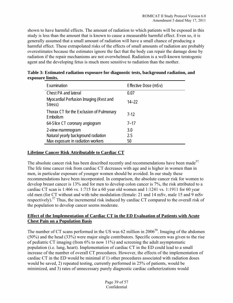
ROMICAT II Study Protocol Version 60 Amendment 5 dated May 17 2011
detection of coronary plaque and stenosis (pixel matrix 512 x 512 FOV 25 cm) 3) one multiphase reformatted dataset of 15 mm thick axial images at 10 increments (10 phases) for single source CT scanners or 5 increments (20 phases) for dual-source CT scanners throughout the cardiac cycle from the onset of the R-wave for the assessment of global and regional LV function in a cine mode and 4) a full field of view with 3 mm thick axial images covering the portions of the thorax acquired during the cardiac CT scan for the assessment of major incidental findings All CT data sets will be transmitted to the CVIC Labrsquos picture archiving and communication system (PACS) via internet using a dedicated routing system (Code of Federal Regulatory compliant) and stored
Image Evaluation Reconstructed data sets will be evaluated for the presence of CAC coronary atherosclerotic plaque and stenosis and global and regional LV function by dedicated highly trained cardiac CT imagers using established criteria22
Step 1mdashCoronary Artery Calcification All non-contrast enhanced CT scans will be assessed for the presence of CAC If present the amount of CAC will be quantified using a dedicated semiautomatic software and expressed as an Agatston Score (AS) as described elsewhere45
Step 2mdashAssessment for Stenosis Contrast-enhanced CT images will be qualitatively evaluated for the presence of coronary atherosclerotic plaque and significant coronary artery stenosis using axial data sets thin slice (5 mm) reconstructions orthogonal and perpendicular to the vessel centerline and short axis cross-sectional reconstructions Assessment will include each of the major epicardial vessels (left main left anterior descending left circumflex and right coronary artery) and their side branches46ndash48
Step 3mdashGlobal and Regional LV Function Global and regional LV function will be assessed visually with the multiphase reformatted dataset in cine mode to correspond to those typically used in echocardiography including LV horizontal long-axis (4-chamber view) vertical long-axis (2-chamber view) LV outflow tract long-axis (parasternal long axis) and short-axis view Global LV function will be reported as normal or impaired (mild moderate or severe) Regional wall motion (RWM) of the myocardium will be assessed to be normal or abnormal (hypokinesia akinesia dyskinesia aneurysm) in short-axis segments of the LV corresponding to the AHAACCASE 17-segment model Hypokinesia will be defined as a segment with impaired thickening and motion akinesia by absent thickening and motion dyskinesia as a paradoxical outward motion of the segment during systole and an aneurysm as a segment with wall thinning and diastolic deformation The abnormality has to be present in at least two contiguous myocardial segments or in one segment visualized in two different views
Step 4mdashMajor Incidental Findings The full field of view will be assessed by a CT reader for the presence of major cardiac and non-cardiac incidental findings such as aortic dissection pericardial effusion pulmonary embolism pneumothorax or mediastinal masses
Page 51 of 57 Confidential
ROMICAT II Study Protocol Version 60 Amendment 5 dated May 17 2011
CT Reporting Cardiac CT Findings
The Cardiac CT Report will be reported and included in the hospitalrsquos electronic medical record which is accessible to all treating physicians and will be the basis for in-hospital patient management The triage and management of the patient will be at the discretion of the ED physician
Image Submissions The protocol-required images must be in DICOM format and submitted to the Cardiovascular Imaging Core Laboratory (CVIC) via the internet using secure File Transfer Protocol (FTP) along with an Imaging Transmittal Worksheet The submission procedure will be detailed in the CVIC site operations manual
Page 52 of 57 Confidential
ROMICAT II Study Protocol Version 60 Amendment 5 dated May 17 2011
Appendix B Adjudication of Events by Clinical Events Committee
Introduction
Because patient safety is one of the concerns in a trial assessing new technology clinical events and serious adverse events will be independently adjudicated by the Clinical Events Committee under the direction of Stephen Wiviott MD This committee will adjudicate events in all subjects who potentially could have a major adverse cardiovascular event (MACE) such as death unstable angina pectoris (UA) myocardial infarction (MI) or coronary revascularization that occur after symptom assessment at patientrsquos initial emergency department (ED) presentation There is a possibility that MACE will occur during index hospitalization For example a second episode of chest pain diagnosed as acute coronary syndrome (ACS) occurring after the chest pain the patient presented with has resolved and all diagnostic tests were negative or recurrence of symptoms with or without hospitalization and re-hospitalization with or without testing
Definitions of Adverse Cardiovascular Events Adverse cardiovascular events comprise cardiovascular death MI unstable angina pectoris (UAP) coronary revascularization andor re-hospitalization that are distinct from the qualifying event (after patientrsquos initial ED presentation)
Definitions of events are listed below
Cardiovascular Death Any sudden cardiac death death due to acute myocardial infarction death due to heart failure death due to stroke and death due to other cardiovascular causes In addition any death without a clear non-cardiovascular cause or a death without known cause will be considered cardiovascular death
MI In Subjects with no recent revascularization in whom normal biomarkers were never elevated or have been documented to return to normal after a qualifying (or recent) MI who meet the following criteria
1) Typical cardiac biomarker rise andor fall AND at least one of the following a) Ischemic discomfort at rest lasing ge10 minutes b) ECG changes indicative of ischemia (ST elevation ge01 mV or ST
depression ge005 mV or new T-wave inversions OR
2) Development of new abnormal Q waves (ge30 msec in duration and ge1 mm in depth) in gt2 contiguous precordial leads or ge2 adjacent limb leads or increase R amplitude in V1-V3 consistent with posterior infarction
OR
3) Pathologic findings of an acute MI
Page 53 of 57 Confidential
ROMICAT II Study Protocol Version 60 Amendment 5 dated May 17 2011
4) Sudden unexpected cardiac death including cardiac arrest often with symptoms suggestive of myocardial ischemia accompanied by presumably new ST elevation or new LBBB or evidence of fresh thrombus in a coronary artery by angiography andor at autopsy but death occurring before blood samples could be obtained or at a time before the appearance of cardiac biomarkers in the blood
In subjects with percutaneous coronary intervention within 48 hours an elevation of CK-MB gt3x ULN distinct from a prior event will be considered to be a procedural MI
In subjects with CABG within 48 hours an elevation of CK-MB gt 10x ULN distinct from a prior event will be considered to be a procedural MI
UA An event not meeting the definition of myocardial infarction and with the following characteristics Chest pain or anginal equivalent at rest or in accelerating pattern AND at least one of the following objective signs
a) New ST-segment changes
b) New TWI
c) Positive stress test with imaging showing ischemia
d) Positive stress test without imaging resulting in increased anginal medication
e) Cath ge70 stenosis or thrombus
Urgent Coronary Revascularization Ischemic discomfort or equivalent meeting the following criteria
a) Lasting ge10 minutes at rest or repeated episodes at rest lasting ge5 minutes considered to be myocardial ischemia upon final diagnosis
AND
b) Prompting coronary revascularization during an unscheduled visit to healthcare facility or during an unplanned (or prolonged) hospitalization for these symptoms
Note Attempted revascularization procedures even if not successful will be counted Potential ischemic events meeting the criteria for myocardial infarction will not be adjudicated as urgent coronary revascularization
Re-hospitalization Coronary ischemia requiring re-hospitalization is defined as an event not meeting the definitions of myocardial infarction or urgent coronary revascularization ischemic discomfort or equivalent meeting the following criteria
a) Lasting ge10 minutes at rest or repeated episodes at rest lasting ge5 minutes or an accelerating pattern of ischemic discomfort (episodes that
Page 54 of 57 Confidential
ROMICAT II Study Protocol Version 60 Amendment 5 dated May 17 2011
are more frequent severe longer in duration and precipitated by minimal exertion) considered to be myocardial ischemia upon final diagnosis
AND
b) Prompting hospitalization (including overnight stay on an inpatient unit) within 48 hours of the most recent symptoms or prolonging hospitalization if occurring during existing hospitalization
AND
c) At least one of the following additional criteria for coronary artery disease andor ischemia
New andor dynamic ST-depression ge005 mV ST-elevation ge01 mV or symmetric T wave inversion ge02 mV on a resting ECG
Definite evidence of ischemia on stress sechocardiography myocardial scintigraphy (eg an area of clear reversible ischemia) or ECG-only stress test (eg significant dynamic ST shift horizontal or downsloping)
Angiographic evidence of epicardial coronary stenosis of ge70 diameter reduction andor evidence for intraluminal arterial thrombus
Note If subjects are admitted with suspected myocardial ischemia and subsequent testing reveals non-cardiac or non-ischemic etiology this will not be adjudicated as meeting this definition Potential ischemic events meeting the criteria for myocardial infarction will not be adjudicated as ischemia requiring hospitalization
Adjudication Process Adverse cardiovascular events (both during index hospitalization and within 28 days) will be independently adjudicated by the Clinical Events Committee (CEC) Potential events will be identified by the sites during a follow-up phone interview at 48-72 hours and 28 days The information collected during the call will be recorded on an ldquoAssessment of MACErdquo Case Report Form which will be submitted to the Data Coordinating Center (DCC) The DCC will request de-identified medical records and source documents supporting the adjudication of the events including but not limited to as discharge summary cardiac consultation notes ECG reports cardiac catheterization reports biomarker laboratory values death certificates autopsy reports etc The DCC will provide copies of the medical record pertaining to the index hospitalization and the potential adverse event for adjudication to the CEC Based on the medical record review and site reported information the adjudicator will complete an ldquoAssessment of MACErdquo Case Report Form which in turn will be submitted to the DCC
Adjudicators are independent board certified or board eligible cardiologists who have received formal documented training based on a CEC Charter and will perform a composite review of the medical records to ascertain the presence or absence of MACE as defined in the CEC charter The adjudication is performed by the TIMI (Thrombolysis In Myocardial Infarction) Study
Page 55 of 57 Confidential
ROMICAT II Study Protocol Version 60 Amendment 5 dated May 17 2011
Group CEC which provides adjudication services for cardiovascular trials performed worldwide Cases will be adjudicated by two independent cardiologists with an experience in cardiovascular event adjudication In case of disagreement the adjudicators will meet discuss the case and attempt to come to consensus If consensus cannot be reached a third adjudicator will cast a tie breaking vote This is a standard accepted technique to assess the presence of clinical events in cardiovascular trials 18 55-58
Discharge Diagnosis from Index Hospitalization The CEC will adjudicate the accuracy of the discharge diagnosis during index hospitalization for the first eight patients for each site followed by a random selection of 10 of the participants Discharge diagnoses are ACS as defined above and consistent with the ACCAHA Guidelines59ndash
61 including MI and UA chest pain of cardiac origin and chest pain of non-cardiac origin with or without a clear differential diagnosis
Clinical sites use different cut-off values and tests to determine MI diagnosis The DCC will provide each sitersquos biomarker cutoffs for each individual case The CEC therefore will adjudicate events in a manner consistent application of site-specific criteria
For each MI identified by the CEC a Type of MI will be assigned using the following guidelines
Type 1 Spontaneous myocardial infarction related to ischemia due to a primary coronary event such as plaque erosion andor rupture fissuring or dissection
Type 2 Myocardial infarction secondary to ischemia due to either increased oxygen demand or decreased supply eg coronary artery spasm coronary embolism anemia arrhythmias hypertension or hypotension
Type 3 Sudden unexpected cardiac death including cardiac arrest often with symptoms suggestive of myocardial ischemia accompanied by presumably new ST elevation or new LBBB or evidence of fresh thrombus in a coronary artery by angiography andor at autopsy but death occurring before blood samples could be obtained or at a time before the appearance of cardiac biomarkers in the blood
Type 4a Myocardial infarction associated with PCI
Type 4b Myocardial infarction associated with stent thrombosis as documented by angiography or at autopsy
Type 5 Myocardial infarction associated with CABG
Page 56 of 57 Confidential
ROMICAT II Study Protocol Version 60 Amendment 5 dated May 17 2011
Appendix C Data and Safety Monitoring Board
The DSMB is responsible for safeguarding the interests of study participants assessing the safety and efficacy of study procedures and for monitoring the overall conduct of the study
The DSMB is an independent group advisory to the Director NHLBI and is required to provide recommendations about starting continuing and stopping the study In addition the DSMB is asked to make recommendations as appropriate to the NHLBI about bull Efficacy of the study intervention (DSMB only) bull Benefitrisk ratio of procedures and participant burden bull Selection recruitment and retention of participants bull Adherence to protocol requirements bull Completeness quality and analysis of measurements bull Amendments to the study protocol and consent forms bull Performance of individual centers and core labs bull Participant safety bull Notification of and referral for abnormal findings
Meetings are usually held approximately twice a year with additional meetings or conference calls scheduled as needed
Page 57 of 57 Confidential
- TABLE OF CONTENTS
- Amendment 1 Summary of Changes
- Amendment 2 Summary of Changes
- Amendment 3 Summary of Changes
- Amendment 4 Summary of Changes
- Amendment 5 Summary of Changes
- Study Synopsis
- Abbreviations
- Part I Study Overview
- Part II Study Description
-
- 1 Background and Significance
-
- 11 Standard Evaluation of Participants with Acute Chest Pain
- 12 Accuracy of Cardiac CT for Detection of Coronary Artery Disease and LV Function
- 13 Accuracy of Cardiac CT to Detect ACS in Participants with Acute Chest Pain
- 14 Cost Effectiveness of Diagnostic Testing in Subjects with Acute Chest Pain
- 15 Significance of the Proposed Clinical Trial
-
- 2 Preliminary Data
-
- 21 Final Results of ROMICAT I
-
- 3 Study ObjectiveSpecific Aims
-
- 31 Primary Aim
- 32 Secondary Aims
- 33 Tertiary Aims
-
- 4 Participant SelectionEligibility Criteria
-
- 41 Inclusion Criteria
- 42 Exclusion Criteria
- 43 Recruitment Screening and Consent
- 44 Randomization
-
- 5 Study Procedures
-
- 51 Index Hospitalization
- 52 48-72 Hour Follow up after ED discharge
- 53 28 Day Follow up
-
- 6 Data Collection
- 7 Statistical Considerations
-
- 71 Primary Aim
- 72 Secondary Aims
- 73 Tertiary Aims
-
- 8 Data Entry and Site Monitoring
-
- 81 Data Collection
- 82 Site Monitoring
-
- 9 Risk and Benefit Assessment
-
- 91 Risks of Nonionic Contrast Agent
- 92 Risks Associated With Radiation
- 93 Other Risks Additional Risks of CT Scans
- 94 Potential Benefits
-
- 10 Ethical Considerations
-
- 101 Inclusion of Women and Minorities
-
- 11 Adverse Event Reporting
- 12 References
-
- Appendix A Cardiac CT Imaging Protocol Guidelinesdagger
- Appendix B Adjudication of Events by Clinical Events Committee
- Appendix C Data and Safety Monitoring Board
-
ROMICAT II Study Protocol Version 60 Amendment 5 dated May 17 2011
Appendix B Adjudication of Events by Clinical Events Committee
Introduction
Because patient safety is one of the concerns in a trial assessing new technology clinical events and serious adverse events will be independently adjudicated by the Clinical Events Committee under the direction of Stephen Wiviott MD This committee will adjudicate events in all subjects who potentially could have a major adverse cardiovascular event (MACE) such as death unstable angina pectoris (UA) myocardial infarction (MI) or coronary revascularization that occur after symptom assessment at patientrsquos initial emergency department (ED) presentation There is a possibility that MACE will occur during index hospitalization For example a second episode of chest pain diagnosed as acute coronary syndrome (ACS) occurring after the chest pain the patient presented with has resolved and all diagnostic tests were negative or recurrence of symptoms with or without hospitalization and re-hospitalization with or without testing
Definitions of Adverse Cardiovascular Events Adverse cardiovascular events comprise cardiovascular death MI unstable angina pectoris (UAP) coronary revascularization andor re-hospitalization that are distinct from the qualifying event (after patientrsquos initial ED presentation)
Definitions of events are listed below
Cardiovascular Death Any sudden cardiac death death due to acute myocardial infarction death due to heart failure death due to stroke and death due to other cardiovascular causes In addition any death without a clear non-cardiovascular cause or a death without known cause will be considered cardiovascular death
MI In Subjects with no recent revascularization in whom normal biomarkers were never elevated or have been documented to return to normal after a qualifying (or recent) MI who meet the following criteria
1) Typical cardiac biomarker rise andor fall AND at least one of the following a) Ischemic discomfort at rest lasing ge10 minutes b) ECG changes indicative of ischemia (ST elevation ge01 mV or ST
depression ge005 mV or new T-wave inversions OR
2) Development of new abnormal Q waves (ge30 msec in duration and ge1 mm in depth) in gt2 contiguous precordial leads or ge2 adjacent limb leads or increase R amplitude in V1-V3 consistent with posterior infarction
OR
3) Pathologic findings of an acute MI
Page 53 of 57 Confidential
ROMICAT II Study Protocol Version 60 Amendment 5 dated May 17 2011
4) Sudden unexpected cardiac death including cardiac arrest often with symptoms suggestive of myocardial ischemia accompanied by presumably new ST elevation or new LBBB or evidence of fresh thrombus in a coronary artery by angiography andor at autopsy but death occurring before blood samples could be obtained or at a time before the appearance of cardiac biomarkers in the blood
In subjects with percutaneous coronary intervention within 48 hours an elevation of CK-MB gt3x ULN distinct from a prior event will be considered to be a procedural MI
In subjects with CABG within 48 hours an elevation of CK-MB gt 10x ULN distinct from a prior event will be considered to be a procedural MI
UA An event not meeting the definition of myocardial infarction and with the following characteristics Chest pain or anginal equivalent at rest or in accelerating pattern AND at least one of the following objective signs
a) New ST-segment changes
b) New TWI
c) Positive stress test with imaging showing ischemia
d) Positive stress test without imaging resulting in increased anginal medication
e) Cath ge70 stenosis or thrombus
Urgent Coronary Revascularization Ischemic discomfort or equivalent meeting the following criteria
a) Lasting ge10 minutes at rest or repeated episodes at rest lasting ge5 minutes considered to be myocardial ischemia upon final diagnosis
AND
b) Prompting coronary revascularization during an unscheduled visit to healthcare facility or during an unplanned (or prolonged) hospitalization for these symptoms
Note Attempted revascularization procedures even if not successful will be counted Potential ischemic events meeting the criteria for myocardial infarction will not be adjudicated as urgent coronary revascularization
Re-hospitalization Coronary ischemia requiring re-hospitalization is defined as an event not meeting the definitions of myocardial infarction or urgent coronary revascularization ischemic discomfort or equivalent meeting the following criteria
a) Lasting ge10 minutes at rest or repeated episodes at rest lasting ge5 minutes or an accelerating pattern of ischemic discomfort (episodes that
Page 54 of 57 Confidential
ROMICAT II Study Protocol Version 60 Amendment 5 dated May 17 2011
are more frequent severe longer in duration and precipitated by minimal exertion) considered to be myocardial ischemia upon final diagnosis
AND
b) Prompting hospitalization (including overnight stay on an inpatient unit) within 48 hours of the most recent symptoms or prolonging hospitalization if occurring during existing hospitalization
AND
c) At least one of the following additional criteria for coronary artery disease andor ischemia
New andor dynamic ST-depression ge005 mV ST-elevation ge01 mV or symmetric T wave inversion ge02 mV on a resting ECG
Definite evidence of ischemia on stress sechocardiography myocardial scintigraphy (eg an area of clear reversible ischemia) or ECG-only stress test (eg significant dynamic ST shift horizontal or downsloping)
Angiographic evidence of epicardial coronary stenosis of ge70 diameter reduction andor evidence for intraluminal arterial thrombus
Note If subjects are admitted with suspected myocardial ischemia and subsequent testing reveals non-cardiac or non-ischemic etiology this will not be adjudicated as meeting this definition Potential ischemic events meeting the criteria for myocardial infarction will not be adjudicated as ischemia requiring hospitalization
Adjudication Process Adverse cardiovascular events (both during index hospitalization and within 28 days) will be independently adjudicated by the Clinical Events Committee (CEC) Potential events will be identified by the sites during a follow-up phone interview at 48-72 hours and 28 days The information collected during the call will be recorded on an ldquoAssessment of MACErdquo Case Report Form which will be submitted to the Data Coordinating Center (DCC) The DCC will request de-identified medical records and source documents supporting the adjudication of the events including but not limited to as discharge summary cardiac consultation notes ECG reports cardiac catheterization reports biomarker laboratory values death certificates autopsy reports etc The DCC will provide copies of the medical record pertaining to the index hospitalization and the potential adverse event for adjudication to the CEC Based on the medical record review and site reported information the adjudicator will complete an ldquoAssessment of MACErdquo Case Report Form which in turn will be submitted to the DCC
Adjudicators are independent board certified or board eligible cardiologists who have received formal documented training based on a CEC Charter and will perform a composite review of the medical records to ascertain the presence or absence of MACE as defined in the CEC charter The adjudication is performed by the TIMI (Thrombolysis In Myocardial Infarction) Study
Page 55 of 57 Confidential
ROMICAT II Study Protocol Version 60 Amendment 5 dated May 17 2011
Group CEC which provides adjudication services for cardiovascular trials performed worldwide Cases will be adjudicated by two independent cardiologists with an experience in cardiovascular event adjudication In case of disagreement the adjudicators will meet discuss the case and attempt to come to consensus If consensus cannot be reached a third adjudicator will cast a tie breaking vote This is a standard accepted technique to assess the presence of clinical events in cardiovascular trials 18 55-58
Discharge Diagnosis from Index Hospitalization The CEC will adjudicate the accuracy of the discharge diagnosis during index hospitalization for the first eight patients for each site followed by a random selection of 10 of the participants Discharge diagnoses are ACS as defined above and consistent with the ACCAHA Guidelines59ndash
61 including MI and UA chest pain of cardiac origin and chest pain of non-cardiac origin with or without a clear differential diagnosis
Clinical sites use different cut-off values and tests to determine MI diagnosis The DCC will provide each sitersquos biomarker cutoffs for each individual case The CEC therefore will adjudicate events in a manner consistent application of site-specific criteria
For each MI identified by the CEC a Type of MI will be assigned using the following guidelines
Type 1 Spontaneous myocardial infarction related to ischemia due to a primary coronary event such as plaque erosion andor rupture fissuring or dissection
Type 2 Myocardial infarction secondary to ischemia due to either increased oxygen demand or decreased supply eg coronary artery spasm coronary embolism anemia arrhythmias hypertension or hypotension
Type 3 Sudden unexpected cardiac death including cardiac arrest often with symptoms suggestive of myocardial ischemia accompanied by presumably new ST elevation or new LBBB or evidence of fresh thrombus in a coronary artery by angiography andor at autopsy but death occurring before blood samples could be obtained or at a time before the appearance of cardiac biomarkers in the blood
Type 4a Myocardial infarction associated with PCI
Type 4b Myocardial infarction associated with stent thrombosis as documented by angiography or at autopsy
Type 5 Myocardial infarction associated with CABG
Page 56 of 57 Confidential
ROMICAT II Study Protocol Version 60 Amendment 5 dated May 17 2011
Appendix C Data and Safety Monitoring Board
The DSMB is responsible for safeguarding the interests of study participants assessing the safety and efficacy of study procedures and for monitoring the overall conduct of the study
The DSMB is an independent group advisory to the Director NHLBI and is required to provide recommendations about starting continuing and stopping the study In addition the DSMB is asked to make recommendations as appropriate to the NHLBI about bull Efficacy of the study intervention (DSMB only) bull Benefitrisk ratio of procedures and participant burden bull Selection recruitment and retention of participants bull Adherence to protocol requirements bull Completeness quality and analysis of measurements bull Amendments to the study protocol and consent forms bull Performance of individual centers and core labs bull Participant safety bull Notification of and referral for abnormal findings
Meetings are usually held approximately twice a year with additional meetings or conference calls scheduled as needed
Page 57 of 57 Confidential
- TABLE OF CONTENTS
- Amendment 1 Summary of Changes
- Amendment 2 Summary of Changes
- Amendment 3 Summary of Changes
- Amendment 4 Summary of Changes
- Amendment 5 Summary of Changes
- Study Synopsis
- Abbreviations
- Part I Study Overview
- Part II Study Description
-
- 1 Background and Significance
-
- 11 Standard Evaluation of Participants with Acute Chest Pain
- 12 Accuracy of Cardiac CT for Detection of Coronary Artery Disease and LV Function
- 13 Accuracy of Cardiac CT to Detect ACS in Participants with Acute Chest Pain
- 14 Cost Effectiveness of Diagnostic Testing in Subjects with Acute Chest Pain
- 15 Significance of the Proposed Clinical Trial
-
- 2 Preliminary Data
-
- 21 Final Results of ROMICAT I
-
- 3 Study ObjectiveSpecific Aims
-
- 31 Primary Aim
- 32 Secondary Aims
- 33 Tertiary Aims
-
- 4 Participant SelectionEligibility Criteria
-
- 41 Inclusion Criteria
- 42 Exclusion Criteria
- 43 Recruitment Screening and Consent
- 44 Randomization
-
- 5 Study Procedures
-
- 51 Index Hospitalization
- 52 48-72 Hour Follow up after ED discharge
- 53 28 Day Follow up
-
- 6 Data Collection
- 7 Statistical Considerations
-
- 71 Primary Aim
- 72 Secondary Aims
- 73 Tertiary Aims
-
- 8 Data Entry and Site Monitoring
-
- 81 Data Collection
- 82 Site Monitoring
-
- 9 Risk and Benefit Assessment
-
- 91 Risks of Nonionic Contrast Agent
- 92 Risks Associated With Radiation
- 93 Other Risks Additional Risks of CT Scans
- 94 Potential Benefits
-
- 10 Ethical Considerations
-
- 101 Inclusion of Women and Minorities
-
- 11 Adverse Event Reporting
- 12 References
-
- Appendix A Cardiac CT Imaging Protocol Guidelinesdagger
- Appendix B Adjudication of Events by Clinical Events Committee
- Appendix C Data and Safety Monitoring Board
-

ROMICAT II Study Protocol Version 60 Amendment 5 dated May 17 2011
are more frequent severe longer in duration and precipitated by minimal exertion) considered to be myocardial ischemia upon final diagnosis
AND
b) Prompting hospitalization (including overnight stay on an inpatient unit) within 48 hours of the most recent symptoms or prolonging hospitalization if occurring during existing hospitalization
AND
c) At least one of the following additional criteria for coronary artery disease andor ischemia
New andor dynamic ST-depression ge005 mV ST-elevation ge01 mV or symmetric T wave inversion ge02 mV on a resting ECG
Definite evidence of ischemia on stress sechocardiography myocardial scintigraphy (eg an area of clear reversible ischemia) or ECG-only stress test (eg significant dynamic ST shift horizontal or downsloping)
Angiographic evidence of epicardial coronary stenosis of ge70 diameter reduction andor evidence for intraluminal arterial thrombus
Note If subjects are admitted with suspected myocardial ischemia and subsequent testing reveals non-cardiac or non-ischemic etiology this will not be adjudicated as meeting this definition Potential ischemic events meeting the criteria for myocardial infarction will not be adjudicated as ischemia requiring hospitalization
Adjudication Process Adverse cardiovascular events (both during index hospitalization and within 28 days) will be independently adjudicated by the Clinical Events Committee (CEC) Potential events will be identified by the sites during a follow-up phone interview at 48-72 hours and 28 days The information collected during the call will be recorded on an ldquoAssessment of MACErdquo Case Report Form which will be submitted to the Data Coordinating Center (DCC) The DCC will request de-identified medical records and source documents supporting the adjudication of the events including but not limited to as discharge summary cardiac consultation notes ECG reports cardiac catheterization reports biomarker laboratory values death certificates autopsy reports etc The DCC will provide copies of the medical record pertaining to the index hospitalization and the potential adverse event for adjudication to the CEC Based on the medical record review and site reported information the adjudicator will complete an ldquoAssessment of MACErdquo Case Report Form which in turn will be submitted to the DCC
Adjudicators are independent board certified or board eligible cardiologists who have received formal documented training based on a CEC Charter and will perform a composite review of the medical records to ascertain the presence or absence of MACE as defined in the CEC charter The adjudication is performed by the TIMI (Thrombolysis In Myocardial Infarction) Study
Page 55 of 57 Confidential
ROMICAT II Study Protocol Version 60 Amendment 5 dated May 17 2011
Group CEC which provides adjudication services for cardiovascular trials performed worldwide Cases will be adjudicated by two independent cardiologists with an experience in cardiovascular event adjudication In case of disagreement the adjudicators will meet discuss the case and attempt to come to consensus If consensus cannot be reached a third adjudicator will cast a tie breaking vote This is a standard accepted technique to assess the presence of clinical events in cardiovascular trials 18 55-58
Discharge Diagnosis from Index Hospitalization The CEC will adjudicate the accuracy of the discharge diagnosis during index hospitalization for the first eight patients for each site followed by a random selection of 10 of the participants Discharge diagnoses are ACS as defined above and consistent with the ACCAHA Guidelines59ndash
61 including MI and UA chest pain of cardiac origin and chest pain of non-cardiac origin with or without a clear differential diagnosis
Clinical sites use different cut-off values and tests to determine MI diagnosis The DCC will provide each sitersquos biomarker cutoffs for each individual case The CEC therefore will adjudicate events in a manner consistent application of site-specific criteria
For each MI identified by the CEC a Type of MI will be assigned using the following guidelines
Type 1 Spontaneous myocardial infarction related to ischemia due to a primary coronary event such as plaque erosion andor rupture fissuring or dissection
Type 2 Myocardial infarction secondary to ischemia due to either increased oxygen demand or decreased supply eg coronary artery spasm coronary embolism anemia arrhythmias hypertension or hypotension
Type 3 Sudden unexpected cardiac death including cardiac arrest often with symptoms suggestive of myocardial ischemia accompanied by presumably new ST elevation or new LBBB or evidence of fresh thrombus in a coronary artery by angiography andor at autopsy but death occurring before blood samples could be obtained or at a time before the appearance of cardiac biomarkers in the blood
Type 4a Myocardial infarction associated with PCI
Type 4b Myocardial infarction associated with stent thrombosis as documented by angiography or at autopsy
Type 5 Myocardial infarction associated with CABG
Page 56 of 57 Confidential
ROMICAT II Study Protocol Version 60 Amendment 5 dated May 17 2011
Appendix C Data and Safety Monitoring Board
The DSMB is responsible for safeguarding the interests of study participants assessing the safety and efficacy of study procedures and for monitoring the overall conduct of the study
The DSMB is an independent group advisory to the Director NHLBI and is required to provide recommendations about starting continuing and stopping the study In addition the DSMB is asked to make recommendations as appropriate to the NHLBI about bull Efficacy of the study intervention (DSMB only) bull Benefitrisk ratio of procedures and participant burden bull Selection recruitment and retention of participants bull Adherence to protocol requirements bull Completeness quality and analysis of measurements bull Amendments to the study protocol and consent forms bull Performance of individual centers and core labs bull Participant safety bull Notification of and referral for abnormal findings
Meetings are usually held approximately twice a year with additional meetings or conference calls scheduled as needed
Page 57 of 57 Confidential
- TABLE OF CONTENTS
- Amendment 1 Summary of Changes
- Amendment 2 Summary of Changes
- Amendment 3 Summary of Changes
- Amendment 4 Summary of Changes
- Amendment 5 Summary of Changes
- Study Synopsis
- Abbreviations
- Part I Study Overview
- Part II Study Description
-
- 1 Background and Significance
-
- 11 Standard Evaluation of Participants with Acute Chest Pain
- 12 Accuracy of Cardiac CT for Detection of Coronary Artery Disease and LV Function
- 13 Accuracy of Cardiac CT to Detect ACS in Participants with Acute Chest Pain
- 14 Cost Effectiveness of Diagnostic Testing in Subjects with Acute Chest Pain
- 15 Significance of the Proposed Clinical Trial
-
- 2 Preliminary Data
-
- 21 Final Results of ROMICAT I
-
- 3 Study ObjectiveSpecific Aims
-
- 31 Primary Aim
- 32 Secondary Aims
- 33 Tertiary Aims
-
- 4 Participant SelectionEligibility Criteria
-
- 41 Inclusion Criteria
- 42 Exclusion Criteria
- 43 Recruitment Screening and Consent
- 44 Randomization
-
- 5 Study Procedures
-
- 51 Index Hospitalization
- 52 48-72 Hour Follow up after ED discharge
- 53 28 Day Follow up
-
- 6 Data Collection
- 7 Statistical Considerations
-
- 71 Primary Aim
- 72 Secondary Aims
- 73 Tertiary Aims
-
- 8 Data Entry and Site Monitoring
-
- 81 Data Collection
- 82 Site Monitoring
-
- 9 Risk and Benefit Assessment
-
- 91 Risks of Nonionic Contrast Agent
- 92 Risks Associated With Radiation
- 93 Other Risks Additional Risks of CT Scans
- 94 Potential Benefits
-
- 10 Ethical Considerations
-
- 101 Inclusion of Women and Minorities
-
- 11 Adverse Event Reporting
- 12 References
-
- Appendix A Cardiac CT Imaging Protocol Guidelinesdagger
- Appendix B Adjudication of Events by Clinical Events Committee
- Appendix C Data and Safety Monitoring Board
-
ROMICAT II Study Protocol Version 60 Amendment 5 dated May 17 2011
Group CEC which provides adjudication services for cardiovascular trials performed worldwide Cases will be adjudicated by two independent cardiologists with an experience in cardiovascular event adjudication In case of disagreement the adjudicators will meet discuss the case and attempt to come to consensus If consensus cannot be reached a third adjudicator will cast a tie breaking vote This is a standard accepted technique to assess the presence of clinical events in cardiovascular trials 18 55-58
Discharge Diagnosis from Index Hospitalization The CEC will adjudicate the accuracy of the discharge diagnosis during index hospitalization for the first eight patients for each site followed by a random selection of 10 of the participants Discharge diagnoses are ACS as defined above and consistent with the ACCAHA Guidelines59ndash
61 including MI and UA chest pain of cardiac origin and chest pain of non-cardiac origin with or without a clear differential diagnosis
Clinical sites use different cut-off values and tests to determine MI diagnosis The DCC will provide each sitersquos biomarker cutoffs for each individual case The CEC therefore will adjudicate events in a manner consistent application of site-specific criteria
For each MI identified by the CEC a Type of MI will be assigned using the following guidelines
Type 1 Spontaneous myocardial infarction related to ischemia due to a primary coronary event such as plaque erosion andor rupture fissuring or dissection
Type 2 Myocardial infarction secondary to ischemia due to either increased oxygen demand or decreased supply eg coronary artery spasm coronary embolism anemia arrhythmias hypertension or hypotension
Type 3 Sudden unexpected cardiac death including cardiac arrest often with symptoms suggestive of myocardial ischemia accompanied by presumably new ST elevation or new LBBB or evidence of fresh thrombus in a coronary artery by angiography andor at autopsy but death occurring before blood samples could be obtained or at a time before the appearance of cardiac biomarkers in the blood
Type 4a Myocardial infarction associated with PCI
Type 4b Myocardial infarction associated with stent thrombosis as documented by angiography or at autopsy
Type 5 Myocardial infarction associated with CABG
Page 56 of 57 Confidential
ROMICAT II Study Protocol Version 60 Amendment 5 dated May 17 2011
Appendix C Data and Safety Monitoring Board
The DSMB is responsible for safeguarding the interests of study participants assessing the safety and efficacy of study procedures and for monitoring the overall conduct of the study
The DSMB is an independent group advisory to the Director NHLBI and is required to provide recommendations about starting continuing and stopping the study In addition the DSMB is asked to make recommendations as appropriate to the NHLBI about bull Efficacy of the study intervention (DSMB only) bull Benefitrisk ratio of procedures and participant burden bull Selection recruitment and retention of participants bull Adherence to protocol requirements bull Completeness quality and analysis of measurements bull Amendments to the study protocol and consent forms bull Performance of individual centers and core labs bull Participant safety bull Notification of and referral for abnormal findings
Meetings are usually held approximately twice a year with additional meetings or conference calls scheduled as needed
Page 57 of 57 Confidential
- TABLE OF CONTENTS
- Amendment 1 Summary of Changes
- Amendment 2 Summary of Changes
- Amendment 3 Summary of Changes
- Amendment 4 Summary of Changes
- Amendment 5 Summary of Changes
- Study Synopsis
- Abbreviations
- Part I Study Overview
- Part II Study Description
-
- 1 Background and Significance
-
- 11 Standard Evaluation of Participants with Acute Chest Pain
- 12 Accuracy of Cardiac CT for Detection of Coronary Artery Disease and LV Function
- 13 Accuracy of Cardiac CT to Detect ACS in Participants with Acute Chest Pain
- 14 Cost Effectiveness of Diagnostic Testing in Subjects with Acute Chest Pain
- 15 Significance of the Proposed Clinical Trial
-
- 2 Preliminary Data
-
- 21 Final Results of ROMICAT I
-
- 3 Study ObjectiveSpecific Aims
-
- 31 Primary Aim
- 32 Secondary Aims
- 33 Tertiary Aims
-
- 4 Participant SelectionEligibility Criteria
-
- 41 Inclusion Criteria
- 42 Exclusion Criteria
- 43 Recruitment Screening and Consent
- 44 Randomization
-
- 5 Study Procedures
-
- 51 Index Hospitalization
- 52 48-72 Hour Follow up after ED discharge
- 53 28 Day Follow up
-
- 6 Data Collection
- 7 Statistical Considerations
-
- 71 Primary Aim
- 72 Secondary Aims
- 73 Tertiary Aims
-
- 8 Data Entry and Site Monitoring
-
- 81 Data Collection
- 82 Site Monitoring
-
- 9 Risk and Benefit Assessment
-
- 91 Risks of Nonionic Contrast Agent
- 92 Risks Associated With Radiation
- 93 Other Risks Additional Risks of CT Scans
- 94 Potential Benefits
-
- 10 Ethical Considerations
-
- 101 Inclusion of Women and Minorities
-
- 11 Adverse Event Reporting
- 12 References
-
- Appendix A Cardiac CT Imaging Protocol Guidelinesdagger
- Appendix B Adjudication of Events by Clinical Events Committee
- Appendix C Data and Safety Monitoring Board
-