
Romanian of Oral PILOT Romanian Journal of Oral...
80
Romanian Journal of Oral Rehabilitation PILOT Number, April 2008 1 Romanian Journal of Oral Rehabilitation PILOT Number, September 2008 Editor in Chief Norina Consuela Forna, Iaşi, România Vice-Editor Viorel Păun, Bucharest, România Senior Associate Editors Pierre Lafforgue, Paris, France Sammi Sandhaus, Lausanne, Switzerland Robert Sader, Germania Zhimon Jacobson, Boston, USA Editorial Board Corneliu Amariei, Constanţa, România Vasile Astărăstoae, Iaşi, România Mihai Augustin, Bucharest, România Grigore Băciuţ, Cluj-Napoca, România Constantin Bălăceanu-Stolnici, Bucharest, România Marc Bolla, Nice, France Dorin Bratu, Timişoara, România Alexandru Bucur, Bucharest, România Eugen Carasevici, Iaşi, România Radu Septimiu Câmpean, Cluj-Napoca, România Virgil Cârligeriu, Timişoara, România Costin Cernescu, Bucharest, România Yves Comissionat, Paris, France Marysette Folliguet, Paris, France Cristina Glavce, Bucharest, România Emilian Hutu, Bucharest, România Constantin Ionescu-Tîrgoviste, Bucharest, România Michel Jourde, Paris, France Veronica Mercuţ, Craiova, România Patrick Missika, Paris, France Ostin Costin Mungiu, Iaşi, România Ady Palti, Kraichtal, Germany Mihaela Păuna, Bucharest, România Phillipe Pirnay, Paris, France Constantin Popa, Bucharest, România Sorin Popşor, Tg. Mureş, România Dorin Ruse, Vancouver, Canada Valeriu Rusu, Iaşi, România Adrian Streinu-Cercel, Bucharest, România Dragoş Stanciu, Bucharest, România Mircea Suciu, Tg. Mureş, România Alin Şerbănescu, Cluj-Napoca, România General Secretary Magda Ecaterina Antohe, Iaşi, România Legislation Committee Delia Barbu, Bucharest, România Technical Committee Andrei Istrate, Bucharest, România Volum realizat în cadrul Casei Editoriale DEMIURG
Transcript of Romanian of Oral PILOT Romanian Journal of Oral...
Romanian Journal of Oral Rehabilitation PILOT Number, April 2008
1
Romanian Journal of Oral Rehabilitation
PILOT Number, September 2008 Editor in Chief Norina Consuela Forna, Iaşi, România
Vice-Editor Viorel Păun, Bucharest, România
Senior Associate Editors Pierre Lafforgue, Paris, France Sammi Sandhaus, Lausanne, Switzerland Robert Sader, Germania Zhimon Jacobson, Boston, USA
Editorial Board Corneliu Amariei, Constanţa, România Vasile Astărăstoae, Iaşi, România Mihai Augustin, Bucharest, România Grigore Băciuţ, Cluj-Napoca, România Constantin Bălăceanu-Stolnici, Bucharest, România Marc Bolla, Nice, France Dorin Bratu, Timişoara, România Alexandru Bucur, Bucharest, România Eugen Carasevici, Iaşi, România Radu Septimiu Câmpean, Cluj-Napoca, România Virgil Cârligeriu, Timişoara, România Costin Cernescu, Bucharest, România Yves Comissionat, Paris, France Marysette Folliguet, Paris, France Cristina Glavce, Bucharest, România
Emilian Hutu, Bucharest, România Constantin Ionescu-Tîrgoviste, Bucharest, România Michel Jourde, Paris, France Veronica Mercuţ, Craiova, România Patrick Missika, Paris, France Ostin Costin Mungiu, Iaşi, România Ady Palti, Kraichtal, Germany Mihaela Păuna, Bucharest, România Phillipe Pirnay, Paris, France Constantin Popa, Bucharest, România Sorin Popşor, Tg. Mureş, România Dorin Ruse, Vancouver, Canada Valeriu Rusu, Iaşi, România Adrian Streinu-Cercel, Bucharest, România Dragoş Stanciu, Bucharest, România Mircea Suciu, Tg. Mureş, România Alin Şerbănescu, Cluj-Napoca, România
General Secretary Magda Ecaterina Antohe, Iaşi, România
Legislation Committee Delia Barbu, Bucharest, România
Technical Committee Andrei Istrate, Bucharest, România
Volum realizat în cadrul Casei Editoriale DEMIURG

Romanian Journal of Oral Rehabilitation PILOT Number, April 2008
2
CUPRINS
FOREWARD (Prof. Univ. Dr. Norina Forna) 3 IMPLICATIONS OF SILICONIC AND POLIAMIDIC BIOMATERIALS IN MAXILO-FACIAL PROSTHESIS Robert Sader, Norina Consuela Forna
4
RELEVANCE OF INTERHUMAN COMMUNICATION IN THE PEDIATRIC DENTISTRY MANAGEMENT Adriana Bălan, Marinela Păsăreanu, Ana Petcu, Carmen Drăgan, Dana Cristiana Rotaru
8
PATIENTS’ KNOWLEDGE AND ATTITUDES TOWARDS INFECTION CONTROL IN THE DENTAL PRACTICE Lucia Bârlean, I. Dănilă , Cristina Dascălu, C. Meriuţă
11
THE IMPORTANCE OF FINDING OUT FACTORS THAT CAN TROUBLE THE MUSCULO-LIGAMENTAR EQUILIBRIUM IN PACIENTS WITH COMPLETE AND EXTENDED PARTIAL EDENTATION D.N. Bosînceanu, Norina Consuela Forna, Sami Sandhaus
14
OPTIMIZING THE CLINICAL DIAGNOSIS IN FETAL ALCOHOL SINDROMA. THE EXPERIENCE OF THE CENTER FOR MEDICAL GENETICS IAŞI Elena Braha, M. Voloşciuc, M. Covic
18
MUSCLES COMPLEX REHABILITATION CRITERIA Mihaela Daniela Brînză, Dieter Wember Mathes, Norina Forna
22
THE EFFECTS OF THE PAIN ENDURED DURING DENTAL TREATMENT Diana Cerghizan, S. Popşor, M. Suciu, A. Kovacs
26
EMDOGAIN AND BIO-OSS BETWEEN HOPE AND REALITY Laura Cîrligeriu, M. Boariu, A. Marinescu, V. Cârligeriu
31
THE PREVALENCE OF HYPODONTIA IN CHILDREN WITH CLEFT AND NONRELATED CONTROLS Claudia Corega, A. Şerbănescu, M.Corega, Mihaela Băciuţ
35
USE OF LASER ANESTHESIA IN DEEP CAVITARY LESIOANS Irina Costache, I. Dănilă
40
MODERN CONCEPTS IN COMPLEX ORAL REABILITATION Yves Commissionat, A. Crăciunescu, Norina Forna
44
SISTEM FOR UNIFIED MEDICAL LANGUAGE A. Crăciunescu, Norina Forna
49
METHODS FOR MULTIVARIATE DATA ANALYSIS IN THE STUDY OF ORAL DISEASES: THE MULTIPLE LINEAR REGRESSION Michel Jourde, Cristina Gena Dascălu, M.Gîrtan, Norina Forna
54
MANDUCATORY MUSCLES ELECTROMYOGRAPHYC EVALUATION TO ELDERLY PEOPLE R.Dima, Maria Ursache, Norina Forna
59
THE ROLE AND IMPORTANCE OF THE CARIES DETECTORS DYES IN EARLY DIAGNOSIS AND TREATMENT OF DENTAL CARIES Pancu Galina, Stoleriu Simona, Andrian Sorin, Gheorghe Angela, Topoliceanu Claudiu, Pancu Ion, Lăcătuşu Ştefan
63
THE ECHOGRAPHY’S CONTRIBUTION IN THE TEMPOROMANDIBULAR ARTHRITIS AT PATIENTS WITH JUVENILE CHRONIC ARTHRITIS Iordache Cristina, Ancuta Codrina, Iordache O., Ancuta E., Pirlia Carmen, Zenaida Surlari, Chirieac Rodica
66
DIAGNOSIS AND PROGNOSIS ASPECTS OF ORAL LESIONS WITH MALIGNANT POTENTIAL Georgiana Macovei, Monica Mihaela Scutariu, D. Frăţilă, Maria Ursache
70
THE BIOLOGICAL INTEGRATION OF REMOVABLE DENTURES IN PARTICULAR CLINICAL CASES WITH MOTORY DISABILITIES Ady Palti, Norina Forna
73

Romanian Journal of Oral Rehabilitation PILOT Number, April 2008
3
FOREWORD
The two sides of the Dental Medicine – reality and ideal
– will create the optimal framework for discussions, controversies, alarm signals in a world governed by exigencies, but at the same time under the sign of the compromise, of the daily, of the social limits.
The scientific kaleidoscope will put us face to face with the vanguard solutions of the Dental Medicine for every clinical entity, reflected in the conferences, the scientific sessions, company presentations and hands-on, at the same time
being presented the daily therapeutic solutions and the difficulties which the practitioner has to surpass in the search for the perfect therapeutic version.
The founding, for the first time in Romania, of a Dental Association for Education constitutes a new beginning for the joining and homogenization of the Romanian Stomatological education fully according to the international standards. At the same time, in the professional field, the bases for the Romanian Association for Oral Rehabilitation will be built, a notable bench-mark for the professional life, promoting the holistic integrative concept for the oral pathology’s approach.
Professor Norina FORNA The President of Romanian Society of Oral Rehabilitation

Romanian Journal of Oral Rehabilitation PILOT Number, April 2008
4
IMPLICATIONS OF SILICONIC AND POLIAMIDIC
BIOMATERIALS IN MAXILO-FACIAL PROSTHESIS
Robert Sader1, Norina Consuela Forna2
1Professor, PhD, Department for Oral, Cranio-Maxillofacial and Facial Plastic Surgery Frankfurt, Germany 2Professor, PhD, University of Medicine and Pharmacy “Gr. T. Popa”, Faculty of Dentistry Iaşi, România.
Abstract: This study regards the fundamental research in silicone materials field used by facial prosthesis and implanto-prosthetic therapy. The essential lucrative directions regarding silicon materials with structural modifications and correspondent associations determine the elaboration of new prosthetic constructions that have an incresed degree of confort in comparison with present stomatologic solutions. Keywords: biomaterials, maxilo-facial prosthesis, silicone, poliamide
Introduction
The terrible clinical reality of the total and subtotal edentulous seen from the impact on the patient’s general status point of view, with extremely serious perturbations upon the body scheme, in relation with the variety of clinical situations and always influenced by present social aspects, all these are just a few directions that argue for the necessity of the present study which is aimed at optimising both the clinical and technological level, with the differentiation of the interrelation between the two sides of the prosthetics therapy. (1, 2)
The variety of facial traumatisms, the tumor pathology resulted in substace loss, are only part of the issues that leave their mark in a mutilating way upon the patients, modifying significantly and sometimes irreversibly their behaviour, from active and social to isolation, these aspects leading without a doubt to the need of diversification of the materials used in the prosthetics solutions for these clinical entities. (3, 4) Matherial and Method
We test in same condition the mechanical properties of new materials and examination the stress distribution on residual alveolar ridge.
An important direction of the study is concerned with the synthesis and analysis of a new siliconic material with various excess materials, these aspects being necessary because of the different types of underlaying tissue. (Image 1) New silicone (synthesized in collaboration with the “Petru Poni” Institute of Macromolecular Chemistry, Iaşi, România) based materials having a higher biocompatibility as compared with those commercially available (Mollosil), have been prepared and used for improvement of the removable dentures’ structure, but also for their lining. The polymeric matrix consisting in a high molecular weight dimethylmethylvinylsiloxane copolymer has been synthesized by cationic ringopening copolymerization of the appropriate cyclic monomers (octamethylcyclotetrasiloxane and methylvinylcyclotetrasiloxane) (Table 1)
Regarding tests of biocompatibilirty, the protocole of introducing test-tubes under the laboratory animals skin comprises the following: Animal species: Domestic Rabbit (Oryctolagus cuniculus), male, 2,5 kg; Anaesthetic: Neuroleptanalgesic: Time 1: Atropina 0,05 mg/kg underepidermic after 5 min; Time 2: Xilasine 3mg/kg intramuscular; After 15 min. Time 3 : Ketamine 20 mg/kg itramuscular. After 10 days a skin biopsy was made on the implanted areas to fiind aut the momentary biocompatibility evaluation. The biopsy samples were fixed in formol and then subdued to histologic techniques and HEA colouring, in order to obtain permanent histologic samples.

Romanian Journal of Oral Rehabilitation PILOT Number, April 2008
5
Results and discussions
The simulation methods are avangardistic and absolutely mandatory in the stage precursory to the practical research steps, giving shape to ways of practical apliability. This way, the final results will be the more pertinent as they were obtained in the conditions of a double set of experimental methods which recreated the clinical situation to be analised.
The 2D analysis presents an advantage in regard of visualisation of the internal tensions in a section, unlike the 3D analysis, in space, which allows only the visualisation of the surface tensions of the system. (Image2)
In order to determine the experimental conditions, a simulator which respects both the mandibular cinematic and dynamic must be projected first. (Image3) In this regard, some considerations of mandibular biomechanics nature must be made as the mandatory starting point for the projection of the simulator. The role of the siliconic materials in the biologic integration is well known, but the structural modifications designed by usc an meet very high values of performance as far as the biocompatibility, chromatic range and sineque non condition in overcoming the facial congenital or achieved flaws are concerned, reaching the state of reconstructive art.
From the clinical point of view, 10 days after implantation we could observe the absence of any inflamatory reaction, sequestration tendency, a marker of body acceptance of the siliconic implant. The histologic samples underline the presence of normal collagenic formations, without the appearence of PMN (polymophonuclear) in the case of synthetised silicones manufactured in collaboration with „Petru Poni” Institute.
The microscopic aspects, marker of immidiate biocompatibility, revealed a reduced limphoplasmocite infiltration, accompanied by sequestration through fibre tissue. (Image 4) General results at the implanting site of siliconic test-tubes with moderated and reduced composition of eogenol show that negative elements represented by giant cells, attributed to test-tube no. 7 which contains siliconic elastomer without silver, are optimised by introducing silver in the same structure.
The structure of the epytheses, which is dictated by the interested substrate implies the combination of two types of materials, the frequently used combination of acrilyte silicon, its adherence being essential, the current research being a conclusive starting point to this point in the field. (Image5)
The association of acrylic resins with copolymers and silicones of different resilience level, and not eluding the combination with antiseptic substances were an experiment and an also answer to the questions: which should be the structure and resilience of revetment materials for removable denture? or which are the conditioning materials for prosthetic implants therapy and for surgical obturator? Conclusions 1. The role of siliconic materials regarding the biologic integration is already known, but the structural modifications that we induced can meet high point in biocompatibility, chromatic range, and it is an essential condition for the overcomming of congenital or aquired facial flaws, reaching the state of reconstructive art. 2. The mathematical simulation is an important step for the choice of the optimal materials regarding the stress that is transmitted on the muco-osseuse support. References 1. Held W., Silicones (2003) Their Science, Production and Major Qualities. Centre european des silicones - report january.

Romanian Journal of Oral Rehabilitation PILOT Number, April 2008
6
2. G. Zappini, A. kammann, W. Wachter (2003) Comparison of fracture tests of denture base materials. The journal of prosthetic dentiatry., 90(6):578-15. 3. N. Forna,V. Burlui (2001) Clinical guidelines and principles in the therapy of partial extended edentation, Apollonia, 470-477. 4. D. C. Jagger, R. G. Jagger, S. M. Allen (2002) An investigation into the transverse and impact strength of high strength denture base acrylic resin. Journal of oral rehabilitation.; 29(2):263-267.
Images Image1- Aspects of siliconic sample of various resilient materials.
Image 2- The evaluation of the internal tension force for a lining
with the siliconic material produced in collaboration with the „Petru Poni” Institute in Iaşi
Image 3- Description of the simulator

Romanian Journal of Oral Rehabilitation PILOT Number, April 2008
7
Image 4- Reduced limfo-histocitary infiltration,
sequestration through fibre tissue
Image 5- Clinical aspect of aplications of our new siliconic material
in lost of substances cases

Romanian Journal of Oral Rehabilitation PILOT Number, April 2008
8
RELEVANCE OF INTERHUMAN COMMUNICATION IN THE
PEDIATRIC DENTISTRY MANAGEMENT
Adriana Bălan, Marinela Păsăreanu, Ana Petcu, Carmen Drăgan, Dana Cristiana Rotaru
Pedodontics Discipline, Faculty of Dental Medicine, Iaşi, Romania
Abstract: The inter-human communication is the first step in adequate therapeutic management for the child-patient. The communication between the dentist, patient and parent is essential and is, of course, fundamental, to the workings of the whole dental team. Our study has been realized on two communities from Iasi: one of 40 preschool children, between 3,6-6,5 years old, and another of 59 school children, between 8,6-10,5 years old. The aims of this study were to outline the key factors that underpin successful communication between patients, parents and members of the dental team, when managing oral diseases in children. When dealing with children the practitioner must take into account the patient’s level of development (both mental and physical) and past dental experience and how these impact upon communication with the child in a dental setting. He also must appreciate the role o the parent or guardian in shaping behaviour and supporting dental health objectives. The practitioner should be aware of effective methods of verbal and written communication for different age groups and of key messages appropriate for children and adolescents, and their parents. Key words: communication, behaviour, health education.
Introduction
The prevention of oral diseases was very little promoted regarding the name, the research and the education. The treatment of dento-periodontal lesions is not a cure for oral diseases, which expects the establishment of some methods to promote the oral health. Few dentists operate in a populational level and follow up the diseases, planning the services and establishing preventive methods in communities.
Oral diseases still have a high frequency and the impact on population and individual level is significant. The modern movement in promotion of oral health was born from the necessity of improving oral health and reducing the inequality between different populational groups [1, 7]. The recognition of the restrictive value of the interventions in oral health, individually, imposed the rediscovery of the populational onset of the OMS promotion methods, that proposes the following definition of the oral health promotion in contemporary practice: "Health promotion is the process of enabling individuals and communities to increase control over the determinants of health and thereby improve their health. Health promotion represents a mediating strategy between people and their environment, combining personal choice and social responsibility for health to create a healthier future" [4].
The aims of this study were to outline the key factors that underpin successful communication between patients, parents and members of the dental team, when managing educational programs to prevent oral diseases in children or when working with little patient in the dental office. Material and methods
This was a prospective study based upon the examination of on two communities from Iasi: one of 40 preschool children, between 3,6-6,5 years old, and another of 59 school children, between 8,6-10,5 years old. Written informed consent from the parents, teachers and involved institutions was obtained.

Romanian Journal of Oral Rehabilitation PILOT Number, April 2008
9
It made a base of personal and demographic data of the subjects’ sample. It used two types of questionnaires: one of general personal records and oral hygiene habits, and another regarding the knowledge and behaviour given the oral health on age groups. The purpose of these questionnaires was to underline the factors that influence the programs of oral health educations and the child’s dental treatment. Results and discussions
Dental health education is considered to be the most important method of controlling tooth decay and periodontal diseases to the level of paediatric population. The control of dental plaque is essential for the control and prevention of oral disease. Health education to improve the effectiveness of oral cleaning is the main approach because regular remove of plaque is the only long-term measure to control dental plaque. There are few effective public dental health education programs. The persons implicated in these programs are dentists, teachers, parents and any professionals complementary to dentistry. The appropriate method is to concentrate on providing people with the skills for informed decision-making and oral hygiene practice. That purpose requires knowledge of health education, communication techniques and theories of health behaviour [3].
The health education has some goals. One is to reinforce and maintain existing positive health behaviours or to change that to a better behaviour that will promote and improve oral child’s health. Another is to facilitate informed decisionmaking and to remove those barriers that inhibit free choice, rather than achieving health-related changes. Therefore health education is concerned with the acquisition of knowledge and understanding, changes in beliefs and attitudes and the acquisition of skills.
The models used in dental health education have serious limitation and are usually ineffective in changing oral hygiene habits for an extended period and to a degree that significantly affected dental and periodontal health. Programs that have been successful in changing dental health behaviour of schoolchildren have been based on one or more of the following models: persuasive communication, behavior modification, “belief-consistency” technique, social learning theory or group dynamics. They include active participation, a high level of teacher cooperation and parent involvement. Teachers, school nurseries, dentists and another dental personnel and health educators should be provided with specific oral hygiene education so that they will provide accurate information and set good examples. There are diverse educational approaches. An oral education program must incorporate a number of diverse educational approaches because individuals have different needs and are at differing stages of behaviour change. Changing behavior necessitates the application of several methods differently to various groups.
Individual instruction, group discussion, mass media and community development methods can be used. These educational methods must be cumulative and consistent. There is no best method. A given combination of methods may be effective for some people but not for others. Encourage modeling and reinforcement.
The practitioner should have an insight into the development of a child’s skill of reasoning and how this impact upon communication with the child patient. “You must be able to communicate with people in order to be a good dentist”. This is an important concept in the dental paediatric practice. Three-way communication between the dentist, patient and parents is essential and fundamental to the workings of the whole dental team [2].
When dealing with the child patient it is important to be aware of the child’s capabilities and understanding in order to tailor treatment appropriately. Children under seven years tend to be egocentric in their thinking and unable to grasp another person’s viewpoint. They may miss many sites in the mouth if brushing unaided and will swallow much of the
Romanian Journal of Oral Rehabilitation PILOT Number, April 2008
10
toothpaste. They have the motor skills to brush their teeth reasonably well though parental assistance is still valuable.
Children aged 7-11 years old can apply reasoning and consider another person’s point of view. Children aged 11 upwards will be able to think in a more abstract way consider different possibilities for action and weigh up alternatives. When interacting with the child patient it is important that most of the communication is directly with the child [5, 6]. Care must be taken to choice of language. Jargon should be avoided. The explanations should be centered on the child and should be accessible, but without being patronizing. It is also important that the practitioner address the child directly and makes eye contact, even if further explanations need to be made to the accompanying adult. It is important to continue to keep in verbal contact with the little patient during the examination.
When communicating with child or adolescent patient it is important to tailor the explanation to the individual. Verbal advice can be supplemented by written advice. Commercially produced leaflets and stickers are available that are specifically targeted at different age groups. Positive reinforcement should be used to praise and encourage good aspects of the child’s behaviour [8]. Parents or guardians can shape their children’s attitudes to dentistry. If the parent’s support is gained then this can have a very positive impact on treatment outcomes. Parental attitudes can influence how often a child brushes [9]. Parents can also positively reinforce the oral health message given in the dental office by using brushing charts with stars or other reward to maintain a child’s interest and encourage compliance. Conclusions
The results of the present investigation reveal that: 1. Communication with the child patient should reflect his age, development stage, understanding and past dental experience. 2. Communication is helped by use a simple language directed at the patient and can be reinforced by targeted written information. 3. Motivation of child patient is an important step in dental treatment. 4. Explanations of disease and treatment should be made to the parents or guardian of child and adolescent patients and their active support encouraged. 5. Dental team members should develop and agree a simple practice policy for communication about patient care, including referral and review procedures. References 1. Chad Wick, Hasey M. T. – Child Taming: How to Cape with Children in Dental Practice. London, Quintessence, 2003, 37-46. 2. Clerehugh V., Tugnoit A., Chapple I.L.C. – Periodontal Management of Children, Adolescents and Young Adult. London, Quintessence, 2004, 159-172. 3. Dailey Y. M., Humphris G. M., Lennan M. – The use of dental anxiety questionnaires a survey of a group of UK dental practitioners. Br.Dent.J., 2001; 190:450-453. 4. Index World Health Organisation. The Ottawa Centre of Health Promotion. Health Promotion 1. Geneva: World Health Organisation; 1986. 5. Kay E., Locker D. – Effectiveness of oral health promotion: a review. Health Education Authority. 6. Maxim A., Bălan A., Păsăreanu M. şi colab. - Tendinţe demografice actuale ale carioactivităţii la copil şi adolescent la nivel naţional. Med. Stomatol 2004; 8 (1): 80-83. 7. Pilot T. – Implications for health screening and public health planning. In: Johnson NW. Ed. Risk markers for oral disease susceptibility and activity. Cambridge University Press, 1991. 8. Sheiham A. - Public Health Approaches to Promoting Periodontal Health in Promoting Children’s Oral Health. Theory & Practice. Quintessence Edition Ltd., 2006, 31-42. 9. Toma V., Maxim A., Pasareanu M., Balan A., Foia L., Savin C. – Particularităţi sistemice la tineri cu afecţiuni ale parodontiului marginal. Vol. 12th Congress of the BaSS Istanbul, 12-14 Aprilie 2007, 45.

Romanian Journal of Oral Rehabilitation PILOT Number, April 2008
11
PATIENTS’ KNOWLEDGE AND ATTITUDES TOWARDS INFECTION CONTROL IN THE DENTAL PRACTICE
Lucia Bârlean, I. Dănilă , Cristina Dascălu, C. Meriuţă
University of Medicine and Pharmacy ”Gr.T.Popa” Iaşi, România Faculty of Dental Medicine, Discipline of Preventive Dentistry
Abstract. Objectiv: This study aims to investigate patients concern and knowledge regarding the cross-infection risk and the infection control methods in the dental practice. Material and methods: The questionnaire-based survey was conducted among 170 patients aged 16 to 68 years. The questionnaire included 20 items related to the medical staff protection equipment, dentist professional appearance and safety protocols in the dental practice. The patients’ answers were analyzed by gender, age and education level. using the SPSS 15.0 statistical package and levels of statistical significance were set at p<0.05. Results: The results revealed that 83,6% of the patients have confidence that the medical staff protects them from catching general illnesses during dental treatment.45,5% of the patients are concerned about the procedures used by the dentist to control cross-infection. Positive responses were associated with traditional professional clothing as the white coat and the name tag. 89,0% of the patients want the dentists to wear rubber gloves, 63,6% agree to face masks and 47,2% to protective eye glasses. Conclusions: The results of the present study prove that most patients trust the dentist in the matter of infection control protocols adopted in the dental office but they claim a better approach in this domain. The medical team has the responsibility to inform the patient on the measures which have been taken to reduce the risk of infection, in order to increase the public confidence in dental care safety. Key words: INFECTION CONTROL, PATIENT ATTITUDE, DENTISTRY.
Introduction
The complex clinical activity carried on in the dental practice is associated with a high risk of transmitting pathogen agents from blood and saliva directly through contact with contaminated products, indirectly through instruments and equipments, as well as by cross-infection.(1)(6).
The population concerns regarding their health status imply a special interest towards infection control during the dental treatment, not only concerning the HIV infection, but also other infectious diseases such as viral hepatitis, tuberculosis or respiratory infections.(2) The patients’ involvement in their own health care represents a strategy of increasing the medical staff responsibility for the safety of the medical act.(4). Material and methods
A questionnaire-based study was conducted among 170 patients in 12 dental offices in Iasi. The survey lot included 37% men and 63% women with ages ranging from 16 to 68 years. The questionnaire comprised 20 questions regarding the protective equipment, professional appearance of the medical team, knowledge concerning diseases that can be transmitted during dental treatments and the procedures with high risk of infection. The data has been analyzed by educational level, age and gender, using the SPSS 15.0 statistical package (levels of statistical significance were set at p<0.05) Results
The data from the questionnaires revealed the fact that the majority of the patients (83,6%) trust the medical staff in protecting them from contracting general diseases. Only

10,9% athey cou
010
2030405060
708090
100
in
(75,8%)applied facial mranging(80,2%)implicaadmit tdifferensure tha
dental tin a smeducatiowith hi(72,7%)
a surgiconly 1,8do not significconsidethe med
fact thamask anare prewithoutequipmtreatme
infectioburs (61
avoid the deuld catch a
75.8
95.5
15
Completetrust
Res
Fig.1.The dn the medic
Men (95) .A percenafter each
mask, surfag from 19 to) and thos
ation is necethat they dnces by genat the infect
The diseatreatments waller ratio, ton manifestgh educatio) .
Concernincal one mad8% of the shave any s
cant high per that the adical act. Ab
The evaluat 89,0% of nd 47,2% toccupied bt significan
ments reducents.
The medons are the 1,8%) .
Ro
ental care bdisease dur
5.2
4.5 9.10
serves No trus
degree of trual staff by g
,5%) showntage of 45,h patient (chace disinfeco 35 years ose with messary ; 1,8o not know
nder, womenion control ases thoughtwere : HIV ituberculosisted a high con were mo
ng the dentde up of a subjects agrpecific pref
percentage(6appearance bout half ofuation of ththe subjectso the protec
by the hair nt differencce the risk
dical instrumendodontic
omanian JouPILOT N
because of thring the den
0
st
Women
Men
ust gender
ed a highe,5% of the hanging thection). Amoold (46,7%)
medium edu% of the suw anything n involvingprocedurest by the patinfection (6s (25,5%) aconcern regore worried
ist clothingwhite blous
ree a short bference in t65,5%), espof the doct
f the subjecthe answerss want the dctive glasseprotection
ces by gek of contr
ments thouc needles (6
urnal of Oral Number, Apr
12
he risk of gntal treatmen
contro
er level of subjects aree glass for ong those, ) and 36 to ucational leubjects are n
about thos twice as m are applied
tients as pre67,3%) , viraand flu (21,8garding the Hd about the
, 52,7% of tse and troublouse overthis domainpecially woor increasets would pre concerning
doctor to wees; a relativwith capelnder or ed
racting vari
ught to hav68,1%), the
0
10
20
30
40
50
60
70
Rehabilitatioril 2008
etting infecnts.
Fig. 2. Patiol procedure
trust in thee interestedoral rinsesthe majori64 years ol
evel (69,2%not interestese procedur
much as mend (fig.2). esenting a hial hepatitis 8%). The suHIV infecti
e infection w
the subjectssers , 23,6%r the casual n, 22,7% oomen and ps the trust oefer the docg the proteear rubber g
ve low perceins. 98,2% ducation leious infect
ve the bigge syringe ne
Yes
63.6
21.2
50
36.4
on
cted and 5,5
ients’involves
e medical sd in infectio, changing ity are yould (39,1%).%) don’t ced in those res . Theren (51,1% to
igh risk of tB(60,0%) a
ubjects withion (84,6%)with a form
s would like% prefer the
closing.20of them beinpersons wiof the patiector to wear ctive equip
gloves, 63,6entage of th
of the inteevel , apptious disea
gest potenteedles (63,6
Not wanting to irritate
6.1 9.1
4
9.1 4
% do not th
vement in in
staff than on control p
rubber gloung active The older
consider thaspects and
e where sigo 27,3%) in
transmissionand C (47,3%h a medium ) while the m of viral h
e the doctore classic go,0% of the ng men.Actith high edent in the qu
an ID card.pment, reve6% agree to he patients erviewed p
preciate thaases during
tial of tran6%) and th
4.5
Women M
hink that
nfection
women protocols oves and
persons subjects
hat their d 10,9% gnificant
making
n during %) and , level of subjects hepatitis
r to wear own and subjects tually, a
ducation, uality of . aled the the face (27,7%)
persons , at those g dental
nsmitting he dental
Men

Romanian Journal of Oral Rehabilitation PILOT Number, April 2008
13
The risk of contracting an infection during the visit to the dental office is associated by patients with lacks in the sterilizing of the instruments (80,0%) and surfaces and equipments disinfecting (54,5%).
The procedures considered to be important for preventing the infection during dental treatments were: dentists’ hands washing (78,2%) , the disinfection of the surfaces in the dental practice after each patient (56,4%) and handling the instruments by the doctor in safe conditions (45,5%). Discussions
The results of the study prove the trust of the patients in the medical staff and in the manner of applying the infection control methods. A low percentage of the interviewed subjects think that during the dental treatment they cannot contract a general disease. This fact demonstrates, especially in men, the lack of knowledge concerning the risk of being exposed. Concernments regarding the procedures used by the dentists to control the infection are expressed particularly by young persons and women, whereas the majority of the old subjects don’t have the necessary knowledge or do not consider that it is of their competence to interfere with the doctor acts. Also, the high level of education inflicts an involvement of the patient in his own health care, with benefic effects over the safety level of the dental treatment.
The majority of the patients want the doctor to use rubber gloves as an essential protective equipment for reducing the risk of infection transmission, the results of our studies being similar with the ones reported in the literature (3),(5). The percentage of the subjects willing to involve in the dental treatment is low revealing the trust granted to the dentist but also the lack of knowledge regarding the risk of infections and the measures needed to prevent it. The way in which the appearance of the staff influences the perception of the patients regarding their competence reflects in the choices of the subjects for a sober appearance, the classic white gown and an ID seen as a mean of committing to the medical act. The subjects with high education consider that the appearance of the doctor increases the quality of the treatment, whereas the majority of the elderly persons do not asses the professional merits of the dentist by the way he is dressed. Conclusions
The medical personnel has the responsibility to inform the patients on the measures used to reduce the risk of diseases transmission and to apply them in an obvious way, in order to reduce the concerns and the avoidance of the dental treatment. The assessment of the patients’ perception regarding the equipments, procedures and protective barriers which are not completely regulated by the law has to be a decisive factor for the compliance of the medical staff in using them in the dental practice according to the European standards concerning the safety of the medical act. References 1. Lill M., Wilkinson T.J., Judging a book by its cover: descriptive survey of patients preferences for doctors
appearance and mode of address, B.M.J. 2005; (331) : 1524-1527 2. Mousa A.A., Mahmoud N.M., Tag El-Din A.M. Knowledge and attitudes of dental patients towards cross-
infection control measures in dental practice , Eastern Mediterranean Health Journal 1997; (3): 263-273. 3. Palenik Ch.J. Strategic Planning for Infection Control The Journal of Contemporary Dental -Practice 2000;
1, (4) : 34-37 4. Shulman E.R., Brehm M.S. Dental clinical attire and infection-control procedures. Patients' attitudes.
J.Am.Dent.Assoc. 2001;132 (4):508-516 5. Guidelines for environmental infection control in health-care facilities: recommendations of CDC and the
Healthcare Infection Control Practices Advisory Committee, CDC MMWR 2003 ; 52 (nr.RR-10).
Romanian Journal of Oral Rehabilitation PILOT Number, April 2008
14
THE IMPORTANCE OF FINDING OUT FACTORS THAT CAN TROUBLE THE MUSCULO-LIGAMENTAR EQUILIBRIUM IN
PACIENTS WITH COMPLETE AND EXTENDED PARTIAL EDENTATION
D.N. Bosînceanu1, Norina Consuela Forna1, Sami Sandhaus2
1University of Medicine and Pharmacy „Gr.T. Popa”, Iaşi Faculty of Dental Medicine, ET Clinic and Therapy, EPI Clinic and Therapy
2Forum Odontologicum, Switzerland
Abstract: In this paper, the extended partial edentation or the complete edentation are looked in terms of the multiple systemic conexions, both inner and outer, that are established between the elements of the stomatognat system.these conexions are responsable for the way in which, the morphological or functional deterioration of one of the components will drawn the alteration of all. Therefore, in this paper we clinicaly establish the signs of muscular disfunction and based on these information we get the incidence of oro-facial muscular disfunctions, in order to work out a complete treatment plan, pursuing to get a complex muscular rehabilitation.
Key words: complete edentation, partial edentation, musculo-ligamentar equilibrium
Introduction
The stomatology of the third millenium acquired new boundries and dimensions, upshot of the developement of the diagnosis and therapy, they themselves being influenced by the modern and complex technology and also by the psycho-social and communication aspects.
The functionality of the stomatognat system depends on many factors which can act on it in the direction of equilibrum and that can anytime be changed, adapting to new situations and circumstances.Among the elements that play a role in the stomatognat system’stability, a special place is held by the muscular factor, the dynamic constituent . Material and method
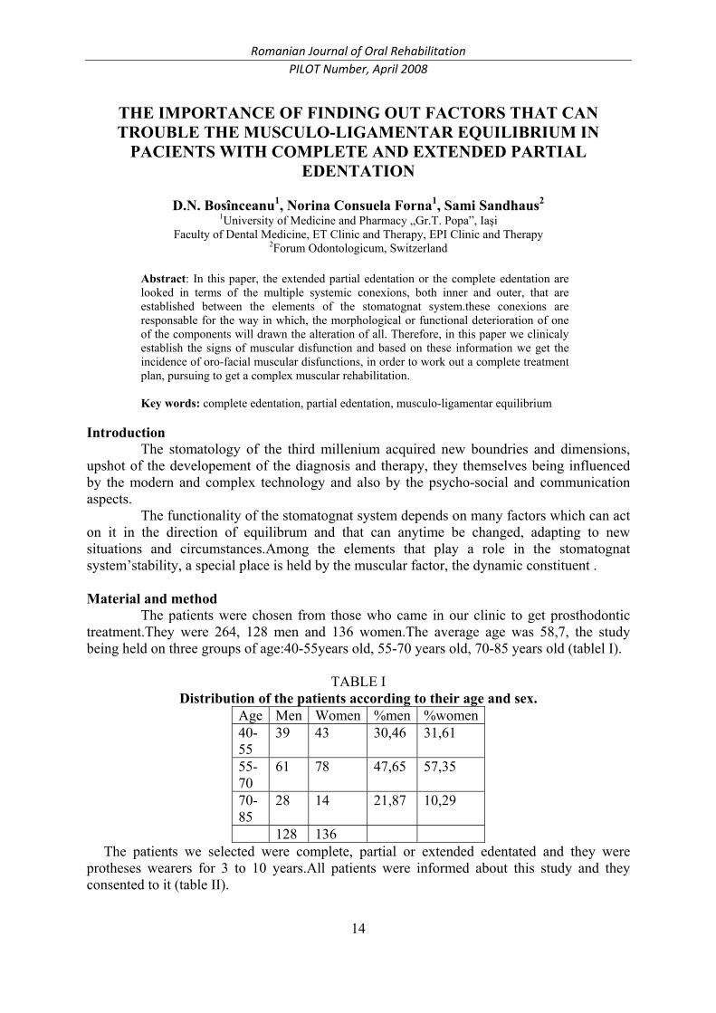
The patients were chosen from those who came in our clinic to get prosthodontic treatment.They were 264, 128 men and 136 women.The average age was 58,7, the study being held on three groups of age:40-55years old, 55-70 years old, 70-85 years old (tablel I).
TABLE I
Distribution of the patients according to their age and sex. Age Men Women %men %women40-55
39 43 30,46 31,61
55-70
61 78 47,65 57,35
70-85
28 14 21,87 10,29
128 136 The patients we selected were complete, partial or extended edentated and they were
protheses wearers for 3 to 10 years.All patients were informed about this study and they consented to it (table II).

Romanian Journal of Oral Rehabilitation PILOT Number, April 2008
15
TABLE II Distribution of the patients according to their type of edentation
Age E.P C.E E.P C.E Men Women Men Women % %
40-55
36 42 3 1 95,12 4,87
55-70
28 37 33 41 46,76 53,23
70-85
6 5 22 9 26,19 73,80
70 84 58 51
To each and every patient was elaborated a clinical report.They were throughly examined and so was every muscular group using the clasic methods of inspection and palpation.
There were investigated the temporals, the masseters as muscles of mastication and the buccinators and the orbiculars as oro-facial ones.
The palpation was made by pressing smoothly the muscles’insertion and tucking the muscles’mass, both in movement and in rest.
During the postural position, the muscles are characterized by a light contraction, that can’t be detected on the electromyography-muscular tonus of posture.This can vary depending on many other factors such as clinical, functional and morphological ones and it will be evaluated considering the relation between the muscles’ osseous insertions and the postural tonus that exists.
First we palpated the masseter and the temporal muscles-the osseous insertions and the masses and then we palpated the oro-facials muscles.Every muscle was examined equably on the right side and on the left.We assesed and wrote down in every patient medical report the trophicity of the muscles and their consitency. The muscular tonus was examined using Netter’s tests.
After the clincal exam of the muscles we examined the protheses, assesing their the maintenance and stability usin the following standards (Tabel III) 0 – Maintenace - Non at all.When it is inserted in the oral cavity is dislocate itself .Stability - Non at all.It’s tipping on the prothetic field. 1 – Minimal maintenance.It has light maintenance when pulled vertically and the same or nothing at all when pull on side . Minimal stability. .It’s tipping moderately on the prothetic field. 2 - Moderately maintenance when pulled vertically and the same or nothing at all when pull on side . Suficient stability. .It’s lightly tipping or not tipping at all on the prothetic field. 3 – Good maintenance.When pulled vertically has maximum maintenance and enough when side forces act. Good stability, without tipping.
The rating of the protheses was made likewise: - minimal stability and maintenance-score <6 - moderately stability and maintenance-score 6-8 - good stability and maintenance-score > 8 Results and discussions
After the clinical exam we found the following grades of tonicity of the masticatory muscles:

Romanian Journal of Oral Rehabilitation PILOT Number, April 2008
16
- at group of age between 40-55 with partial extended edentation from 78 cases :22 of them had normal tonicity for masseters, 28 with normal tonicity for the temporals , 33 with normal tonicity for the orbiculars and 29 for the buccinators , 15 with hipertonicity for the masseters, 17 with hipertonicity for the temporals, 12 with hipertonicity for the orbiculars and 8 with buccinators hipertonics and with hipotonicity we found 41 cases for masseters and temporals, 33 for the buccinators and the orbiculars
- at the same group of age but in casese of complete edentation from 4 cases we found one case with normal tonicity in masseters, 2 cases in temporals and orbiculars and one with normal tonicity in buccinators
- at group of age between 55-70 with partial extended edentation from 65 cases : 12 of
them had normal tonicity for masseters, 15 with normal tonicity for the temporals , 21 with normal tonicity for the orbiculars and 14 for the buccinators , 7 with hipertonicity for the masseters, 8 with hipertonicity for the temporals, 10 with hipertonicity for the orbiculars and 6 with buccinators hipertonics and with hipotonicity we found 46 cases for masseters and 42 for temporals, 45 for the buccinators and 34 for the orbiculars
- at the same group of age but in casese of complete edentation from 74 cases we found:8 of them had normal tonicity for masseters, 10 with normal tonicity for the temporals , 13 with normal tonicity for the orbiculars and 8 for the buccinators , 3 with hipertonicity for the masseters, and the temporals, 4 with hipertonicity for the orbiculars and 1 with buccinators hipertonics and with hipotonicity we found 63 cases for masseters and 61 for temporals, 64 for the buccinators and 57 for the orbiculars.
- at group of age between 70-85 with partial extended edentation from 11 cases we found:
2 of them had normal tonicity for masseters and the temporals , 3 with normal tonicity for the orbiculars and 2 for the buccinators , 3 with hipertonicity for the masseters and the temporals, 2 with hipertonicity for the orbiculars and 1 with buccinators hipertonics and with hipotonicity we found 6 cases for masseters ,for temporals and for the orbiculars and 8 for the buccinators
- at the same group of age but in casese of complete edentation from 31 cases we found: 3 of them had normal tonicity for masseters and for the temporals , 2 with normal tonicity
0
20
40
60
80
100
28.235.89
42.337.17
18.46 23.0732.3
21.5318.18 18.1827.27
18.18
40 - 55 ani
55 - 70 ani
70 - 85 ani0
20
40
60
80
100
25
50 50
25
10.81 13.51 17.5610.819.67 9.67 6.45 6.45
40 - 55 ani
55 - 70 ani
70 - 85 ani
0
20
40
60
80
100
19.23 21.715.38
10.2510.76 12.3 15.389.23
27.27 27.2718.18
9.09
40 - 55 ani
55 - 70 ani
70 - 85 ani
0
20
40
60
80
100
25 25 25 25
4.05 4.05 5.4 2.73.22 3.22 6.45 3.22
40 - 55 ani
55 - 70 ani
70 - 85 ani

Romanian Journal of Oral Rehabilitation PILOT Number, April 2008
17
for the orbiculars and the buccinators , 1 with hipertonicity for the masseters, and the temporals and buccinators, 2 with hipertonicity for the orbiculars and with hipotonicity we found 27 cases for masseters ,for temporals and for the orbiculars, 28 for the buccinators
Conclusions
1. Between the morphological bone structure and the muscles of the stomatognat system there is quit an equilibrium, always changing according to the adaptation of the two systems, the muscular activity being directly influenced by the integrity of every element of the stomatognat system
2.The dishomeostasis of the stomatognat system as a result of edentation is just a step on the way of this complex diseases, the changes that took place being irreversible.Therefore, the group of muscles affected can influence the relationships between the two maxilla and can also change the mandible’s movements in old wearers of protheses.
3. The great variety of the stomatognat system’s changes as a result of edentation and ageing requires a thoroughly investigation of each and every case, in order to track down as soon as possible every muscular disfunction.
4. All patients found during the clinical exam to have a muscular disorder must be investigated to set up a complex treatment, monitorized even after the prothese would be over
5.As a result of the clinical and paraclinical investigations , we determined a high rate of muscular disfunction in the group of old wearers, with the stability and maintenance of the protheses affected .The disfunction was asserted by means of hiper and hipotonicity of the muscular masses. In the group of recently edentated patients the changes were less visible than in the group of old wearers , in which the body tried to adjust to the edentation.We also found muscles that weren’t yet affected by the changes of the stomatognat system.
6. The change of the muscular tonus and of the mandibular movements are signs and symptoms that lead us to a diagnosis of muscular disfunction and are elements that will influence the prosthodontic treatment. References 1. Ash M. M., Current concepts in the etiology, diagnosis and treatment of TMJ and muscle dysfunction,
1986, J. of Oral Rehab, 13, 1-20. 2. Burlui V., Forna N., Clinica şi terapia edentaţiei parţial întise, Ed.Apollonia, Iaşi, 2004. 3. Costa E., Sindromul de disfuncţie mandibulară, Ed.Ştiinţifică şi Enciclopedică, Bucureşti, 1987. 4. Korfage J.A., Koolstra J.H., Lagenbach G.E. and Van Eijden T.M., Fiber type composition of the human
jaw muscle-role of hybrid fibres and factors responsible for interindividual variation, J.Dent Rest., 2005:84:9(784-793).
5. Lejoyeux J., Proteza totală vol I+vol II, Ed.Medicală, Bucureşti, 1968 6. Travell J.G., Simons D.G., Myofascial pain and dysfunction, The Trigger Point Manual, Williams &
Wilkins, 1983 7. Tsai C.M., Chou S.L., Gale E.N., McCall W.D. Jr., Human masticatory activity and jaw position under
experimental stress, J.Oral Reab., 29(1):44-51, 2002 8. Wheeler A.H., Myofascial pain disorder, Theory to therapy, Drugs. 64(1):45-62,2004
0
20
40
60
80
100
52.5642.3 42.3
52.56
70.7264.61
52.3
69.23
54.54 54.54 54.54
72.72
40 - 55 ani
55 - 70 ani
70 - 85 ani0
20
40
60
80
100
50
25 25
50
85.13 82.4377.02
86.4887.09 87.09 87.09 90.32
40 - 55 ani
55 - 70 ani
70 - 85 ani

Romanian Journal of Oral Rehabilitation PILOT Number, April 2008
18
OPTIMIZING THE CLINICAL DIAGNOSIS IN FETAL ALCOHOL
SINDROMA. THE EXPERIENCE OF THE CENTER FOR MEDICAL GENETICS IAŞI
Elena Braha, M. Voloşciuc, M. Covic
University of Medicine and Pharmacy “Gr.T. Popa” Iaşi Faculty of Dental Medicine, Medical Genetics Dept.
Abstract: Fetal alcohol spectrum disorder (FASD) is a term that combines a characteristic dysmorphic face, growth retardation, mental and behavioral effects that can occur when the fetus is exposed to ethanol during gestation. FASD is a teratogenic syndrome. The diagnoses under the FASD umbrella are fetal alcohol syndrome (FAS), partial FAS (p-FAS) and alcohol- related neurodevelopmental disorder (ARND). The diagnostic process consists of prenatal alcohol exposure screening, careful physical examination and a correct neurobehavioral assessment. We used for clinical diagnosis a Diagnostic Guideline suggested in 2005 by Medical Canadian Association. We selected 8 patients from 8615 total patients evaluated in Iasi medical genetics centre during 5 years. For 7 patients we established the FAS diagnostic and for 1 patient p-FAS. We had no ARND diagnosis. In conclusion the Canadian guideline is very useful in clinical evaluation and correct diagnosis. The limits of this study consist in unrecognized the syndrome by the physicians, a difficult anamnesis for alcohol exposure during pregnancy and the maternal education limited. Early diagnosis is associated with better long-term outcomes. The diagnostic assessment for prenatal alcohol exposure is a diagnosis for the affected individual, their mother, and other possibly affected family members. The primary care physician is a key person for prevention of continued alcohol exposure. Key words: Fetal alcohol spectrum disorder, teratogenic syndrome, Canadian diagnosis guideline
Introduction
Prenatal alcohol exposure and its role were not recognized until 1968 by Lemoine. Fetal alcohol syndrome (FAS) was first reported in the international literature by Smith and Jones in 1973. FAS has been estimated to occur in at least 2/1000 livebirths and fetal alcohol spectrum disorder (FASD) in 1% in USA. [Christina Chambers, 2006] The effects from gestational alcohol exposure lie on undesirable effects (fetal alcohol spectrum disorder). Some children might be severely affected (FAS), whereas others may have no apparent effects. The alcohol effects on the embryo are dose related and there appears to be no safe period for alcoholic consummation during pregnancy. The pathophysiologic basis for alcohol embryopathy appears to be related to genetic polymorphism for alcohol dehydrogenase. FAS could be an acetaldehyde embryopathy. [Abel, 1996]
The epidemiological studies indicate the alcohol as the most frequent cause of mental retardation. This disease is serious moreover it can be prevented with the elimination of alcohol consumption during pregnancy. It is recommended to a pregnant woman to avoid any alcohol consummation during pregnancy. [Wilkie, 1997] Fetal alcohol spectrum disorder (FASD) is a wide spectrum that encompasses fetal alcohol syndrome (FAS), partial FAS (p-FAS) and alcohol-related neurodevelopmental disorder (ARND). [Chudley, 2005]
Clinically FAS is characterize by: prenatal and/or postnatal growth retardation (W and H < - 2SD), distinct facial appearance (short palpebral fissures, strabismus, ptosis , low nasal bridge , midface hypoplasia, flat philtrum, thin upper lip, posterior rotation of the ears) and central nervous system (CNS) dysfunction (mild mental retardation, microcephaly, hyperactivity in childhood or attention deficit/hyperactivity disorder). The patients could have other anomalies: cleft of palate in 15%, congenital heart malformations (VSD, ASD),

Romanian Journal of Oral Rehabilitation PILOT Number, April 2008
19
(“railroad track” appearance of the ears) and dermatogliphic anomalies (“hockey stick” palmar creases). Materials and methods
Centers for Disease Control and Prevention (CDC) and Institute of Medicine (IOM) published a guideline for clinical diagnosis and for establish FAS subcategories diagnosis. The study methodology and questionnaire were developed after consultation the guideline proposed by Health Canada National Advisory Committee in 2005. These criteria will be utilized for our patients evaluation (table I). [Chudley, 2005]
TABLE I. FASD diagnostic criteria
Criteria
FAS p-FAS ARND
Growth impairment yes no no Facial dysmorphy: (1) Short palpebral Fissures <10 percentil (2) Smooth or flattened philtrum (score 4-5) (3) Thin upper lip (score 4-5)
All 3 are present
2 of the 3 are present
None are present
Brain injury Minimum of 3 CNS domains impaired*
Minimum of 3 CNS domains impaired*
Minimum of 3CNS domains impaired*
Prenatal alcohol exposure
Confirmed or Unconfirmed
Confirmed Confirmed
Key domains assessed for CNS deficit*: hard and soft neurological signs, brain structure (including microcephaly), cognition, communication, academic achievement, memory, executive functioning and abstract reasoning, adaptive behaviour, social skills, social communication, attention span, activity level, distractibility
The smoothness of the philtrum and the thinness of the upper lip are assessed
individually on a scale of 1 to 5 (1 = unaffected, 5 = most severe) using a lip-philtrum guide (figure 1). It must be accentuated the importance of a correct medical photo. The patient must have a relaxed facial expression, because a smile can alter lip thinness and philtrum smoothness.
Fig. 1 Lip-Philtrum Guide (Chudley, 2005)

Romanian Journal of Oral Rehabilitation PILOT Number, April 2008
20
We selected 8 patients with the alcohol fetal spectrum diagnosis (3 boys, 5 girls) from 8615 total patients evaluated in Iasi medical genetics centre during 5 years (2000 – 2004). Their age was between 5 months and 13 years 2 months.
The majority of patients come from rural aria (6 cases) and only 2 cases come from urban aria. We notice the predominance of patients from rural aria, partial motivated by the medical education absence, restricted access to it, parents behaviours imitation from childhood (including alcohol consummation), mother belief that a mild alcohol use as wine is indeed healthy. The mothers from cities aria hardly admit that used alcohol during pregnancy because culpability feeling.
We have to note by the beginning that the small number of cases could be explained by ignorance of fetal alcohol spectrum clinical features. Most of the patients did not come up to a correct diagnosis. To all our patients we applied the diagnosis criteria presented above, trying to evaluate their clinical utility.
Fig. 2 The age distribution of the
patients
Fig. 3 Facial dysmorphy presented to our patients
Results and discussions
Growth retardation (prenatal or postnatal height or weight, or both, at or below the 10th percentile) was relieved to all patients. Was regards the dysmorphic face we observed the predominance of 5 score for philtrum and upper lip. CNS involvement was following using head circumference, psychological exam and occasionally EEG (Figure 2 – 4).
Fig. 4 Central nervous system abnormalities
Regarding alcohol exposure only in 6 cases we could proved it using maternal anamnesis. Physical findings manifest in other organs or systems were: cardiovascular anomalies (atrial septal defect in 2 cases), hypospadias (1 case), hydrocel (1 case), inguinal hernia (1 case) and cutaneous angioma (1 case). Finally we establish fetal alcohol syndrome diagnosis for 7 patients and partial – alcohol fetal syndrome for 1 patient.
Towards a better recognize diagnosis, management and prevention of the syndrome we will present a clinical case.
A case of 13 years 2 months old girl is described. The girl comes from rural aria and she was referred for genetic exam because dysmorphic face and mental retardation. The patient is the third child of a healthy, non-consanguineous couple, 40 years age the mother

Romanian Journal of Oral Rehabilitation PILOT Number, April 2008
21
and 45 years age the father in the fecundation moment. The mother is known as alcoholic. The pregnancy evolution was normal. The delivery occurred in hospital but we have no medical records. The postnatal evolution reveals growth and mental retardation.
Clinical exam notes mild growth retardation (W = -2.17 SD, H = - 1.6 SD), mycrocephaly (OFP = -5.53 SD), facial dysmorphy with short palpebral fissures, low nasal bridge with septal deviation, microstoma with flat philtrum (score 4), thin upper lip (score 4), dental anomalies (hypodontia with upper canine absence, malocclusion), bilateral V finger clinodactyly, dermatogliphic anomalies (typical “hockey stick” palmar creases), cardiac anomalies (ASD), mental retardation. (Figure 5)
Paraclinic and other specialities examinations show: ASD (echocardiography 2D), mild mental retardation (IQ 44), normal EKG and normal abdominal echography.
a) b) c) Fig. 5 Facial dysmorphy for AAI patient and the mother clinical appearance
a) new-born b) 6 years 7 months c) 13 years 2 months
Based on anamnesis, clinical findings and paraclinicaly exams we can establish the diagnosis of alcohol fetal syndrome.
Familial history does not reveal other cases. Medical management for the patient includes cardiac and dentistry follow up; special needs school and annual medical examination. We must keep in mind the syndrome prevention although the mother is 53 years old; the prevention will strive to ensure that the entire family member understands that drinking alcohol can have hazardous consequences, particularly during pregnancy. Conclusions
In conclusion clinical diagnosis of fetal alcohol syndrome, implementation educational strategies for patients, selective prevention interventions target people who are at greater risk for alcohol using during pregnancy and started an education programme about teratogenic effects of the alcohol are extremely important.
Medical management of the patients with FAS includes: public education particularly for the young population at high risk for changing attitudes regarding alcohol use; selective prevention interventions target women who drink alcohol and are in the reproductive age; improvement the diagnosis of the possible affected new-born or child; an early assessment process to stop the complications; public support programs for children with FAS that provide access to school, recreational, and social activities. Such a medical management plan minimizes risk factors for lifelong negative consequences and promotes protective factors that maximize developmental potential. [Chudley, 2005]
A late intervention may have lifelong negative consequences for patients with FAS, including disrupted school experiences, legal problems, incarceration, mental health problems, substance abuse problems, inappropriate sexual behaviour, dependent living, and poor employment history. Although the anomalies associated with FAS were permanent some of them could be change by an early medical intervention. Keys to working successfully with children with FAS are to insure access to appropriate rehabilitation services

Romanian Journal of Oral Rehabilitation PILOT Number, April 2008
22
(physical, speech, behavioural, mental health, occupational), to ensure that school curricula are balanced with vocational training and they have skills of daily living (e.g., personal hygiene, money management). Selective references 1. Christina Chambers, Keith Vaux, Fetal Alcohol Syndrome, http://www.emedicine.com/ped/topic767.htm,
octombre 2006 2. Abel EL., Moderate drinking during pregnancy: cause for concern?, Clin Chim Acta, 1996 Mar 15; 246(1-
2): 149-54 3. Wilkie S., Global overview of drinking recommendations and guidelines. AIM Digest Suppl 1997 Jun; 2-4 4. Albert E. Chudley, Julianne Conry, Jocelynn L. Cook, Christine Loock, Ted Rosales and Nicole LeBlanc,
Fetal alcohol spectrum disorder: Canadian guidelines for diagnosis, CMAJ, March 1, 2005; 172 (5_suppl), S1-S21

Romanian Journal of Oral Rehabilitation PILOT Number, April 2008
23
MUSCLES COMPLEX REHABILITATION CRITERIA
Mihaela Daniela Brînză1, Dieter Wember Mathes2, Norina Forna1
1University of Medicine and Pharmacy „Gr.T. Popa” Iaşi Faculty of Dental Medicine, EPR Clinic and Therapy
2Private Practicioner Germany, Associated Professor – University of Medicine and Pharmacy „Gr.T. Popa” Iaşi
Abstract: Treatment strategies based on scientific evidence in muscles rehabilitation have as immediate purpose pain amelioration, re-establishing a normal function of stomatognatic system, and last but not least improving of life conditions. Muscles dysfunctional diagnosis, choosing and performing different dental treatment which can induce disturbances in occlusal or intermaxilary relations, makes from diagnosis and treatment a responsibility issue. Numerous techniques and methods for a total management of patient to muscles rehabilitation interfere either with diagnosis either with all treatment steps, and are made due to scientific criteria and principles. The purpose of the study is to identify, appreciate and summarise all criteria for muscles rehabilitation by applying them to a study group for diagnosis and treatment. If the treatment is not correct or quick applied the muscular dysfunction will perpetuate and will damage the other elements of the system. In conclusion, muscles rehabilitation interfere with all diagnosis and treatment steps, reconditioning methods even that are time consuming can be applied with minimum equipment and are one actual necessity of dental treatment. Key words: complex rehabilitation, muscles treatment, criteria.
Introduction
Systemic theory of dysfunctional syndrome indicates the muscular dysfunction as a major factor in producing of disease [1,2]. Dysfunctional syndrome theories promotes holistic approach of masticator system and it’s pathology but, in many cases treatment, despite necessity of being complex and well organized, is mostly for symptoms and signs of muscle dysfunction, not an etiologic one. Due to changes in muscles contraction and coordination and modified mandible pathways the muscle gets tired, painful and the local circulation is diminished.
Despite of it’s importance in maintaining system homeostasis muscles complex rehabilitation methods are forgotten or badly applied, the context of general and local rehabilitation aren’t use. Myotherapy is a complex of methods well integrated in different treatments and possible to individualize and future promote programs for health have to include methods for muscle rehabilitation [3].
Total management of patient is based on diagnosis decision, foundation for all treatment, no matter if the problem regards dental diseases, temporo-mandible diseases or muscles dysfunctions. Muscles rehabilitation is an important step of complex rehabilitation and the treatment methods are scientific and based on criteria and principles. Even to establish a diagnosis of muscles dysfunction we use these criteria [2].
The aim of the study is to reveal all possible treatment therapies for muscles rehabilitation as a part of oral complex rehabilitation, the necessity of using this methods in the same time with other complex treatment for stomatognatic system and also the proportion of treatment applicability on a study group and the result.
Material and methods
Observational analytical study was made on 2817 patients 1058 women and 883 men with age between 16 and 89 years old, presents for dental treatment in Prosthetic Department of Dental Medical Faculty between 1-st January 2001 and 1-st October 2004. Results of clinic and paraclinic exam were recorded in observation file, the inclusion criteria

Romanian Journal of Oral Rehabilitation PILOT Number, April 2008
24
in study being presence of muscles dysfunction sings and symptom and exclusion criteria presence of other systemic elements dysfunction (temporo-mandibular joint, third molar pathology). After applying these criteria we modify the study lot to 1072 patients (594 women and 478 men) which fit the criteria. Results and discussions
Complex rehabilitation of muscles in context of bio-psycho-social criteria permits to explain all interconnection which appears when one element of stomatognatic system is disturbed (anatomical or functional) and has as effects directly and immediate appearance of muscles dysfunction. These disturbances on biologic level (pain, muscles spasm, facial asymmetry) is perceived as cortical projection modification and the immediate result will be behavioral and psychological modifications (intensity and frequencies of these manifestations is directly related with cultural experience and medical knowledge) [5].
Exist also the reverse of this explanation easy to understand especially to bruxism patient when daily tension can induce tonus modification [4].
Systemic criteria is related with diagnosis and treatment and permits stomatognatic system approach by respecting patient individuality and interrelation between muscles dysfunction and other systemic elements. Analytical and global analysis criteria are fundamentals for diagnosis. Analytical examination offer information regarding muscles tonus, volume, contraction, presence of pain. But this evaluation can’t appreciate if cranio-mandible relation or mandible dynamics pattern are modified facts that makes diagnosis more difficult. Global examination offers this type of information so association of these two methods is necessary. For example, is impossible to appreciate occlusal or cranio-mandible diagnosis as long as the patient has a muscle spasm. If the spasm is not observed diagnosis will be incorrect and the treatment plan also so is necessary to use relaxation methods before establishing diagnosis until muscles are in normal condition of function.
Therapeutically a hierarchy criteria is a moment which can’t be standardized and related with patient individuality. Muscles therapy interfere all treatment steps: starting with sanitary education (posture position, head position), general preparation (treatment of general disease that can influence neuro-muscles disease by temporizing or contraindicate dental treatment), local preparation (specific step for muscles treatment methods).
Temporizing therapy criteria imposes emergency, provisional or transitory treatment to obtain tissue conformation.
Prophylactic criteria include general and local prophylactic methods to avoid muscles overload by sedentary work [4]. Biologic criteria is also applied in muscles complex rehabilitation all methods respecting systemic homeostasis. Easy treatment methods can became dangerous when are applied incorrect by inducing irreversible modification in muscles structure. Cure criteria in muscles treatment means etiologic factors removal which can permit morphological and functional systemic’s element restoration.
Immediate intervention criteria offer to doctor possibility to cache the disease at it’s debut and to use treatment methods with good effects in short time. Subordinate to these criteria is the temporized therapy criteria which don’t allow any intervention to stomatognatic system until homeostasis is obtained. Temporary or provisional prosthesis permit new cranio-mandible relation and implicit muscles relaxation and make the transition to a correct relation. Simultaneity therapy criteria allow using muscles therapy in the same time with other dental therapy (prosthetic therapy, cranio-mandible therapy and muscles relaxation methods). Minim invasive criteria are also applied in muscles complex rehabilitation either as investigation either as therapeutic aspects.
The muscular relaxation techniques are multiple and are specific for each step of dental treatment. Can be an emergencies treatment or a curative treatment, etiologic and

Romanian Journal of Oral Rehabilitation PILOT Number, April 2008
25
symptomatic, in many cases the methods are superposing. The emergency treatment was applied to 116 patients (10, 82 %) with analgesic drugs and high efficiency immediate or in 24 hours.
Etiologic treatment it doesn’t mean only muscular relaxation, it also mean a series of therapeutic measures for local and loco-regional tissue re-equilibrium, addressed to reestablish general and systemic homeostasis and to allow normal function of stomatognatic system. Etiologic treatment was applied to 483 patients (40,85%), myo-kinetic exercises and muscles stretch seems to be effective but not in singular therapy and not in short period of time.
Symptomatic treatment is for muscle signs and most of the time is superposing the etiologic treatment, removal of etiologic factors having as effect abolish or diminishing muscles signs. This treatment was applied to 483 patients, the same which also had etiologic treatment. Conclusion
Muscles complex rehabilitation is based on scientific criteria and treatment methods are applied in establishing diagnosis and treatment. Applying one single treatment methods by unique action mechanism is insufficient to obtain muscles complex rehabilitation. Etiologic treatment associate with symptomatic therapy due to rehabilitation criteria will induce muscles relaxation and permit other systemic elements treatment.
Muscles complex rehabilitation treatment became a general rule applicable to all patients that will receive a dental treatment.
Grouped as new elements for oral complex rehabilitation, postural exercise and muscles re-equilibration (in tonus changes or in case of affected functions) the drawing and systematization of a new treatment schema with time controllably efficiency, with possibility of individualization due to case specifics and also integration of muscles complex therapy in routine of dental treatment, are not only new aspects for dental practicing, but also offer the scientific characteristics and the approach of study goals represents a warranty for results validity. BIBLIOGRAPHY 1. Burlui V., Malrelatiile cranio-mandibulare, Ed. Apollonia, Iasi, 2002, 118-243 2. Burlui V., Morăraşu C., Gnatologie, Ed. Apollonia, Iasi, 2000, 37-59 3. Fricton, J. R., Behavioral and psychosocial factors in chronic craniofacial pain. Anes. Prog., 32(1):7-12,
1985. 4. Glaros AG, Williams K, Lausten L. The role of parafunctions, emotions and stress in predicting facial pain.
J Am Dent Assoc.;136(4):451-458, 2005. 5. Tschernitschek H, Fink M. Applied kinesiology in medicine and dentistry - a critical review Wien Med
Wochenschr.155(3-4):59-64, 2005

Romanian Journal of Oral Rehabilitation PILOT Number, April 2008
26
THE EFFECTS OF THE PAIN ENDURED DURING DENTAL
TREATMENT
Diana Cerghizan, S. Popşor, M. Suciu, A. Kovacs University of Medicine and Pharmacy Tg. Mures
Department of Prosthetic Dentistry and Oral Rehabilitation
Abstract: It has been acknowledged for many years that human pain perception is made up of multiple dimensions, including a sensory aspect and an emotional/affective quality aspect (Price, 1988). Researchers have shown that some “pain” stimuli are associated with high levels of emotionality/affect (for example, cancer pain), whereas other “pain” stimuli can produce relatively low levels of emotional distress (for example, labour pain) (Price et al., 1987). These findings indicate that people can experience very different emotional responses to very similar levels of stimuli intensity, depending on their perception of the event (Gracely, Kwilosz, 1988). Assessment of clinical pain response requires the use of measurement scales designed to capture the different dimensions of pain perception (Logan, 1995). The target of this study is to prove the connection between previous pain and anticipating pain. This study is a part of a larger research project , and the results presented here are only preliminary, they can modify with the advancement of the study (ex. rising patient number). Keywords: pain, dental treatment
INTRODUCTION
The dental treatments usually are associated by the patient with pain and anxiety. Is proved that painful therapeutic procedures are the most important reason of generating pain and anxiety during a dental treatment.
An early negative dental experience is probably the most stated single cause for dental anxiety (Locker et al., 1996, 1999). However, a negative dental experience does not necessarily lead to dental anxiety. The 'latent inhibition' theory, for instance, states that a history of positive or neutral dental experiences may serve as a buffer against the development of traumatic associations or experiences (Davey, 1989). As a consequence, high levels of anxiety or fear are developed less easily. Conversely, an early negative dental experience can serve as a one-shot conditioner and may leave a patient with feelings of anxiety. Fear of dental pain is a highly relevant concept in dental pain research and, moreover, in dentistry (van Wijk and Hoogstraten, 2003). Whereas anxiety and fear can be seen as a state of distress in anticipation or in the presence of a perceived danger, respectively, fear of pain can be seen as a state of distress related to a very specific type of stimulus, namely, pain (Gower, 2004). Research suggests that anxious people tend to overestimate anticipated pain. Moreover, individuals tend to overestimate the intensity of aversive events in general, including such events as fear. Therefore, people who are predisposed to respond fearfully to pain are at an increased risk of ending up in a vicious circle of anxiety, fear of pain, and avoidance of dental treatment (van Wijk, Hoogstraten, 2005). MATERIAL AND METHOD
This study is based on a questionnaire created by us, which includes general data’s about the patient (age, sex, studies), and also contains four questions , which are helping us to determine, if the patient had any painful experiences during the dental treatment, if he’s anticipating the pain, or if he is avoiding the appointments because of pain.

Romanian Journal of Oral Rehabilitation PILOT Number, April 2008
27
At the same time we determined the patient’s anxiety level using the Dental Anxiety Scale (DAS) questionnaire. DAS contains four questions about different situations which are occurring during the dental treatment. Every question is rated between 1 (no anxiety) and 5 (very anxious), the final score can alternate between 4 and 20. A result higher than 15 is the proof for a high level of anxiety.
The patient’s selection was based on the next criteria’s: 1. patients older than 18 2. patients who had contact with one or more dentist’s before the start of the study 3. we used only the fully completed questionnaires After a selection made using this criteria’s it resulted a lot of 247 persons with age
between 18 and 79 (M = 38,03), 179 (72,47 %) female and 69 (27,53%) male. Using the DAS we confirmed that the majority of the patients with painful
experiences in the past are subject of high or even severe level of anxiety. RESULTS
The questionnaire carry out by us presents questions with closed answer (yes, no), codified by entering them in statistical analysis charts, done by GraphPad InStat 3 and NCSS software’s.
Out of 247 questioned patients 60 % said that they endured painful dental treatments in the past and also 60|% said that they during a dental treatment are waiting for the appearance of the pain. For statistical analysis we used the Fisher test and the results showed that is a very significant association between pain in the past and anticipating pain (p< 0,0001) The association is significant both statistically and scientifically to (OR = 3.951, CI = 95%, 2,298 – 6,794) (fig. 1).
durerea din antecedente si anticiparea dureriiColumn Totals
Columnsanticipeaza nu anticipeaza
140
130
120
110
100
90
80
70
60
50
40
30
20
10
Fig. 1 Correlation between pain in the past and anticipating pain
After dividing on age groups we observed a extremely significant positive
association, both from statistically or scientifically points of view , between pain in the past and anticipating pain at patients with ages between 18 -30 (n= 81) years and 41 – 50 (n= 47) years with p=0,0005 (OR = 5.600 95% CI: 2,088 to 15,017), or in case of p=0,0006 (OR = 12 95% CI:2,685 to 53,636). At patients with ages between 18 and 30 years we could prove statistically significant correlation between anticipating pain and avoiding dental treatment (p=0,0024) (Fig. 2).

Romanian Journal of Oral Rehabilitation PILOT Number, April 2008
28
Fig. 2 Pain anticipation and ages
The statically reading of the results showed that between pain in the past and
avoiding dental treatments exists a positive association, but statistical insignificant (p=0,08 OR = 1,743 95% CI: 0,9635 to 3,151). This helped us to conclude that to obtain accurate results we need a larger lot of patients. CONCLUSIONS
In 1984 Wall and Melzack said “ Pain always is one-sided. Every individual is learning the signification of this word by the experiences he starts to have from his first years. Without doubts is a sensation with organic origins, but this sensation always is apprehended like an unlike one, which makes from this an emotional experience”
For many patients, fear of dental pain and avoidance of dentistry are synonymous (Freeman, 1991). Moreover, clinicians report that managing some patients’ pain and distress can be a frustrating task (Lindsay, Jackson 1995).
From this lot of patients 60% (n=149) had in the past dental treatments involving pain. This result has to put the practitioners to think how they can avoid pain, because pain could be the starting or the aggravating factor of the dental anxiety.
The high number of patients who had a positive answer to the first question from our questionnaire shows us that practitioners are not giving enough significance to the symptom of pain, resulting an absence of interest in trying to challenge the pain. Between pain in the past and avoiding dental treatments exists a positive association, but to determine the statistically and scientifically magnitude we have to rise the number of questioned patients. We can claim that any pain endured during the dental treatment remains printed in the patients memory, making them to think on possible pain at their following appointment. 73% (n=109) out of the patients who experienced painful dental treatments , are believing that at the next appointment pain can show up again. A number of 40 patients are waiting for pain to show up at their next appointment, even they never experienced painful dental treatments. This situation is making us to associate pain with the dental treatment.
It often is assumed that aging results in loss of pain sensitivity. Although some efforts have been made to study the effects of aging on pain perceptions, the results are not conclusive. Experimental studies of acute pain responses do not show significant age-related alteration in the pain perceptions of healthy elderly subjects (Harkins et al., 1994). It has been proposed that differences in acute pain responses between younger and older patients (Lash et al., 1997) may be a result of changes in pathophysiology (for example, neural conductivity) rather than changes in the pain perception itself (Harkins et al., 1990; Heft et al., 1996). It is
0.0
26.7
53.3
80.0
da
nu
Romanian Journal of Oral Rehabilitation PILOT Number, April 2008
29
not clear, however, from the literature whether these changes in pathophysiology influence both affective pain and sensory intensity in the elderly.
During our study we observed that patients with age between 18 – 30 years are avoiding dental treatments because of the pain which can show up during the dental treatment. Patient older than 50 years are not avoiding dental treatments. One of the main reasons of this can be that painful experiences are fading during the years in patients memory.
New evidence suggests that there are differences in pain perceptions between men and women (Riley et. al. 1998, Unruh et al. 1999). Although, most studies suggest that women have greater pain sensitivity than men, there are inconsistencies in the literature (Eli et al., 1996). These inconsistencies suggest that the type of pain stimuli may influence perceived pain differences between men and women (Fillingim, 1998). In addition, the influence of aging on these reported sex differences has yet to be clarified.
In our study because of the lower number of questioned male patients we couldn’t determine a precise correlation between pain in the past , anticipating pain and avoiding dental treatments.
Our own experience is showing that the majority of patients are favoring different methods to fight pain showing up during the oral rehabilitation treatments.
Patients avoiding dental treatments usually presents a poor oral health, and at the end they will need elaborate oral rehabilitation treatments.
This work was supported by the CNCSIS – Ministry of Education and Research, Romania, Project Td No 417/2007 BIBLIOGRAPHY
1. Davey GC, Dental phobias and anxieties: evidence for conditioning processes in the acquisition and modulation of a learned fear. Behav Res Ther 1989, 27:51–58
2. Eli I, Bar-Tal Y, Zvu F, Korff E., Effect of biological sex differences on the perception of acute pain stimulation in the dental setting. Pain Res Manage 1996;1(4):201–6.
3. Fillingim RB, Maixner W, Kincaid S, Silva S., Sex differences in temporal summation but not sensory-discriminative processing of thermal pain. Pain 1998;75:121–7.
4. Freeman R., The role of memory on the dentally anxious patient’s response to dental treatment. Ir J Psychological Med 1991;8(2):110–5.
5. Gower PL, editor (2004). Psychology of fear. New York: Nova Science Publishers, Inc 6. Gracely RH, Kwilosz DM, The Descriptor Differential Scale: applying psychophysical principles to
clinical pain assessment. Pain 1988;35:279–88. 7. Harkins SW, Kwentus J, Price DD, Pain and suffering in the elderly. In: Bonica JJ, ed. The
management of pain. 2nd ed. Philadelphia: Lea & Febiger; 1990:552–9. 8. Heft MW, Cooper BY, O’Brien KK, Hemp E, O’Brien R., Aging effects on the perception of noxious
and non-noxious thermal stimuli applied to the face. Aging (Milano) 1996;8(1):35–41. 9. Lasch H, Castell DO, Castell JA., Evidence for diminished visceral pain with aging: studies using
graded intraesophageal balloon distension. Am J Physiol 1997;272(1 Pt 1):G1–3. 10. Lindsay SJ, Jackson C, Fear of routine dental treatment in adults: its nature and management. Psychol
Health 1993;8:135–53. 11. Locker D, Liddell A, Dempster L, Shapiro D., Age of onset of dental anxiety. J Dent Res 1999, 78:790–
796 12. Locker D, Shapiro D, Liddell A., Negative dental experiences and their relationship to dental anxiety.
Community Dent Health 1996, 13:86–92. 13. Logan HL, Baron RS, Kohout F. Sensory focus as therapeutic treatments for acute pain. Psychosom
Med 1995; 57:475–84. 14. Lyndsay C. Bare, B.A.; Lauren Dundes, M.H.S., Sc.D. Strategies for Combating Dental Anxiety J Dent
Educ. 68(11): 1172-1177 2004 15. Price DD, Harkins SW, Baker C. Sensory-affective relationships among different types of clinical pain
and experimental pain. Pain 1987;28:297–307. 16. Price DD. Psychological and neural mechanisms of pain. New York: Raven; 1988:2–70. 17. Riley JL III, Robinson ME, Wise EA, Myers CD, Fillingim RB Sex differences in the perception of
noxious experimental stimuli: a meta-analysis. Pain 1998;74:181–7.

Romanian Journal of Oral Rehabilitation PILOT Number, April 2008
30
18. Unruh AM, Ritchie J, Merskey H. Does gender affect appraisal of pain and pain coping strategies? Clin J Pain 1999;15:31–40.
19. van Wijk AJ, Hoogstraten J (2003). The fear of dental pain questionnaire; construction and validity. Eur J Oral Sci 111:12–18
20. van Wijk A.J. and J. Hoogstraten. Experience with Dental Pain and Fear of Dental Pain J Dent Res 84(10):947-950, 2005
21. Wall PD, Melzack R. Geriatric pain. In: Wall PD, Melzack R, eds. Textbook of pain. 3rd ed. London: Churchill Livingstone; 1994:769–84.

Romanian Journal of Oral Rehabilitation PILOT Number, April 2008
31
EMDOGAIN AND BIO-OSS BETWEEN HOPE AND REALITY
Laura Cîrligeriu, M. Boariu, A. Marinescu, V. Cârligeriu
University of Medicine and Pharmacy Timisoara
Abstract: In the last years the treatment of periodontal disease has gain new meanings thanks to new developed materials that helped the surgical treatment. The developers of these materials promise a lot in the field of guided tissue regeneration, but in order to know how much of these is true we tried some of them in clinical tests. The most frequently used materials by us was Emdogain and Bio-Oss and made a study (similar studies was conducted by Sculeanu et.al.) to demonstrate how much of these materials properties was hope and how much was reality. The aim of the study was to evaluate the clinical and radiographic outcome using two different materials in the treatment of periodontal bone defects based on guided tissue regeneration techniques (2). Our study included 40 subjects with generally healthy status with periodontitis ilness in advanced stage. Depending of the materials used in the treatment our patients were divided into two equal groups. The group number I, was treated with a bovine-derived hydroxyapatite xenograft (Bio-Oss, ) combined with a resorbable collagen membrane (COREM, Poneti) was used. In the second group was used enamel matrix derivative (Emdogain® Gel). Before the surgical treatment a clinical and radiographic examinations were performed, and the following parameters were evaluated: plaque index, gingival index, probing pocket depth, clinical attachment level, radiographic defect depth, and defect width. At the patients with bad oral hygiene the plaque and gingival index was haing high values. All clinical and radiographic parameters (except plaque index) were significantly reduced after treatment in both groups . No great differences were revealed between the two groups of patients in examined parameters after treatment. The results demonstrated that the treatment of periodontal bone defects with both materials leads to similar and significant improvements in clinical and radiographic parameters. The exception was made by the patients with bad oral hygiene. Key Words: emdogain, bio-oss, guided tissue regeneration, osteoregenerative materials
Introduction:
In the last years the treatment of periodontal disease has gain new meanings thanks to new developed materials that helped the surgical treatment. The developers of these materials promised a lot in the field of guided tissue regeneration, but in order to know how much of these is true we tried some of them in clinical tests. The most frequently used materials by us was Emdogain and Bio-Oss and made a study to demonstrate how much of these materials properties was hope and how much was reality. The aim of the study was to evaluate the clinical and radiographic outcome using two different materials in the treatment of periodontal bone defects. Material and methods
The study included 40 generally healthy subjects with advanced periodontitis (20 women, 20 men), aged 35–60, supra- and subgingival scaling and root planing were carried out during a 3-month preoperative period in all the patients, who also received oral hygiene instructions.. The patients were divided into two groups, 20 in each, according to the material used. In the first group, a resorbable collagen membrane (COREM, Poneti) and a bovine-derived xenograft (Bio-Oss®; Geistlich, Wolhusen, Switzerland, Bioos, Cerasorb) was used. The other group of patients was treated with enamel matrix derivative (Emdogain®; Biora, Malmö, Sweden).
Clinical and radiographic examinations were performed prior to, and 1 year after surgery. A periodontal probe (WHO Probe, CPITN) was used for clinical examinations prior and 1 year after surgery. At the same time the radiographic examination was made. Clinical

Romanian Journal of Oral Rehabilitation PILOT Number, April 2008
32
were evaluated the folowing parameters: plaque index (PI - Sillness& Löe), gingival index (GI - Löe& Sillness), probing pocket depth (PPD, in mm), clinical attachment level (CAL, in mm). The measurements were made on folowing sites mesiovestibular, vestibular, distovestibular, mesiooral, oral, distooral.
Radiographic examination used two intraoral radiographs taken with customized film holders and long-cone paralleling technique. The following parameters were measured; (1) defect depth (the vertical distance between the crest of the alveolar process and the jonction site of the root surface and normal alveolar bone, in mm), (2) defect width (the horizontal distance between the root surface and the most coronal part of the alveolar process, in mm).
Only patients with deepest site of gingival pocket was considered for analysis. This selection was made using the folowing criteria: PI<1, PPD≥6 mm and radiographic defect depth≥3 mm. Bone defects chosen for analysis were similar in size and shape, and were found at homonymous teeth in the two groups to be compared. Both groups of patients have presented periodontal pockets (22 with two walls, 8 with three walls, 5 circular bone defects and the remaining five other defects).
In both groups of patients, the surgical procedures were performed under local anaesthesia (4% Ubistesin). Following an incision in the gingival pocket, a mucoperiosteal flap was raised vestibularly and lingually. Vertical incisions were placed only if necessary for adequate access to the surgical site or to achieve complete coverage of the membrane with the mucoperiosteal flap (1). Granulation tissue was removed from the bone defect, and the root surface was cleaned and planed using hand and rotative instruments depending of the site defect. The surgical area was then rinsed with physiological saline. In the first group, the root surface was etched for 2 min with neutral 24% EDTA to remove the smear layer. Then the site was rinsed again with physiological saline and Emdogain was applied starting at the bottom of the defect. Finally, the flap was repositioned coronally and sutured tightly with non-resorbable sutures. In the second group of patients, the bone defect was filled with Bio-Oss and then covered with the resorbable Bio-Gide membrane. The membrane was fixed using resorbable sutures. The sutures were removed 14 days after the surgery. All patients received antiobiotics – 1 g amoxiclav twice a day for 1 wk. Moreover, the patients were instructed to avoid mechanical tooth brushing in the region involved for 4 wk, to chew carefully for 4 wk, and to rinse the oral cavity with 0.2% chlorhexidine solution (Plak-Out) twice daily for 6 wk. Four weeks after the procedure the patients started gentle tooth cleaning using the roll technique. Postsurgical appointments were scheduled at 2, 4 and 6 wk, and then every 2 months. During the first year after surgery, only professional supragingival tooth cleaning was carried out. Results and discussions
In all patients postoperative healing was uneventful. No inflammatory complications were observed, except a few case of patients with bad oral hygiene.
The mean PI values in the Bio-Oss and the Emdogain groups at baseline were identical. Following the treatment PI remained unchanged in both groups. The mean GI values in the two groups before treatment were not significantly different. The mean GI values after treatment were significantly reduced in both groups, and the values were not statistically different between the two groups. Likewise, PPD and CAL values were initially similar between the two groups. After surgery, the mean PPD and CAL values significantly decreased in both groups.
The differences in radiographic parameters between the two groups of patients before treatment was not significant. The mean radiographic parameters (depth and width of bone defects) were significantly reduced after both Bio-Oss and Emdogain treatment.

Romanian Journal of Oral Rehabilitation PILOT Number, April 2008
33
We will try to exemplify this using the RX from two patients. First patient was treated with Emdogain. It presents periodontal pockets around 1.7 and 1.8 with a probing depth of 8 mm, 4.7 presents a lvl 4 furcation, and around 4.2 and 4.3 periodontal pockets with a probing depth of 5 mm (Figure 1). Also around 2.2-2.3 periodontal pockets with a probing depth of 6 mm and 2.7-2.8 periodontal pockets with a probing depth of 7 mm. The second Rx (Figure 2) was taken after 1 year and it shows a bone gain of 5-6 mm.
Fig. 1 Fig. 2 This patient was treated with Bio-Oss. It presents periodontal pockets around 1.7-
1.6 1.3, 2.4-2.5 with a probing depth of 6 mm (Figure 3). The second Rx was taken after 1 year and it shows a bone gain of 5-6 mm (Figure 4).
Fig. 3 Fig. 4 Conclusions
Based on the above data and our own experience it appears that the use of Bio-Oss combined with resorbable membrane and the regenerative tehnique with Emdogain are comparable in the aspect of clinical and radiografically assessed periodontal healing and ofered the bone regeneration promised by their developer. The reults of our study are overlaping with data reported by other authors, who demonstrated a similar improvement in radiographic parameters. In some cases we couldn`t reach the bone regeneration desired because of the patient bad hygiene and other healing factors that are not related to the proprieties of the material used. Emdogain aplication caused pocket depth reduction and improvement in the periodontal atachement level, which statistically shows significant reduction in gingival pocket depth by at least 4 mm after 1 year period.
This results corespund with those of others authors (Sculean et al., Heden et al). In conclusion the results of our study are overlaping with another study (Sculean et.
al. and Melonig et. al.) (3) and demonstrated the treatment of periodontal defect with Bio-Oss and Emdogain leads to improvement in clinical and radiographic parameters and are a reality. References 1. Carranza F.A., Newman M.G., Clinical periodontology, Philadelphia, W.B. Saunders, 1996 2. Chiantella GC & Sculean A. Intrabony defects: Active principle for periodontal regeneration. Dent Cadmos
1998;1:45–51.

Romanian Journal of Oral Rehabilitation PILOT Number, April 2008
34
3. Sculean A, Donos N, Blaes A, Lauermann M, Reich E, Brecx M. Comparison of enamel matrix protein and bioabsorbable membranes in the treatment of intrabony periodontal defects. A split-mouth study. J Periodontol 1999; 70: 255–262.
4. Sculean A, Reich E, Chiantella GC, Brecx M. Treatment of intrabony periodontal defects enamel matrix protein derivative: a raport of 32 cases. Int J Periodontics Restorative Dent 1999; 19: 157–163.

Romanian Journal of Oral Rehabilitation PILOT Number, April 2008
35
THE PREVALENCE OF HYPODONTIA IN CHILDREN WITH CLEFT
AND NONRELATED CONTROLS
Claudia Corega, A. Şerbănescu, M.Corega, Mihaela Băciuţ University of Medicine and Pharmacy ”Iuliu Haţieganu” Cluj-Napoca, Romania
Abstract: The aim of this study was to compare the occurrence of hypodontia, dental age, and asymmetric dental development in children with cleft with a non- sibling control group. The study sample consisted of 30 children with cleft (aged 7.2 to 17.1 years) and 60 controls without cleft (aged between 7 and 18.8 years). Hypodontia, dental age, and asymmetric dental development were assessed on panoramic radiographs of the children with cleft and the control children without cleft. The cleft (p.001) group showed a significantly higher frequency of hypodontia and a significantly higher occurrence (cleft p.01) of asymmetric dental development, compared with the control group. Only a small, but insignificant delay in dental development could be found in the cleft group. The cleft subjects showed a significantly higher occurrence of hypodontia and asymmetric dental development than the non-cleft control group. This may suggest a genetic component for the occurrence of hypodontia and asymmetric dental development. Key words: cleft, hypodontia, tooth formation
Introduction
Some dental traits such as hypodontia, supernumerary teeth, peg-shaped teeth, dental delay and dental asymmetry occur with higher frequency in individuals affected with cleft lip, cleft palate, or both (Ranta, 1986). The literature includes a large number of studies dealing with tooth formation in patients with cleft with a range of findings. Several studies report a delayed formation of the permanent teeth (Bailit et al., 1968; Ranta, 1972, 1982; Harris and Hullings, 1990; Brouwers and Kuijpers-Jagtman, 1991). Other studies report only a delayed dental development in boys until the age of 9 years (Prahl- Andersen, 1978; Prahl-Andersen et al., 1979). In the study of Loevy and Aduss (1988), early development in boys with clefts was observed. Left-to-right differences in tooth formation are also greater in children with cleft (Ranta 1973; Harris and Hullings, 1990). The incidence of hypodontia away from the cleft area in individuals is also markedly increased as compared with the population without cleft (Haataja et al., 1971; Ranta, 1986; Jiroutova and Mu¨ llerova, 1994). In particular, hypodontia most frequently involves the second premolars in the upper and lower jaw and the upper lateral incisor on the noncleft side (Ranta, 1986).
Some studies (Jordan et al., 1966; Schroeder and Green, 1975) report an increase in dental aberrations such as abnormal shape of teeth and supernumerary or missing teeth in siblings of children with cleft, compared with the general population.
However, these studies were only descriptive with little statistical analysis and in the meantime the dental age was not investigated. Investigations of Adams and Niswander (1967) and Bhatia (1972) support the idea that the same etiological factors that cause the formation of the cleft can affect the development of the dentition. Significant associations of some patients with cleft lip and palate with tranforming growth factor alpha and retinoic acid receptor loci (Chevenix-Trench et al., 1992) were found.
Since there are few studies on children with a cleft, the aim of the present study was to compare hypodontia, dental delay, and asymmetric dental development in children affected with cleft lip or palate with a group of control children. Materials and methods
Sample Selection

Romanian Journal of Oral Rehabilitation PILOT Number, April 2008
36
The cleft group consisted of 30 children (20 girls and 10 boys), aged 7 years 2 months to 17 years 1 month (mean age 10 years 2 months). All were of Caucasian origin with nonsyndromic clefting. Twenty of these children had a complete cleft lip and palate, 6 children showed an isolated cleftpalate, and only 4 children had a cleft lip with cleft alveolar process. They were all enrolled for treatment at the Department of Orthodontics at the University of Medicine and Pharmacy ”Iuliu Hațieganu” Cluj-Napoca, Romania. The nonsibling control group consisted of 60 children (40 girls and 20 boys) whose age ranged from 7 years to 18 years 9 months (mean age 11 years 3 months). At the time of the orthopantomogram, none had been treated orthodontically. The children of the noncleft sibling and control groups were of Caucasian origin and were nonsyndromic.
Method An orthopantomogram was taken of each child to assess the frequency of
hypodontia and the dental maturation (dental age). The sample for evaluating the frequency of hypodontia consisted of 30 children with cleft.
Dental age was calculated using the method of Demirjian and Goldstein (1976). A computer system and individual data sheets were used to train the evaluators in scoring the stages of development correctly and consistently. Individual radiologic appearances of the seven permanent teeth on the left side of the mandible were evaluated according to developmental criteria. Development of each tooth was categorized into one of eight stages. These individual scores were entered into a clinical evaluation program, which converted them, depending on the sex of the child, into a maturation and dental age score. Panoramic X-rays, which showed a full maturation score, or bilateral agenesis or extraction of at least one tooth in the lower jaw were excluded. Thus the final sample for evaluating the dental development consisted of 30 children affected with namely 20 with cleft lip and palate, 6 with a cleft palate, and 4 with a cleft lip and alveolus. In order to assess the reliability of this method, the scores of 30 children were measured twice with an interval of 1 month by two examiners as a pilot study.
To investigate the symmetry of permanent tooth formation, individual tooth developmental stages of seven left and right mandibular teeth were compared. A pair of teeth was regarded as having undergone asymmetrical development when the tooth development stage of the left tooth deviated from that of the antimeric tooth by at least one developmental stage.
The panoramic X-rays were also studied for congenitally missing teeth outside the cleft region (excluding the lateral incisor in the upper jaw on the cleft side). A tooth germ was considered to be congenitally missing if it was absent on the X-ray, although the child’s age would have supported its being radiographically detectable (Haavikko, 1970). The presence of the preceding deciduous tooth was in most cases a supporting criterion for the diagnosis of hypodontia. When the deciduous tooth was missing, the patient’s file was reviewed and the patient was interviewed in order to exclude the possibility of an extraction.
All data were transferred to Microsoft Excel 97 (Microsoft Corporation, Redmond, Washington) for statistical analysis.
For each patient, missing teeth, the difference between dental and chronological age, the dental delay compared with the controls as well as the asymmetry of dental development were assessed.
For each group (cleft group and control group), the means and the standard deviations of dental age, chronological age, differences between dental and chronological age and dental delay of the cleft compared with the controls were calculated. Differences between the groups were analyzed using the unpaired t test and the F test for equality of variances.

Romanian Journal of Oral Rehabilitation PILOT Number, April 2008
37
The chi-square test was used in order to test differences (frequency) in hypodontia and dental asymmetry among the two groups.
Probabilities less than .05 were considered to be statistically significant. Results
Error of Method No statistically significant differences were found between the means of the intra-
and interobserver set of measurements. The intraobserver measurements yielded a correlation of 0.988, which was almost
equal to the correlation of the interobserver measurements: 0.994. The measurement error for the dental age was at most one developmental stage.
Hypodontia In the group of 30 children with cleft, 15 children (50%) showed hypodontia of one
or more teeth outside the cleft region.A total of 17 teeth were absent (upper/lower jaw 10/7). In the control group of 60 children, 6 children (10%) showed hypodontia of one or more teeth. A total of 9 teeth were absent (upper/lower jaw 6/3).Compared with the nonsibling controls, the cleft group showed a highly significant increase in frequency of hypodontia (p .001).
Hypodontia involved mostly the second premolars of the upper and lower jaw and the upper lateral incisor on the contralateral side to the cleft. The most frequently missing teeth in all the groups were the second premolars. No significant difference in hypodontia between the upper and lower jaw or any significant sex differences were found.
Comparison of the Dental and Chronological Age The cleft group had a mean dental age of 10.2 years, which was 0.25 years (3
months) greater than the mean chronological age of 9.11 years of this group. The control group showed a mean dental age of 11.3 years, which was 0.3 years (4
months) older than the mean chronological age of 10.11 years. Asymmetric Tooth Formation In the group of 30 cleft children, 25 (50%) were found to have one or more
asymmetrically developing pair of teeth and in the control group, 17 of 60 children (28.33%) showed asymmetric tooth development. The cleft group showed significantly more asymmetrical dental formation, compared with the control group (chi-square: cleft-control p .01). In each group, the premolars most frequently exhibited asymmetric development. Discussion
The aim of this study was to compare dental development among a cleft and a control group.
Sample size precluded comparison of scores for different cleft types, which would also influence results.
In the cleft group, some of the children had been treated orthodontically. According to Fanning (1962), orthodontic treatment can influence the eruption but not the root formation of the teeth.
Teeth close to the cleft are likely to have various malformations because of some additional environmental factors (Ranta, 1986). Since this study was interested in the genetic issues in hypodontia of children with cleft, we excluded hypodontia in the cleft area.
The most frequently missing teeth on the noncleft side were the premolars and the maxillary lateral incisor. This is in agreement with Ranta (1986). Our findings show a certain gradation in frequency of hypodontia among the two groups: the cleft group shows the highest frequency of hypodontia outside the cleft region (34.5%), followed by the control group (22.6%). This frequency of hypodontia outside the cleft region is in accordance with

Romanian Journal of Oral Rehabilitation PILOT Number, April 2008
38
previous studies (Weise and Erdmann: 1967; 28% in unilateral cleft lip and palate, 17.9% in bilateral cleft lip and palate; Ranta 1983: 31.5% in isolated cleft palate). Concerning the dental development, we preferred to use the method of Demirjian and Goldstein (1976), which uses the teeth of the lower jaw so that local (environmental) factors such as surgical trauma are excluded. We found no significant differences in mean (dental-chronological) age among the two groups. Compared with the controls, the cleft groups show an insignificant mean relative dental delay. Ranta (1986) estimated the delay in tooth formation to var y from 0.3 years to 0.7 years according to the severity of the cleft and the hypodontia. Tooth formation was delayed longer in the more severe cleft cases and in the subgroups with severe hypodontia. This is in agreement with the mean dental delay of 0.2 years reported in this study.
With the method of Demirjian and Goldstein, however, we were not able to assess dental age in cases of multiple missing teeth, which were often severe cleft cases.
Concerning the dental age assessment, a consistent overestimation of 3.5 months was found in all groups using the method of Demirjian and Goldstein. This confirms the results of other studies, which found an overestimation from 6 to 10 months with Demirjian and Goldstein’s method (Ha¨ gg and Matson, 1985; Staaf et al., 1991). Given this consistent overestimation in all groups (greater overestimation in the control group than in the cleft group), one could wonder whether the cleft group are really as different as results indicate.
No gender difference could be discovered with Demirjian and Goldstein’s method because the conversion of the maturity score into a dental age is dependent on the sex. Significant differences were found in the frequency of asymmetric dental development between the cleft group and the control group. This agrees with the results of several other studies that found a significantly higher frequency of asymmetric dental development in children with cleft (Ranta, 1973, 1986). We should be careful with these results, given the reliability of the method (useable within one development stage). Conclusions
The cleft group showed findings which were significantly different from the control individuals.
The children with cleft demonstrated a significantly higher frequency of hypodontia and a significantly higher frequency of dental asymmetries together with a small but nonsignificant mean dental delay relative to controls without cleft.
The results of this study suggest that some genetic factors for clefting and tooth development have some relationship. References 1. Adams MS, Niswander JD. Development ‘‘noise’’ and a congenital malfor- mation. Genet Res.
1967;10:313–317. 2. Bailit HL, Doykos JD, Swanson LT. Dental development in children with cleft palates. J Dent Res.
1968;47:664. 3. Bhatia SN. Genetics of cleft lip and palate. Br Dent J. 1972;132:95–103. 4. Brouwers HJM, Kuijpers-Jagtman AM. Development of permanent tooth length in patients with unilateral
cleft lip and palate. Am J Orthod Dentofac Orthop. 1991;99:543–549. 5. Chevenix-Trench G, Jones K, Green AC, Duffy DL, Martin NG. Cleft lip with or without cleft palate:
associations with transforming growth factor alpha and retinoic acid receptor loci. Am J Hum Genet. 1992;51:1377–1385.
6. Demirjian A, Goldstein H. New systems for dental maturity based on seven and four teeth. Hum Biol. 1976;3:411– 421.
7. Fanning EA. Effect of extraction of deciduous molars on the formation and eruption of their successors. Angle Orthod. 1962;32:44 –53.
8. Fraser FC. The genetics of cleft lip and cleft palate. Am J Hum Genet. 1970; 22:336–352.

Romanian Journal of Oral Rehabilitation PILOT Number, April 2008
39
9. Haataja J, Haavikko K, Ranta R. Hypodontia and supernumerar y teeth in Finnish children affected with facial clefts. Suom Hammasla¨ ak Toim. 1971;67: 303–311.
10. Haavikko K. The formation and the alveolar and clinical eruption of the permanent teeth. An orthopantomographic study. Acad. Diss., Helsinki. Suom Hammasla¨ ak Toim. 1970;66:103–170.
11. Ha¨gg U, Mattson L. Dental maturity as indicator of chronological age: the accuracy and precision of three methods. Eur J Orthod. 1985;7:25–34.
12. Harris EF, Hullings JG. Delayed dental development in children with isolated cleft lip and palate. Arch Oral Biol. 1990;35:469 – 473.
13. Jiroutova O, Mu¨ llerova Z. The occurrence of hypodontia in patients with cleft lip and/or palate. Acta Chirurg Plast. 1994;36:53–56.
14. Jordan RE, Kraus BS, Neptune CM. Dental abnormalities in the deciduous and permanent dentition of individuals with cleft lip and palate. Cleft Palate J. 1966;3:22–55.
15. Loevy HT, Aduss H. Tooth maturation in cleft lip, cleft palate, or both. Cleft Palate J. 1988;25:343–347. 16. Prahl-Andersen B. The dental development in patients with cleft lip and palate. Trans Eur Orthod Soc.
1978;155–161. 17. Prahl-Andersen B, Kowalski CW, Heyendaal PHJ. eds. A Mixed Longitudinal, Interdisciplinary Study of
Growth and Development. New York: Academic Press, 1979. 18. Ranta R. A comparative study of tooth formation in the permanent dentition of Finnish children with cleft
lip and palate. Proc Finn Dent Soc. 1972;68: 58–66. 19. Ranta R. A review of tooth formation in children with cleft lip/palate. Am J Orthod Dentofac Orthop.
1986;90:11–18. 20. Ranta R. Asymmetric tooth formation in the permanent dentition of cleft-af- fected children. Scand J Plast
Reconstr Surg. 1973;7:59 –63. 21. Ranta R. Comparison of tooth formation in non-cleft and cleft-affected children with and without
hypodontia. J Dent Child. 1982;49:197–199. 22. Ranta R. Hypodontia and delayed development of the second premolars in cleft palate children. Cleft Palate
J. 1983;20:163–165. 23. Schroeder DC, Green LJ. Frequency of dental trait anomalies in cleft, sibling and non-cleft groups. J Dent
Res. 1975;54:802–807. 24. Staaf V, Mo¨ rnstad H, Welander U. Age estimation based on tooth development: a test of reliability and
validity. Scand J Dent Res. 1991;99:281–286. 25. Weise W; Erdmann P. Anomalien der Zahnzahl and Zahnform im bleibenden Gebiss bei Lippen-Kiefer-
Gaumen-Spalten. Zahnaerztl Rundsch. 1967;76: 357–372
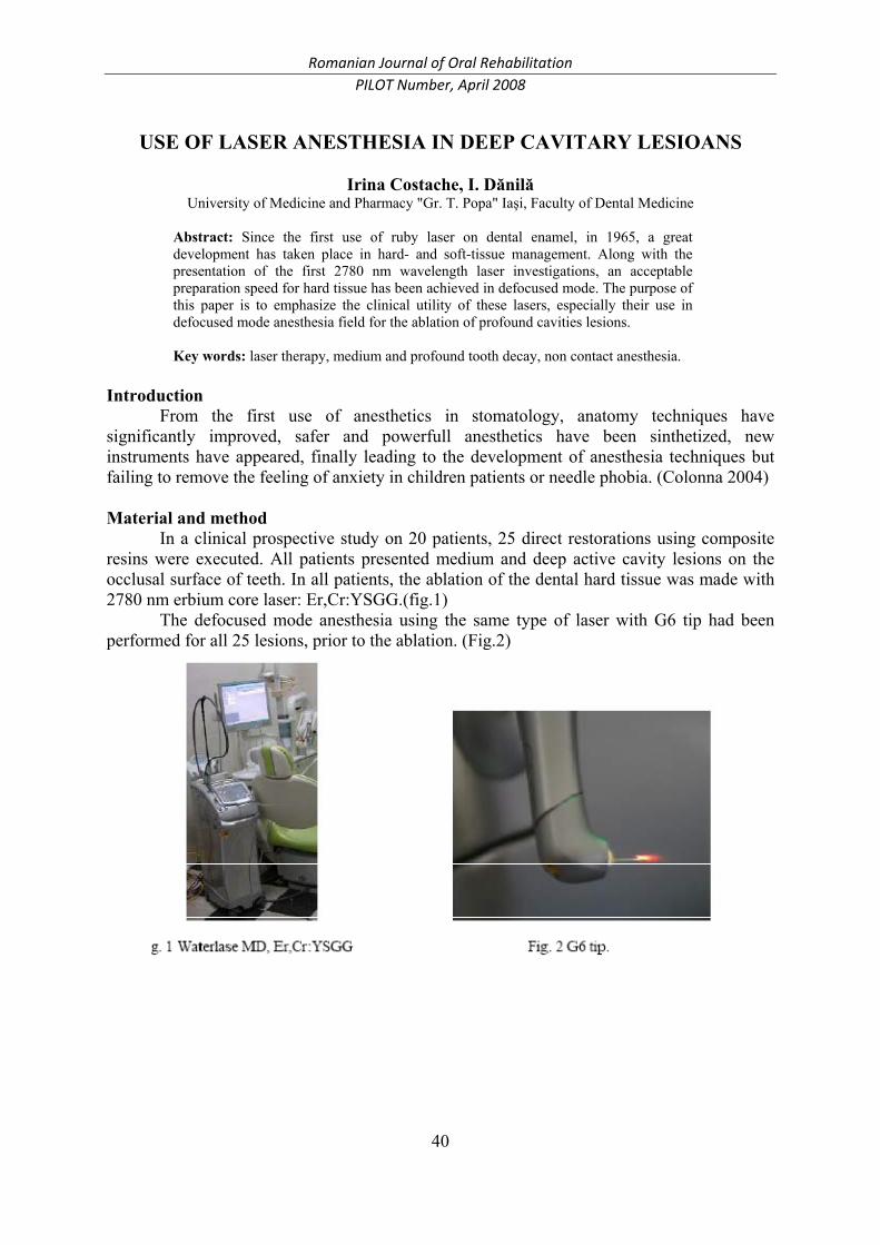
Romanian Journal of Oral Rehabilitation PILOT Number, April 2008
40
USE OF LASER ANESTHESIA IN DEEP CAVITARY LESIOANS
Irina Costache, I. Dănilă
University of Medicine and Pharmacy "Gr. T. Popa" Iaşi, Faculty of Dental Medicine
Abstract: Since the first use of ruby laser on dental enamel, in 1965, a great development has taken place in hard- and soft-tissue management. Along with the presentation of the first 2780 nm wavelength laser investigations, an acceptable preparation speed for hard tissue has been achieved in defocused mode. The purpose of this paper is to emphasize the clinical utility of these lasers, especially their use in defocused mode anesthesia field for the ablation of profound cavities lesions. Key words: laser therapy, medium and profound tooth decay, non contact anesthesia.
Introduction
From the first use of anesthetics in stomatology, anatomy techniques have significantly improved, safer and powerfull anesthetics have been sinthetized, new instruments have appeared, finally leading to the development of anesthesia techniques but failing to remove the feeling of anxiety in children patients or needle phobia. (Colonna 2004) Material and method
In a clinical prospective study on 20 patients, 25 direct restorations using composite resins were executed. All patients presented medium and deep active cavity lesions on the occlusal surface of teeth. In all patients, the ablation of the dental hard tissue was made with 2780 nm erbium core laser: Er,Cr:YSGG.(fig.1)
The defocused mode anesthesia using the same type of laser with G6 tip had been performed for all 25 lesions, prior to the ablation. (Fig.2)

Romanian Journal of Oral Rehabilitation PILOT Number, April 2008
41
Two defocused mode anesthesia techniques were used: 1. the reference values for
the first technique were P = 5w, air = 90% and water = 90%, G6 tip placed at a 20 mm distance from the occlusal surface of the tooth, for 90 s (fig.3). 2. The reference values for the second technique were P = 1,25w, air = 15%, water =15%, G6 tip placed at 5 mm distance from the enamel-cement junction, 20 sec oral and 20 sec vestibular.(fig.4) (Walinski 2004, Colonna 2004)
The intraoperatory sensitivity determined the application of a single anesthesia tecnique or both tecniques simultaneously. 1 = absent sensitivity; 2 = mild sensitivity, with the use of single anesthesia technique; 3 = persistent sensitivity, with the use of both defocused techniques.

Romanian Journal of Oral Rehabilitation PILOT Number, April 2008
42
Results
From 20 patients with ages between 7 and 14, 2 of them presented no intraoperatory sensitivity. 18 medium cavity lesions were treated under a single anestesia tecnique; the patients presented mild intraoperatory sensitivity. During the laser ablation of the hard dental tissue from 5 deep cavity lesions, both anesthesia techniques were used, one in addition to the other. For every restoration it was used hydrokinetic laser Er,Cr : YSGG with 2780 nm wavelength; no local anestesia was necessary but only laser anesthesia. Discussion

Romanian Journal of Oral Rehabilitation PILOT Number, April 2008
43
Comparing with the conventional method for treating cavity lesions, we can assert that laser therapy eliminates the need for local anesthesia, laser anesthesia completing the minimum invasive therapy of these lesions with no intraoperatory sensitivity when using one or both anesthesia techniques (Hochman 2007, Jacobson 2003). Er,Cr :YSGG laser therapy has proven itself to be an easy technique, with no vibrations and no pressure upon dental tissue, with no thermic effects, exciding the affected tissue selectively and with precission of mikrons (fig. 5,6,7)(Vitale 2007, Goodchild 2005). Conclusions
Er, Cr:YSGG can be succesfully used for the preparation of the cavities, avoiding the heating of the dental pulp due to its ablation mechanism, resulting rough, clean surfaces cut with precision, with minimal tissue loss, and prohibit the pain response most hard tissue procedures can be completed without the aid of local anesthesia, the laser anesthesia technique been used to eliminate the intraoperatory sensibility and remove the feeling of anxiety in children patients or needle phobia. References 1. Hochman M. N., Single - Tooth Anesthesia: Pressure – Sensing Technology Provides Innovative Advancement in the Field of Dental Local Anesthesia, Compendium, April 2007; 28(4): 186- 193. 2. Hochman M. N., O noua tehnologie a anesteziei unidentare şi continuarea dezvoltării anesteziei intraligamentare, Quintessence International România, iunie 2007, 3, 3, (223 – 230). 3. Jacobson B., Laser pediatric crowns performed without anesthesia: a contemporary techique, J. Clin. Pediatr. Dent, 2003 Fall. 4. Vitale M. C., Actualities of Laser Applications in Pediatric Dentistry, plenary lection – Quintessence international Romania – Dedicated edition to International Conference on Lasers in Medicine, 2ⁿ edition, Actualities and Prospectives in Laser Therapy in Dentistry and Other Medical Specialities, Timisoara 5-7 iulie 2007. 5. Goodchild J. H., Feck A. S., The use of anxiolytic medications to supplement local anesthesia in the anxious patient,Compendium martie 2005; 26(3): 183 – 190, martie 2005. 6. Walinski J. Cristopher, Waterlase Anesthesia Procedure, plenary lection, WCLI, Laser Analgesia and Desensitization Congress – 24 octombrie 2004. 7. Colonna Mark, The Rabbit technique plenary lection, WCLI, Laser Analgesia and Desensitization Congress – 24 octombrie 2004.

Romanian Journal of Oral Rehabilitation PILOT Number, April 2008
44
MODERN CONCEPTS IN COMPLEX ORAL REABILITATION
1Yves Commissionat, 2A. Crăciunescu, 2Norina Forna
1University Paris VI, Paris, France 2University of Medicine and Pharmacy "Gr. T. Popa" Iasi
Faculty of Dental Medicine, Discipline of Extended Partial Edentation
Abstract: The adult edentulous patients have, in most of the situations, a complex clinical status with a large number of problems which must be approached in order to obtain optimal therapeutic results. Covering the area between diagnostic and treatment, between "planning" and "doing" can be difficult in many cases, not because of the clinician lack of theoretical or practical training or the absence of the technological means, but because of the complex decisional processes that must be accomplished in dealing with a great number of variables which need evaluation and assessment and of the various technological solutions available on the market. Cognitive processes involved in diagnostic and establishing a treatment plan have major impact on therapeutic variability for the patient. The modern computing systems that can easily work with large amount of date and can produce high resolution graphical solutions are very useful in the complex decisional process that clinician must cover between diagnostic and treatment plan. Keywords: complex oral reabilitation, cognitive process, clinical decision suport systems
Introduction
The adult edentulous patients have, in most of the situations, a complex clinical status with a large number of problems which must be approached in order to obtain optimal therapeutic results. Covering the area between diagnostic and treatment, between "planning" and "doing" can be difficult in many cases, not because of the clinician lack of theoretical or practical training or the absence of the technological means, but because of the complex decisional processes that must be accomplished in dealing with a great number of variables which need evaluation and assessment and of the various technological solutions available on the market.
Adding to this, the aggressive marketing of the big companies it is a reality for the dental practitioner, their policies being especially a commercial component of the factories that are producing technological treatment solutions and not a scientific or a patient orientated medical one.
In generally, available information can provide diagnostic possibilities for different clinical conditions and strategies to apply a treatment plan, without almost any references to the cognitive procedures covering the path between complex diagnostic and treatment plan.
The process of developing a treatment based on the large quantity of information taken from the patient becomes a second nature for the specialist from the university clinics, results of years of clinical experience, studying and research. This process is generally automated, without any awareness about the cognitive process involved in it. The treatment plan just became the best therapeutic solution for the clinical case given, based on the analysis of the particular situation.
For dental practitioner, who perhaps has not the same clinical and scientific experience, the process of developing a treatment plan can be, if not difficult, at least resources consuming activity, resources among which the most important is time. All this in a private practice where the main aim of offering to the patients the best medical treatment solutions must be completed with efficiency in using available resources.
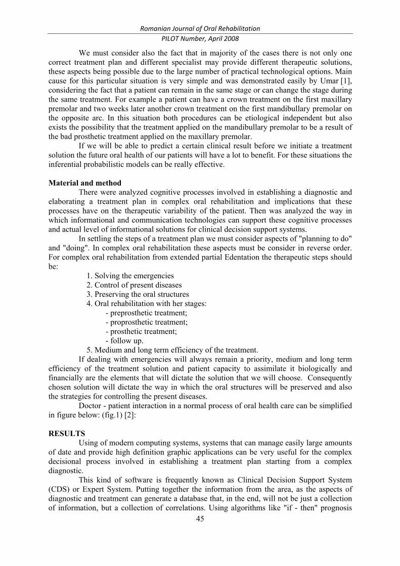
Romanian Journal of Oral Rehabilitation PILOT Number, April 2008
45
We must consider also the fact that in majority of the cases there is not only one correct treatment plan and different specialist may provide different therapeutic solutions, these aspects being possible due to the large number of practical technological options. Main cause for this particular situation is very simple and was demonstrated easily by Umar [1], considering the fact that a patient can remain in the same stage or can change the stage during the same treatment. For example a patient can have a crown treatment on the first maxillary premolar and two weeks later another crown treatment on the first mandibullary premolar on the opposite arc. In this situation both procedures can be etiological independent but also exists the possibility that the treatment applied on the mandibullary premolar to be a result of the bad prosthetic treatment applied on the maxillary premolar.
If we will be able to predict a certain clinical result before we initiate a treatment solution the future oral health of our patients will have a lot to benefit. For these situations the inferential probabilistic models can be really effective. Material and method
There were analyzed cognitive processes involved in establishing a diagnostic and elaborating a treatment plan in complex oral rehabilitation and implications that these processes have on the therapeutic variability of the patient. Then was analyzed the way in which informational and communication technologies can support these cognitive processes and actual level of informational solutions for clinical decision support systems.
In settling the steps of a treatment plan we must consider aspects of "planning to do" and "doing". In complex oral rehabilitation these aspects must be consider in reverse order. For complex oral rehabilitation from extended partial Edentation the therapeutic steps should be:
1. Solving the emergencies 2. Control of present diseases 3. Preserving the oral structures 4. Oral rehabilitation with her stages:
- preprosthetic treatment; - proprosthetic treatment; - prosthetic treatment; - follow up.
5. Medium and long term efficiency of the treatment. If dealing with emergencies will always remain a priority, medium and long term
efficiency of the treatment solution and patient capacity to assimilate it biologically and financially are the elements that will dictate the solution that we will choose. Consequently chosen solution will dictate the way in which the oral structures will be preserved and also the strategies for controlling the present diseases.
Doctor - patient interaction in a normal process of oral health care can be simplified in figure below: (fig.1) [2]: RESULTS
Using of modern computing systems, systems that can manage easily large amounts of date and provide high definition graphic applications can be very useful for the complex decisional process involved in establishing a treatment plan starting from a complex diagnostic.
This kind of software is frequently known as Clinical Decision Support System (CDS) or Expert System. Putting together the information from the area, as the aspects of diagnostic and treatment can generate a database that, in the end, will not be just a collection of information, but a collection of correlations. Using algorithms like "if - then" prognosis

Romanian Journal of Oral Rehabilitation PILOT Number, April 2008
46
can be made, or we can build inferential judgments like "if these conditions are present this result can be forecasted".
Fig. 1 Stages of doctor - patient interaction
These systems are based on causal and probabilistic judgments and theoretical
decision patterns. Later they were developed using "Bayes methods", methods that can provide decisional models based on partial argumentation in conditions of uncertainty. This means that each statement has a numeric attribute or value which signifies level of certainty given to them in a certain stage of knowledge. Later these parameters are processed according to the laws of probabilistic.
Only a few mathematical models were developed for dental medicine, and particularly for complex oral rehabilitation field. One example of expert system is ORAD® (Oral Radiographic Differential Diagnosis), software developed for clinical and radiological assessment for patients with bone lesions.
Fig. 2 Working procedures for ORAD® system
Another example is development of the system for cavity model simulation. This
system examines the influence of time important factors on the gain of oral health after aproximal restoration for posterior teeth. This simulation model was described by Downer and Moles [3] and proved that after coronary restorative treatment oral health improvement benefits are not automatically achieved.
There is also an application that can be used for detecting initial stage cavity called Logicon Caries Detector® (Logicon Inc., USA);
Kordass and Gaertner [4] use the results of single tooth 3D scan and combine virtual models of a complete model of dental arches and their relations with information from a device that analyses the mandibular movements. These elements provide a simulation of the mandibular movements and diagnostic of ocluzal contact and interferences.

Romanian Journal of Oral Rehabilitation PILOT Number, April 2008
47
Fig. 3 Working procedures for Logicon Caries Detector® system
Another application of information technologies are the systems SimPlantTM developed by MaterialiseTM and NobelGuide® developed by NobelBiocare® using the core of the PROCERA® software.
Fig. 4 Working procedures for MaterialiseTM system
Page, Krall and Martin have developed an analytic instrument useful for the
assessment of periodontal risk and stage of periodontal disease, cavities and risk of oro-maxilo-facial cancer - Previser Risk Calculator® - (fig.3) producing an application of information technologies easy to use by the dental practitioners who have internet access.
Fig. 5 Working procedures for Previser Risk Calculator® system Conclusions
1. The necessity to use information and communication technologies is more and more important every day in actual context of oral health care systems.

Romanian Journal of Oral Rehabilitation PILOT Number, April 2008
48
2. Development of clinical decision support systems specific for dental medicine is very important for the future evolution in the field.
3. Possibilities offered by the software application are very important for medical, therapeutic and managerial performances.
REFERENCES 1. Umar H. A Markov Process in Clinical Dentistry: the Prosthodontic Cycle. In: Lemke HU, Vannier MV,
Inamura K, Farman AG, Doi K, editors. Computer Assisted Radiology and Surgery: Proceedings of the 15th International Congress and Exhibition CARS 2001. Amsterdam, The Netherlands: Elsevier Science B.V., 2001;649-653
2. The Journal of Contemporary Dental Practice, Volume 3, No. 1, February 15, 2002 3. Downer MC, Moles DR. Health gain from restorative dental treatment evaluated by computer simulation.
Community Dent Health. 1998 Mar;15(1):32-9 4. Kordass B, Gaertner Ch. The virtual articulator - concept and development of VR-tools to analyze the
dysfunction of dental occlusion. In: Lemke HU, Vannier MV, Inamura K, Farman AG, Doi K, editors. Computer Assisted Radiology and Surgery: Proceedings of the 15th International Congress and Exhibition CARS 2001. Amsterdam, Netherlands: Elsevier Science B.V., 2001; 649-653.

Romanian Journal of Oral Rehabilitation PILOT Number, April 2008
49
SISTEM FOR UNIFIED MEDICAL LANGUAGE
A. Crăciunescu, Norina Forna
“Gr. T. Popa” University of Medicine and Pharmacy Iaşi Faculty of Dental Medicine – Department of Prosthetic Dentistry
Abstract: For some time now, dentistry isn’t just a traditional medical discipline in which the patient receives the necessary treatment by using medical equipment or medical devices. The development of the CDS (Clinical Decision Support) systems offers the most appropriate reliance necessary for using the information technology in dentistry. This speciality recognizes a high number of types of information that need to be structured and conceptualized. Also, it needs a reason, expression and execution parallel based on the unified medical language system similar to the one used in general medicine. This survey has evaluated the medical practician’s attitude regarding the achievement of a sistem that includes standard terms for defining the oral and dental affections in a virtual environment and has taken into account the impact that this kind of system would have upon the development of computer science dentistry applications. The need for a unified dentistry medical language is a clear requirement for the practician and when it combines with the possibility of improving the efficiency by implementing the CDS systems. Key words: clinical decision support, dental informatics
Introduction
The calculus systems are now a part of the activity of every dentist, and the number of users and the frequency of use show an exponential growth. Without exagerating the role of such computer appliances which will never replace the practician’s therapeutic decision, we can say that their utility will be demonstrated through the higher effectiveness reached for the resources used in solving a clinical situation:
a) The analysis of a large amount of information in an extremely short period of time; b) Ensuring a strict analysis of all the variables established through the source code, without having any doubt that an element of most value for the future treatment plan has been omitted; c) Guiding the doctor towards a specific area of therapeutical solutions, his decision being afterwards facilitated by the analysis of a small number of variables; d) The possibility of using calculus facilities as analysis elements, operation that the practician can not acomplish by himself:
- the analysis of the efforts in the structures of the gnato-prosthetic apparatus and at the level of the elements of the stomatognat system using the finished elements method; - the analysis of the radiological exams using breakthrough imaging methods;
e) The ulterior extremely easy access to all the information that lead to a certain therapeutical decision and the possibility of doing a statistical analysis which can improve the analysis algorithms included in the source code of the computer systems. Like in medicine, a new field has appeared: that of the “dental informatics” or
“stomatological informatics”. It combines the software technologies with dentistry in order to offer a base of research and to solve the daily problems of the medical nurse through different applications. Informational technologies had begun to be used as support for clinical decisions in the late 70s and the early 80s. These early expert systems used statistical information and a structure based on known information [1, 2]. The decision support systems improved considerably in the early 90s as a consequence to the new reason strategies in conditions of uncertainty [3, 4].

Romanian Journal of Oral Rehabilitation PILOT Number, April 2008
50
A lot of studies were carried out in order to determine if an artificial system can differentiate between begginer level strategies and expert level strategies used in the complex medical diagnosis. After such an artificial system was ready, the artificial neural network systems showed the ability of indentifying a small number of solutions at an expert level [5]. The continuous feedback provided by the practician during routine actions can be collected and it can lead to an improvement of the system’s performances. [6]
The errors and the lack of coherence that are present in the cognitive reasonings of the experts which develop such artificial systems are only seemingly. Nowadays, the procedure means of a decisional process and its decomposal to simpler components are very well defined and known. [7,8,9]
The different possibilities and requirements for such expert systems for the dentistry field have been defined in a study in 1993 conducted by Feldman and co. [10], completed later on by studies carried out by White in 1995 [11] and Ben and co. in 1998 [12]. In the initial stages, the expert systems were developed for the histopathological diagnosis of the salivary glands neoplasms [163], the orthodontic [13] or the cavity lesions [14] diagnosis, in dental radiology [11], oral surgery [15] and the design for the prosthetic devices [16].
Using relational data bases for stocking and manipulating large amounts of data regarding the patients is very efficient today and it provides a quick and easy statistical analysis and any type of documents.
There are a lot of applications available today which can be useful for solving current problems. [17]
It’s obvious that there are different demands and problems which dental informatics can solve at this point and a lot of problems who’s solution lie in the future. Nevertheless, it is a clear fact that defining a standard medical language and creating applications which can implement this language are critical conditions for the development of dental informatics. Material and method
The study was conducted upon a group of 124 dentists from the district of Iasi, with ages between 25 and 60 years, using the questionnaire method. The questionnaire includes information regarding the doctor’s age, professional degree, the form of exercising his proffesion and the length of service. Results

Romanian Journal of Oral Rehabilitation PILOT Number, April 2008
51

Romanian Journal of Oral Rehabilitation PILOT Number, April 2008
52
Conclusions
From the analysis of the answers the following conclusions can be deduced:

Romanian Journal of Oral Rehabilitation PILOT Number, April 2008
53
1. Although a significant percent of the questioned doctors confronted with comunicating medical difficulties due to the differences of language (88%), the notion of „Unified Medical Language System” is familiar to only a small number of the questioned subjects (40%). 2. Subsequently, very large percentages of the questioned appreciate as „Very important” and „Important” the existence of a „Unified Medical Language System” and of informatical system which would sustain the current clinical activity and the research in the dental medicine field. 3. We can state that although the access to the Internet is very well spread among doctors (83%), and the research done using this source of information is frecquent (60% of the questioned use it daily), using informatical solutions as a support of the current activity is still a problem (23% of the questioned use these systems very little of don’t use them at all).
In this context, we can say that the development of a comunication platform in the form of a „Unified Medical Language System” is a need proven not only by the scientifical benefits it can generate, but also by the doctors’ needs in clinical activity, who are forced to maintain a permanent contact with the information development and the researches in the field.
Also, we can determine that the benefits of the expert systems for current practice aren’t sufficiently known, their use not being widespread. References 1. Cowell R. G., Dawid A. P., Lauritzen S. L., Spiegelhalter D. J. Probabilistic Networks and Expert Systems. New York, NY: Springer Verlag Inc., 1999. 2. Shachter R. D., Heckerman D. E. Thinking backward for knowledge acquisition. AI Magazine 1987; Fall issue: 55-61. 3. Morgan M. G., Henrion M. Uncertainty: A guide to dealing with uncertainty in quantitative risk and policy analysis. Reprint ed. Cambridge: Cambridge University Press, 1992. 4. Tversky A., Shafir E. The disjunction effect in choice under uncertainty. Psychological Science 1992; 3:305-309. 5. Eisner J., Chasteen J. E., Schleyer T., et. al. The Computer Based Oral Health Record. American Fund for Dental Health, 1993. 6. Kahn M. G., Dunagan W. C., Steib S. A., Monitoring expert system performance using continuous user feedback. J Am Med Inform Assoc. 1996 May-Jun; 3(3):216-23. 7. Shachter R. D., Heckerman D. E. Thinking backward for knowledge acquisition. AI Magazine 1987; Fall issue: 55-61. 8. Druzdzel M. J., Flynn R. R. Decision Support Systems. In: Kent A, editor. Encyclodpedia of Library and Information Science, in press. Marcel Dekker, Inc., 2000. 9. Henrion M., Breese J. S., Horvitz E. J. Decision analysis and expert systems. AI Magazine 1991 12:64-91. 10. Reichert A., Sadan B. A., Bengtsson S., et. al. Design of an oral health information system based upon a computer based dental record. Jerusalem, Israel: Freund, 1993. 11. White S. C. Decision-support systems in dentistry. J Dent Educ. 1996 Jan;60(1):47-63. Review. 16 12. Benn D. K., Dankel D. D., Kostewicz S. H. Can low accuracy disease risk predictor models improve health care using decision support systems? In: Chute CG, (ed.). American Medical Informatics Association, AMIA '98 Annual Symposium. Philadelphia: Hanley & Belfus, 1998: 577-581 13. Hammond R. M., Freer T. J. Application of a case-based expert system to orthodontic diagnosis and treatment planning: a review of the literature. Aust Orthod J. 1996 Oct;14(3):150-3. Review. 14. Benn D. K., Clark T. D., Dankel D. D. 2nd, et. al. Practical approach to evidence-based management of caries. J Am Coll Dent. 1999 Spring; 66(1):27-35. 15. Brickley M. R., Shepherd J. P. Comparisons of the abilities of a neural network and three consultant oral surgeons to make decisions about third molar removal. Br Dent J. 1997 Jan 25; 182(2):59- 63. 16. Hammond P., Wells P., Modgil S. Animating Medical and Safety Knowledge. In: Horn W, Shahar Y, Lindberg G, Andreassen S, Wyatt J, (eds.). Artificial Intelligence in Medicine. Proceedings AIMDM '99 Berlin-Heidelberg, Germany: Springer-Verlag, 1999 443-447.97. 17. Lackey A. D. Computers in the Dental Treatment Room. DDRT [2nd edition], 17-19. 1994.

Romanian Journal of Oral Rehabilitation PILOT Number, April 2008
54
METHODS FOR MULTIVARIATE DATA ANALYSIS IN THE STUDY
OF ORAL DISEASES: THE MULTIPLE LINEAR REGRESSION
Michel Jourde1, Cristina Gena Dascălu2, M.Gîrtan2, Norina Forna2 1Academy of Dental Surgery, Paris, France
2Faculty of Dental Medicine, University of Medicine and Pharmacy „Gr.T.Popa”, Iaşi, România
Abstract: During the statistical analysis of medical data, in many situations it is necessary to identify the multiple correlations established between the studied parameters. In this purpose, one of the most useful methods is to build a model of multiple regression, which allows the modeling of a dependant variable values having at least the ordinal type, based on its linear relation with more than one independent variables satisfying the same restriction, called predictors. The multiple linear regression model is a generalization of the simple linear regression model, which identifies the parameters of an equation with n variables y = b0+b1x1+b2x2+…+ bnxn+e, based on which we can find the predictors that have a statistically significant influence over the dependant variable. We used this model to identify, on a set of 202 patients having different types of oral lesions, the biochemical analysis which can be eventually correlated with the oral diagnosis. We found that the values of leucocytes, hemoglobin and hematocrit are significant for the general oral diagnosis, the cholesterol and glucose values for the oral lesion type, and the hemoglobin for the periodontal disease. The identified predictors are useful for further data processing. Key words: multivariate analysis, multiple linear regression, oral diagnosis
Introduction
In many cases, in scientific researches, it is necessary to analyze the correlations between more than two parameters, in order to detect the internal influences between data. In such cases it is absolutely necessary to choose the right method to study the assumed correlations – because the parameters nature defines in fact the path we are going to follow. There are a few main possibilities to analyze the multiple correlations between data: the regressional models, the principal components analysis, the discriminant analysis, the clustering. The regressional analysis is the easiest available method between these, which tries to find a very clear pattern for data variation: one parameter is interpreted as being “dependant”, so it will be influenced in its variation by all the other parameters. The only problem that remains in this case are to find an appropriate mathematic model for this influences schema; the regressional analysis works with a few mathematic models: the linear regression, the curve estimation, the binary and the multinomial logistic models, the ordinal regression, the probit regression and the non-linear regression. The difference between these models consists not only in their mathematical fundaments, but, more important, in the data types for which there are available.
When all the studied parameters are quantitative and we intend to study in which way one of these parameters, defined as “dependant”, is influenced by all the other, the easiest way to quantify this influence is to build a model of linear multiple regression and to check how well it fits with the real studied phenomenon.
Material and methods
The linear regression method is used when we need to model the values of a dependant variable according with the values of at least two independent variables, also called “predictors”, using the equation of a straight line. The main requirement that must be fulfilled by all the variables involved in the model is that these variables must have at least

Romanian Journal of Oral Rehabilitation PILOT Number, April 2008
55
the scale type – but the model behaves best when all the variables are quantitative (Draper, 1981).
The linear regression model assumes that there is a linear relationship between the dependent variable and each predictor, described in the following formula:
yi = b0 + b1xi1 + ... + bpxip + ei , where:
yi is the value of the i-th case of the dependent scale variable p is the number of predictors bj is the value of the j-th coefficient, j ∈ {0, 1, ..., p} xij is the value of the i-th case of the j-th predictor ei is the error in the observed value for the i-th case
We can notice that we are dealing here with an equation of 1st degree, with p variables; b0 is the intercept or the model-predicted value of the dependent variable when the value of every predictor is equal to 0 (the point where the line intersects the Oy axes, in a representation using a Cartesian coordinates system). The error term ei must fulfill also the following conditions (Draper, 1981):
- Its distribution is normal, with a mean of 0; - Its variance is constant across cases and independent of the variables in the model; - Its value for a given case is independent of the values of the variables in the model
and of its values term for other cases. When we build the multiple linear regression model, we must follow a few steps. First, it is necessary to check the model fit (Norusis, 2004). In this purpose, the
ANOVA test is used, and the F statistic is calculated; if the F value is statistically significant (p ≤ 0.05), it follows that the model fits well with the analyzed data, and using it is better than guessing the mean.
Secondly, the correlation coefficients R and R squared are calculated; these coefficients show also how well the model works: R squared, for example, shows the percentage from the dependant variable’s variation which is explained by the model.
Then, for each predictor involved in the model, we calculate its unstandardized and standardized coefficients and its significance level (expressed using the t statistic); in this way we can separate, from all the predictors involved in the model, only the significant ones – in order to eliminate further from the model the non-significant predictors (variables which do not contribute too much to the model). At this step we can also find the relative importance of each significant predictor, which varies proportionally with its standardized coefficient Beta.
At this step it is also good to calculate the part and partial correlation coefficients (Norusis, 2004); these coefficients help us to detect the possible multicollinearity problems. Such problems appear when the part and the partial correlations are very different by the zero-order correlation – which means that a large amount in the variance of the dependant variable that is explained by the analyzed predictor is also explained by the other predictors – so the predictors are “collinear” and their effects are overlapped. Another coefficient calculated at this step is the tolerance – or the percentage of the variance in a given predictor that cannot be explained by the other predictors; small tolerances show that large amounts in the variance in a given predictor are explained also by the other predictors, so again the multicollinearity is present, and large tolerances show that the multicollinearity is absent. Finally the multicollinearity is also measured using a Variance Inflation Factor (VIF) – that regards the standard error of the regression coefficients; a VIF factor greater than 2 is usually considered problematic, being a clue for predictors multicollinearity.

Romanian Journal of Oral Rehabilitation PILOT Number, April 2008
56
There are also a few diagnostics tests especially designed for collinearity (Weisberg, 1985):
- in the predictors matrix, the eigenvalues are calculated; if these values are close to 0, it means that the predictors are highly intercorrelated, and therefore, small changes in the data values may lead to large changes in the estimates of the coefficients;
- in the same matrix, the condition indices are also computed, as the squared roots of the ratios of the largest eigenvalue to each successive eigenvalue; values greater than 15 indicate a possible problem with collinearity; greater than 30, a serious problem.
The last step of the analysis regards the collinearity removing, as long as this is possible. In order to do this, the easiest way is to rerun the model using the z scores for all the variables involved (predictors, as well as the dependant variable) instead of their direct values. In this way, only the most useful predictors will be included in the model. Results
On a set of 202 patients having different types of oral lesions we will try to identify the biochemical analysis which can be eventually correlated with the oral diagnosis using the multiple linear regression model.
In the first step of our analysis, we will use in our model the following variables as predictors: glucose, hemoglobin, monocytes, thrombocytes, lymphocytes, eosinophils, cholesterol, creatinine, leukocytes, red blood cells, neutrophils, hematocrit. The dependant variable is the oral diagnosis, defined as a scale variable which represents the sum of the following binary variables regarding the oral health status: dental mobility, gingival hyper growing, gingival retraction, decay lesions, periodontal disease. Therefore, the 0 value for this variable means health, and its value grows proportionally with the number of new identified symptoms in the oral area.
The ANOVA test (tab. I) shows that the multiple linear regression model doesn’t fit significantly with the analyzed data, but its level of significance is quite close to the threshold value, so it is useful to continue the analysis. The correlation coefficients R and R squared (tab. II) come to strengthen the previous observation, through their low values: only 13.7% from the oral diagnosis variation is covered by the regressional model.
TABLE I. The ANOVA test for model’s fitting (1st study)
Model Sum of Squares df Mean Square F Sig.
1 Regression 36.791 12 3.066 1.648 .087 Residual 232.484 125 1.860 NS Total 269.275 137
TABLE II. The global correlation coefficients of the model (1st study)
Model R R Square Adjusted R
Square Std. Error of the Estimate
1 .370 .137 .054 1.364
Anyway, we will still try to identify which are the significant predictors of this model. Checking the significance levels of all the predictors (tab. III), we notice that the only significant predictors for the model are Leukocytes, Hemoglobin and Hematocrit. According to the Beta coefficient values, it follows that the most important predictor is Hematocrit (β = -0.661), followed by Hemoglobin (β = 0.584). The less important predictor is Leukocytes (β =

Romanian Journal of Oral Rehabilitation PILOT Number, April 2008
57
0.254). Checking the tolerance values, we can see that most of them are very different of 0; similarly, the VIF factor is bigger than 2 in only 5 cases from all 12 – so, there are not important problems concerning the predictors collinearity.
TABLE III. The predictors coefficients and significance levels (1st study)
Model
Unstandardized Coefficients
Standardized Coefficients
t
Sig.
Correlations Collinearity
Statistics
B Std.
Error Beta Zero-order
Partial Part
Tole rance VIF
1 (Constant) 3.723 2.606 1.429 .156 Leukocytes .182 .077 .254 2.366 .020 .188 .207 .197 .600 1.667 Red blood cells -.446 .433 -.141 -1.029 .306 -.106 -.092 -.086 .366 2.730 Hemoglobin .633 .259 .584 2.443 .016 -.075 .213 .203 .121 8.277 Hematocrit -.212 .083 -.661 -2.559 .012 -.130 -.223 -.213 .104 9.652 Thrombocytes .000 .001 -.020 -.209 .835 .057 -.019 -.017 .726 1.377 Neutrophils -.008 .021 -.065 -.383 .703 .041 -.034 -.032 .242 4.127 Lymphocytes -.014 .023 -.091 -.597 .552 -.062 -.053 -.050 .297 3.362 Monocytes -.068 .063 -.105 -1.079 .283 -.126 -.096 -.090 .733 1.364 Eosinophile -.011 .046 -.022 -.229 .819 -.003 -.020 -.019 .783 1.276 Cholesterol .001 .003 .024 .271 .787 .039 .024 .023 .895 1.117 Creatinine .508 .690 .084 .736 .463 .002 .066 .061 .536 1.866 Glucose .005 .004 .110 1.236 .219 .144 .110 .103 .877 1.140
We will repeat the analysis, in order to find the influence of the same set of
predictors (glucose, hemoglobin, monocytes, thrombocytes, lymphocytes, eosinophils, cholesterol, creatinine, leukocytes, red blood cells, neutrophils, hematocrit) over another dependant variable - oral lesion type. This variable also has the scale type, with 5 values corresponding to increased gravity diagnosis: 0 – no lesions; 1 – epithelial-conjunctive hyperplasia; 2 – chronic ulceration; 3 – prosthesis stomatitis; 4 – precancerous lesions.
TABLE IV. The ANOVA test for model’s fitting (2nd study)
Model Sum of Squares df Mean Square F Sig.
1 Regression 21.215 12 1.768 2.083 .022 Residual 106.089 125 .849 SS Total 127.304 137
TABLE V. The global correlation coefficients of the model (2nd study)
Model R R Square Adjusted R
Square Std. Error of the Estimate
1 .408 .167 .087 .921
This time, the ANOVA test (tab. IV) shows that the multiple linear regression model fits significantly with the analyzed data (p = 0.022 – SS), even if the correlation coefficients R and R squared (tab. V) still have low values: only 16.7% from the oral lesion type variation is covered by the regressional model.
TABLE VI.

Romanian Journal of Oral Rehabilitation PILOT Number, April 2008
58
The predictors coefficients and significance levels (2nd study)
Model
Unstandardized Coefficients
Standardized Coefficients
t
Sig.
Correlations Collinearity
Statistics
B Std.
Error Beta Zero-order Partial Part
Tole rance VIF
1 (Constant) 2.372 1.760 1.347 .180 Leukocytes -.069 .052 -.139 -1.318 .190 -.087 -.117 -.108 .600 1.667 Red blood cells .160 .293 .074 .546 .586 .005 .049 .045 .366 2.730 Hemoglobin -.103 .175 -.138 -.588 .558 .018 -.053 -.048 .121 8.277 Hematocrit .004 .056 .020 .080 .936 .020 .007 .007 .104 9.652 Thrombocytes .001 .001 .123 1.284 .202 .020 .114 .105 .726 1.377 Neutrophils -.016 .014 -.195 -1.175 .242 -.102 -.105 -.096 .242 4.127 Lymphocytes -.018 .016 -.173 -1.156 .250 -.016 -.103 -.094 .297 3.362 Monocytes .001 .042 .001 .014 .989 .086 .001 .001 .733 1.364 Eosinophile .050 .031 .149 1.618 .108 .235 .143 .132 .783 1.276 Cholesterol .006 .002 .214 2.478 .015 .153 .216 .202 .895 1.117 Creatinine .873 .466 .209 1.872 .064 .109 .165 .153 .536 1.866 Glucose -.007 .003 -.215 -2.462 .015 -.189 -.215 -.201 .877 1.140
The VIth table shows the significant predictors of the model: this time, only
Cholesterol and Glucose have this property – followed by the Creatinine predictor, whose significance level is very close by the threshold (p = 0.064). The most important predictor is Glucose (β = -0.215), but the other two are also very close: Cholesterol - β = 0.214 and Creatinine - β = 0.209. The tolerance values are again high, and the VIF factor is again bigger than 2 in 5 cases from all 12 (but not for the predictors identified as significant); therefore, we don’t have again problems concerning the predictors collinearity. Conclusions
The multiple linear regression model is useful to identify the correlations between more than two parameters; it allows to select from large set of predictors only the significant ones, and also to classify them according to their importance in the variance of the dependant variable. This method can be successfully used as a preliminary step in data analysis, because the model usually must be rebuilt – including only the significant predictors or, even better, studying only the simple regression line – with a single predictor and the dependant variable – which allows to identify exactly the corresponding correlation coefficients. References 1. Draper, N. R., H. Smith, Applied regression analysis, 2nd ed. John Wiley and Sons, New York,1981. 2. Norusis, M., SPSS 13.0 Guide to Data Analysis. Prentice Hall, Inc., Upper Saddle-River, N.J., 2004. 3. Norusis, M., SPSS 13.0 Statistical Procedures Companion. Prentice Hall, Inc., Upper Saddle-River, N.J.,
2004. 4. Weisberg, S., Applied Linear Regression, 2nd ed. John Wiley and Sons, New York,1985.

Romanian Journal of Oral Rehabilitation PILOT Number, April 2008
59
MANDUCATORY MUSCLES ELECTROMYOGRAPHYC
EVALUATION TO ELDERLY PEOPLE
R.Dima, Maria Ursache, Norina Forna University of Medicine and Pharmacy „Gr.T.Popa”, Iaşi, Faculty of Dental Medicine
Abstract: Between maxilla bone morphology and muscles function is a fragile equilibrium, all manducatory muscles suffer a continuous process of remodelling for a better fit in functional mechanic regime. Path and developmental functional growth of manducatory muscles are changing form childhood to elderly persons. If any dysfunctional discrepancies take place between stomatognatic system elements re-establishing the functional equilibrium is made either by remodelling the bone, either by changing in muscles functions, most likely and most frequent. The purpose of the study is to identify the signals changes in manducatory muscles electric activity for an elderly group population which will help for a better diagnosis and a correct therapy. In conclusion, electromyographic activity to elderly people is modified by reducing amplitudes and increased time period of potential motor unit signals, period with no electric activity. Meanwhile the contraction and relaxing period in also increased, all elements contributing to establish muscular dysfunction diagnosis, more frequent to older people. Key words: manducatory muscles, electromiographic activity, elderly people
Introduction
The dysfunctional syndrome of the stomatognatic system presents not only a complex etiopathogeny, but also a symptomatic polymorphic picture which requires investigations taking into consideration both its subjective and objective aspects, from the point of view of a clinical and paraclinical examination [5]. The multitude of the functions accomplished by the stomatognatic system involves a complex neuromuscular activity, its changes having effects on the constituent elements and on the existent interrelations at this level as well. The excitability, electric activity, tonicity and contractility modifications are particular to different neuro-muscular diseases (pareses, paralyses, atrophies), and appear more frequently at the elderly persons, having as a consequence the affectation of the posture and the triple muscular antagonism with clinical implications in generating the mandible - cranium malreactions [2].
The galvano-faradic electrodiagnosis and cronaximetrical methods associated with the electromyography ones used for detection and stimulo-detection (the nervous influx rate) allow an efficient exploration of the excitable systems of the neuro-muscular structure type and that is why we have considered advisable to paraclinically evaluate the stomatognatic system musculature at the elderly persons by studying the surface electric activities. Material and method
For the electromyography investigation I have selected 32 patients, 13 men and 19 women, with different ages ranging between 60 and 82, patients who presented a suggestive clinical picture with the established diagnosis of stomatognatic system dysfunctional syndrome.
The selection criteria for our study included the presence of the muscular dysfunctions associated or not with the affectations of the other elements of the stomatognatic system. The exclusion criteria included the presence of the previous muscular dysfunction for which the patient had treatment, the presence of diseases due to the wisdom tooth. We underline the fact that it has not been any differences regarding the clinical data selection since the same clinical examination approach has been used, and the perspective on the mentioned pathology and its characteristic symptoms and signs is the same, materialized in

Romanian Journal of Oral Rehabilitation PILOT Number, April 2008
60
the perspective of the systemic organization of the stomatognatic system elements. We have chosen for recording the action biopotentials at the level of the manducatory muscles (right and left) and the temporal ones, anterior beam (right and left), these being the most affected ones in the muscular dysfunctions, the recordings being made in the relation of posture, the patient sitting on a chair. Results and discussions
The stomatognatic system musculature at the elderly persons presents a gradual decrease of the active mass up to 30% and the muscular mass loss takes place as an effect of the fiber volume reduction and it is doubled by the accumulation of lipofuscine and the increase in lipid content. The number of muscular cells as part of a muscle remains invariable since our birth, but their volume is different, according to the muscles, age, physical effort and nutrition. Simultaneously with the reduction of the active muscular fiber length, the extracellular space increases, being filled with supportive conjunctive tissue.
The electric muscular manifestations are recorded as biopotentials by means of coaxial or surface electrodes, obtaining the elementary or global electromyography.
The clinical utility of the surface electromyography, even if controversial, is demonstrated in the case of histological and physiological studies of the mechanical muscular dysfunctions. The potentials which appear at the muscular level can be detected by surface and intramuscular electrodes and can be recorded after an adequate amplification.
By means of the electromyography recording we can get data referring to four parameters of the trapped biocurrents: the signal shape, amplitude, duration and frequency. The surface electromyography has many and important applications, but also a lot of limits which have to be known. The surface electromyography can be easily used, but special care should be taken when it comes to its interpretation which should depend on that particular clinical situation. Recent data from the literature emphasize the fact that through a correct analysis of the electromyography trajectory detected through noninvasive and painless methods it is possible the supervision and quantitative evaluation of the biochemical products resulted in a muscular fiber during the sustained contraction[1,3].
After the clinical examination, we have discovered at the level of the investigated group the presence of the following muscular symptoms and signs: muscular pain – present at 12 patients, being located at the level of the muscles under study, during the palpation of the muscular mass and its insertion as well as when there is the tendency of mobilizing the mandible and with a different intensity; muscular hypertonia – present at 3 patients, demonstrated by the increase in its consistency located at the level of the manducatory muscle unilateral at two patients and bilateral at one patient; muscular tiredness at 23 patients, demonstrated by their incapacity of making certain repetitive movements of the mandible, without the appearance of a certain muscular or articulator pain, these patients also complaining about a modified general mood including asthenia, a reduced physical resistance at the effort, hypotomy and muscular hypotrophy present at 26 patients; the modification of the mandible dynamic course at 17 patients, for the lateral movement right – left, with the asymmetry of the two trajectories (the trajectory is limited by the side with spasm and muscular pain), in the protruzy movement , the mandible deviates towards the affected side and the opening of the mouth is limited due to the spasm and pain. These modifications appear due to the unequal and asymmetrical muscular contraction, which causes the mandible deviation from its normal movement trajectory. The paraclinical evaluation by means of electromyography: The presence of inactive bioelectric activity at all the patients, with values ranging from
1.4 and 6.9 mV, the higher values were obtained at the patients with muscular hypertonia exacerbated by the usage of an unilateral mastication or by the lack of plate (fig.1);

Romanian Journal of Oral Rehabilitation PILOT Number, April 2008
61
High value asymmetries for the quantitative parameters, with important differences on the left- right side 1.2 – 4 mV at 28 patients, where the affectation of the muscular component (pain, increased muscular pain) is unilateral, the values are lower on the healthy side (fig.2);
The decrease in the amplitude of the bioelectric discharges, much above the normal value at 23 patients (fig.3);
Uncoordinated contractions of the manducatory and temporal muscles (right and left) in maximum intercuspal position - at 22 patients (fig.4).
Fig. 1 Presence of inactive Fig. 2 High value asymmetries for the quantitative bioelectric activity parameters, with important differences on the
left- right side Fig. 3 The decrease in the amplitude Fig. 4 Uncoordinated contractions of temporal muscles of the bioelectric discharges, much (right and left) in maximum intercuspal position under the normal value
We have found the existence of a correlation between the values of the action potentials at different muscular groups and the existent symptoms, obtaining action potentials with increased amplitude on the side where the symptoms were more severe. The interpretation of the results obtained by means of electromyography is associated with the structure of the electrodes and their location, the area and shape of the surface electrode which determines the number of active motor units, according to the number of muscular fibers, the location of the electrode on muscle surface, respecting the lateral edge of the muscle, which determines the possible interferences detected by the electrode, the orientation of the detection surface, respecting the muscular fiber which affects the value of the electric signal rate and consequently the amplitude and frequency of the signal.
The surface electromyography has also its disadvantages generated by a series of factors which cannot be controlled by the present technology, and that is: the number of active motor units at a certain moment which affects the amplitude of the detected signal, the fiber composition which produces the pH modifications at the level of the interstitial fluid during contraction, the sanguine flux in the muscle which determines the metabolic elimination rate during contraction, the fiber diameter which influences the amplitude and the conduction rate of the action potentials which form the signal, the profoundness and location of active muscular fibers; this relation determines the spatial filtration and consequently the amplitude and frequency of the detective signal, the quantity of tissue between the surface of the muscle and the electrode which affects the spatial filtration of the signal[4].

Romanian Journal of Oral Rehabilitation PILOT Number, April 2008
62
Conclusions The modifications of excitability, electric activity, tonicity and contractility are
frequently met at the elderly persons, having as an effect the affectation of the posture and the triple muscular antagonism with clinical implications in generating mandible cranium malrelations but also in the systemic postural modifications at the level of the scapular and pelvine belt.
The electromyography at the elderly persons presents the decrease in the amplitude of the muscular contraction, the appearance of the muscular pain which makes the periods of absolute and relative refractivity increase, whereas the duration of the latency, contraction and relaxation is longer.
There have not been emphasized significant variations of these parameters according to sex. Noticeable differences have been noticed by analyzing the muscular force, at the male subjects, being observed a higher value of this parameter in similar recording conditions.
The muscular tiredness, frequently noticed by means of electromyography, diminishes the excitability, the power and duration of the muscular contraction, by decreasing the number of motorii units involved in the motor activity. Also, the amplitude of each contraction is diminished by tiredness, by both the decrease in the number of the stimulated muscular fibers and the reduction of the shortening capacity of each fiber. Besides the characteristic signal of the muscular tiredness, defined by the reversible diminution of its activity, we can add the reduction of the movement precision and the presence of a tremble (as a consequence of the nervous tiredness). In order to emphasize the muscular tiredness, we can follow the electromyography trajectory during the sustained contraction (we can notice especially the flattening of the curve and the modification of the shape).
The electromyography examination by means of the surface electromyography of the muscular function at the elderly persons has many advantages since it is not invasive, it is easily achieved if the methodology is known, but also many disadvantages because it collects simultaneously electrical information from many motor units which makes the interpretation of the results less facile and compulsory in the context of a complex evaluation of the stomagtonatic system. BIBLIOGRAPHY 1. Audette JF, Wang F, Smith H. Bilateral activation of motor unit potentials with unilateral needle
stimulation of active myofascial trigger points. Am J Phys Med Rehabil.; 83(5):368-74, 2004. 2. Barton E, Morris C. Mechanisms and strategies to counter muscle atrophy. J Gerontol A Biol Sci Med
Sci.;58(10):M923-6, 2003. 3. Dahlstrom, L.: Electromyographic Studies of Craniomandibular Disorders: A Review of the Literature. J
Oral Rehabil 1989,16: 1-20. 4. Gross MD, Ormianer Z, Moshe K, Gazit E. Integrated electromyography of the masseter on incremental
opening and closing with audio biofeedback: a study on mandibular posture. Int J Prosthodont.;12 (5):419-25, 1999.
5. Ursache M, V. Burlui. Purdu A. Semiologie stomatologică, Ed. Apollonia, Iaşi, 1997, 15-34.

Romanian Journal of Oral Rehabilitation PILOT Number, April 2008
63
THE ROLE AND IMPORTANCE OF THE CARIES DETECTORS DYES
IN EARLY DIAGNOSIS AND TREATMENT OF DENTAL CARIES
Pancu Galina1, Stoleriu Simona1, Andrian Sorin1, Gheorghe Angela1, Topoliceanu Claudiu1, Pancu Ion2, Lăcătuşu Ştefan1
1Departament Restorative Dentistry-Cariology, Faculty of Dental Medicine, UMF “Gr.T.Popa”, Iasi 2Private Dentistry Medilife, Iassy, România
Abstract: Despite the progresses of the modern dentistry, the carious disease is still affecting a large number of peoples. The caries detectors would be helpful for diagnosis of early enamel caries as well as dentinal caries. The study focused on the role of caries detectors on objective criteria of assessment of the caries preparation, the early diagnosis of the incipient caries as well as monitorisation of the remineralising processes. The study used product Color-test of the Vladmiva (Rusia): solution and gel. The study was performed on 25 patients age 15-38 with medium and high level of cariogenic status. The statistical results show the practical importance of the caries detectors for the conservative treatment of the dental caries, with different degree of penetration in dental tissues. It also allow the monitorisation of the success of the non-invasive or minimal invasive treatment. The use of the caries detectors allow minimal preparation of the dental issues, accordingly to modern principles of the actual dentistry. Key words: incipient caries, caries detectors, remineralisation therapy.
Introduction
Although many dentists know modern principles of dental caries therapy, the use of dental caries indicators is very limited in current practice. Today is more important that restorative dentistry to stand on minimal invasive approach, without idle sacrifices of healty dental tissues. The role of the research is to highlight dental caries in incipient stages, with caries detectors dyes and to monitor their evolution after remineralisation therapy. Materials and methods The caries detector dyes Color-test (Vladmiva, Rusia) is used in study. The diagnostic and monitorisation of the incipient dental caries were made through Borovschii-Axamit method. This method allows us to assess depth and surface of demineralisation area. In study were included 36 patients and 65 demineralisation focuses (white-spot). These demineralisation areas were divided in 4 lots: lot 1- 18 teeth (10 patients) with remineralisation therapy by fluor gel; lot 2- 15 teeth (8 patients) with remineralisation therapy by calcium, phosphat and fluor; lot 3- 17 teeth (12 patients) with remineralisation therapy by calcium-phosphat gel; lot 4 (6 patients)- 15 teeth without remineralisation therapy (witness lot). The patients were monitorised for 12 months, with assessment periodes at 6 and 12 months. Results
The evolutions of the caries detector dyes intensity and of the demineralisation surfaces in the four lots are synthesised in tables I and II.
TABLE I. The evolution of the caries detector dyes
LOT 1 – F
(Average values)
LOT 2 – CaFP
(Average values)
LOT 3 – CaP
(Average values)
LOT 4 – witnes
(Average values
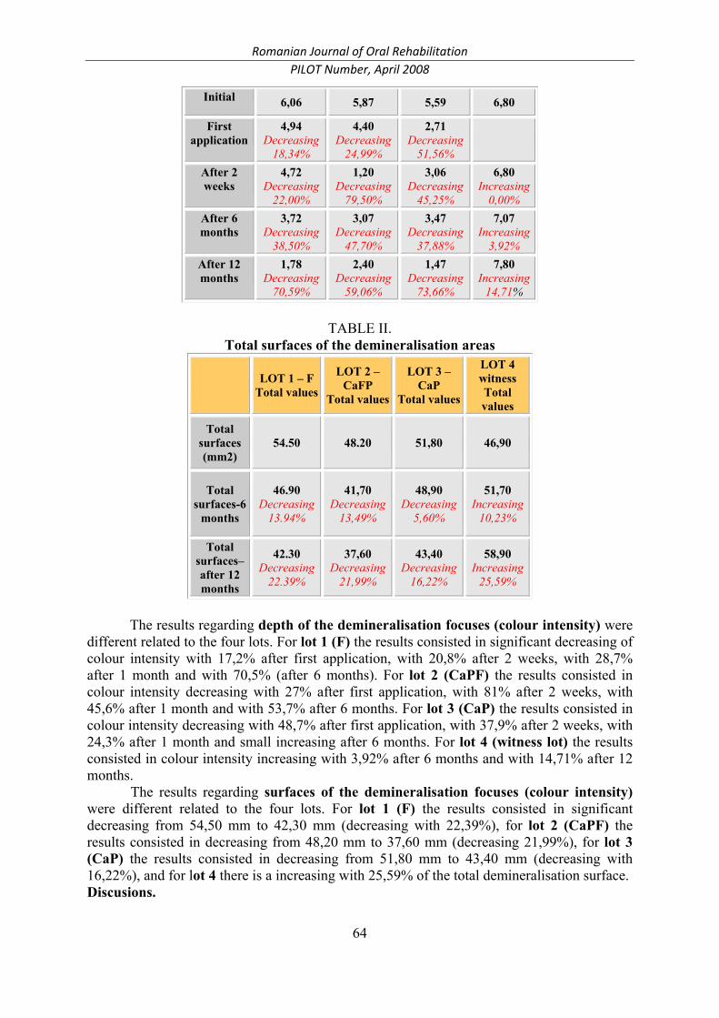
Romanian Journal of Oral Rehabilitation PILOT Number, April 2008
64
Initial 6,06 5,87 5,59 6,80
First application
4,94 Decreasing
18,34%
4,40 Decreasing
24,99%
2,71 Decreasing
51,56%
After 2 weeks
4,72 Decreasing
22,00%
1,20 Decreasing
79,50%
3,06 Decreasing
45,25%
6,80 Increasing
0,00% After 6 months
3,72 Decreasing
38,50%
3,07 Decreasing
47,70%
3,47 Decreasing
37,88%
7,07 Increasing
3,92% After 12 months
1,78 Decreasing
70,59%
2,40 Decreasing
59,06%
1,47 Decreasing
73,66%
7,80 Increasing
14,71%
TABLE II.
Total surfaces of the demineralisation areas
LOT 1 – FTotal values
LOT 2 – CaFP
Total values
LOT 3 – CaP
Total values
LOT 4 witness Total values
Total surfaces (mm2)
54.50 48.20 51,80 46,90
Total surfaces-6
months
46.90 Decreasing
13.94%
41,70 Decreasing
13,49%
48,90 Decreasing
5,60%
51,70 Increasing
10,23%
Total surfaces– after 12 months
42.30 Decreasing
22.39%
37,60 Decreasing
21,99%
43,40 Decreasing
16,22%
58,90 Increasing
25,59%
The results regarding depth of the demineralisation focuses (colour intensity) were
different related to the four lots. For lot 1 (F) the results consisted in significant decreasing of colour intensity with 17,2% after first application, with 20,8% after 2 weeks, with 28,7% after 1 month and with 70,5% (after 6 months). For lot 2 (CaPF) the results consisted in colour intensity decreasing with 27% after first application, with 81% after 2 weeks, with 45,6% after 1 month and with 53,7% after 6 months. For lot 3 (CaP) the results consisted in colour intensity decreasing with 48,7% after first application, with 37,9% after 2 weeks, with 24,3% after 1 month and small increasing after 6 months. For lot 4 (witness lot) the results consisted in colour intensity increasing with 3,92% after 6 months and with 14,71% after 12 months. The results regarding surfaces of the demineralisation focuses (colour intensity) were different related to the four lots. For lot 1 (F) the results consisted in significant decreasing from 54,50 mm to 42,30 mm (decreasing with 22,39%), for lot 2 (CaPF) the results consisted in decreasing from 48,20 mm to 37,60 mm (decreasing 21,99%), for lot 3 (CaP) the results consisted in decreasing from 51,80 mm to 43,40 mm (decreasing with 16,22%), and for lot 4 there is a increasing with 25,59% of the total demineralisation surface. Discusions.

Romanian Journal of Oral Rehabilitation PILOT Number, April 2008
65
Caries detector dyes are useful for early detection of the incipient dental caries in pits, fissures and smooth dental surfaces (Lăcătuşu Şt.., Ismail AI). The retention of the caries detector dyes allows precise assessment of the depth and surface for demineralisation areas, through assessment of the colour intensity and use of a graph paper (Ржанов Е.А. şi colab.). The caries detector dyes are an useful instrument for detection of secondary dental caries and fissures or microfractures (Andrian S., Cureachina N.V.). The scientific progress of the modern dentistry allows more effective caries detector dyes that can be visible in special spectrum. In fact, these caries detector dyes make possible a less invasive treatment (Andrian S., Ржанов Е.А.). Conclusions The caries detector dyes are extremely useful in early detection of incipient dental caries located in pits, fissures and smooth dental surfaces. References
1. Andrian Sorin Tratamentul minim invaziv al cariei dentare , Editura, Princeps Edit, Iaşi 2002, pag. 94-95.
2. Andrian Sorin, Lăcătuşu Ştefan., Caria dentară, protocoale şi tehnici. Ed. Apollonia, Iaşi, 1999. 3. Cureachina N.V., Savelieva N. A. Profilaxia stomatologică. Mascva, Izdatelistvo Mediţinscaia cniga,
2005, pag. 35-55,104. 4. Ismail AI. Clinical diagnosis of precavitated carious lesions. Community Dental Oral Epidemiol
1997;25:13-23 5. Lăcătuşu ŞT., Dănilă I, Ghiorghe A, Iovan G, Pendefunda V, Solomon S. Caria fisurală: diagnostic,
aspecte morfopatologice. Rez.Com. Ses. Şt. “30 de ani de Învăţământ stomatologic ieşean”, Iaşi, 1.03.1996, 100, 3-4, 193
6. Ржанов Е.А. Минимально-инвазивное лечение кариеса зубов. // Клиническая стоматология. – 2005, №1 – с. 24-27.

Romanian Journal of Oral Rehabilitation PILOT Number, April 2008
66
THE ECHOGRAPHY’S CONTRIBUTION IN THE
TEMPOROMANDIBULAR ARTHRITIS AT PATIENTS WITH JUVENILE CHRONIC ARTHRITIS
Iordache Cristina, Ancuta Codrina, Iordache O., Ancuta E., Pirlia Carmen,
Zenaida Surlari, Chirieac Rodica Ergonomics, Faculty of Dental Medicine, University of Medicine and Pharmacy “Gr.T.Popa”, Iasi, Romania
Abstract: The EULAR classification defines the CJA as a systemic manifestation affecting both sexes before 16 years old and registers 3 clinical forms: the systemic disease (Still’s disease), oligo- and poly-articular. Ultrasonography has remarkable contribution in the diagnosis of musculoskeletal apparatus’ lesions. Due to the advantages (non-invasive, low cost, no negative effects and contraindications), this method allows the static and dynamic evaluation of the hard and soft structures. The purpose of the paper: to assess TMJ involvement in CJA patients by ultrasonography and to define characteristic patterns of the disease. Material and method: the research was conducted on 2 patients with CJA, hospitalized in Rheumatology Department, Rehabilitation Hospital of Iasi during 01.01.2005-31.07.2006. Assessment was done according to a standard protocol in all patients and included: inflammatory and immune parameters and a complex, both static and dynamic, ultrasound evaluation of TMJ. Descriptive statistical analysis was done using SPSS-11. Results and discussions 86.36% patients featured either inflammatory-destructive or degenerative types ultrasound lesions, the majority of them (59.09%) being inflammatory and destructive. Conclusions a non-invasive, easy, low cost and easily reproducible method, ultrasonography, reconfirmed its diagnostic function in the small joints evaluation, including TMJ. Key words: TMD, ULTRASONOGRAPHY, TMJ, CJA
Introduction
The temporomandibular joint or temporomaxilar joint, the main component of the stomatognat system, is the only mobile joint in the cephalic extremity, the most developed and the most complex body joint, so that Gouppille Philippe (1988) used to name it also the most challenging joint of the human body.
The EULAR classification defines CJA as a systemic disease affecting both sexes before 16 years old; 3 major clinical forms are actually known including the systemic form of the disease (Still’s disease), the olygo-articular and the poly-articular form.
About 14-65% of CJA present with TMJ involvement as reported by Goupille in 1995; in 17% of cases (Grosfeld, 1973 and Mayne, 1969) and 63 % according to Ronnig, 1981, distinct types of lesions are described because the inflammatory synovitis leads to both destruction of the articular components (especially the condyle) and deformation of the region.
In the last two decades the imagistic diagnostic of the TMJ recorded important progresses assuring by this both the differentiation of the entities causing the chronic pain and disability in the oro-facial area and the results’ evaluation of the conservatory and surgical therapy.
The ultrasonography has a remarkable contribution in the diagnosis of the musculo-skeletal lesions. Through its main advantages including non-invasive, low cost, no

Romanian Journal of Oral Rehabilitation PILOT Number, April 2008
67
negative effects and contraindications, this method allows the static and dynamic evaluation of the hard and soft structures.
Objectives: to assess the TMJ involvement in CJA patients by ultrasound and to define characteristic patterns of the disease. Material and method
22 patients diagnosed with CJA as defined by EULAR criteria were enrolled in the present research. All patients were admitted in Rheumatology Department, Rehabilitation Hospital of Iasi during 01.01.2005-31.07.2006.
The main inclusion criteria were: both static and dynamic pain in the TMJ area (during the examination or in the history); either morning or after rest TMJ stiffness; difficulties in mouth opening or closing; trismus; articular noises (including crepitations, cracments).
The following exclusion criteria were taken into account: cranio-facial traumas; tumors and infections of the facial and cranial area; patients with neoplasia with other localizations. 32 patients with CJA, defined according to EULAR criteria, were included in the present study. The selected patients were clinically, biologically and echographically assessed using a 10MHz probe. Results and discussions
TMJ ultrasound was based on a standard protocol allowing imaging in the antero-superior compartment, sagital and frontal planes.
The patient was supine, the transducer being placed directly on the TMJ area, parallel with the long axis of the mandibular ram. The transducer was oriented also parallel to Camper line (that connects the nose with the tragus). The movement in this way allowed the identification and the measurement of a hypoecogenic space, homogenous situated between the mandibular condyle and the lateral part of the articular eminence (patient with shout mouth), that corresponds to the disk.
Both static and dynamic ultrasound TMJ examination were done, with Doppler color SAOT ultrasound device with a 10MHz probe. The following ultrasonic evaluation score was used:
- condyle shape: N (normal) = 0; flat (F) = 1, extended lysis (L) = 2; - articular space: N ( >0.5) = 0; <0.5 = 1; - synovial fluid: the presence of the articular liquid in a high amount (hypoechogenic):
LA = 2 - condyle mobility (trajectory, ampleness): normal (M0) = 0; moderately affected (M1)
= 1, severely affected (M2) = 2; - condyle contour: linear (CL) = 0; sclerous (CSc) = 2; jagged (CD)=3; - temporal bone contour: linear (CL)=0; sclerous (CSc)=1; jagged (CD)=2; - erosions: one (E1) = 1; > 1 (E2) = 2; - geodes: one (G1)=1; >1 (G2) =2; - osteoporosis: OP =1; - discal anomalies: the displacement (DD) = 1; morphological changes (MMD) = 2; - osteophytes: bone = 2;
Both inflammatory–destructive and degenerative lesions have been reported by ultrasound evaluation of TMJ in more than 86.36% cases; among them, 59.09% were of inflammatory –destructive types as shown in graphic no 1 and table 1.
Table 1: Ultrasound examination of TMJ involvement

Romanian Journal of Oral Rehabilitation PILOT Number, April 2008
68
Results N % Normal 3 13.64 Inflammatory lesions 10 45.45 Destructive lesions 3 13.64 Destructive lesions + osteoarthritis 2 9.09 Osteoarthritis 4 18.18
13,64
45,45
13,649,09
18,18
0
10
20
30
40
50
Normal Infl.les. Destr.les. Destr.les.+arth Arthrosis Graphic no.1: The frequency of the affections detected in ultrasound
Conclusions
1. Sonography, an easy, non-invasive, low cost and easily reproducible method has reconfirmed its diagnostic place in the evaluation of the small joints relieving the TMJ lesions;
2. Ultrasound examination used in this study to a significant number of patients, can be considered as an useful diagnostic means for the TMJ pathology; we appreciate that the method must be indicated even of first and unique intention in the actual exploration, due to the advantages it has;
3. The technique has relieved pathological modifications of inflammatory, destructive and degenerative type.
References 1. Aldescu C: Radiologie pentru studenţi şi medici stomatologi, Ed. Polirom, Iasi, 1998 2. Brown A K, O’Connor J P, Roberts T E, Emery P, si colab: Ultrasonography for rheumatologist: the, development of specific competency based educational outcomes, Ann rheum Dis, 2006, 65, (5): 629637 3. Burlibaşa C, Nimigean V: „Anatomia funcţională a ATM”, Editura Cernea, Bucuresti, 1993 4. Emshoff R, Bertram S, Rudisch A si colab: The diagnostic value of ultrasonography to determine the temporomandibular joint disc position, Oral Surg Oral Med Oral Pathol Oral Radiol Endod, 1997, 84: 688- 696 5. Filipppuci E, Iagnoco A, Salaffi F, Cerioni A, Valesini G, Grassi W: Power Doppler sonography monitoring of synovial perfusionat the wrist joints in patients with rheumatoid arthritis treated with adalimumab, Ann Rheum Dis, 65, 11: 1433- 1438 6. Kuseler A, Pedersen TK, Herlin T si colab: Contrast enhanced magnetic residence imaging as a method to diagnose early inflammatory changes in tha temporomandibular joint in children with juvenile chronic arthritis, J Rheumatol 1998, 25: 1406-1412 7. Arthographic findings in the temporomandibular joint in patients with rheumatic disease, J Oral Maxillofac Surg, 1989, 47: 780-784 8. Larheim T: Current trends in temporomandibular joint imaging, Oral Surgery Oral Medicine Oral Pathology, 1995, 80 (5): 555-576. 9. Larheim T, Haanes HR, Ruud AF: Mandibular growth temporomandibular joint changes, and dental occlusion in juvenil rheumatoid arthritis: a 17-year followup study, Scand J Rheumatol 1981, 10: 225-233 10. Martini G, Bacciliero U, Tregnaghi A, si colab: Insolated temporomandibular synovitis as unique presentation of juvenile idiopathic arthritis, J Rheumatol 2001, 28: 1689-1692 11. Mock D: The differential diagnosis of temporomandibular disorders, Journal of Orofacial Pain, 1999, 13 (4): 246-250 12. Ogus H, Toller P : L′articulation temporo-mandibulaire, Editura Masson, Paris, 1985

Romanian Journal of Oral Rehabilitation PILOT Number, April 2008
69
13. Petty R E, Southwood TR, Manners P, Baum J Glass DN si colab: International League of Assotiations for Rheumatology Classification of Juvenile Idiopathic Arthritis; second revision, Edmonton, 2001, J Rehumatol, 2004, 31: 390- 392 14. Scheunemann I, Dannecker G E, Roth J : Phalangeal bone ultrasound is of limited value in patients with juvenile idipathic arthritis, Rheumatology, 2006, 45, (8): 1125- 1129 15. Schramm I, Teuscher T, Witt E : Involvement of the temporomandibular joint in juvenile idiopatic arthritis, Arthritis & Rheum 2001, vol 44: 453-458 16. Schanberg L, Gil K, Antony K, Yow E, Rochon J: Pain, stiffness and fatigue in juvenile polyarticular Arthritis: Contemporaneus stressful events and mood as predictors, Arthritis & Rheumatism, 2005, 52, 94): 1196-1205 17. Stone M, Bergin D, Whelan B si colab: Power Doppler ultrasound assessment of rheumatoid hand synovitis, J Rheum, 2001, 29:1979-1982 18. Strunk J, Stube K, Muller- Ladner U, Lange U: Three dimensional power Doppler ultrasonogrphy confirms early reduction of synovial perfusion after intrarticular steroid injection, Ann Rheum Dis, 2006, 65 (3): 411- 413 19. Svensson B, Adell R, Kopp S: Temporomandibular disorders in juvenile chronic arthritis patients, Swed Dent J, 2000, 24: 83-92 20. Twilt M, Schulten A J M, Nicolas P, Dulger si colab: Facioskeletal changes in children with juvenile idiopathic arthritis, Ann Rheum Dis, 2006, 65, (6): 823- 826

Romanian Journal of Oral Rehabilitation PILOT Number, April 2008
70
DIAGNOSIS AND PROGNOSIS ASPECTS OF ORAL LESIONS WITH
MALIGNANT POTENTIAL
Georgiana Macovei, Monica Mihaela Scutariu, D. Frăţilă, Maria Ursache Oro-dental Diagnosis Department, Faculty of Dental Medicine , UMF “GR.T.Popa” Iaşi
Abstract: Oral prevalence studies are important to know the state of health and the needs of treatment. Oral epithelial dysplasia is not associated with any specific clinical appearance. The presence of epithelial dysplasia may be even more important in predicting malignant development than the clinical characteristics. Three major problems are attached to the importance of epithelial dysplasia in predicting malignant development: the diagnosis is essentially subjective, it seems that not all lesions exhibiting dysplasia will eventually become malignant and some may even regress, and carcinoma can develop from lesions in which epithelial dysplasia was not diagnosed previously. The challenge within the field of oral pre-cancer is to predict which lesions will eventually develop into carcinoma .There is a real need to improve the histologic assessment of epithelial dysplasia or, since epithelial dysplasia does not seem to be
invariably associated with or even a necessary prerequisite for malignant development, it may be necessary to develop other methods for predicting the malignant potential of pre-malignant lesions. The use of molecular biological markers for predicting malignant
transformation of oral pre-malignant lesions is intriguing and rapidly evolving. Conventional clinical and histopathological characteristics are still the most important parameters for the prediction of malignant transformation in oral pre-malignant lesions in routine diagnostic oral pathology and require a careful oral examination for optimal management . Key words: epithelial dysplasia, malignant potential,oral lesions
The first suggestions of an association between a benign oral mucosal lesion and the
subsequent development of oral malignancy were reported in the mid-nineteenth century. Sir James Paget of London wondered in 1851 about the cancer-producing potential of pipe smoker's palate or "leukokeratosis," and in 1870 he clearly implied that oral "ichthyosis" (white keratotic plaque) was a significant precursor to lingual carcinoma. (Schwimmer E. 1877)
The concept of oral precancer now clearly assumes that only some of the premalignant lesions will actually transform /degenerate into invasive carcinoma
Oral soft tissue lesions with malignant potential are considered by the risc level : proliferative verrucous leukoplakia, nicotine palatinus in reverse smokers, erythroplakia, oral submucous fibrosis, erythroleukoplakia, granular leukoplakia, laryngeal keratosis, actinic cheilosis, smooth, thick leukoplakia, lichen planus (erosive forms), smooth, red tongue of Plummer-Vinson disease, smokeless tobacco keratosis, smooth, thin leukoplakia. Our review will debate diagnosis and prognosis aspects of most usual pre-malign lesions: leukoplakia and erythroplakia. Erythroplakia is a clinical term for a chronic red mucosal macule which, like leukoplakia, cannot be given another specific diagnostic name and cannot be attributed to traumatic, vascular or inflammatory causes. Such lesions are less common than white precancers but very careful observation will reveal erythroplakia in association with a many early invasive oral carcinomas. Erythroplakia may also be associated with leukoplakia (erythroleukoplakia), and in mixed red and white lesions it is the red portion that is more worrisome than the white. Most cases of erythroplakia are diagnosed on the mucosa of the lateral and ventral tongue, the oral floor and the soft palate. It typically presents as carcinoma in situ, severe epithelial dysplasia or superficially invasive carcinoma under the microscope evaluation.

Romanian Journal of Oral Rehabilitation PILOT Number, April 2008
71
Leukoplakia is a precancerous mucous membrane condition manifested by white patches that develops on the tongue, gums, or the inside of the cheek as a response to chronic irritation (dentures, smoking or alcohol abuse). Leukoplakias are divided into homogeneous and non-homogeneous types (Axéll et al., 1984, 1996). Proliferative verrucous leukoplakia is one of the very aggressive type of lesion which almost invariably develops into malignancy Silverman and Gorsky, 1997). The clinical type of leukoplakia has a bearing on the prognosis, since the non-homogeneous leukoplakias containing an erythematous, nodular, and/or verrucous component have a higher malignant potential than the homogeneous ones (Gupta et al., 1989; Schepman et al., 1998). Studies have shown that between 1 and 18% of oral pre-malignant lesions will develop into oral cancer (Silverman et al., 1976; Gupta et al., 1980).
There is considerable uncertainty as to whether or not all clinically detectable lesions characterized as precursors will eventually develop into carcinoma. When evaluating studies on the outcome of pre-malignant lesions after a follow-up period, including studies on the usability of molecular markers, one must recognize that the outcome can be influenced by treatment intervention which in turn can affect the reliability of the results obtained. Usualy, lesions exhibiting epithelial dysplasia, at least of moderate or severe grade, are excised, to diminish the risk for further malignant development. The same applies to lesions localized in presumed risk sites or in patients (heavy smokers, drinkers) at high risk for cancer development. The risk of malignant development may not change significantly in treatment of oral pre-malignant lesions by excision (McCartan, 1998; Schepman et al., 1998).
The presence of epithelial dysplasia is accepted as one of the most important predictors of malignant development in pre-malignant lesions Oral epithelial dysplasia is not associated with any specific clinical appearancebut the cellular changes as atypia, increased nuclear-cytoplasmic ratio, an increased number of mitotic figures including abnormal mitoses and mitoses occurring in the middle and upper parts of the epithelium, nuclear hyperchromatism, and enlarged nuclei should be of interest. Conventional clinical and histopathological aspects are not optimal for decisions on management, which is influenced
by the perceived risk of malignant development (Lind, 1987). Markers for determining future cancer prognosis development in oral pre-malignant
lesions from molecular point of view are: genomic markers, including DNA content (ploidy), chromosome aberrations (allelic loss or gain), and changes in the expression of oncogenes and tumor suppressor genes (p53); proliferation markers; differentiation markers, including keratins and carbohydrate antigens. This field of research is rapidly evolving, and new potential predictive markers are probably on the horizon. The use of molecular biological markers for predicting malignant transformation of oral pre-malignant lesions is intriguing and rapidly evolving. So far, these studies have not demonstrated methods that are readily applicable for routine diagnostic work.
Conventional clinical (subtype of leukoplakia) and histopathological (presence or absence of epithelial dysplasia) characteristics are still the most important parameters for the prediction of malignant transformation in oral pre-malignant lesions in routine diagnostic. Leukoplakia is often associated with progression to cancer when its surface becomes thickened and rough or granular, when its surface becomes verruciform, or when red areas of minimal keratin production are interspersed amongst the background of thickened keratin (erythroleukoplakia, speckled leukoplakia)
In future studies, it may be important to evaluate the combined significance of several markers and/or clinical and histological variables for their prognostic value.

Romanian Journal of Oral Rehabilitation PILOT Number, April 2008
72
References: 1. Axéll T, Holmstrup P, Kramer IRH, Pindborg JJ, Shear M (1984). International seminar on oral leukoplakia and associated lesions related to tobacco habits. Community Dent Oral Epidemiol 12:145–154. 2. Axéll T, Pindborg JJ, Smith CJ, van der Waal I (1996). Oral white lesions with special reference to precancerous and tobacco-related lesions: conclusions of an international symposium held in Uppsala, Sweden, May 18-21 1994. International Collaborative Group on Oral White Lesions. J Oral Pathol Med25:49–54. 3. Gupta PC, Bhonsle RB, Murti PR, Daftary DK, Mehta FS, Pindborg JJ (1989). An epidemiologic assessment of cancer risk in oral precancerous lesions in India with special reference to nodular leukoplakia. Cancer 63:2247–2252 4. Gupta PC, Mehta FS, Daftary DK, Pindborg JJ, Bhonsle RB, Jalnawalla PN, et al. (1980). Incidence rates of oral cancer and natural history of oral precancerous lesions in a 10-year follow-up study of Indian villagers. Community Dent Oral Epidemiol8:283–333. 5. Lind PO (1987). Malignant transformation in oral leukoplakia. Scand J Dent Res 95:449–455 6. McCartan B (1998). Malignant transformation of leukoplakia. Oral Surg Oral Med Oral Pathol Oral Radiol Endod 85:348–349 7. Schepman KP, van der Meij EH, Smeele LE, van der Waal I (1998). Malignant transformation of oral leukoplakia: a follow-up study of a hospital-based population of 166 patients with oral leukoplakia from The Netherlands. Oral Oncol34:270–275. 8. Schwimmer, E. Die idiopathischen Schleimhautplauques der Mundhohle (Leukoplakia buccalis). Arch Dermat Syph 9:611-570, 1877 9. Silverman S Jr, Gorsky M (1997). Proliferative verrucous leukoplakia: a follow-up study of 54 cases. Oral Surg Oral Med Oral Pathol Oral Radiol Endod84:154–157

Romanian Journal of Oral Rehabilitation PILOT Number, April 2008
73
THE BIOLOGICAL INTEGRATION OF REMOVABLE DENTURES IN
PARTICULAR CLINICAL CASES WITH MOTORY DISABILITIES Ady Palti1, Norina Forna2
1 ICOI President, Germany 2University of Medicine and Pharmacy “Gr. T. Popa” Iasi, Romania, Faculty of Dental Medicine
Abstract: The presence of motory disabilities of upper limbs imposes a complex approach in partial edentulism therapy, the high precission solutions being excluded even if the particularities of clinical case recommend them. This study follows the practical identification of therapeutical alternatives, according to the clinical case particularities and the general health state, characterized by various types of upper limbs disabilities. A group of 25 patients have been clinical and paraclinical investigated, the clinical-biological parameters being essential in the final therapeutical decision. Multiple and various considerents plead for the flexible alternatives despite skeletized partial removable dentures with special elements of maintaining, support and stability. The patients with upper limbs motory disabilities, diagnosed with partial extended edentation require specific therapeutical solutions represented by flexible appliances that allow both a superior comfort and an easy handling, decidedly determining their biological integration. The biological integration of removable dentures depends on real possibilities of handling them in the upper limbs motory disabilities context. Keywords: motory disabilities, biological integration, removable dentures.
Introduction
The presence of motory disabilities of upper limbs imposes a complex approach in partial edentulism therapy, the high precision solutions being excluded even if the particularities of clinical case recommend them. (1) Purpose
This study follows the practical identification of therapeutical alternatives, according to the clinical case particularities and the general health state, characterized by various types of upper limbs disabilities. (2) The final decision is influenced by the minutious paraclinical investigations. (3) Material and method
A group of 25 patients have been clinical and paraclinical investigated, the clinical and the biological parameters being essential in the final therapeutical decision. Multiple and various considerents plead for the flexible alternatives despite skeletized partial removable dentures with special elements of maintaining, support and stability.
1875%
417%
28%
Masticatory disfuncti on
Physionomical disfunctionothers

Romanian Journal of Oral Rehabilitation PILOT Number, April 2008
74
Fig. 1 The percentage of persons referred to a specialist due to various disorders and malfunctions
Fig. 2 Index regarding the males / females prevalence
Fig. 3 Index regarding the age prevalence
Masticatory malfunctions encountered in a percent of 75, being folowed by
physionomical disorders in a percent of 17 and various other afections in a percent of 8. (Fig.1). This study referes to a wide range of age categories, most of the cases being focused on 50- 60 segment covering the whole spectrum of associate oral and general patologies such as arthrotic and rheumatoid affections (Fig. 3, Fig. 4).
Fig. 4 Table of edentulism related ethiology
Fig. 5 Table of the general pathology main aspects
8
6
10
decays
periodontal diseases
others
17%
25%
37%
21%
30-40 40-50 50-60 >60
feminine66,66%masculine33,34%
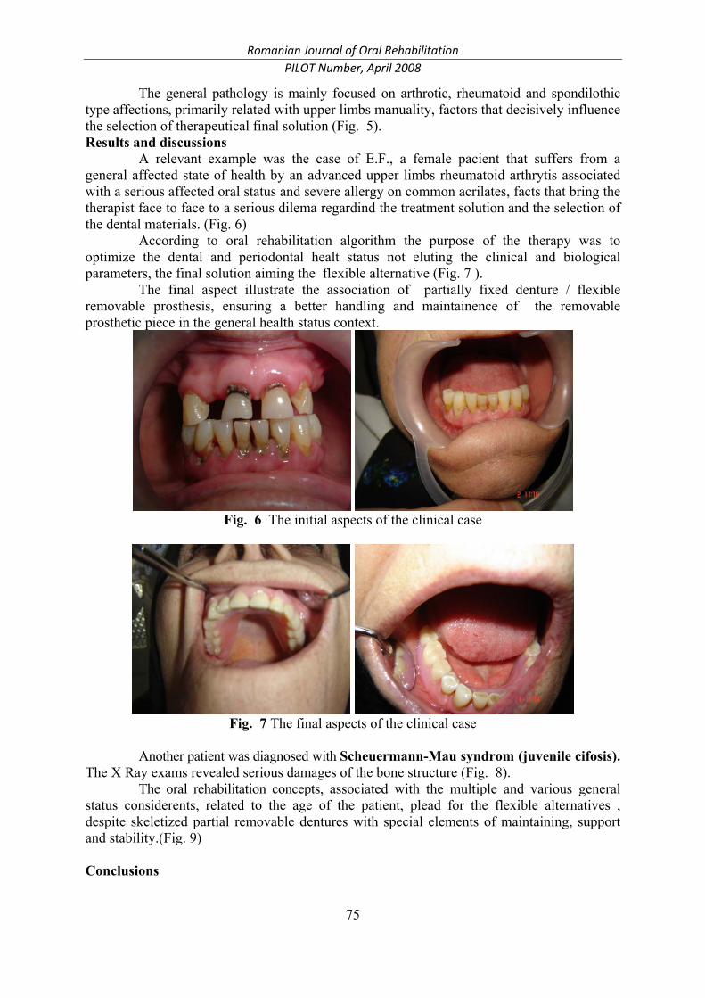
Romanian Journal of Oral Rehabilitation PILOT Number, April 2008
75
The general pathology is mainly focused on arthrotic, rheumatoid and spondilothic type affections, primarily related with upper limbs manuality, factors that decisively influence the selection of therapeutical final solution (Fig. 5). Results and discussions
A relevant example was the case of E.F., a female pacient that suffers from a general affected state of health by an advanced upper limbs rheumatoid arthrytis associated with a serious affected oral status and severe allergy on common acrilates, facts that bring the therapist face to face to a serious dilema regardind the treatment solution and the selection of the dental materials. (Fig. 6)
According to oral rehabilitation algorithm the purpose of the therapy was to optimize the dental and periodontal healt status not eluting the clinical and biological parameters, the final solution aiming the flexible alternative (Fig. 7 ).
The final aspect illustrate the association of partially fixed denture / flexible removable prosthesis, ensuring a better handling and maintainence of the removable prosthetic piece in the general health status context.
Fig. 6 The initial aspects of the clinical case
Fig. 7 The final aspects of the clinical case
Another patient was diagnosed with Scheuermann-Mau syndrom (juvenile cifosis).
The X Ray exams revealed serious damages of the bone structure (Fig. 8). The oral rehabilitation concepts, associated with the multiple and various general
status considerents, related to the age of the patient, plead for the flexible alternatives , despite skeletized partial removable dentures with special elements of maintaining, support and stability.(Fig. 9) Conclusions

Romanian Journal of Oral Rehabilitation PILOT Number, April 2008
76
1. The biological integration of removable dentures depends on real possibilities of handling them in the upper limbs motory disabilities context.
2. The evaluation of the whole general pathology mainly focused on arthrotic, rheumatoid and spondilothic type affections, primarily related with upper limbs manuality, decisively influences the selection of therapeutical final solution alternatives and may possibly contribute to the improvement of the oral health status on this type of patients.(4)
Fig. 8 The initial aspects of the clinical case
Fig. 9 The final aspects of the clinical case
References 1. D.C.Jagger, R.G. Jagger , S.M.AllenAn investigation into the transverse and impact strength of high
strength denture base acrylic resin. Journal of oral rehabilitation. (2002); 29(2):263-267.

Romanian Journal of Oral Rehabilitation PILOT Number, April 2008
77
2. G. Zappini, A. Kammann, W. WachterComparison of fracture tests of denture base materials. The journal of prosthetic dentistry, (2003), 90(6):578-15.
3. N.Forna,, V. Burlui (2001) Clinical guidelines and principles in the therapy of partial extended edentation, Apollonia, 470-477.
4. D.C. Jagger, A. Harrison (1999) The effect of chopped poly (methyl methacrylate) Fibers on Some properties of Acrylic Resin Denture Base Material. The International Journal of prosthodontics, 12(6):542 – 546.

Romanian Journal of Oral Rehabilitation PILOT Number, April 2008
78

Romanian Journal of Oral Rehabilitation PILOT Number, April 2008
79

Romanian Journal of Oral Rehabilitation PILOT Number, April 2008
80


