Roberta Ness, MD, MPH University of Pittsburgh Ovarian Cancer: Reproductive Factors and Beyond.
Upload
truonghanhCategory
view
220download
0
Review Article
Role of Minimally Invasive Surgery in Ovarian Cancer
Farr R. Nezhat, MD*, Tanja Pejovic, MD, Tamara N. Finger, MD, and Susan S. Khalil, MDFrom the Divisions of Gynecologic Oncology and Minimally Invasive Gynecologic Surgery, Department of Obstetrics and Gynecology, St. Luke’s andRoosevelt Hospitals, New York, New York (Drs. Nezhat, Finger, and Khalil), and Department of Gynecologic Oncology, Oregon Health and ScienceUniversity, Portland, Oregon (Dr. Pejovic).
ABSTRACT The standard treatment of ovarian cancer includes upfront surgery with intent to accurately diagnose and stage the disease andto perform maximal cytoreduction, followed by chemotherapy in most cases. Surgical staging of ovarian cancer traditionallyhas included exploratory laparotomywith peritonealwashings, hysterectomy, salpingo-oophorectomy, omentectomy,multipleperitoneal biopsies, and possible pelvic and para-aortic lymphadenectomy. In the early 1990s, pioneers in laparoscopic surgeryused minimally invasive techniques to treat gynecologic cancers, including laparoscopic staging of early ovarian cancer andprimary and secondary cytoreduction in advanced and recurrent disease in selected cases. Since then, the role of minimallyinvasive surgery in gynecologic oncology has been continually expanding, and today advanced laparoscopic and robotic-assisted laparoscopic techniques are used to evaluate and treat cervical and endometrial cancer. However, the important ques-tion about the place of the minimally invasive approach in surgical treatment of ovarian cancer remains to be evaluated andanswered. Overall, the potential role of minimally invasive surgery in treatment of ovarian cancer is as follows: i) laparoscopicevaluation, diagnosis, and staging of apparent early ovarian cancer; ii) laparoscopic assessment of feasibility of upfront surgicalcytoreduction to no visible disease; iii) laparoscopic debulking of advanced ovarian cancer; iv) laparoscopic reassessment inpatients with complete remission after primary treatment; and v) laparoscopic assessment and cytoreduction of recurrent dis-ease. The accurate diagnosis of suspect adnexal masses, the safety and feasibility of this surgical approach in early ovariancancer, the promise of laparoscopy as the most accurate tool for triaging patients with advanced disease for surgery vs upfrontchemotherapy or neoadjuvant chemotherapy, and its potential in treatment of advanced cancer have been documented andtherefore should be incorporated in the surgical methods of every gynecologic oncology unit and in the training programsin gynecologic oncology. Journal of Minimally Invasive Gynecology (2013) 20, 754–765! 2013 AAGL. All rights reserved.
Keywords: Cytoreduction; Laparoscopy; Ovarian cancer; Robotic-assisted laparoscopy; Staging
DISCUSS You can discuss this article with its authors and with other AAGL members athttp://www.AAGL.org/jmig-21-1-JMIG-D-13-00205R1
Use your Smartphoneto scan this QR codeand connect to thediscussion forum forthis article now*
* Download a free QR Code scanner by searching for ‘‘QRscanner’’ in your smartphone’s app store or app marketplace.
Ovarian cancer is the fifth most common cancer inwomen in the United States. The American Cancer Societyestimated that in 2012, epithelial ovarian cancer would bediagnosed in .21 000 women and that .15 500 would dieof the disease. The risk of a woman developing ovarian can-
cer is 1:70. More than a half of affected women are agedR65 years at diagnosis, and most are white [1]. Ovarian can-cer is extraordinarily difficult to diagnose at an early stagebecause of the insidious nature of its symptoms and lackof markers and tests for early detection. Subsequently, ovar-ian cancer typically is not diagnosed until it is in stage III orIV. Early stages of the disease are diagnosed in only 15% to20% of women, and in these patients the prognosis is muchbetter, with 5-year survival.90%. Histologic features of thedisease in these patients are primarily endometrioid andclear cell carcinomas, with a background of endometriosis[2,3]. In most patients with typical stage III disease, the5-year survival rate is 46% [4]. Most lesions in these patientsare poorly differentiated serous carcinomas, and althoughthe term implies that the ovary is the site of origin, recent
The authors have no commercial, proprietary, or financial interest in theproducts or companies described in this article.Corresponding author: Farr R. Nezhat, MD, Divisions of GynecologicOncology and Minimally Invasive Gynecologic Surgery, Department ofObstetrics and Gynecology, St. Luke’s and Roosevelt Hospitals, 1000 TenthAve, Ste 10-C, New York, NY 10019.E-mail: [email protected]
Submitted April 3, 2013. Accepted for publication April 26, 2013.Available at www.sciencedirect.com and www.jmig.org
1553-4650/$ - see front matter ! 2013 AAGL. All rights reserved.http://dx.doi.org/10.1016/j.jmig.2013.04.027

research has suggested that the fallopian tube and the perito-neal cavity can be the sources of the disease [5].
The standard treatment of ovarian cancer includes upfrontsurgery with intent to accurately diagnose and stage the dis-ease and to perform maximal cytoreduction, followed bytaxanes and platinum–based combination chemotherapy inmost patients [6]. Surgical staging of ovarian cancer tradi-tionally has included exploratory laparotomywith peritonealwashings, hysterectomy, salpingo-oophorectomy, omentec-tomy, multiple peritoneal biopsies, and possible pelvic andpara-aortic lymphadenectomy. When preservation of fertil-ity is desired and the disease seems to be confined to 1 ovary,preservation of the uterus and contralateral ovary is oftenpossible.
In the early 1990s, pioneers of laparoscopic surgery usedminimally invasive techniques to treat gynecologic cancers,including laparoscopic staging of early ovarian cancer. In se-lected cases, laparoscopic primary and secondary cytoreduc-tion was reported [7–9]. Since then, the role of minimallyinvasive surgery in gynecologic oncology has beencontinually expanding, and today advanced laparoscopictechniques are used to evaluate and treat cervical andendometrial cancer. The important question about the roleof the minimally invasive approach in surgical treatmentof ovarian cancer remains to be evaluated and answered.Overall, the potential role of minimally invasive surgery inovarian cancer is in the following categories: i)laparoscopic evaluation, diagnosis, and staging of apparentearly ovarian cancer; ii) laparoscopic debulking ofadvanced ovarian cancer; iii) laparoscopic assessment offeasibility of upfront optimal surgical cytoreduction; iv)laparoscopic reassessment in patients with completeremission after primary treatment; and v) laparoscopicassessment and cytoreduction of recurrent disease.
Laparoscopic Evaluation, Diagnosis, and Staging ofApparent Early Ovarian Cancer
Early ovarian cancer is defined as cancer limited to 1 orboth ovaries corresponding to FIGO stage I. Traditionally,staging surgery has been performed via a vertical midlineincision, which provides excellent exposure of the upperabdomen, diaphragmatic surfaces, and pelvis. Meticuloussurgical staging leads to upstaging in 16% to 35% of pre-sumed early-stage ovarian carcinoma [10].
Several retrospective and case series reports have demon-strated the feasibility and safety of a laparoscopic approachin management of early-stage ovarian cancer [11–15]. Thesestudies have shown that laparoscopy is associated withperioperative benefits such as decreased blood loss, fasterreturn of bowel function, and shorter hospital stay. Inaddition, the studies have supported the concept thatlaparoscopy may offer an advantage in management ofearly-stage ovarian cancer by enabling better visualizationof difficult areas such as the anterior abdominal wall, sub-diaphragmatic areas, peritoneal surfaces, obturator spaces,
and anterior and posterior cul-de-sacs, as well as magnifica-tion and detection of smaller lesions that may be missed atperioperative imaging and even during laparotomy. Retro-spective evidence in early ovarian cancer has revealedsimilar recurrence rates after laparoscopic and open stagingprocedures, suggesting that the laparoscopic techniquedoes not compromise the outcome of early-stage ovariancarcinoma [14].
Nezhat et al [15] have reported a case series of 36 patientswith presumed early-stage adnexal cancers who underwentlaparoscopic staging/restaging. The lesions included 20 in-vasive epithelial tumors, 11 borderline tumors, and 5 non-epithelial tumors. The mean number of peritoneal biopsieswas 6, of para-aortic nodes was 12.23, and of pelvic nodeswas 14.84. Eighty-three percent of patients underwent lapa-roscopic omentectomy. At final pathologic analysis, lesionswere upstaged in 7 patients. Postoperative complications in-cluded 1 small bowel obstruction, 2 pelvic lymphoceles, and1 lymphocele cyst. Three patients had recurrence of disease.At mean follow-up of 55.9 months, all patients were alivewithout evidence of disease. That study represents one ofthe largest series, with the longest follow-up, of laparoscopicstaging of early-stage adnexal tumors and illustrates that lap-aroscopic staging of these lesions, when performed by gyne-cologic oncologists experienced in advanced laparoscopy,seems to be feasible and comprehensivewithout compromis-ing survival [15].
Tumor rupture during laparoscopic surgery to treat earlyovarian cancer is of great concern. Although not proved inprospective studies, intraoperative tumor rupture will leadto immediate upstaging of the disease and may cause spreadof tumor cells, compromising the prognosis. Although theseconcepts and observations seem logical, the ultimate test viarandomized clinical trial is needed to determine whether theoutcomes of laparoscopic and open surgical staging of earlyovarian cancer are equivalent. Considering the low incidenceof early ovarian cancer and the rapid changes in technology,it is questionable whether such study will be initiated. Atpresent, all efforts should be made to reduce the incidenceof tumor contamination of the abdominal cavity, includingliberal use of a laparoscopic bag and controlled aspiration,and minimizing the risk of rupture [16].
Minimally Invasive Surgery for Cytoreduction ofAdvanced Ovarian Cancer
There is a paucity of data on laparoscopic cytoreductivesurgery for advanced ovarian cancer. The first report of suc-cessful videolaparoscopic cytoreduction of advanced ovar-ian cancer was a case series that included 3 patients, all ofwhom underwent successful total laparoscopic primary orsecondary cytoreduction [8].
In 2010, Nezhat et al [17] reported a series of 32 patientswith advanced ovarian, fallopian tube, or primary peritonealcancer who underwent laparoscopic evaluation for debulk-ing. In 17 of the 32 patients, the disease was successfully
Nezhat et al. Minimally Invasive Surgery in Ovarian Cancer 755

debulked at laparoscopy, with 88% of optimal cytoreduc-tion. At mean follow up of 19.2 months, 9 patients werewithout evidence of the disease. Compared with the groupwho underwent laparotomy, the laparoscopic approach hadresulted in minimal blood loss and a shorter hospital stay.No patients developed port-site metastasis, and time to dis-ease recurrence in the laparoscopic group was not inferiorto that in the laparotomy group.
In another retrospective series of 25 patientswith presumedstage III/IV primary ovarian cancer undergoing laparoscopic-assisted cytoreduction, Fanning et al [18] reported successfulcytoreduction in 23 patients (92%). Two procedureswere con-verted to laparotomybecause of extensive omental disease andbulky metastasis surrounding the rectosigmoid colon, res-pectively. In all 25 patients, the lesion was cytoreduced to,2 cm, and 36% had no residual disease. Median operativetimewas 2.3 hours, and blood losswas 340mL.Median lengthof staywas 1 day.Medianvisual analog scale pain scorewas 4,which was discomforting. Six patients (24%) had postopera-tive complications; however, none were grade 3 or 4. Medianoverall survival was 3.5 years. The authors concluded thatlaparoscopic cytoreduction for primary advanced ovariancancer can be successful and result in minimal morbidityand acceptable survival [18].
Hand-Assisted Laparoscopy
Krivak et al [19] reported 25 patients with ovarian carci-noma who underwent surgical staging and cytoreduction viahand-assisted laparoscopy. Six patients had apparent ad-vanced ovarian cancer at referral, and of the 19 patientswith presumed early-stage cancer, the disease was upgradedin 5 patients on the basis of retroperitoneal lymph node in-volvement. In 3 the disease had metastasized to other pelvicstructures, and 2 had microscopic disease in the omentum.Twenty-two surgical procedures were completed via hand-assisted laparoscopy, and 3 procedures required conversionto laparotomy for completion of debulking. Operating timewas variable, ranging from 81 to 365 minutes. Mean hospitalstay was 1.8 days for the 22 patients who underwent success-ful hand-assisted laparoscopic evaluation. Complicationrates were low, with 3 complications requiring repeatoperation or hospitalization. The authors concluded thathand-assisted laparoscopy may be applicable in the initialmanagement of early-stage and advanced ovarian carci-noma, enabling thorough evaluation of peritoneal and retro-peritoneal structures and surgical cytoreduction, with theadvantages of minimally invasive surgery [19].
Robotic-Assisted Laparoscopy
Recently, there have been reports of use of robotic-assisted surgery in patients with advanced ovarian cancer.In a retrospective case-control study, Magrina et al [20] com-pared 25 patients with ovarian cancer undergoing a robotic-assisted approach with 27 similar patients undergoing
conventional laparoscopy and 119 undergoing laparotomy.In the respective groups, 60%, 75%, and 87% of patientswere found to have FIGO stage III/IV disease, and theremaining patients had FIGO stage I/II disease. Mean esti-mated blood loss was 164 mL vs 267 mL vs 1307 mL(p , .001), and length of hospital stay was 4 vs 3 vs 9days (p , .001), both significantly less in the robotic-assisted and conventional laparoscopic group comparedwith the laparotomy group. Node counts were similar inthe 3 groups. Operative time was significantly longer inthe robotic-assisted group, 315 vs 254 vs 261 minutes(p 5 .009). Overall survival was similar in the 3 groups,67.1% vs 75.6% vs 66.0% (p5 .08). The rates of intraoper-ative and postoperative complications were similar in the 3groups. Patients were also subdivided and compared accord-ing to the extent and number of major procedures. The au-thors concluded that in patients undergoing primary tumorexcision of epithelial ovarian cancer alone or with 1 addi-tional major surgery, robotic-assisted and videolaparoscopyare preferable to laparotomy. They also concluded that over-all survival is not influenced by the type of surgical approachbut by the extent of debulking (complete vs incomplete).Similar to the report by Nezhat et al [17], no patients inthe robotic-assisted or laparoscopy groups developed port-site metastases, which they believed was likely due in partto early initiation of chemotherapy.
Laparoscopic Assessment of Feasibility of UpfrontOptimal Surgical Cytoreduction
The mainstay of treatment for advanced invasive epithelialovarian cancer is ideally cytoreduction to no visible disease(microscopic) followed by platinum-based combination che-motherapy [6], which is associated with the best survival[21–24]. However, cytoreduction to microscopic disease isnot possible in all patients at the initial surgery. To increasethe rate of complete or optimal debulking and to limitperioperative morbidity, neoadjuvant chemotherapy withinterval cytoreduction has emerged as an alternative toprimary surgery [25]. This strategy does not seem to compro-mise survival, in particular if surgery is performed early,within 42 days of completion of neoadjuvant chemotherapy[25,26]. Therefore, the primary issue is how to best evaluatethe optimal resectability of advanced ovarian cancer. Despiteimprovements in computed tomography, magnetic resonanceimaging, and positron emission tomography [27] and in tumormarkers, resectability of intraperitoneal disease remains diffi-cult to determine. Several predictive models have been pro-posed on the basis of clinical findings, imaging [27], andCA125 serum concentration; however, false-positive ratesrange from 5% to 37% [28], and therefore addition of surgicallaparoscopic evaluation can be useful [29].
To assess resectability of advanced ovarian cancer, pa-tients should be selected with the goal of achieving optimalprimary cytoreduction, ie, no visible disease or microscopicdisease. Laparoscopy has been used to assess the status of
756 Journal of Minimally Invasive Gynecology, Vol 20, No 6, November/December 2013
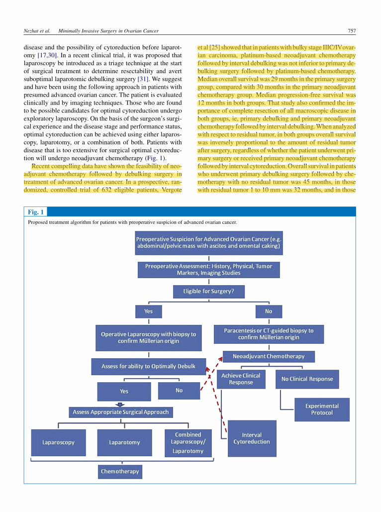
disease and the possibility of cytoreduction before laparot-omy [17,30]. In a recent clinical trial, it was proposed thatlaparoscopy be introduced as a triage technique at the startof surgical treatment to determine resectability and avertsuboptimal laparotomic debulking surgery [31]. We suggestand have been using the following approach in patients withpresumed advanced ovarian cancer. The patient is evaluatedclinically and by imaging techniques. Those who are foundto be possible candidates for optimal cytoreduction undergoexploratory laparoscopy. On the basis of the surgeon’s surgi-cal experience and the disease stage and performance status,optimal cytoreduction can be achieved using either laparos-copy, laparotomy, or a combination of both. Patients withdisease that is too extensive for surgical optimal cytoreduc-tion will undergo neoadjuvant chemotherapy (Fig. 1).
Recent compelling data have shown the feasibility of neo-adjuvant chemotherapy followed by debulking surgery intreatment of advanced ovarian cancer. In a prospective, ran-domized, controlled trial of 632 eligible patients, Vergote
et al [25] showed that in patientswith bulky stage IIIC/IVovar-ian carcinoma, platinum-based neoadjuvant chemotherapyfollowed by interval debulking was not inferior to primary de-bulking surgery followed by platinum-based chemotherapy.Median overall survival was 29months in the primary surgerygroup, compared with 30 months in the primary neoadjuvantchemotherapy group. Median progression-free survival was12 months in both groups. That study also confirmed the im-portance of complete resection of all macroscopic disease inboth groups, ie, primary debulking and primary neoadjuvantchemotherapy followedby interval debulking.When analyzedwith respect to residual tumor, in both groups overall survivalwas inversely proportional to the amount of residual tumorafter surgery, regardless of whether the patient underwent pri-mary surgery or received primary neoadjuvant chemotherapyfollowedby interval cytoreduction.Overall survival in patientswho underwent primary debulking surgery followed by che-motherapy with no residual tumor was 45 months, in thosewith residual tumor 1 to 10 mm was 32 months, and in those
Fig. 1
Proposed treatment algorithm for patients with preoperative suspicion of advanced ovarian cancer.
Nezhat et al. Minimally Invasive Surgery in Ovarian Cancer 757

with residual tumor.10mmwas 26months. Overall survivalin these 3 groups who underwent neoadjuvant chemotherapywas 38, 27, and 25 months, respectively. Thus that trial con-firmed the importance of optimal surgical cytoreduction re-gardless of whether surgery is performed before or afterneoadjuvant chemotherapy. Therefore, we believe that in se-lected cases in which neoadjuvant triage is offered to patientsand the disease after completionof chemotherapy is less exten-sive, laparoscopic debulking surgery may be performed(Fig. 1).
Laparoscopic Reassessment or Second-Look Surgery
In the past, second-look surgery was suggested as part ofthe therapeutic triage in patients with advanced ovarian can-cer. Today this procedure is performed primarily in clinicaltrials or in selected cases with uncertain clinical responseof patients. The minimally invasive approach had beenused in second-look assessment in patients with a completeclinical response to platinum-based combination chemother-apy. Disease recurrence after negative second-look surgerywas reported to be similar for laparoscopy and laparotomy,with laparoscopy having the advantages of less morbidity,shorter operative time, shorter hospital stay, and lower totalhospital charges [32].
Laparoscopic Assessment and Cytoreduction ofRecurrent Disease
The role of secondary cytoreduction surgery to treat recur-rent ovarian carcinoma is debatable. Recently, several authorshave suggested some criteria including isolated recurrence,lack of ascites, and optimal debulking at the primary surgeryas indications for secondary debulking [33,34]. In theseselected cases, laparoscopic secondary cytoreduction hasbeen reported in case reports and series, with acceptableresults insofar as efficacy and outcomes [8,35–38] (Fig. 2).
Trinh et al [36] reported on 36 consecutive patients withasymptomatic chemosensitive stage III/IV ovarian cancerwho had previously undergone debulking via laparotomyfollowed by chemotherapy, and when CA 125 concentrationwas elevated, underwent laparoscopic debulking. Preopera-tive abdominal/pelvic computed tomography yielded nor-mal findings. Operative laparoscopy was performed usingan open technique in the left upper quadrant, and tumorswere debulked laparoscopically using the loop electrosurgi-cal excision procedure and argon beam coagulation. Of the36 patients, laparoscopic debulking was successful in 34(94%), without requiring laparotomy. Of those 34 patients,all visible disease was resected at laparoscopy in 32(94%), and surgical complications occurred in 2 patients(6%). Median operative time was 2.6 hours, median bloodloss was 70 mL, and median hospital stay was 1 day.Seventy-four percent of patients had a complete responseafter laparoscopic debulking and chemotherapy, with amedian progression-free survival of 1.1 years. The authors
concluded that laparoscopic debulking using the loop elec-trosurgical excision procedure and argon beam coagulationseems feasible (94%), successful (94%), and safe (complica-tions in 6%) [36].
Nezhat et al [37] reported a retrospective analysis of a pro-spective case series of 23 patients with recurrent ovarian, fal-lopian tube, or primary peritoneal cancer who were deemedappropriate candidates for laparoscopic debulking. The pa-tients underwent exploratory videolaparoscopy, biopsy,and laparoscopic secondary/tertiary cytoreduction betweenJune 1999 and October 2009. Of the 23 procedures, only 1was converted to laparotomy. Seventeen patients (77.3%)had stage IIIC disease at the time of initial diagnosis, and20 (90.9%) underwent laparotomy for primary debulking.Median blood loss was 75 mL, median operative time was200 minutes, and median hospital stay was 2 days. No intra-operative complications occurred. One patient (4.5%) hadpostoperative ileus. Eighteen patients (81.8%) with recurrentdisease underwent optimal cytoreduction to ,1 cm. Overa median follow-up of 14 months, 12 patients had no evi-dence of disease, 6 were alive with disease, and 4 had diedof the disease. Median disease-free survival was 71.9months. The authors concluded that in a well-selected pop-ulation, laparoscopy is technically feasible and can beused for optimal cytoreduction in patients with recurrentovarian, fallopian, or primary peritoneal cancer [37] (Fig. 2).
Surgical Technique
Conventional Laparoscopy
Closed or open transumbilical or left upper quadrant(Palmer point) entry using a Veress needle is used mostoften. A 0-degree laparoscope, and at times a 30-degree lap-aroscope, is used via a port placed 4 to 5 cm supraumbili-cally. We use three 5- to 12-mm ancillary ports in themid–lower abdomen when the primary lesion is below thepelvic brim. Additional upper abdominal ports can be placedfor extensive upper abdominal debulking. Pelvic washingsare collected for cytologic analysis, and parietal and visceralperitoneal surfaces of the deep pelvis and middle and upperabdominal cavities are thoroughly inspected (Fig. 3, A–D).Any suspect growth is biopsied. In the case of normal visualexploration, random peritoneal biopsies are performed in thepouch of Douglas, pelvic and abdominal parietal perito-neum, paracolic gutters, hemidiaphragms, and mesentery.Small and large bowel can also be carefully inspected lapa-roscopically. Running the small bowel can be accomplishedfrom the ileocecal valve to the ligament of Treitz using 2atraumatic bowel graspers. When conservative treatment isconsidered, biopsy of the contralateral ovary is performedonly in the case of suspect growth on imaging studies or atlaparoscopy. In this context, dilation and curettage are per-formed so as not to miss a possible endometrial spread ora synchronous tumor [3]. Every attempt should be made toprevent rupture of a suspect adnexal mass in the abdomen,
758 Journal of Minimally Invasive Gynecology, Vol 20, No 6, November/December 2013

including choosing unilateral adnexectomy rather than ovar-ian cystectomy, limited manipulation of the mass, use ofnon-traumatic graspers, and preventive coagulation to avertbleeding that may obscure identification of the cleavageplanes. Additional safety measures include removal of thespecimen exclusively using a laparoscopic bag and controlof the bag integrity once extracted. Laparoscopy is intrinsi-cally limited by the size of the port incisions. Even when theincision is enlarged, a puncture is required to remove largemasses. If the puncture can be located within an endobagand the integrity of the bag is preserved, the procedure isconsidered safe and preferable.
To achieve an infracolic omentectomy, the patient isplaced supine in a dorsal lithotomy position. The primarysurgeon stands between the patient’s legs, with the firstand second assistants standing on each side of the patient.The position of the primary surgeon can be changed depend-ing on port placement and the tissue being manipulated or
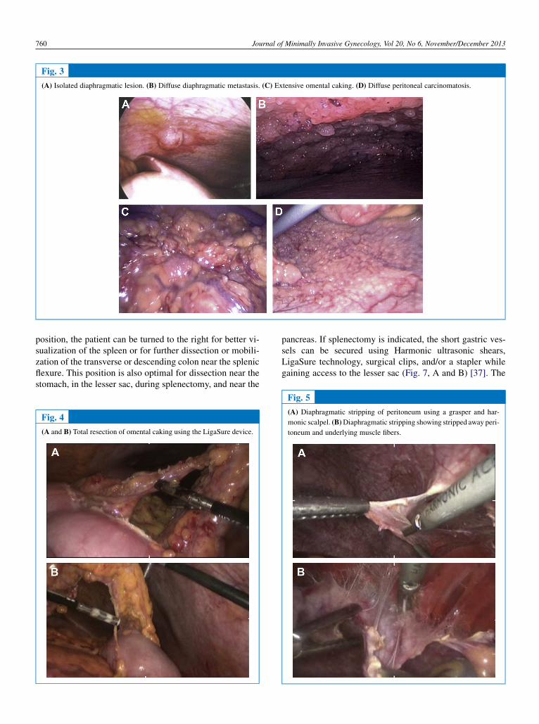
resected. The omentum is excised from the inferior marginof the transverse colon using a Harmonic scalpel (EthiconEndo-Surgery, Cincinnati, OH), a linear stapler, or any ofthe electrosurgical blood vessel sealing devices. TheHarmonic scalpel and LigaSure technology (Covidien,Mansfield, MA) are superior for omentectomy because ofminimal fume formation, ease and speed of use, and lackof protruding staple edges (Fig. 4, A and B). The omentumspecimen can also be removed using an endobag.
In patients with extensive upper abdominal disease, it isour practice to place a 5- to 12-mm port in the middle to up-per right side of the abdomen and a 5-mm port in the left up-per abdomen. This position is also optimal for diaphragmaticablation and stripping (Fig. 5, A and B), supracolic omentec-tomy, and resection of further upper abdominal disease. Di-aphragmatic ablation can be performed using a CO2 laser,PlasmaJet (Plasma Surgical, Ltd., Oxfordshire, UK)(Fig. 6, A and B), and argon beam coagulator. From this
Fig. 2
Proposed treatment algorithm for selection of patients with preoperative suspicion of recurrent ovarian cancer.
Nezhat et al. Minimally Invasive Surgery in Ovarian Cancer 759
position, the patient can be turned to the right for better vi-sualization of the spleen or for further dissection or mobili-zation of the transverse or descending colon near the splenicflexure. This position is also optimal for dissection near thestomach, in the lesser sac, during splenectomy, and near the
pancreas. If splenectomy is indicated, the short gastric ves-sels can be secured using Harmonic ultrasonic shears,LigaSure technology, surgical clips, and/or a stapler whilegaining access to the lesser sac (Fig. 7, A and B) [37]. The
Fig. 3
(A) Isolated diaphragmatic lesion. (B) Diffuse diaphragmatic metastasis. (C) Extensive omental caking. (D) Diffuse peritoneal carcinomatosis.
Fig. 4
(A and B) Total resection of omental caking using the LigaSure device.
Fig. 5
(A) Diaphragmatic stripping of peritoneum using a grasper and har-
monic scalpel. (B)Diaphragmatic stripping showing stripped away peri-
toneum and underlying muscle fibers.
760 Journal of Minimally Invasive Gynecology, Vol 20, No 6, November/December 2013

spleen can then be mobilized from its attachments and liga-ments. The splenic vessels can be cut and the spleen re-moved using the laparoscopic stapling device (EthiconEndo-Surgery). The patient can be turned to the left for fur-ther dissection near the liver, porta hepatis, and ascendingcolon. The position can be further manipulated to aid inbowel resection of the small and transverse colon.
Transperitoneal pelvic and/or para-aortic lymph node dis-section is performed while the patient is in the Trendelen-burg position. For pelvic lymph node retrieval, the primarysurgeon stands on either side of the patient, facing the mon-itors, which are positioned on both sides of the patient’s legs.The first assistant stands across from the surgeon, and thesecond assistant is at the bottom of the table, between the pa-tient’s legs. The peritoneum overlying the psoas muscle isincised from the round ligament to the base of the infundib-ular pelvic ligament. External and internal iliac vessels, theirmajor branches, and the ureter are delineated, and the avas-cular spaces are developed. After delineation of the anatomyof the pelvic sidewall, sampling or complete removal of allnodal packets along the external and internal iliac vesselsand the obturator fossa is performed. Lymph nodes are re-trieved through the suprapubic trocar sleeve (10–12 mm),avoiding contamination of the abdominal wall [16].
For para-aortic lymph node dissection, the room setupand trocar placement are similar as for pelvic lymphadenec-tomy, with the laparoscope introduced through the supraum-bilical or suprapubic port. Pelvic operations such ashysterectomy, salpingo-oophorectomy, and any resectionof metastatic disease are performed using the same portsas for pelvic lymph adenectomy [16].
Robotic-Assisted Laparoscopy
There are limitations when using the current computer-enhanced telesurgery called the robotic platform (IntuitiveSurgical, Inc., Sunnyvale, CA) for staging and cytoreductionin ovarian cancer. Once the robot is docked for pelvicsurgery, it is more difficult to access the upper abdomenwithout having to undock and reposition the robot or addadditional ports enable performance of the procedure. TheSociety of Gynecologic Oncology consensus statement onrobotic-assisted surgery commented that it is poorly suited
for treatment of advanced ovarian cancer because of its lim-itation in gaining upper abdominal access with conventionaltrocar placement for pelvic surgery [39].
We have developed a hybrid technique in which both con-ventional laparoscopy and the robot are used in the surgicalmanagement of ovarian cancer. This surgical technique and
Fig. 6
(A) Hydrodissection over the diaphragm. (B) Diaphragmatic ablation using the PlasmaJet device.
Fig. 7
(A) Short gastric vessels are secured using the laparoscopic stapler
while gaining access to the lesser sac during laparoscopic splenectomy
because of metastasis to the parenchyma. (B) Splenectomy is performed
using an articulating stapler.
Nezhat et al. Minimally Invasive Surgery in Ovarian Cancer 761

its use in patients with early and advanced ovarian cancer isdescribed as follows for laparoscopic management of bothpelvic and upper abdominal disease.
An incision is made in either the left upper quadrant or 4to 5 cm above the umbilicus, a Veress needle is introduced,and pneumoperitoneum is established. After adequate pneu-moperitoneum is obtained, a 5- or 8-mm primary port is in-serted in the left upper quadrant. If pneumoperitoneum isestablished supraumbilically, a 12-mm trocar and sleeveare introduced into the supraumbilical port. After assessingthe abdominopelvic cavity, either a 10- or 12-mm port is in-troduced into the right upper quadrant, and two 8-mmrobotic ports are introduced 8 to 10 cm lateral to the umbili-cus bilaterally (Fig. 8). Further peritoneal inspection isperformed via conventional laparoscopy, and peritonealwashings or aspiration of any existing ascites are obtainedand sent for cytologic analysis.
If there is disease in the upper abdomen and pelvis, surgeryis begun using conventional laparoscopy to perform theomentectomy and upper abdominal debulking (Figs. 5–7).This is performed via use of the supraumbilical port for thecamera and other ports for introduction of instruments. Anyupper abdominal debulking is performed as described forconventional laparoscopy. This same approach can be usedif there is no upper abdominal disease and only infracolicomentectomy is performed as part of surgical staging.
Any abdominal and pelvic adhesions that interfere withproper use of the robotic platform are lysed using conven-tional laparoscopy. After the upper abdominal portion is per-formed, the robotic apparatus is docked on the patient’s leftside, using the supraumbilical port for the camera and thebilateral robotic 8-mm ports. The posterior parietal perito-neum over the right common iliac artery is incised, and ret-roperitoneal dissection is completed cephalad to above theinferior mesenteric artery (Fig. 9).We use the electrosurgicalspatula or scissors for cutting and bipolar forceps for achiev-
ing hemostasis. The left and right upper assist ports are usedfor introduction of ancillary instruments for traction, tissueremoval, suction, and irrigation (Fig. 10). Pelvic lymphade-nectomy, hysterectomy, salpingo-oophorectomy, and anytumor debulking are performed with the same instrumentsas used before or, at times, standard blood vessel sealingdevices such as the LigaSure technology for ligation of theinfundibular ligaments and hysterectomy (Figs. 11 and12). If access to the para-aortic lymph nodes above the infe-rior mesenteric artery is not possible using the robotic plat-form, this portion of the operation is performed usinga conventional laparoscopic approach after undocking therobot. In some instances, the location of the camera is movedfrom the supraumbilical port to the right upper abdomen toachieve this goal.
After the uterus is transected, it is removed from thevagina along with the omentum and any other specimens,which are confined to an endocatch bag. The vaginal cuffis closed in 2 layers. After complete hemostasis is achieved,the robotic apparatus is undocked. If hysterectomy is notbeing performed, all bulky specimens can be removed viaa small mini-laparotomy using laparoscopic bags.
Cystoscopy is routinely performed to ensure that there isno damage to the bladder or bilateral ureters. The posteriorcul-de-sac is filled with fluid and air is injected into the rec-tum to above the sigmoid colon to ensure that the bowel isintact. Trocars are removed and port sites closed in a routinemanner.
In our experience in 20 women who underwent surgeryusing our hybrid technique of conventional laparoscopyand robotic-assisted laparoscopy, 21 surgical procedureswere performed: 10 for early-stage disease, and 11 for ad-vanced or recurrent disease (6 advanced and 5 recurrent).Of the 10 procedures for early-stage disease, mean patientage was 42.3 years (range, 29–55 years), body mass index
Fig. 8
Port placement in hybrid technique.
Fig. 9
Robotic dissection exposes the bifurcation of the common iliac artery
and mobilizes the right ureter laterally for para-aortic lymph node dis-
section.
762 Journal of Minimally Invasive Gynecology, Vol 20, No 6, November/December 2013
was 32.1 (range, 17–65), estimated blood loss was 212.5 mL(range, 50–1000 mL), operative time was 306.1 minutes(87–639), and length of stay was 1.6 days (1–2). Mean num-ber of pelvic lymph nodes dissected was 10.3 (range, 5–18),and of para-aortic lymph nodes dissected was 8.6 (range, 3–12). There were no intraoperative complications or intrao-perative transfusions, and 2 postoperative complications.One patient was readmitted on postoperative day 9 becauseof a wound infection, and another was readmitted because offever of unknown origin, which resolved with intravenousantibiotic therapy. Of the 11 patients operated on becauseof advanced and/or recurrent ovarian cancer, mean agewas 63.9 years (range, 39–92 years), body mass index was29.7 (22.1–37.2), estimated blood loss was 129.1 mL (20–400), operative time was 238 minutes (103–477), and lengthof stay was 3.8 days (1–17). Of these 11 patients, 1 under-went a second-look procedure. In 9 patients, disease was cy-
toreduced to no visible disease, and in 1 to ,0.5 cm. Therewere no intraoperative complications or blood transfusions,but 3 postoperative complications. Two postoperative com-plications occurred in the same patient, which where portsite cellulitis and a peritoneal vaginal fistula. The other pa-tient underwent a second operation on postoperative day 3because of a bowel perforation, and was the only patient ad-mitted to the intensive care unit in either the early or ad-vanced disease groups [40].
Upper abdominal primary cytoreduction can also be per-formed with the robot in reverse docking. This can be ac-complished by undocking the robot, rotating the operatingtable, and redocking the robot at the patient’s head. Upperabdominal secondary cytoreduction in isolated upperabdominal recurrent disease can also be accomplished bydocking the robot off of one of the patient’s shoulders, de-pending on the side of disease, using the same port place-ment as shown in Figs. 8 and 13A. Readjustment of theports can be performed according to the pathologic findings.
Holloway et al [38] reported the case of a 60-year-oldwoman with recurrent platinum-sensitive ovarian cancerwith an isolated 3.4-cm lesion noted on the dome of the righthepatic lobe at computed tomography and positron emissiontomography. Upper abdominal tumor resection was accom-plished by placing the patient in a 10-degree reverse Trende-lenburg position, rotated to the left 10 degrees, and dockingthe robot over her right shoulder (Fig. 13B). During theoperative procedure, adhesions from the liver to the dia-phragm were separated, the hepatic lesion was excised,and diaphragmatic involvement of the cancer was noted.Full-thickness resection of the diaphragmatic lesion wasperformed, and the diaphragm was closed primarily usingrunning No. 1 polypropylene (Prolene) suture. Estimatedblood loss was 100 mL, total operative time was 137minutes, and console time was 82 minutes. Pathologic an-alysis revealed margins and washings negative for disease.There were no important intraoperative complications.
Fig. 10
Robotic para-aortic lymphadenectomy.
Fig. 11
Robotic dissection of the right pelvic sidewall exposes the iliac vessels
and the obturator fossa for pelvic lymphadenectomy.
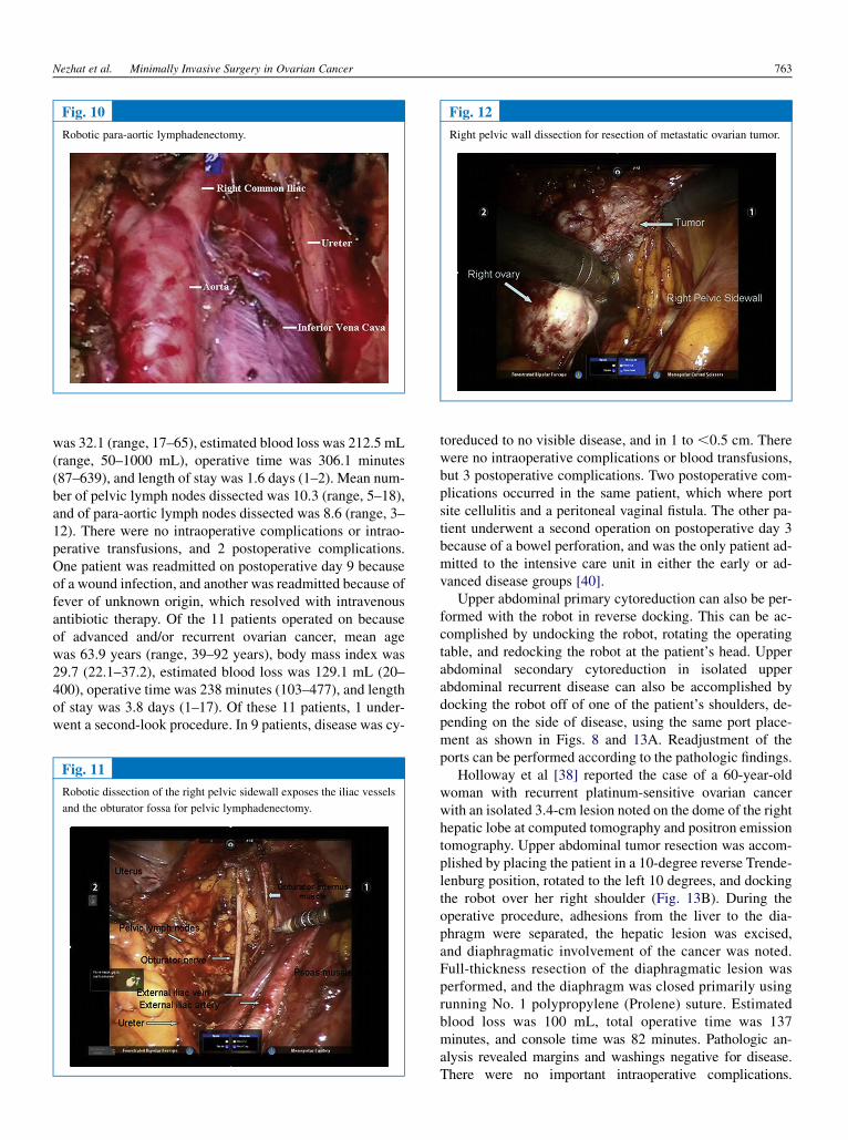
Fig. 12
Right pelvic wall dissection for resection of metastatic ovarian tumor.
Nezhat et al. Minimally Invasive Surgery in Ovarian Cancer 763

Cytology-negative pleural effusion developed, which wassuccessfully drained via thoracentesis on postoperative day4, and subsequently chemotherapy was started at 4 weekspostoperatively (Fig. 13, A and B).
Bowel resection can be performed at conventional lapa-roscopy and robotically using the appropriate port and robotplacement. In case of the need for mid-abdominal debulkingsuch as appendectomy or ileocecal resection, mobilizationof the bowel is performed using the robotic platform, and ap-pendectomy or bowel resection is performed using a staplingdevice introduced through the right upper abdominal port.Anastomosis can be performed either in situ or extracorpo-really by extending the supraumbilical incision after pelvictumor debulking and undocking the robot. This approachcan be used for segmental transverse colon resection and re-anastomosis to achieve optimal cytoreduction. For rectosig-moid colon resection and anastomosis, we use a 12-mm portin the right lower abdomen for introduction of the staplingdevice. This is especially true for a bulky lesion involvingthe rectosigmoid colon. Using a laparoscopic 60-mm gastro-intestinal anastomosis stapler, a rectosigmoid resection canbe performed proximally and distally. Once the proximalsigmoid colon is appropriately mobilized, this end can bebrought out through a widened incision in the right lowerquadrant or a lower middle incision, or transvaginally alongwith the specimen. An anvil can then be placed and securedusing a purse string suture. The anvil and proximal sigmoidcolon are then brought back into the pelvis, and an end-to-end anastomosis can be performed using a stapler passedthrough the rectum. Once the device is properly activated,it is important to test the integrity of the anastomosis. Thiscan be accomplished by clamping the proximal colon usinga bowel grasper, filling the pelvis with lactated Ringer solu-tion, and insufflating the rectum with air while observinglaparoscopically. The anastomosis can be alternatively or ad-ditionally examined by filling the rectum with indigo car-mine and observing for leakage [37].
In conclusion, with the continued advancement in endo-scopic techniques and instrumentation, laparoscopy hasemerged as a feasible alternative to open laparotomy in man-aging gynecologic malignant disease. Theminimally invasivesurgical approach and its use in ovarian cancer diagnosis andtreatment continues to evolve and broaden.
Accurate diagnosis of suspect adnexal masses, the safetyand feasibility of this surgical approach in early ovarian can-cer, the promise of laparoscopy as the most accurate tool fortriaging patients with advanced disease for surgery vs upfrontchemotherapy or neoadjuvant chemotherapy, and its potentialfor treatment of advanced cancer are documented and there-fore should be incorporated in the surgical arsenal of everygynecologic oncology unit and training programs in gyneco-logic oncology. The potential role of minimally invasive sur-gery in advanced and recurrent ovarian, fallopian tube, andprimary peritoneal cancer are described in Figures 1 and 2.
The promise of minimal incisions and shorter recoverytime, coupled with an increased number of skilled laparo-scopic surgeons and a team approach in well-equipped oper-ating rooms, add to the potential of introducing theseapproaches in ovarian cancer treatment without the contextof a clinical trial. However, women with ovarian cancerand our clinical field would be best served if our collectiveeffort is focused on designing and conducting randomizedphase III clinical trials to adequately and definitively addressthese clinical situations.
Until now, surgery followed by chemotherapy has beenthe standard treatment of advanced-stage ovarian cancers.However, this has not significantly improved survival andprognosis in these women. The future depends on findingthe different developmental pathways of this disease, findingappropriate chemotherapy and biological agents, targetingspecific tumor cell types, and individualizing therapies.The majority of ovarian cancer can be treated medically in-stead of surgically and in the future, if surgery does playa role, it will not be as aggressive as it is today.
Fig. 13
Robotic port placement during secondary cytoreduction of liver and diaphragmatic lesion. (A) The 12-mm port is placed superolateral to the umbilicus,
12 cm below the mid-right costal margin, and one 12-mm assistant port is placed midline above the umbilicus and another in the right upper quadrant. Three
8.5-mm robotic ports are placed as shown. (B) Patient placed in a 10-degree Trendelenburg position, rotated 10 degrees to the left, and the robot is docked
over the right shoulder.
764 Journal of Minimally Invasive Gynecology, Vol 20, No 6, November/December 2013

References
1. American Cancer Society. Cancer Facts and Figures, 2011. Atlanta,GA: American Cancer Society; 2011.
2. Pearce CL, Templeman C, Rossing MA, et al. on behalf of the OvarianCancer Association Consortium. Association between endometriosisand risk of histological subtypes of ovarian cancer: a pooled analysisof case-control studies. Lancet Oncol. 2012;13:385–394.
3. Deligdisch L, Penault-Llorca F, Schlosshauer P, Altchek A, Peiretti M,Nezhat F. Stage I ovarian carcinoma: different clinical pathologicpatterns. Fertil Steril. 2007;88:906–910.
4. Jemal A, Siegel R, Xu J,Ward E. Cancer Statistics, 2010 [published cor-rection appears in CA Cancer J Clin. 2011;61:133-134]. CA Cancer JClin. 2010;60:277–300.
5. Crum CP, McKeon FD, Xian W. BRCA, the oviduct, and the space andtime continuum of pelvic serous carcinogenesis. Int J Gynecol Cancer.2012;22:S29–S34.
6. Katz VL, Lentz GM, Lobo RA, Gershenson DM. ComprehensiveGynecology. 5th ed. Philadelphia, PA: Mosby Elsevier; 2007.
7. Querleu D, Leblanc E. Laparoscopic infrarenal para-aortic lymph nodedissection for restaging of carcinoma of the ovary or fallopian tube.Cancer. 1994;73:1467–1471.
8. Amara DP, Nezhat C, Teng N, Nezhat F, Nezhat C, Rosati M. Operativelaparoscopy in the management of ovarian cancer. Surg LaparoscEndosc. 1996;6:38–45.
9. Liu CS, Nagarsheth NP, Nezhat FR. Laparoscopy and ovarian cancer:a paradigm change in the management of ovarian cancer? J MinimInvasive Gynecol. 2009;16:250–262.
10. Stier EA, Barakat RR, Curtin JP, Brown CL, Jones WB, Hoskins WJ.Laparotomy to complete staging of presumed early ovarian cancer.Obstet Gynecol. 1996;87:737–740.
11. Childers JM, Lang J, Surwit EA, Hatch KD. Laparoscopic surgicalstaging of ovarian cancer. Gynecol Oncol. 1995;59:25–33.
12. Angioli R, Muzii L, Battista C, et al. The role of laparoscopy in ovariancarcinoma. Minerva Gynecol. 2009;61:35–43.
13. Tozzi R, Schneider A. Laparoscopic treatment of early ovarian cancer.Curr Opin Obstet Gynecol. 2005;17:354–358.
14. Weber S, McCann CK, Boruta DM, Schorge JO, Growdon WB. Lapa-roscopic surgical staging of early ovarian cancer. Rev Obstet Gynecol.2011;4:117–122.
15. Nezhat FR, Ezzati M, Chuang L, et al. Laparoscopic management ofearly ovarian and fallopian tube cancers: surgical and survival outcome.Am J Obstet Gynecol. 2009;200:83–85.
16. Sternchos J, Finger T, Mahdavi A, Nezhat F. Laparoscopic managementof ovarian, fallopian tube and primary peritoneal cancer. In: Nezhat C,Nezhat F, Nezhat C, editors. Nezhat’s Video-Assisted and Robotic-Assisted Laparoscopy and Hysteroscopy. 4th ed. Cambridge, MA:Cambridge University Press; 2013. p. 508–525.
17. Nezhat FR, DeNoble SM, Liu CS, et al. The safety and efficacy of lap-aroscopic surgical staging and debulking of apparent advanced stageovarian, fallopian tube, and primary peritoneal cancers. JSLS. 2010;14:155–168.
18. Fanning J, Yacoub E, Hojat R. Laparoscopic-assisted cytoreduction forprimary ovarian cancer: success, morbidity and survival. GynecolOncol. 2011;123:47–49.
19. Krivak TC, Elkas JC, Rose GS, et al. The utility of hand-assisted lapa-roscopy in ovarian cancer. Gynecol Oncol. 2005;96:72–76.
20. Magrina JF, Zanagnolo V, Noble BN, Kho RM, Magtibay P. Roboticapproach for ovarian cancer: perioperative and survival results andcomparison with laparoscopy and laparotomy. Gynecol Oncol. 2011;121:100–105.
21. Bristow RE, Tomacruz SR, Armstrong DK, Trimble EL, Montz FJ.Survival effect of maximal cytoreductive surgery for advanced ovariancarcinoma during the platinum era: a meta analysis. J Clin Oncol. 2002;20:1248–1259.
22. Winter WE III, Maxwell GL, Tian C, et al. Gynecologic OncologyGroup Study. Prognostic factors for stage III epithelial ovarian cancer:
a Gynecologic Oncology Group Study. J Clin Oncol. 2007;25:3621–3627.
23. Winter WE III, Maxwell GL, Tian C, et al. Gynecologic OncologyGroup. Tumor residual after surgical cytoreduction in prediction ofclinical outcome in stage IV epithelial ovarian cancer: a GynecologicOncology Group Study. J Clin Oncol. 2008;26:83–89.
24. Du Bois A, Reuss A, Pujade-Lauraine E, Harter P, Ray-Coguard I,Pfisterer J. Role of surgical outcome as prognostic factor in advancedepithelial ovarian cancer: a combined exploratory analysis of 3 prospec-tively randomized phase 3 multicenter trials; by the Arbeitsgemein-schaft Gynaekologische Onkologie Studiengruppe Ovarialkarzinom(AGO-OVAR) and the Groupe d’ Investigateurs Nationaux pour lesEtudes des Cancers l’Ovaire (GINECO). Cancer. 2009;115:1234–1244.
25. Vergote I, Trop!e CG, Amant F, et al. European Organization for Researchand Treatment of Cancer-Gynaecological Cancer Group; NCIC ClinicalTrials Group. Neoadjuvant chemotherapy or primary surgery in stageIIIC or IVovarian cancer. N Engl J Med. 2010;363:943–953.
26. Nezhat F, Olfer L. The Role of Minimally Invasive Surgery in OvarianCancer [letter to editor]. Int J Gyencol Cancer. 2013;23:782–783.
27. Gemer O, Gdalevich M, Ravid M, et al. A multicenter validation ofcomputerized tomographymodels as predictors of non-optimal primarycytoreduction of advanced epithelial ovarian cancer. Eur J Surg Oncol.2009;35:1109–1112.
28. Gemer O, LurianM, GdalevichM, et al. A multicenter study of CA 125level as a predictor of non-optimal primary cytoreduction of advancedepithelial ovarian cancer. Eur J Surg Oncol. 2005;31:1006–1010.
29. Kang S, Park SY. To predict or not to predict? the dilemma of predictingthe risk of suboptimal cytoreduction in ovarian cancer. Ann Oncol.2011;22(Suppl 8):viii23–viii28.
30. Fagotti A, Ferrandina G, Fanfani F, et al. A laparoscopy-based score topredict surgical outcome in patients with advanced ovarian carcinoma:a pilot study. Ann Surg Oncol. 2006;13:1156–1161.
31. Rutten MJ, Gaarenstroom KN, Van Gorp T, et al. Laparoscopy to pre-dict the result of primary cytoreductive surgery in advanced ovariancancer patients (LapOvCa-trial): a multicentre randomized controlledstudy. BMC Cancer. 2012;12:31.
32. Abu-Rustum NR, Barakat RR, Siegel PL, Venkatraman E, Curtin JP,Hoskins WJ. Second-look operation for epithelial ovarian cancer: lap-aroscopy or laparotomy? Obstet Gynecol. 1996;88:549–553.
33. Schorge JO, Wingo SN, Bhore R, Heffernan TP, Lea JS. Secondarycytoreductive surgery for recurrent platinum-sensitive ovarian cancer.Int J Gynaecol Obstet. 2010;108:123–127.
34. Frederick PJ, McQuinn L, Milam MR, et al. Preoperative factors pre-dicting survival after secondary cytoreduction for recurrent ovariancancer. Int J Gynecol Cancer. 2011;21:831–836.
35. Chi DS, Abu-Rustum NR, Sonoda Y, et al. Laparoscopic and hand-assisted laparoscopic splenectomy for recurrent and persistent ovariancancer. Gynecol Oncol. 2006;101:224–227.
36. Trinh H, Ott C, Fanning J. Feasibility of laparoscopic debulking withelectrosurgical loop excision procedure and argon beam coagulator atrecurrence in patients with previous laparotomy debulking. Am J ObstetGynecol. 2004;190:1394–1397.
37. Nezhat FR, Denoble SM, Cho JE, et al. The safety and efficacy of videolaparoscopic surgical debulking of recurrent ovarian, fallopian tube,and primary peritoneal cancers. JSLS. 2012;16:511–518.
38. Holloway RW, Brudie LA, Rakowski JA, Ahmad S. Robotic-assistedresection of liver and diaphragm recurrent ovarian carcinoma: descrip-tion of technique. Gynecol Oncol. 2011;120:419–422.
39. Ramirez PT, Adams S, Boggess JF, et al. Robotic-assisted surgery in gy-necologic oncology: a Society ofGynecologic Oncology consensus state-ment. Developed by the Society of Gynecologic Oncology’s ClinicalPractice Robotics Task Force. Gynecol Oncol. 2012;124:180–184.
40. Nezhat F, Khalil S, Finger T. Combined conventional and robotic-assisted laparoscopy for staging and debulking of early, advanced andrecurrent ovarian, fallopian tube and primary peritoneal cancer: a hybridtechnique. Submitted.
Nezhat et al. Minimally Invasive Surgery in Ovarian Cancer 765