ROLE OF LYMPHOSCINTIGRAPHY FOR SELECTIVE SENTINEL ...
24
Chapter 2 ROLE OF LYMPHOSCINTIGRAPHY FOR SELECTIVE SENTINEL LYMPHADENECTOMY Roger F. Uren, Robert B. Howman-Giles, David Chung, John F. Thompson* Nuclear Medicine and Diagnostic Ultrasound, RPAH Medical Centre and Discipline oj Medicine, The University of Sydney, Sydney, NSW, Australia and The Sydney Melanoma Unit, Royal Prince Alfred Hospital, Camperdown, NSW and Discipline of Surgery*, The University of Sydney, Sydney, NSW, Australia Abstract: An essential prerequisite for a successful sentinel node biopsy (SNB) procedure is an accurate map of the pattern of lymphatic drainage from the primary tumor site. The role of lymphoscintigraphy(LS) in SNB is to provide such a map in each patient. This map should indicate not only the location of all sentinel nodes but also the number of SNs at each location. Such mapping can be achieved using 99m Tc-labeled small particle radiocolloids, high- resolution collimators with minimal septal penetration, and imaging protocols that detect all SNs in every patient regardless of their location. This is especially important in melanoma patients, since high-quality LS can identify the actual lymphatic collecting vessels as they drain into each SN. The SN is not always found in the nearest node field and is best defined as "any lymph node receiving direct lymphatic drainage from a primary tumor site." Reliable clinical prediction of lymphatic drainage from the skin or breast is not possible. Patterns of lymphatic drainage from the skin are highly variable from patient to patient, even from the same area of the skin. Unexpected lymphatic drainage has been found from the skin of the back to SNs in the triangular intermuscular space and in some patients through the posterior body wall to SNs in the para-aortic, paravertebral, and retroperitoneal areas. Lymphatic drainage from the head and neck frequently involves SNs in multiple node fields, and can occur from the base of the neck up to nodes in the occipital or upper cervical areas or from the scalp down to nodes at the neck base, bypassing many other node groups. Lymphatic drainage from the upper limb can be directly to SNs above the axilla. Drainage to the epitrochlear region from the hand and arm is more common than was previously thought as is drainage to the popliteal region from the foot and leg.
Transcript of ROLE OF LYMPHOSCINTIGRAPHY FOR SELECTIVE SENTINEL ...

Chapter 2
ROLE OF LYMPHOSCINTIGRAPHY FOR SELECTIVE SENTINEL LYMPHADENECTOMY
Roger F. Uren, Robert B. Howman-Giles, David Chung, John F. Thompson* Nuclear Medicine and Diagnostic Ultrasound, RPAH Medical Centre and Discipline oj Medicine, The University of Sydney, Sydney, NSW, Australia and The Sydney Melanoma Unit, Royal Prince Alfred Hospital, Camperdown, NSW and Discipline of Surgery*, The University of Sydney, Sydney, NSW, Australia
Abstract: An essential prerequisite for a successful sentinel node biopsy (SNB) procedure is an accurate map of the pattern of lymphatic drainage from the primary tumor site. The role of lymphoscintigraphy(LS) in SNB is to provide such a map in each patient. This map should indicate not only the location of all sentinel nodes but also the number of SNs at each location. Such mapping can be achieved using 99mTc-labeled small particle radiocolloids, high-resolution collimators with minimal septal penetration, and imaging protocols that detect all SNs in every patient regardless of their location. This is especially important in melanoma patients, since high-quality LS can identify the actual lymphatic collecting vessels as they drain into each SN. The SN is not always found in the nearest node field and is best defined as "any lymph node receiving direct lymphatic drainage from a primary tumor site."
Reliable clinical prediction of lymphatic drainage from the skin or breast is not possible. Patterns of lymphatic drainage from the skin are highly variable from patient to patient, even from the same area of the skin.
Unexpected lymphatic drainage has been found from the skin of the back to SNs in the triangular intermuscular space and in some patients through the posterior body wall to SNs in the para-aortic, paravertebral, and retroperitoneal areas. Lymphatic drainage from the head and neck frequently involves SNs in multiple node fields, and can occur from the base of the neck up to nodes in the occipital or upper cervical areas or from the scalp down to nodes at the neck base, bypassing many other node groups. Lymphatic drainage from the upper limb can be directly to SNs above the axilla. Drainage to the epitrochlear region from the hand and arm is more common than was previously thought as is drainage to the popliteal region from the foot and leg.

16
Interval nodes, which lie along the course of a lymphatic vessel between a melanoma site and a recognised node field, are not uncommon especially on the trunk. Drainage across the midline of the body is quite frequent on the trunk and in the head and neck region.
In breast cancer, although dynamic imaging is usually not possible, an early postmassage image will also often visualize the lymphatic vessels leading to the SN allowing them to be differentiated from any second tier nodes. Small radiocolloid particles are also needed to achieve migration from peritumoral injections sites and LS allows accurately detection of SNs outside the axilla, which occur in about 50% of patients. These nodes may lie in the internal mammary chain, the supraclavicular region, or the interpectoral region. Intramammary interval nodes can also be SNs in some patients. The location of the cancer in the breast is not a reliable guide to lymphatic drainage, since lymph flow often crosses the center line of the breast.
Micrometastatic disease can be present in any SN regardless of its location, and for the SNB technique to be accurate all true SNs must be identified and removed in every patient. LS is an important first step in ensuring that this goal is achieved.
LYMPHOSCINTIGRAPHY, CANCER, SENTINEL LYMPH NODE BIOPSY
There is now general consensus that accurate SNB requires close cooperation between nuclear medicine physician, surgeon and histopathologist. Nuclear medicine's role in this technology is to provide an accurate map of the pattern of lymphatic drainage from the primary tumor site so that the location of every SN can be marked on the overlying skin in each patient, regardless of the location of the node. The pursuit of this goal has led to the discovery of several new lymphatic drainage pathways from the skin1"5 and a better understanding of the patterns of lymph drainage from the breast. Several factors are critical if such an accurate map of lymphatic drainage is to be obtained. These include an understanding of the physiology of lymphatic flow, the use of an appropriate small particle radiocolloid, the use of high-resolution collimators or other methods to reduce septal penetration from the activity at the injection site and the application of imaging protocols that will enable the detection of all true SNs in every patient even though SNs may lie in unexpected places.

17
PHYSIOLOGY OF LYMPHATIC FLOW
We know that there are several factors in the clinical setting that increase lymphatic flow and several that decrease it. Lymph flow is increased by heat, massage, inflammation, movement of the part, and an increase in hydrostatic pressure within the lumen of the lymphatic collecting vessel.6
Lymph flow is decreased by cold, lack of movement, and external pressure.1
When performing SNB some of these factors may affect the accuracy of the lymphatic drainage map obtained on LS and after blue dye injection at surgery. The patient must be kept warm in the operating theatre to encourage movement of the blue dye and massage can be a very useful intervention to enhance flow. In the breast, which is not as richly served with lymphatics as the skin, massage is a vital post-injection intervention to ensure entry of the radiocolloid into the initial lymphatic capillary and subsequent visualization of the SN. Even light external pressure dramatically reduces lymph flow so that any swab placed over the injection site should be only lightly applied and patients should be encouraged to exercise the relevant part of their body between the early and delayed images to further enhance flow. The increased intraluminal hydrostatic pressure in lymphatics of the lower limb which accompanies standing also increases lymph flow from this area.6
It should also be remembered that the velocity of lymphatic flow is not uniform throughout the body and in fact varies systematically from different areas of the skin.7
Table 1. Lymph Flow Rates From Cutaneous Sites Region Average Flow(cm/min)
Head and neck Anterior trunk Posterior trunk Arm and shoulder Forearm and hand Thigh Leg and foot
1.5 2.8 3.9 2.0 5.5 4.2
10.2
This information can be useful in timing blue dye injection prior to surgery and is also relevant in terms of the incidence of tracer movement to second tier nodes beyond the SN.8 The faster the lymph flow, the more second tier nodes are seen. Second tier nodes are thus more common in the groin than elsewhere in the body since the fastest lymph flows are seen from the leg and foot.

18
Lymph nodes are not passive mechanical filters and radiocolloids are trapped and retained in SNs by an active physiological process which involves opsonization of the colloid and causes it to be recognized as foreign. This process may occur in the collecting vessel itself or in the subcapsular sinus of the SN. The opsonized radiocolloid is then phagocytosed by the macrophages and tissue histiocytes which line the subcapsular sinus and other sinuses of the lymph node.9 These processes take time and can be overwhelmed if too many radiocolloid particles reach the SN per unit time. When this occurs it will be apparent on dynamic imaging that there is movement of tracer on to second tier nodes. This process occurs in the first 10 to 15 minutes after radiocolloid injection, when the initial bolus of particles reaches the SN. In most patients there is very little further migration of the radiocolloid particles from this point on and the pattern of uptake seen at 15 minutes is essentially the same as it is at 2 hours and 24 hours. This is not a function of particle size. The frequent assertion that large particles stay in the SN while small ones do not is incorrect. Even with the small particles we have used in our patients, 99mTc antimony sulfide colloid, the SN remains the only "hot" node in many patients and this is so up to 24 hours postinjection.
As a guide, there is usually a correlation between the number of lymphatic collecting vessels seen on dynamic or early imaging and the number of SNs present in that patient.1 This is not always the case, however, and a single lymphatic collecting vessel sometimes divides to reach two or more SNs. This occurs especially in the groin for leg lesions. The reverse may also occur and two or more collecting vessels may converge to meet a single SN. This occurs most often in the axilla.
Figure 1. (A) Summed 10 minute-dynamic sequence in the anterior projection over the right groin following intradermal injection of 99mTc antimony sulfide colloid at a melanoma excision biopsy site on the right leg. A single lymphatic collecting vessel passes to the right groin where it bifurcates to meet two SNs. A faint second lymph vessel joins this dominant vessel just below the groin. Some second tier nodes are also seen above the two SNs. (B) The summed dynamic sequence in the anterior projection following the intradermal injection of tracer at a melanoma excision biopsy site over the left costal margin. Three lymphatic collecting vessels pass to the left axilla where they converge to meet a single SN.

19
The path taken by a collecting vessel on its way to a draining node field varies from patient to patient and from skin site to skin site. These pathways can sometimes be extremely complex and tortuous.1 The collecting vessels usually travel in the subcutaneous fat layer and generally do not penetrate the deep fascia until a node field such as the groin or axilla is reached.
THE SENTINEL NODE
Definition: "A sentinel lymph node is any lymph node which receives lymph drainage directly from a tumor site." l
An SN is not just the first node seen on dynamic imaging, since there may be multiple separate lymph channels that have different rates of lymph flow. If they drain to different nodes these are all SNs, regardless of the time taken for the lymph containing the radiocolloid to reach them. An SN is also not necessarily the closest node to the primary site. Lymphatic vessels can bypass many nodes and even whole node fields before reaching an SN.
The best way to identify an SN on LS is therefore to see the lymphatic collecting vessel on dynamic imaging as it drains directly to the SN (Fig. One). This is the same lymphatic collecting vessel that the surgeon sees, stained blue, in the operative field during SN surgery. In order to visualize the lymphatic collecting vessels there must be adequate numbers of radiocolloid particles in the lymph fluid during the early dynamic phase, and this requires the use of small particle radiocolloids as previously emphasized.
APPROPRIATE RADIOCOLLOID
A radiocolloid must gain access to the lumen of the initial lymphatics under physiological conditions to allow accurate mapping of lymphatic drainage. The ideal radiocolloid for LS and SNB is one which readily enters the initial lymphatic capillary following interstitial injection, and moves freely through the lymphatic vessels to the draining SN where it is retained. If this occurs, the SN can be detected at the time of surgery if this is within 24 hours. Beyond that time the radioactivity in the SN will become undetectable using a gamma detecting probe due to radioactive decay.
When the microanatomy of the lymphatic vessels is considered, it is apparent that small particle radiocolloids best enter the lymphatic capillaries. The lymphatic endothelial cells which line the walls of the initial lymphatic capillaries overlap each other over a significant distance and there is a 10 to

20
25 nm gap between the cells through which material can freely pass to enter the lumen of the lymphatic. Small particle radiocolloids such as 99mTc antimony sulfide colloid (particle size 5-15 nm),1 nanocolloid of albumin labeled with 99mTc(particle size 3-80 nm),11 filtered 99mTc sulfur colloid(100 nm filter, particle size 5-100 nm)12 and 99mTc rhenium sulfide colloid(particle size around 50 nm)13 will all pass through this gap to some degree under physiological conditions. The gap between the endothelial cells can be increased by massage and this will allow some larger particles to enter the lumen of the lymphatic but usually in insufficient numbers to allow visualization of the lymphatic vessel itself. This is an important part of dynamic LS as the SN can be clearly identified if the lymphatic collecting vessel can be seen passing directly to it. Visualizing only a series of "hot spots" without seeing the vessels makes identification of the true SN problematic and largely speculative. This then leads to definitions of the SN based on the activity in the node compared to background or to other "hot" nodes in the node field. This is imprecise and usually leads to some second tier nodes being removed as SNs, an undesirable result when one of the rationales for the SNB technique is the reduced morbidity associated with minimal surgery.
The poor migration from the injection site that occurs with large particle radiocolloids means that a larger percentage of patients will not have an SN identified on LS. This is particularly the case in the breast with peritumoral injections. Faced with this dilemma and with no small particle colloid available as an alternative, many began injecting away from the tumor in order to radiolabel a node in the axilla. This is intuitively an undesirable approach. Intradermal or subdermal injections over the tumor site or peri-areolar injections do make the radiolabeling of an axillary node more likely when using large particle colloids compared with peritumoral injections. There is also evidence that the same node in the axilla will usually be radiolabeled regardless of the site of injection,14 though this may not always be the case. However, these injection sites away from the tumor certainly do not provide a full map of the pattern of lymphatic drainage from the breast cancer because the skin rarely drains to the nonaxillary sentinel nodes such as those in the internal mammary and supraclavicular regions, or to intramammary interval nodes and interpectoral nodes. Our data suggest that SNs are seen in these areas in almost 50% of patients with breast cancer.1
Thus, to obtain an accurate map of lymphatic drainage and therefore allow the identification of all true SNs in every patient we recommend small particle radiocolloids and peritumoral injections (or injection around the excision biopsy site) for both melanoma and breast cancer.
At the SN2002 conference in Yokohama, data were presented that raised questions about the best particle size to use when performing LS and SNB on visceral organs such as those in the gastrointestinal tract. Using 99mTc phytate(with a particle size of 500 nm) and Kitajima15 showed that the

21
detected SNs accurately staged the regional node fields with an average of about five SNs being seen for colon and stomach cancer. When using smaller particles they found extra nodes were radiolabeled. The disadvantage of the large particles was that a 2-hour delay was required between tracer injection and LS or surgery with a gamma detecting probe. This meant that tracer injection had to be performed endoscopically at a different time from blue dye, which was injected during surgery. It is possible that lymph nodes draining visceral structures have a functional anatomy that is different from that found in peripheral lymph nodes. Such nodes may be less efficient at retaining radiocolloid particles, perhaps because their phagocytic capacity is less well developed. This may mean that larger particles are required for SNB in these areas. We have noticed that interval nodes tend to be more "porous" to small particle radiocolloid than other lymph nodes and in our breast cancer patients it is routine to see a string of second tier internal mammary nodes superior to the sentinel node which is seen directly receiving the lymphatic collecting vessel. Ege16 and Kaplan et al.17 years ago showed that almost the full chain of internal mammary nodes could be displayed using an injection of 99mTc antimony sulfide colloid in the upper posterior rectus sheath (an observation that was probably the source of the erroneous assumption that small particle radiocolloids always pass rapidly through the first node they meet-the SN). There is thus considerable evidence that lymph nodes throughout the body vary in their ability to trap radiocolloid particles and it is therefore possible that larger particles will prove preferable for SNB in patients with cancer of the visceral organs.
HIGH RESOLUTION COLLIMATORS
Regardless of the radiocolloid used, the majority of the injected dose for lymphatic mapping will remain at the site of injection. Even the best small particle colloids such as 99mTc antimony sulfide colloid show only 5-8% migration to the sentinel node so that 92-95% remains at the injection site while with 99mTc sulfur colloid 99% remains at the injection site.18 Quite often in melanoma patients and almost always in breast cancer and other tumors this means that the injected activity remains in the field of view. Since the SN contains a small amount of activity compared with the injection site in order to visualize the sentinel nodes the image will need to be digitally enhanced so that even the faintest uptake is seen. With many high resolution collimators, especially folded metal collimators, this digital enhancement of the image will cause star artifact to become apparent which may obscure possible SNs. This collimator star artifact is caused by septal penetration of the collimator. When using digital enhancement, which is

22
necessary when trying to highlight small areas of low activity, this "star" can bloom on the image and completely obscure true sentinel nodes in the field.
To avoid this we use a superhigh-resolution collimator. This is microcast and not made using the folded metal approach. Such collimators minimize star artifact since septal penetration is less than 1% at an energy of 140 keV, the energy of the gamma ray emitted by 99mTc. If a superhigh-resolution collimator is unavailable we would recommend using a medium-energy collimator. Though this will result in loss of resolution it will prevent star artifact and should allow better detection of sentinel nodes in areas outside the axilla especially in breast cancer patients.
An alternate approach in melanoma and breast cancer patients who have an accessible site of injection is to attempt to shield this activity using lead sheets. This will be effective if carefully performed but is cumbersome and time consuming and thus not a practical solution for most busy nuclear medicine departments. It is also not an option in most cancers of visceral origin with the exception of cervical cancer.
A low level of septal penetration is especially important in LS for patients with visceral organ cancer. Such patients require tomographic imaging and any star artifact will completely obscure SNs near the injection site. This is a common situation in stomach, colon, prostate, and cervical cancer. If no superhigh-resolution microcast collimator is available, it would be preferable to use the medium-energy collimator.
IMAGING PROTOCOLS
Imaging protocols for LS should be designed to detect all SNs in every patient. In breast cancer patients this is relatively straightforward, since anterior and lateral views will suffice in all except those with a lower outer quadrant lesion in whom posterior views should also be obtained to detect the rare occurrence of drainage to posterior intercostal nodes. In melanoma patients, however, the situation is more complex and requires a full understanding of the unusual patterns of lymphatic drainage that can occur from the skin.1 In the trunk, posterior and lateral views are required for back lesion sites and a check should be made for intra-abdominal drainage. The head and neck region, particularly the nape of the neck area can also be challenging and superior oblique or vertex views are usually required to ensure that SNs are not obscured by the injected activity, a situation which is common on straight AP views in this part of the body.
In patients with cancer of the GI tract or other visceral organs, three-dimensional SPECT imaging will be required and even just displaying a movie of the raw projection data will allow determination of the best angle to view the SN separate from the injection site when the two are close to

23
each other. Star artifact during SPECT imaging will be further reduced using a continuous rotation acquisition rather than step and shoot and by using OSEM during reconstruction rather than the back projection method.
LYMPHOSCINTIGRAPHY METHOD
MELANOMA At the Sydney Melanoma Unit, LS to locate SNs in patients with
melanoma involves the intradermal injection of a radiocolloid around the melanoma site or excision biopsy site1'19 Injections of 5-10 MBq in 0.05-0.1 ml/injection are used and typically four injections are required, though this will depend on the primary melanoma size. Following tracer injection, dynamic imaging is performed to follow the lymphatic collecting vessels until they reach the draining SNs. An image should be acquired as the vessels reach the node field so that SNs directly receiving the channels can be identified and distinguished from any second tier nodes that may be seen. This phase of the study usually takes 10-20 minutes.
Delayed scans are performed 2-2.5 hours later, at which time all regions that could possibly drain the primary melanoma site are examined with 5 tolO-minute static images. Appropriate lateral, posterior, oblique or vertex views are also acquired as necessary to define the exact location of all sentinel nodes. We routinely use a transmission source on all delayed images to highlight the body outline and these images are especially useful when performing a retrospective review of the scans. We often repeat delayed scans without the transmission source however, as in some patients a faint sentinel node in a new node field will be obscured by the scattered activity from the source. (Most of the scans used as illustrations in this article have been acquired without the transmission source for this reason, and the body outlines have been added later.)
The surface location of all SNs is marked on the overlying skin with an "X" of indelible ink. A permanent point tattoo of carbon black can also be applied and is a useful locator to guide clinical or ultrasound follow up over subsequent years. The depth of the sentinel node from the skin mark is measured in an orthogonal view with a radioactive marker placed on the skin mark. The depth can then be measured off the film directly or by using electronic calipers. Some centers use a gamma probe in the nuclear medicine suite to further aid localization of the sentinel node. In whatever fashion the patient and the scan data are presented to the operating surgeon, it is essential that the method of presentation is completely understood. The surgeon must be familiar with the appearance of your scans so that reference to them can be made while searching for the SN during surgery. This very

24
close communication and understanding between nuclear medicine and surgical colleagues is invaluable if the SNB method is to be accurate, and we regularly have communication directly from the operating suite between surgeon and nuclear medicine physician if there is an unusual lymphatic drainage pattern to be clarified.
The pattern of drainage seen in an individual patient is surprisingly reproducible and in the small number of patients who have had the study repeated we have invariably seen exactly the same SNs though there has been some variation in the relative intensity of tracer uptake by the nodes.
Figure 2. (A) Anterior and posterior 2-hour postinjection delayed LS images performed on two occasions 3 days apart on a patient with an upper back melanoma. The same drainage pattern is seen with SNs in the axilla bilaterally though there are slight differences in the intensity of the nodal uptake. (B) Two-hour postinjection delayed LS images anteriorly over the groin and posteriorly over the popliteal fossa in the same patient on two occasions 7 days apart. The lesion site was on the lower right thigh laterally. A single SN is seen in the right groin on both scans though it is much fainter on the second study and faint second tier nodes are seen on the first scan in the groin above the SN.
Rt Anterior Lt Lt Posterior Rt Rt Anterior Lt Lt Posterior Rt
This is the protocol we have successfully used in over 3000 patients with cutaneous melanoma. More detailed descriptions of our technique and imaging protocols are recorded elsewhere.1'19
Whenever possible the lymphatic mapping should be done prior to wide local excision of the primary melanoma, as this disrupts lymph drainage pathways and may cause nonmigration of the tracer or the identification of lymph nodes that are not true SNs.

BREAST CANCER
25
In breast cancer we use ultrasound to locate the tumor and inject at four points at 3, 6, 9, and 12 o'clock around it at the depth of the center of the mass. Each injection contains 5-20 MBq of 99mTc antimony sulfide colloid in a volume of 0.2ml, with the higher activity being used if surgery is to be performed the following day. The patient then performs massage in a rotary fashion for 5 minutes, thus we do not acquire a dynamic sequence in breast cancer patients. Immediately postmassage, 5 tolO-minute anterior and lateral static images are acquired, and will often show the lymphatic collecting vessel as it drains directly to the SN or SNs. A posterior view is also performed for lower outer quadrant lesions to check for SNs in the posterior intercostal region. If no movement of tracer is seen on these early images, a further 5 minutes of massage is performed by the patient. Delayed static images for 5-10 minutes are performed 2 hours later in the anterior and lateral projections. The surface location of all SNs is then marked on the skin as described previously for melanoma patients.
With our protocol in about 3% of patients, no movement of tracer from the peritumoral injection site is seen on the delayed 2-hour images and we then place a single intradermal injection of 5 MBq 99mTc antimony sulfide colloid in the skin overlying the tumor. Imaging is then performed over the following 20-30 minutes until a radiolabeled axillary node is observed. The surface location of this node is then marked on the skin as the likely SN in the axilla.
LYMPHATIC DRAINAGE OF THE SKIN
In 1984 the Sydney Melanoma Unit began performing lymphatic mapping using 99mTc antimony sulfide colloid to find the draining node fields in patients with intermediate thickness melanomas located in the so-called "ambiguous zones" prior to elective dissection of the relevant node field. Over a 6-year period we performed about 200 studies.19
As soon as Morton and colleagues described successful SNB in melanoma patients using injection of blue dye20 we began to apply the method described above to locate the SNs using LS the day before surgery. This meant that all patients with intermediate thickness melanomas were studied, regardless of the site of the lesions on the skin. Since our examinations required us to track down every SN and not just to image in standard positions we began to observe drainage to lymph nodes in completely unexpected places.1 Some were in new node fields not previously known to drain the skin. We quickly began to appreciate that there was unambiguous

26
drainage from very few sites on the skin, and that without preoperative LS, accurate SNB was simply not possible in many patients. This variability in lymph drainage and drainage to SNs in unexpected places has also been observed by others.21"23
We have performed lymphatic mapping in 3280 patients with cutaneous melanoma up to October 2002, and have accumulated a large body of data relating to common and uncommon cutaneous lymphatic drainage pathways. All of these studies were undertaken by a small group of nuclear medicine physicians and were not performed by trainees. The surgical correlation and SNBs were all performed by a group of specialist melanoma surgeons. The following is a detailed description of the patterns of lymphatic drainage that we have observed.
PATTERNS OF LYMPH DRAINAGE FROM THE SKIN
Posterior Trunk Melanoma sites on the posterior trunk included axillary drainage in 91%
of 1086 patients. Flow to the groin occurred in 11% of patients with back lesions. Drainage across the midline of the patient to contralateral SNs occurred in 35% of patients with back melanomas, and 20% showed drainage over the shoulders to SNs in the neck.
Further unexpected drainage that was seen included lymphatic pathways draining to the triangular intermuscular space(TIS) lateral to the scapula, behind the axilla,24 and pathways that passed through the posterior body wall directly to SNs in the retroperitoneal, paravertebral and intercostal areas.25
The most common of these unexpected pathways is drainage from the skin of the back to an SN in the TIS.24 We have observed this drainage pathway in 11% of our patients with back melanomas. The lymphatic pathway then passes anteriorly, following the course of the circumflex scapular vessels, into the posterior part of the axilla. This will mean that in some patients tracer will pass on from an SN in the TIS to a second tier node in the axilla. We have seen this occur in several patients. Without accurate lymphatic mapping with LS this could lead to a radiolabeled second tier node being mistakenly identified as the SN and removed from the axilla while the true SN in the TIS remained in the patient. Histological examination of this radiolabeled axillary node will give a false picture of the lymph node status in this patient. This would occur if a gamma probe-only approach was used to find and remove radiolabeled nodes from the axilla, or if the LS imaging protocol was inadequate. Older protocols called only for anterior views of the axilla, but posterior and lateral views are required to identify the SNs in this unexpected location since attenuation of the photons as they pass through the patient's body will mean that nodes in the TIS may

27
not be seen at all in an anterior view. Drainage to an SN in the TIS often occurs along with drainage to an SN in another node field, but we have seen eight patients with exclusive drainage to a SN in this unexpected location.
The second unexpected lymphatic drainage pathway that we have observed draining the skin of the back is one which passes directly through the posterior body wall to SNs in the paravertebral, para-aortic, retroperitoneal, or intercostal areas.25 This drainage pattern is usually to intra-abdominal sites, but we have also seen paravertebral nodes and intercostal nodes in the thorax as SNs draining the skin of the back. The skin sites which may drain via this unexpected pathway are concentrated mainly in the posterior loin area. We have observed this pathway in 3% of patients with back melanomas, making it much less common than the pathway draining to the TIS. If we consider only the posterior loin area, however, we find drainage via this pathway in 24% of patients. Again drainage to SNs in these unexpected areas is usually accompanied by drainage to SNs in expected node fields (the axilla and/or groin) but we have encountered four patients who had exclusive drainage to SNs in these areas with no drainage whatsoever to axillary or groin nodes.26 The importance of identifying drainage to SNs in the paravertebral, para-aortic, retroperitoneal, and intercostal areas is that metastatic disease in one of these nodes represents locoregional metastasis, not systemic disease.
Most patients with melanoma sites on the posterior trunk however, do drain to SNs in the expected node fields (the axilla and/or groin), but drainage to combinations of node fields is also very common and will be missed without detailed preoperative lymphatic mapping with LS(Figure 3). Careful imaging is required, including vertex or lateral oblique views, to ensure all SNs are identified around the base of the neck, since such nodes are often obscured by injection site activity in straight anterior or posterior views.
Figure 3. Summed dynamic sequence (top left) and 2-hour delayed scans in the anterior, posterior, and left lateral projection following the injection of 99mTc antimony sulfide colloid around the melanoma excision biopsy site on the back just to the left of midline. SNs are present in the left axilla but a separate SN is also seen in the left infraclavicular area.
Lt Posterior Rt Rt Rnterior Lt
Lt Posterior Rt Lt Lateral Delayed Delayed j
28
Interval nodes, which are nodes that lie along the course of a lymphatic collecting vessel between a primary site and a draining node field, were seen as SNs more commonly on the back than elsewhere in our patients with melanoma.
Anterior Trunk Lymphatic drainage from the skin of the anterior trunk is generally to
expected node fields and there tends to be less frequent passage of lymph vessels across the midline than on the posterior trunk. In 244 anterior trunk melanoma patients, 83% included drainage to the axilla and 19% included drainage to the groin. Contralateral drainage occurred in 20% of patients. Lymph drainage also occurs to neck nodes from the anterior trunk, just as it occurs from the posterior trunk (Figure 4). Drainage to interval nodes is less common than on the back.
We did detect one new unexpected drainage pathway which passes from the periumbilical skin to an SN which lies in the subcutaneous fat over the costal margin.27 The lymphatic pathway then passes medially and through the chest wall to internal mammary nodes on the same side as the costal margin node. Thus the SN in these patients is the costal margin node with an internal mammary node receiving drainage as a second tier node. In fact, we have seen an internal mammary node as an SN for the skin of the anterior trunk in only two patients. One had undergone lymph node dissection of his ipsilateral axilla 20 years earlier as treatment for lymphoma. This presumably caused an alternative lymphatic drainage pathway to open up. The other patient had undergone an extensive excision biopsy of a melanoma in the epigastrium and showed drainage to an SN in the right internal mammary chain as well as a left axillary SN.
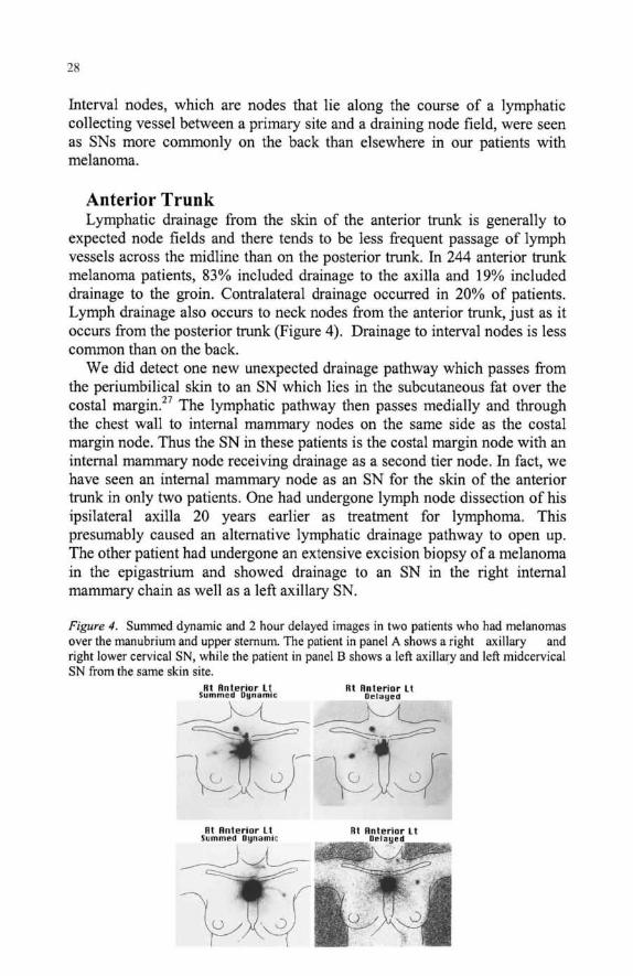
Figure 4. Summed dynamic and 2 hour delayed images in two patients who had melanomas over the manubrium and upper sternum. The patient in panel A shows a right axillary and right lower cervical SN, while the patient in panel B shows a left axillary and left midcervical SN from the same skin site.
Rt anterior Lt Summed Dynamic
Rt Anterior Lt Delayed
Rt Rnterior Lt Summed Dynamic
Rt Rnterior Lt Delayed

29
Head and Neck The head and neck is a challenging area for accurate lymphatic mapping,
both for nuclear medicine physicians and for surgeons. Drainage to multiple SNs is common1'28 and the nodes are often small. The draining SNs often lie very near or sometimes immediately beneath the melanoma site. Detection of such nodes is thus extremely difficult on LS and sometimes impossible. However, if care is taken and such limitations are understood, accurate lymphatic mapping and reliable SNB can be achieved in the head and neck region just as elsewhere in the body.
Lymphatic drainage from the skin of our 578 head and neck melanoma patients is shown in Table 2. As we have found elsewhere, clinical prediction of lymphatic drainage in the head and neck is unreliable and 33% of patients drain to node sites discordant with clinical prediction.28
Table 2. Head and neck melanoma- sentinel node sites («=578)
Location of SN Cervical
Occipital Parotid Postauricular Interval node Contralateral
Level I Level II Level III Level IV Level V (Supraclavicular)
% 20 55 13 9
33 10 8
33 17 5
10
This is often to postauricular nodes from the skin of the face and anterior scalp. Such nodes are not usually excised in elective neck dissections for melanoma. Drainage also occurs across the midline and we have seen this in 10% of patients with head and neck melanomas. Such a contralateral node can in the occasional patient be the only site of micrometastatic disease. Lymph drainage also may occur from the base of the neck upwards to SNs in the upper cervical or occipital area. Again we have seen patients with this pattern in whom the only positive SN was an occipital node even though other SNs were present in the axilla, upper cervical area and lateral neck base. Drainage is also seen regularly from the upper scalp directly down to SNs at the base of the neck or in the supraclavicular region. Lymphatic vessels reaching these SNs are thus completely bypassing all the nodes in the upper and midcervical areas as well as the preauricular (parotid) nodes,

30
occipital and postauricular nodes. This reinforces the important concept that the SN is not simply the closest node to the primary melanoma site.
Upper Limb Lymph drainage from the skin of the upper limb is to the axilla, as
expected, in almost all patients. However, that is often not the complete picture. Drainage to SNs in the epitrochlear region is more common than previously thought and we observed drainage to this site in 20% of patients with melanomas located on the forearm and hand. We also have detected direct drainage to nonaxillary SNs (in the supraclavicular region, interpectoral region, lateral neck base, and TIS) in 6% of our 608 patients with upper limb melanomas.1 These patients also usually had an SN in the axilla and the lymph drainage to these unexpected sites occurred via a separate, discrete lymph vessel. Relying exclusively on gamma probe guided removal of axillary SNs in these patients would very likely have missed these other SNs. Accurate lymphatic mapping with LS is thus imperative.
An "interval" SN is regularly seen lying medially in the arm about half way between the shoulder and elbow. We have seen one patient who had drainage exclusively to this interval node in the mid inner arm, so that it was the only SN.
Lower Limb In our 764 patients with lower limb melanomas, drainage was always to
the ipsilateral groin unless there had been prior surgery to the groin nodes. In this circumstance drainage to the contralateral groin may occur and we have found micrometastases in such contralateral groin SNs.29
Lymph drainage from the foot and leg may also occur to popliteal SNs and we have observed this in 38 of 518(7%) patients with melanomas in these areas (Figure 5). The melanoma sites draining to popliteal SNs are quite variable and it is not just the skin of the lateral heel which drains here, as was previously thought.30 Skin sites on the posterior calf as well as the dorsum and sole of the foot can drain here. The anterior leg less commonly drains to the popliteal fossa.

31
Figure 5. (A) Summed dynamic and 2-hour delayed images in a patient with a melanoma on the right upper shin. Drainage occurs to a single SN in the right popliteal fossa and to several SNs in the right groin. (B) Summed dynamic image anteriorly over the knees with an early image anteriorly over the groin and a 2-hour delayed posterior image over the popliteal area and anterior image over the groin in a patient with a melanoma excision biopsy site on the left upper shin. An interval node is seen in the left leg as well as nodes in the left popliteal fossa and a single SN in the left groin (see upper right early image). Both of these patients show second tier nodes in the groin above the SNs and the patient in panel B has an unusually large amount of tracer in these second tier nodes. This is related to rapid lymph flow.
Bt ( I n t e r i o r Lt Summed Dynamic
Lt P o s t e r i o r Rt Delayed
Rt A n t e r i o r Lt Summed Dynamic
Rt A n t e r i o r Lt I n i t i a l
Interval Nodes Interval nodes can be SNs and we have seen 10 patients in whom they
were the only SNs. When present they must be detected and removed if an SNB procedure is to be accurate. We have shown these interval nodes, when SNs, contain micrometastases with the same incidence as SNs found in standard node fields.31 We found interval nodes in 7% of patients overall and they are more common on the trunk(12% posterior trunk and 8% anterior trunk) than in the head and neck(5%) or upper limb(4%), while they are rare in the lower limb(0.5%). In a large multicenter study McMasters and colleagues32 also found that in melanoma patients interval nodes were positive for metastases with the same frequency as SNs in standard node fields. In their 13 patients with a positive interval node it was the only positive SN in 11 patients (85%).
Although interval nodes may be found at any point along the course of a lymphatic collecting vessel they are more common in certain locations, such as the midaxillary line, the upper back, and the medial aspect of the mid upper arm.

32
Interval nodes remain "hot" on delayed scans as they retain the radiocolloid, though it is noticeable that much of the radiocolloid reaching an interval node passes on almost immediately to second tier nodes. They thus seem to be more "porous" to radiocolloids than SNs in standard node fields or in other unexpected node sites.
Lymphatic Lakes Unlike interval nodes, lymphatic lakes do not need to be examined during
the SNB procedure, because they are simply focal dilatations of lymphatic collecting vessels. They are seen during LS as focal areas of increased tracer retention along the course of lymphatic collecting vessels during the dynamic early postinjection phase of the study.1 The activity rapidly passes onwards in the lymph vessel, however, so that they are not visible on delayed scans performed 2 hours later. These should not be mistaken for interval nodes, which retain tracer and are therefore "hot" on delayed scans.
PATTERNS OF LYMPHATIC DRAINAGE FROM THE BREAST
The patterns of lymphatic drainage from the breast have been well documented in the past33'34 and more recent studies have confirmed these findings.35 Lymphatic drainage from breast tissue passes directly not only to SNs in the axilla but also to SNs in the internal mammary chain, supra- and infraclavicular regions, interpectoral region or to intramammary interval nodes. Flow to posterior intercostal nodes has also been described33 although we have not observed this phenomenon.
In our 640 patients with breast cancer the primary tumors were located as shown in Figure 6. The location of the SNs is shown in Table 3. Most patients (94%) displayed an SN in the axilla and these patients had an average
Table 3. Breast cancer—sentinel node sites
Location Number (%)
Axilla 578 (94%) Internal mammary 249 (40%) Supraclavicular 048 (7.8%) Infraclavicular 007 (1.1%) Interpectoral 008 (1.2%) Interval node 053 (8.6%) SN outside axilla 286 (46 %) Exclusive nonaxillary SN 030 ( 5 %) No drainage 022 (3.4%)
33
of 1.45 SNs in the axilla. It is noteworthy that 46% of patients showed drainage to an SN outside the axilla (Figure 7). The demonstration of such lymph flow to SNs outside the axilla is one of the important contributions LS makes to patients with breast cancer. The commonest nonaxillary site for SNs is the internal mammary chain where we have found SNs in 40% of patients overall. This figure is higher than that generally reported for internal mammary node drainage but is almost identical to the 38% of internal mammary drainage found by Shimazu and colleagues (36) when they used subtumoral injections of radiocolloid.
Figure 6. The location of primary breast cancers.
m E
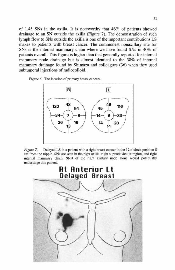
Figure 7. Delayed LS in a patient with a right breast cancer in the 12 o'clock position 8 cm from the nipple. SNs are seen in the right axilla, right supraclavicular region, and right internal mammary chain. SNB of the right axillary node alone would potentially understage this patient.

34
We also believe the depth of injection is critically important in obtaining a true map of lymph drainage from the tumor site. Our injections are given under ultrasound control with the needle tip at the depth of the center of the lump so that the tracer is deposited at this depth or slightly deeper. Injections given more superficially will not demonstrate drainage to internal mammary SNs because the initial lymphatic capillaries that drain to internal mammary nodes lie in the posterior part of the mammary gland. Clearly, if the SNB method is to be accurate, all true SNs in every patient must be examined. An SNB procedure that targets only axillary SNs, based on our data, potentially understages 46% of breast cancer patients. The status of the internal mammary nodes in breast cancer is the second most important prognostic indicator and a positive internal mammary SN has an adverse effect on prognosis regardless of axillary node status.37 Excision biopsy of SNs found in the internal mammary chain is associated with low morbidity and has been shown to improve staging and change treatment strategies.38'39
Like others, we have found that the pattern of lymphatic drainage in a particular patient cannot be predicted clinically on the basis of the location of the tumor in the breast tissue. Tumors that were located entirely in outer quadrants of the breast showed drainage to internal mammary SNs in 29% of cases, while tumors located entirely in inner quadrants drained to SNs in the axilla in 86%. Thus, lymph flowed across the centerline of the breast in 46% of patients. We have observed flow across the midline of the patient to contralateral nodes that were second tier nodes in the internal mammary chain and supraclavicular region but we have not yet seen drainage to a contralateral second tier node in the opposite axilla. We have also not yet seen drainage to a sentinel node on the side contralateral to the primary breast cancer. Table 4 shows the drainage patterns seen in 419 patients based on the location of the primary tumor according to the four breast quadrants.
As mentioned earlier, when internal mammary drainage was observed there was usually a string of second tier nodes seen above the SN, reinforcing our view that "visceral" lymph nodes inside body cavities have a different physiology and do not retain radiocolloid as effectively as do nodes in the standard node fields which drain the skin.

35
Table 4. Lymph flow patterns by breast quadrant (419 patients)
SN location
Axilla Internal mammary Supraclavicular Infraclavicular Interpectoral Interval node SN outside axilla No drainage
UOQ («=236)
227 (98%) 054 (23%) 022 ( 9%) 003 ( 1%) 002 ( 1%) 013 ( 6%) 072(31%) 005 ( 2%)
Site of breast cancer
UIQ (n=99)
79 (86%) 60 (65%) 07 ( 8%) 01 ( 1%) 02 ( 2%) 06 ( 6%) 60 (65%) 07 ( 7%)
LOQ r«=54)
54 (100%) 28 ( 52%) 01 ( 2%) 00 ( 0%) 00 ( 0%) 05 ( 9%) 30 ( 56%) 00 ( 0%)
LIQ C«=30)
25 (89%) 14 (50%) 02 ( 7%) 00 ( 0%) 01 ( 3%) 01 ( 3%) 16(57%) 02 ( 7%)
SN,sentinel node; U,upper; L,lower; 0,outer; I,inner.
CONCLUSIONS
Lymphatic drainage of the skin is highly variable from patient to patient, even when the same region of the body is being examined. The path taken by lymphatic collecting vessels is unpredictable, as is the ultimate location of the draining SN or SNs as several recent studies have confirmed.40"42
Lymphatic drainage of the breast is also not clinically predictable and passes to SNs outside the axilla in a significant number of patients.
Preoperative LS with small particle radiocolloids allows lymphatic vessels to be visualized as they drain directly to SNs. Careful imaging technique will thus allow all true SNs to be identified in each patient, even if these SNs lie outside standard node fields or are interval SNs lying between the primary site and a node field. By providing more accurate nodal staging this is an important contribution to the management of patients with cancer.
We now know that clinical prediction of the pattern of lymph drainage in an individual patient is unreliable and inaccurate. We also know that we now have an accurate method of mapping lymph drainage in every patient, which can make the difficulties associated with clinical prediction irrelevant. This technique which provides an accurate map of lymph drainage in each patient

36
can thus have a direct and important impact on the clinical management of patients with solid tumors that drain to lymph nodes. The technique has proven accurate in melanoma and breast cancer and is now being applied to an increasing number of other solid tumors.
ACKNOWLEDGMENTS
The authors would like to thank all the nuclear medicine technologists whose skill and dedication were so important to the success of our lymphatic mapping studies. These include Kim Ioannou, Nicholas Trpezanovski, Angelique Ngyuen, Tracey Smith, Sally Raymond, and Ian Dyer. We thank the other nuclear medicine physicians who have added their enthusiasm to our work, including Drs. Robert Mansberg, John Roberts, Reginald Hutcherson, and Elizabeth Bernard. We also thank the surgeons of the Sydney Melanoma Unit for referring their patients and providing the surgical correlation and validation of the lymphatic drainage patterns we have documented, including Professor William McCarthy, Dr. Michael Quinn, Dr. Kerwin Shannon, Associate Professor Christopher O'Brien, Dr. Andrew Spillane, Dr. Robyn Saw and Dr. Jonathan Stretch and also the breast surgeons who have collaborated with us including Professor David Gillett, Associate Professor Stuart Renwick, Dr. Andrew Spillane, Dr. Owen Ung, Dr. Hugh Carmalt, Dr. Fred Niesche, Associate Professor Richard West and Dr. Robert Claxton.
REFERENCES
1. Uren RF, Thompson JF, Howman-Giles RB: Lymphatic drainage of the skin and breast: Locating the sentinel nodes. Amsterdam, Harwood Academic Publishers, 1999.
2. Thompson JF, Uren RF, Shaw HM, et al: The location of sentinel lymph nodes in patients with cutaneous melanoma. New insights into lymphatic anatomy. J Am Coll Surg 1999; 189: 195-206.
3. McMasters KM, Chao C, Wong SL, et al: Interval sentinel lymph nodes in melanoma. Arch Surg 2002; 137:543-549.
4. O'Toole GA, Hettiaratchy S, Allan R, et al: Aberrant sentinel nodes in malignant melanoma. Br J Plast Surg 2000; 53: 415-417.
5. Statius Muller MG, Hennipman FA, van Leeuwen PAM, et al: Unpredictability of lymphatic drainage patterns in melanoma patients. Eur J Nucl Med 2002; 29: 255-261.
6. Sjoberg T, Steen S: Contractile properties of lymphatics from the human lower leg. Lymphology 1991; 24: 16-21.
7. Uren RF, Howman-Giles RB, Thompson JF: Variation in cutaneous lymphatic flow rates. Ann Surg Oncol 1997; 4: 279-280.

37
8. Uren RF, Howman-Giles RB, Thompson JF: Demonstration of second tier lymph nodes during preoperative lymphoscintigraphy for melanoma: Incidence varies with primary tumour site. Ann Surg Oncol 1998; 5: 517-521.
9. Nopajaroonsri C, Simon GT: Phagocytosis of colloidal carbon in a lymph node. Am J Pathol 1971; 65:25-42.
10. Leak LV: Lymphatic vessels, in Cardiovascular system, lymphoreticular and hematopoietic system. Johannessen, JV (ed). New York, McGraw-Hill, ppl59-183,1980.
11. Kapteijn BAE, Nieweg OE, Muller SH, et al: Validation of gamma probe detection of the sentinel node in melanoma. J Nucl Med 1997; 38: 362-366.
12. Alazraki NP, Eshima D, Eshima LA, et al: Lymphoscintigraphy, the sentinel node concept, and the intraoperative gamma probe in melanoma, breast cancer, and other potential cancers. Semin Nucl Med 1997; 27: 55-67.
13. Bergqvist L, Strand S-E, Persson BRR: Particle sizing and biokinetics of interstitial lymphoscintigraphic agents. Semin Nucl Med 1983; 8:9-19.
14. Maza S, Valencia R, Geworski L, Zander A, Guski H, Winzer KJ, Munz DL. Peritumoural versus subareolar administration of technetium-99m nanocolloid for sentinel lymph node detection in breast cancer: preliminary results of a prospective intra-individual comparative study. Eur J Nucl Med Mol Imaging 2003; 30:651-656.
15. Kitagawa Y, Kitajima M. Gastrointestinal cancer and sentinel node navigation surgery. J Surg Oncol 2002; 79: 188-193.
16. Ege GN. Internal mammary lymphoscintigraphy in breast carcinoma: A study of 1072 patients. Int J Radiat Oncol Biol Physl977; 2:755-761.
17. Kaplan WD, Andersen JW, Siddon RL, et al. The three dimensional localization of internal mammary lymph nodes by radionuclide lymphoscintigraphy. J Nucl Med 1988; 29:473-478.
18. Strand SE, Persson BRR: Quantitative lymphoscintigraphy I: basic concepts for optimal uptake of radiocolloids in the parasternal lymph nodes of rabbits. J Nucl Med 1979; 20: 1038-1046.
19. Uren RF, Howman-Giles RB, Shaw HM, et al: Lymphoscintigraphy in high risk melanoma of the trunk: predicting draining node groups, defining lymphatic channels and locating the sentinel node. J Nucl Med 1993; 34: 1435-1440.
20. Morton DL, Wen D-R, Wong JH, et al: Technical details of intraoperative lymphatic mapping for early stage melanoma. Arch Surg 1992; 127: 392-399.
21. Norman J, Cruse W, Espinosa C, et al: Redefinition of cutaneous lymphatic drainage with the use of lymphoscintigraphy for malignant melanoma. Am J Surg 1991;162:432-437.
22. Eberbach MA, Wahl RL: Lymphatic anatomy: functional nodal basins. Ann Plast Surg 1989; 22: 25-31.
23. Leong SP, Achtem TA, Habib FA, et al: Discordancy between clinical predictions versus lymphoscintigraphic and intraoperative mapping of sentinel lymph node drainage of primary melanoma. Arch Dermatol 1999; 135: 1472-1476.
24. Uren RF, Howman-Giles RB, Thompson JF, et al: Lymphatic drainage to triangular intermuscular space lymph nodes in melanoma on the back. J Nucl Med 1996; 37: 964-966.
25. Uren RF, Howman-Giles RB, Thompson JF: Lymphatic drainage from the skin of the back to intra-abdominal lymph nodes in melanoma patients. Ann Surg Oncol 1998;5:384-387.

38
26. Uren RF, Howman-Giles RB, Thompson JF, et al: Exclusive lymphatic drainage from a melanoma on the back to intraabdominal lymph nodes. Clin Nucl Med 1998; 23: 71-73.
27. Uren RF, Howman-Giles RB, Thompson JF, et al: Lymphatic drainage from periumbilical skin to internal mammary nodes. Clin Nucl Med 1995; 20: 254-255.
28. O'Brien CJ, Uren RF, Thompson JF, et al: Prediction of potential metastatic sites in cutaneous head and neck melanoma using lymphoscintigraphy. Am J Surg 1995; 170:461-466.
29. Thompson JF, Saw RP, Colman MH, et al: Contralateral groin node metastasis from lower limb melanoma. Eur J Cancer 1997; 33: 976-977.
30. Clouse ME, Wallace S, eds. Lymphatic Imaging: Lymphography, computed tomography and scintigraphy. 2nd ed. Williams & Wilkins: Baltimore. 15-21, 1985.
31. Uren RF, Howman-Giles R, Thompson JF, et al: Interval nodes. The forgotten sentinel nodes in patients with melanoma. Arch Surg 2000; 135:1168-1172.
32. McMasters KM, Chao C, Wong SL, et al: Interval sentinel lymph nodes in melanoma. Arch Surg 2002; 137:543-549.
33. Turner-Warwick RT: The lymphatics of the breast. Br J Surg 1959; 46:574-582 . 34. Vendrell-Torne E, Setain-Quinquer J, Domenech-Torne FM: Study of normal
mammary lymphatic drainage using radioactive isotopes. J Nucl Med 1972; 13:801-805.
35. Uren RF, Howman-Giles RB, Thompson JF et al: Mammary lymphoscintigraphy in breast cancer. J Nucl Med 1995; 36: 1775-1780.
36. Shimazu K, Tamaki Y, Taguchi T, et al. Lymphoscintigraphy visualization of internal mammary nodes with subtumoral injection of radiocolloid in patients with breast cancer. Ann Surg 2003; 237: 390-398.
37. Veronesi U, Marubini E, Mariani L, Valagussa P, Zucali R: The dissection of internal mammary nodes does not improve the survival of breast cancer patients. 30-year results of a randomized trial. Eur J Cancer 1999; 35:1320-1325.
38. Tanis PJ, Nieweg OE, Valdes Olmos RA, Peterse JL, Rutgers EJ, Hoefnagel CA, Kroon BB. Impact of non-axillary sentinel node biopsy on staging and treatment of breast cancer patients. Br J Cancer 2002; 87: 705-710.
39. Noguchi M. Relevance and practicability of internal mammary sentinel node biopsy for breast cancer. Breast Cancer 2002; 9: 329-336.
40. Thompson JF, Uren RF, Shaw HM, et al. The location of sentinel lymph nodes in patients with cutaneous melanoma. New insights into lymphatic anatomy. J Am Coll Surg 1999; 189: 195-206.
41. O'Toole GA, Hettiaratchy S, Allan R, Powell BWEM. Aberrant sentinel nodes in malignant melanoma. Br J Plast Surg 2000; 53: 415-417.
42. Statius Muller MG, Hennipman FA, van Leeuwen PAM, Pijpers R,Vuylsteke RJCLM, Meijer S.Unpredictability of lymphatic drainage patterns in melanoma patients, Eur J Nucl Med . 2002; 29: 255-261.