Role of interventional radiology in the management of ...PICTORIAL REVIEW Role of interventional...
9
PICTORIAL REVIEW Role of interventional radiology in the management of complications after pancreatic surgery: a pictorial review Giovanni Mauri & Chiara Mattiuz & Luca Maria Sconfienza & Vittorio Pedicini & Dario Poretti & Fabio Melchiorre & Umberto Rossi & Fabio Romano Lutman & Marco Montorsi Received: 18 September 2014 /Revised: 1 November 2014 /Accepted: 18 November 2014 /Published online: 17 December 2014 # The Author(s) 2014. This article is published with open access at Springerlink.com Abstract Pancreatic resections are surgical procedures asso- ciated with high incidence of complications, with relevant morbidity and mortality even at high volume centres. A mul- tidisciplinary approach is essential in the management of these events and interventional radiology plays a crucial role in the treatment of patients developing post-surgical complications. This paper offers an overview on the interventional radiolog- ical procedures that can be performed to treat different type of complications after pancreatic resection. Procedures such as percutaneous drainage of fluid collections, percutaneous transhepatic biliary procedures, arterial embolisation, venous interventions and fistula embolisation are viable treatment options, with fewer complications compared with re-look surgery, shorter hospital stay and faster recovery. A selection G. Mauri : L. M. Sconfienza Servizio di Radiologia, IRCCS Policlinico San Donato, San Donato Milanese, Milano, Italy L. M. Sconfienza e-mail: [email protected] G. Mauri Dipartimento Aziendale di Oncologia, Struttura Complessa di Radiologia Oncologica Interventistica, Azienda Ospedaliera Ospedale di Circolo di Busto Arsizio, Varese, Italy C. Mattiuz Facoltà di Medicina e Chirurgia, Scuola di Specializzazione in Radiodiagnostica, Università degli Studi di Milano, Milano, Italy e-mail: [email protected] L. M. Sconfienza Dipartimento di Scienze Biomediche per la Salute, Università degli Studi di Milano, San Donato Milanese, Milano, Italy V. Pedicini : D. Poretti : F. R. Lutman Dipartimento di Diagnostica per Immagini, IRCCS Istituto Clinico Humanitas, Rozzano, Italy V. Pedicini e-mail: [email protected] D. Poretti e-mail: [email protected] F. R. Lutman e-mail: [email protected] F. Melchiorre Dipartimento di Diagnostica per Immagini, Azienda Ospedaliera San Paolo, Milano, Italy e-mail: [email protected] U. Rossi Dipartimento di Scienze Diagnostiche, Azienda Ospedaliera San Carlo Borromeo, Milano, Italy e-mail: [email protected] M. Montorsi Dipartimento di Chirurgia, Azienda Ospedaliera Humanitas (Humanitas Research Hospital), Rozzano, Italy e-mail: [email protected] G. Mauri (*) Servizio di Radiologia, IRCCS Policlinico San Donato, Piazza Malan 2, 20097 San Donato Milanese, Milano, Italy e-mail: [email protected] Insights Imaging (2015) 6:231–239 DOI 10.1007/s13244-014-0372-y
Transcript of Role of interventional radiology in the management of ...PICTORIAL REVIEW Role of interventional...

PICTORIAL REVIEW
Role of interventional radiology in the managementof complications after pancreatic surgery: a pictorial review
Giovanni Mauri & Chiara Mattiuz & Luca Maria Sconfienza & Vittorio Pedicini &Dario Poretti & Fabio Melchiorre & Umberto Rossi & Fabio Romano Lutman &
Marco Montorsi
Received: 18 September 2014 /Revised: 1 November 2014 /Accepted: 18 November 2014 /Published online: 17 December 2014# The Author(s) 2014. This article is published with open access at Springerlink.com
Abstract Pancreatic resections are surgical procedures asso-ciated with high incidence of complications, with relevantmorbidity and mortality even at high volume centres. A mul-tidisciplinary approach is essential in the management of theseevents and interventional radiology plays a crucial role in thetreatment of patients developing post-surgical complications.This paper offers an overview on the interventional radiolog-
ical procedures that can be performed to treat different type ofcomplications after pancreatic resection. Procedures such aspercutaneous drainage of fluid collections, percutaneoustranshepatic biliary procedures, arterial embolisation, venousinterventions and fistula embolisation are viable treatmentoptions, with fewer complications compared with re-looksurgery, shorter hospital stay and faster recovery. A selection
G. Mauri : L. M. SconfienzaServizio di Radiologia, IRCCS Policlinico San Donato,San Donato Milanese, Milano, Italy
L. M. Sconfienzae-mail: [email protected]
G. MauriDipartimento Aziendale di Oncologia, Struttura Complessa diRadiologia Oncologica Interventistica, Azienda OspedalieraOspedale di Circolo di Busto Arsizio, Varese, Italy
C. MattiuzFacoltà di Medicina e Chirurgia, Scuola di Specializzazione inRadiodiagnostica, Università degli Studi di Milano, Milano, Italye-mail: [email protected]
L. M. SconfienzaDipartimento di Scienze Biomediche per la Salute, Università degliStudi di Milano, San Donato Milanese, Milano, Italy
V. Pedicini :D. Poretti : F. R. LutmanDipartimento di Diagnostica per Immagini,IRCCS Istituto Clinico Humanitas, Rozzano, Italy
V. Pedicinie-mail: [email protected]
D. Porettie-mail: [email protected]
F. R. Lutmane-mail: [email protected]
F. MelchiorreDipartimento di Diagnostica per Immagini,Azienda Ospedaliera San Paolo, Milano, Italye-mail: [email protected]
U. RossiDipartimento di Scienze Diagnostiche,Azienda Ospedaliera San Carlo Borromeo, Milano, Italye-mail: [email protected]
M. MontorsiDipartimento di Chirurgia, Azienda Ospedaliera Humanitas(Humanitas Research Hospital), Rozzano, Italye-mail: [email protected]
G. Mauri (*)Servizio di Radiologia, IRCCS Policlinico San Donato,Piazza Malan 2, 20097 San Donato Milanese, Milano, Italye-mail: [email protected]
Insights Imaging (2015) 6:231–239DOI 10.1007/s13244-014-0372-y

of cases of complications following pancreatic surgery man-aged with interventional radiological procedure are presentedand discussed.Teaching Points• Interventional radiology is crucial to treat complicationsafter pancreatic surgery
• Percutaneous drainage of collections can be performedunder ultrasound or computed tomography guidance
• Percutaneous biliary procedures can be used to treat biliarycomplications
• Venous procedures can be performed effectively throughtranshepatic or transjugular access
• Fistulas can be treated effectively by percutaneousembolisation
Keywords Interventional radiology .
Duodenopancreasectomy . Complication . Embolisation .
Biliary drainage
Introduction
Pancreatic resection is a surgical procedure associated withsignificant morbidity and mortality even at specialised high-volume centres [1–3]. Although in recent years refinements insurgical technique and perioperative management have led toa reduction in perioperative mortality, the incidence of post-operative complications (including intra-abdominal abscessesand leakages [4–9], biliary complications [10–14] andvascular complications [15–18]) still remains high.Moreover, in case of complications, surgical re-operationis associated with a high mortality rate [1–3]. In a large seriesof 650 patients, Yeo et al. [2] reported an overallmortality of 1.4 % and a morbidity of 41 %, with a meanlength of hospital stay of 13 days. They reported re-operation in 26/650 patients (4 %) and identified the absenceof reoperation as an independent predictor of prolonged sur-vival [2].
Interventional radiology (IR) provides a minimally inva-sive alternative for managing post-surgical complications[19, 20]. Several different IR procedures, such as percuta-neous drainage, aspiration of abscesses or fluid collections[4, 5], percutaneous transhepatic biliary drainage [6], andarterial embolisation [21–23] have been introduced in clin-ical practice to treat post-surgical complications. IR proce-dures are an alternative approach to manage post-surgicalcomplications less invasive than surgical re-intervention, andmay lead to a reduction in hospital stay and re-operation rate[10, 11].
In the present pictorial review, we offer an overview on theIR procedures that can be performed to treat different types ofcomplications after pancreatic resection.
Percutaneous drainage
Intra-abdominal collections and abscesses represent the mostcommon complication following pancreatic surgery [1–3].Once an intra-abdominal collection is identified, in most casesit is possible to place a percutaneous drainage under imageguidance. When the collection is well visible using ultrasound(US), US-guided percutaneous drainage placement is gener-ally the preferred choice, as US is widely available, easy tohandle and allows for real-time monitoring of the drainageplacement, being also free from ionising radiation [24]. Whenthe collection is located deep in the abdomen and is not wellseen at US, computed tomography (CT) generally offers goodanatomical definition to guide the safe placement of a percu-taneous drainage. Drainage placement can be performed usingthe trocar or Seldinger technique. In the trocar technique, thedrainage catheter containing a trocar needle is inserted directlyinto the collection. The trocar technique provides a fast de-ployment of the drainage that can be extremely helpful incritically ill or agitated patients. The Seldinger techniqueimplies multiple steps: the collection is punctured with a smallcalibre needle, different calibre guidewires are inserted and thedrainage is then advanced up to the collection over theguidewire. The Seldinger technique is particularly helpfulwhen there is a small window to reach the collection, astypically happens when a retroperitoneal collection has to bedrained through an anterior approach. The main disadvantageof this technique is that it may require considerably longertime than the trocar technique. The correct location of theneedle or drainage can be confirmed by aspiration of a smallamount of material. Furthermore, a small amount of iodinatedcontrast may be injected through the catheter to identify thepresence of an underlying fistula.
In a series of 373 subjects who underwent pancreatico-duodenectomy reported by Zink et al. [5], percutaneous drainagewas required in 22.2 % of patients after surgery. They report animmediate technical and overall success rate of 97.6 and 79.6%,respectively.
A case of a patient with fluid collection after pancreatico-duodenectomy successfully managed with percutaneous CTguided drainage is shown in Fig. 1.
Percutaneous transhepatic biliary procedures
In patients treated by pancreaticoduodenectomy with Roux-en-Y bilio-enteric reconstruction, endoscopical access to thebiliary tree is not feasible. Thus, in this kind of patient, apercutaneous transhepatic route is the only approach to thebiliary system in case of biliary complications. The access tothe biliary system is obtained by puncturing a peripheral bileduct under US and/or fluoroscopic guidance.
232 Insights Imaging (2015) 6:231–239

Percutaneous biliary procedures can be particularly helpfulin postoperative biliary leaks. They have been reported inabout 3–4 % of cases after pancreatic surgery [1–3]. Thesecomplications are often associated with others, in particularwith pancreatic fistula and fluid collections. In patients whodevelop such complication after surgery, percutaneoustranshepatic biliary drainage—by allowing for a bile diversionfrom the site of the fistula—has been proven to be feasible andeffective in the majority of cases [6, 20, 21]. The positioningof an occlusion balloon to obstruct the biliary duct above the
site of the fistula, by allowing for complete external drainageof the bile, may represent a valuable option to treat post-surgical biliary leaks [7, 10]. A different and more recenttechnique is to use a covered stent that can close the bile leakand subsequently be retrieved percutaneously. Gwon et al.[25] used this approach successfully in 11 patients with post-operative bile leak, with no recurrence in the 1-year follow-up.A case of a patient with post-surgical biliary leak successfullytreated with percutaneous transhepatic biliary drainage isshown in Fig. 2.
Fig. 1 Use of a percutaneousdrainage to treat a post-pancreaticoduodenectomy fluidcollection. a CT scan shows post-pancreaticoduodenectomyretroperitoneal fluid collection(asterisks). b The fluid collection(asterisks) is punctured with asmall needle under CT guidance.c A percutaneous drainage(arrow) is inserted into thecollection (asterisks) using theSeldinger technique. d At the endof the treatment completeresolution of the collection isobtained
Fig. 2 Use of a percutaneous transhepatic biliary drainage to treat a post-surgical biliary leak. a Percutaneous colangiography demonstrates a biliaryleak (arrow) and a biliary drainage (white arrows) is inserted via segment
III left lobe approach; RBT right biliary tree, LBT left biliary tree, SB smallbowel. b Final result with complete healing of the leak and absence ofcontrast leakage; RBT right biliary tree, LBT left biliary tree, SB bowel
Insights Imaging (2015) 6:231–239 233

After bilio-pancreatic surgery, stricture of a biliary ductmay represent a serious complication, [26]. Surgical re-intervention is associated with morbidity and mortality ratesas high as 28 and 2.6 % respectively [26]. Percutaneoustreatments represent an effective alternative to surgery in thetreatment of such complication [27–29]. To achieve resolutionof the stenosis, subsequent larger biliary drainage cathetermay be inserted and left in place and balloon dilation can beperformed. However, recurrence of stenosis may occur in upto 29–58 % of cases [27–29], and multiple treatment sessionsmay be required. Stents are rarely used in the treatment ofbenign strictures, as they have to be removed after a periodbecause the tube itself may stimulate inflammatory reaction,fibrosis and stone formation. A novel option may be repre-sented by the use of biodegradable biliary stents, which may
improve the long-term patency rate, without requiring a sub-sequent procedure for removal [13].
A case of a patient with a post-surgical biliary stricturesuccessfully treated with balloon dilation and placement of abiodegradable biliary stent is shown in Fig. 3.
Arterial embolisation
Postoperative intra-abdominal arterial haemorrhage is still oneof the most serious complications, with a reported incidencebetween 1.5 and 15 %, and a reported mortality rate of 20–50 % [15–17]. Haemorrhage is defined as ‘early’ when occur-ring within 24 h after surgery, while ‘late’ haemorrhages occurafter 24 h [30]. Early haemorrhage requires immediate
Fig. 3 Percutaneous treatment ofa benign stricture of the commonbile duct by a bioabsorbablebiliary stent. a Percutaneoustranshepatic cholangiographydemonstrates remarkably dilatedbile ducts (asterisks). Theobstruction can be clearly spotted(white arrows). b Bilioplastyprocedure. The balloon (blackarrows) is fully inflated to dilatethe stricture. Radiopaque contrastagent can be detected in the bowel(B); asterisks dilated bile ducts. cA bioabsorbable stent ispositioned to keep the strictureopen. The stent is radiolucent, buttwo platinum, radiopaquemarkers can be seen(black arrowheads); asterisksdilated bile ducts, B bowel. d Thestent is fully expanded andradiopaque bile can be seenflowing through the patentcommon bile duct(white arrowheads); black arrowsradiopaque stent markers; Bbowel. e US follow-up at3 months demonstrating goodvisibility of the stent(arrowheads) that was correctlyexpanded, and no dilationof the intrahepatic biliary ducts;L liver
234 Insights Imaging (2015) 6:231–239

laparotomy, as it is generally caused by a technical failure orunderlying coagulopathy, while the most appropriate manage-ment of delayed haemorrhage still remains controversial. Inhaemodynamically stable patients, CT is crucial to address thesuspicion of a delayed haemorrhage and is extremely usefulprior to angiography as it may avoid it or guide it. Inunstable patients, direct visceral angiography has beensuggested as the best method to elucidate the site of bleeding,with the advantage of sparing time for a subsequent immediateintra-arterial treatment.
Transcatheter arterial embolisation has been reported to besafe and effective, with a reported success rate of 50–100 %,gaining acceptance for the treatment of intra-abdominal bleed-ing [15–17]. Once the site of bleeding has been identified,several different techniques may be used to stop bleeding andachieve haemostasis. In case of terminal vascularisation, aproximal embolisation of the bleeding vessel may be enoughto achieve the haemostasis, while in the presence of collat-erals, it is crucial to embolise both the inflow and outflowvessels in order to avoid re-bleeding (“isolation technique”)[22]. Different materials are nowadays available to obtainvascular occlusion, and have to be chosen according to the
desired type of vascular occlusion (transient or permanent).Transient embolisation, generally required due to a traumatichaemorrhage, is reached by resorbable materials, which al-lows for restoring the blood flow in a variable window of time(inter alia: autologous blood clot, gelatine or fibrin sponge).The main advantage of such materials is to avoid definitiveocclusion of the treated vessel. However, a non-negligible riskof re-bleeding has to be considered once the material has beenabsorbed. Permanent embolisation is reached using non-resorbable materials (polyvynilic alcohol, bucrilate, metalliccoils or detachable balloons) that induce a permanent vesselocclusion. At the present, an ideal material does not exist, thusthe choice of the most appropriate embolic material and tech-nique for embolisation, crucial to minimise failure and com-plications, requires the presence of a IR team with highexperience and the availability of several different materials.
Stent grafting of the artery at the site of bleeding has beenproposed as an alternative or in addition to embolisation, withthe advantage of maintaining the patency of the end organ,thus minimising the risk of ischaemia deriving from emboli-sation [21]. A recent meta-analyisis comparing laparotomyand transarterial embolisation for the management of delayed
Fig. 4 Arterial embolisation of apost-surgical haemorrhage.a CT scan showing contrastextravasation (arrow) from theceliac trunk at the level of thebifurcation between hepatic andsplenic artery; V inferior venacava, A abdominal aorta.bAngiographic appearance of theactive bleeding with contrastextravasation (arrows); asteriskceliac trunk, black arrowheadsplenic artery, white arrowheadhepatic artery. c Angiographyafter embolisation with coils(arrows) demonstrating absenceof contrast extravasation at thelevel of previous bleeding.d CT scan at 2 years,demonstrating absence of contrastextravasation at the level ofprevious bleeding and preservedpatency of proper hepatic artery(white arrow), even in thepresence of part of a coil in itslumen (arrowhead); black arrowclustered metallic coils in thefocus of arterial lesion properlyembolised
Insights Imaging (2015) 6:231–239 235
postoperative haemorrhage found a reduction in mortality(43 % vs 20 %) and morbidity (77 % vs 35 %) in the IRgroup, even if not reaching the statistical significance. Authors
conclude that the appropriate treatment pathway for latehaemorrhage ultimately will be decided by the clinical statusof the patient and the institution preference [16].
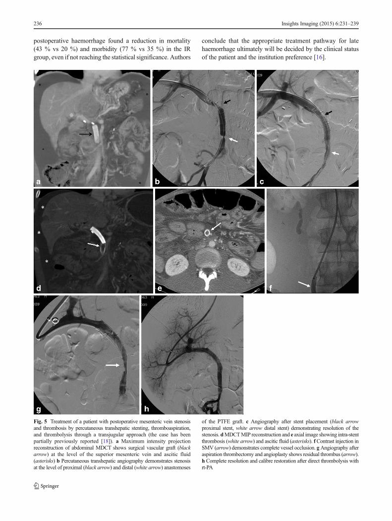
Fig. 5 Treatment of a patient with postoperative mesenteric vein stenosisand thrombosis by percutaneous transhepatic stenting, thromboaspiration,and thrombolysis through a transjugular approach (the case has beenpartially previously reported [18]). a Maximum intensity projectionreconstruction of abdominal MDCT shows surgical vascular graft (blackarrow) at the level of the superior mesenteric vein and ascitic fluid(asterisks) b Percutaneous transhepatic angiography demonstrates stenosisat the level of proximal (black arrow) and distal (white arrow) anastomoses
of the PTFE graft. c Angiography after stent placement (black arrowproximal stent, white arrow distal stent) demonstrating resolution of thestenosis. dMDCTMIP reconstruction and e axial image showing intra-stentthrombosis (white arrow) and ascitic fluid (asterisks). f Contrast injection inSMV (arrow) demonstrates complete vessel occlusion. gAngiography afteraspiration thrombectomy and angioplasty shows residual thrombus (arrow).h Complete resolution and calibre restoration after direct thrombolysis withrt-PA
236 Insights Imaging (2015) 6:231–239

A case of transcatheter arterial embolisation for a post-surgical haemorrhage is shown in Fig. 4.
Venous interventions
In some cases, post-surgical complications may involve theportal and mesenteric veins, which may develop stenosis orthrombosis after surgical treatment. Moreover, with the recentadvancement of vascular reconstruction with polytetrafluo-roethylene graft, this occurrence may happen more frequentlyin the future. Percutaneous endovascular treatment, such astransjugular portosystemic shunt, direct and indirect thrombol-ysis, stenting and mechanical thrombectomy, described mainlyin liver-transplant patients, may represent a valuable option inthe management of these conditions after pancreatic surgery [2,5, 18]. The access to the portal systemmay be achieved througha transhepatic route, with the direct image guided puncture of aperipheral portal branch, or through a transjugular approach, bypuncturing the portal system from the hepatic veins.Transhepatic access is generally easier than transjugular accessand represents the first choice strategy in patients with normalcoagulation parameters. In patients with thrombosis of the portal
or mesenteric vein, in which anticoagulation treatment has beenundertaken, the best choice is represented by the transjugularapproach, in order to minimise the risk of peritoneal haemor-rhage correlated with direct liver puncture [18]. Once the accessto the portal system has been achieved, it is possible to performballoon dilation and stenting of post-surgical strictures using theseveral different commercially available devices. In case ofthrombosis, direct trombo-aspiration through a catheter or infu-sion of trombolitic agents has been reported as a feasible andeffective treatment [18].
A case of post-surgical mesenteric vein stenosis and throm-bosis treated percutaneously is shown in Fig. 5.
Fistula embolisation
Fistulas are the most common complication following pancre-atic surgery after presence of fluid collections [1–3]. The mostfrequent type is pancreatic fistula, followed by enteric andbiliary fistulas [1–3]. This kind of complication is frequentlyassociated with others, in particular the presence of an intra-abdominal fluid collection. Recent reports highlighted howthis kind of complication can be successfully managed
Fig. 6 Successful percutaneousembolisation with cyanoacrilicglue of a postoperative fistula. a,b Contrast injection through apreviously placed percutaneousdrainage (white arrowhead) at thelevel of a fluid collection(asterisk) demonstrating a fistula(black arrow) with acommunication (small blackarrowhead) with the biliarysystem and bowel (B); large blackarrowhead percutaneoustranshepatic biliary drainage. c Aguidewire (small whitearrowhead) is advanced throughthe percutaneous drainage (largewhite arrowhead) into the fistula(black arrow); asterisk fluidcollection, black arrowheadpercutaneous transhepatic biliarydrainage, B bowel. d Amicrocatheter (white arrowhead)is advanced over the wire at thelevel of the fistula (black arrow),and glue is injected (white arrow);asterisk fluid collection, blackarrowhead percutaneoustranshepatic biliary drainage, Bbowel
Insights Imaging (2015) 6:231–239 237

without surgery in over 90 % of patients [9]. In the non-operative management of such complications, IR plays acrucial role.
In presence of a fluid collection, a percutaneously placeddrainage may be enough to treat the collection, and in severalcases to allow for the spontaneous closure of the fistula [4, 5]. Ifthe main component of a fistula is biliary material, thetranshepatic insertion of a biliary drainage, by diverting the bilefrom the site of the fistula, may be enough to determine thefistula closure. In cases with persisting biliary leak, the place-ment of an occlusion balloon above the fistula, by interruptingcompletely the bile flow towards the site of the fistula, has beenreported as an effective tool [7–10].
Some authors reported the direct embolisation of the siteof the fistula as a feasible and effective procedure.Reaching perfectly the site of the fistula is crucial toperform a correct procedure. This can be done through apreviously placed surgical drainage, through an image-guided percutaneously placed drainage, or even through atranshepatic approach. Once the site of the fistula has beenreached, several different materials can be used to performembolisation, including ethanol, particles or different kindof glues [31–33]. Inter alia, cyanoacrilic glues seem torepresent optimal materials for fistula percutaneous treat-ment, due to their high adhesive and haemostatic propertiesand fast polymerisation [31].
A case of embolisation of a fistula with cyanoacrilic glue isshown in Fig. 6.
Conclusions
IR plays an increasing, crucial role in the multidisciplinarymanagement of complications after pancreatic surgery, pro-viding a minimally invasive therapy also in critical patients,reducing recovery times and avoiding re-operation morbidity.IR procedures such as percutaneous drainage of fluid collec-tions, percutaneous transhepatic biliary procedures, arterialembolisation, venous interventions and fistula embolisationare viable treatment options and have been reported as feasi-ble, safe and effective techniques with fewer complicationscompared with re-look surgery, with a shorter hospital stayand faster recovery in the management of complications afterpancreatic surgery.
Conflict of interest Giovanni Mauri declares sponsored research andconsulting from Esaote S.p.A, Italy. All other authors declare no conflictsof interest.
Open Access This article is distributed under the terms of the CreativeCommons Attribution License which permits any use, distribution, andreproduction in any medium, provided the original author(s) and thesource are credited.
References
1. Buchler MW,Wagner M, Schmied BM, Uhl W, Friess H, Z’GraggenK (2003) Changes in morbidity after pancreatic resection: toward theend of completion pancreatectomy. Arch Surg 138(12):1310–1314
2. Yeo CJ, Cameron JL, Sohn TA et al (1997) Six hundred fiftyconsecutive pancreaticoduodenectomies in the 1990s: pathology,complications, and outcomes. Ann Surg 226(3):248–257
3. Sohn TA,YeoCJ, Cameron JL et al (2000) Resected adenocarcinomaof the pancreas-616 patients: results, outcomes, and prognostic indi-cators. J Gastrointest Surg 4(6):567–579
4. Gervais DA, Fernandez-del Castillo C, O’Neill MJ, Hahn PF,Mueller PR (2001) Complications after pancreatoduodenectomy:imaging and imaging-guided interventional procedures.Radiographics 21(3):673–690
5. Zink SI, Soloff EV, White RR et al (2009) Pancreaticoduodenectomy:frequency and outcome of post-operative imaging-guided percutane-ous drainage. Abdom Imaging 34(6):767–771
6. Cozzi G, Severini A, Civelli E et al (2006) Percutaneous transhepaticbiliary drainage in the management of postsurgical biliary leaks inpatients with nondilated intrahepatic bile ducts. Cardiovasc InterventRadiol 29(3):380–388
7. Pedicini V, Poretti D, Mauri G et al (2010) Management ofpost-surgical biliary leakage with percutaneous transhepaticbiliary drainage (PTBD) and occlusion balloon (OB) in pa-tients without dilatation of the biliary tree: preliminary results.Eur Radiol 20(5):1061–1068
8. Winter JM, Cameron JL, Yeo CJ, Lillemoe KD, Campbell KA,Schulick RD (2008) Duodenojejunostomy leaks afterpancreaticoduodenectomy. J Gastrointest Surg 12(2):263–269
9. Shrikhande SV, D’Souza MA (2008) Pancreatic fistula after pancre-atectomy: evolving definitions, preventive strategies and modernmanagement. World J Gastroenterol 14(38):5789–5796
10. Cozzaglio L, Cimino M, Mauri G et al (2011) Percutaneoustranshepatic biliary drainage and occlusion balloon in themanagement of duodenal stump fistula. J Gastrointest Surg15:1977–1981
11. Kim JH, Ko GY, Sung KB et al (2008) Bile leak following livingdonor liver transplantation: clinical efficacy of percutaneoustranshepatic treatment. Liver Transpl 14:1142–1149
12. Mauri G, Sconfienza LM (2014) Few comments on “Defining treat-ment and outcomes of hepaticojejunostomy failure followingpancreaticoduodenectomy”. J Gastrointest Surg 18(4):880–881
13. Mauri G, Michelozzi C, Poretti D et al (2013) Bioabsorbable biliarystent implantation in the treatment of benign bilioplastic-refractorybiliary strictures: preliminary experience. Eur Radiol 23(12):3304–3310
14. Mauri G, Sconfienza LM (2013) A few thoughts on “Interventionalradiology in the management of benign biliary stenoses, biliary leaksand fistulas: a pictorial review” Insights. Imaging 4(2):253
15. Blanc T, Cortes A, Goere D et al (2007) Hemorrhage afterpancreaticoduodenectomy: when is surgery still indicated? Am JSurg 194(1):3–9
16. Limongelli P, Khorsandi SE, Pai M et al (2008) Management ofdelayed postoperative hemorrhage after pancreaticoduodenectomy:a meta-analysis. Arch Surg 143(10):1001–1007
17. Yekebas EF, Wolfram L, Cataldegirmen G et al (2007)Postpancreatectomy hemorrhage: diagnosis and treatment: an analysisin 1669 consecutive pancreatic resections. Ann Surg 246(2):269–280
18. Mauri G, Monti L, Pedicini V (2011) Interventional management ofin-stent thrombosis after superior mesenteric vein stenting. EJVESExtra 22(3):27–29
19. Baker TA, Aaron JM, Borge M, Pierce K, Shoup M, Aranha GV(2008) Role of interventional radiology in the management of com-plications after pancreaticoduodenectomy. Am J Surg 195(3):386–390
238 Insights Imaging (2015) 6:231–239

20. Sohn TA, Yeo CJ, Cameron JL et al (2003) Pancreaticoduodenectomy:role of interventional radiologists in managing patients and complica-tions. J Gastrointest Surg 7(2):209–219
21. Lee HG, Heo JS, Choi SH, Choi DW (2010) Management of bleed-ing from pseudoaneurysms following pancreaticoduodenectomy.World J Gastroenterol 16(10):1239–1244
22. Rossi UG, Seitun S, Ferro C (2013) Endovascular embolization of athird jejunal artery aneurysm: isolation techinique using the Amplatzervascular plug 4. Catheter Cardiovasc Interv 81(6):1049–1052
23. Miura F, Asano T, Amano H et al (2009) Management of postoper-ative arterial haemorrhage after pancreato-biliary surgery accordingto the site of bleeding: re-laparotomy or interventional radiology. JHepatobiliary Pancreat Surg 16(1):56–63
24. Sconfienza LM,Mauri G, Grossi F et al (2013) Pleural and peripherallung lesions: comparison of US- and CT-guided biopsy. Radiol266(3):930–935
25. GwonDI, Ko GY, Sung KB, Kim JH, Yoon HK (2011) Percutaneoustranshepatic treatment of postoperative bile leaks: prospective evalu-ation of retrievable covered stent. J Vasc Interv Radiol 22(1):75–83
26. Shimada H, Endo I, Shimada K, Matsuyama R, Kobayashi N,Kubota K (2012) The current diagnosis and treatment of benignbiliary stricture. Surg Today 42:1143–1153
27. Mueller PR, vanSonnenberg E, Ferrucci JT Jr,Weyman PJ, Butch RJ,Malt RA et al (1986) Biliary stricture dilatation: multicenter review ofclinical management in 73 patients. Radiol 160(1):17–22
28. Born P, Rosch T, Bruhl K et al (1999) Long-term results of endo-scopic and percutaneous transhepatic treatment of benign biliarystrictures. Endosc 31(9):725–731
29. Krokidis M, Orgera G, Rossi M, Matteoli M, Hatzidakis A (2013)Interventional radiology in the management of benign biliary steno-ses, biliary leaks and fistulas: a pictorial review. Insights Imaging4:77–84
30. Wente MN, Veit JA, Bassi C et al (2007) Postpancreatectomy haem-orrhage (PPH): an International Study Group of Pancretic Surgery(ISGPS) definition. Surg 142(1):20–25
31. Mauri G, Sconfienza LM, Fiore B et al (2013) Post-surgical entericfistula treatment with image-guided percutaneous injection ofcyanoacrylic glue. Clin Rad 68(1):59–63
32. Sconfienza LM, Mauri G (2014) Interventional options to treat post-operative duodenal stump leaks. Cardiovasc Intervent Radiol. doi:10.1007/s00270-014-0937-7
33. Mtsumoto T, Iwaki K, Hagino Y et al (2002) Ethanol injectiontherapy of an isolated bile duct associated with a biliary-cutaneousfistula. J Gastroenterol Hepatol 17(7):807–810
Insights Imaging (2015) 6:231–239 239