Role of immunohistochemistry in diagnosis of - Journal of Clinical
4
J Clin Pathol 1985;38:845-848 Role of immunohistochemistry in diagnosis of nasopharyngeal tumours J BOSQ,* KC GATTER,* C MICHEAU,t DY MASON* From the *Nuffield Department of Pathology, John Radcliffe Hospital, Headington, Oxford, and the tDepartement d'Anatomie Pathologique, Histopathologie "A", Institut Gustave Roussy, 94800 Villejuif, France SUMMARY To evaluate the usefulness of immunocytochemistry in the differential diagnosis of nasopharyngeal tumours, 35 undifferentiated nasal carcinomas were examined with a panel of monoclonal antibodies against a wide variety of epithelial and non-epithelial antigens. The results were compared with those obtained from a series of nasopharyngeal tumours comprising three squamous cell carcinomas, six lymphomas, one rhabdomyosarcoma, and one yolk sac tumour. All of the carcinomas were positive with at least one of the antiepithelial markers and negative for the leucocyte common antigen and were clearly distinguishable from the nasopharyngeal lym- phomas, which gave the opposite staining pattern. It was concluded that such a panel of mono- clonal antibodies would be extremely useful for the differential diagnosis of nasopharyngeal tumours, particularly with small or technically inadequate biopsies. Most malignancies of the head and neck are easily recognisable as squamous cell carcinomas and, therefore, present few problems for histological diagnosis. The exception is malignancy of the nasopharynx, where many of the tumours are undif- ferentiated carcinomas that can be difficult to distin- guish from other neoplasms such as malignant lym- phoma. Although these tumours are rare in most of Europe and the United States, they are relatively common in north Africa, South East Asia, and southern China. The present study was undertaken to establish the value of a panel of monoclonal antibodies in the diagnosis of so called undifferentiated carcinomas of nasopharyngeal type. A series of routinely pro- cessed nasopharyngeal carcinomas was compared with a small number of other nasopharyngeal tumours of both epithelial and non-epithelial type with which they might be confused morphologically. Material and methods TISSUE SAMPLES Forty six nasopharyngeal biopsies were obtained from the files of the histopathology departments of the Institut Gustave Roussy and the John Radcliffe Hospital. All the biopsies had been routinely pro- Accepted for publication 10 April 1985 cessed by formalin fixation and paraffin embedding. The biopsies were classified as follows: thirty five undifferentiated carcinomas of nasopharyngeal type; three squamous cell carcinomas (two well and one poorly differentiated); six non-Hodgkin's lym- phomas (three lymphoblastic, one centroblastic, and two immunoblastic); one rhabdomyosarcoma; one metastasis from a yolk sac tumour. IMMUNOHISTOLOGICAL STAiNING Sections were immunostained either by an immunoperoxidase procedure in three stages or by the immunoalkaline phosphatase antialkaline phosphatase procedure.'2 IMMUNOHISTOCHEMICAL REAGENTS Table 1 shows the details of the monoclonal anti- bodies used in this study. Peroxidase conjugated swine antirabbit immunoglobulin and peroxidase conjugated rabbit antimouse immunoglobulin were obtained from Dakopatts A/S. Alkaline phosphat- ase antialkaline phosphatase immune complexes were prepared according to the method described by Cordell et al.2 Diaminobenzidine tetrahydroch- loride, naphthol-AS-MX phosphate, and fast red were obtained from the Sigma Chemical Company. Tris buffered saline was prepared by adding a tenth volume of 05M Tris hydrochloric acid buffer (pH 7.6) to 015M saline. 845 copyright. on 6 December 2018 by guest. Protected by http://jcp.bmj.com/ J Clin Pathol: first published as 10.1136/jcp.38.8.845 on 1 August 1985. Downloaded from
Transcript of Role of immunohistochemistry in diagnosis of - Journal of Clinical
J Clin Pathol 1985;38:845-848
Role of immunohistochemistry in diagnosis ofnasopharyngeal tumours
J BOSQ,* KC GATTER,* C MICHEAU,t DY MASON*
From the *Nuffield Department ofPathology, John Radcliffe Hospital, Headington, Oxford, and thetDepartement d'Anatomie Pathologique, Histopathologie "A", Institut Gustave Roussy, 94800 Villejuif,France
SUMMARY To evaluate the usefulness of immunocytochemistry in the differential diagnosis ofnasopharyngeal tumours, 35 undifferentiated nasal carcinomas were examined with a panel ofmonoclonal antibodies against a wide variety of epithelial and non-epithelial antigens. The resultswere compared with those obtained from a series of nasopharyngeal tumours comprising threesquamous cell carcinomas, six lymphomas, one rhabdomyosarcoma, and one yolk sac tumour. Allof the carcinomas were positive with at least one of the antiepithelial markers and negative forthe leucocyte common antigen and were clearly distinguishable from the nasopharyngeal lym-phomas, which gave the opposite staining pattern. It was concluded that such a panel of mono-clonal antibodies would be extremely useful for the differential diagnosis of nasopharyngealtumours, particularly with small or technically inadequate biopsies.
Most malignancies of the head and neck are easilyrecognisable as squamous cell carcinomas and,therefore, present few problems for histologicaldiagnosis. The exception is malignancy of thenasopharynx, where many of the tumours are undif-ferentiated carcinomas that can be difficult to distin-guish from other neoplasms such as malignant lym-phoma. Although these tumours are rare in most ofEurope and the United States, they are relativelycommon in north Africa, South East Asia, andsouthern China.The present study was undertaken to establish the
value of a panel of monoclonal antibodies in thediagnosis of so called undifferentiated carcinomasof nasopharyngeal type. A series of routinely pro-cessed nasopharyngeal carcinomas was comparedwith a small number of other nasopharyngealtumours of both epithelial and non-epithelial typewith which they might be confused morphologically.
Material and methods
TISSUE SAMPLESForty six nasopharyngeal biopsies were obtainedfrom the files of the histopathology departments ofthe Institut Gustave Roussy and the John RadcliffeHospital. All the biopsies had been routinely pro-
Accepted for publication 10 April 1985
cessed by formalin fixation and paraffin embedding.The biopsies were classified as follows: thirty fiveundifferentiated carcinomas of nasopharyngealtype; three squamous cell carcinomas (two well andone poorly differentiated); six non-Hodgkin's lym-phomas (three lymphoblastic, one centroblastic, andtwo immunoblastic); one rhabdomyosarcoma; onemetastasis from a yolk sac tumour.
IMMUNOHISTOLOGICAL STAiNINGSections were immunostained either by animmunoperoxidase procedure in three stages or bythe immunoalkaline phosphatase antialkalinephosphatase procedure.'2
IMMUNOHISTOCHEMICAL REAGENTSTable 1 shows the details of the monoclonal anti-bodies used in this study. Peroxidase conjugatedswine antirabbit immunoglobulin and peroxidaseconjugated rabbit antimouse immunoglobulin wereobtained from Dakopatts A/S. Alkaline phosphat-ase antialkaline phosphatase immune complexeswere prepared according to the method described byCordell et al.2 Diaminobenzidine tetrahydroch-loride, naphthol-AS-MX phosphate, and fast redwere obtained from the Sigma Chemical Company.Tris buffered saline was prepared by adding a tenthvolume of 05M Tris hydrochloric acid buffer (pH7.6) to 015M saline.
845
copyright. on 6 D
ecember 2018 by guest. P
rotected byhttp://jcp.bm
j.com/
J Clin P
athol: first published as 10.1136/jcp.38.8.845 on 1 August 1985. D
ownloaded from
846
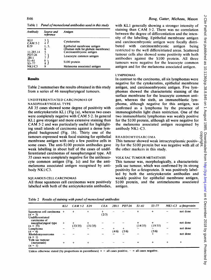
Table 1 Panel ofmonoclonal antibodies used in this study
Antibody Source and Antigenreference
CAM 5324 CytokeratinsE29 1, 5, Epithelial membrane antigen
6 (Human milk fat globule membrane)11.285.14 5 Carcinoembryonic antigenPD7/26 7 Leucocyte common antigen.2B11 7
Sl3-6 8 S100 proteinNK1/C3 9 Melanoma associated antigen
Results
Table 2 summarises the results obtained in this studyfrom a series of 46 nasopharyngeal tumours.
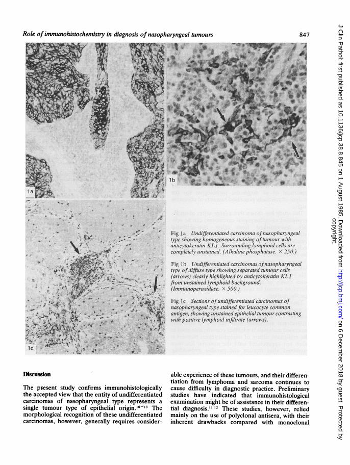
UNDIFFERENTIATED CARCINOMAS OFNASOPHARYNGEAL TYPEAll 35 cases showed some degree of positivity withthe anticytokeratin KL1 (Fig la), whereas two caseswere completely negative with CAM 5 2. In generalKL1 gave stronger and more extensive staining thanCAM 5 2 and was particularly useful for highlight-ing small islands of carcinoma against a dense lym-phoid background (Fig. lb). Thirty one of thetumours expressed weak focal staining for epithelialmembrane antigen with only a few positive cells insome cases. The anti-S100 protein antibodies gaveweak labelling in about half of the cases of undif-ferentiated carcinomas of nasopharyngeal type. All35 cases were completely negative for the antileuco-cyte common antigen (Fig. ic) and for the anti-melanoma associated antigen recognised by anti-body NK1/C3.
SQUAMOUS CELL CARCINOMASAll three squamous cell carcinomas were positivelylabelled with both of the anticytokeratin antibodies,
Bosq, Gatter, Micheau, Mason
with KL1 generally showing a stronger intensity ofstaining than CAM 5-2. There was no correlationbetween the degree of differentiation and the inten-sity of the labelling. Epithelial membrane antigenand carcinoembryonic antigen were focally distri-buted with carcinoembryonic antigen beingrestricted to the well differentiated areas. Scatteredtumour cells also showed some positivity with bothantibodies against the 5100 protein. All threetumours were negative for the leucocyte common
antigen and for the melanoma associated antigen.
LYMPHOMASIn contrast to the carcinoma, all six lymphomas were
negative for the cytokeratins, epithelial membraneantigen, and carcinoembryonic antigen. Five lym-
phomas showed the characteristic staining of thesurface membrane for the leucocyte common anti-gen, whereas the sixth, an immunoblastic lym-phoma, although negative for this antigen, was
confirmed as a lymphoma by the presence ofimmunoglobulin light chain restriction. One of thetwo immunoblastic lymphomas was weakly positivefor the S100 protein, although all were negative forthe melanoma associated antigen recognised byantibody NK1-C3.
RHABDOMYOSARCOMAThis tumour showed weak intracytoplasmic positiv-ity for the SiO protein but was negative with all ofthe other markers in this study.
YOLK SAC TUMOUR METASTASISThis tumour was, morphologically, a characteristicyolk sac tumour, which was confirmed by its strongpositivity for ca-fetoprotein. It was positively label-led by both the anticytokeratin antibodies andweakly positive for epithelial membrane antigen,S100 protein, and the antimelanoma associatedantigen.
Table 2 Results ofstaining with panel ofmonoclonal antibodies
KLI CAM 5-2 E29 CEA 2BII PD7126 SI-61 S3-77 NKI-C3 a-fetoprotein
Squamous cell carcinoma + + + + - + + not done(n= 3) (2/3)Undifferentiatedcarcinoma ofnasopharyngeal type + + + - - + + not done
(n = 35) (33/35) (31/35) (14/35) (19/35)Lymphoma - - - - + + - - not done(n = 6) (4/6) (5/6) (5/6)Rhabdomyosarcoma - - - - - - - + not done(n = 1)Yolk sac tumour
(metastasis) + + + - + + + +(n = 1)
Unless otherwise stated (by proportions in parentheses) + = all cases positive, - all cases negative.
copyright. on 6 D
ecember 2018 by guest. P
rotected byhttp://jcp.bm
j.com/
J Clin P
athol: first published as 10.1136/jcp.38.8.845 on 1 August 1985. D
ownloaded from
Role of immunohistochemistry in diagnosis of nasopharyngeal tumours
I
la
._
dr~~~~~~~~U
-. v**
_-D , 5, t
- ai.
Discussion
The psesent study confirms immunohistologicallythe accepted view that the entity of undifferentiatedcarcinomas of nasopharyngeal type represents asingle tumour type of epithelial origin.'0-'3 Themorphological recognition of these undifferentiatedcarcinomas, however, generally requires consider-
Fig la Undifferentiated carcinoma ofnasopharyngealtype showing homogeneous staining oftumour withanticytokeratin KLI. Surrounding lymphoid cells arecompletely unstained. (Alkaline phosphatase. x 250.)
Fig lb Undifferentiated carcinomas ofnasopharyngealtype ofdiffuse type showing separated tumour cells(arrows) clearly highlighted by anticytokeratin KLIfrom unstained lymphoid background.(Immunoperoxidase. x 500.)
Fig Ic Sections ofundifferentiated carcinomas ofnasopharyngeal type stained for leucocyte commonantigen, showing unstained epithelial tumour contrastingwith positive lymphoid infiltrate (arrows).
able experience of these tumours, and their differen-tiation from lymphoma and sarcoma continues tocause difficulty in diagnostic practice. Preliminarystudies have indicated that immunohistologicalexamination might be of assistance in their differen-tial diagnosis.' 12 These studies, however, reliedmainly on the use of polyclonal antisera, with theirinherent drawbacks compared with monoclonal
847
copyright. on 6 D
ecember 2018 by guest. P
rotected byhttp://jcp.bm
j.com/
J Clin P
athol: first published as 10.1136/jcp.38.8.845 on 1 August 1985. D
ownloaded from
848
antibodies, and contained no reliable agents for therecognition of lymphomas in routinely processedmaterial. We used several recently produced mono-clonal antibodies against both epitheliaP4 and lym-phoid antigens,7 which are relatively well preservedin routinely fixed and processed material.The clinical usefulness of this panel of monoclonal
antibodies has been established in our laboratory ona series of 120 routinely processed tumours ofuncertain origin, which were referred by otherpathologists owing to diagnostic difficulties.'4 Ofthese 120 tumours, 105 could be clearly identified as
either lymphomas (79 cases) or carcinomas (28cases), with four melanoma/seminoma and neuro-blastoma. This left only seven tumours whose trueidentity could not be established. Clinical follow upof these patients showed that at three years therewas a marked difference in prognosis of about 25%between the patients with lymphoma and those withcarcinoma. It was therefore suggested that such apanel of monoclonal antibodies should be part of thearmament of all diagnostic pathologists for theanalysis of routinely processed tumours of uncertainorigin.
This study has shown that a large series of undif-ferentiated nasopharyngeal carcinomas can be reli-ably distinguished from other non-epithelialtumours of the nasopharynx, including malignantlymphomas. The use of a panel of monoclonal anti-bodies is especially valuable as most types of tumourcan be recognised by positive staining as well as theabsence of markers associated with other types.Although epithelial membrane antigen was a reli-able epithelial marker in this study, its presence hasbeen shown in a few lymphoid tumours,6 and ittherefore cannot be relied on alone as an epithelialspecific marker.These findings are particularly relevant in practice
as nasopharyngeal biopsies are often small and con-siderably distorted by crush artefact. That thesemonoclonal antibodies work reliably in routinelyprocessed materials is a further considerable asset as
the malignant nature of nasopharyngeal biopsies isoften not appreciated until the material has beenexamined after fixation and paraffin embedding.This means that the diagnostic dilemma as towhether a nasopharyngeal tumour is a carcinoma ora lymphoma, with its consequent effect on thetreatment and prognosis of the patient, can now bereliably solved by any pathologist augmenting theroutine histological examination with immuno-cytochemistry using such a panel of monoclonalantibodies.
Bosq, Gatter, Micheau, Mason
This work was supported by the Association pour laRecherche sur le Cancer, the Wellcome Trust, andthe Leukaemia Research Fund. We thank all thosewho generously donated the monoclonal antibodiesused and Lesley Watts for typing the manuscript.
Referenc
Gatter KC, Falini B, Mason DY. The use of monoclonal anti-bodies in histopathological diagnosis. In: Anthony P,MacSween R, eds. Recent advances in histopathology. Vol 12.London: Churchill Livingstone, 1984:35-67.
2 Cordell JL, Falini B, Erber WN, etal. Immunoenzymatic label-ling of monoclonal antibodies using immune complexes ofalkaline phosphatase and monoclonal anti-alkaline phos-phatase (APAAP complexes). Histochem Cytochem 1984;32:219-29.
3Viac J, Reano A, Brochier J, Staquet MJ, Thivolet J. Reactivitypattern of a monoclonal antikeratin antibody (KL1). J InvestDermatol 1983;81:351-54.
4Makin CA, Bobrow LG, Bodmer WF. Monoclonal antibody tocytokeratin for use in routine histopathology. J Clin Pathol1984;37:975-83.
Gatter KC, Abdulaziz Z, Beverley P, etal. Use of monoclonalantibodies for the histopathological diagnosis of human malig-nancy. J Clin Padhol 1982;35:1253-67.
6 Delsol G, Stein H, Pulford KAF, etal. Human lymphoid cellsmay express epithelial membrane antigen. Implications for thediagnosis of human neoplasms. Lancet 1984:ii; 1124-9.
' Warnke RA, Gatter KC, Falini B, etal. Diagnosis of humanlymphoma with monoclonal anti-leucocyte antibodies. NewEngl J Med 1983; 309:1275-81.
Vanstapel MJ, Peeters B, Cordell J, et al. Production andidentification of monoclonal antibodies directed against andantigenic determinent common to the a and ,3 chain of S100.Lab Invest 1985; 52:232-8.
Mackie RM, Campbell I, Turbitt ML. Use of NK1C3 mono-clonal antibody in the assessment of benign and malignantmelanocytic lesions. J Clin Pathol 1984;37:367-72.
10 Svoboda D, Kirchner F, Shanmugaratnam K. The fine structureof nasopharyngeal carcinoma. In: Muir CS, ShanmugaratnamK, eds. Cancer ofthe nasopharynx (VICC Monograph series.)Copenhagen: Munksgaard, 1967:163-71.
"Madri JA, Barwick KW. An immunohistochemical study ofnasopharyngeal neoplasms using keratin antibodies. Epithelialversus non-epithelial neoplasms. Am J Surg Pathol1982;6: 143-9.
2 Gusterson BA, Mitchell DP, Warburton HJ, Carter RL. Epithel-ial markers in the diagnosis of nasopharyngeal carcinoma: animmunocytochemical study. J Clin Patdol 1983; 36:628-31.
3 Shi Sr, Goodman ML, Bhan AK, Pilch BZ, Chen LB, Sun TT.Immunohistochemical study of nasopharyngeal carcinomausing monoclonal keratin antibodies. Am J Pathol1984; 117:53-63.
4 Gatter KC, Alcock C, Heryet A, Mason DY. The clinical impor-tance of analysing tumours of uncertain origin by immunohis-tological techniques. Lancet (in press).
Requests for reprints to: KC Gatter, Nuffield Departmentof Pathology, John Radcliffe Hospital, Headington,Oxford OX3 9DU.
copyright. on 6 D
ecember 2018 by guest. P
rotected byhttp://jcp.bm
j.com/
J Clin P
athol: first published as 10.1136/jcp.38.8.845 on 1 August 1985. D
ownloaded from