Role of HE4 and ROMA Score in Diagnosis of Ovarian Cancer ...
12
Received: August 08, 2019; Accepted: April 19, 2020 Role of HE4 and ROMA Score in Diagnosis of Ovarian Cancer in Egyptian Women Nahla I. El-Attar*, MD, Lamiaa Mahmoud Kamel*, MD, Amany Abdelghany**, MD, Rasha Haggag***, MD, Heba F. Taha*** ,♦ , MD, Entesar R. Mahdy**, MD, Nadia M. Madkour**, MD *Department of Clinical Pathology, Faculty of Medicine, Zagazig University, Zagazig, Egypt **Department of Obstetrics and Gynecology, Faculty of Medicine, Zagazig University, Zagazig, Egypt ***Department of Medical Oncology, Faculty of Medicine, Zagazig University, Zagazig, Egypt Abstract Background: Risk of ovarian malignancy algorithm (ROMA) combining human epididymis secretory protein 4 (HE4) and CA125 is a novel score, specific for epithelial ovarian cancer (EOC). Method: Our cohort prospective study aimed to evaluate the role of HE4 and ROMA score in the diagnosis of EOC. We determined CA125 and HE4 serum levels in 56 premenopausal women with ovarian mass (38 women with benign ovarian mass and 18 women with malignant ovarian mass), 56 postmenopausal women with ovarian mass (20 women with benign ovarian mass and 36 women with malignant ovarian mass), and 56 healthy women as control. Results: Serum CA125 and HE4 and ROMA score were significantly higher among postmenopausal group compared with premenopausal and control groups (P< 0.001), and the median serum CA125 and HE4 and ROMA levels were statistically higher among malignant lesions compared with benign lesions and control group (P< 0.001). The sensitivity and specificity of HE4 and ROMA vs. CA125 in discriminating ovarian cancer from benign ovarian tumor was (88% and 98% vs. 90%) and (97% and 99% vs. 80%), respectively. ROMA had better sensitivity and specificity compared to CA125 and HE4 in premenopausal and postmenopausal women (P <0.001) In premenopausal patients, there was a statistically significant difference regarding the area under the curve (AUC) of ROMA vs. CA125 (P=0.004) and ROMA vs. HE4 (P =0.02). Conclusion: ROMA score showed a better performance in comparison with either CA125 or HE4 alone in premenopausal patients. HE4 and ROMA score significantly differentiated early from late stage ovarian cancer. Keywords: Ovarian cancer, Prognosis, ROMA score, CA125, HE4 ♦ Corresponding Author: Heba F. Taha, MD Medical Oncology Department, Faculty of Medicine, Zagazig University, Zagazig, Egypt Tel: +20552381878 Email: [email protected] Original Article Middle East Journal of Cancer; January 2021; 12(1): 28-39 Please cite this article as: El- Attar NI, Kamel LM, Abdelghany A, Haggag R, Taha HF, Mahdy ER, Madkour NM. Role of HE4 and ROMA score in diagnosis of ovarian cancer in Egyptian women. Middle East J Cancer. 2021;12(1):28- 39. doi: 10.30476/mejc.2020. 82770.1100.
Transcript of Role of HE4 and ROMA Score in Diagnosis of Ovarian Cancer ...

Received: August 08, 2019; Accepted: April 19, 2020
Role of HE4 and ROMA Score in Diagnosis
of Ovarian Cancer in Egyptian Women
Nahla I. El-Attar*, MD, Lamiaa Mahmoud Kamel*, MD,
Amany Abdelghany**, MD, Rasha Haggag***, MD, Heba F. Taha***,♦, MD,
Entesar R. Mahdy**, MD, Nadia M. Madkour**, MD
*Department of Clinical Pathology, Faculty of Medicine, Zagazig University, Zagazig, Egypt
**Department of Obstetrics and Gynecology, Faculty of Medicine, Zagazig University, Zagazig, Egypt
***Department of Medical Oncology, Faculty of Medicine, Zagazig University, Zagazig, Egypt
Original Article
Middle East Journal of Cancer; October 2018; 9(4): 288-294
Abstract Background: Risk of ovarian malignancy algorithm (ROMA) combining human
epididymis secretory protein 4 (HE4) and CA125 is a novel score, specific for epithelial ovarian cancer (EOC).
Method: Our cohort prospective study aimed to evaluate the role of HE4 and ROMA score in the diagnosis of EOC. We determined CA125 and HE4 serum levels in 56 premenopausal women with ovarian mass (38 women with benign ovarian mass and 18 women with malignant ovarian mass), 56 postmenopausal women with ovarian mass (20 women with benign ovarian mass and 36 women with malignant ovarian mass), and 56 healthy women as control.
Results: Serum CA125 and HE4 and ROMA score were significantly higher among postmenopausal group compared with premenopausal and control groups (P< 0.001), and the median serum CA125 and HE4 and ROMA levels were statistically higher among malignant lesions compared with benign lesions and control group (P< 0.001).
The sensitivity and specificity of HE4 and ROMA vs. CA125 in discriminating ovarian cancer from benign ovarian tumor was (88% and 98% vs. 90%) and (97% and 99% vs. 80%), respectively.
ROMA had better sensitivity and specificity compared to CA125 and HE4 in premenopausal and postmenopausal women (P <0.001)
In premenopausal patients, there was a statistically significant difference regarding the area under the curve (AUC) of ROMA vs. CA125 (P=0.004) and ROMA vs. HE4 (P =0.02).
Conclusion: ROMA score showed a better performance in comparison with either CA125 or HE4 alone in premenopausal patients. HE4 and ROMA score significantly differentiated early from late stage ovarian cancer.
Keywords: Ovarian cancer, Prognosis, ROMA score, CA125, HE4
♦Corresponding Author:
Heba F. Taha, MD Medical Oncology Department, Faculty of Medicine, Zagazig University, Zagazig, Egypt Tel: +20552381878 Email: [email protected]
Original Article
Middle East Journal of Cancer; January 2021; 12(1): 28-39
Please cite this article as: El-Attar NI, Kamel LM, Abdelghany A, Haggag R, Taha HF, Mahdy ER, Madkour NM. Role of HE4 and ROMA score in diagnosis of ovarian cancer in Egyptian women. Middle East J Cancer. 2021;12(1):28-39. doi: 10.30476/mejc.2020. 82770.1100.

HE4 and ROMA Score in Diagnosis of Ovarian Cancer
Middle East J Cancer 2020; 12(1): 28-39 29
Introduction
Ovarian cancer is the sixth most prevalent cancer in women and the most common cause of mortality arising from female pelvic malignancy.1 In 2014, the annual cases of ovarian cancer went beyond 220,000.2
The incidence rate increases with age in postmenopausal status. The crude incidence rate changes from 4.7 per 100,000 in women <50 years of age to 29.6 per 100,000 in the age group 50-64.3
The symptoms of ovarian cancer are similar to some other benign conditions;4 therefore, ovarian cancer is diagnosed at advanced stages. The five-year survival in the early stages of ovarian cancer ranges from 80 to 93%; however, in the late stages, it is less than 30%.5
The use of CA125 with ultrasonography for early diagnosis of ovarian cancer is a diagnostic approach; nonetheless, it has certain drawbacks, including low sensitivity and specificity.6
Other such malignancies as endometrium, endocervix, lung cancer, and lymphomas can express abnormal CA125 serum levels.7 High serum level of CA 125 may also be found in other benign gynecologic conditions such as ovarian cysts, myomas, and endometriosis and other benign diseases such effusions, liver, or renal failure.8 CA125 concentrations may fluctuate throughout the menstrual cycle and pregnancy.9
Human epididymis secretory protein 4 is a precursor of human epididymis protein encoded by a gene located in chromosome 20q12-13.1.10
It is a member of the whey acidic protein gene family.11 Human epididymis secretory protein 4 (HE4) is frequently overexpressed in ovarian cancers.12
HE4 had a significantly higher diagnostic specificity compared with CA125, and the combination of CA125 and HE4 improved the detection of ovarian cancer in all stages and histological types.13
This finding encouraged the development of a double marker algorithm combining HE4 and CA125 with the pre- and postmenopausal statuses of the patient, known as risk of ovarian malignancy algorithm (ROMA).8
The objective of our study was to assess the diagnostic significance and performance of HE4 level and ROMA score in comparison with CA125 among women with epithelial ovarian cancer (EOC) and benign gynecological diseases.
Patients and Methods
We conducted our cohort prospective study at Obstetrics and Gynecology, Medical Oncology and Clinical Pathology departments, Zagazig University Hospitals, Faculty of Medicine, Egypt, between December 2014 and December 2015.
The study included 168 patients with malignant or benign ovarian mass consecutively admitted at obstetrics and gynecology and medical oncology departments. The data required for the study included patients’ personal, menstrual, and obstetric history, grade, stage, histologic type, and follow-up data; we collected the information from the patients’ files in the shared departments. Each subject signed a written informed consent and the study conformed with the World Medical Association (Declaration of Helsinki), printed in the British Medical Journal (18 July 1964). The Ethics Committee of Zagazig University further approved the present study (Ethics code: 1453).
We divided the patients into three groups: i) 56 apparently healthy women as control; ii) 56 premenopausal women with ovarian mass, where 38 women had benign ovarian mass (25 patients with ovarian cyst, five patients with fibroadenoma, five patients with cystadenoma, and three patients with teratoma) and 18 women had malignant ovarian mass; and iii)56 postmenopausal women with ovarian mass, where 20 women had benign ovarian mass (15 patients with ovarian cyst and five patients with fibroadenoma) and 36 women had malignant ovarian mass. The inclusion criteria were EOC, benign gynaecological disease, and written informed consent for participation.The exclusion criteria were inability to provide consent, pregnancy, other malignancies and diseases affecting HE4 level (chronic kidney disease, renal failure, kidney fibrosis, systemic lupus erythematosus) and women with previous bilateral oophorectomy.
We considered the women to be post-

Nahla I. El-Attar et al.
Middle East J Cancer 2020; 12(1): 28-3930
menopausal under the following conditions: cessation of menses for >1 year prior to investigation, age >55, or unknown date of the last menstrual period.
5 mL of venous blood sample was collected from all subjects and divided into 2 mL for CBC
on EDTA vaccutainer and 3 mL on plain vaccutainer; it was left for 30 minutes at 37°C to be clotted; the sample was then centrifuged at 3000 rpm for 10 minutes to separate the serum for an immediate analysis of serum glucose, liver, and kidney functions, hepatitis B,C markers as
Figure 1. (A) CA 125 (U/mL) level, (B)HE4 (Pm/L) level, and (c) ROMA (%) score increased in postmenopausal women in the studied groups.

HE4 and ROMA Score in Diagnosis of Ovarian Cancer
Middle East J Cancer 2020; 12(1): 28-39 31
routine preoperative laboratory investigations; CA125 was further measured by electrochemi-luminesence on Cobas e411, Roche. The remaining serum was stored at -80c until further analysis of HE4 via Enzyme Linked Immunosorbent Assay, Fujirebio.
Statistical analysis
To analyze the data, we employed SPSS software version 22.0 (IBM SPSS Statistics for Windows, Version 22.0. Armonk, NY: IBM Corp, 2013) and Epi-Info 7 (CDC, 2014) . ROMA score:
We calculated ROMA using the following algorithms: Premenopausal:
PI (predictive index) = -12+2.38 * LN(HE4)+
0.0626 *LN(CA125) Postmenopausal:
PI (predictive index) = -8.09+1.04 * LN(HE4)+ 0.732 * LN(CA125) Afterwards, we calculated ROMA-value (predictive value) using the following equation: ROMA (%) =ePI/(1+ePI) * 100.8 * We defined menopausal status as the absence of periods for more than one year.
We plotted receiver operating characteristics (ROC) curves for the optimal cut-off values of CA125 and HE4 and ROMA score, predictive of malignant ovarian tumors, as well as, sensitivity and specificity.
Table 1. Demographic data and comparison of histopathological findings and ROMA score of the patient’s groups Variable Premenopausal Postmenopausal Control P LSD
(n=56) (n=56) (n=56)
No % No % No % Parity
Virgin 11 19.6 0 0 4 7.1 <0.001 No children 2 3.6 1 1.8 1 1.8 Nullipara 2 3.6 0 0 6 10.8 Multipara 41 73.2 55 98.2 45 80.3 Menstruation
Pre-menopausal 56 100 0 0 28 50 <0.001 Post-menopausal 0 0 56 100 28 50 Histopathology
Benign 38 67.9 20 35.7 - - <0.001 Malignant 18 32.1 36 64.3 - - Age (years) ≤0.05a
Mean ± SD 34.41 ± 9.38 59.76 ± 6.03 45.36 ± 15.15 >0.05b >0.05c
CA 125 (U/ml)
Median 85.4 323 24.45 <0.001 ≤0.001a IQ-Range 44.1-908.2 74.7-3255.1 17.4-29.5 ≤0.05b
≤0.001c HE4 (Pm/L) ≤0.001a
Median 125 300 71 <0.001 ≤0.05b IQ-Range 90.8-165 122-345.5 60.8-93 ≤0.001c ROMA (%) ≤0.001a
Median 32.7 59 17.6 <0.001 ≤0.05b IQ-Range 21.5-42.3 42.5-88.9 12.6-22.9 ≤0.001c (a) premenopausal group versus postmenopausal group, (b) premenopausal group versus control group, (c)postmenopausal group versus control; LSD=Least significant difference; ROMA: Risk of ovarian malignancy algorithm, HE4: Human epididymis secretory protein 4.; CA125:Cancer antigen 125; P value statistical significance (≤0.05)

Nahla I. El-Attar et al.
Middle East J Cancer 2020; 12(1): 28-3932
Results Table 1 shows the demographic data and
menstrual history of the studied groups. Histopathological findings revealed that in the premenopausal group, there were 38 cases (67.9%) with benign ovarian mass and 18 cases (32.1%)
with malignant ovarian mass. In the postmenopausal group; on the other hand, there were 20 cases (35.7%) with benign ovarian mass and 36 cases (64.3%) with malignant ovarian mass. This means that malignancy was more frequent in postmenopausal women (Table 1);
Figure 2. (A) CA 125 (U/ml) level increased in malignant lesions compared with benign lesions and control group; (B)HE4 (Pm/L) level increased in malignant lesions compared to benign lesions and control group; and (c) ROMA (%) score increased in malignant lesions in comparison with benign lesions and control group.
HE4 and ROMA Score in Diagnosis of Ovarian Cancer
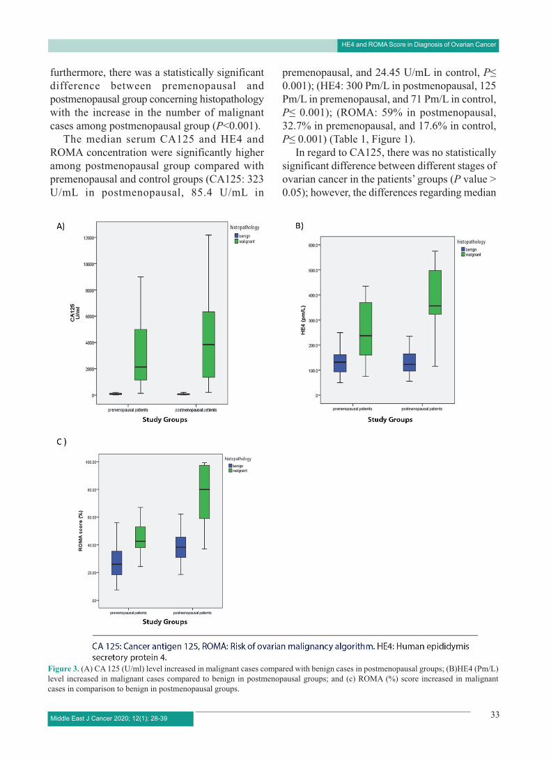
furthermore, there was a statistically significant difference between premenopausal and postmenopausal group concerning histopathology with the increase in the number of malignant cases among postmenopausal group (P<0.001).
The median serum CA125 and HE4 and ROMA concentration were significantly higher among postmenopausal group compared with premenopausal and control groups (CA125: 323 U/mL in postmenopausal, 85.4 U/mL in
premenopausal, and 24.45 U/mL in control, P≤ 0.001); (HE4: 300 Pm/L in postmenopausal, 125 Pm/L in premenopausal, and 71 Pm/L in control, P≤ 0.001); (ROMA: 59% in postmenopausal, 32.7% in premenopausal, and 17.6% in control, P≤ 0.001) (Table 1, Figure 1).
In regard to CA125, there was no statistically significant difference between different stages of ovarian cancer in the patients’ groups (P value > 0.05); however, the differences regarding median
Middle East J Cancer 2020; 12(1): 28-39 33
Figure 3. (A) CA 125 (U/ml) level increased in malignant cases compared with benign cases in postmenopausal groups; (B)HE4 (Pm/L) level increased in malignant cases compared to benign in postmenopausal groups; and (c) ROMA (%) score increased in malignant cases in comparison to benign in postmenopausal groups.

Nahla I. El-Attar et al.
serum HE4 and ROMA were statistically significant (P value ≤0.05) (Table 2).
The median serum CA125 and HE4 and ROMA concentration were statistically higher among malignant lesions compared with benign lesions and control group (CA125: 3000 U/mL in malignant lesions, 69.3 in benign lesions, and 24.45 in control, P ≤0.001); (HE4: 347 Pm/L in malignant lesions, 128 Pm/L in benign lesions, and 71 Pm/L in control group P≤0.001); (ROMA 65.5% in malignant lesions, 33.9% in benign lesions, 17.6% in control group, P≤ 0.001) (Table 2, Figures 2 and 3).
Performance of HE4 and ROMA compared with CA125 is better in discriminating ovarian cancer from benign ovarian tumor with sensitivity (88% and 98% vs. 90%), specificity (97% and 99% vs. 80%), negative predictive values (90.3% and 98.3% vs. 90.2%), and positive predictive values (95.7% and 98% vs. 79.3%) (Table 3).
ROMA had a better performance compared with CA125 and HE4 with sensitivity (88.9% vs.
83.3% and 83.3%), specificity (100% vs. 71.1 % and 78.9%), positive predictive value (100% vs. 57.7% and 65.2%), and negative predictive value (95.0% vs. 90.0% and 90.9%) in premenopausal women and sensitivity (98.0% vs. 85.0% and 95.0%), specificity (97.0% vs. 100% and 100%), positive predictive value (97.1% vs. 100% and 100%), and negative predictive value (94.7% vs. 79.2% and 90.5%) in postmenopausal women (Tables 4 and 5).
The ROC curve for the diagnosis of ovarian cancer in patients’ groups showed that the area under the curve of CA 125, HE 4, and ROMA score were 0.94 (P<0.001), 0.93 (P<0.001), and 0.96 (P<0.001), respectively (Table 3).
This ROC curve also showed that the area under the curve of CA 125, HE 4, and ROMA score was 0.79 (P<0.001), 0.82 (P<0.001), and 0.93 (P<0.001), respectively, in premenopausal women (Table 4).
The area under the curve of CA 125, HE 4, and ROMA score were 0.95 (P<0.001), 0.97 (P<0.001),
Middle East J Cancer 2020; 12(1): 28-3934
Table 2. CA125, HE4, and ROMA scores in different stages of malignant lesions and according to histopathology Stages CA 125 (U/mL) HE 4 (Pm/L) ROMA (%)
Median Median Median
Range Range Range
I 230.1 160.0 34.0 1557.0 – 8974.0 125.0 – 325.0 41.0 – 60.0
II 3712.5 340.0 46.5 1458.8 – 6232.5 266.3 – 399.8 35.3 – 73.7
III 2588.0 497.5 50.5 1835.7 - 5412.3 291.0 – 553.5 45.0 – 71.7
IV 7486.9 515.0 88.0 1951.0 – 9227.3 360.0 – 522.0 79.0 – 93.2
P-value >0.05 ≤0.05 ≤0.05 Histopathology Benign lesions 69.3 128 33.9 (n=58) 26.7-124.5 94.5-165 21-41.6 Malignant lesions 3000 347.5 65.5 (n=54) 1142.5-5653.7 173-432 47-92.4 Control group 24.45 71 17.6 (n=56) 17.4-29.5 60.8-93 12.6-22.9 P-value ≤0.001 ≤0.001 ≤0.001 LSD ≤0.001d ≤0.05d ≤0.05(d)
≤0.05e ≤0.001e ≤0.05(e) ≤0.001f ≤0.001f ≤0.001(f)
P value statistical significance (≤0.05); (d) Benign group versus malignant group, (e) Benign group versus control group, (f) Malignant group versus control group.; ROMA: Risk of ovarian malignancy algorithm, HE4: Human epididymis secretory protein 4.; CA125: Cancer antigen 125; LSD=Least significant difference

HE4 and ROMA Score in Diagnosis of Ovarian Cancer
and 0.98 (P<0.001), respectively, in postmenopausal women (Table 5).
Comparison of ROC area under the curve (AUC) of the three parameters (CA125, HE4, and ROMA) showed that the AUC of ROMA was higher in all studied groups; however, there was no statistically significant difference between the AUC of the three markers regarding all patients and postmenopausal patient groups; nevertheless, in premenopausal patients, there was a statistically significant difference between AUC of ROMA vs. CA125 (P =0.004) and ROMA vs. HE4 (P=0.02) (Table 6).
Discussion
Our study attempted to define the AUC value of ROMA (combining HE4 and CA125) in early detection of ovarian cancer premenopausal patients; in these subjects, contrary to postmenopausal women, there was a statistically significant difference between AUC of ROMA vs. CA125 and ROMA vs. HE4.
Several new markers have been suggested for early detection of ovarian cancer. HE4 is one of the markers, also known as the core epitope protein 2 (WFD2) belonging to the protease inhibiting family.14
Detected in patients with ovarian cancer, the HE4 gene product is an N-glycosylated protein secreted into the extracellular environment. HE4 was elevated in more than 50% of ovarian cancer patients; however, they had no detected CA125 levels.15
In our study, we found that median serum CA125, in different stages of ovarian cancer, showed no statistically significant difference between stages (P>0.05); however, the median serum HE4 and ROMA showed statistically significant difference among different stages
(P≤0.05). Similarly, Jafari-Shobiri et al.16 found no
statistically significant difference between the average CA125 in patients with stage I-II (early stage) ovarian cancer in comparison to patients with stage III-IV (advanced stage) ovarian cancer (P=0.062). In patients with stage I-II ovarian cancer, the average HE4 was statistically significant compared with stage III-IV (P=0.04). However, they observed no statistically significant difference concerning ROMA between the stage I-II and III-IV ovarian cancer patients (P=0.25).
Contrary to the present study, Molina et al.17
reported that CA125 was related to the stage of the disease with significantly higher concentration in advanced stages III-IV compared with stage I-II; the difference might be attributed to the different population and number of cases. Similar to our results, they found that HE4 was higher in advanced stages compared with early stages.
We found that the median serum CA125 and HE4 and ROMA concentration were significantly higher among postmenopausal group compared with premenopausal and control groups (CA125: 323 U/mL in postmenopausal, 85.4 U/mL in premenopausal, and 24.45 U/mL in control, P≤0.001); (HE4: 300 Pm/L in postmenopausal, 125Pm/L in premenopausal, and 79.5 Pm/L in control, P<0.001); (ROMA: 59% in postmenopausal, 32.7% in premenopausal, and 17.6% in control, P≤0.001). Similarly, Fujiwara et al.18 reported that the median serum levels of HE4 and ROMA were significantly higher in postmenopausal group compared with premenopausal group; in contrast to our results, they observed that the median serum levels of CA125 were significantly higher in premenopausal than in postmenopausal regarding both benign and early stage EOC groups; however, they were
Middle East J Cancer 2020; 12(1): 28-39 35
Table 3. Performance of CA 125, HE 4, and ROMA score among patients, malignant vs. benign, in pre- and postmenopausal patients Marker Cut-off AUC Sens. Spec. +PV -PV Accuracy P-value
CA125 (U/ml) 66.3 0.94 90.0% 80.0% 79.3% 90.2% 84.4% <0.001(HS) HE 4 (Pm/L) 86.5 0.93 88.0% 97.0% 95.7% 90.3% 92.7% <0.001(HS) ROMA (%) 22.0 0.96 98.0% 99.0% 98.0% 98.3% 98.2% <0.001(HS) Sens.: sensitivity, Spec.: specificity, +PV: positive predictive value, - PV: negative predictive value, AUC: area under curve, ROMA: Risk of ovarian malignancy algorithm, CA125: Cancer antigen 125, HE4: human epididymis secretory protein 4.

Nahla I. El-Attar et al.
significantly higher in postmenopausal subjects in late stage EOC.
Jafari-Shobeiri et al.16 reported that the median serum CA125 and HE4 and ROMA concentration were higher (though not significantly) among postmenopausal group in comparison with premenopausal group (CA125: 437.5 U/mL in postmenopausal and 219.06 U/mL in premenopausal, P=0.17); (HE4: 357.1 Pm/L in postmenopausal and 204.1 Pm/L in premenopausal, P=0.2); (ROMA: 47.8% in postmenopausal and 86.01% in premenopausal, P=0.16).
On the other hand, Molina et al.17 found that CA125 serum levels were significantly higher in premenopausal women than in postmenopausal women (P=0.001). Higher concentrations of HE4 were detected in postmenopausal women, but the data were not statistically significant. They also found that ROMA was higher in premenopausal women compared with postmenopausal women in control group; nonetheless, it was higher in postmenopausal women in both benign and malignant groups.
In the present study the median serum CA125 and HE4 and ROMA concentration were statistically higher among malignant lesions compared with benign lesions and control group (CA125: 3000 U/mL in malignant lesions, 69.3 in benign lesions, and 24.45 in control, P≤0.001); (HE4: 347 Pm/L in malignant lesions, 128 Pm/L in benign lesions, and 71 Pm/L in control group, P≤0.001); (ROMA 65.5% in malignant lesions, 33.9% in benign lesions, 17.6% in control group, P≤0.001). This finding is in agreement with Fujiwara et al.18 who reported that the median serum CA125 andHE4 and ROMA concentration were statistically higher among malignant lesions compared with benign lesions and control group (CA125: 61.2 U/mL in type 1 EOC and 567.2
U/mL in type 2 EOC, 21.9 in benign lesions, and 11.9 in control); (HE4: 65.8 Pm/L in type 1 EOC and 310.9 Pm/L in type 2 EOC, 40.8 Pm/L in benign lesions, and 38.9 Pm/L in control group); (ROMA 24.8% in type 1 EOC and 92.4% in type 2 EOC, 5.6% in benign lesions, 4.6% in control group); Zheng et al.19 also reported that the median serum levels of CA125 and HE4 were statistically higher in malignant ovarian tumor group in comparison with benign and healthy groups (CA125: 78.95 IU/L in malignant group, 22.35 IU/L in benign group, and 20.00 IU/L in healthy group); (HE4: 93.23 Pm/L in malignant group, 25.42 Pm/L in benign group, and 24.00 Pm/L in healthy group).
Also, consistent with our results, Molina et al.17 observed that the median serum level of CA125 and HE4 were statistically higher in malignant group compared with benign and healthy groups (CA125: 185 U/mL in malignant group, 21 U/mL in benign group, and 15U/mL in healthy control group); (HE4: 406 Pm/L in malignant group, 46.5 Pm/L in benign group, and 54 Pm/L in healthy control group); (ROMA 90.1% in malignant group, 12.3% in benign group, 25.8% in healthy control group). The median serum levels of HE4 and ROMA were higher in the healthy control group than in the benign group, which is different from the present results.
Compared with CA125, HE4 and ROMA had a better performance in differentiating ovarian cancer from benign ovarian tumor; this is evident from cut-off suggested by ROC curve analysis with sensitivity (88% and 98% vs. 90%), specificity (97% and 99% vs. 80%), negative predictive values (90.3% and 98.3% vs. 90.2%), and positive predictive values (95.7% and 98% vs. 79.3%); Molina et al.17 reported that based on their suggested cut-off by ROC curve analysis, HE4 and ROMA score had a better performance
Middle East J Cancer 2020; 12(1): 28-3936
Table 4. Performance of CA 125, HE 4, and ROMA score in the diagnosis of ovarian cancer in premenopausal patients Marker Cut-off AUC Sens. Spec. +PV -PV Accuracy P-value
CA125 (U/ml) 53.3 0.79 83.3% 71.1% 57.7% 90.0% 75.0% <0.001** HE 4 (Pm) 64.0 0.82 83.3% 78.9% 65.2% 90.9% 80.4% <0.001** ROMA (%) 19.3 0.93 88.9% 100% 100% 95.0% 96.4% <0.001** Sens.: sensitivity, Spec.: specificity, +PV: positive predictive value, - PV: negative predictive value, AUC: area under curve, ROMA: Risk of ovarian malignancy algorithm, CA125: Cancer antigen 125, HE4: human epididymis secretory protein 4.

HE4 and ROMA Score in Diagnosis of Ovarian Cancer
regarding sensitivity (79.3% and 90.1% vs. 82.9%), specificity (98.9% and 87.7% vs. 70.9%), and negative (92.5% and 95.8% vs. 91.4%) and positive predictive values (96.7% and 74% vs. 52.6%) compared with CA125 in differentiating ovarian cancer from benign ovarian tumor.
We found that in premenopausal women, ROMA offered improved performance compared with CA125 and HE4 in regard to sensitivity (88.9% vs. 83.3% and 83.3%), specificity (100% vs. 71.1% and 78.9%), positive predictive value (100% vs. 57.7% and 65.2%), and negative predictive value (95.0% vs. 90.0% and 90.9%). Similarly, Chan et al. reported a ROMA specificity of 87.45 % compared with CA125 (63.2%) and a positive predictive value of 34.69% compared with CA125 (16.8%) in premenopausal women.20 Clemente and Benitez. also found a ROMA specificity of 75% compared with CA125 (55%) and a positive predictive value of (19%) compared with CA125 (15%) in premenopausal women.21
On the other hand, Molina et al.17 reported the following data regarding ROMA performance compared with CA125 and HE4 in premenopausal women: sensitivity (74.1% vs. 74.1% and 63.0%), specificity (88.9% vs. 69.0% and100%), positive predictive value (44.4% vs. 22.2% and 100%), and negative predictive value (96.6% vs. 95.7% and 95.8%). This difference might be ascribed to different populations, sample sizes, and methods of detection as they used chemiluminescent enzyme immunoassay on the Architect Analyzer.
Concerning postmenopausal women, our results compared ROMA performance with CA125 and HE4: sensitivity (98.0% vs. 85.0% and 95.0%), specificity (97.0% vs. 100% and100%), positive predictive value (97.1% vs. 100% and 100%), and negative predictive value (94.7% vs. 79.2% and 90.5%); Clemente and
Benitez., also reported a ROMA specificity of (70%) compared with CA125 (90%) and a positive predictive value of (42%) compared with CA125 (68%) in postmenopausal women.21
In contrast, Molina et al.17 found the following data concerning the performance of ROMA compared with CA125 and HE4: sensitivity (95.2% vs. 85.7% and 84.5%), specificity (83.1% vs. 78.0% and 94.9%), positive predictive value (88.9% vs. 84.7% and 96.0%), and negative predictive value (92.5% vs. 79.3% and 81.2).
In the present work, in all patients with ovarian cancer, the AUC of CA125, HE4 , and ROMA were 0.94, 0.93, and 0.96, respectively; however, in premenopausal group, the AUCs were 0.79, 0.82, and 0.93, while in postmenopausal group, the AUCs were 0.95, 0.97, and 0.98, respectively.
These results are not in agreement with Cho et al.22 who reported that in patients with ovarian cancer, the AUCs of CA125, HE4, and ROMA were 0.75, 0.85, and 0.86, respectively. In premenopausal subjects, the AUCs were CA125 (0.76), HE4 (0.74), and ROMA (0.74), while in postmenopausal group, the AUCs were CA125 (0.72), HE4 (0.83), and ROMA (0.79).
Many studies reported that after comparing the ROC-AUCs of the different tumor markers, HE4 and CA125 performed similarly except in the postmenopausal patients, in whom CA125 performed better.10, 23-25 That is in contrast to our results, possibly due to the difference in populations and sample sizes.
Based on the ROC area under the curve, we compared ROMA, HE4, and CA125 values for the diagnosis of malignant ovarian tumors; we detected that in all patients, there was no statistically significant difference between AUC of the three markers. Regarding premenopausal patients, there was a statistically significant
Middle East J Cancer 2020; 12(1): 28-39 37
Table 5. Performance of CA 125, HE 4, and ROMA score in the diagnosis of ovarian cancer in postmenopausal patients Marker Cutoff AUC Sens. Spec. +PV -PV Accuracy P-value
CA125 (U/ml) 82.1 0.95 85.0% 100% 100% 79.2% 90.7% <0.001** HE4 (Pm) 74.5 0.97 95.0% 100% 100% 90.5% 96.3% <0.001** ROMA (%) 27.0 0.98 98.0% 97.0% 97.1% 94.7% 96.3% <0.001** Sens.: sensitivity, Spec.: specificity, +PV: positive predictive value, - PV: negative predictive value, AUC: area under curve, ROMA: Risk of ovarian malignancy algorithm, CA125: Cancer antigen 125, HE4: human epididymis secretory protein 4.

Nahla I. El-Attar et al.
difference between AUC of CA125 vs. ROMA and HE4 vs. ROMA. There was no statistically significant difference between the AUC of the three markers in postmenopausal patients.
However, Van Gorp et al.26 found that in the ROC area under the curve in all patients and premenopausal groups, there was only statistically significant difference between AUC of HE4 vs. ROMA; however, in postmenopausal group, there was a highly significant difference between the AUC of HE4 vs. CA125 and HE4 vs. ROMA and a non-significant difference between AUC of CA125 vs. ROMA.
Based on the results of this paper, it is highly important to use these markers for an early detection of early stage ovarian cancer with better performance in premenopausal women, enhancing the management of ovarian cancer patients. The selection of these markers is an important issue in developing countries.
The limitation of our study was the small number of cases, which has to be further extended to include a higher number of cases.
Conclusion
HE4 and ROMA score improved the diagnostic ability for ovarian cancer compared with CA125 alone. In premenopausal patients, ROMA score showed better performance compared with either CA125 or HE4 alone. HE4 and ROMA scores significantly differentiated early from late stage ovarian cancer.
Conflict of Interest
None declared.
References 1. Garcia M, Jemal A, Ward EM, Center MM, Hao Y,
Siegel RL, et al. Global Cancer Facts & Figures 2007. Atlanta, GA: American Cancer Society, 2007. Available
from: https://www.cancer.org/content/dam/cancer-org/research/cancer-facts-and-statistics/global-cancer-facts-and-figures/global-cancer-facts-and-figures-2007.pdf.
2. Jayson GC, Kohn EC, Kitchener HC, Ledermann JA. Ovarian cancer. Lancet. 2014;384(9951):1376-88. doi: 10.1016/S0140-6736(13)62146-7.
3. Ferlay J, Shin HR, Bray F, Forman D, Mathers C, Parkin DM. Estimates of worldwide burden of cancer in 2008: GLOBOCAN 2008. Int J Cancer. 2010;127(12):2893-917. doi: 10.1002/ijc.25516.
4. Jemal A, Siegel R, Ward E, Murray T, Xu J, Thun MJ. Cancer statistics, 2007. CA Cancer J Clin. 2007;57(1):43-66.
5. Heintz AP, Odicino F, Maisonneuve P, Quinn MA, Benedet JL, Creasman WT, et al. Carcinoma of the ovary. FIGO 26th Annual Report on the Results of Treatment in Gynecological Cancer. Int J Gynaecol Obstet. 2006;95 Suppl 1:S161-92.
6. Bast RC Jr, Badgwell D, Lu Z, Marquez R, Rosen D, Liu J, et al. New tumor markers: CA125 and beyond. Int J Gynecol Cancer. 2005;15 Suppl 3:274-81.
7. Anastasi E, Granato T, Marchei GG, Viggiani V, Colaprisca B, Comploj S, et al. Ovarian tumor marker HE4 is differently expressed during the phases of the menstrual cycle in healthy young women. Tumour Biol. 2010;31(5):411-5. doi: 10.1007/s13277-010-0049-1.
8. Moore RG, McMeekin DS, Brown AK, DiSilvestro P, Miller MC, Allard WJ, et al. A novel multiple marker bioassay utilizing HE4 and CA125 for the prediction of ovarian cancer in patients with a pelvic mass. Gynecol Oncol. 2009;112(1):40-6. doi: 10.1016/j. ygyno.2008.08.031.
9. Nolen B, Velikokhatnaya L, Marrangoni A, De Geest K, Lomakin A, Bast RC Jr, et al. Serum biomarker panels for the discrimination of benign from malignant cases in patients with an adnexal mass. Gynecol Oncol. 2010;117(3):440-5. doi: 10.1016/j.ygyno.2010.02.005.
10. Hellström I, Raycraft J, Hayden-Ledbetter M, Ledbetter JA, Schummer M, McIntosh M, et al. The HE4 (WFDC2) protein is a biomarker for ovarian carcinoma. Cancer Res. 2003;63(13):3695-700.
11. Bouchard D, Morisset D, Bourbonnais Y, Tremblay GM. Proteins with whey-acidic-protein motifs and cancer. Lancet Oncol. 2006;7(2):167-74.
12. Drapkin R, von Horsten HH, Lin Y, Mok SC, Crum CP, Welch WR, et al. Human epididymis protein 4
Middle East J Cancer 2020; 12(1): 28-3938
Table 6. Comparison of ROC area under the curve (AUC) of the three parameters (CA125, HE4, and ROMA) CA125 vs. HE 4 CA125 vs. ROMA HE 4 vs. ROMA
All patients 0.8 (NS) 0.7 (NS) 0.4 (NS) Premenopausal 0.6 (NS) 0.004 (S) 0.02 (S) Postmenopausal 0.2 (NS) 0.08 (NS) 0.7 (NS) NS: no statistical significance (>0.05), S: statistical significance (≤0.05), ROMA: Risk of ovarian malignancy algorithm. CA125: Cancer antigen 125. HE4: Human epididymis
secretory protein 4. ROC: Receiver operating characteristic.

HE4 and ROMA Score in Diagnosis of Ovarian Cancer
(HE4) is a secreted glycoprotein that is overexpressed by serous and endometrioid ovarian carcinomas. Cancer Res. 2005;65(6):2162-9.
13. Escudero JM, Auge JM, Filella X, Torne A, Pahisa J, Molina R. Comparison of serum human epididymis protein 4 with cancer antigen 125 as a tumor marker in patients with malignant and nonmalignant diseases. Clin Chem. 2011;57(11):1534-44. doi: 10.1373/clinchem.2010.157073.
14. Zhang P, Wang C, Cheng L, Zhang P, Guo L, Liu W, et al. Comparison of HE4, CA125, and ROMA diagnostic accuracy: A prospective and multicenter study for Chinese women with epithelial ovarian cancer. Medicine (Baltimore). 2015;94(52):e2402. doi: 10.1097/MD.0000000000002402.
15. Moore RG, Brown AK, Miller MC, Skates S, Allard WJ, Verch T, et al. The use of multiple novel tumor biomarkers for the detection of ovarian carcinoma in patients with a pelvic mass. Gynecol Oncol. 2008;108(2):402-8.
16. Jafari-Shobiri M, Parizad M, Nazari F, Ouladsaheb-madarek E, Sayyah-Melli M, Mostafa-Gharabaghi P, et al. Diagnostic value of HE4, CA125 and risk of ovarian malignancy algorithm in detecting ovarian cancer. International Journal of Women’s Health and Reproduction Sciences. 2015;3(4):208-11.
17. Molina R, Escudero JM, Augé JM, Filella X, Foj L, Torné A, et al. HE4 a novel tumour marker for ovarian cancer: comparison with CA 125 and ROMA algorithm in patients with gynaecological diseases. Tumour Biol. 2011;32(6):1087-95. doi: 10.1007/s13277-011-0204-3.
18. Fujiwara H, Suzuki M, Takeshima N, Takizawa K, Kimura E, Nakanishi T, et al. Evaluation of human epididymis protein 4 (HE4) and Risk of Ovarian Malignancy Algorithm (ROMA) as diagnostic tools of type I and type II epithelial ovarian cancer in Japanese women. Tumour Biol. 2015;36(2):1045-53. doi: 10.1007/s13277-014-2738-7.
19. Zheng LE, Qu JY, He F. The diagnosis and pathological value of combined detection of HE4 and CA125 for patients with ovarian cancer. Open Med (Wars). 2016;11(1):125-32. doi: 10.1515/med-2016-0024.
20. Chan KK, Chen CA, Nam JH, Ochiai K, Wilailak S, Choon AT, et al. The use of HE4 in the prediction of ovarian cancer in Asian women with a pelvic mass. Gynecol Oncol. 2013;128(2):239-44. doi: 10.1016/j. ygyno.2012.09.034.
21. Clemente, J, Benitez, G. A retrospective cohort study to validate CA125 and the combination of CA125 and HE4 using the ROMA in assessing the risk for ovarian malignancy in women diagnosed with an adnexal mass in Makati Medical Centre. BJOG. 2015;122: 133-4.
22. Cho HY, Park SH, Park YH, Kim HB, Kang JB, Hong SH, et al. Comparison of HE4, CA125, and risk of ovarian malignancy algorithm in the prediction of
ovarian cancer in Korean women. J Korean Med Sci. 2015;30(12):1777-83. doi: 10.3346/jkms.2015.30. 12.1777.
23. Scholler N, Crawford M, Sato A, Drescher CW, O'Briant KC, Kiviat N, et al. Bead-based ELISA for validation of ovarian cancer early detection markers. Clin Cancer Res. 2006;12(7 Pt 1):2117-24.
24. Montagnana M, Lippi G, Ruzzenente O, Bresciani V, Danese E, Scevarolli S, et al. The utility of serum human epididymis protein 4 (HE4) in patients with a pelvic mass. J Clin Lab Anal. 2009;23(5):331-5. doi: 10.1002/jcla.20340.
25. Andersen MR, Goff BA, Lowe KA, Scholler N, Bergan L, Drescher CW, et al. Use of a Symptom Index, CA125, and HE4 to predict ovarian cancer. Gynecol Oncol. 2010;116(3):378-83. doi: 10.1016/j.ygyno. 2009.10.087.
26. Van Gorp T, Cadron I, Despierre E, Daemen A, Leunen K, Amant F, et al. HE4 and CA125 as a diagnostic test in ovarian cancer: prospective validation of the Risk of Ovarian Malignancy Algorithm. Br J Cancer. 2011;104(5):863-70. doi: 10.1038/sj.bjc.6606092.
Middle East J Cancer 2020; 12(1): 28-39 39