
ROLE OF GLYPICAN-6 AND NG2 AS METASTASIS PROMOTING...
80
UNIVERSITA’ DEGLI STUDI DI PARMA Dottorato di ricerca in Fisiopatologia Sistemica Ciclo XX ROLE OF GLYPICAN-6 AND NG2 AS METASTASIS PROMOTING FACTORS Coordinatore: Chiar.mo Prof. Ezio Musso Tutor: Chiar.mo Prof.Roberto Perris Dottoranda: Katia Lacrima Anni Accademici 2005-2008
Transcript of ROLE OF GLYPICAN-6 AND NG2 AS METASTASIS PROMOTING...

UNIVERSITA’ DEGLI STUDI DI PARMA
Dottorato di ricerca in Fisiopatologia Sistemica
Ciclo XX
ROLE OF GLYPICAN-6 AND NG2 AS
METASTASIS PROMOTING FACTORS
Coordinatore: Chiar.mo Prof. Ezio Musso Tutor: Chiar.mo Prof.Roberto Perris
Dottoranda: Katia Lacrima
Anni Accademici 2005-2008

To IndyTo IndyTo IndyTo Indy

L'anima libera e' rara, ma quando la vedi la riconosci: soprattutto perché provi un senso di benessere, quando gli sei vicino.
(Charles Bukowski)

Index
1
Summary ……………………………………………………..................................................... 3
1. Introduction …………………………………………………………………………………… 5
1.1. Proteoglycans (PGs)………………………………………………………………......... 6
1.2. Membrane associated proteoglycans……………………………………………..…... 8
1.3. Syndecans……………………………………………………………………………..…. 9
1.4. Glypicans……………………………………………………………………………...….. 11
1.5. GPC6……………………………………………………………………………….…….. 13
1.6. NG2/CSPG4……………………………………………………………………….…….. 14
1.7. Metastasis………………………………………………………………………….….…. 16
1.8. Soft Tissue Sarcoma (STS)…………………………………………………………….. 17
1.9. Membrane PGs and tumour……………………………………………………………. 18
1.10. Membrane PGs in sarcoma…………………………………………………………….. 24
2. Material and Methods ……………………………………………………………………… 26
2.1. Cell Culture……………………………………………………………………….………. 27
2.2. RNA extraction……………………………………………………………………….…. 28
2.3. Real Time quantitative PCR……………………………………………….………….. 28
2.4. DNA extraction…………………………………………….……………………………. 30
2.5. Plasmids and Transfection………………………………………….………………… 30
2.6. Western Blotting………………………………………………………………………... 31
2.7. Preparation of ECM substrates………………………………………….…………… 32
2.8. Immunostaining……………………………………….………………………………… 33
2.9. FACs analysis…………………………………………………………………………… 33
2.10. Cell proliferation assays……………………………………………………………… 33
2.11. Adhesion and spreading assays…………………………………………………….. 33
2.12. Cell Migration assays…………………………………………………………………. 34

Index
2
3. Results………………………………………………………………………………….. 35
3.1. Constitutive PG mRNA pattern…………………………………………………………. 36
3.2. Model cells diverse pattern of surface PGs……………………………………………. 38
3.3. GPC6 subcellular localization…………………………………………………………… 39
3.4. Effect of GPC6 overexpression on cell morphology………………………………….. 40
3.5. Changes of PGs profile upon modulation of GPC6 overexpression ……………….. 41
3.6. Role of GPC6 in cell-ECM interactions………………………………………………… 41
3.7. Haptotactic migration of 143B GPC6 overexpression cells …………………………. 44
3.8. Kinetics of cell motility displayed by 143B 143B GPC6 overexpression cells……... 44
3.9. Intracellular fate of transduced GPC6………………………………………………….. 46
3.10. NG2 and collagen type VI expression in sarcoma cells…………………………….. 48
3.11. NG2 involvement in the adhesion and spreading of sarcoma cells on collagen
type VI.substrates……………………………………………………………….……… 48
3.12. NG2 involvement in the movement of sarcoma cells on collagen type VI
substrate ………………………………………………………………………………….. 52
4. Discussion…………………………………………………………………………… .. 55
5. Acknowledgements………………………… …………………………….………… 60
6. Reference………………………………………… ……………………………………. 62

Summary
3
Summary

Summary
4
Cell surface proteoglycans (PGs) are key molecules in the regulation of tumour progression and
metastasis formation. Eleven primary surface PGs, including syndecans-1-4, glypicans-1-6 and
NG2, are currently known to act as mediators of the cancer cell’s interaction with the host
microenvironment, with the potential to function synergically or antagonistically in the promotion of
tumour growth and spreading. Using soft-tissue sarcomas as a model system we have observed
that a given tumour cell may constitutively and coincidently express on average 3-5 of the 11
surface PGs, suggesting that diverse combinations of surface PGs may dictate the behaviour of
cancer cells in different manners. To start to investigate the PG surface profiles as pro- and anti-
tumorigenic we delineated strategies to modify the PG expression of 143B osteosarcoma cells by
stable gene transduction and by examining how these modifications affected the cells adhesive
and migratory capabilities in response to selected ECM substrates, and endothelial monolayers. To
date there are no notice about GPC6 implications either in cell-ECM interactions or in the
behaviour motility of cells. For these reasons 143B cells were stable transfected for overexpressing
GPC6 showed a modulation of relative expression of all surface PGs respect to vector control
cells. GPC6 overexpression induces morphological modification in 143B cells which are seen as
changes in the organization of actin filaments containing protrusions similar to
fillopodia/lamellipodia and an increase of motility cells in monodimensional assay which could be in
relationship with cytoskelatal reorganization. Moreover these cells showed a spreading ability and
the lower invasion ability on different ECM molecules that could be correlated by surface PG profile
modulation. Since NG2 as a cell surface ligand for collagen type VI has been postulated to be
involved in tumor progression, we have also examinated the interaction of the NG2+ and NG2-
sarcoma cells with collagen type VI and other ECM molecules. Sarcoma cells were NG2 abrogated
by RNAi or cells immunosorted for NG2 expression were found to exhibit a rather elective,
impaired ability to adhere and migrate on purified collagen type VI. These findings confirmed that
the NG2 was capable of mediating tumour cell adhesion and migration through interaction with this
specific collagen. The outcome of these investigations provide a first evidence that NG2 may
represent a unique, malignancy promoting factor in several types of soft-tissue sarcomas and that
defined surface PGs pattern differentially control tumour progression, with some profiles being
specifically associated with an aggressive behaviour, whereas others with a more benign
phenotype.

Introduction
5
1. Introduction

Introduction
6
1.1. Proteoglycans (PGs)
Proteoglycans (PGs) are glycoproteins that can be found on the cell surface and
extracellular matrix (ECM) where they mediate critical interactions between cells and their
environment. They consist of a core protein with one or more covalently attached
glycosaminoglycan (GAG) chain(s). PGs are classified both on the basis of their
localization and on the type of the core protein and GAG-chain composition (Fig. 1 ).
Heparan sulphate (HS) and chondroitin sulphate (CS) chains are bound to membrane
associated PGs such as glypicans, syndecans and NG2. PGs found in the extracellular
matrix (ECM) may also carry keratin (KS) and dermatan sulphate (DS) GAG chains.
Next to collagens, PGs constitute a major class of extracellular matrix (ECM)/cell surface
components known to be involved in both primary physiological and pathological
phenomena. In vertebrates, PGs are among the first ECM constituents to be produced
during embryonic development and are often aberrantly expressed in a variety of inherited
and acquired disorders. Surface expression of certain PGs also appears to be an early
embryonic event and, paradoxically, due to the altered transcription/translation patterns
that these PGs exhibit, they have been identified as potential diagnostic/prognostic and
therapeutic targets in diverse disease states. Several cell surface-associated PGs, and at
least one ECM PG, are widely expressed throughout embryonic and adult life of
invertebrates, underscoring the highly evolutionary conserved nature of these
Figure 1 . Simplified diagram showing the major PG families.

Introduction
7
macromolecules and their multivalent biological role. Based upon its direct involvement in
cell–cell and cell–ECM interactions, this gene family has been strongly implicated in the
regulation of cell movement (Table 1 )
Table 1. Predicted functional traits of PGs involved in the regulation of cell movement1 (From Cattaruzza and Perris, 2005)
PROTEOGLYCAN
Haptotactic
ECM component 2
Non
permissive directional
cue 3
ECM
linker of direction-promoting
cues 4
Shedded motility-
promoting factor 5
Cryptic motility-
promoting factor 6
Shedded motility-inhibitor 7
Cell
surface interactor 8
Enhancer of
signal transduction 9
Cell-ECM
co-receptor 10
Versican V0-V2
X
(X)
X
Versican V3 X (X)
Aggrecan X X
Neurocan X X
Brevican (X) X X
CD44 (X) X X X
Perlecan X X X
Decorin X X
Biglycan X
Fibromodulin X
Keratokan X
Lumican (X) X
NG2 (X) X X X
Syndecan-1 X X X X
Syndecan-2 X X X X
Syndecan-3 (X) (X) X
Syndecan-4 (X) X X X
Glypican-1 X
Glypican-2 X X X X
Glypican-3 X X
TENB2 X
Neuroglycan-C X
Phosphocan X X X
1Based upon both published and unpublished observations; 2Acting alone or in combination with other ECM components as a haptotactic motility factor; 3Regulator of directionality of cell migration by acting as a non-permissive ECM substrate component; 4Capable of sequestering chemotactic molecules and cell growth- and motility-promoting factors in the ECM; 5Enhancer of cell motility when shedded from the cell surface; 6Promoter of cell motility following proteolysis; 7Acting as motility-inhibiting factor when shedded from the cell surface; 8Engaded in multivalent interactions with cell surface components directly or indirectly involved in the control of cell motility; 9Directly mediating signal transduction cascades involved in the regulation of cell movement; 10Specifically serving as co-receptor for ECM components and thereby implicated in the regulation of cell movement.
However, how PGs actually affect this process is only partially understood and in some
instances controversial. It is currently believed that both normal and tumour cells may
Introduction
8
adapt their modality of locomotion according to the environment through which they
migrate and by adjusting their migratory strategy from ‘‘path generating’’ to ‘‘path finding’’.
To accomplish this, cells co-ordinately rearrange the surface organization of integrins,
PGs, cell adhesion molecules, and membrane-bound metalloproteinases (Wolf et al.,
2003). Thus, intricate patterns of cooperation between PGs and other cell surface
components may be envisioned and the dissection of these interplays is fundamental to
understand how cell movement is regulated
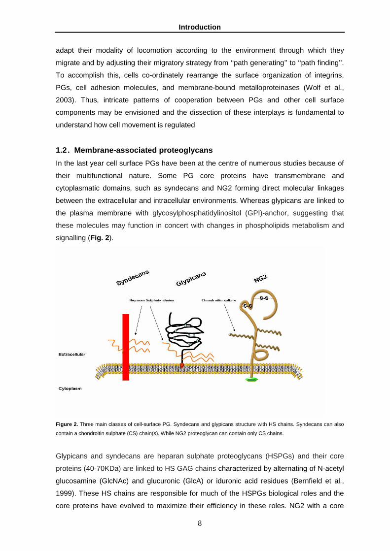
1.2 . Membrane-associated proteoglycans
In the last year cell surface PGs have been at the centre of numerous studies because of
their multifunctional nature. Some PG core proteins have transmembrane and
cytoplasmatic domains, such as syndecans and NG2 forming direct molecular linkages
between the extracellular and intracellular environments. Whereas glypicans are linked to
the plasma membrane with glycosylphosphatidylinositol (GPI)-anchor, suggesting that
these molecules may function in concert with changes in phospholipids metabolism and
signalling (Fig. 2).
Figure 2. Three main classes of cell-surface PG. Syndecans and glypicans structure with HS chains. Syndecans can also
contain a chondroitin sulphate (CS) chain(s). While NG2 proteoglycan can contain only CS chains.
Glypicans and syndecans are heparan sulphate proteoglycans (HSPGs) and their core
proteins (40-70KDa) are linked to HS GAG chains characterized by alternating of N-acetyl
glucosamine (GlcNAc) and glucuronic (GlcA) or iduronic acid residues (Bernfield et al.,
1999). These HS chains are responsible for much of the HSPGs biological roles and the
core proteins have evolved to maximize their efficiency in these roles. NG2 with a core

Introduction
9
protein of 550 and a size of the full molecule of 600KDa is a unique chondroitin sulphate
transmembrane proteoglycan, characterized of alternating sugars (N-acetylgalactosamine
(GalNAc) and glucuronic acid. Unlike to HSPGs, NG2 PGs core protein can be expressed
on the cell surface either with or without chondroitin sulphate GAG chains, placing NG2 in
the category of part-time proteoglycans (Stallcup et al., 1990). Membrane PGs regulate
the distribution of extracellular signalling molecules such as morphogens and chemokines,
and modulate signalling events at the cell surface that influence cell fate determination,
proliferation, adhesion and motility. They participate in endocytosis and vesicular
trafficking, regulating the movement of molecules between intracellular and extracellular
compartments. Several of them have assumed important roles as prognostic factors
and a few of them are now contemplated for therapeutic applications. Literature data
and our preliminary observations indicate that given tumour cells may constitutively
and coincidently express on average 3-5 surface PGs, suggesting that given
combinations of surface PGs may diversely dictate the behaviour of cancer cells,
during distinct phases of tumour progression.
1.3. Syndecans
Syndecans are transmembrane heparan sulphate and comprise a family of four distinct
genes: SDC1, 2, 3 and 4. Their chromosomal locations, exon organization, and sequence
relationships with the single Drosophila syndecan (D syndecan) suggest that the gene
family arose by gene duplication and divergent evolution from a single ancestral gene,
and that SDC-1 and SDC3 and SDC2 and SDC4 represent subfamilies (Bernfield et
al.1999; Fig. 3 ). Each gene product is a single type I membrane-spanning protein with an
apparently extended extracellular domain of variable size that contains covalently
attached HS chains distal from the plasma membrane.

Introduction
10
Figure 3. Dendrogram suggests that the D-Sdc gene is derived from a common ancestor of the four
mammalian syndecan genes and that SDC1 and Synd3 form one subfamily and SDC2 and SDC4 form
another. (Modified from Spring et. ,1994)
Syndecan core proteins (40-50 KDa) contain three different functional domains: the
extracellular, cytoplasmatic and transmembrane domain. The extracellular domain
(ectodomain) is among the most rapidly diverging vertebrate proteins with the exception of
their regions for GAG attachment, cell interaction, proteolytic cleavage, and
oligomerization. Ectodomain contains two regions for GAGs attachment (Esko et al.,
1996; Bourdon et al., 1989). SDC1, 3 and 4 also contain Ser-Gly sequence near the
plasma membrane that may serve as attachment sites for CS (Kokenyesi et al., 1994).
Transmembrane domain is evolutionarily relatively stable and only a few amino acids
differ among the vertebrate sequences. It contains the regions for interactions with other
membrane proteins and for localization to distinct membrane compartments.
Transmembrane domain may also interact within the plane of the membrane with proteins
involved in cell spreading, sensitive to protein tyrosine kinase inhibitors (Lebakken et al,
1996). Despite of the molecular volume of their HS chain it is possible to find these PGs in
oligomers form, especially in dimmers. Both syndecans bearing small HS chains (i.e.
produced by keratinocytes) and syndecans devoid of HS chains are inclined to form
oligomers Sanderson et al., 1992). Oligomerization would enhance the proximity between
syndecan core proteins, enlarging their interaction surface, and increasing the probability
of interaction with other membrane proteins (Klemm et al, 1998). The cytplasmatic
domains contain two invariant regions, a membrane proximal common region (C1)
containing a serine and a tyrosine and a C-terminal common region (C2), separated by a
region (V) of variable length and composition. The C2 region shows an EFYA sequence at
the C-terminus that can bind to the PDZ domains present in intracellular proteins. PDZ

Introduction
11
domains organize and assemble protein complexes on the inner surface of the plasma
membrane and are thought to link membrane components to the underlying actin-
containing cytoskeleton. The variable (V) region is distinct for each of the 4 family
members, suggesting functional difference between these PGs (Bernfield et al., 1999).
The function of this domain is largely unknown except for SDC4, where it is responsible
for the assembly of SDC4 tetramers with phosphatidylinositol 4,5-bisphosphate (PIP2)
and activated protein kinase C-α (PKC-α) in focal adhesions (Woods et al, 2001;
Rapraeger et al., 1998). Instead SDC2 V region serve only as substrata for PCKα (Itano et
al., 1996) Syndecans can be involved in growth control, cell spreading, cellular
recognition, cellular adhesion, and signalling and. Their function as co-receptors with
integrins and cell-cell adhesion molecules (fibronectin, vitronectin, laminins, and the
fibrillar collagens; Bernfield et al, 1999; Thodeti et al., 2003). All syndecans studied can be
shed from the cell surface by proteolytic cleavage near the plasma membrane (Fitzgerald
et al., 2000). Shedding of the syndecan ectodomains is highly regulated and can be
accelerated by various cellular effectors (thrombin and plasmin), which, interestingly, do
not usually bind HS. Syndecans expression levels are regulated during development in
morphogenesis, cell differentiation and they can be altered in tumorigenesis, progression
and metastasis process (Sanderson et al., 2001).
1.4. Glypicans
The first member of the family of GPI-anchored heparan sulphate (HS)-substituted PGs,
later named glypicans (GPC), was identified some 15 years ago (David G. et al., 1990).
Glypicans comprise a family of at least six distinct genes in mammals GPC-1,-2,-3,-4,-5,-6
(Filmus 1998, Watanabe 1995 ) two in Drosophila, called Dally and Dally-like (Nakato et
al., 1995; Baeg et al., 2001) and one C. elegans, called knypek (Fransson et al. 2004). All
glypicans have an N-terminal signal sequence; an ~60-70 kDa globular domain with a
characteristic pattern of 14 highly conserved cysteine residues (CRD region), with the HS
attachment sites; and finally a C-terminal sequence involved in formation of a GPI for
linking to the plasma membrane. Except for the HS attachment sequences, the HS-
bearing juxtamembrane regions are the most divergent sequences in glypican core
proteins (Bernfield et al., 1999). The structures of each glypican are extremely well
conserved across species (more than 90% identical when comparing glypicans from
different vertebrate species; Fig. 4 ).

Introduction
12
Figura 4. Diagrams showing the derived core protein domain organization, locations of putative GAG attachment sites,
dendrogram and aligned sequences of human glypican-1 through -6 and dally, the Drosophila glypican homolog (from
Bernfield 1999)
The hypothesis of different subfamilies is further strengthened by an analysis of the
genomic structures of the corresponding genes as follows: GPC1, GPC2, GPC4, and
GPC6 consist of nine exons, whereas GPC3 and GPC5 contain only eight exons
(Veugelers et al., 1998; Huber et al., 1997). The GPC1, 2, 4, and 6 form a separate group
with high homology (40–60%) to each other but only 20% identity to the other group
comprising GPC3 and GPC5 (also 40% identical to each other). Interestingly, GPC6 is
most homologous to GPC4 (both proteins are 63% identical). This suggests glypicans
arose from a series of gene and genome duplications and may herald extension of the
gene family by additional members and gene clusters (Bernfield et al., 1999). Whether
members of the same subfamily share some common functions and members belonging
to different subfamilies have different functions remains to be established. Glypicans are
linked to membrane lipid without penetring the bilayer. Currently, the functional
significance of attaching proteins through a GPI anchor has not been clearly established.
One of these possible roles is the targeting of GPI-anchored proteins to specific micro-
domains within the cell membrane called ‘rafts’ (Brown et al., 1992) that can facilitate
interactions with specific intracellular signalling molecules in the absence of a cytoplasmic
domain (Ilangumuran et.al., 2000). GPI anchors may also be to provide a system of
regulated release of proteins to the extracellular environment with releasing from cell
surfaces by proteolytic cleavage and by the action of a phosphatidyl inositol-specific
phospholipase C (Ishihara et al, 1997). Moreover, the anchor is reported to mediate the

Introduction
13
turnover of cell surface components by rapid endocytosis and transport to lysosomes
(Yanagashita et al., 1998). In general, glypicans are expressed predominantly during
development (De Cat et al., 2001), the expression levels have been shown to change in a
stage- and tissue-specific manner suggesting that glypicans are involved in
morphogenesis (Saunders et al.,1998; Pellegrini et al.,1998; De Cat et al.,2001; Table 2)
Table 2. The glypican family during embryogenesis and in adult tissues (adapted from Fico et al., 2007).
In general, glypicans interaction of with multiple proteins may be due to the fact that the
length and modifications of the heparan sulphate chains are cell-type-specific (Nurcombe
et al., 1993). It has also be seen that Glypicans regulate the signalling of morphogenesis
through Wnt, Hedgehog (Hh), bone morphogenetic proteins (BMPs), and fibroblast growth
factors (FGFs; Yayon et al., 1991). In fact GPC1 and GPC3 are able to sequester a
variety of growth in particular they can link TGFβ, BMP and Wnt family controlling their
signalling events. Consistent with a proposed role in development, the expression
patterns of these glypicans often coincide with those of growth factors, such as FGFs and
BMPs and their receptors (Veugelers et al., 1999).
1.5. GPC6
Till 1999 five members of the glypican family have been identified in vertebrates. Bio-
informatics analyses have led to the identification of a sixth member of the human
glypican gene family and of its mouse orthologue. As might have been expected from the
characterization of the other members, the human and mouse forms of Gpc-6 are highly
Name Original Designation
Expression in Embryo
Expression in Adult
Reference
Glypican 1 Glypican Bone, bone marrow, muscle epidermis, kidney
Most tissues David et al., 1990; Litwack et al., 1994
Glypican 2 Cerebroglycan Nervous system Not detected Stipp et al., 1994; Ivins et al., 1997
Glypican 3 OCI-5 Most tissues Ovary, mammary gland, mesothelium, lung, kidney
Filmus et al., 1998; Pellegrini et al., 1998; Li et al., 1997; Filmus (unpublished observations)
Glypican 4 K-glypican Brain, kidney, lung Most tissues Watanabe et al., 1995; Veugelers et al., 1998; Siebertz et al., 1999
Glypican 5 Brain, lung, liver, kidney, limb
Brain Veugelers et al., 1997; Saunders et al., 1997
Glypican 6 Many tissues, including liver and kidney
Many tissues including ovary, kidney, liver, and intestine
Paine-Saunders et al., 1999; Veugelers et al., 1999

Introduction
14
similar in structure, with 96% identity (Fig. 5). Besides GPC6 is high similar to GPC4 with
63% identity.
Based on similarities in sequence and gene organization, GPC1, 2, 4, and 6 appear to
define a subfamily of glypicans, differing from the subfamily comprising so far GPC3 and
GPC5. The GPC6 gene maps close to the GPC5 gene on human chromosome 13q32,
whereas GPC4 maps to chromosome Xq26, where it flanks GPC3, the gene encoding the
glypican which so far is most homologous to GPC5. Northern blotting indicates that GPC6
mRNA is widespread, with prominent expressions in human fetal kidney and adult ovary.
In situ hybridization studies localize GPC6 to mesenchymal tissues in the developing
mouse embryo (Sanderson at al., 1996). High expressions occur in smooth muscle cells
lining the aorta and other major blood vessels and in mesenchymal cells of the intestine,
kidney, lung, tooth, and gonad (Veuglers et al.,1999). There is no evidence about
molecular interaction of GPC6 in cell spreading and cell invasion, so GPC6 was
overexpressed in sarcoma cells for studying its involvement in adhesion and migration of
cells.
1.6. NG2/CSPG4
NG2 is a chondroitin sulphate (CS) membrane-spanning protein that interacts with
macromolecules on both sides of the plasma membrane. NG2 has an extensive
extracellular domain of 2195 amino acids and a much smaller cytoplasmic domain of 76
amino acids. A single 25-residue transmembrane domain divides the core protein into a
relatively short 76 amino acid cytoplasmic tail and an extensive 2225-residue extracellular
domain (Fig. 6 ).
Figure 5 . The lengths of the lines in this tree are directly proportional to the predicted genetic distances between
individual sequences. These results indicate that glypican-4 and glypican-6 are in fact more closely related than any
other pair of vertebrate glypicans previously described.

Introduction
15
Figure 6. The N-terminal globular domain 1 is stabilized by intramolecular disulfide bonds (the actual number of disulfides is
not known). The central domain 2 contains both the type VI collagen-binding site and the single chondroitin sulphate chain
(irregular line). The membrane-proximal globular domain 3 contains at least two sites for proteolytic processing of NG2. The
cytoplasmic tail contains a PDZ-binding motif a proline-rich segment, and several potential sites for threonine
phosphorylation .
The extracellular domain of the NG2 core protein contains three subdomains: an N-
terminal globular domain (domain 1), a central extended domain that has the sites for GAG
attachment (domain 2), and a juxtamembrane domain (domain 3). Domain 1 has a
globular conformation stabilized by intramolecular disulphide bonds (Tillet et al., 1997;
Burg et al., 1997); domain 2 contains the sites for binding CS chains (Stallcup et al, 2001)
and it contains α-helical site for collagen type V and VI binding (Tillet, 1997; Burg 1997);
domain 3 is the globular juxtamembrane site for proteolysis of NG2 that leads to its
cleavage and release from the cell surface (Stallcup et al, 2001). At the extreme C-
terminus, a QYWV sequence fits the pattern for a PDZ-binding motif (Songyang et al.,
1997) and may be responsible for interaction of NG2 with MUPP1, a multi- PDZ domain-
containing cytoplasmic scaffolding protein (Barritt et al., 2000). NG2 is expressed by
immature progenitor cells in several different developmental lineages, including
oligodendrocyte progenitors, chondroblasts, and pericytes/smooth muscle cells
(Nishiyama et al., 1991; Grako et al., 1995; 1999; Burg et al., 1999; Schlingemann et al.,
1990). The expression pattern of NG2 on immature progenitor cells suggested that the
NG2 might contribute to processes such as cell proliferation and motility which are critical
to progenitor biology. In fact NG2 is often re-expressed by tumour cells, which are usually
characterized by increased proliferation and migration.

Introduction
16
1.7. Metastasis
Tumour cell invasion and metastasis is highly dependent on dynamic changes in the
adhesion and migration of transformed and malignant cells. Tumour metastasis consists
of a series of biological processes that move tumour cells from the primary neoplasm to a
distant location (Fig. 7).
Figure 7. The tumour metastatic process (From Patricia S Steeg, 2006) At the primary tumour site, tumor cells invade into the lymphatics or directly into the
circulation. Once in the bloodstream, tumour cells must survive and avoid immune attack
to extravasate. Arrest is most often by size restriction in capillary beds but can involve
specific adhesive interactions. The process by which tumour cells form micrometastases
and then progressively growing, vascularized macrometastases in a distant organ is
termed metastatic colonization. Metastatic colonization involves reciprocal interactions
between tumour cells and the microenvironment of the distant organ, and can pause for
periods of dormancy. One of the most enduring observations in metastasis research was
published in 1889 by Stephen Paget. Describing tumour cells as the “seed” and the host
environment as the “soil,” Paget hypothesized that their interaction determines metastatic
outcome: “When a plant goes to seed, its seeds are carried in all directions; but they can
only live and grow if they fall on congenial soil”. This observation predicted that the tissue

Introduction
17
environment, composed of a myriad of specialized cell types, extracellular matrices and
cells recruited to the site, may facilitate tumour metastasis and contribute to the organ
selectivity sometimes seen in metastatic colonization. Complex and redundant pathways
involving the tumour cell and the microenvironment mediate tumour invasion at the
primary site, survival and arrest in the bloodstream, and progressive outgrowth at a distant
site.
Understanding these pathways and their dynamic interactions will help identify promising
molecular targets for cancer therapy and key obstacles to their clinical development.
1.8. Soft Tissue Sarcoma (STS)
Sarcomas are a heterogeneous group of rare tumours that arise predominantly from the
embryonic mesoderm. The various sarcomas include bone sarcomas (osteosarcomas and
chondrosarcomas), Ewing’s sarcomas, peripheral primitive neuroectodermal tumours, and
soft tissue sarcomas, which are the most frequent. In 2004, approximately 8,680 new
cases are expected to be diagnosed in the United States, and 3,660 deaths from soft
tissue sarcomas are predicted, accounting for 0.63% of all cases and 1.15% of deaths
from cancer (Jemal et al, 2004). Currently, more than 50 histologic types of soft tissue
sarcoma have been identified (Table 3 ), but the most common are malignant fibrous
histiocytoma (28%), leiomyosarcoma (12%), liposarcoma (15%), synovial sarcoma (10%),
and malignant peripheral nerve sheath tumours (6%; Coindre et al., 2001).
Rhabdomyosarcoma is the most common soft tissue sarcoma of childhood.
Table 3. Histologic Subtypes of Soft Tissue Sarcoma
Histological Subtypes n % Malignant Fibrous Histiocytoma 349 28 Liposarcoma 188 15 Leiomyosarcoma 148 12 Unclassified sarcoma 140 11 Synovial sarcoma 125 10 Malignant Peripheral Nerve Shealth Tumor 72 6 Rhabdomyosarcoma 60 5 Fibrosarcoma 38 3 Ewing’s Sarcoma 25 2 Angiosarcoma 25 2 Osteosarcoma 14 1 Ephiteliod Sarcoma 14 1 Chondrosarcoma 13 1 Clear cell Sarcoma 12 1 Alveolar Soft Part Sarcoma 7 1 Malignant Hemangiopericytoma 5 0,5
Modified from Coindre et al.
As for most types of tumours, the presence of metastasis at diagnosis, or the evolving of
such lesions with time, catastrophically reduces the probability of survival. Factors
predicting the formation of metastasis in soft-tissue sarcoma patients are not known and
similarly obscure remains the modes through which metastases form in these individuals.

Introduction
18
1.9. Membrane PGs and tumour
One of the most classical experiments demonstrating the importance of cell surface PGs
in tumour progression, and in particular the role of HS-carrying PGs, was originally
performed by Jeffrey Esko and collaborators (Esko et al., 1988). This study highlighted
that cells genetically engineered to be defective in the HS biosynthesis, and thereby
harbouring a compositional deficit of their surface HSPGs, failed to form tumours both in
vitro and in vivo. These findings were strongly corroborated by comparative analyses of
the GAG structures present on the surface of tumour cells with different metastatic
potentials (Cattaruzza et al., 2005). In most solid tumours, especially those of epithelial
origin, clinically orientated surveys have shown a certain prognostic correlation between
the overall levels of surface PGs expression and disease-free survival (Table 4).
Table 4. Potential clinical applications of membran e proteoglycans in the field of oncology (358 papers; 1990-2007)
PG Diagnostic Predictive/prognostic
SDC1/CD138 Mesothelioma Carcinoma: breast, oral squamous cell, pancreatic, cervical, small cell lung, prostate, ovarian
Hodgkin’s lymphoma Myeloma PEL Endometrial cancer Intrahepatic cholangiocarcinoma Mesothelioma
Syndecan-2 Mesothelioma ?
Syndecan-3 ? ?
Syndecan-4 ? ?
GPC-1 ? Pancreatic carcinoma
GPC-2 ?
GPC-3 Hepatocarcinoma (Melanoma)
Hepatocarcinoma
GPC-4 ? ?
GPC-5 ? ?
GPC-6 ? ?
NG2 MLL ALL ?
With the possible exception of breast carcinoma, the initial dysplastic phases of epithelial
tumorigenic involve a pronounced loss of syndecan surface expression, which occurs at
both transcriptional and translational level and is believed to be a prerequisite to attain a
migratory phenotype (Matsumoto et al., 1997; Day et al., 1999). Subsequently, an
accumulation of syndecans in the stromal compartment accompanies an enhanced
invasiveness of the malignant cells and a poor prognosis (Bayer-Garner et al., 2000;
Wicksten et al., 2001; Zellweger et al., 2003; Barbareschi et al., 2003; Leivonen et al.,
2004). Of the four syndecan family members, SDC1 (Fig. 8) is the most extensively
studied and was the first HSPG to be identified and cloned. SDC1 is down regulated in a
variety of cancer tissues (Wiksten, J. P, 2001, Nakanishi, K, 1999) and the loss of SDC1

Introduction
19
expression renders the tumour cells less adhesive, thereby increasing their potential to
metastasize. Instead augmented SDC1 expression is a hallmark of malignancy in multiple
myeloma, several types of lymphomas and certain leukaemia. In B cell lymphomas, cell
surface-bound SDC1 assists the cells in their spreading, cytoskeletal reorganization and
signal transduction (Lebakken et al., 2000; McQuade and Rapraeger, 2003). This since, in
these pathological entities, SDC1 expression seems to inhibit cell–ECM interactions,
haptotactic motility and invasion of three-dimensional matrices/cell monolayers
(Liebersbach and Sanderson, 1994; Liu et al., 1998; Langford et al., 2005; Spessotto et
al., 2001), while favouring cell–cell adhesions through a re-targeted cell surface
redistribution to uropods of polarized cells (Børset et al., 2000).
Conversely, SDC2 seems to be a characterizing constitutive component of metastatic
colon and lung carcinoma cells, where it contributes, alone, or in cooperation with SDC4,
to their recognition and stabilization of contacts with ECM molecules serving as potent
migratory substrates (Kusano et al., 2000; Contreras et al., 2001; Park et al., 2002;
Munesue et al., 2002). The role of SDC2 in cell migration has been less investigated, but
several reports indicate that it may positively regulate cell migration, since it is normally
highly expressed in cells on migratory conditions (Fig. 9 ). Upregulation of SDC4 has been
noted in hepatocellular carcinomas and malignant mesotheliomas and this over
expression may correlate with increased tumour cell proliferation (Beauvais et al., 2004).
Figure 8. Schematic overview of the putative molecular functions of SDC1. Through its HS chains and core protein the PG
may link to ECM components and cooperate with integrins in cell-ECM interactions and/or form oligomeric complexes
activating integrin-independent cytoskeletal rearrangements and signal transduction. SDC1 molecules are also
endocytosed and recycled through specific pathways that coincidently may activate additional signal transduction events.
Introduction
20
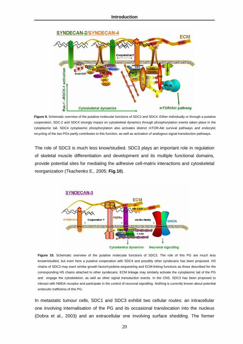
The role of SDC3 is much less know/studied. SDC3 plays an important role in regulation
of skeletal muscle differentiation and development and its multiple functional domains,
provide potential sites for mediating the adhesive cell-matrix interactions and cytoskeletal
reorganization (Tkachenko E., 2005; Fig.10).
In metastatic tumour cells, SDC1 and SDC3 exhibit two cellular routes: an intracellular
one involving internalisation of the PG and its occasional translocation into the nucleus
(Dobra et al., 2003) and an extracellular one involving surface shedding. The former
Figure 9. Schematic overview of the putative molecular functions of SDC2 and SDC4. Either individually or through a putative
cooperation, SDC-2 and SDC4 strongly impact on cytoskeletal dynamics through phosphorylation events taken place in the
cytoplasmic tail. SDC4 cytoplasmic phosphorylation also activates distinct mTOR-Akt survival pathways and endocytic
recycling of the two PGs partly contributes to this function, as well as activation of analogous signal transduction pathways.
Figure 10. Schematic overview of the putative molecular functions of SDC3. The role of this PG are much less
known/studied, but even here a putative cooperation with SDC4 and possibly other syndecans has been proposed. HS
chains of SDC3 may exert similar growth factor/cytokine-sequestring and ECM-linking functions as those described for the
corresponding HS chains attached to other syndecans. ECM linkage may similarly activate the cytoplasmic tail of the PG
and engage the cytoskeleton, as well as other signal transduction events. In the CNS, SDC3 has been proposed to
interact with NMDA receptor and participate in the control of neuronal signalling. Nothing is currently known about potential
endocytic trafficking of this PG.

Introduction
21
translocation routing may reflect a ‘‘functionally unrelated’’ consequence of the loss of
cell–cell contacts upon neoplastic transformation, though the precise function of cytosolic
and intranuclear syndecans still remains to be defined. It is similarly unknown whether
syndecan internalisation is a spontaneous auto-removal process, or whether it is
associated with the transformation-induced endocytosis of putative syndecan ligands. On
the other hand, release of the extracellular syndecan ectodomain involves a well-known
phenomenon of surface shedding driven by MT1-MMP-mediated cleavage of syndecan
and having a potentially multivalent role. Through its activation of the Wnt signalling
pathway syndecan shedding may contribute to the process of tumorigenic (Alexander et
al., 2000) and, via this participation in signal transduction events and the activation of the
avβ3 integrin (Beauvais et al., 2004), it may stimulate cell migration (Endo et al., 2003). In
addition, highly invasive carcinoma and melanoma cells seem to produce syndecans with
a diverse glycanation pattern, which, irrespective of shedding phenomena, confers to
these PGs the unique ability to mediate the cells’ interaction with laminin(s) (Salmivirta et
al., 1994; Engbring et al., 2002). Thus, differential glycosylation of cell surface HSPGs
may provide tumour cells with an adjunct to the repertoire of their ECM interactive
capabilities needed to accomplish optimal migrations. Glypicans may also exert
counteracting roles during neoplastic transformation and tumour progression. The
expression of glypicans seems to be involved in cell proliferation and migration and some
of these proteins can play a double role acting both stimulating and inhibiting proliferation,
in relation to tissue or cellular context (Dietrich et al.,1977). GPC1 expression is up-
regulated in pancreatic cancer cells and in breast cancer. Similarly to the situation with
SDC1, high GPC3 expression seems to protect transformed cells from converting into a
strongly malignant phenotype. This since mammary carcinoma cells engineered to
overexpress GPC3 show reduced local invasiveness and develop fewer metastatic lung
lesions in murine tumour models (Peters et al., 2003). Accordingly, indirect evidence in
the literature suggest that in hepatocellular carcinoma, GPC3 may be released from the
developing tumour cells during early phases of their transformation and malignant
progression and may be absent from the poorly differentiated, metastatic cells (Fig. 11)

Introduction
22
Figure 11. Schematic overview of the putative molecular functions of GPC1 and GPC3. Similarly to syndecans through their
HS chains they are believed to sequester a variety of growth factors and additionally have been shown to also immobilize
TGFβ and BMP antagonists and Wnt family members, thereby participating indirectly in the control of TGF and Wnt
signalling events. There no or very few molecules that are known to specifically associate with the core proteins of these
PGs, but their are postulated to be involved in cell-ECM interactions. Endocytosis of GPC1 has been documented and the
PG may also act intracellularly. For unknown reasons GPC3 may promote, rather than inhibit apoptosis in certain cell types.
GPC5 has recently been found to be up regulated in rhabdomyosarcomas where it seems
to influence malignancy (Williamson et al., 2007; Fig. 12 ).
Figure12 . Schematic overview of the putative molecular functions of the GPC5. As for most other glypicans the biological
function of the PG is still obscure, but it is the first glypican to be discovered to be upregulated in soft-tissue sarcomas (i.e.
rhabdomyosarcomas). Both HGF and FGF family growth factor and ECM linkage are presently the candidate functions of
the GPC5.

Introduction
23
Conversely, virtually nothing is known about the role of other, i.e. GPC2, 4 and 6 in
tumour development. In highly aggressive neoplasia of the nervous system and in
melanoma, NG2 seems to play a fundamental role in conferring to the cells the invasive
and metastatic potential (Chekenya et al., 2002). In fact, if NG2-negative melanoma cells
are transduced with NG2, their ability to give rise to lung metastasis is substantially
increased (Burg et al., 1998). It is also expressed by a number of different types of
tumours, including melanoma (Real et al., 1985), glioblastoma (Schrappe et al., 1991),
chondrosarcoma (Leger et al., 1994), and myeloid leukemia (Smith et al., 1996). NG2
appears to be important for potentiating cell motility (Burg et al., 1997,1998: Fang et al.,
1999; Eisenmann et al., 1999) and for modulating cellular responses to growth factors
(Grako et al., 1995, 1999; Nishiyama et al., 1996b; Goretzki et al., 1999), properties which
are critical for the proliferation and migration of both immature progenitor cells and tumour
cells. Instead, NG2 interactions with ECM molecules regulate signalling pathways
involved in cell proliferation and motility (Iida et al.,1995 ; Tillet et al., 2002; Fig. 13 ).
Linkage of NG2 to ECM components (and its putative interaction with cell surface
molecules) triggers cytoskeletal rearrangements (Majumdar et al, 2003) that may, in part,
be mediated by the multi-PDZ domain adapter molecule MUPP1. In addition, PKCa-
mediated phosphorylation of the threonine residue at position 2256 of the cytoplasmic
NG2 tail allows the clustering of the PG at filodopial tips (Lin et al., 1996a,b).
Simultaneous molecular interactions involving the cytoplasmic portion of NG2 and the
formation of integrin-mediated focal adhesions trigger FAK phosphorylation and down-
stream activation of Rac-1 and cdc42 to further control actin microfilament and
microtubular dynamics in the filopodial and lamellopodial protrusions of the migrating cell
(Majumdar et al, 2003). These signalling cascades also involve activation of the kinase
Ack-1 and a FAK-independent activation of ERK.
Figure 13 . NG2 involvment in regulation of adhesion, migration and invasion of tumour cells. During cancer cell migration,
seems to be the underpinning for the FAK/p130cas-mediated activation of cdc42 and Rac1 implicated in the control of
Introduction
24
microtubular dynamics. NG2 may also modulate the activity of certain integrins, in particular α3β1 and α4 β1 and electively
link to ECM components, where the primary ligand is collagen type VI.
The proposed tumour stimulating action of the NG2 is associated with its ability to function
as a docking receptor for PDGF-AA (Stallcup, 2002) and various members of the FGF
family (Cattaruzza et al., 2008). It may be potentiate the effect of growth factors by
sequestering them at the cell surface and presenting them to their respective signalling
receptors. Also matrix metalloproteinases (MMPs) are another ligand for NG2. In
particular membrane-type 3 matrix metalloproteinase (MT3-MMP) forms a complex with
NG2 that is critical for the ability of melanoma cells to degrade and invade a type I
collagen-containing matrix (Iida et al., 2001). However, NG2 is also strongly implicated in
the neoangiogenic process by sequestering angiostatin and supporting pericyte sprouting
and tubular formation (Goretzki et al., 2000; Chekenya et al., 2002; Ozerdem et al., 2003;
2004; Virgintino et al., 2007a; b) but the precise mode by which NG2 confers to tumour
cells invasive and metastatic capabilities and, thereby, influences progression of tumours
remains to be fully clarified. By virtue of its direct association with the actin cytoskeleton
(Lin et al., 1996a; b; Fang et al., 1999; Barritt et al., 2000), a possible mechanism by
which NG2 may affect tumour development and spreading is by promoting the tumour
cells interplay with neighbouring host cells and ECM constituents. This function is believed
to be exerted through, on one hand, an association of the NG2 with galectin-3 and
integrins α3β1 and α4β1 (Iida et al., 1992; 1995; Eisenmann et at., 1999; Fukushi et al.,
2004), and on the other by assisting tumour cells in their linking to specific collagens, such
as collagen type VI (; Stallcup et al., 1990; Nishiyama and Stallcup, 1993; Burg et al.,
1996; 1997; Tillet et al., 1997; 2002), produced endogenously or by the stromal
compartment. In mesenchymal tumours collagen type VI transcription seems to be down-
regulated as a consequence of neoplastic transformation (Schenker and Trueb, 1998),
whereas it is upregulated in the stromal tissue of breast cancer lesions (Iyengar et al.,
2005). Remodelling of collagen type VI-containing matrices may contribute to the
acquisition of drug resistance in ovarian tumours (Sherman-Baust et al., 2003), probably
because it counteracts the drug-activated apoptotic machinery of the cells and triggers
intracellular signals responsible for cell survival (Rühl et al., 1999a; b). It remains,
however, unknown whether Col VI also promotes tumour spread and metastasis formation
and, if so, through which mechanisms.
1.10. Membrane PGs in sarcoma metastasis
In the last years extensive progress has been made in the identifying of molecules
implicated either cell-ECM interactions, cell-cell interactions or in adhesion and motility
mechanism (Akiyama et al.1993, Yamada et al., 1995). PGs and integrins are molecules

Introduction
25
that are fundamentally important for mediating cell adhesion (Akiyama et al.1993, Albeda
et al., 1990, Hynes et al., 1992). In fact PG binding sites are often expressed in close
proximity to integrin binding domains within ECM molecules or cell surface adhesion
molecules suggest that cellular recognition of the ECM might involve the formation of
receptor clusters on the plasma membrane that include both cell surface PGs and
integrins. It has been seen that NG2 controls tumour progression in melanoma and
sarcoma (Benassi et al., 2009) locally and distantly by accentuating growth responses, by
mediating the tumour cell-host microenvironment interaction, by promoting
neoangiogenesis, and by conferring enhanced drug resistance. In fact NG2 is highly
expressed in melanoma lesions and metastases of soft-tissue sarcomas patients show a
>5-fold increase in the expression of NG2 when compared to the primary lesion(s) of
these individuals (Benassi et al., 2009). Since NG2 proteoglycan was identified as cell
surface ligand for collagen type VI (Stallcup et al., 1990; Midwood and Salter, 2001) we
went to consider as interaction of NG2-collagen type VI was implicated in tumour
progression. To address this possibility we have examined the NG2 and collagen type VI
expression patterns in primitive and secondary lesions of sarcoma patients and addressed
the cellular and molecular mechanisms through which the interaction of these molecules
may propagate tumorigenesis and induce metastasis formation. Instead, the only one
evidence about HSPG involvement in sarcoma tumour is given by GPC5 in
rhabdomyosarcoma. It has been seen that amplification of GPC5 in rhabdomyosarcoma
and its expression plays a role in tumour development (Williamson et al, 2007). GPC5
might be used for therapeutic intervention, as a potential modulator of multiple growth
factors, therapies that reduce the function of GPC5 could affect multiple tumorigenic
pathways. Instead potential immunotherapeutic strategies have recently been described
involving the use of GPC3 (Motomura et al., 2006) and GPC5 might represent a similarly
appropriate target for this approach in rhabdomyosarcoma.

Material and Methods
26
2. Material and Methods

Material and Methods
27
2.1. Cell culture
Estabilished sarcoma and melanoma cell lines (Table 4) A375 cell line were obtained from
ATCC and whereas a number of sarcoma cell lines were estabilished from surgical
specimens obtained from sarcoma patient tretated at CRO Institute (Centre of Oncologic
Reference, Aviano (PN), Italy). All cells were cultured in DMEM (Dulbecco’s modified
Eagle’s medium; 1.0 g/L Glucose; BioWitakker) supplemented with Pen/Strep, 2mM L-
Glutamine and 10% (v/v) fetal bovine serum (FBS, Gibco).
Table 4. List of sarcoma cell lines used
CELL LINE HYSTOTYPE
SK-UT-1 LEIOMYOSARCOMA (uterus)
SKL-MS-1 LEIOMYOSARCOMA (vulva)
Saos2 OSTEOSARCOMA (bone)
143B OSTEOSARCOMA (bone)
MG63 OSTEOSARCOMA (bone)
SW982 SYNOVIAL SARCOMA (synovium)
HT1080 FIBROSARCOMA (connective tissue)
MES-SA UTERINE SARCOMA (uterus)
Hs913T FIBROSARCOMA (metast.to lung)
RD RHABDOMYOSARCOMA (muscle)
SAR 91266 FIBROSARCOMA
A204 RHABDOMYOSARCOMA (muscle)
DMR-SN-8.4.98 LEIOMYOSARCOMA
NTI-LMS-5 LEIOMYOSARCOMA
GCT PLEOMORPH
MALIGNANT HISTIOCYTOMA
SJRH30 RHABDOMYOSARCOMA (muscle)
NTI-LS-1 LIPOSARCOMA
NTI-LMS-3 LEIOMYOSARCOMA
NTI-MFH-3 MALIGNANT FIBROHISTIOCYTOMA
NTI-PNS-1 PNS SARCOMA
NTI-LMS-3 LEIOMYIOSARCOMA
NTI-MFH-1 MALIGNANT FIBROHISTIOCYTOMA
NTI-MS-1 MULLERIAN SARCOMA
SW872 LIPOSARCOMA
NTI-LS-2 LIPOSARCOMA
NTI-LS-3 LIPOSARCOMA
NTI-LS-4 LIPOSARCOMA
NTI-OS-1 OSTEOSARCOMA
NTI-LMS-6 LEIOMYOSARCOMA

Material and Methods
28
The sarcoma cell line GCT was provided by Istituto Zooprofilattico Sperimentale della
Lombardia e dell’Emilia Romagna (Brescia, Italy) and maintained in DMEM medium (1.0 g/L
Glucose) with 10% FBS. Non-manipulated, mock-transfected and NG2 stably transduced
(Burg et al., 1998) murine B16-F10 melanoma cells were grown in DMEM with 10% FBS.
Twenty-one-mer siRNA probes against human NG2, scrambled versions of these probes
were synthesized with the Dicer siRNA Generation kit (Gene Therapy Systems Inc., San
Diego, CA) as previously described (Cattaruzza et al., 2009). In addition siRNA probes
against NG2 and integrin β1 were obtained through Ambion (Austin, TX). Human Umbelical
Vein endothelial cell (HUVEC) were isolated from umbelical cords (ARCI Ospedale S.Maria)
as previously described (Balconi et al., 1986; ). HUVEC were manteined till 4 passage in
medium M199 (Gibco) supplemented with Pen/Strep, 20% FBS, 50mg/ml heparin (Sigma)
and 50ng/ml endothelial cell growth (ECG, Sigma). Human Iliac Venous and Arterial
Endothelial Cells (HIAEC), Human Pulmonary Artery Endothelial Cells (HPAEC) and Human
Coronary Artery Endothelial Cells (HCAEC) were cultured in Endothelial Cell Growth Medium
MV (Cambrex) supplemented with Pen/Strep, 2%FBS, 0.1ng/ml ECG, 1µg/ml Hydrocortison
and 1ng/ml basic Fibroblast Factor . Human Lymphatic Endothelial Cells (HLEC; ScienCell)
were cultured in Endothelial Cell Medium (ECM, ScienCell) supplemented with Pen/Strep,
5%FBS and ECG. All the cells were incubated at 37°C in a humidified atmosphere of 5 %
CO2.
2.2. RNA extraction
Total RNA was extracted from sarcoma cells lines and peripheral blood lymphocytes
obtained after informed consent using TRIzol (Invitrogen) according to the manufacturer’s
instructions. To control no DNA contamination was performed a PCR with primers for RLP41
on reverse-transcribed without RNA as substrate.
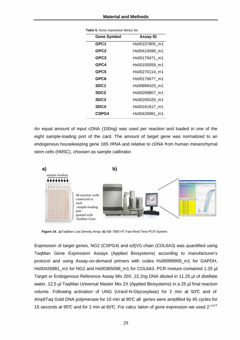
2.3. Real time quantitative PCR
Quantitative PCR reaction was performed on cDNA by ABI PRISM 7900 Sequence Detector
and ABI PRISM 7900 HT Fast Real-Time PCR System (PE Applied Biosystems), with
TaqMan technology. Expression of target genes GPC1, GPC2, GPC3, GPC4, GPC5, GPC6,
SDC1, SDC2, SDC3, SDC4 and CSPG4 was quantified using TaqMan Low Density Array
(TLDA) based on the fluorogenic 5’ nuclease assay (Fig. 14). The assay were chosen
among thr TaqMan Gene Expression Assay library (Table 5 ) and the TLDA was runned on
ABI 7900 HT Fast Real-Time PCR System.
Material and Methods
29
Table 5. Gene expression library list
Gene Symbol Assay ID
GPC1 Hs00157805_m1
GPC2 Hs00415099_m1
GPC3 Hs00170471_m1
GPC4 Hs00155059_m1
GPC5 Hs00270114_m1
GPC6 Hs00170677_m1
SDC1 Hs00896423_m1
SDC2 Hs00299807_m1
SDC3 Hs00206320_m1
SDC4 Hs00161617_m1
CSPG4 Hs00426981_m1
An equal amount of input cDNA (100ng) was used per reaction and loaded in one of the
eight sample-loading port of the card. The amount of target gene was normalized to an
endogenous housekeeping gene 18S rRNA and relative to cDNA from human mesenchymal
stem cells (hMSC), choosen as sample calibrator.
Figure 14. .a )TaqMan Low Density Array; b) ABI 7900 HT Fast Real-Time PCR System.
Expression of target genes, NG2 (CSPG4) and α3(VI) chain (COL6A3) was quantified using
TaqMan Gene Expression Assays (Applied Biosystems) according to manufacturer’s
protocol and using Assay-on-demand primers with codes Hs99999905_m1 for GAPDH,
Hs00426981_m1 for NG2 and Hs00365098_m1 for COL6A3. PCR mixture contained 1.25 µl
Target or Endogenous Reference Assay Mix 20X, 22.2ng DNA diluted in 11.25 µl of distillate
water, 12.5 µl TaqMan Universal Master Mix 2X (Applied Biosystems) in a 25 µl final reaction
volume. Following activation of UNG (Uracil-N-Glycosylase) for 2 min at 50°C and of
AmpliTaq Gold DNA polymerase for 10 min at 95°C all genes were amplified by 45 cycles for
15 seconds at 95°C and for 1 min at 60°C. For calcu lation of gene expression we used 2-∆∆CT
sample-loading
48 reaction wells connected to each sample-loading port spotted with TaqMan Gene
a) b)

Material and Methods
30
comparative method. The amount of NG2, COL6A3 was normalized to an endogenous
reference (GAPDH) and relative to a calibrator (cDNA from healthy lymphocytes).
2.4. DNA extraction
Clones genomic DNA was prepared using InstaGeneTM Matrix 6% (Bio-Rad Laboratories)
according to the manufacturer’s instructions. DNA quality extraction was done by PCR
amplifying the housekeeping gene RLP41 Forward primer GGAGGCCACAGGAGCAGAAA
and Reverse primer TGTCACAGGTCCAGGGCAGA.
2.5. Plasmids and Transfection
143B osteosarcoma cells were stably transfected both with pDisplay GPC-6-HA/pDisplay
empy vector and with pEGFP-C1-GPC6/ pEGFP-C1 empty vectors. pDisplay vector
contained human cDNA of Glypican-6 Hemagglutinin A (HA)-tagged. The expression vector
pDisplay (Invitrogen) containing ORF cDNA of GPC-6 and Hematoglutinin A (HA )-tagged
and the box for G418 resistence (Geneticin/Neomycin). It was gently provided by Guido
David (University of Leuve-CME). pEGFP-C1-GPC-6 vector was obtained cloning GPC-6
cDNA in pEGFP-C1 by PCR reaction with two specific primer- restriction- adapters 3’-SmaI
CAGATCCTCTTCTGAGATGAG and 5’ BclI- GAGGGTGATCAGCCCAGCCGGCCAGATCC.
The PCR product was cut with BclI (New England Biolabs) and SmaI (New England Biolabs)
and purified from the agarose gel using the “QIAquick Gel Extraction Kit” (QIAGEN) in
according to manifacture’s. pEGFP-C1 vector (BD Biosciences) was linearized with BclI and
SmaI for cDNA GPC-6 ligation using T4 DNA Ligase (Biolabs; Pashley C et al., 2003). In
order to select stable transfected cells it was necessary to determine the right concentration
of G418. For this reason a dose-response curve was performed in 143B cells in response to
different concentration of G418, from 100ug/ml up to 1mg/ml. The lower concentration which
kill all cells in two weeks was 500 µg/ml. Cells were transfected with pDisplay-GPC6 vector,
pEGFP-C1-GPC-6 and their respective empty vectors using metafectene-pro (Biontex,
Germany) in according to the manufacturer’s instructions. After twenty-four hours from
transfection the cells were trypsinized and seeded on 10cm dish at low density (6000cells
per dish) and growth in selection condition with G418 (500 µg/ml) for two weeks. For
selecting monoclonal cells the colony were picked with p200 Gilson and were expanded;
clones were screened by PCR amplification for Amp gene Forward primer
GTGTCGCCCTTATTCCCTTT and Reverse primer GGCACCTATCTCAGCGATCT. The
positive clones were also screened for GPC -6 expression wither by real time PCR and
immunonostaining and FACS.
Sarcoma cell lines were also transiently or stably transfected using Lipofectamine Plus as a
delivery vehicle with a plasmid containing either the cytoplasmic tail and membrane-spanning

Material and Methods
31
domain of the PG, or the membrane spanning domain plus the entire ectodomain. Human
NG2 cDNA clones B, C and D (Plusckhe et al.,1996) were cut with XhoI and SacI, SacI and
HindIII, HindIII and BamHI, respectively. These fragments were ligated and inserted into the
pEGFP-N1 vector (Clontech Laboratories Inc.), and the sequence of the entire insert
comprising bases 4030-7216 of the CSPG4 sequence reported with the NCBI accession
number X96753 was verified by automated DNA sequencing. Dominant-negative-like mutant
cells named NG2cyto were generated by transient transfection of the cells previously stably
transduced to express cells with the 3’-end-directed NG2 siRNA probe 1279 to specifically
abrogate the constitutively produced NG2 and spare the transduced deletion construct.
Dominant-negative mutants named NG2extra were generated by stable transfection of the
same cell lines with a GFP-plasmid containing the entire extracellular portion and the
transmembrane domain of human NG2. For this purpose, NG2 cDNA clones H, G and F 29
were cut with XhoI and ApaI, ApaI and BamHI, BamHI and HindIII, respectively, and inserted
between the XhoI and HindIII sites in pEGFP-N1 vector (BD Biosciences). Total cDNA from
A375 melanoma cells was used as the template to amplify the sequence corresponding to
nucleotides 2230-5025, the fragment was inserted into a pGEM-T vector (Promega), and its
sequence verified by automated DNA sequencing. The construct was then subcloned into the
pEGFP-N1 expression vector containing fragments H, G and F as described above (BD
Biosciences). Cells stably transfected with this plasmid were transiently transfected with a 5’-
end-directed NG2 siRNA probe (Cattaruzza et., 2008) to differentially eliminate the
endogenous NG2 without affecting the transduced deletion construct. In both types of
dominant-negative mutants, relative levels of expression of the full-length endogenous NG2,
following siRNA knock-down, versus transduced truncated NG2 were determined at the
mRNA level by quantitative real-time PCR as described above and by FACS and
immunoblotting (Cattaruzza et al., 2009). Deletion constructs lacking different segments of the
putative collagen-binding region of the ectodomain were as previously described (Burg et al.,
1997; Tillet et al., 1997).
2.6. Western blotting
Cells were solubilized in RIPA lysis buffer (Tris-HCl (pH 7.4) 50mM, NaCl 150mM, P-40 1%,
Na-deoxycholate 0.5%, SDS 0.1%, EDTA 2mM, leupeptin 50µM, aprotinin 2µg/ml, soybean
trypsin inhibitor 2µg/ml, pepstatinNa3VO4 1µg/ml, NaF 1mM, Pefabloc SC 0.8mM) after
collection from a 90mm dish and double washing with PBS1X. The protein content in the
different samples was determined using Bradford method (Bradford et al., 1976; Zor et al.,
1995). Surnatant (400 µl) were incubated overnight in -20°C with 2.5 volum es of cool 100%
methanol. The precipitate was washed in cool 100% acetone and recovered for dissolving in
sterile water and 20µl of concentrated media were denatured with 2X sample buffer (final

Material and Methods
32
concentration Tris-HCl (pH 7.4) 0.065M, Glycerol 10.5%, SDS(10%) 21%, Bromophenol
Blue(0.05%) 6.5% ). Lysates (30 µg) and surnatant concentrated were fractionated by SDS–
PAGE using 8% polyacrylamide gels, based upon the expected molecular weight.
Supernatant of stable cell lines were collected after 24 hours of culture incubation in DMEM
serum free and concentrated utilizing methanol protocol (Burbach et al., 2003). The resolved
proteins were blotted to a nitrocellulose membrane (Amersham Pharmacia) by semi-dry
electric transfer, and the membranes were blocked for 1 h in TBS buffer (20 mM Tris–HCl pH
7.6, 137 mM NaCl) containing 5% blotting-grade non-fat milk. Membranes were incubated
with primary antibodies diluted in milk with 0.1% Tween 20 overnight. The following
antibodies were used: anti-HA High Affinity (1:2000; clone 3F10, Roche Diagnostics); anti-
GFP (1:5000; Fitzerald); anti-actin (1:3000; Sigma Aldrich). Membranes were washed three
times in TBS for 5 min each and then incubated in TBS containing the appropriate
horseradish peroxidase conjugated anti-mouse, anti-rat or anti-rabbit secondary antibodies
for 1 h. The membranes were washed three times for 5 min each in TBS with 0.1% Tween 20
and then processed for enhanced chemiluminescence detection according to the
manufacturer's instructions (Amersham Life Science). Equal loading of samples was
confirmed by probing for actin.
2.7. Preparation of ECM substrates
Coverlips and inserts traswell were coating with different ECM molecules (20 µg/ml).
Fibronectin, laminin-1 and vitronectin were dissolved in 0.05 M bicarbonate buffer pH 9,6
(Spessotto et al., 2001) for coating the surfaces for about 1 hour a room temperature and
37°C for vitronectin. Coverslips and the upperside of transwell were washed three time with
PBS. Collagen molecules were dissolved in 0.02N of acetic acid for coating coverslips and
inserts transwell for about 1 hour at room temperature and left them to dry underflow
continuous, in according to the manufacturer’s instructions. Whether upper side and under
side of transwell were coated with Matrigel (BD Biosciences, San Jose, CA). In the latter
case, Matrigel was diluted in cold serum-free DMEM medium at a concentration of 0.5 mg/ml
and 50 µl/well dispensed on top of the porous membrane. The Matrigel solution was then
incubated for 1 hr at 37°C to form a semisolid thin gel across the membrane.
2.8. Immunostaining
Sarcoma cells line were seeded in 24 well coated glass coverslips. Cells were fixed with 4%
paraformaldehyde (PFA) for 15 min and then were washed three times with PBS for 10
minutes. Exogenous GPC6 was detected with anti-HA rat (1:300; Roche) or anti-GFP rabbit
(1:500; Sigma), in PBS with 10% NGS and 0.1% Triton-X 100 at 4°C overnight. Binding of
primary antibody was revealed with specific FITC-conjugated secondary antibody, antirat or

Material and Methods
33
antirabbit Alexa 488 (1:100; Invitrogen), in PBS with 10% NGS and 0,1 % Triton-X 100. F-
actin was visualized with TRITC conjugated phalloidin (Sigma) for 15 minutes at room
temperature. Endosomes was detected using anti-Rab5 (abcam; 1:100), in PBS with
10%NGS and 1%Triton-X 100 overnight at 4°C and foll owed by incubation with secondary
antibody TRITC conjugated antirabbit (1:100, Sigma) in PBS with 10% NGS and 1%Triton-X
100 for 1 hour at room temperature. NG2 was visualized using anti-NG2 monoclonal
antibody, in PBS with 10% NGS, 0.1%Triton-X 100 overnight at 4°C followed by incubation
with TRITC conjugated antimouse (1:100), 10%NGS and Tricton-X 100 for 1 hour at room
temperature. Nuclei were counterstained with Hoechst 33258 (Sigma) for 20 minutes a room
temperature. Samples were washed three times in PBS and mounted with Mowiol 4-88
(Calbiochem-BD Biosciences) supplemented with 2.5 mg/ml DABCO anti-fading reagent
(Sigma-Aldrich).
2.9. FACs analysis
FACS analyses were carried out with the anti-NG2 PE-conjugated antibody 7.1 (Beckman-
Coulter) and anti-GPC6 goat antibody (R&D) for 40 minutes at 4°C. The secondary antibody
conjugated-PE was left 30 minutes at 4°C. For negat ive controls, IgG isotypes were used at
the same concentration as the primary antibody. Immunosorting of NG2+ and NG2- sarcoma
cells was accomplished MACS-based magnetic bead separation using either the anti-NG2
antibody 9.2.27 (Millipore Corporation) or one of our recently generated anti-NG2 monoclonal
antibodies mAb 2161D3 followed by incubation with goat anti-mouse IgG microbeads
(Milteny Biotech, Inc). Relative efficiency of the immunosorting procedure and approximate
yield of NG2+ cells was on average 23.4% for SW982 cells, 13.6% for HT1080 cells, 28.4%
for SK-LMS-1 cells and 18.7% for SKUT-1 cells.
2.10. Cell proliferation assays
For cell growth curves, cells were seeded in triplicate in 6-well culture plates at a density of 1
x 105 cells per well. After 24, 48, 72, 96, hours. Cell number and cell viability were determined
daily using a Burker chamber (Beckman Coulter-Z2) and the trypan blue dye exclusion test.
2.11. Adhesion and spreading assays
Adhesion of siRNA treated and untreated cells to various purified ECM components was
examined using a previously detailed cell adhesion assay denoted CAFCA (Centrifugal
Assay for Fluorescence-based Cell Adhesion; Spessotto et al., 2001), which allows for both
qualitative and quantitative parameters of cell-substratum adhesion to be established. For
the assessment of cell attachment to purified ECM molecules, sarcoma cells were seeded in

Material and Methods
34
serum-free medium and allowed to adhere to different substrates for 30 minutes and fixed
with 4% of PFA. Subsequently the cells were washed three time with PBS and
immunostained. Assessment of cell adhesion to ECM molecules was carried out in multiple
independent adopting 15-20 different representative fields of the substrate with discernable
matrix deposits. The cells were counted under epifluorescence Nikon eclipse E600
microscope
2.12. Cell Migration assays
FATIMA assay (Fluorescence-Assisted Transmigration, Invasion and Motility Assay) was
performed using transwell insert (Falcon HTS FluoroBlock™ Inserts, BD) for evaluating cells
migration in response to ECM molecules and a monolayer of endothelial cells. Scratch assay
was performed using ibidi Culture-Insert (Ibidi Culture System) for evaluating cells motility.
Cell migration, invasion and transendothelial assays were carried out using a modified
version of FATIMA (Fluorescence-Assisted Transmigration, Invasion and Motility Assay;
Spessotto et al., 2001). Transwell inserts (Fig. 15 ) with 8 µm pore size membranes were
coated 20 µg/ml using purified ECM molecules as previously described.
Figure 15. Schematic overview of transwell insert
Cells were previously labeled with Cell Tracker ORANGE and FAST DiTIM (Molecular
Probes) in according to the manufacturer’s instructions. 100000 cells /ml in 200µl DMEM 1%
FBS were seeded on membrane into the upper compartment coated with ECM)molecules.
The lower chamber contained DMEM 10%. After 16 hours the cells were fixed in PFA 4%
and counted using inverted fluorescent Nikon Eclipse TS 40x. In several experiments
invasion assays were performed counting nine fields for each transwell (Fig. 16) In others
the percentage of transmigrated and “nonmigrated” cells was assessed using the
SPECTRAFluor microplate fluorometer (TECAN Group, Maennedorf, Switzerland).
Haptotactic migration and invasion assay of siRNA treated and untreated cells in response to
purified EMC molecules and Matrigel was examined by real-time videomicroscopy. It was
done with an inverted phase contrast microscope (Leica) equipped with an on-stage mini-
chamber providing routine incubation conditions (37°C, 5% CO 2). Phase contrast images
0.8mm
Upper chamber
Microporus membrane (8µm)
Lower chamber

Material and Methods
35
were taken in 5-minutes intervals, contrasted digitally and exported into conventional image
analyses software for elaboration and presentation.
Figure 16. The scheme shows the methodology for counting migrated cells.
Transendothelial assays were performed realizing and endothelial monolayer on the
under side of transwell. 300000 endothelial cells for ml previously stained with Orange
CellTracker were seeded on the upper side and left in incubator for about 2 hours.
Transwells were again reverse in wells of 24 well plate filled with 800µl of endothelial
medium. The confluence of the endothelial cell monolayers was reached after 72 hours
(Fig. 17).
Figure 17. The monolayer of endothelial cells was verified inverted fluorescent Nikon Eclipse TS (40x).
100000 sarcoma cells per ml were previously labeled with Green CellTracker (Molecular
Probes) and seeded into the insert. Kinetic curves of migrated cells was performed
counting cells in different times using inverted fluorescent microscope.

Material and Methods
36
Scratch assays was performed using Ibidi Culture System for making linear scratches of
200 µm thick. Cells were seeded into each well at concentration of 3x10⁵ cell/ml in 70 µl
DMEM 1% FBS and were incubated at 37° and 5% CO 2 C. After about 22 hours the cells
reached confluence and the insert was removed by using sterile tweezers with the result
of two defined monolayer with a scratch of 200 µm thick (Fig. 18).
Figure 18. Steps for the Wound healing assays using Ibidi Culture System.
The wounds are observed using inverted phase contrast microscopy. Images were taken
using 10x magnification at regular intervals over the course of 12–24 hours. Images were
analyzed by digitally drawing lines (using Adobe Photoshop) averaging the position of the
migrating cells at the wound edges. The cell migration distance was determined by
measuring the width of the wound divided by two and by subtracting this value from the
initial half-width of the wound (Valster et al., 2005).

Results
35
3. Results

Results
36
3.1. Constitutive PG mRNA pattern
In order to understand the implication of PG surface profiles in the malignancy traits of
established cell lines and cells isolated in the our laboratory a screening was performed
on 31 sarcoma cells for defining the constitutive pattern of NG2, glypican and syndecan.
PG surface were revealed in each sarcoma cell were showed to have a different and
particular PG pattern (Table 6).
Table.6 . PG Expression pattern in sarcoma cells.
Cells CSPG4 GPC1 GPC2 GPC3 GPC4 GPC5 GPC6 SDC1 SDC2 SDC3 SDC4 GCT + + + + + + HT1080 + + + + + + + + Hs913T nd1 + + + + NTI-FS-1 + + + + + + + + MES-SA + + + + + SK-LMS-1 + + + + + + SK-UT-1 + + + + + + NTI-LMS-1 + + NTI-LMS-3 nd + + + + + + NTI-LMS-5 + + + + + + + NTI-LMS-6 + + + + + + NTI-LMS-8 nd + nd + nd + + + + SW872 nd + + + + + NTI-LS-1 + + + + + + NTI-LS-2 + NTI-LS-3 + nd + nd + + NTI-LS-4 + + + + + + + + NTI-LS-5 + + + + + + NTI-MFH-1 nd + + + + NTI-MFH-3 nd + + + + NTI-MFH-4 + + + + NTI-MS-1 + + + + + + + + 143B + + + + + + + + + + MG63 + + + + SAOS2 + + + + + NTI-OS-1 + + + + + + + + + + RD + + + + + + + A204 nd + + + + + SJRH30 + + + + + + + + + + + SW982 (+)2 + + + + + + NTI-PNS-1 nd + + + + + +
(1 nd: not determined; 2 (+): to confirm)
This first qualitative analysis were showed an average of 5-8 PGs surface in fibrosarcoma
cells. NG2, GPC1, SDC1, SDC3 e SDC4 were always expressed whereas GPC2 and
GPC3 were not revealed and GPC4 was expressed only in HT1080 cells. An average of
6-7 PGs surface were identified in leiomysarcoma, SDC3 and SDC4 were always
expressed whereas GPC2 and GPC3 were not detected; NTI-LMS-1 cells express only
two genes (SDC3 and SDC4). In liposarcoma cells, GPC2, GPC3 and GPC5 were not

Results
37
expressed and GPC4 and SDC2 were expressed respectively only in NTI-LS-4 cells . An
average of 4-5 PGs surface was detected in malignant hystocitoma. GPC2, GPC3, GPC4
and GPC5 were not expressed and NG2 was detected only in NTI-MFH-4 cells. In
osteosarcoma hystotype 10 PG on total of 11 was identified in 143B and NTI-OS-1 cells
even if have got a different genetic profile; whereas NG2, SDC2 and SDC4 were always
expressed. In rhabdomyosarcoma, NG2, SDC2 and SDC4 were always expressed
whereas 11 PGs were detected in SJRH30 cells.
Sarcoma cells were tumor of mesenchymal origin and their PG expression were
compared to cDNA of hMSC cells (calibrator sample) after normalizing to endogenous
housekeeping gene 18S rRNA. The relative expression of surface PGs were showed in
Fig. 19 with the exception of GPC2, GPC3, GPC5 and SDC1 because they were not
exhibited in calibrator sample.
CSPG4 GPC1 GPC2 GPC3 GPC4 GPC5 GPC6 SDC1 SDC2 SDC3 SDC4
GCT
HT1080
NTI-FS-1
MES-SA
SKL-MS-1
SK-UT-1
NTI-LMS-1
NTI-LMS-5
NTI-LMS-6
NTI-LS-1
NTI-LS-2
NTI-LS-3
NTI-LS-4
NTI-LS-5
NTI-MFH-4
NTI-MS-1
143B
MG63
SAOS2
NTI-OS-1
RD
SJRH30 Figure 19. Relative gene expression of sarcoma cells grouped in hystotypes. TaqMan low-density arrays results: gene expression level down-regulate �ddd , up-regulate �ddd or unchanged �ddd in comparison to hMSC as sample calibrator; �ddd indicate that PG was not expressed, �ddd indicate that PG was not expressed in both cells and sample calibrator, �ddd indicate that was not possible to calculate relative expression.

Results
38
The comparison of the PGs relative expression showed different PGs pattern respect to
reference sample in fibrosarcoma cells, in fact all PGs were down regulated in HT1080
cells whereas NG2, GPC1 and SDC4 were up regulated in NTI-FS-1 cells. In
leiomyosarcoma cells all PGs expressed are down regulate and NG2 expression was
50% less than in the sample calibrator (data no show). In liposarcoma cells, NG2
expression was up-regulated in NTI-LS-4 and NTI-LS-5 cells, GPC6 and SDC2 were up
regulated respectively in NTI-LS-4 and NTI-LS-1 cells, instead the other PGs were down
regulated in comparison to calibrator sample. In osteosarcoma cells all PGs were down
regulated with the exception of NG2 that was up regulated in MG63 and Sa-os-2 cells
respect to sample calibrator.
3.2. Model cells with diverse pattern of surface PG s
This preliminary screening has permitted us to identify 24 different model cells (Fig. 20 )
with diverse combinations of surface PGs. It was been found that sarcoma cells may
constitutively and coincidently expressed from 4 to 8 of the 11 surface PGs with the
exception of NTI-LS-2, 143B and SRJH30 cells.
Figure. 20. Overview of 24 model cells characterized by their PG profile
To start to investigate the PG surface profiles as pro- and anti-tumorigenic we delineated
strategies to modify the PG expression of 143B osteosarcoma cells by stable gene

Results
39
transduction and by examining how these modifications affected the cells adhesive and
migratory capabilities in response to selected ECM substrates, and endothelial
monolayers. For this aim, 143B were stable transfected for overexpressing GPC6 that
was chosen as a first PG to modulate since there is currently no knowledge about its
involvement in tumor behaviour.
3.3. GPC6 subcellular localization
143B cells were stable transfected with GPC6 using pDisplayGPC6 vector or pEGFP C1-
GPC6 vector to produce overepressing cells. GPC-6 N-terminal was fused respectively
with HA or eGFP as reporter genes. GPC6 overexpression (143BGPC6+) was detected
by qPCR (data not shown), immunostaining and Western blotting (Fig. 21).
Figure 21 . 143B GPC6 transfected with pEGFPC1GPC6 vector . A). GPC6 detection by immunostaining using anti-GFP
and the nuclei were counterstained with Hoechst 33258 and examinated by fluorescent microscope; B). GPC6 expression
together with GFP protein, the fusion product was about 100KDa; 143B mock (control) and two different selected clones
143B clone 109, 143B clone 111; actin was shown as control loading.
A detail analysis of 143B GPC6 overexpressing cells by confocal laser microscopy and
flow cytometry confirmed the localization of GPC6 on surface (Fig.22 ).

Results
40
.
Figure 22. GPC-6 localization on membrane surface. A). GPC6-HA detection using immunostaining with anti-HA and nuclei
was counterstained with TO-PRO-3. B). GPC6 overexpression was detected using anti-GPC6 in comparison with control.
3.4. Effect of GPC6 overexpression on cell morpholo gy
In order to investigate the relationship between GPC6 overexpression and cell shape, the
143B GPC6 overexpressing cells were stained with phalloidin-TRITC to reveal an actin
filaments organization (F-actin) (Fig. 26 ).
Figure26. Cells morphology was observed with phallodin staining that highlighted actin filament protrusion. GPC6 was
detected with anti-HA and nuclei were counterstained with Hoechst 33258 (60x).

Results
41
143B GPC6 overexpressing cells have showed a markedly different cellular morphology
than the vector control cell line. Morphological changes induced by GPC6 overexpression
could be associated with changes in the organization of their actin filaments. In particular,
phalloidin staining revealed that 143BGPC6 overexpressing cells showed protrusions
similar to fillopodia/lamellipodia. Moreover it also possible to observe a more
concentrations of F-actin in the attachment sites of the cells to the substrate, suggesting a
more pronounced focal contact formation.
3.5. Changes of PG profiles upon modulation of GPC6 overexpression
Modulation of surface PG profile was demonstrated in tumour cells (Timar et al., 1990). To
that end, for analyzing forced expression of GPC6 in 143B cells we also investigated the
relative expression of PGs surface compared to 143B vector control cells.(Fig. 27 )
Figure 27. PGs surface modulation by qPCR. The level of relative expression was calculated using 143b vector control cells
as calibrator sample after normalizing to endogenous housekeeping gene 18S rRNA
It was observed that the PG surface profile in 143B GPC6 overexpressing cells was
modified when compared to control vector cells. First of all 143BGPC6+ showed an
evident over expression of GPC6 (about 500 fold) respect to 143B vector control. GPC1,
GPC2, GPC4 and SDC1 had a little increased of expression respect to control. Whereas it
was interesting to observe an evident over expression of GPC5, about 570 folder than the
control. The misexpression GPC6 on a given background of surface PGs brought an
evident modulation of the other PGs.
3.6. Role of GPC6 in cell-ECM interactions
Modulation of surface PGs profile and changes in morphology of 143B overexpressing
cells had led us to investigate how those alterations could affect the adhesive capabilities
of this cells. A preliminary screening was made analyzing the spreading of 143BGPC6

Results
42
overexpressing cells on different ECM molecules (fibronectin, vitronectin, laminin-1,
collagen type III and V). Cell spreading was evaluated observing the distribution of actin
filaments staining (F-actin) of 143BGPC6+ cells in response to ECM substrates and
uncoated surface (as a control; Fig. 28).
A
B
C
D

Results
43
Figure 28. Adhesion and spreading ability of 143BGPC6+ cells on different molecules substrates: collagene type III (A), collagene type V (B), vitronectin (C), fibronectin (D), laminin-1 (E), uncoated surface (F). The percentage of adhesion 143BGPC6+ was compared to 143B vector control. Valuation of cell adhesion was performed counting about 1000 cells in several fields (average of percentage of cells adhesion to the substrate). The assay was made in triplicate (* p<0,05; ** p<0,01)
The behaviour of 143B overexpressing cells could be concatenated to different surface
PGs profile. 143B GPC6 overexpressing cells showed different ability to adhere on each
ECM molecules in particular on laminin-1 and collagene III substrates. Moreover the
spreading of 143BGPC6+ cells was lowest than the vector control cells on uncoated
surface. It was interesting to look the lacking of stress fibres in both cells on vitronectin
and uncoated substrates.
F
E

Results
44
3.7. Haptotactic migration of 143BGPC6 overexpressi ng cells
Migration ability of 143BGPC6 overexpressing cells was evaluated in response to different
ECM molecules (fibronectin, laminin-1, vitronectin and collagen type III and V). The upper
side of transwell was coated with ECM molecules and the migration of cells was estimated
after 16 hours to ensure that migration could not be affected from cell proliferation. The
migration of 143BGPC6 overexpressing cells was compared with 143B vector control cells
(Fig. 29)
Figure 29. Number of cells migrates in response to ECM molecules. The assay was performed using HTS fluoroblock transwells. Cells were previously labelled with OrangeCell Tracker and the number of cells migrates were counted in nine different fields, using fluorescent inverted microscope (40x). The experiments were made in duplicate (histograms shows an average of experiments in duplicate, * p<0,05).
There was not visualized difference in cells migration but the motility of 143BGPC6
overexpressing cells in response to ECM molecules was resulted to be significantly
different on collagene III and on vitronectin substrates.
3.8. Kinetics of cell motility displayed by 143BGPC 6 overexpressing cells
To study the motility of 143B GPC6 overexpressing cells was performed a wound healing
assay (or scratch assay) using Ibidi Culture System as previously described in material
and methods. The wounds were observed using inverted phase contrast microscopy.
Images were taken at regular intervals 0, 3, 5, 7, and 24 hours (Fig.30) and analyzed for
realizing kinetics curve. 143BGPC6+ cells motility was compared to 143B vector control
cells

Results
45
Figure 30 . Kinetics migration of 143BGPC6+ cells and control cells. (A). The trend of migration was detected using inverted phase contrast microscopy (10x). Scratch assay was made in duplicate and the kinetic curves were showed the same trend; (B). Kinetic curves of 143BGPC6+ and control cells (averages was made by measuring the width of the wound in 5 different points of captured images using draw line of adobe Photoshop).
GPC6 overexpression in 143B cells has permitted the closure of the wound more quickly
than the control cells. Moreover it was possible to observe how the cell motility was
independent from cell proliferation.
A
B

Results
46
3.9. Intracellular fate of transduced GPC6
Modified expression of GPC6 has been observed in 143BGPC6+ cells which could be
attributable to shedding or to enhanced endocytocis of the PG. For detecting an eventual
proteolysis of GPC6 a preliminary analysis was made going to look the GPC6 expression
level in culture medium. The culture medium of 143BGPC6 overexpressing cells was
concentrate (clone 109 and 111) and the protein lysate was used as control (Fig. 31).
Figure 31. Western blotting of GPC6-GFP protein levels in surnatant cultured cells. A). In order to loading surnatant:
concentrated of 143B, 143BGPC6+ clone1, 143BGPC6+ clone2, 143BGPC6GFP+ clone 109, 143BGPC6GFP+ clone 111
and protein lysate of 143B GPC6GFP+ clone 111 B). Ponceau S staining shows protein loading
From this first approach it was possible to observe the lacking of GPC6 protein in culture
medium showing that GPC6 recycling could have another way. Since GPI-anchored
protein were internalized and transported in recycling endosomes the fate of GPC6 was
studied analysing the endosome vesicles with Rab proteins (Rab-5; Fig.32 ).
A
B

Results
47
Figure 32. Colocalization of GPC6 and Rab-5 (arrow). There was not Rab-5 expression in cells where GPC6 has ceased to
be delivered. Images were taken by fluorescent microscope (60x).
Preliminary analyses showed a correlation between expression of GPC6 and Rab-5
suggesting a high endocytotic activity of GPC6. Rab-5 was not revealed in 143B that not
expresses GPC6. This hypothesis will be validated through confocal laser microscope.

Results
48
3.10. NG2 and collagen type VI expression in sarcom a cells
Since NG2 and collagen type VI cooperate in tumour progression (Stallcup et al., 1990)
we have studied how NG2-collagen type VI interactions may influence the behaviour of
sarcoma cells. We have screened the expression of α3(VI) (collagen type VI) and NG2 in
sarcoma cells by qPCR and FACs for looking if there was a relationship between NG2
and collagne VI expression in sarcoma cells. A375 melanoma cells were used as
reference (Table 8 )
Table 8. Expression of NG2 and collagene VI in sarcoma cells
Different expression of NG2 was detected in cells, SW872 cells were showed a high
expression of NG2 (99,4%) than the other cells. High expression levels were detected
also in SK-LM-S-1 (88,5%), MG63 (83,6%), NTI-OS-2 (81,2%) cells. Low levels of NG2
were observed in NT-LS-2 (0,9%), NT-LS-3 (1,7%), NTI-MFH-2 (1,0%) and NTI-LMS-4
(2,3%) cells. Collagen type VI was expressed in SK-LM-S-1, SK-UT-1, GCT, HT1080,
SW872 and NTi-LMS-1 cells. High expression levels of collagene VI were detected in NTI-
FS-1 cells.
3.11. NG2 involvement in the adhesion and spreading of sarcoma cells on
collagen type VI substrates
In order to investigate NG2 involvement in sarcoma cells adhesion on collagen type VI we
performed adhesion assays using CAFCA methodology which allows a quantitative
analysis of cell-substrate interactions. We have chosen sarcoma cells which exhibited
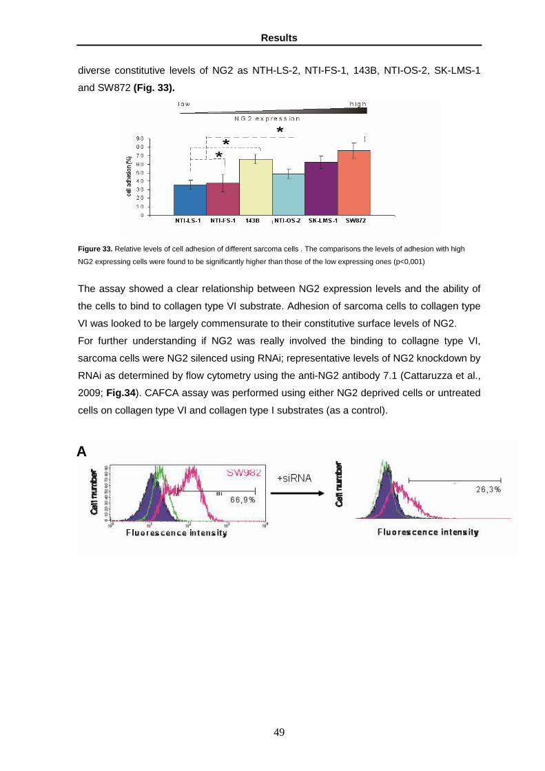
Results
49
diverse constitutive levels of NG2 as NTH-LS-2, NTI-FS-1, 143B, NTI-OS-2, SK-LMS-1
and SW872 (Fig. 33).
Figure 33. Relative levels of cell adhesion of different sarcoma cells . The comparisons the levels of adhesion with high
NG2 expressing cells were found to be significantly higher than those of the low expressing ones (p<0,001)
The assay showed a clear relationship between NG2 expression levels and the ability of
the cells to bind to collagen type VI substrate. Adhesion of sarcoma cells to collagen type
VI was looked to be largely commensurate to their constitutive surface levels of NG2.
For further understanding if NG2 was really involved the binding to collagne type VI,
sarcoma cells were NG2 silenced using RNAi; representative levels of NG2 knockdown by
RNAi as determined by flow cytometry using the anti-NG2 antibody 7.1 (Cattaruzza et al.,
2009; Fig.34). CAFCA assay was performed using either NG2 deprived cells or untreated
cells on collagen type VI and collagen type I substrates (as a control).
A

Results
50
0
20
40
60
80
100
A375 SK-UT-1 SK-LMS-1 A375 SK-UT-1 SK-LMS-1 A375 SK-UT-1 SK-LMS-1 A375 SK-UT-1 SK-LMS-1
FN VN LN-511 (LN-10) Col IVC
ell a
dhes
ion
(%)
Untreated + siRNA
*
*
Figure 34 . Cell adhesion after silencing of NG2. A). NG2 silenced cells were verified by flow cytometry. An exemplified for
the synovial sarcoma cell line SW982. Largely comparable abrogation levels were obtained in the other sarcoma cell lines;
B). Comparing of adhesion of siRNA treated and untreated cells on different matrices quantified using CAFCA system;
p<0,01 C). Quantification of CAFCA system. Images were captured using camera connected phase-contrast microscope
(40x); p<0,01.
Comparison of adhesion of siRNA treated and untreated cells to collagen type VI
underlined that NG2 abrogation significantly reduces both adhesion and spreading of the
cells on this substrate and this effect seems rather specific for this collagen since little or
no inhibition was seen on other representative ECM components.
Since the sarcoma cell lines used for this study were a mixed population of NG2 positive
and NG2 negative cells, SK-UT-1 cells has immunosorted with MACS-based magnetic
beads separation (Cattaruzza et al., 2009). Relative efficiency of the immunosorting
B
C

Results
51
procedure and approximate yield of NG2+ was an average 18,7% for SK-UT-1 cells.
CAFCA assay was performed for SK-UT-1 NG2+ and SK-UT-1 NG2- immunosorted cells
on collagen type VI, fibronectin and collagen type I substrates (Fig. 35 ).
Figura 35. Adhesion of SK-UT-1 NG2+ and NG2- cells imnusorted. A). Representative views of attachment and spreading
of NG2+ and NG2- cells. Cells on the indicated substrates as revealed by phalloidin-Texas red staining. B). Corresponding
quantitative analysis of cell adhesion to the same substrates; * p<0,01.
Comparing the adhesion of two subpopulations on collagen type VI has underlined
different binding and spreading of NG2+ and NG2- cells on substrates. Whereas adhesion
to collagen type I and fibronectin was not distinguishable.

Results
52
3.12. NG2 involvement in the movement of sarcoma ce lls on collagen type VI
substrate
Since NG2 influences the migration of tumour cells we tested migration of sarcoma cells
with different surface levels of NG2 on different ECM substrates (Fig. 36 ).
Figure 36 . Relative levels of cell migration of different sarcoma cell lines exhibiting diverse constitutive levels of NG2
expression. In several pair-wise comparisons the levels migration of high NG2 expressing cells were found to be
significantly higher than those of the low expressing ones (*, p<0,001)
It was interesting to note again sarcoma cells showed a migration ability in response to
collagen type VI substrates that closely correlated with the endogenous levels of NG2
surface expression. In order to examine if NG2 was really involved in migration, sarcoma
cells were NG2 silenced with siRNA. We have chosen to analysis the haptotactic
migration of treated and untreated SK-LMS-1 cells because they had a high migration
percentage and easily detectable. The migration of SK-LMS-1 silenced and untreated
cells were monitored on collagen type IV, collagen type VI and collagen type I (as a
control; Fig 37 ).
Figure 37 . Haptotactic migration of treated siRNA and untreated cells on substrates. Selectively reduced haptotactic
movement of RNA-mediated NG2 knockdown.

Results
53
A 3-dimensional migration assay was performed by coating the transwell upper side with
Matrigel. Collagene type VI was embedded in polymeric collagen type I substrates and
Matrigel (Fig. 38 ).
.
Figure 38. Reduced migration of sarcoma cells in response to Collagene VI after NG2 knockdown when examined in a 3D
setting. Haptotatic migration was detected using SPCTRAfluor fluorometerI (p<0,001). Images were captured using camera
connected to confocal microscopy
Also in this case RNAi-mediated NG2 silencing strongly compromised invasion of cells to
move through collagene VI-containing Matrigel, in comparison to their counterpart.
We examinated the interaction between sarcoma cells and different endothelial cells
(HUVEC, HLEC, HCAEC and HIAEC). Transendothelial migration was performed using
SK-UT-1 NG2+ and NG2- immunosorted. The kinetics of transmigration was examinated
in different times (0, 2, 5, 8, 24 and 30 hours; Fig. 39 ).

Results
54
Figure 39. Transmigration assay. A). Kinetics of migration of two NG2 SK-UT-1 subpopulation; the assay was performed in
duplicated; B). SK-UT-1 NG2+ and NG2- immunosorted cells were previously labelled with GreenCell Tracker. The
endothelial monolayers were labelled with OrangeCellTracker..
The results showed a different transmigration behaviour of SK-UT-1 NG2+ and NG2-
immunosorted cells only on HUVEC but not in others endothelial monolayer. It was
interesting to observe that the percentage of two SK-UT-1 subpopulation migrates in the
others endothelial monolayer less (about 10%) than SK-UT-1 NG2+ (about 25%) on
HUVEC monolayer.

Discussion
55
4. Discussion

Discussion
56
Cell surface PGs are multifunctional molecules with roles in adhesion and growth factor signalling
and are gradually emerging as key regulators of tumour progression. There are currently 11
membrane-bound PGs known in man and a certain number of them have been proposed to
influence tumorigenesis by acting upon local tumour growth and/or by contributing to the metastatic
processes. Increased or de novo induced expression of both secreted and cell surface-associated
PGs may be a characteristic traits of invasive tumours solid. However paradoxically, these
macromolecules often exhibited counteracting tumour growth-promoting and tumour progression-
inhibiting functions depending on the tumour type in which they are mixepressed (Nurcombe et al.,
1993; Cattaruzza et al., 2008). Nevertheless aberrant distribution of PGs observed in various
tumours has incited a wealth of investigations on their potential diagnostic-prognostic, as well as
therapeutic role. Several of these macromolecules are assuming important roles as prognostic
factors and few are approaching the clinics. Given the dual tumour-promoting and tumour-
suppressing role that cells surface PGs may play it is interesting to investigate whether different
combinations of cells surface PGs actually synergize to promote tumour progression or act
antagonistically to impede tumour growth and metastasis formation. The mode by which certain
cell surface PGs may participate in the tumorigenic process has in some cases been extensively
investigated (Fico et al., 2007; Sanderson et al.,2007; Munusue et., 2007; Williamson et. al, 2007)
several have been found to exert a potential antineoplastic role by interfering with cell surface
components regulating cell growth and maintenance of a differentiated phenotype. Since there is
no information about surface PGs may affect the malignancy in sarcomas we have performed a
preliminary screening for determining the surface PG profile in established cell lines and cells
isolated from patients by qPCR and immunocitochemistry. This screening has allowed us to
establish 24 surface PG pattern in sarcoma cells with an average of 5-8 PGs preserved on each
cell type with the exception of 11 PGs expresses in SJRH30 rhabdomyosarcoma cells, 10 PGs
detected in 143B and NTI-OS-5 osteosarcoma cells at the transcript level and only 1 surface PG
expressed by NTI-LS-2 leyomiosarcoma cells. Literature data and our preliminary observations
suggest that given combinations of surface PGs may diversely dictate the behaviour of cancer cells
during distinct phases of tumour progression,. In fact our surface PG screening show how the
same surface PG profile have been identified in cells belong to different hystotypes which could be
in direct relation to the aggressive behaviour of the cells. Characteristically, in progressing tumours
two patterns emerged: loss or neoexpression of surface PG depending on the initial expression
pattern of the cell type of origin (Timar et al, 2002). Altered expression of surface PGs was
detected in sarcomas respect with hMSC cells which could be the putative cell of origin of these
tumours. In this study we aimed the asserting hypothesis that well-defined cell surface PG profiles
may influence in specific way cancer cell motility, spreading and host microenvironment
interactions. The relative expression of surface PGs in 143B osteosarcoma cells showed down
regulation of all PGs. Till to day there are no notice about GPC6 implications either in cell-ECM
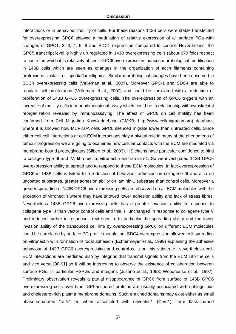
Discussion
57
interactions or in behaviour motility of cells. For these reasons 143B cells were stable transfected
for overexpressing GPC6 showed a modulation of relative expression of all surface PGs with
changes of GPC1, 2, 3, 4, 5, 6 and SDC1 expression compared to control. Nevertheless, the
GPC5 transcript level is highly up regulated in 143B overexpressing cells (about 670 fold) respect
to control in which it is relatively absent. GPC6 overexpression induces morphological modification
in 143B cells which are seen as changes in the organization of actin filaments containing
protrusions similar to fillopodia/lamellipodia. Similar morphological changes have been observed in
SDC4 overexpressing cells (Velleman et al., 2007). Moreover GPC-1 and SDC4 are able to
regulate cell proliferation (Velleman et al., 2007) and could be correlated with a reduction of
proliferation of 143B GPC6 overexpressing cells. The overexpression of GPC6 triggers with an
increase of motility cells in monodimensional assay which could be in relationship with cytoskelatal
reorganization revealed by immunoassaying. The effect of GPC6 on cell motility has been
confirmed from Cell Migration Knowledgebase (CMKB; http://www.cellmigration.org) database
where it is showed how MCF-10A cells GPC6 silenced migrate lower than untreated cells. Since
either cell-cell interactions or cell-ECM interactions play a pivotal role in many of the phenomena of
tumour progression we are going to examinee how cellular contacts with the ECM are mediated via
membrane-bound proteoglycans (Silbert et al., 2003). HS chains have particular confidence to bind
to collagen type III and -V, fibronectin, vitronectin and laminin-1. So we investigated 143B GPC6
overexpression ability to spread and to respond to these ECM molecules. In fact overexpression of
GPC6 in 143B cells is linked to a reduction of behaviour adhesion on collagene III and also on
uncoated substrates, greater adhesion ability on laminin-1 substrate than control cells. Moreover a
greater spreading of 143B GPC6 overexpressing cells are observed on all ECM molecules with the
exception of vitronectin where they have showed lower adhesion ability and lack of stress fibres.
Nevertheless 143B GPC6 overexpressing cells has a greater invasion ability in response to
collagene type III than vector control cells and this is unchanged in response to collagene type V
and reduced further in response to vitronectin. In particular the spreading ability and the lower
invasion ability of the transduced cell line by overexpressing GPC6 on different ECM molecules
could be correlated by surface PG profile modulation. SDC4 overexpression allowed cell spreading
on vitronectin with formation of focal adhesion (Echtermeyer et al., 1999) explaining the adhesive
behaviour of 143B GPC6 overexpressing and control cells on this substrate. Nevertheless cell-
ECM interactions are mediated also by integrins that transmit signals from the ECM into the cells
and vice versa [90-91] so it will be interesting to observe the existence of collaboration between
surface PGs, in particular HSPGs and integrins (Juliano et al., 1993; Woodhouse et al., 1997).
Preliminary observation reveals a partial disappearance of GPC6 from surface of 143B GPC6
overexpressing cells over time. GPI-anchored proteins are usually associated with sphingolipid-
and cholesterol-rich plasma membrane domains. Such enriched domains may exist either as small
phase-separated "rafts" or, when associated with caveolin-1 (Cav-1), form flask-shaped

Discussion
58
plasmalemmal invaginations called caveolae, which are involved in signal transduction and special
forms of non-clathrin-dependent endocytosis mediated by Cav-1-containing endosomes, also
called caveosomes (Drab et al., 2001). GPC1 has been associated with lipid rafts, caveolae,
caveosomes, or endosomes transporting cargo from the plasma membrane to the Golgi. Using
anti-Rab5 as a marker for late endosomes (Barbero et al, 2002), colocalization with GPC6 are
seen in small vesicles in cytoplasm. This observation will be validated with more detailed analysis
using confocal laser microscope.
To dissect the molecular mechanisms that may govern the neoplastic behaviour of sarcoma cells
with different relative surface levels of the above glypicans we will select the model cells that
through the combination of our in vitro and in vivo assays have emerged as those with the
strongest and poorest aggressive phenotype and on these we are performed combined antibody
array- and immunoblotting-based phosphorylation analyses (adopting platforms provided custom
by Kinexus Bioinformatic Corporation, Canada).
The idea that NG2-positive cells may be particularly aggressive was sustained by comparing the in
vivo behaviour of NG2-positive and NG2-negative subsets implanted into immunodeficient mice:
the former sarcoma cells grew into a defined mass more rapidly and more extensively than the
latter ones (Cattaruzza et al., 2009). This observation is consistent with our in vitro findings
showing sustained anchorage-independent growth of NG2+ cells and recalls the potential influence
of the PG in the control of cell survival and proliferation. In fact, in a separate, recent study from
our group we demonstrate that NG2 profoundly affects mitogenic responses of sarcoma cells to
FGF family members (Cattaruzza et al., 2008). However, since some of the NG2+ cells
investigated here failed to engraft and grow in nude mice, the pro-tumorigenic role of NG2 may not
be restricted to a mere control of cell proliferation. Conversely, we hypothesize that high surface
levels of NG2 may confer a more malignant behaviour in sarcoma cells by controlling multiple
facets of the tumourigenic process. Analyses of the in situ distribution of the primary ECM ligand of
NG2 revealed that up regulation of the NG2 in neoplastic cells of primary sarcoma lesions was
accompanied by a corresponding augmented host synthesis and deposition of the collagen within
the lesion (Cattaruzza et al, 2009). It seems that an overabundance of the collagen in the host
microenvironment did not seem to constitute an effective prognostic/predictive factor. A restricted
expression of Collagen type VI in the tumour stroma was also consistent with previous
observations indicating that mesenchymal tumours may loose the expression of the collagen upon
neoplastic transformation (Schenker and Trueb, 1998). Our present data support this notion and
show that only a few of the sarcoma cell lines examined in this study displayed detectable levels of
Collagene VI transcription and translation. Anyway high levels of Collagen type VI deposition in
NG2-rich metastatic lesions suggested to us that the previously reported NG2-collagene type VI
molecular interaction could play a crucial role in tumour cell-host microenvironment interplays, not
Discussion
59
only during local growth but also during the dissemination and homing phases to reach the putative
metastatic sites. We examinated the interactions of NG2- and NG2+ sarcoma cells with collagen
type VI and other ECM molecules. To support this hypothesis sarcoma cells were NG2 abrogated
by RNAi and immunosorted for NG2 expression were found to exhibited a rather elective impaired
ability to adhere and migrate on purified collagene type VI. These findings confirmed that the NG2
was able of mediating tumour cell adhesion and migration on Collagene type VI substrates with a
direct interaction with the collagen and largely precluded that the NG2 acted solely as a modulator
of the function of collagen-binding integrins (in our case of the α2β1 integrin; Chatterjee et al.,
2008). Furthermore, these data strongly supported the notion that enhanced growth and metastatic
behaviour of NG2-expressing sarcoma and melanoma cells could be directly mediated by a local
interaction with collagen type VI present within the tissues/organs infiltrated by metastasizing cells
or overproduced and secreted within the tumour lesions by stroma-forming cells. These preliminary
linear findings corroborated the role of the NG2-ollagene type VI interplay in the potential
regulation of tumour cell spread. NG2 may represent a unique, independent prognostic factor in
several types of soft-tissue sarcomas in which its relative levels of expression in primary lesions
are additionally capable of predicting the future occurrence of post-surgical metastases. Further
multicenter studies are now in progress to establish whether NG2 may be proposed in the routine
clinical practice and in which sarcoma types, else than those examined herein, it may be used as a
metastasis predicting factor.

Acknowledgements
60
5. Acknowledgements

Acknowledgements
61
L'arte e la scienza sono libere e libero ne è l'ins egnamento.
Articolo 33 della Costituzione italiana
Carissimi,
spero di non deludere e di non escludere nessuno!
E ora…apriamo le danze con i rigraziamenti:
al mio ‘tutor’ Prof. Roberto Perris per avermi in parte aiutata nel lavoro di questi lunghissimi 4 anni.
In particolar modo lo ringrazio per il suo sostegno morale;
al President Prof.ssa Ileana Ferrero per il sostegno scientifico e per la sua solare presenza;
a Silvia e Luisa che mi hanno accompagnata e aiutata in questo lavoro di stesura, il vostro
supporto scientifico stato davvero importante. Ops, dimenticavo: “Cavolo mi si è impallato
Photoshop e ora non riesco più a croppare le foto!!”.(Ho evitato di scrivere gli intercalari che ho
usato quella serata);
alla dolce Nicoletta e Giorgia per il loro supporto scientifico;
anche alla dott.ssa Elena Garusi, dott.ssa Silvia Affo e dott. Luca Bruni che hanno contributo al
lavoro insieme alla dott.ssa Silvia Rossi;
a Elisa, Alice e Alba per avere reso le giornate in laboratorio più divertenti;
Grazie agli amici di Parma, di Lodi e di tutto il mondo.
Grazie a mamma, papà, Angelo e Anita.
Questo lavoro lo dedico ad una persona speciale Patrick Indy Stroppa, fondatore della coopertiva
sociale greencoop (www.greencoop.ch), che questa breve vita mi ha permesso di conoscere
anche se per poco tempo. Indy, spero che tu abbia raggiunto la libertà che tanto hai desiderato, un
pezzo del mio cuore è volato via con te. Arrivare al traguardo senza di te è stato molto difficile.
Mm, bzzzzz……
Grazie
Katia Lacrima

Reference
62
6. Reference

Reference
63
Albeda SM, Buck CA .(1990). Integrins and other cell adhesion molecules. FASEB J 4:2869-2879.
Alexander, C.M., Reichsman, F., Hynes, M.T., Lincecum, J., Becker, K.A., Cumberledge, S., Bernfield,
M. (2000). Syndecan-1 is required for Wnt- 1-induced mammary tumorigenesis in mice. Nat. Genet.
25, 329–332.
Akiyama SK, Yamada KM. (1993). Introduction: adhesion molecules in cancer: Part I. Semin Cancer
Biol 4:215-218.
Baeg, G.H., Lin, X., Khare, N., Baumgartner, S., and Perrimon, N. (2001). Heparan sulfate
proteoglycans are critical for the organization of the extracellular distribution of Wingless.
Development. 126:87
Barritt, D. S., Pearn, M. T., Zisch, A. H., et al. (2000). The multi-PDZ domain protein MUPP1 is a
cytoplasmic ligand for the membrane-spanning proteoglycan NG2. J. Cell Biochem. 79, 213–224.
Balconi G, Dejana E. (1986). Cultivation of endothelial cells: limitations and perspectives. Med
Biol.;64(5):231-45. Review.
Bayer-Garner, I.B., Dilday, B., Sanderson, R.D., Smoller, B.R. (2000). Syndecan-1 expression is
decreased with increasing aggressiveness of basal cell carcinoma. Am. J. Dermatopathol. 22, 119–
122.
Barbareschi, M., Maisonneuve, P., Aldovini, D., Cangi, M.G., Pecciarini, L., Angelo Mauri, F.,
Veronese, S., Caffo, O., Lucenti, A., Palma, P.D., Galligioni, E., Doglioni, C. (2003). High syndecan-1
expression in breast carcinoma is related to an aggressive phenotype and to poorer prognosis.
Cancer 98, 474– 483
Barbero G, Pergamenshchik VM. (2002). Intermediate periodic "saddle-splay" nematic phase in the
vicinity of a nematic-smectic-A transition. Phys Rev E Stat Nonlin Soft Matter Phys.; Nov;66
Beauvais, D.M., Rapraeger, A.C. (2004). Syndecans in tumor cell adhesion and signalling,
Reproductive Biology and Endocrinology, 2: 1-12.
Benassi MS, Pazzaglia L, Chiechi A, Alberghini M, Conti A, Cattaruzza S, Wassermann B, Picci P,
Perris R. (2009). NG2 expression predicts the metastasis formation in soft-tissue sarcoma patients. J
Orthop Res. Jan;27(1):135-40.
Bernfield M, Kokenyesi R, Kato M, Hinkes MT, Spring J, Gallo RL, Lose EJ. (1992). Biology of the
syndecans: a family of transmembrane heparan sulfate proteoglycans. Annu Rev Cell Biol. 8:365-93.
Review.

Reference
64
Bernfield, M., Gotte, M., Park, P.W., Reizes O.,Marilyn L. Fitzgerald, Lincecum J., and Zako M. (1999).
Funcion of cell surface heparan sulphate proteoglycans. Annual Review of Biochemistry, 68: 729-777.
Bonaldo, P., Braghetta P., Zanetti, M., Piccolo, S., Volpin, D., Bressan, G.M. (1998). Collagen type VI
deficiency induces early onset myopathy in the mouse: an animal model for Bethlem myopathy. Hum.
Mol. Genet. 7, 2135-2140.
Børset, M., Hjertnern, Ø., Yaccoby, S., Epstein, J., Sanderson, R.D. (2000). Syndecan-1 is targeted to
the uropods of polarized myeloma cells where it promotes adhesion and sequesters heparin-binding
proteins. Blood 96, 2528– 2536.
Bourdon MA, Ruoslahti E. Tenascin mediates cell attachment through an RGD-dependent receptor.
(1989). J Cell Biol. Mar;108(3):1149-55.
Bradford, M. M. (1976) A Rapid and Sensitive Method for the Quantitation of Microgram Quantities of
Protein Utilizing the Principle of Protein-Dye Binding. Anal. Biochem. 72:248-254.
Burbach, B.J., Friedl A, Mundhenke C, Rapraeger AC. (2003). Syndecan Western Blot Procedure.
(2003). Matrix Biology 22:163-177.
Burg, M.A., Tillet, E., Timpl, R., Stallcup, W.B. (1996). Binding of the NG2 proteoglycan to type VI
collagen and other extracellular matrix molecules. J. Biol. Chem. 271, 26110-26116.
Burg, M. A., Nishiyama, A., and Stallcup, W. B. (1997). A central segment of the NG2 proteoglycan is
critical for the ability of glioma cells to bind and migrate toward type VI collagen. Exp. Cell Res., 235,
254–264.
Burg, M., Grako, K. and Stallcup, W. (1998). Expression of the NG2 proteoglycan enhances the
growth and metastatic properties of melanoma cells. J. Cell Physiol. 177, 299-312
Burg, M., Pasqualini, R., Arap, W., Ruoslahti, E. and Stallcup, W. (1999). NG2 proteoglycan-binding
peptides target tumor neovasculature. Cancer Res. 59, 2869-2874.
Brown D.A. (1992). Interactions between GPI-anchored proteins and membrane lipids. Trends Cell
Biol. Nov;2(11):338-43.
Cattaruzza S, Perris R. (2005). Proteoglycan control of cell movement during wound healing and
cancer spreading. Matrix Biol. Sep;24(6):400-17. Review
Cattaruzza, S., Nicolosi, P.A., Perris, R., (2008), Proteoglycans in the control of tumor growth and
metastasis formation, Connect Tissue Res.; 49 (3): 225-9 18661348
Cattaruzza S., Braghetta P., Nicolosi PA. Pazzaglia L., Benassi MS., Bertani N., Lacrima K.,Stallcup
WB., Malatesta P., Colombatti A., Picci P., Bonaldo P:, and Perris R. (2009). NG2 proteoglycan

Reference
65
induced malignancy is linked to its controllo of cancer cell movement through interaction with host
collagen type VI. In press
Chekenya, M., Hjelstuen, M., Enger, P., Thorsen, F., Jacob, A. L., Probst, B., Haraldesth, O.,
Pilkington, G., Butt, A., Levine, J.M. et al. (2002). NG2 proteoglycan promotes angiogenesis-
dependent tumor growth in CNS by sequestering angiostatin. FASEB J. 16, 586-588.
Coindre JM, Terrier P, Guillou L, Le Doussal V, Collin F, Ranchère D, Sastre X, Vilain MO, Bonichon
F, N'Guyen Bui B. Predictive value of grade for metastasis development in the main histologic types of
adult soft tissue sarcomas: a study of 1240 patients. (2001). From the French Federation of Cancer
Centers Sarcoma Group. Cancer. May 15;91(10):1914-26.
Contreras, H.R., Fabre, M., Grane´s, F., Casaroli-Marano, R., Rocamora,N., Garcia Herreros, A.,
Reina, M., Vilaro´ , S. (2001). Syndecan-2 expression in colorectal cancer-derived HT-29 M6 epithelial
cells induces a migratory phenotype. Biochem. Biophys. Res. Commun.286, 742– 751.
David G. Heparan sulphate proteoglycans of human fibroblasts. (1990). Biochem Soc Trans.
Oct;18(5):805-7. Review
Day, R.M., Hao, X., Ilyas, M., Daszak, P., Talbot, I.C., Forbes, A. (1999). Changes in the expression of
syndecan-1 in the colorectal adenoma–carcinoma sequence. Virchows Arch. 434, 121– 125.
De Cat B, David G. Developmental roles of the glypicans. Semin Cell Dev Biol. (2001). Apr;12(2):117-
25. Review
De Cat B, Muyldermans SY, Coomans C, Degeest G, Vanderschueren B, Creemers J, Biemar F,
Peers B, David G. Processing by proprotein convertases is required for glypican-3 modulation of cell
survival, Wnt signaling, and gastrulation movements. (2003). J Cell Biol. Nov 10;163(3):625-35.
Dobra, K., Nurminen, M., Hjerpe, A. (2003). Growth factors regulate the expression profile of their
syndecan co-receptors and the differentiation of mesothelioma cells. Anticancer Res. 23, 2435– 2444.
Drab M, Verkade P, Elger M, Kasper M, Lohn M, Lauterbach B, Menne J, Lindschau C, Mende F, Luft
FC, Schedl A, Haller H, Kurzchalia TV. (2001). Loss of caveolae, vascular dysfunction, and pulmonary
defects in caveolin-1 gene-disrupted mice. Science; Sep 28;293(5539):2449-52.
Echtermeyer F, Baciu PC, Saoncella S, Ge Y, Goetinck PF.(1999). Syndecan-4 core protein is
sufficient for the assembly of focal adhesions and actin stress fibers. J Cell Sci.; 112 ( Pt 20):3433-41.
Eisenmann, K., McCarthy, J., Simpson, M., Keely, P., Guan, J., Tachibana, K., Lim, L., Manser, E.,
Furcht, L. and Iida, J. (1999). Melanoma chondroitin sulfate proteoglycan regulates cell spreading
through Cdc42, Ack-1, and p130cas. Nat. Cell Biol. 1, 507-513

Reference
66
Endo, K., Takino, T., Miyamori, H., Kinsen, H., Yoshizaki, T., Furukawa, M., Sato, H. (2003). Cleavage
of syndecan-1 by membrane type matrix metalloproteinase-1 stimulates cell migration. J. Biol. Chem.
278, 40764– 40770.
Engbring, J.A., Hoffman, M.P., Karmand, A.J., Kleinman, H.K. (2002). The B16F10 cell receptor for a
metastasis-promoting site on laminin-1 is a heparan sulfate/chondroitin sulfate-containing
proteoglycan. Cancer Res. 62, 3549– 3554.
Esko, J.D., Rostand, K.S., Weinke, J.L. (1988). Tumor formation dependent on proteoglycan
biosynthesis. Science 241, 1092–1096.
Esko JD, Zhang L. Influence of core protein sequence on glycosaminoglycan assembly. (1996). Curr
Opin Struct Biol. Oct;6(5):663-70. Review.
Fang, X., Burg, M.A., Barritt, D., Dahlin-Huppe, K., Nishiyama, A., Stallcup, W.B. (1999). Cytoskeletal
reorganization induced by engagement of the NG2 proteoglycan leads to cell spreading and migration.
Mol. Biol. Cell 10, 3373-3387.
Fico A, Maina F, Dono R. (2007). Fine-tuning of cell signalling by glypicans. Cell Mol Life Sci.; Dec 18.
[Epub ahead of print]
Filmus, J., Church, J., and Buick, R.N. (1988). Isolation of a cDNA corresponding to a developmentally
regulated transcript in rat intestine. Mol. Cell. Biol. 8:4243–4249.
Filmus J. Glypicans in growth control and cancer. (2001). Glycobiology Mar;11(3):19R-23R. Review.
Fitzgerald ML, Wang Z, Park PW, Murphy G, Bernfield M. Shedding of syndecan-1 and -4
ectodomains is regulated by multiple signaling pathways and mediated by a TIMP-3-sensitive
metalloproteinase. (2000). J Cell Biol. Feb 21;148(4):811-24.
Fukushi, J., Makagiansar, I.T., Stallcup, W.B. (2004). NG2 proteoglycan promotes endothelial cell
motility and angiogenesis via engagement of galectin-3 and �3�1 integrin. Mol. Biol. Cell 15, 3580-
3590
Fransson LA, Belting M, Cheng F, Jönsson M, Mani K, Sandgren S. (2004). Novel aspects of glypican
glycobiology. Cell Mol Life Sci. May;61(9):1016-24. Review.
Goretzki, L., Burg, M., Grako, K. and Stallcup, W. (1999). High affinity binding of bFGF and PDGF-AA
to the core protein of the NG2 proteoglycan. J. Biol. Chem. 274, 16831-16837.
Goretzki, L., Lombado, C.R., Stallcup, W.B. (2000). Binding of the NG2 proteoglycan to kringle
domains modulates the functional properties of angiostatin and plasmin(ogen). J. Biol. Chem. 275,
28625-28633

Reference
67
Grako, K. A. and Stallcup, W. (1995). Participation of the NG2 proteoglycan in rat aortic smooth
muscle cell responses to platelet-derived growth factor. Exp. Cell Res. 221, 231-240
Grako, K., Ochiya, T., Barritt, D., Nishiyama, A. and Stallcup, W. (1999). PDGF a-receptor is
unresponsive to PDGF-AA in aortic smooth muscle cells from the NG2 knockout mouse. J. Cell Sci.
112, 905-915.
Huber R, Crisponi L, Mazzarella R, Chen CN, Su Y, Shizuya H, Chen EY, Cao A, Pilia G. Analysis of
exon/intron structure and 400 kb of genomic sequence surrounding the 5'-promoter and 3'-terminal
ends of the human glypican 3 (GPC3) gene. (1997). Genomics. Oct 1;45(1):48-58.
Hynes RO (1992). Integrins: versatility, modulation and signalling in cell adhesion. Cell 69:11-24.
Ilangumaran S, He HT, Hoessli DC. Microdomains in lymphocyte signalling: beyond GPI-anchored
proteins. (2000). Immunol Today. Jan;21(1):2-7. Review.
Iida, J., Skubitz, P.N., Furcht, L.T., Wayner, E.A., McCarthy J.B. (1992). Coordinate role of cell surface
chondroitin sulphate proteoglycan and alpha 4 beta 1 integrin in mediating melanoma cell adhesion to
fibronectin. J. Cell. Biol. 118, 431-44
Iida J, Meijne AM, Spiro RC, Roos E, Furcht LT, McCarthy JB. Spreading and focal contact formation
of human melanoma cells in response to the stimulation of both melanoma-associated proteoglycan
(NG2) and alpha 4 beta 1 integrin. (1995). Cancer Res. May 15;55(10):2177-85.
Iida, J., Pei, D., Kang, T., Simpson, M.A., Herlyn, M., Furcht, L.T., McCarthy J.B. (2001). Melanoma
chondroitin sulphate proteoglycan regulates matrix metalloproteinase-dependent human melanoma
invasion type I collagen. J. Biol. Chem. 276, 18786-18794.
Ishihara M, Fedarko NS, Conrad HE. Transport of heparan sulfate into the nuclei of hepatocytes.
(1986). J Biol Chem. Oct 15;261(29):13575-80.
Ishihara M, Kariya Y, Kikuchi H, Minamisawa T, Yoshida K. (1997). Importance of 2-O-sulfate groups
of uronate residues in heparin for activation of FGF-1 and FGF-2. J Biochem. Feb;121(2):345-9.
Itano N, Oguri K, Nagayasu Y, Kusano Y, Nakanishi H, et al. (1996). Phosphorylation of a membrane-
intercalated proteoglycan, syndecan-2, expressed in a stroma-inducing clone from a mouse Lewis
lung carcinoma. Biochem. J. 315:925–30
Iyengar, P., Espina, V., Williams, T.W., Lin, Y., Berry, D., Jelicks, L.A., Lee, H., Temple, K., Graves,
R., Pollard, J., et al. (2005). Adipocyte-derived collagen VI affects early mammary tumor progression
in vivo, demonstrating a critical interaction in the tumor/stroma microenvironment. J. Clin. Invest. 115,
1163-1176.

Reference
68
Jemal A, Tiwari RC, Murray T, Ghafoor A, Samuels A, Ward E, Feuer EJ, Thun MJ; American Cancer
Society. Cancer statistics. (2004). CA Cancer J Clin. Jan-Feb;54(1):8-29. Review.
Juliano RL, Varner JA (1993) Adhesion molecules in cancer: the role of integrins. Curr Opin Cell Biol
5:812–818
Klemm JD, Schreiber SL, Crabtree GR. Dimerization as a regulatory mechanism in signal
transduction. (1998). Annu Rev Immunol;16:569-92. Review
Kokenyesi R, Bernfield M. (1994). Core protein structure and sequence determine the site and
presence of heparan sulfate and chondroitin sulfate on syndecan-1. J Biol Chem. Apr
22;269(16):12304-9
Kurosawa N, Chen GY, Kadomatsu K, Ikematsu S, Sakuma S, Muramatsu. (2001). Glypican-2 binds
to midkine: the role of glypican-2 in neuronal cell adhesion and neurite outgrowth. Glycoconj J. 2001
Jun;18(6):499-507.
Kusano, Y., Oguri, K., Munesue, S., Ishihara, M., Saiki, I., Yonekura, H., Yamamoto, H., Okayama, M.,
(2000). Participation of syndecan-2 in the induction of stress fiber formation in cooperation with
integrin a5β1: structural characteristics of heparan sulfate chains with avidity to COOH-terminal
heparin-binding domain of fibronectin. Exp. Cell Res. 256, 434–444.
Langford, J.K., Yang, Y., Kieber-Emmons, T., Sanderson, R.D. (2005). Identification of an invasion
regulatory domain within the core protein of syndecan-1. J. Biol. Chem. 280, 3467– 3473.
Lebakken CS, Rapraeger AC. Syndecan-1 mediates cell spreading in transfected human
lymphoblastoid (Raji) cells. (1996). J Cell Biol. Mar;132(6):1209-21.
Lebakken, C.S., McQuade, K.J., Rapraeger, A.C. (2000). Syndecan-1signals independently of h1
integrins during Raji cell spreading. Exp.Cell Res. 259, 315– 325.
Leger, O., Johnson-Leger, P., Jackson, E., Coles, B. and Dean, C. (1994). The chondroitin sulfate
proteoglycan NG2 is a tumour specific antigen on the chemically-induced rat sarcoma HSN. Int. J.
Cancer 58, 700-705.
Leivonen, M., Lundin, J., Nordling, S., von Boguslawski, K., Haglund, C. (2004). Prognostic value of
syndecan-1 expression in breast cancer. Oncology 67, 11 – 18.
Liebersbach, B.F., Sanderson, R.D. (1994). Expression of syndecan-1 inhibits cell invasion into type I
collagen. J. Biol. Chem. 269, 20013– 20019.
Lin, X.H., Dahlin-Huppe, K., Stallcup, W.B. (1996a). Interaction of the NG2 proteoglycan with the actin
cytoskeleton. J. Cell. Biochem. 63, 463-477.

Reference
69
Lin, X.M., Grako, K.A., Burg, M.A., Stallcup, W.B. (1996b). NG2 proteoglycan and the actin-binding
protein fascin define separate populations of actin-containing filopodia and lamellipodia during cell
spreading and migration. Mol. Biol. Cell 7, 1977-1993.
Majumdar M, Vuori K, Stallcup WB. Engagement of the NG2 proteoglycan triggers cell spreading via
rac and p130cas. (2003). Cell Signal. Jan;15(1):79-84.
Matsumoto, A., Ono, M., Fujimoto, Y., Gallo, R.L., Bernfield, M.,Kohgo, Y. (1997). Reduced
expression of syndecan-1 in human hepatocellular carcinoma with high metastatic potential. Int. J.
Cancer 74, 482– 491
Matsuda K., Maruyama H., Guo F., Kleeff K., Itakura J., Matsumoto Y. et al. (2001). Glypican-1 is
overexpressed in human breast cancer and modulates the mitogenic effects of multiple heparin-
binding growth factors in breast cancer cells. Cancer Res. 61: 5562–5569
McQuade, K.J., Rapraeger, A.C. (2003). Syndecan-1 transmembrane and extracellular domains have
unique and distinct roles in cell spreading. J. Biol. Chem. 278, 46607– 46615
Motomura Y, Senju S, Nakatsura T, et al. Embryonic stem cell-derived dendritic cells expressing
glypican-3, a recently identified oncofetal antigen, induce protective immunity against highly metastatic
mouse melanoma, B16-10. (2006). Cancer Res;66:2414
Munesue, S., Kusano, Y., Oguri, K., Itano, N., Yoshitomi, Y., Nakanishi, H., Yamashina, I., Okayama,
M. (2002). The role of syndecan-2 in regulation of actin-cytoskeletal organization of Lewis lung
carcinomaderived metastatic clones. Biochem. J. 363, 201–209.
Munesue S, Yoshitomi Y, Kusano Y, Koyama Y, Nishiyama A, Nakanishi H, Miyazaki K, Ishimaru T,
Miyaura S, Okayama M, Oguri K. (2007). A novel function of syndecan-2, suppression of matrix
metalloproteinase-2 activation, which causes suppression of metastasis.. J Biol Chem.; Sep
21;282(38):28164-74.
Nanki N, Fujita J, Yang Y, Hojo S, Bandoh S, Yamaji Y, Ishida T. (2001). Expression of oncofetal
fibronectin and syndecan-1 mRNA in 18 human lung cancer cell lines. Tumour Biol 22: 390–396.
Nakanishi, K., Yoshioka, N., Oka, K., and Hakura, A. (1999). Int. J. Cancer 80, 527-532.
Nakato H, Futch TA, Selleck SB. The division abnormally delayed (dally) gene: a putative integral
membrane proteoglycan required for cell division patterning during postembryonic development of the
nervous system in Drosophila. (1995). Development. Nov;121(11):3687-702.
Nishiyama, A., Dahlin, K., Prince, J., Johnstone, S. and Stallcup, W. (1991a). The primary structure of
NG2, a novel membrane-spanning proteoglycan. J. Cell Biol. 114, 359-371.

Reference
70
Nishiyama, N., Stallcup, W.B. (1993). Expression of NG2 proteoglycan causes retention of type VI
collagen on the cell surface. Mol. Biol. Cell 4, 1097-1108.
Nishiyama, A., Lin, X., Giese, N., Heldin, C. and Stallcup, W. (1996a). Colocalization of NG2
proteoglycan and PDGF alpha-receptor on O2A progenitor cells in the developing rat brain. J.
Neurosci. Res. 43, 299-314.
Nishiyama, A., Lin, X., Giese, N., Heldin, C. and Stallcup, W. (1996b). Interaction between NG2
proteoglycan and PDGF alpha-receptor on O2A progenitor cells is required for optimal response to
PDGF. J. Neurosci. Res. 43, 315-330
Nurcombe V, Ford MD, Wildschut JA, Bartlett PF. (1993). Developmental regulation of neural
response to FGF-1 and FGF-2 by heparan sulfate proteoglycan. Science.; Apr 2;260(5104):103-6.
Ozerdem, U., Stallcup, W.B. (2003). Early contribution of pericytes to angiogenic sprouting and tube
formation. Angiogenesis 6, 241-249.
Ozerdem, U., Stallcup, W.B. (2004). Pathological angiogenesis is reduced by targeting pericytes via
the NG2 proteoglycan. Angiogenesis 7, 269-276.
Paget, S. The distribution of secondary growths in cancer of the breast. (1889). Lancet 1, 571–573.
Paine-Saunders S, Viviano BL, Saunders S. (1999). GPC6, a novel member of the glypican gene
family, encodes a product structurally related to GPC4 and is colocalized with GPC5 on human
chromosome 13. Genomics; May 1;57(3):455-8.
Patricia S Steeg. Tumor metastasis: mechanistic insights and clinical challenges. (2006). Nature
Medicine. 12, 895 - 904
Park H, Kim Y, Lim Y, Han I, Oh ES. (2002). Syndecan-2 mediates adhesion and proliferation of colon
carcinoma cells. J Biol Chem 277: 29730–29736.
Pellegrini M., G. Pilia, S. Pantano, F. Lucchini, M. Uda, M. Fumi, A. Cao, D. Schlessinger, A.
Forabosco. (1998). Dev. Dyn. 213 431–439
Perris, R., Krotoski, D., Bronner-Fraser, M. (1991). Collagens in avian neural crest development:
distribution in vivo and motility-promoting ability in vitro. Development 113, 969-984.
Perris, R., Kuo, H.-J., Glanville, R., Leibold, S., Bronner-Fraser, M. (1993). Neural crest cell interaction
with collagen type VI is mediated by cooperative interaction sites within triple-helix and globular
domains. Exp. Cell Res. 209, 103-117.

Reference
71
Peters, M.G., Farias, E., Colombo, L., Filmus, J., Puricelli, L., Bal de Kier Joffe, E. (2003). Inhibition of
invasion and metastasis by glypican-3 in a syngeneic breast cancer model. Breast Cancer Res. Treat.
80, 221–232
Pluschke, G., Vanek, M., Evans, A., Dittmar, T., Schmid, P., Itin, P., Filardo, EJ., Reisfeld, RA. (1996).
Molecular cloning of a human melanoma-associated chondroitin sulphate proteoglycan. Proc. Natl.
Acad. Sci. USA. 93, 9710-90715.
Rapraeger, A.C., Otto, V.L. (1998), Molecular interactions of the syndecans core proteins, Current
Opinion in Cell Biology, 10: 620-628.
Real, F., Houghton, A., Albino, A., Cordon-Cardo, C., Melamed, M., Oettgen, H. and Old, L. (1985).
Surface antigens of melanomas and melanocytes defined by mouse monoclonal antibodies: specificity
analysis and comparison of antigen expression in cultured cells and tissues. Cancer Res. 45, 4401-
4411
Rühl, M., Johannsen, M., Atkinson, J., Manski, D., Sahin, E., Somasundaram, R., Riecken, E. O.,
Schuppan, D. (1999a). Soluble collagen VI induces tyrosine phosphorylation of paxillin and focal
adhesion kinase and activates the MAP kinase erk2 in fibroblast. Exp. Cell Res. 250, 548-557.
Rühl, M., Sahin E., Johannsen, M., Somasundaram, R., Manski, D., Riecken, E.O., Schuppan, D.
(1999b). Soluble collagen VI drives serum-starved fibroblasts through S phase and prevents apoptosis
via down-regulation of Bax. J. Biol. Chem. 26, 34361-34366
Sabatelli, P., Bonaldo, P., Lattanzi, G., Braghetta, P., Bergamin, N., Capanni, C., Mattioli, E.,
Columbaro, M., Ognibene, A., Pepe, G., Bestini, E., Merlini, L., Maraldi, N.M., Squarzoni, S. (2001).
Collagen VI deficiency affects the organization of fibronectin in the extracellular matrix of cultured
fibroblasts. Matrix Biol. 20, 475-486
Salmivirta, M., Mali, M., Heino, J., Hermonen, J., Jalkanen, M. (1994). A novel laminin-binding form of
syndecan-1 (cell surface proteoglycan) produced by syndecan-1 cDNA transfected NIH-3T3 cells.
Exp. Cell Res. 215, 180– 188.
Sanderson RD, Hinkes MT, Bernfield M. (1992). Syndecan-1, a cell-surface proteoglycan, changes in
size and abundance when keratinocytes stratify. J Invest Dermatol.; Oct;99(4):390-
Sanderson, R.D. (2001). Heparan sulfate proteoglycans in invasion and metastasis, Semin. Cell Dev.
Biol. 12, 89-98.
Sanderson RD, Yang Y. 2007. Syndecan-1: a dynamic regulator of the myeloma microenvironment.
Clin Exp Metastasis (Epub ahead of print)

Reference
72
Saunders S, Bernfield M. (1988). Cell surface proteoglycan binds mouse mammary epithelial cells to
fibronectin and behaves as a receptor for interstitial matrix. J Cell Biol. Feb;106(2):423-30.
Schenker, T., Trueb, B. (1998). Down-regulated proteins of mesenchymal tumor cells. Exp. Cell Res.
239, 161-168.
Sherman-Baust, C.A., Weeraratna, A.T., Rangel, L.B.A., Pizer, E.S., Cho, K.R., Schwartz, D.R.,
Shock, T., Morin, P.J. (2003). Remodeling of the extracellular matrix through overexpression of
collagen VI contributes to cisplatin resistance in ovarian cancer cells. Cancer Cell 3, 377-386.
Schlingemann, R., Rietveld, F., de Waal, R., Ferrone, S. and Ruiter, D. (1990). Expression of the high
molecular weight melanoma-associated antigen by pericytes during angiogenesis in tumors and in
healing wounds. Am. J. Pathol. 136, 1393-1405.
Schrappe, M., Klier, F., Spiro, R., Waltz, T., Reisfeld, R. and Gladson, C. (1991). Correlation of
chondroitin sulfate proteoglycan expression on proliferating brain capillary endothelial cells with the
malignant phenotype of astroglial cells. Cancer Res. 51, 4986-4993
Silbert JE, Sugumaran G (2003) A starting place for the road to function. Glycoconj 19:227–237
Smith, F. O., Rauch, C., Williams, D. E., March, C. J., Arthur, D., Hilden, J., Lampkin, B. C., Buckley,
J. D., Buckley, C. V., Woods, W. G. et al. (1996). The human homologue of rat NG2, a chondroitin
sulphate proteoglycan, is not expressed on the cell surface of normal hematopoietic cells but is
expressed by acute myeloid leukemia blasts from poor-prognosis patients with abnormalities of
chromosome band 11q23. Blood 87, 1123- 1133
Songyang, Z., Fanning, A. S., Fu, C., et al. (1997) Recognition of unique carboxyl- terminal motifs by
distinct PDZ domains. Science 275, 73–77.
Spessotto, P., Giacomello, E., Perris, R. (2001). Improving fluorescence-based assays for the in vitro
analysis of cell adhesion and migration. Mol. Biotech. 18, 1-16.
Spessotto P, Yin Z, Magro G, Deutzmann R, Chiu A, Colombatti A, Perris R. (2001) .Laminin isoforms
8 and 10 are primary components of the subendothelial basement membrane promoting interaction
with neoplastic lymphocytes. Cancer Res. Jan 1;61(1):339-47.
Spring J, Paine-Saunders SE, Hynes RO, Bernfield M. (1994. ). Drosophila syndecan: conservation of
a cell-surface heparan sulfate proteoglycan.. Proc Natl Acad Sci U S A. Apr 12;91(8):3334-8.
Stallcup, W.B., Dahilin, K., Healy, P. (1990). Interaction of the NG2 chondroitin sulfate proteoglycan
with type VI collagen. J. Cell Biol. 111, 3177-3188.

Reference
73
Stallcup, W. B., and Dahlin-Huppe, K. (2001). Chondroitin sulfate and cytoplasmic domain-dependent
membrane targeting of the NG2 proteoglycan promotes retraction fiber formation and cell polarization.
J. Cell Sci. 114, 2315–2325
Stallcup, W.B. (2002). NG2 proteoglycan: past insights and future prospects. J. Neurocytol. 31, 423-
435.
Sung Y. K., Hwang S. Y., Park M. K., Farooq M., Han I. S., Bae H. I. et al. (2003) Glypican-3 is
overexpressed in human hepatocellular carcinoma. Cancer Sci. 94: 259–262
Spring J, Paine-Saunders SE, Hynes RO,Bernfield M. (1994). Proc. Natl. Acad. Sci. USA 91:3334–38
Tillet, E., Ruggiero, F., Nishiyama, A., and Stallcup, W. B. (1997). The membrane- spanning
proteoglycan NG2 binds to collagens V and VI through the central nonglobular domain of its core
protein. J. Biol. Chem. 272, 10,769–10,776
Tillet, E., Gential, B., Garrone, R., Stallcup, W.B. (2002). NG2 proteoglycan mediates beta-1 integrin-
independent cell adhesion and spreading on collagen VI. J. Cell. Biochem. 86, 726-736.
Tímár J, Lapis K, Dudás J, Sebestyén A, Kopper L, Kovalszky I. Proteoglycans and tumor
progression: Janus-faced molecules with contradictory functions in cancer. (2002). Semin Cancer Biol.
Jun;12(3):173-86. Review.
Thodeti, C.K., Albrechtsen, R., Grauslund, M., et al. (2003). ADAM12/ syndecan-4 signaling promotes
beta(1) integrin-dependent cell spreading through protein kinase C alpha and RhoA, Journal of
Biological Chemistry, 278: 9576-9584
Tkachenko E., Rhodes J.M., Simons M. (2005), Syndecans: New kids on the signaling block. Circ.
Res. 96, pp. 488–500
Topczewsky, J., Sepich, D. S., Myers, D. C., Walker, C., Amores, A., Lele, Z., Hammerschmidt, M.,
Postlethwait, J., and Solnica-Krezel, L. (2001). Dev. Cell 1, 251–26
Valster A, Tran NL, Nakada M, Berens ME, Chan AY, Symons M. Cell migration and invasion assays.
(2005). Methods. Oct;37(2):208-15
Velleman SG, Coy CS, McFarland DC. (2007). Effect of syndecan-1, syndecan-4, and glypican-1 on
turkey muscle satellite cell proliferation, differentiation, and responsiveness to fibroblast growth factor
2. Poult Sci. Jul;86(7):1406-13.
Veugelers, M.&David, G. (1998).The glypicans: a family of GPI-anchored heparan sulfate
proteoglycanswith a potential role in the control of cell division. Trends Glycosci. Glycotechnol. 10,
145:152

Reference
74
Veugelers, M., De Cat, B., Ceulemans, H., Bruystens, A.M., Coomans, C., Durr, J., Vermeesch, J.,
Marynen, P., and David, G. (1999) Glypican-6, a new member of the glypican family of cell surface
proteoglycans. J. Biol. Chem., 274, 26968–26977.
Virgintino, D., Girolamo, F., Errede, M., Capobianco, C., Robertson, D., Stallcup, W.B., Perris, R.,
Roncali, L.. (2007a). An intimate interplay between precocious, migratine pericytes and endothelial
cells governs human fetal brain angiogenesis. Angiogenesis 10, 35-45.
Virgintino, D., Ozerdem, U., Girolamo, F., Roncali, L., Stallcup, W.B., Perris, R. (2007b). Reversal of
cellular roles in angiogenesis: implications for anti-angiogenic therapy. J. Vasc. Res. 45, 129-131.
Watanabe, K., Yamada, H., and Yamaguchi, Y. (1995). K-glypican: a novel GPI-linked heparan sulfate
proteoglycan that is highly expressed in developing brain and kidney. J. Cell Biol. 130:1207–1218.
Wiksten JP, Lundin J, Nordling S, Lundin M, Kokkola A, von Boguslawski K, Haglund C. (2001).
Epithelial and stromal syndecan-1 expression as predictor of outcome in patients with gastric cancer.
Int J Cancer 95: 1–6.
Williamson D, Selfe J, Gordon T, Lu YJ, Pritchard-Jones K, Murai K, Jones P, Workman P, Shipley J.
(2007). Role for amplification and expression of glypican-5 in rhabdomyosarcoma. Cancer Res.; Jan
1;67(1):57-65.
Woodhouse EC, Chuaqui RF, Liotta LA (1997) General mechanisms of metastasis. Cancer 80:1529–
1537
Wolf, K., Mazo, I., Leung, H., Engelke, K., von Adrian, U.H., Deryugina, E.I., Strongin, A.Y., Bro¨cker,
E.-B., Friedl, P. (2003). Compensation mechanism in tumour cell migration: mesenchymal –amoeboid
transition after blocking of pericellular proteolysis. J. Cell Biol. 160, 267–277
Woods, A. and Couchman, J. R. (1994). Syndecan-4 heparan sulphate proteoglycan is a selectively
enriched and widespread focal adhesion component, Mol. Biol. Cell 5, 183-192
Woods A, Couchman JR. (2001). Syndecan-4 and focal adhesion function.. Curr Opin Cell Biol.
Oct;13(5):578-83. Review
Yamada KM, Miyamoto S. (1995). Integrin transmembrane signalling and cytoskeletal control. Curr
Opin Cell Biol 7:681-689
Yanagishita, M. (1998). Cellular catabolism of heparan sulfate. proteoglycans. Trends Glycosci.
Glycotechnol. 10, 57–63.
Yayon, A., Klagsbrun, M., Esko, J.D., et al. (1991). Cell surface heparinlike molecules are required for
binding of basic fibroblast growth factor to its high affinity receptor, Cell, 64: 841-848.

Reference
75
Zellweger T, Ninck C, Mirlacher M, Annefeld M, Glass AG, Gasser TC, Mihatsch MJ, Gelmann EP,
Bubendorf L. (2003). Tissue microarray analysis reveals prognostic significance of syndecan-1
expression in prostate cancer. Prostate 55: 20–29.
Zor, T. and Selinger, Z. (1995). Linearization of the Bradford protein assay increases its sensitivity:
theoretical and experimental studies. Anal. Biochem. 236:302-8