The Role of Cardiac Magnetic Resonance for Arrhythmic Risk ...
19
Role of Advanced Cardiac Magnetic Resonance Imaging in Atypical Cardiomyopathies such as
Stress-Induced Cardiomyopathie and Left-Ventricular Non-Compaction
Cardiomyopathy
Oliver Strohm1,2, Abdullah Shehab3 and Anwer Qureshi4 1University of Calgary,
2Center for Cardiology and Pediatric Cardiology Baden-Baden, 3UAE University,
4Tawam Hospital, Al Ain, 1Canada
2Germany 3,4United Arab Emirates
1. Introduction
Magnetic Resonance Imaging (MRI) is a medical imaging technique that has been in clinical use since the 1980’s, and has advanced significantly in the last decade. MRI scans are usually performed at magnetic field strengths of 1.5 to 3.0 Tesla. Such a magnetic field is at least 25,000 times stronger than that of the earth and is required to align and modify the behaviour of unevenly charged protons in the patient’s body. Brief radio frequency pulses with specific properties are applied, which change the axis of rotation (precession) in the protons. The protons rapidly resume their original precession angle and emit a small signal, which is used to compute images. Importantly, the behaviour of resuming their previous state is strongly dependent on the molecular environment; in other words: different tissue composition creates different image contrasts (14-16) The main advantages of MRI techniques include their completely non-invasive nature, lack of any harmful radiation or radioactivity, and the lack of need for Iodine-based contrast agents. It is considered to be free of any harm to patients, but – since metal is affected in a magnetic field – it may not be performed if the patient carries certain magnetic material such as specific electronic devices or some metallic implants. The most commonly performed MRI scans are of the head, brain, spine, large joints and abdomen (15). Cardiac applications of MRI have been applied since the late 1980’s and have evolved into a robust, clinically valid application due to recent advances in both hardware and software. CMR provides a variety of valuable diagnostic information including anatomy and function of the heart, flow of blood, and perfusion and viability of the myocardium. Its most unique advantage is the ability to visualize tissue characteristics selectively based on molecular environments, adding a new and valuable piece of information to medical testing
www.intechopen.com
Cardiomyopathies – From Basic Research to Clinical Management
426
Contrast-enhanced cardiac magnetic resonance imaging (CMR) allows for a non-invasive assessment of the tissue composition using contrast-free (T2, e.g. STIR) and contrast-enhanced techniques (early and late Gadolinium enhancement). Together with a functional assessment, it can be used to determine the acuity of e.g. inflammatory diseases and provide a non-invasive follow-up tool.
1.1 CMR techniques used Functional imaging with high-resolution sequences such as SSFP cines allow to assess the whole left and right ventricles and calculate volumes, ejection fraction and mass. Myocardial wall stress can be calculated from these. Assessment of valvular function is needed and may require additional flow studies for the calculation of regurgitation fraction of aortic and mitral valve. Assessment of the pericardium can be done on the functional images, too. T2-weighted images with fat suppression allow assessing myocardial water content, thus allowing to assess the stage of disease. To provide an “internal standard”, skeletal muscle is used as control; an SI-ratio of more than 2.0 is considered abnormal. T1-weighted images allow to demonstrate acute inflammatory changes including increased extracellular volume and membrane integrity. As in T2-weighted imaging a skeletal muscle is used as “internal standard”; an enhancement – ratio (myocardial enhancement / muscle enhancement) of more than 4.0 is considered abnormal. The body-coil is used to obtain homogenous SI through the images, short axis or axial images are selected to optimize image quality. Newer sequences may improve image quality and allow for the use of multi-element coils. Late Gadolinium enhancement allows to non-invasively diagnose irreversible damage in the myocardium (e.g. fibrosis, infarcts). Due to the specific location in ischemic damages (starts at the subendocardial layer), it is easy to distinguish non-ischemic damages (as in myocarditis) from ischemic problems. Combining T2-information, early and late enhancement, CMR is able to safely assess the acuity and reversibility of the disease process in non-ischemic Cardiomyopathies and inflammatory processes (16).
2. Left ventricular non-compaction
2.1 Introduction Left ventricular non-compaction (LVNC) is a distinct cardiomyopathy resulting from arrest of fetal development of the heart (1). This leads to altered myocardial architecture that is seen as a two layered myocardium with a thin, compacted epicardial layer and a thick, non-compacted endocardial region. The non-compacted myocardial region is comprised of prominent trabeculations and deep intertrabecular recesses that directly communicate with the left ventricular cavity (2, 3) .The condition may present without any associated cardiac malformation and is then labelled isolated left ventricular non compaction (LVNC). Non compacted myocardium is also seen in conjunction with other cardiac abnormalities including cyanotic congenital heart disease, Ebstein’s anomaly and other cardiomyopathies. Clinical presentation in LVNC is seen with congestive heart failure, ventricular arrhythmia and systemic thromboembolism. The condition is listed as an unclassified cardiomyopathy in the WHO and European Society of Cardiology classification of cardiomyopathies (4, 5) and as a primary genetic cardiomyopathy in the American Heart Association classification (6).
www.intechopen.com
Role of Advanced Cardiac Magnetic Resonance Imaging in Atypical Cardiomyopathies such as Stress-Induced Cardiomyopathie and Left-Ventricular Non-Compaction Cardiomyopathy
427
2.2 Pathophysiology Early intrauterine myocardial perfusion is through direct diffusion of nutrients from the left ventricular (LV) cavity to the myocardium. The presence of deep trabecular recesses filled with blood facilitates this process. Development of coronary circulation in the second trimester provides direct blood supply to the myocardium with consequent compaction of extensive intrauterine trabeculations of the myocardial wall. Failure in this process results in a non-compacted myocardium (1). The exact mechanism for this is not known.
2.3 Inheritance Both sporadic and familial forms are described. In the latter an autosomal dominant or X-linked inheritance with genetic heterogeneity is reported. Mutations in MYH7 encoding β-myosin heavy chain is seen in patients with Ebstein’s anomaly and LVNC (7). In X-linked inheritance mutation of G4.5 TAZ gene with abnormality of tafazzin is reported (8, 9). This can be seen with or without Barth syndrome.
2.4 Prevalence The presence of significant non compaction is estimated at 1:2.000 in the general population (10). The condition is, however, more prevalent in heart failure patients. More frequent use of cardiac imaging in clinical practice has increased recognition of this condition.
2.5 Clinical presentation This is variable from asymptomatic individuals to those with severe disease presenting with heart failure, ventricular arrhythmia and systemic thromboembolism (9, 11-13). Non cardiac features may include facial dysmorphism and neuromuscular disorders.
2.6 Diagnostic criteria Trabeculation in the LV wall is seen even in healthy volunteers. To separate benign LV trabeculation from pathological LVNC following diagnostic criteria is proposed. - Echo: ratio of non-compacted to compacted myocardium in end-systole of > 2:1 (14) - Cardiac MRI: ratio of non-compacted to compacted myocardium in end-diastole of >
2.3:1 (15)
2.7 Cardiac MRI in the diagnosis of LVNC Cardiovascular imaging is central to the diagnosis of left ventricular non compaction. Compared to echocardiography better resolution of cardiac MRI makes it a preferred imaging modality particularly in those with limited echo windows. Additionally, non-compacted segment may be confined to the LV apex where echo has inherent imaging problems. Cardiac MRI is also reliable in distinguishing LVNC from other causes of LV apical deformity including apical variant of hypertrophic cardiomyopathy, endomyocardial fibrosis and apical thrombus.
2.8 Cardiac MRI imaging protocol LV morphology and function is evaluated with cine images using a steady-state free precession (SSFP) technique. Images are usually displayed in three to six long axes and in contiguous short axis projections.
www.intechopen.com
Cardiomyopathies – From Basic Research to Clinical Management
428
After baseline imaging post Gadolinium delayed enhancement (LGE) is performed to assess myocardial fibrosis.
2.9 Myocardial morphology on cardiac MRI Besides pathological LVNC myocardial trabeculation is also seen in normal healthy individuals as well as in patients with hypertrophic and dilated cardiomyopathies. In LVNC distribution of non-compacted myocardium can be accurately assessed by MRI. There is involvement of the LV apex along with more frequent non compaction of the mid and apical segments of inferior and lateral wall. Non-compaction of the right ventricular wall is seen in some of these individuals. However, it is the severity of non-compaction rather than the distribution that distinguishes pathological LVNC from other disorders (15). A ratio of non-compacted to compacted myocardium of > 2.3 in diastole has as a sensitivity of 86% with a very high specificity of 99% and positive and negative predictive values of 75% and 99% respectively. In contrast to echocardiography where systolic frame images are used to measure the relationship of non-compacted to compacted myocardium, diastolic frame images are used in MRI. This is possible because of improved spatial resolution of MRI. In systole, thickening of compacted myocardium may account for the altered relationship of compacted and non-compacted myocardium and this may explain the lower ratio of non-compacted to compacted myocardium of > 2:1 used in echocardiography (15). Others have suggested that a trabeculated LV mass of > 20% of the total LV mass separates LVNC from other causes of LV trabeculation both benign and that seen in dilated and hypertrophic cardiomyopathy with a sensitivity and specificity of > 90%(16).
2.10 Myocardial kinetics and function Cine short-axis images are used for calculation of ejection fraction (EF) using Simpson’s method by tracing end diastolic and end systolic volumes in all imaging planes. The spectrum of myocardial function may range from normal to severe systolic dysfunction. Quantitative assessment of left ventricular function by cardiac MRI is the current reference
standard and can be compared longitudinally. However, in patients with LVNC interobserver and intraobserver reproducibility may be affected, depending on whether the
inner contour (interface between the LV cavity and non-compacted myocardium) or outer contour (interface between the compacted and non-compacted myocardium) are used to
calculate LV volumes. Latter may have the effect of increasing left ventricular end-diastolic volume and falsely overestimating volumetric EF (17). Therefore, additional qualitative
information of LV function from review of myocardial kinetics on cine images is suggested.
2.11 Tissue characterization-late gadolinium enhancement Late Gadolinium enhancement (LGE), a marker of myocardial fibrosis, was seen in 23 out of 42 patients (55%) in a study conducted by Nucifora, et al. The degree of LGE in their study correlated with clinical symptoms, LV systolic dysfunction and arrhythmia (18). In another study Dodd et al. demonstrated a correlation between trabecular LGE and LV dysfunction (19).
2.12 Clinical management This is largely supportive with standard heart failure therapy. Heart transplantation remains an option in patients with advanced disease. Ventricular arrhythmia is not directly
www.intechopen.com

Role of Advanced Cardiac Magnetic Resonance Imaging in Atypical Cardiomyopathies such as Stress-Induced Cardiomyopathie and Left-Ventricular Non-Compaction Cardiomyopathy
429
related to severity of LV dysfunction and a prophylactic ICD is recommended. Anticoagulation to prevent thromboembolic complications is recommended, particularly in patients with severe contractile dysfunction. Family members of affected individual should be screened and counseled.
2.13 Summary Left ventricular non-compaction is a rare cardiomyopathy arising from developmental
arrest of the left ventricular myocardium. It has distinct morphometric appearance with a
two layer myocardium-a compacted and a non-compacted zone. Sporadic and familial
forms are described. The triad of heart failure, arrhythmia and systemic thromboembolism
are feared complications. In milder forms the patient may be completely asymptomatic.
Increased awareness of this disorder and improvement in cardiovascular imaging has
contributed to greater recognition of this condition. Cardiac MRI with its improved spatial
resolution can conclusively establish the diagnosis and the unique property of LGE on
cardiac MRI can identify severe forms of this disorder which may have both therapeutic and
prognostic implications.
3. Stress-induced or “Tako-Tsubo” cardiomyopathy
3.1 Introduction This cardiomyopathy is a transient and reversible cardiomyopathy that was first reported in
Japan by Dote, et al., in 1991 (1). Clinical presentation may be indistinguishable from acute
coronary syndrome, invariably necessitating coronary angiography for exclusion of
obstructive coronary artery disease. Prevalence is in 1-2% of patients undergoing coronary
angiography for acute coronary syndrome. Complimentary imaging modalities including
echocardiography and cardiac MRI are helpful in diagnosis and in monitoring clinical
recovery. Absence of delayed hyperenhancement on cardiac MRI is particularly important
in differentiating this condition from ischemic and other types of non-ischemic
cardiomyopathy and acute myocarditis (2). Based on morphologic features of the left
ventricle, presumed causative role of stress and catecholamine excess and transient nature of
the contractile dysfunction, other nomenclature used to describe this cardiomyopathy
include ampulla cardiomyopathy, stress cardiomyopathy or catecholamine cardiotoxicity
and transient left ventricular apical ballooning syndrome.
3.2 Pathophysiology Distinct pattern of contractile abnormality is noted in the left ventricle. In the typical case
the LV apex is dyskinetic and expanded and may be associated with hyperdynamic
contractility of the basal LV segments. The shape of left ventricle in systole resembles a
Japanese octopus trap (Takotusbo), which has a narrow neck and a wide base (1).
The condition is associated with markedly elevated circulating catecholamine, which is assumed to be central in the pathophysiology of this condition though exact mechanism at the cellular level is not fully understood. In a report by Wittstein, et al., two to three times higher plasma catecholamine concentrations were found in 13 patients with transient LV apical ballooning syndrome compared with 7 controls hospitalized for acute MI with Killip class III heart failure (3). Other proposed mechanisms include neurogenic stunned myocardium, coronary vasospasm, microvasculature dysfunction and altered cellular
www.intechopen.com

Cardiomyopathies – From Basic Research to Clinical Management
430
metabolism (4, 8-11). Preponderance of females afflicted by this condition is unclear. Estrogen deficiency in the post-menopausal state may play a role. Of particular interest, in other conditions with elevated catecholamine levels like subarachnoid hemorrhage, segmental wall motion abnormality is also predominantly seen in women (12). A reverse pattern of contractile abnormality with apical sparing has also been reported (13).
3.3 Clinical features The presentation is typically following intense emotional or physical stress. The
condition is predominantly seen in post-menopausal female with more than 80% of
patients being female and more than 90% above the age of 50 years. Presentation may be
clinically indistinguishable from ACS including ST segment elevation on ECG and rise in
cardiac biomarkers. Chest pain and mild dyspnea are most common features
encountered in more than 70% of patients. More significant heart failure and some
degree of LVOT tract obstruction from hyperdynamic contraction of basal LV segments
may be encountered in 15-20% of patients. Complications including cardiogenic shock
may complicate the clinical course in up to 5% of patients. This may be secondary to
either severe LV systolic dysfunction or obstruction of LVOT or both. Systemic
thromboembolism from apical LV thrombus is reported. Ventricular arrhythmia is seen
in 1-2% of patients (1-14).
Although presentation is typically encountered in the setting of profound emotional or
physical stress, this characteristic trigger can be absent in up to a third of patients (14). Right
ventricular involvement in takotsubo is seen in 25-30% of patients and is associated with a
more complicated clinical course (15). It is encountered in patients with more severe LV
involvement (16). However, isolated right ventricular takotsubo has been reported (17).
3.4 Diagnosis History of intense emotional or physical stress and a typical pattern of left ventricular contractile dysfunction on cardiac imaging are suggestive of the diagnosis. Invariably, however, coronary angiography is required in the acute setting to exclude obstructive coronary artery disease. Both echocardiography and cardiac MRI provide information on the distinct morphologic and contractile dysfunction of takotsubo cardiomyopathy. However, cardiac MRI is instrumental in conclusively distinguishing takotsubo cardiomyopathy from other conditions.
3.5 Cardiac MRI in the diagnosis of Takotsubo cardiomyopathy 3.5.1 Assessment of myocardial morphology and contractility Imaging sequence includes Standard TrueFISP (fast imaging with steady-state precession) cine images acquisition in 3 long-axis slices and 11 to 15 short-axis slices, 7 mm in thickness with a 3-mm interslice gap, achieving full ventricular coverage. Apical ballooning with or without mid myocardial contractile dysfunction and with basal sparing can be easily appreciated. In addition hyperdynamic contraction of basal segments with LVOT obstruction can be identified. The above-described pattern of contractile dysfunction in takotsubo cardiomyopathy does not fit a coronary artery distribution and is helpful in differentiating this condition from acute coronary syndrome. Left ventricular apical thrombus, which can complicate the clinical course, can be diagnosed using the long axis cine projections and LGE images.
www.intechopen.com

Role of Advanced Cardiac Magnetic Resonance Imaging in Atypical Cardiomyopathies such as Stress-Induced Cardiomyopathie and Left-Ventricular Non-Compaction Cardiomyopathy
431
Right ventricular involvement is seen in some patients. Because of the difficulty in assessing RV morphology and function by echocardiography, cardiac MRI is a preferred modality for identification of right ventricular involvement (15-17) and in quantification of RV dysfunction. However, cautious interpretation of RV dysfunction by MRI is advised in view of some degree of projection dependent contractile dysfunction seen even in normal subjects (18-19). Sequential cardiac imaging study will show normalization of contractile dysfunction within a few days to weeks.
a b
c
Fig. 1. Long axis and short axis SSFP images in diastole (a: 4-chamber view long axis; b: 2-chamber view long axis; c: short axis at mid ventricular level) in a patient with genetically proven non-compaction cardiomyopathy. Note increased end-diastolic volume and increased
www.intechopen.com
Cardiomyopathies – From Basic Research to Clinical Management
432
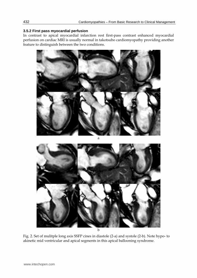
3.5.2 First pass myocardial perfusion In contrast to apical myocardial infarction rest first-pass contrast enhanced myocardial perfusion on cardiac MRI is usually normal in takotsubo cardiomyopathy providing another feature to distinguish between the two conditions.
a
b
Fig. 2. Set of mulitple long axis SSFP cines in diastole (2-a) and systole (2-b). Note hypo- to akinetic mid ventricular and apical segments in this apical ballooning syndrome.
www.intechopen.com
Role of Advanced Cardiac Magnetic Resonance Imaging in Atypical Cardiomyopathies such as Stress-Induced Cardiomyopathie and Left-Ventricular Non-Compaction Cardiomyopathy
433
3.5.3 Tissue characterization – Myocardial edema on t2-weighted images Myocardial edema following myocardial injury, ischemic or non-ischemic, can be detected
using T2-weighted short tau inversion recovery (T2-STIR) images obtained in basal, mid, and
apical short-axis, vertical long-axis, 3-chamber, and horizontal long-axis planes (20).
Elevated T2- signal consistent with myocardial edema is seen in patients with takotsubo
cardiomyopathy. T2 signal is highest in myocardium with the most impaired function and
resolves over time (21-22). Myocardial edema on T2-STIR sequence is also a hallmark of
ischemic myocardium prior to irreversible myocardial injury (23). In contrast to ischemic
myocardium, myocardial edema in patients with takotsubo cardiomyopathy may represent
catecholamine-induced myocardial stunning due to microvascular dysfunction (2).
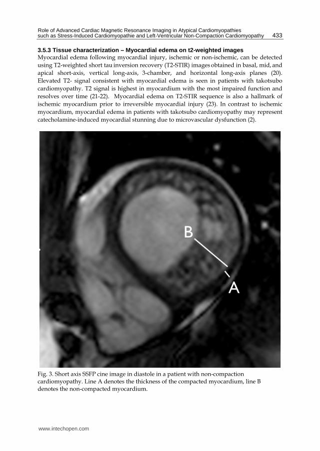
Fig. 3. Short axis SSFP cine image in diastole in a patient with non-compaction cardiomyopathy. Line A denotes the thickness of the compacted myocardium, line B denotes the non-compacted myocardium.
www.intechopen.com

Cardiomyopathies – From Basic Research to Clinical Management
434
3.5.4 Tissue characterization – Late gadolinium enhancement (LGE) on T1-weighted Images Gadolinium concentrates in region of damaged myocardium and appears bright on T1-weighted images (24). Cine images using a segmented gradient-echo sequence (6-mm slice thickness) is obtained in multiple short-axis views every 10 mm covering the whole left ventricle (LV). Ten to 15 min after injection of a Gadolinium-based contrast agent, images are acquired in the same orientation as the cine images using a segmented inversion-recovery gradient-echo pulse sequence. Abnormal myocardium is usually bright on T1-weighted images. LGE is absent in patients with takotsubo cardiomyopathy (2, 3, 5, 25). This indicates viable myocardium and is particularly useful in distinguishing it from myocardial infarction, other types of non-ischemic cardiomyopathy and myocarditis. In a prospective study using CMR conducted by Eitel, et al., 6.100 patients presenting with ACS underwent coronary angiography and left ventriculography. In 59 patients with normal coronary angiography and typical contractile pattern of takotsubo cardiomyopathy, cardiac MRI was performed. Using strict criteria of absence of any late Gadolinium enhancement, takotsubo cardiomyopathy was diagnosed in 38. In the remaining 21 patients, based on the pattern of delayed hyperenhancement, a diagnosis of ischemic heart disease was made in 13 and of myocarditis in 8 patients (2). However, in a study of 15 patients with takotsubo cardiomyopathy, low intensity, patchy, mid-myocardial LGE was seen in 5 out of 15 patients. Increase in extracellular collagen was reported to be a plausible mechanism for this finding (26).
3.5.5 Coronary magnetic resonance angiography (MRA) Coronary MRA though quite helpful in delineating the course of proximal coronary arteries (27), is not feasible for assessment of obstructive coronary artery disease and hence cannot be substituted for invasive or CT angiography (28).
3.6 Management Treatment is supportive. ß-blockers are the mainstay in blunting catecholamine cardiotoxicity. This should be used cautiously to avoid worsening of heart failure. In patients with cardiogenic shock inotropic and IABP support may be required. However, the possibility of exacerbating LVOT obstruction with these measures requires careful monitoring and appropriate adjustment of therapy including discontinuation of inotropes and IABP support (29). ACE-I are beneficial and warfarin is recommended till left ventricular recovery is complete. In addition to these therapeutic measures, emotional or physical precipitating factors should be addressed. Prognosis is usually very good with complete or near complete recovery of LV contractile dysfunction. For prevention of rare recurrent episodes of takotsubo even after removal of initial trigger, indefinite neurohormonal modulation with ß-blockers and ACE-I merits consideration (30).
3.7 Summary Takotsubo cardiomyopathy presents with clinical features that can be indistinguishable from acute coronary syndrome. However, distinct myocardial contractile dysfunction on echocardiography or cardiac MRI, absence of obstructive CAD on invasive angiography and exposure to an emotional or physical stressor is helpful in initially distinguishing it from ACS. Furthermore, cardiac MRI with normal first-pass contrast enhanced rest myocardial
www.intechopen.com
Role of Advanced Cardiac Magnetic Resonance Imaging in Atypical Cardiomyopathies such as Stress-Induced Cardiomyopathie and Left-Ventricular Non-Compaction Cardiomyopathy
435
perfusion, reversible myocardial edema in regions of contractile dysfunction and absence of late gadolinium enhancement is strongly indicative of the diagnosis of takotsubo cardiomyopathy. Resolution of contractile dysfunction, days to weeks after initial presentation, is confirmatory of the diagnosis.
4. References
4.1 Part one references [1] Sedmera D, Pexieder T, Vuillemin M, Thompson RP,Anderson RH. Developmental
patterning of the myocardium. Anat Rec 2000; 258: 319–337. [2] Chin TK, Perloff JK, Williams RG, Jue K, Mohrmann R. Isolated noncompaction of left
ventricular myocardium. A study of eight cases. Circulation 1990;82:507-513. [3] Dusek J, Ostadal B, Duskova M. Postnatal persistence of spongy myocardium with
embryonic blood supply. Arch Pathol 1975;99:312-317. [4] Richardson P, McKenna W, Bristow M, Maisch B, Mautner B, O’Connell J, Olsen E, Thiene G,
Goodwin J.Report of the 1995 World Health Organisation/International Society and Federation of Cardiology Task Force on the definition and classification of cardiomoypathies. Circulation 1996; 93:841–842.
[5] Perry Elliott, Bert Andersson, Eloisa Arbustini, Zofia Bilinska, Franco Cecchi, Philippe Charron, Olivier Dubourg, Uwe Kühl, Bernhard Maisch, William J. McKenna, Lorenzo Monserrat, Sabine Pankuweit, Claudio Rapezzi, Petar Seferovic, Luigi Tavazzi, Andre Keren. Classification of the cardiomyopathies: a position statement from the European society of cardiology working group on myocardial and pericardial diseases. Eur Heart J (2008) 29 (2): 270-276
[6] Barry J. Maron, MD, Chair; Jeffrey A. Towbin, MD, FAHA; Gaetano Thiene, MD; Charles Antzelevitch, PhD, FAHA; Domenico Corrado, MD, PhD; Donna Arnett, PhD, FAHA; Arthur J. Moss, MD, FAHA; Christine E. Seidman, MD, FAHA; James B. Young, MD, FAHA. Contemporary Definitions and Classification of the Cardiomyopathies. An American Heart Association Scientific Statement from the Council on Clinical Cardiology, Heart Failure and Transplantation Committee; Quality of Care and Outcomes Research and Functional Genomics and Translational Biology Interdisciplinary Working Groups; and Council on Epidemiology and Prevention. Circulation. 2006;113:1807-1816.
[7] Mutations in the Sarcomere Gene MYH7 in Ebstein Anomaly / Clinical Perspective.Alex V. Postma, Klaartje van Engelen, Judith van de Meerakker, Thahira Rahman, Susanne Probst, Marieke J.H. Baars, Ulrike Bauer, Thomas Pickardt, Silke R. Sperling, Felix Berger, Antoon F.M. Moorman, Barbara J.M. Mulder, Ludwig Thierfelder, Bernard Keavney, Judith Goodship, and Sabine Klaassen. Circ Cardiovasc Genet. 2011;4:43-50
[8] Ichida F, Tsubata S, Bowles KR, Haneda N, Uese K, Miyawaki T, Dreyer WJ, Messina J, Li H, Bowles NE, Towbin JA. Novel Gene Mutations in Patients with Left Ventricular Noncompaction or Barth Syndrome. Circulation 2001;103;1256-1263.
[9] Pignatelli RH, McMahon CJ, Dreyer WJ, Denfield SW, Price J, Belmont JW, et al. Clinical characterization of left ventricular noncompaction in children: a relatively common form of cardiomyopathy. Circulation 2003;108:2672–8
[10] Ritter M, Oechslin E, Sutsch G, Attenhofer C, Schneider J, Jenni R. Isolated noncompaction of the myocardium in adults. Mayo Clin Proc 1997;72:26-31.
www.intechopen.com
Cardiomyopathies – From Basic Research to Clinical Management
436
[11] Oechslin EN, Attenhofer Jost CH, Rojas JR, Kaufmann PA, Jenni R. Long-term follow-up of 34 adults with isolated left ventricular noncompaction: a distinct cardiomyopathy with poor prognosis. J Am Coll Cardiol 2000; 36: 493–500.
[12] Ichida F, Hamamichi Y, Miyawaki T, Ono Y, Kamiya T, Akagi T, et al., Clinical features of isolated noncompaction of the ventricular myocardium. J Am Coll Cardiol 1999; 34: 233–240.
[13] Murphy RT, Thaman R, Blanes JG, Ward D, Sevdalis E, Papra E, Kiotsekolglou A, Tome MT, Pellerin D, McKenna WJ, Elliott PM. Natural history and familial characteristics of isolated left ventricular non-compaction. European Heart Journal (2005) 26, 187–192.
[14] Jenni R, Oechslin E, Schneider J,Attenhofer Jost C, Kaufmann PA. Echocardiographic and pathoanatomical characteristics of isolated left ventricular non-compaction: a step towards classification as a distinct cardiomyopathy. Heart 2001;86:666-671.
[15] Petersen SE, Selvanayagam JB, Wiesmann F, Robson, MD, Francis JM, Anderson RH, Watkins H, Neubauer S. Left ventricular non-compaction: insights from cardiovascular magnetic resonance imaging J Am Coll Cardiol 2005;46:101-105
[16] Jacquier A, Thuny F, Jop B, Giorgi R, Cohen F, Gaubert J, Vidal V, Bartoli JM, Habib G, Moulin G. Measurement of trabeculated left ventricular mass using cardiac magnetic resonance imaging in the diagnosis of left ventricular non-compaction. Eur Heart J (2010) 31 (9): 1098-1104.
[17] Papavassiliu T, Kuhl HP, Schroder M, Suselbeck T, Bondarenko O, Bohm CK, Beek A, Hofman MM, van Rossum AC. Effect of endocardial trabeculae on left ventricular measurements and measurement reproducibility at cardiovascular MR imaging. Radiology 2005;236:57–64.
[18] Nucifora G, Aquaro GD, Pingitore A, Masci PG, Lombardi M: Myocardial fibrosis in isolated left ventricular non-compaction and its relation to disease severity. Eur J Heart Fail (2011) 13 (2): 170-176.
[19] Dodd JD, Holmvang G, Hoffmann U, et al. Quantification of left ventricular noncompaction and trabecular delayed hyperenhancement with cardiac MRI: correlation with clinical severity. Am J Roentgenol 2007;189:974-980.
4.2 Part two references [1] Dote K, Sato H, Tateishi H, Uchida T, Ishihara M. Myocardial stunning due to
simultaneous multivessel coronary spasms: a review of 5 cases. J Cardiol 1991;21: 203–214.
[2] Eitel I, Behrendt F, Schindler K, Kivelitz D, Gutberlet M, Schuler G, Thiele H. Differential diagnosis of suspected apical ballooning syndrome using contrast-enhanced magnetic resonance imaging. Eur Heart J (2008) 29 (21): 2651-2659.
[3] Wittstein IS, Thiemann DR, Lima JA, Baughman KL, Schulman SP, Gerstenblith G, et al. Neurohumoral features of myocardial stunning due to sudden emotional stress. N Engl J Med. 2005;352:539–548.
[4] Abe Y, Kondo M, Matsuoka R, Araki M, Dohyama K, Tanio H. Assessment of clinical features in transient left ventricular apical ballooning. J Am Coll Cardiol. 2003;41:737–742.
www.intechopen.com
Role of Advanced Cardiac Magnetic Resonance Imaging in Atypical Cardiomyopathies such as Stress-Induced Cardiomyopathie and Left-Ventricular Non-Compaction Cardiomyopathy
437
[5] Sharkey SW, Lesser JR, Zenovich AG, Maron MS, Lindberg J, Longe TF, Maron BJ. Acute and Reversible Cardiomyopathy Provoked by Stress in Women From the United States. Circulation 2005;111:472-479.
[6] Bybee KA, Prasad A. Stress-related cardiomyopathy syndromes Circulation 2008;118:397-40.
[7] Sharkey SW, Windenburg DC, Lesser JR, Maron MS, Hauser RG, Lesser JN, Haas TS, Hodges JS, Maron BJ. Natural History and Expansive Clinical Profile of Stress (Tako-Tsubo) Cardiomyopathy. J. Am. Coll. Cardiol., January 26, 2010; 55(4): 333 – 341.
[8] Bybee KA, Prasad A, Barsness GW, et al. Clinical characteristics and thrombolysis in myocardial infarction frame counts in women with transient left ventricular apical ballooning syndrome. Am J Cardiol 2004;94:343-346.
[9] Sansen V, Holvoet G. Takotsubo cardiomyopathy presenting as multivessel coronary spasm syndrome: case report and review of the literature. Acta Cardiol 2007;62:507-511.
[10] Afonso L, Bachour K, Awad K, Sandidge G. Takotsubo cardiomyopathy: pathogenetic insights and myocardial perfusion kinetics using myocardial contrast echocardiography Eur J Echocardiogr 2008;9:849-854
[11] Kurisu S, Inoue I, Kawagoe T, Ishihara M, Shimatani Y, Nishioka K, Umemura T, Nakamura S, Yoshida M, Sato H. Myocardial perfusion and fatty acid metabolism in patients with tako-tsubo-like left ventricular dysfunction. J Am Coll Cardiol. 2003 Mar 5;41(5):743-8.
[12] Mayer SA, Lin J, Homma S, et al. Myocardial injury and left ventricular performance after subarachnoid hemorrhage Stroke 1999;30:780-786.
[13] Hurst RT, Prasad A, Askew III JW, Sengupta PP, Tajik AJ. Takotsubo Cardiomyopathy: A Unique Cardiomyopathy with Variable Ventricular Morphology. J. Am. Coll. Cardiol. Img., June 1, 2010; 3(6): 641 - 649.
[14] Gianni M, Dentali F, Grandi AM, Sumner G, Hiralal R, Lonn E. Apical ballooning syndrome or takotsubo cardiomyopathy: a systematic review. European Heart Journal (2006) 27, 1523–1529.
[15] Elesber AA, Prasad A, Bybee KA, Valeti U, Motiei A, Lerman A, Chandrasekaran K, Rihal CS, Transient Cardiac Apical Ballooning Syndrome: Prevalence and Clinical Implications of Right Ventricular Involvement. J Am Coll Cardiol, 2006; 47:1082-1083.
[16] Haghi D, Athanasiadis A, Papavassiliu T, Suselbeck T, Fluechter S, Mahrholdt H, Borggrefe M, Sechtem U. Right ventricular involvement in Takotsubo Cardiomyopathy. Eur Heart J (2006) 27, 2433–2439.
[17] Mrdovic I, Kostic J, Perunicic J, Asanin A, Vasiljevic Z, and Ostojic M. Right Ventricular Takotsubo Cardiomyopathy. J. Am. Coll. Cardiol., April 20, 2010; 55(16): 1751 - 1751.
[18] Sievers B, Addo M, Franken U, Trappe HJ. Right ventricular wall motion abnormalities found in healthy subjects by cardiovascular magnetic resonance imaging and characterized with a new segmental model. J Cardiovasc Magn Reson 2004;6:601–608.
[19] Fritz J, Solaiyappan M, Tandri H, MD, Bomma C, Genc A, Claus D. Claussen CD, Lima JAC, Bluemke DA.Right Ventricle Shape and Contraction Patterns and Relation to Magnetic Resonance Imaging Findings. J Comput Assist Tomogr 2005;29:725–733.
www.intechopen.com
Cardiomyopathies – From Basic Research to Clinical Management
438
[20] Simonetti OP, Finn JP, White RD, Laub G, Henry DA. ‘Black blood’ T2-weighted inversion-recovery MR imaging of the heart. Radiology 1996;199:49–57.
[21] Abdel-Aty H, Cocker M, Friedrich MG. Myocardial edema is a feature of Tako-Tsubo cardiomyopathy and is related to the severity of systolic dysfunction: Insights from T2-weighted cardiovascular magnetic resonance. Int J Cardiol. 2009 Feb 20;132(2):291-3.
[22] Joshi SB, Chow T, Herzka DA, Zeman PR, Cooper HA, Lindsay J, Fuisz AR. Cardiovascular magnetic resonance T2 signal abnormalities in left ventricular ballooning syndrome. Int J Cardiovasc Imaging: Vol. 26, No. 2, 227-232.
[23] Abdel-Aty H, Cocker M, Meek C, Tyberg JV, Friedrich MG. Edema as a very early marker for acute myocardial ischemia: a cardiovascular magnetic resonance study J Am Coll Cardiol 2009;53:1194-120.
[24] Saeed M, Wendland MF, Yu KK, Lauerma K, Li HT, Derugin N, Cavagna FM, Higgins CB. Identification of myocardial reperfusion with echoplanar magnetic resonance imaging: discrimination between occlusive and reperfused infarction. Circulation 1994; 90:1492–1501.
[25] Teraoka K, Kiuchi S, Takada N, Hirano M, Yamashina A. Images in cardiovascular medicine. No delayed enhancement on contrast magnetic resonance imaging with Takotsubo cardiomyopathy. Circulation. 2005;111:e261–e262
[26] Rolf A, Nef HM, Mollmann H, et al. Immunohistological basis of the late gadolinium enhancement phenomenon in tako-tsubo cardiomyopathy. Eur Heart J. 2009;30:1635-1642.
[27] Taylor AM, Thorne SA, Rubens MB, Jhooti P, Keegan J, Gatehouse PD,Wiesmann F, Grothues F, Somerville J, Pennell DJ. Coronary artery imaging in grown up congenital heart disease: complementary role of magnetic resonance and x-ray coronary angiography. Circulation. 2000;101:1670 –1678.
[28] Bluemke DA, Achenbach S, Budoff M, Gerber TC, Gersh B, Hillis LD, Hundley WG, Manning WJ, Printz BF, Stuber M, Woodard PK. Noninvasive coronary artery imaging: magnetic resonance angiography and multidetector computed tomography angiography: a scientific statement from the American Heart Association Committee on Cardiovascular Imaging and Intervention of the Council on Cardiovascular Radiology and Intervention, and the Councils on Clinical Cardiology and Cardiovascular Disease in the Young. Circulation. 2008;118:586–606.
[29] Echocardiographic Guidance in Treatment of Cardiogenic Shock Complicating Transient Left Ventricular Apical Ballooning Syndrome. Good CW, Hubbard CR, Harrison TA, Qureshi A: J Am Coll Cardiol Img 2009 March 2; (3):372-374.
[30] Dec, GW. Recognition of the Apical Ballooning Syndrome in the United States. Circulation: 2005;111:388-390.
www.intechopen.com
Cardiomyopathies - From Basic Research to Clinical ManagementEdited by Prof. Josef Veselka
ISBN 978-953-307-834-2Hard cover, 800 pagesPublisher InTechPublished online 15, February, 2012Published in print edition February, 2012
InTech EuropeUniversity Campus STeP Ri Slavka Krautzeka 83/A 51000 Rijeka, Croatia Phone: +385 (51) 770 447 Fax: +385 (51) 686 166www.intechopen.com
InTech ChinaUnit 405, Office Block, Hotel Equatorial Shanghai No.65, Yan An Road (West), Shanghai, 200040, China
Phone: +86-21-62489820 Fax: +86-21-62489821
Cardiomyopathy means "heart (cardio) muscle (myo) disease (pathy)". Currently, cardiomyopathies aredefined as myocardial disorders in which the heart muscle is structurally and/or functionally abnormal in theabsence of a coronary artery disease, hypertension, valvular heart disease or congenital heart diseasesufficient to cause the observed myocardial abnormalities. This book provides a comprehensive, state-of-the-art review of the current knowledge of cardiomyopathies. Instead of following the classic interdisciplinarydivision, the entire cardiovascular system is presented as a functional unity, and the contributors explorepathophysiological mechanisms from different perspectives, including genetics, molecular biology,electrophysiology, invasive and non-invasive cardiology, imaging methods and surgery. In order to provide abalanced medical view, this book was edited by a clinical cardiologist.
How to referenceIn order to correctly reference this scholarly work, feel free to copy and paste the following:
Oliver Strohm, Abdullah Shehab and Anwer Qureshi (2012). Role of Advanced Cardiac Magnetic ResonanceImaging in Atypical Cardiomyopathies such as Stress-Induced Cardiomyopathie and Left-Ventricular Non-Compaction Cardiomyopathy, Cardiomyopathies - From Basic Research to Clinical Management, Prof. JosefVeselka (Ed.), ISBN: 978-953-307-834-2, InTech, Available from:http://www.intechopen.com/books/cardiomyopathies-from-basic-research-to-clinical-management/contrast-enhanced-cardiac-magnetic-resonance-imaging-cmr-in-the-identification-of-unclassified-cardi