RNAi in Budding Yeastbartellab.wi.mit.edu/publication_reprints/Drinnenberg_Science09.pdf · RNA...
7
27. M. T. Holder, J. A. Anderson, A. K. Holloway, Syst. Biol. 50, 978 (2001). 28. M. A. Noor, J. L. Feder, Nat. Rev. Genet. 7, 851 (2006). 29. Drosophila 12 Genomes Consortium, Nature 450, 203 (2007). 30. We thank G. Kalay, X. Heng, M. Weisel, A. Ratnala, and E. Larimore for technical assistance; P. Mena and B. McAllister for sharing unpublished data (including the images shown in fig. S7) and lines of D. americana; C. Dick for use of the Mixer Mill, M. Rebeiz for sharing an unpublished in situ protocol; N. Rosenberg, G. Coop, and other colleagues for discussions of incomplete lineage sorting and introgression; and J. Gruber, J. Coolon, A. Cooley, G. Kalay, D. Yuan, B. McAllister, A. Kopp, B. Prud'homme, and N. Gompel for comments on the manuscript. Supported by NSF grant DEB140640485, the Margaret and Herman Sokol Endowment for Faculty and Graduate Student Research Projects, and the University of Michigan. P.J.W. is an Alfred P. Sloan Research Fellow. DNA sequences are available from GenBank (accession numbers GQ457336 to GQ457453). Supporting Online Material www.sciencemag.org/cgi/content/full/326/5952/540/DC1 Materials and Methods Figures S1 to S9 Tables S1 to S3 References 28 May 2009; accepted 13 August 2009 10.1126/science.1176980 RNAi in Budding Yeast Ines A. Drinnenberg, 1,2 * David E. Weinberg, 1,2,3 * Kathleen T. Xie, 1,2,3 * Jeffrey P. Mower, 4 † Kenneth H. Wolfe, 4 Gerald R. Fink, 1,3 David P. Bartel 1,2,3 ‡ RNA interference (RNAi), a gene-silencing pathway triggered by double-stranded RNA, is conserved in diverse eukaryotic species but has been lost in the model budding yeast Saccharomyces cerevisiae. Here, we show that RNAi is present in other budding yeast species, including Saccharomyces castellii and Candida albicans. These species use noncanonical Dicer proteins to generate small interfering RNAs, which mostly correspond to transposable elements and Y′ subtelomeric repeats. In S. castellii, RNAi mutants are viable but have excess Y′ messenger RNA levels. In S. cerevisiae, introducing Dicer and Argonaute of S. castellii restores RNAi, and the reconstituted pathway silences endogenous retrotransposons. These results identify a previously unknown class of Dicer proteins, bring the tool of RNAi to the study of budding yeasts, and bring the tools of budding yeast to the study of RNAi. R NA-silencing pathways contribute to trans- poson silencing, viral defense, DNA elim- ination, heterochromatin formation, and posttranscriptional repression of cellular genes (1, 2). In the simplest form of silencing, known as RNA interference (RNAi), the ribonuclease III (RNaseIII) endonuclease Dicer successively cleaves double-stranded RNA (dsRNA) into small interfering RNAs (siRNAs), which are loaded into the effector protein Argonaute to guide the cleav- age of target transcripts (1, 3). RNAi arose in an early eukaryotic ancestor and appears to have been conserved throughout most of the fungal kingdom (4, 5) (Fig. 1A). A prominent excep- tion is Saccharomyces cerevisiae, a budding yeast that lacks recognizable homologs of Argonaute, Dicer, and RNA-dependent RNA polymerase (RdRP), which in some RNAi pathways produces dsRNA. Indeed, RNAi has been presumed lost in all budding yeasts. Despite this perceived loss, Argonaute genes are present in some budding yeasts (6, 7), including Saccharomyces castellii and Kluyveromyces polysporus (both close relatives of S. cerevisiae) and Candida albicans [the most common yeast pathogen of humans (8)] (Fig. 1A). The presence of these genes in budding yeast has been enigmatic, because other RNAi genes, especially Dicer, have not been found in these species. A similar conundrum appears in prokaryotes, in which certain bacteria have Argonaute homologs yet lack the other genes associated with RNAi or related RNA- silencing pathways (9). siRNAs in budding yeasts. To search for RNA silencing in budding yeast, we looked for short-guide RNAs, isolating 18- to 30-nucleotide (nt) RNAs from S. castellii, K. polysporus, and C. albicans and preparing sequencing libraries representing the subset of small RNAs with 5′- monophosphates and 3′-hydroxyls (10), which are the chemical features of Dicer products. The small RNAs of S. castellii and K. polysporus were most enriched in 23-RNAs beginning with U, and those of C. albicans were most enriched in 22-nt beginning with A or U (Fig. 1B). These biases were reminiscent of those observed for Argonaute-bound guide RNAs of animals, plants, and other fungi (11–13). Analogous RNAs were not found in S. cerevisiae, as expected for a species lacking RNAi (Fig. 1B). Although some reads from the Argonaute- containing yeasts mapped to ribosomal RNA (rRNA) and transfer RNA (tRNA) and presum- ably represented degradation intermediates of abundant RNAs, many reads clustered at other types of genomic loci. The loci generating the most reads had sequence homology to repetitive elements, including long terminal repeat retro- transposons (Ty elements), LINE (long interspersed nuclear element) –like retrotransposons (Zorro ele- ments), and subtelomeric repeats (Y′ elements) (Fig. 1C and table S1). Loci of S. castellii were also particularly enriched in long inverted re- peats; these palindromic loci generated most of the reads with homology to Ty elements (Fig. 1, C and D). In S. cerevisiae, essentially all the reads appeared to represent degradation fragments of rRNA, tRNA, and mRNA. The reads matching inverted repeats suggested origins from paired regions of transcripts that folded back on themselves to form hairpins (Fig. 1D). These inferred hairpins had 100- to 400-bp (base pair) stems, with loops ranging from 19 to >1600 nt. In regions of imperfect duplex, where reads could be mapped unambiguously, the small RNAs tended to match only one genomic strand, which further supported the idea that they orig- inated from hairpin transcripts (Fig. 1D, bottom). Other reads did not map to inverted repeats and, instead, mapped uniquely to both genomic strands in a pattern suggesting that they originated from long bimolecular duplexes involving transcripts from both strands. Most siRNAs of the fission yeast Schizosac- charomyces pombe correspond to the outer repeats of the centromeres and direct heterochromatin for- mation and maintenance (14). We therefore ex- amined whether any of our sequenced small RNAs matched centromeres. Of the three Argonaute- containing species from which we sequenced (Fig. 1B), only C. albicans had annotated centromeres, and almost none (<0.001%) of our C. albicans reads matched these genomic loci. Also arguing against a function analogous to that in S. pombe is the lack in budding yeasts of recognizable orthologs of the histone H3 lysine 9 (H3K9) methyltransferase Clr4 and recognizable homologs of RdRP, Tas3, Chp1, and the heterochromatin protein HP1-like chromodomain protein Swi6—proteins all nec- essary for RNAi-dependent heterochromatin in S. pombe (14). When mapped to the genome, the end of one 23-nt RNA was often next to the beginning of another 23-nt RNA, which suggested that endo- nuclease cleavage simultaneously generated the 3′ terminus of one small RNA and the 5′ terminus of the next. Consistent with this hypothesis, systematic analysis of the intervals spanning the mapped ends of all 23-nt RNA pairs revealed a clear phasing interval of 23 nt (Fig. 1E). Such phasing implied successive cleavage, beginning 1 Whitehead Institute for Biomedical Research, 9 Cambridge Center, Cambridge, MA 02142, USA. 2 Howard Hughes Medical Institute, Massachusetts Institute of Technology, Cambridge, MA 02139, USA. 3 Department of Biology, Mas- sachusetts Institute of Technology, Cambridge, MA 02139, USA. 4 Smurfit Institute of Genetics, Trinity College Dublin, Dublin 2, Ireland. *These authors contributed equally to this work. †Present address: Center for Plant Science Innovation and Department of Agronomy and Horticulture, University of Nebraska, Lincoln, NE 68588, USA. ‡To whom correspondence should be addressed. E-mail: [email protected] 23 OCTOBER 2009 VOL 326 SCIENCE www.sciencemag.org 544 RESEARCH ARTICLES
Transcript of RNAi in Budding Yeastbartellab.wi.mit.edu/publication_reprints/Drinnenberg_Science09.pdf · RNA...
27. M. T. Holder, J. A. Anderson, A. K. Holloway, Syst. Biol.50, 978 (2001).
28. M. A. Noor, J. L. Feder, Nat. Rev. Genet. 7, 851 (2006).29. Drosophila 12 Genomes Consortium, Nature 450, 203
(2007).30. We thank G. Kalay, X. Heng, M. Weisel, A. Ratnala, and
E. Larimore for technical assistance; P. Mena andB. McAllister for sharing unpublished data (including theimages shown in fig. S7) and lines of D. americana;C. Dick for use of the Mixer Mill, M. Rebeiz for sharing an
unpublished in situ protocol; N. Rosenberg, G. Coop, andother colleagues for discussions of incomplete lineage sortingand introgression; and J. Gruber, J. Coolon, A. Cooley,G. Kalay, D. Yuan, B. McAllister, A. Kopp, B. Prud'homme,and N. Gompel for comments on the manuscript. Supportedby NSF grant DEB140640485, the Margaret and HermanSokol Endowment for Faculty and Graduate Student ResearchProjects, and the University of Michigan. P.J.W. is an AlfredP. Sloan Research Fellow. DNA sequences are available fromGenBank (accession numbers GQ457336 to GQ457453).
Supporting Online Materialwww.sciencemag.org/cgi/content/full/326/5952/540/DC1Materials and MethodsFigures S1 to S9Tables S1 to S3References
28 May 2009; accepted 13 August 200910.1126/science.1176980
RNAi in Budding YeastInes A. Drinnenberg,1,2* David E. Weinberg,1,2,3* Kathleen T. Xie,1,2,3* Jeffrey P. Mower,4†Kenneth H. Wolfe,4 Gerald R. Fink,1,3 David P. Bartel1,2,3‡
RNA interference (RNAi), a gene-silencing pathway triggered by double-stranded RNA, isconserved in diverse eukaryotic species but has been lost in the model budding yeastSaccharomyces cerevisiae. Here, we show that RNAi is present in other budding yeast species,including Saccharomyces castellii and Candida albicans. These species use noncanonical Dicerproteins to generate small interfering RNAs, which mostly correspond to transposable elements andY′ subtelomeric repeats. In S. castellii, RNAi mutants are viable but have excess Y′ messengerRNA levels. In S. cerevisiae, introducing Dicer and Argonaute of S. castellii restores RNAi, and thereconstituted pathway silences endogenous retrotransposons. These results identify a previouslyunknown class of Dicer proteins, bring the tool of RNAi to the study of budding yeasts, and bringthe tools of budding yeast to the study of RNAi.
RNA-silencing pathways contribute to trans-poson silencing, viral defense, DNAelim-ination, heterochromatin formation, and
posttranscriptional repression of cellular genes(1, 2). In the simplest form of silencing, knownas RNA interference (RNAi), the ribonucleaseIII (RNaseIII) endonuclease Dicer successivelycleaves double-stranded RNA (dsRNA) into smallinterfering RNAs (siRNAs), which are loaded intothe effector protein Argonaute to guide the cleav-age of target transcripts (1, 3). RNAi arose in anearly eukaryotic ancestor and appears to havebeen conserved throughout most of the fungalkingdom (4, 5) (Fig. 1A). A prominent excep-tion is Saccharomyces cerevisiae, a budding yeastthat lacks recognizable homologs of Argonaute,Dicer, and RNA-dependent RNA polymerase(RdRP), which in some RNAi pathways producesdsRNA. Indeed, RNAi has been presumed lostin all budding yeasts. Despite this perceived loss,Argonaute genes are present in some buddingyeasts (6, 7), including Saccharomyces castellii
and Kluyveromyces polysporus (both closerelatives of S. cerevisiae) and Candida albicans[the most common yeast pathogen of humans(8)] (Fig. 1A). The presence of these genes inbudding yeast has been enigmatic, because otherRNAi genes, especially Dicer, have not beenfound in these species. A similar conundrumappears in prokaryotes, in which certain bacteriahave Argonaute homologs yet lack the othergenes associated with RNAi or related RNA-silencing pathways (9).
siRNAs in budding yeasts. To search forRNA silencing in budding yeast, we looked forshort-guide RNAs, isolating 18- to 30-nucleotide(nt) RNAs from S. castellii, K. polysporus, andC. albicans and preparing sequencing librariesrepresenting the subset of small RNAs with 5′-monophosphates and 3′-hydroxyls (10), whichare the chemical features of Dicer products. Thesmall RNAs of S. castellii and K. polysporuswere most enriched in 23-RNAs beginning withU, and those of C. albicans were most enrichedin 22-nt beginning with A or U (Fig. 1B). Thesebiases were reminiscent of those observed forArgonaute-bound guide RNAs of animals, plants,and other fungi (11–13). Analogous RNAs werenot found in S. cerevisiae, as expected for aspecies lacking RNAi (Fig. 1B).
Although some reads from the Argonaute-containing yeasts mapped to ribosomal RNA(rRNA) and transfer RNA (tRNA) and presum-ably represented degradation intermediates ofabundant RNAs, many reads clustered at othertypes of genomic loci. The loci generating themost reads had sequence homology to repetitive
elements, including long terminal repeat retro-transposons (Ty elements), LINE (long interspersednuclear element)–like retrotransposons (Zorro ele-ments), and subtelomeric repeats (Y′ elements)(Fig. 1C and table S1). Loci of S. castellii werealso particularly enriched in long inverted re-peats; these palindromic loci generated most ofthe reads with homology to Ty elements (Fig. 1,C andD). In S. cerevisiae, essentially all the readsappeared to represent degradation fragments ofrRNA, tRNA, and mRNA.
The readsmatching inverted repeats suggestedorigins from paired regions of transcripts thatfolded back on themselves to form hairpins (Fig.1D). These inferred hairpins had 100- to 400-bp(base pair) stems, with loops ranging from 19 to>1600 nt. In regions of imperfect duplex, wherereads could be mapped unambiguously, the smallRNAs tended to match only one genomic strand,which further supported the idea that they orig-inated from hairpin transcripts (Fig. 1D, bottom).Other reads did not map to inverted repeats and,instead, mapped uniquely to both genomic strandsin a pattern suggesting that they originated fromlong bimolecular duplexes involving transcriptsfrom both strands.
Most siRNAs of the fission yeast Schizosac-charomyces pombe correspond to the outer repeatsof the centromeres and direct heterochromatin for-mation and maintenance (14). We therefore ex-aminedwhether any of our sequenced small RNAsmatched centromeres. Of the three Argonaute-containing species fromwhichwe sequenced (Fig.1B), only C. albicans had annotated centromeres,and almost none (<0.001%) of ourC. albicans readsmatched these genomic loci. Also arguing againsta function analogous to that in S. pombe is the lackin budding yeasts of recognizable orthologs ofthe histone H3 lysine 9 (H3K9) methyltransferaseClr4 and recognizable homologs of RdRP, Tas3,Chp1, and the heterochromatin protein HP1-likechromodomain protein Swi6—proteins all nec-essary for RNAi-dependent heterochromatin inS. pombe (14).
When mapped to the genome, the end of one23-nt RNA was often next to the beginning ofanother 23-nt RNA, which suggested that endo-nuclease cleavage simultaneously generated the 3′terminus of one small RNA and the 5′ terminusof the next. Consistent with this hypothesis,systematic analysis of the intervals spanning themapped ends of all 23-nt RNA pairs revealed aclear phasing interval of 23 nt (Fig. 1E). Suchphasing implied successive cleavage, beginning
1Whitehead Institute for Biomedical Research, 9 CambridgeCenter, Cambridge, MA 02142, USA. 2Howard HughesMedical Institute, Massachusetts Institute of Technology,Cambridge, MA 02139, USA. 3Department of Biology, Mas-sachusetts Institute of Technology, Cambridge, MA 02139,USA. 4Smurfit Institute of Genetics, Trinity College Dublin,Dublin 2, Ireland.
*These authors contributed equally to this work.†Present address: Center for Plant Science Innovation andDepartment of Agronomy and Horticulture, University ofNebraska, Lincoln, NE 68588, USA.‡To whom correspondence should be addressed. E-mail:[email protected]
23 OCTOBER 2009 VOL 326 SCIENCE www.sciencemag.org544
RESEARCH ARTICLES
at preferred starting points. Moreover, pairs fromopposite strands had the same phasing interval butin a register 2 nt offset from that of the same-strand pairs. Together, the phasing and offsetimplied successive cleavage of dsRNA with a2-nt 3′ overhang—the classic biogenesis of endog-enous siRNAs by Dicer (3). Therefore, the smallRNAs that appeared to derive from regions ofdsRNA, i.e., those mapping in clusters to thearms of predicted hairpins and those mapping inclusters to both genomic strands, were classifiedas siRNAs.
Dicer in budding yeasts. The presence ofsiRNAs in Argonaute-containing budding yeastsimplied that each of these species also had aDicer-like activity. To assay for this activity, wemonitored processing of a long dsRNA added towhole-cell extracts (15). Extracts from S. castellii,K. polysporus, and C. albicans—but not fromS. cerevisiae—contained an activity that produced22- to 23-nt RNAs, each preferentially fromdsRNA rather than from single-stranded RNA
(Fig. 2A). Moreover, for each extract the small-RNA length matched that of the most abundantlength observed in vivo (Figs. 1B and 2A).
Despite the observedDicer-like activity, a genewith the domain architecture of knownDicers wasnot found in any budding yeast genome (Fig. 1A)(7). Because we had evidence for cleavage ofdsRNA with 2-nt 3′ overhangs, a hallmark ofRNaseIII activity, we relaxed the search criteriato consider any gene with an RNaseIII domain.S. cerevisiae had only one gene, RNT1, with arecognizable RNaseIII domain. RNT1 helps pro-cess rRNA and other noncoding RNAs (16), andpresumed orthologs were found throughout thefungal kingdom. S. castellii had a secondRNaseIIIdomain–containing gene, and a potential ortho-log of this gene was found in each of the otherArgonaute-containing budding yeasts (Fig. 1A).Anticipating that this second gene encoded theDicer of budding yeasts, we named it DCR1.
To test whether the Dicer candidate is requiredfor siRNA accumulation, we deleted DCR1 in
S. castellii—the closest relative to S. cerevisiaeamong the sequenced Argonaute-containing spe-cies. This procedure required establishing strainsand protocols to better enable molecular geneticanalysis in this species (15, 17). In the Ddcr1mutant, siRNAs failed to accumulate (Fig. 2B,fig. S2, and table S1). Deletion of the Argonautehomolog, which we named AGO1, also reducedsiRNA accumulation, as expected if loadinginto Argonaute protected siRNAs from degra-dation (Fig. 2B, fig. S2, and table S1). For bothmutants, ectopically expressing the deleted generescued siRNA accumulation (Fig. 2B). Theseresults indicate that the core components of en-dogenous RNAi pathways—Dicer, Argonaute,and siRNAs—are present in some species of thebudding yeast clade.
In S. pombe and other fungi, known Dicergenes resemble those in plants and animals, com-plete with tandem RNaseIII domains, two or threedsRNA-binding domains (dsRBDs), a PAZdomain,and an N-terminal helicase domain (15, 18, 19)
RNA length (nt)
Saccharomyces cerevisiae
Candida glabrata
Kluyveromyces polysporus
Saccharomyces bayanus
Saccharomyces castellii
Kluyveromyces lactisAshbya gossypiiCandida albicansCandida tropicalisDebaryomyces hansenii
Neurospora crassaMagnaporthe griseaAspergillus nidulansSchizosaccharomyces pombeSchizosaccharomyces japonicusCryptococcus neoformansCoprinus cinereusUstilago maydisUstilago hordei Rhizopus oryzae
Ψ
AgoDcr RdRP
–– * –
–+ * –+ * –– – –– – –+ * –+ * –
–
+ + ++ + ++ + ++ + ++ + ++ + ++ + +– – –+ + ++ + +
Bud
ding
yea
st
Pichia stipitis– *
–– *
0
5
10
15
20
25
14 15 16 17 18 19 20 21 22 23 24 25 26 27 28 29 30
AUGC
14 15 16 17 18 19 20 21 22 23 24 25
0
5
10
15
14 15 16 17 18 19 20 21 22 23 24 25 26 27 28 29 30
5 nt identity
eaisiverec .Ssnacibla .C
Per
cent
rea
dsP
erce
nt r
eads
suropsylop .Kiilletsac .S
14 15 16 17 18 19 20 21 22 23 24 25
26 27 28 29 30
26 27 28 29 30RNA length (nt)
BA
C
'
– –
Saccharomyces pastorianus Ψ–– –
Fra
ctio
n of
rea
ds
0%
10%
20%
30%
40%
50%
60%
70%
80%
90%
100%
K. polys
porus
C. albica
ns
S. cerevis
iae
S. caste
llii
Sm
all-RN
A clusters
ZorroLPF familyCTA2 familyY'Ty
Palindromes
tRNArRNA
OtherORFs
Other
Palindromic Ty
ORFs
0
5
10
15
20
25
0
5
10
15
5' armD
Rea
ds
sc712:108200-1088003' arm Loop
10080604020
020406080
100
4080120160200
0
Enclosed pairs per position
Secondary structure
siRNA plus strandsiRNA minus strand
Uni
que
read
s
10080604020
020406080
100
4080120160200
0
Enclosed pairs per position
~170 nt
~170 nt ~140 nt ~170 nt
~170 nt ~140 nt ~170 nt
23 nt 23 nt 23 nt 23 nt
Sam
e st
rand
Opp
osite
str
and
Sm
all-R
NA
5' e
nd p
airs
(x1
00,0
00)
E
1
2
3
4
5
6
4321
Distance(nt)10 20 30 40 50 60 70 80 90
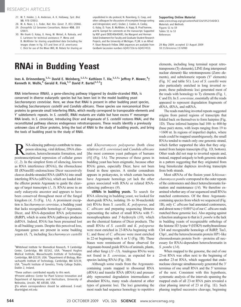
Fig. 1. Endogenous siRNAs in budding yeasts. (A) Cladogram showingBasidiomycota (blue), Zygomycota (gray) and Ascomycota, subdivided intoSaccharomycotina (budding yeasts, orange), Pezizomycotina (yellow), andTaphrinomycotina (green) (35, 36). The presence of canonical RNAi genes isindicated (+) [(4, 5) and references therein]. All genomes had an RNT1ortholog, and several others had a second RNaseIII domain–containing gene(*), which has Dicer activity in S. castellii. Pseudogenes are indicated (Y).S. bayanus, which had a Dicer but not an Argonaute gene, appeared to lacksiRNAs (fig. S1). (B) Length distribution of genome-matching sequencingreads representing small RNAs with the indicated 5′ nucleotide. Readsmatching rRNA and tRNA are excluded. (C) Classification of loci to which
21- to 23-nt RNAs map, considering those that map to clusters in a patternsuggestive of siRNAs separately from those that do not. (D) A palindromicregion generating siRNAs in S. castellii. 5′ Termini of 22- to 23-nt RNAs weremapped to the genome, and counts (normalized to the number of genomicmatches) are plotted for the plus and minus genomic strands. The topconsiders all reads; the bottom considers those matching the genome at onlyone locus. The predicted structure of the (–)-strand transcript is represented asa mountain plot (37). (E) Distribution of the genomic intervals separating the5′ termini of sequenced 23-nt RNAs from S. castellii. Plotted is the frequencyof each interval, when considering all pairs of reads less than 100 nt apart(excluding reads matching rRNA and tRNA).
www.sciencemag.org SCIENCE VOL 326 23 OCTOBER 2009 545
RESEARCH ARTICLES
(Fig. 2C). In budding yeasts, DCR1 has twodsRBDs, but only a single RNaseIII domain, andno helicase or PAZ domains. Because RNaseIIIdomains work in pairs to nick both strands of anRNA duplex (19, 20), we suspect that S. castelliiDcr1 acts as a homodimer. Dicers of insects, plants,and mammals, which already have two RNaseIIIdomains, do not homodimerize but do form het-erodimeric complexes with cofactors that provideadditional dsRBDs (21–23). A homodimericS. castellii Dcr1 complex would already havefour dsRBDs, which might obviate the need forsuch a cofactor.
Except for its second dsRBD, the domainarchitecture of the budding yeast Dicer re-sembled that of RNT1 rather than that ofcanonical Dicer genes (Fig. 2C). Furthermore,the amino acid sequence of its RNaseIII domainwas more similar to that of the RNT1 RNaseIIIdomain than to that of any previously identifiedDicer RNaseIII domain (Fig. 2D). These obser-vations suggest that budding yeast Dicer mighthave emerged from a duplication of RNT1 earlyin the budding yeast lineage, perhaps coincidentwith the loss of canonical Dicer. The unusualancestry and domain structure of DCR1 mightexplain why its activity, and thus RNAi moregenerally, went undetected for so long in buddingyeast.
Biochemical analyses of Dcr1 and Ago1.Dicing activity of S. castellii extracts was lost inthe Ddcr1 mutant and restored by Dcr1 over-expression (Fig. 2E). To determine whether Dcr1is active in the absence of S. castellii cofactors,we expressed the protein in S. cerevisiae andE. coli (Fig. 2E). Expression in E. coli conferredrobust activity, indicating that S. castellii Dcr1 issufficient to dice dsRNA at precise intervals. Inother Dicers, the PAZ domain is an essential com-ponent of a molecular ruler that imparts cleavageprecision (19). The budding yeast Dicers, whichlack this domain, must achieve this measuringfunction differently.
To establish a biochemical link between Ago1and the siRNAs of S. castellii, we sequenced thesmall RNAs that copurified with tagged Ago1expressed from its native promoter. Comparedwith the input RNA, the population of Ago1-associated RNAs was even more enriched for 22-to 23-nt RNAs and was depleted in matches toboth rRNA and tRNA, with concomitant en-richment for matches to palindromes, Ty ele-ments, and Y′ elements (fig. S3 and table S2).These biochemical results supported the geneticlink betweenAGO1 and the siRNAs (Fig. 2B) andprovided a set of small RNAs suitable for an-notating the siRNA-producing loci of S. castellii(table S3).
The impact of RNAi on the S. castelliitranscriptome. To investigate the molecular con-sequences of RNAi,we performed high-throughputsequencing of polyadenylated RNA [mRNA-Seq(24)] from wild-type, Dago1, and Ddcr1 strains(table S4). The two annotated open reading frames(ORFs) that changed most in RNAi deletion
strains were also the two with the highest densityof antisense siRNA reads (Fig. 3A, red points).One was the consensus Y′ ORF (fig. S5), whichincreased more than sevenfold in both deletionmutants. The other was an ORF within a palin-dromic Ty fragment, which increased more thanfourfold in the Ddcr1mutant but less in theDago1mutant. For other ORFs, transcript-abundancechanges were modest and not correlated withsiRNA density (fig. S6), although changes inDago1 andDdcr1mutants did correlate with eachother (R2 = 0.39) (Fig. 3A). This correlationmight reflect a general response to the loss ofRNAi (although we cannot exclude contributions
of a common response to the hygromycin- andkanamycin-resistance genes used to delete AGO1and DCR1, respectively).
Because many siRNAs mapped antisense toor outside of ORFs, the mRNA-Seq data reveal-ing the S. castellii polyadenylated transcriptomeenabled the systematic identification of siRNAprecursor transcripts. We focused on three typesof siRNA precursors: sense-antisense transcriptpairs fromORF loci, partially overlappingmRNAs,and transcripts producing the most siRNA-likereads, regardless of annotation.
The potential for dsRNA composed of sense-antisense transcripts fromORF loci was indicated
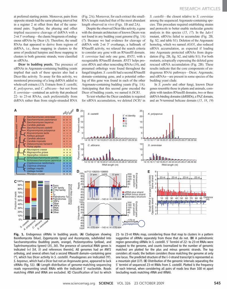
Fig. 2. The Dicer of budding yeast. (A) In vitro processing of radiolabeled dsRNA or single-stranded RNA(ssRNA) in extracts from the indicated budding yeast species. Products were resolved on a denaturing gel.The fraction of product normalized to that observed with dsRNA is indicated below as a percentage. (B)RNA blot probing for an endogenous siRNA (sc1056) in the indicated deletion and rescue strains. The blotwas reprobed for U6 small nuclear RNA, and the siRNA percent signal normalized to that of U6 isindicated below. (C) Domain architectures of representative Dicer proteins and the two S. castellii proteinscontaining an RNaseIII domain. (D) Maximum-likelihood tree based on amino acid alignment of RNaseIIIdomains from Dicer proteins and Rnt1 homologs. Orange shading highlights budding yeast Dicercandidates indicated by asterisks in Fig. 1A. Budding yeast species encoding Argonaute are listed in red.Bootstrap values higher than 50% are shown. (E) In vitro dicing in extracts from recombinant S. castellii(S. cas), S. cerevisiae (S. cer), or E. coli strains with the indicated deletions and additions, analyzed as in (A).
23 OCTOBER 2009 VOL 326 SCIENCE www.sciencemag.org546
RESEARCH ARTICLES

by widespread low-level antisense transcriptionof ORFs, with antisense mRNA-Seq tags map-ping to over half of all annotated ORFs. More-over, small RNAs mapped antisense to nearlyone-third of ORFs (Fig. 3A) and, as a class, werereduced in RNAi mutants and enriched by Ago1immunoprecipitation (fig. S3 and table S2).Supporting a precursor-product relation, the abun-dance of the sense-antisense duplexes (inferredfrom mRNA-Seq data) correlated with that of
small RNAs deriving from these loci (fig. S7).The most striking example of siRNAs arisingfrom sense-antisense transcript pairs was withinthe Y′ ORF, which was most affected by the lossof the RNAi machinery (Fig. 3, A and B). Y′elements are conserved protein-coding repeats.In S. cerevisiae, they are located near both endsof most chromosomes (25) (fig. S7), and syn-teny suggests analogous locations in S. castellii(26). The S. castellii elements had a robustly ex-
pressed antisense transcript with many siRNAsmapping to the region of sense-antisense overlap(Fig. 3B).
We considered partially overlapping mRNAsas another potential source of siRNA-generatingdsRNA, after using the mRNA-Seq data to ex-tend the 5′ and 3′ boundaries of 5297 S. castelliiprotein-coding transcripts. Although only 1% ofdivergent transcript pairs and 7% of tandem tran-script pairs overlapped, 78% of convergent tran-script pairs overlapped (Fig. 3C) [median overlapof 92 nt at their 3′ ends (fig. S7)]. At least 43% ofthese convergent and overlapping gene pairs (com-posing 9% of all gene pairs) generated DCR1-dependent siRNAs in the region of overlap (Fig.3C and fig. S3); one such pair is illustrated (Fig.3D). A recent study reported pervasive over-lapping transcripts in S. cerevisiae (27). Our re-sults revealing analogous overlap in S. castelliishow that, in contrast to previous speculation, thisphenomenon is not restricted to RNAi-deficientorganisms and is an ancestral feature of theseSaccharomyces species (15).
We next inferred precursor transcripts with-out considering whether or not they overlappedORFs (table S5). A hiddenMarkovmodel analyz-ing the Ago1-associated small RNAs identifiedthe genomic loci producing abundant siRNAs,and analysis of the mRNA-Seq data from Ddcr1strains revealed the corresponding transcripts. Inaddition to recovering the more prolific ORF-overlapping siRNA precursors, this analysisidentified the transcript illustrated in Fig. 1Dand transcripts of 84 other non–protein-codingsiRNA-generating genes of S. castellii [annotatedas NCS1–NCS85 (tables S3 and S5)]. Transcriptsproducing fewer siRNAs in RNAi-competentcells changed modestly, but similarly, in bothdeletion mutants (Fig. 3E), as observed whenanalyzing only ORF transcripts (Fig. 3A). Tran-scripts producing the most siRNAs, which werepredominantly from palindromic loci, increaseddramatically in the Ddcr1 mutant but were rela-tively unchanged in the Dago1 mutant (Fig. 3Eand table S5), which indicated that Dcr1 alonewas sufficient to reduce these transcripts to wild-type levels. This mode of posttranscriptionaldown-regulation may be unique to palindromictranscripts, which can fold into hairpin struc-tures that are ideal Dcr1 substrates but refractoryto intermolecular pairing with Ago1-associatedsiRNAs.
Taken together, our results indicate that morethan one thousand genomic loci in S. castellii gen-erate siRNAs. The consequences of siRNAs derivedfrom the widespread antisense and overlappingtranscription in S. castellii are unknown. With theexception of the Y′ mRNA, the loss of the RNAimachinery did not substantially affect the levelsof mRNAs corresponding to these siRNAs (Fig.3A and fig. S6). Perhaps in other growth conditionsthe regulatory impact of non-Y′ siRNAs might bemore pronounced. The specificity for Y′-elementregulation could arise from requiring both anabundance of antisense siRNAs and the ability
Fig. 3. The impact of RNAi on the S. castellii transcriptome. (A) Strand-specific mRNA-Seq analysis ofannotated ORF transcripts in wild-type (WT) and RNAi-mutant strains. Plotted is the log2 ratio of transcriptabundance in Dago1 versus wild-type (x axis) and Ddcr1 versus wild-type (y axis). Colors indicate thedensity (reads per kilobase) of antisense small (22- to 23-nt) RNAs that copurified with Ago1. A Ty ORFfragment (annotated as Scas_712.50) embedded within a palindromic siRNA-producing locus is indicated(square). Annotated Y′-element ORFs were replaced by one consensus Y′ ORF (triangle, fig. S5). Becausethe mRNA-Seq protocol included poly(A) selection, which retains the 3′ but not 5′ fragments of cleavedmRNAs, we calculated full-length transcript abundance using tags mapping to the 5′ half of each ORF.Similar trends were observed when we used tags mapping across the ORF (fig. S4). (B) Analysis of theS. castellii Y′ element. The numbers of siRNA 5′ ends (small RNAs) and mRNA tags (mRNA-Seq) mappingto the consensus Y′ element are plotted for each position (sense, above axis; antisense, below axis). (C)Gene-pair organization and overlap in S. castellii. (Inner ring) Fraction of neighboring annotated ORFswith the indicated orientation; (middle ring) fraction of transcript pairs with overlapping 3′ ends(convergent), overlapping 5′ ends (divergent), or continuous transcription in between (tandem); (outerring) fraction of convergent transcript pairs generating siRNAs in the overlapping region. (D) A pair ofconvergent transcripts that generate siRNAs in the region of overlap. Plots are as in (B). (E) mRNA-Seqanalysis of inferred siRNA-generating transcripts. The plot is as in (A), with the same colors to indicatesiRNA-read density and shapes to indicate transcripts mapping to Y′ elements (triangle), palindromes(square), and others (diamonds).
www.sciencemag.org SCIENCE VOL 326 23 OCTOBER 2009 547
RESEARCH ARTICLES

to base pair with a target transcript. Althoughpalindromic loci generate many siRNAs, the hair-pin structure of these transcripts might blockpairingwith siRNAs, and although codingmRNAsare relatively unstructured, most generate only lowlevels of siRNAs. These two requirements wouldexplain the observed impact of RNAi on the S.castellii transcriptome.
Engineering RNAi in S. castellii. To confirmthat siRNAs can silence a gene in S. castelliiand to create tools for monitoring RNAi in bud-ding yeast, we generated two constructs (strongand weak) designed to silence a green fluorescentprotein (GFP) reporter gene (Fig. 4A). Both si-lencing constructs were under the control of aninducible promoter, and each was integrated into
the chromosomes of wild-type, Dago1, and Ddcr1strains expressing GFP. The two constructs andtwo induction conditions produced a gradient ofGFP siRNAs (Fig. 4B). In cells containing bothAGO1 and DCR1, the amount of GFP silenc-ing, as measured by fluorescence-activated cellsorting (FACS), corresponded to the level ofGFP siRNAs, with the highest level of siRNA
Fig. 4. Engineering RNAi inS. castellii and S. cerevisiae.(A) Schematic for silencing ofa GFP reporter. The strong si-lencing construct included in-verted repeats of a gfp fragmentand was designed to producea hairpin transcript (38). Theweak silencing construct con-tainedone copy of the fragment,which is transcribed conver-gently to produce dsRNA. (B)RNA blot probing for siRNAsantisense to GFP, by usingtotal RNA from the indicatedS. castellii strains with inte-grated empty vector (Ø) orsilencing construct (strong orweak), either induced with ga-lactose (+) or uninduced (–).The blot was reprobed for U6small nuclear RNA. (C) FACShistograms showing GFP fluo-rescence in the indicated S.castellii strains expressing theindicated silencing constructs.(D) RNA blot probing for siRNAsantisense to GFP in S. cerevisiaestrains expressing either no S.castellii genes (WT) or the indi-cated integrated S. castellii genes, and either the strong (St), the weak (Wk), orno (Ø) silencing construct. The blot was reprobed for U6 small nuclear RNA. (E)FACS histograms showing GFP fluorescence in the indicated S. cerevisiae strainsexpressing the indicated silencing constructs. All strains were induced; silencingfrom uninduced constructs was similar for the strong construct andundetectable for the weak construct (fig. S9). (F) RNA blot probing for GFPmRNA in the indicated S. cerevisiae strains expressing the indicated silencing
constructs. The blot was reprobed for PYK1 mRNA as a loading control. (G)Silencing an endogenous gene. S. cerevisiae strains containing nonfunctionaland functional URA3 genes (ura3 and URA3, respectively) and expressing theindicated S. castellii genes and either the diagrammed hairpin construct (Hp) orno silencing construct (Ø) were tested for Ura3p expression by plating serialdilutions on complete medium (SC), medium lacking uracil (SC–Ura), andmedium containing 5-FOA (to which cells producing Ura3p are sensitive).
Fig. 5. Silencing of Ty1retrotransposons byRNAiinS. cerevisiae. (A) Immu-noblot probing for Ty1Gag protein (p45) andits precursor (p49) (39)in S. cerevisiae strainsexpressing the indicatedS. castellii genes. Strains
B
rRNA
Ty1
E
p49
actin
Gag-p45
30°C 20°C
AGO1DCR1
––
–+
++
––
–+
++
30°C 20°C
AGO1DCR1
––
–+
++
––
–+
++
A D AGO1DCR1
––
–+
++
U6
Probe 1
U6
Probe 2
WT + DCR1
+ DCR1 + AGO1 + DCR1 + AGO1
5-FOA–His
WT + DCR1
5-FOA
mRNA-Seq
Tag
coun
ts (
log 2)
C
10
(A)NTyA-TyB polyprotein
2 21
1.0 kb
10
0
5
5
were grown under standard (30°C) or transposition-inducing (20°C) conditions. The blot wasreprobed for actin. (B) RNA blot probing for Ty1 mRNA, analyzing the same cultures as in(A). Ethidium bromide–stained rRNA is shown. (C) mRNA-Seq analysis of S. cerevisiae Ty1elements. The average numbers of mRNA tags to a consensus Ty1 element are plotted(sense, above axis; antisense, below axis). The schematic shows a Ty1 transcript (purple) andelement, with long terminal repeats as black triangles. Locations of the two probes used in(D) are indicated. (D) RNA blot probing for siRNAs processed from endogenous Ty1 dsRNA.The blot was reprobed for U6 small nuclear RNA. (E) HIS3-marked Ty1 transposition assay.
Galactose-induced S. cerevisiae strains expressing the indicated S. castellii genes were tested for transposition by growth on plates with 5-FOA and lacking histidine(5-FOA–His). Cells grow without histidine and are resistant to 5-FOA when the HIS3-marked Ty1 element has transposed into the genome and the URA3-markedplasmid carrying the original HIS3-marked element has been lost (33). Also shown is growth on media selective for plasmid loss but not transposition (5-FOA).
23 OCTOBER 2009 VOL 326 SCIENCE www.sciencemag.org548
RESEARCH ARTICLES
production repressing fluorescence to back-ground autofluorescence (Fig. 4C). As expected,silencing depended onDCR1 for siRNA produc-tion and on AGO1 for siRNA function (Fig. 4, Band C). These results confirmed that siRNAscould function to silence a gene and demon-strated that the targeted transcript could origi-nate from a locus distinct from that producing thesiRNAs.
Reconstitution of RNAi in S. cerevisiae. Ourobservation that some budding yeasts closely re-lated to S. cerevisiae contain a functional RNAipathway suggested that the S. cerevisiae lineagelost RNAi recently and that perhaps introducingthe two RNAi proteins found in S. castellii—Ago1 and Dcr1—could restore the pathway. Totest this possibility, we used a GFP-reporter sys-tem based on our S. castellii system. GFP-positivestrains of S. cerevisiae were generated that ex-pressed either the strong, the weak, or no silencingconstruct. Introducing Dcr1 was sufficient to gen-erate some GFP siRNAs from the weak constructand abundant GFP siRNAs from the strong si-lencing construct (Fig. 4D).WhenAgo1 andDcr1were both present, we observed intermediate si-lencing with the weak construct and robust si-lencing with the strong construct (Fig. 4E), withthe decrease in fluorescence accompanied by adecrease in mRNA factor of >100 (Fig. 4F andfig. S10). Moreover, a hairpin construct target-ing URA3 reduced growth in the absence ofuracil and enabled growth on 5-fluoroorotic acid(5-FOA), which demonstrated that the RNAipathway reconstituted in S. cerevisiae can silencean endogenous gene with phenotypic consequences(Fig. 4G).
The ability to reconstituteRNAi in S. cerevisiaeby using only Ago1 and Dcr1 raises the pos-sibility that the S. castellii RNAi pathway re-quires only these two proteins. This simplicitywould make budding yeast RNAi distinct fromall known RNAi pathways, which use addition-al proteins involved in, for example, Argonauteloading [e.g., R2D2 in Drosophila melanogaster(28)] or maturation of the silencing complex[e.g., QIP in Neurospora crassa (29)]. The fourdsRBDs that would be present in a Dcr1 homo-dimer might explain the absence of a separateloading factor. Alternatively, overexpression ofAgo1, Dcr1, and a hairpin precursor might besufficient to enact RNAi in S. cerevisiae, butthey might require additional factors for effi-cient silencing when expressed at physiologicallevels in S. castellii. Another possibility is thatthe reconstituted pathway uses components thathave been maintained in S. cerevisiae since itsrecent loss of RNAi.
RNAi and transposon silencing. The Dago1and Ddcr1 mutants of S. castellii were viable,with no obvious growth disadvantage in minimalor rich media at a range of temperatures; noobserved decrease in mating, sporulation, orchromosome stability; and no altered sensitiv-ity to a replication inhibitor (hydroxyurea) or tomicrotubule-destabilizing agents (thiobendazole
and benomyl). However, both Dago1 and Ddcr1mutants had difficulty retaining introduced plas-mids, which showed that the loss of RNAi hasdetectable phenotypic consequences (fig. S11).
We suspected that budding yeast RNAi mightalso silence transposable elements. RNAi and re-lated processes silence and eliminate transposonsin other eukaryotes (2), and a large fraction of ourbudding yeast siRNAs corresponded to transpos-able elements. For example,most S. castellii siRNAsmapped to fragments of Ty retrotransposons(Fig. 1C). Despite the abundance of Ty fragments,indicative of former activity in the S. castelliilineage (fig. S12), we have not yet found an activeretrotransposon in the current, albeit incomplete,S. castellii genome sequence. Therefore, to testthe effect of RNAi on transposition, we turned tothe RNAi-competent S. cerevisiae strain.
Comparedwith the strain with no RNAi genesor the one with only DCR1, the RNAi-competentstrain had much less Ty1 Gag protein and mRNA(Fig. 5, A and B). The dsRNA triggering thisrepression of protein and mRNA from native Ty1elements could have come from elements express-ing their own antisense transcripts (30) or fromneighboring elements or fragments oriented withpotential to produce convergent or hairpin tran-scripts (31). Analysis of published mRNA-Seqdata from S. cerevisiae (32) revealed regions withmany tags antisense to Ty1 elements (Fig. 5C), andthese regions produced siRNAs in S. cerevisiaestrains containing DCR1 (Fig. 5D). To examinewhether RNAi can suppress retrotransposition,we ectopically expressed a Ty1 element markedwith HIS3, which enabled transposition to bedetected as plasmid-independent complementa-tion of histidine auxotrophy (33). Consistent withour molecular findings for endogenous elements,the RNAi-competent strain permitted much lesstransposition (Fig. 5E). These results, combinedwith our sequencing data (Fig. 1C), indicate thata major role of budding yeast RNAi is to silencetransposons.
Adding the minimal RNAi components con-ferred transposon silencing to a species normallylacking the RNAi pathway. The recipient strainhad no obvious abnormalities, whereas endoge-nous transposon protein and mRNA were bothdrastically reduced, which illustrates the ability ofRNAi to preferentially target transposon genesrather than other cellular genes. Although specif-ic for transposable elements, the pathway ap-pears general for any element requiring an RNAtranscript—including those it had not previouslyencountered—by exploiting internally initiated anti-sense transcripts, as well as the intrinsic propensityof these elements to generate hairpin and conver-gent transcripts as their genomic load increases.
Concluding remarks. We have uncovered anRNAi pathway present in several different bud-ding yeast species that appears distinct from thewell-characterized pathway of fission yeast. Thetwo known components of the pathway have apatchy phylogenetic distribution among buddingyeasts (Fig. 1A), indicating that the pathway can
be lost easily. Indeed, if transposon silencing is thecritical function of the RNAi pathway, then a spe-cies in which transposons have been completelysilenced for a long evolutionary period is likely tolose all intact elements and thereby lose selectionto retain the RNAi pathway, opening the door toreinvasion. Perhaps also contributing toRNAi lossis its potential inhibition of dsRNA viruses andtheir associated satellite dsRNAs. In S. cerevisiae,the M satellite element of the reovirus-like L-Avirus encodes a secreted toxin that kills neighbor-ing cells lacking element-encoded immunity (34).If cells that have lost RNAi are better able to retainthis system, theymight have a selective advantagedespite having lost an efficient transposon-defensepathway.
With the discovery and characterization of thebudding yeast pathway, RNAi can be used as atool to silence genes in S. cerevisiae, S. castellii,and presumably other budding yeasts. RNAimightbe particularly useful in C. albicans, an obligatediploid for which both gene deletions and geneticscreens are not trivial (8). Even in S. cerevisiae,RNAi might have advantages for repressing re-petitive gene families. RNAi also enables an in-ducible repression system that might provide analternative to existing technologies, which in-volve either nonphysiological expression of thegene of interest (e.g., the galactose-glucose system)or generation of temperature-sensitive mutations.Perhapsmore important, the tools of budding yeastcan now be applied to the study of RNAi, eitherby developing reagents to investigate the endog-enous pathway in S. castellii or by applying exist-ing technologies to examine the reconstitutedpathway in S. cerevisiae. As we anticipate a pro-ductive future for RNAi research in budding yeasts,we note that if, in the past, S. castellii rather thanS. cerevisiae had been chosen as the model bud-ding yeast, the history of RNAi research wouldhave been dramatically different.
References and Notes1. Y. Tomari, P. D. Zamore, Genes Dev. 19, 517 (2005).2. C. D. Malone, G. J. Hannon, Cell 136, 656 (2009).3. T. A. Farazi, S. A. Juranek, T. Tuschl, Development 135,
1201 (2008).4. H. Nakayashiki, N. Kadotani, S. Mayama, J. Mol. Evol. 63,
127 (2006).5. J. D. Laurie, R. Linning, G. Bakkeren, Curr. Genet. 53, 49
(2008).6. D. R. Scannell et al., Proc. Natl. Acad. Sci. U.S.A. 104,
8397 (2007).7. M. Axelson-Fisk, P. Sunnerhagen, Comparative Genomics:
Using Fungi as Models (Topics in Current Genetics,Springer, Heidelberg, Germany, vol. 15, 2006), pp. 1–28.
8. J. Berman, P. E. Sudbery, Nat. Rev. Genet. 3, 918(2002).
9. T. M. Hall, Structure 13, 1403 (2005).10. A. Grimson et al., Nature 455, 1193 (2008).11. N. C. Lau, L. P. Lim, E. G. Weinstein, D. P. Bartel, Science
294, 858 (2001).12. T. A. Montgomery et al., Cell 133, 128 (2008).13. M. Buhler, N. Spies, D. P. Bartel, D. Moazed, Nat. Struct.
Mol. Biol. 15, 1015 (2008).14. S. I. Grewal, S. Jia, Nat. Rev. Genet. 8, 35 (2007).15. Materials and methods are available as supporting
material on Science Online.16. B. Lamontagne, S. Larose, J. Boulanger, S. A. Elela,
Curr. Issues Mol. Biol. 3, 71 (2001).
www.sciencemag.org SCIENCE VOL 326 23 OCTOBER 2009 549
RESEARCH ARTICLES

17. E. Astromskas, M. Cohn, Yeast 24, 499 (2007).18. E. Bernstein, A. A. Caudy, S. M. Hammond, G. J. Hannon,
Nature 409, 363 (2001).19. I. J. MacRae, J. A. Doudna, Curr. Opin. Struct. Biol. 17,
138 (2007).20. H. Zhang, F. A. Kolb, L. Jaskiewicz, E. Westhof,
W. Filipowicz, Cell 118, 57 (2004).21. Q. Liu et al., Science 301, 1921 (2003).22. F. Vazquez, V. Gasciolli, P. Crete, H. Vaucheret,
Curr. Biol. 14, 346 (2004).23. T. P. Chendrimada et al., Nature 436, 740 (2005).24. R. Lister et al., Cell 133, 523 (2008).25. E. J. Louis, J. E. Haber, Genetics 131, 559 (1992).26. K. P. Byrne, K. H. Wolfe, Genome Res. 15, 1456
(2005).27. U. Nagalakshmi et al., Science 320, 1344 (2008).28. Y. Tomari, C. Matranga, B. Haley, N. Martinez, P. D. Zamore,
Science 306, 1377 (2004).29. M. Maiti, H. C. Lee, Y. Liu, Genes Dev. 21, 590 (2007).30. J. Berretta, M. Pinskaya, A. Morillon, Genes Dev. 22, 615
(2008).
31. J. M. Kim, S. Vanguri, J. D. Boeke, A. Gabriel, D. F. Voytas,Genome Res. 8, 464 (1998).
32. N. T. Ingolia, S. Ghaemmaghami, J. R. S. Newman,J. S. Weissman, Science 324, 218 (2009).
33. D. J. Garfinkel, M. F. Mastrangelo, N. J. Sanders,B. K. Shafer, J. N. Strathern, Genetics 120, 95 (1988).
34. R. B. Wickner, Microbiol. Rev. 60, 250 (1996).35. S. M. Hedtke, T. M. Townsend, D. M. Hillis, Syst. Biol. 55,
522 (2006).36. D. A. Fitzpatrick, M. E. Logue, J. E. Stajich, G. Butler,
BMC Evol. Biol. 6, 99 (2006).37. M. Zuker, P. Stiegler, Nucleic Acids Res. 9, 133 (1981).38. A. Sigova, N. Rhind, P. D. Zamore, Genes Dev. 18, 2359
(2004).39. K. Kawakami et al., Genetics 135, 309 (1993).40. We thank A. Hochwagen, G. Ruby, and H. Guo for helpful
discussions; V. Agarwal for protein homology modeling;D. Garfinkel for Ty1-VLP antiserum and other reagents;M. Cohn and A. Regev for yeast strains; W. Johnstonand the Whitehead Genome Technology Core forhigh-throughput sequencing; and S. Pääbo for academic
advising. Supported by Science Foundation Ireland andthe Irish Research Council for Science, Engineering, andTechnology (K.H.W.), NIH grants GM040266 (G.R.F.),GM0305010 (G.R.F.), and GM067031 (D.P.B.), anNSF graduate research fellowship (D.E.W.), and aBoehringer-Ingelheim Fonds predoctoral fellowship(I.A.D.). D.P.B. is an Investigator of the Howard HughesMedical Institute. The Gene Expression Omnibusaccession number for all the mRNA-Seq and small-RNAcloning data is GSE17872.
Supporting Online Materialwww.sciencemag.org/cgi/content/full/1176945/DC1Materials and MethodsFigs S1 to S12Tables S1 to S8References
28 May 2009; accepted 31 August 2009Published online 10 September 2009;10.1126/science.1176945Include this information when citing this paper.
REPORTS
Probing the Magnetic Field ofLight at Optical FrequenciesM. Burresi,1* D. van Oosten,1 T. Kampfrath,1 H. Schoenmaker,1R. Heideman,2 A. Leinse,2 L. Kuipers1
Light is an electromagnetic wave composed of oscillating electric and magnetic fields, theone never occurring without the other. In light-matter interactions at optical frequencies, themagnetic component of light generally plays a negligible role. When we “see” or detect light, onlyits electric field is perceived; we are practically blind to its magnetic component. We used conceptsfrom the field of metamaterials to probe the magnetic field of light with an engineered near-fieldaperture probe. We visualized with subwavelength resolution the magnetic- and electric-fielddistribution of propagating light.
As light interacts with matter, the forceexerted by the electric field on a chargeis c/v larger than the force applied by
the magnetic field, where v is the velocity of thecharge and c is the speed of light. As a result, theresponse of a material to a magnetic field—itsmagnetic susceptibility—is a factor 10−4 smallerthan the ease with which it is polarized, the di-electric susceptibility (1). Only when the chargesmove extremely fast, such as in relativistic plas-mas (2, 3), can the magnetic and electric cou-pling become comparable. In atomic systems,even though the magnetic dipole coupling is ex-tremely weak, it is important for fundamental testsof the standard model of particle physics (4). Mag-netic light-matter interaction has been accomplishedusing artificial “magnetic” atoms. By tailoring
the geometry of such subwavelength metallo-dielectric structures (so-called metamaterials),effective magnetic coupling is achievable in themicrowave regime (5, 6). Photonic nanostruc-tures that resonantly respond to the magneticfield at optical frequencies can now be fabricated(7–11). This magnetic resonance can be exploitedto study fascinating phenomena, such as negativeindex of refraction (10), super-lensing (12), andcloaking (13, 14). Whereas many advances havebeen made in the control of light-matter couplingby magnetic means, the possibility of directlyprobing the magnetic field at optical frequencieshas not yet been explored. The ability to directlyprobe the magnetic field of light would of coursebe beneficial to studies on metamaterials.
We used a near-field aperture probe, designedfollowing split-ring resonator concepts, to detectthemagnetic field at optical frequencies. Our probewas used to map the amplitude and phase of themagnetic field of propagating light. By simulta-neously measuring the electric field as well, bothconstituent components of light (that is, the mag-netic and the electric) can be mapped with sub-wavelength resolution.
We fabricated a nanostructuredmetallo-dielectricprobe to detect the magnetic field at optical fre-quencies. A subwavelength aperture was createdat the end of a tapered aluminum-coated single-mode fiber bymeans of focused ion-beammilling(15). An air gap of 40 nm was opened with fo-cused ion-beam milling in the coating. Such asplit-probe is shown in the lower image of Fig.1B. We compare the optical signals measuredwith a split-probe with those measured with astandard, cylindrically symmetric, coated probe(Fig. 1B, top) that was used in the past (16, 17). Toensure that we probed the magnetic field ratherthan some signal caused by other electric-fieldcomponents, a well-characterized single-modeSi3N4 ridge waveguide was used as a test structure(18, 19). Linearly polarized light from a diode laser,tuned to a wavelength of 1550 nm, was coupled tothe transverse electric (TE) mode of the wave-guide. A fraction of the light in the waveguide isreflected by the end-facet, which sets up a standingwave inside the waveguide. Any standing wavewill exhibit a spatial amplitude modulation in boththe electric- and magnetic-field components of thelight. The modulation of the amplitudes of the elec-tric and magnetic components are always shifted inspace with respect to each other by half a period(20). This modulation is therefore ideal to dem-onstrate that the magnetic field is being probed.
A phase- and polarization-sensitive near-fieldmicroscope was used for scanning and collectingthe light (Fig. 1A) (21). The aperture of the probecouples the evanescent field of the light prop-agating through the waveguide to the probe fiber(22). The light coupled into the probe fiber ismixed with light in the reference branch of aMach-Zehnder interferometer. Subsequently, thetwo orthogonal polarizations in the fiber were sep-arated by a polarizing beamsplitter and simulta-neously detected with a heterodyne scheme. Byraster-scanning the probe 20 nm above the sam-ple, the amplitude and the phase distribution of
1Center for Nanophotonics, Stichting voor FundamenteelOnderzoek der Materie (FOM) Institute–FOM Institute forAtomic and Molecular Physics (AMOLF), Science Park 104,1098 XG Amsterdam, Netherlands. 2LioniX B.V., Universityof Twente, de Veldmaat 10, 7500 AH Enschede, Netherlands.
*To whom correspondence should be addressed. E-mail:[email protected]
23 OCTOBER 2009 VOL 326 SCIENCE www.sciencemag.org550


![RNA interference (RNAi) [aka post-transcriptional gene silencing (PTGS)]](https://static.fdocuments.us/doc/165x107/5681446b550346895db0fc8c/rna-interference-rnai-aka-post-transcriptional-gene-silencing-ptgs-56945a2ebdd61.jpg)