
RNA Secondary Structure Biological Functions & … Secondary Structure -Biological Functions &...
42
7.91 / 20.490 / 6.874 / HST.506 7.36 / 20.390 / 6.802 C. Burge Lecture #10 March 13, 2014 RNA Secondary Structure - Biological Functions & Prediction 1
Transcript of RNA Secondary Structure Biological Functions & … Secondary Structure -Biological Functions &...

7.91 / 20.490 / 6.874 / HST.506 7.36 / 20.390 / 6.802
C. Burge Lecture #10
March 13, 2014
RNA Secondary Structure -Biological Functions & Prediction
1

Hidden Markov Models of Genomic & Protein Features
• Hidden Markov Model terminology
• Viterbi algorithm
• Examples
- CpG Island HMM
- TMHMM (transmembrane helices)
2

“Trellis” Diagram for Viterbi Algorithm Position in Sequence →
1 … i-2 i-1 i i+1 i+2 …
T … A T C G C … A
Hid
den
Sta
tes →
Full set of possible transitions from position i to i+1
3
L

CpG Island HMM
Genome
Island
Pii = 0.999
Pgg = 0.99999 Pig = 0.001
Pgi = 0.00001
…
C G A TCpG Island: 0.3 0.3 0.2 0.2Genome: 0.2 0.2 0.3 0.3
A C T C G A G T A
“EmissionProbabilities” bj(k)
“Transitionprobabilities” aij
“Initiationprobabilities” π Rabiner notation
j
Pg = 0.99, Pi = 0.01
4

More Viterbi Examples
What is the optimal parse of the sequence for the CpG island HMM defined previously?
• (ACGT)10000
• A1000C80T1000C20A1000G60T1000
Powers of 1.5:
N = 20 40 60 80
(1.5)N = 3x10³ 1x10⁷ 3x10¹⁰ 1x10¹⁴
5

Real World HMMs
6

“Profile HMM” with insertions/deletions
Of course, can have insertion/ deletion states for HMM models of DNA/RNA as well
7
© Cold Spring Harbor Laboratory Press. All rights reserved. This content is excluded from our Creative
Commons license. For more information, see http://ocw.mit.edu/help/faq-fair-use/.

A. Krogh et al. J. Mol. Biol. 2001
Correctly predicts ~97% of transmembrane helices according to authors
8
© Center for Biological Sequence Analysis. All rights reserved. This content is excluded from ourCreative Commons license. For more information, see http://ocw.mit.edu/help/faq-fair-use/.

Architecture of TMHMM
9
Courtesy of Biomedical Informatics Publishing Group. Used with permission.
Source: Chaturvedi, Navaneet, Sudhanshu Shanker, et al. "Hidden Markov Model for the Prediction of
Transmembrane Proteins using MATLAB." Bioinformation 7, no. 8 (2011): 418.

Optimal Parse
TMHMM Output for Mouse Chloride Channel CLC6
Transmembrane inside outside
Pos
terio
r Pro
babi
lity
10

RNA Secondary Structure
• Biological examples of RNA structure
• Predicting 2o structure by covariation
• Predicting 2o structure by energy minimization
Readings
NBT Primer on RNA folding, Z&B Ch. 11.9
11

RNA Secondary and Tertiary Structure Example: tRNA
12
© source unknown. All rights reserved. This content is excluded from our Creative
Commons license. For more information, see http://ocw.mit.edu/help/faq-fair-use/.

RNA Secondary Structure Notation
Parentheses notation
..(((…..)))……((((……..............)).))…
Arc (‘rainbow’) notation
………………………………………….
What do these structures look like?
What is the difference between these two structures?
13

14
© American Association for the Advancement of Science. All rights reserved. This content is excluded
from our Creative Commons license. For more information, see http://ocw.mit.edu/help/faq-fair-use/.
Source: Cate, Jamie H., Marat M. Yusupov, et al. "X-ray Crystal Structures of 70S Ribosome Functional
Complexes." Science 285, no. 5436 (1999): 2095-104.

Ribosome at 7 Å with tRNAs
Slide courtesy of Rachel Green
15
© American Association for the Advancement of Science. All rights reserved. This content is excluded
from our Creative Commons license. For more information, see http://ocw.mit.edu/help/faq-fair-use/.
Source: Cate, Jamie H., Marat M. Yusupov, et al. "X-ray Crystal Structures of 70S Ribosome Functional
Complexes." Science 285, no. 5436 (1999): 2095-104.

Can build useful structures out of RNA
The exit channel for the growingpolypeptide
Slide courtesy of Rachel Green
16
© American Association for the Advancement of Science. All rights reserved. This content is excluded
from our Creative Commons license. For more information, see http://ocw.mit.edu/help/faq-fair-use/.
Source: Ban, Nenad, Poul Nissen, et al. "The Complete Atomic Structure of the Large Ribosomal
Subunit at 2.4 Å Resolution." Science 289, no. 5481 (2000): 905-20.

RNA/protein distribution on the 50S ribosome
fettucini = RNA linguini = protein
17
© American Association for the Advancement of Science. All rights reserved. This content is excluded
from our Creative Commons license. For more information, see http://ocw.mit.edu/help/faq-fair-use/.
Source: Ban, Nenad, Poul Nissen, et al. "The Complete Atomic Structure of the Large Ribosomal
Subunit at 2.4 Å Resolution." Science 289, no. 5481 (2000): 905-20.

The ribosome is a ribozyme
Nearest proteins and distances to active site (Å)
Slide courtesy of Rachel Green
18
© American Association for the Advancement of Science. All rights reserved. This content is excludedfrom our Creative Commons license. For more information, see http://ocw.mit.edu/help/faq-fair-use/.Source: Nissen, Poul, Jeffrey Hansen, et al. "The Structural Basis of Ribosome Activity in Peptide BondSynthesis." Science 289, no. 5481 (2000): 920-30.

What are the practical applications of knowing the ribosome structure?
Antibiotics!
19
© sources unknown. All rights reserved. This content is excluded from our Creative
Commons license. For more information, see http://ocw.mit.edu/help/faq-fair-use/.

ncRNAs: Challenges for Computational Biology
• Prediction of ncRNA structure
• Identification of ncRNA genes
• Prediction of ncRNA functions
20

RNA 2o structure by covariation /compensatory changes
Seq1: A C G A A A G U
U A G U A A U A
A G G U G A C U
C G G C A A U G
G U G G G A A C
Seq2:
Seq3:
Seq4:
Seq5:

Mutual information statistic for pair of columns in a multiple alignment
( i, j ) ( i, j ) f
= x, yMij ∑ f x, y log2 f ( i ) ( j ) x, y
x f y ( i, j)f x, y = fraction of seqs w/ nt. x in col. i, nt. y in col. j
f ( i ) = fraction of seqs w/ nt. x in col. ix
sum over x, y = A, C, G, U
Mij is maximal (2 bits) if x and y individually appearat random (A,C,G,U equally likely), but perfectlycovary (e.g., always complementary) Could use other measure of dependence (e.g., chi-square statistic)
22

Inferring 2o structure from covariation
23
© sources unknown. All rights reserved. This content is excluded from our CreativeCommons license. For more information, see http://ocw.mit.edu/help/faq-fair-use/.

What is needed for accurate inference of RNA secondary structure
by covariation?
• Secondary structure more highly conserved than primary sequence
• Sufficient divergence between homologs for many variations to have occurred, but not so much that can’t be aligned
• Sufficient number of homologs sequenced
24

Classes of Non-coding RNAs
• tRNAs • RNaseP
• rRNAs • SRP RNA
• UTRs • tmRNA
• snRNAs • miRNAs
• snoRNAs • lncRNAs
• prok. terminators • riboswitches … …
25

Energy Minimization Approach
ΔGfolding = Gunfolded - Gfolded
There are typically many possible folded states
- assumption that minimum energy state(s) will be occupied
ΔG = ΔH - TΔS
Enthalpy favors folding
Entropy favors unfolding
What environmental variables affect RNA folding?
26

How Do Energy Minimization Algorithms Work?
Consider Simple Model: Base Pair Maximization
Scoring System:
+1 for base pair (C:G, A:U)
0 for anything else
Maximizing score equivalent to minimizing folding free energy for a model which assigns same enthalpy to all allowed base pairs (and ignores details such as base stacking, loops, entropy)
Nussinov algorithm: recursive maximization of base pairing
27

Recursive Maximization of Base Pairing Given an RNA sequence of length N
Define S(i,j) to be the score of the best structure for the subsequence (i, j)
Notice that S(i,j) can be defined recursively in terms of optimal scores of smaller subsequences of the interval (i,j)
There are four possible ways that the score of the optimal structure on (i,j) can relate to scores of optimal structures of nested subsequences:
j-1 j
i+1 i
ji i+1 i j-1 j i k k+1 j
S(i+1,j-1) S(i+1,j) S(i,j-1) S(i,k) S(k+1,j)
1. i,j pair 2. i unpaired 3. j unpaired 4. bifurcation
Eddy, Nature Biotech. 2004
28
Courtesy of Macmillan Publishers Limited. Used with permission.
Source: Eddy, Sean R. "How do RNA Folding Algorithms Work?" Nature Biotechnology 22, no. 11 (2004): 1457-8.

Base Pair Maximization Algorithm
S(i,j) = score of the optimal structure for the subsequence (i, j)
S(i+1,j-1) + 1 (if i,j base pair)
S(i+1,j) (i is unpaired)
S(i,j) = max
S(i,j-1) (j is unpaired)
max(i<k<j) S(i,k) + S(k+1,j) (bifurcation)
1) Initialize an N x N matrix S with S(i,i) = S(i,i-1) = 0 2) Fill in S(i,j) matrix recursively from the diagonal up and to the right
(keep track of which choice was made at each step)
3) Trace back from S(1,N) (upper right corner of matrix) to diagonal to determine optimal structure
29

Dynamic Programming for Base Pair Maximization
Eddy, Nature Biotech. 2004
30
Courtesy of Macmillan Publishers Limited. Used with permission.
Source: Eddy, Sean R. "How do RNA Folding Algorithms Work?" Nature Biotechnology 22, no. 11 (2004): 1457-8.

Base Pair Maximization Algorithm Issues
• What is computational complexity of algorithm? (for sequence of length N)
Answer: Memory - O(N2) Time - O(N3)
• Can it handle pseudoknots?
Answer: No. Pseudoknots invalidate recursion for S(i,j)
31
© source unknown. All rights reserved. This content is excluded
from our Creative Commons license. For more information, see
http://ocw.mit.edu/help/faq-fair-use/.

Viral Pseudoknots
and “Kissing loops”
Baranov et al. Virology 2005
32
Courtesy of Elsevier, Inc., http://www.sciencedirect.com. Used with permission.Source: Baranov, Pavel V., Clark M. Henderson, et al. "Programmed Ribosomal Frameshifting inDecoding the SARS-CoV Genome." Virology 332, no. 2 (2005): 498-510.

3’…CCAUUCAUAG…5’ ||||||
5’…CGUGAGU…3’
RNA Energetics I Free energy contributions to helix formation come from:
• base pairing:
G A G
C >
U >
U
• base stacking:
G p A | |C p U
Base stacking contributes more to free energy than base pairing
33
© American Chemical Society. All rights reserved. This content is excluded from
our Creative Commons license. For more information, see
http://ocw.mit.edu/help/faq-fair-use/.
Source: Mohan, Srividya, Chiaolong Hsiao, et al. "RNA Tetraloop Folding Reveals
Tension Between Backbone Restraints and Molecular Interactions." Journal of the
American Chemical Society 132, no. 36 (2010): 12679-89.

RNA Energetics IFree energy contributions from:
G A G • base pairing: > >
C U U
• base stacking: G p A
5' --> 3' UX
are combined in AY
3' <-- 5’
Doug Turner’s Energy Rules: X
Matrix for each X,Y stacking on Y A
A .
C G . .
U-1.30
each possibly base pair or free end
C G U
.
. -0.90
. -2.40 -2.10 . . -1.30
. -1.00 .
| |C p U
34

RNA Energetics II Other Contributions to Folding Free Energy
• Hairpin loop destabilizing energies - a function of loop length
• Interior and bulge loop destabilizing energies - a function of loop length
• Terminal mismatch and base pair energies
35

RNA Energetics III Folding by Energy Minimization
A more complex dynamic programming algorithm is used - similar in spirit to the Nussinov base pair maximization algorithm
Gives: • minimum energy fold • suboptimal folds (e.g., five lowest ΔG folds) • probabilities of particular base pairs • full partition function
Accuracy: ~70% of base pairs correct
36
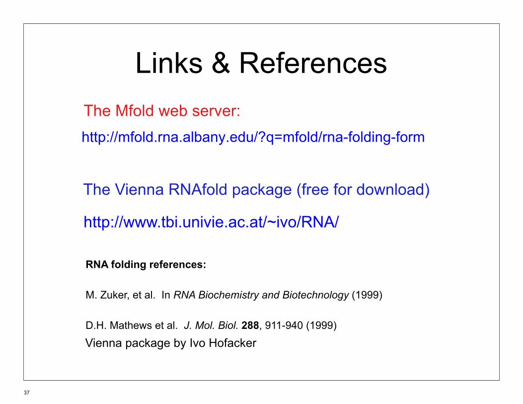
Links & References
The Mfold web server: http://mfold.rna.albany.edu/?q=mfold/rna-folding-form
The Vienna RNAfold package (free for download)
http://www.tbi.univie.ac.at/~ivo/RNA/
RNA folding references:
M. Zuker, et al. In RNA Biochemistry and Biotechnology (1999)
D.H. Mathews et al. J. Mol. Biol. 288, 911-940 (1999)
Vienna package by Ivo Hofacker
37

RNA Secondary Structure Prediction by Energy Minimization Summary
• Assumes folding energy decomposable into independent contributions of small units of structure
• Algorithms are guaranteed to find minimal free energy structure defined by the model
• In practice, algorithms predict ~70% of bp correct
• Errors result from
- imprecision of the model/parameters
- differences between in vitro and in vivo conditions
- in vivo structure may not always have minimum free energy
38

Sample Mfold Output (Human U5 snRNA)
dG = -34.6 kcal/mol
5’ 3’
Minimum free energy structure
Energy dot plot
39
© source unknown. All rights reserved. This content is excluded from our Creative
Commons license. For more information, see http://ocw.mit.edu/help/faq-fair-use/.© Washington University. All rights reserved. This content is
excluded from our Creative Commons license. For moreinformation, see http://ocw.mit.edu/help/faq-fair-use/.

Energy dot plot for a lysine riboswitch
40
© Washington University. All rights reserved. This content is excluded from our Creative
Commons license. For more information, see http://ocw.mit.edu/help/faq-fair-use/.

Function of the lysine riboswitch
Lysine interacts with the junctional core of the riboswitch and is specifically recognized through shape-complementarity within the elongated binding pocket and through several direct and K+-mediated hydrogen bonds to its charged ends.
Controls expression of enzymes involved in biosynthesis and transport of lysine
Serganov et al. Nature 2008. Caron et al PNAS 2012
41
© source unknown. All rights reserved. This content is excluded from our Creative
Commons license. For more information, see http://ocw.mit.edu/help/faq-fair-use/.

MIT OpenCourseWarehttp://ocw.mit.edu
7.91J / 20.490J / 20.390J / 7.36J / 6.802J / 6.874J / HST.506J Foundations of Computational and Systems BiologySpring 2014
For information about citing these materials or our Terms of Use, visit: http://ocw.mit.edu/terms.


![Naturally occurring triterpene Lupane exerts anticancer ... · biological functions [1]. These naturally occurring compounds fall in broad categories of primary and secondary metabolites.](https://static.fdocuments.us/doc/165x107/5fd8c7dbf631af59543bd391/naturally-occurring-triterpene-lupane-exerts-anticancer-biological-functions.jpg)


