RNA polymerase II contributes to preventing...
15
Article RNA polymerase II contributes to preventing transcription-mediated replication fork stalls Irene Felipe-Abrio, Juan Lafuente-Barquero, María L García-Rubio & Andrés Aguilera * Abstract Transcription is a major contributor to genome instability. A main cause of transcription-associated instability relies on the capacity of transcription to stall replication. However, we know little of the possible role, if any, of the RNA polymerase (RNAP) in this process. Here, we analyzed 4 specific yeast RNAPII mutants that show different phenotypes of genetic instability including hyper- recombination, DNA damage sensitivity and/or a strong depen- dency on double-strand break repair functions for viability. Three specific alleles of the RNAPII core, rpb1-1, rpb1-S751F and rpb9Δ, cause a defect in replication fork progression, compensated for by additional origin firing, as the main action responsible for instabil- ity. The transcription elongation defects of rpb1-S751F and rpb9Δ plus our observation that rpb1-1 causes RNAPII retention on chro- matin suggest that RNAPII could participate in facilitating fork progression upon a transcription-replication encounter. Our results imply that the RNAPII or ancillary factors actively help prevent transcription-associated genome instability. Keywords DNA damage response; DNA replication; double-strand break repair; genome instability; RNAPII Subject Categories Chromatin, Epigenetics, Genomics & Functional Genomics; DNA Replication, Repair & Recombination; Transcription DOI 10.15252/embj.201488544 | Received 20 March 2014 | Revised 29 September 2014 | Accepted 8 October 2014 Introduction Genome integrity is essential for cell cycle progression, development and differentiation. In addition to the DNA lesions occurring sponta- neously or induced by external genotoxic agents, cellular processes such as DNA replication, repair, recombination or transcription could affect the stability of the genome (Aguilera & Go ´mez- Gonza ´lez, 2008). Consequently, cells have developed a complex network to coordinate these processes and guarantee genome integ- rity and cell proliferation. Deficiencies in this coordination result in a variety of diseases ranging from severe genetic disorders to cancer predisposition and accelerated aging. Conflicts between replication and transcription can cause an increase in DNA breaks as a consequence of replication fork (RF) stalling and collapse leading to recombination and chromosome rearrangements in a process termed transcription-associated recombination (TAR) (Gaillard et al, 2013b). The fact that replication is the major source of recombino- genic DNA breaks and that recombination with the sister chromatid represents the major double-strand break (DSB) repair pathway during the S/G2 phase of the cell cycle supports this model (Aguilera & Garcı ´a-Muse, 2012). Consistently, TAR is mainly seen when transcription occurs in the S-phase and has been related to RF progression impairment caused by transcription (Prado & Aguilera, 2005; Aguilera & Go ´ mez-Gonza ´lez, 2008; Merrikh et al, 2012). Colli- sions between the replisome and the transcription machinery could lead to DNA breaks that would rely on recombinational repair to allow RF restart (Branzei & Foiani, 2010; Labib & De Piccoli, 2011). An increasing number of studies have tried to understand the role of replication and recombination factors in transcription-associated genome instability both in prokaryotes and in eukaryotes (Kim & Jinks-Robertson, 2012; Gaillard et al, 2013b). The way by which bacteria deal with collisions between the replication and transcrip- tion apparatuses has been extensively studied, since co-directional collisions between the replisome and the RNAP are inevitable. This is due to the fact that the bacterial genome contains only one repli- con; the rate of replication is much faster than that of transcription, and replication is not limited to one defined cell cycle phase. Escheri- chia coli contains three helicases, Rep, UvrD and DinG, which might promote replication of DNA bound to proteins such as the RNAP. Destabilization of transcription complexes can suppress the viability defect observed in repDC33 DuvrD cells, indicating that the Rep– DnaB interaction facilitates resolution of transcription–replication conflicts (Atkinson et al, 2010). Moreover, DinG, Rep and UvrD are essential for efficient replication across highly transcribed regions in vivo (Baharoglu et al, 2010; Boubakri et al, 2010). Backtracking of RNAP at an obstacle has also been implicated in genome instability mediated by transcription–replication collisions (Dutta et al, 2011). RNAP mutants that reduce the frequency of RF stalling have been described in bacteria (McGlynn & Lloyd, 2000; Trautinger & Lloyd, 2002). It has been suggested that such mutants form less stable complexes with template DNA, thus decreasing the probability of collisions with replisomes. In eukaryotes, several factors have been described as being involved in TAR, which in a number of cases proved to be a result from collisions between transcription and replication. These include Centro Andaluz de Biología Molecular y Medicina Regenerativa CABIMER, Universidad de Sevilla, Seville, Spain *Corresponding author. Tel: +34 954468372; E-mail: [email protected] ª 2014 The Authors The EMBO Journal 1 Published online: December 1, 2014
Transcript of RNA polymerase II contributes to preventing...
Article
RNA polymerase II contributes to preventingtranscription-mediated replication fork stallsIrene Felipe-Abrio, Juan Lafuente-Barquero, María L García-Rubio & Andrés Aguilera*
Abstract
Transcription is a major contributor to genome instability. A maincause of transcription-associated instability relies on the capacityof transcription to stall replication. However, we know little of thepossible role, if any, of the RNA polymerase (RNAP) in this process.Here, we analyzed 4 specific yeast RNAPII mutants that showdifferent phenotypes of genetic instability including hyper-recombination, DNA damage sensitivity and/or a strong depen-dency on double-strand break repair functions for viability. Threespecific alleles of the RNAPII core, rpb1-1, rpb1-S751F and rpb9Δ,cause a defect in replication fork progression, compensated for byadditional origin firing, as the main action responsible for instabil-ity. The transcription elongation defects of rpb1-S751F and rpb9Δplus our observation that rpb1-1 causes RNAPII retention on chro-matin suggest that RNAPII could participate in facilitating forkprogression upon a transcription-replication encounter. Our resultsimply that the RNAPII or ancillary factors actively help preventtranscription-associated genome instability.
Keywords DNA damage response; DNA replication; double-strand break
repair; genome instability; RNAPII
Subject Categories Chromatin, Epigenetics, Genomics & Functional
Genomics; DNA Replication, Repair & Recombination; Transcription
DOI 10.15252/embj.201488544 | Received 20 March 2014 | Revised 29
September 2014 | Accepted 8 October 2014
Introduction
Genome integrity is essential for cell cycle progression, development
and differentiation. In addition to the DNA lesions occurring sponta-
neously or induced by external genotoxic agents, cellular processes
such as DNA replication, repair, recombination or transcription
could affect the stability of the genome (Aguilera & Gomez-
Gonzalez, 2008). Consequently, cells have developed a complex
network to coordinate these processes and guarantee genome integ-
rity and cell proliferation. Deficiencies in this coordination result in
a variety of diseases ranging from severe genetic disorders to cancer
predisposition and accelerated aging. Conflicts between replication
and transcription can cause an increase in DNA breaks as a
consequence of replication fork (RF) stalling and collapse leading to
recombination and chromosome rearrangements in a process
termed transcription-associated recombination (TAR) (Gaillard et al,
2013b). The fact that replication is the major source of recombino-
genic DNA breaks and that recombination with the sister chromatid
represents the major double-strand break (DSB) repair pathway
during the S/G2 phase of the cell cycle supports this model
(Aguilera & Garcıa-Muse, 2012). Consistently, TAR is mainly seen
when transcription occurs in the S-phase and has been related to RF
progression impairment caused by transcription (Prado & Aguilera,
2005; Aguilera & Gomez-Gonzalez, 2008; Merrikh et al, 2012). Colli-
sions between the replisome and the transcription machinery could
lead to DNA breaks that would rely on recombinational repair to
allow RF restart (Branzei & Foiani, 2010; Labib & De Piccoli, 2011).
An increasing number of studies have tried to understand the role
of replication and recombination factors in transcription-associated
genome instability both in prokaryotes and in eukaryotes (Kim &
Jinks-Robertson, 2012; Gaillard et al, 2013b). The way by which
bacteria deal with collisions between the replication and transcrip-
tion apparatuses has been extensively studied, since co-directional
collisions between the replisome and the RNAP are inevitable. This
is due to the fact that the bacterial genome contains only one repli-
con; the rate of replication is much faster than that of transcription,
and replication is not limited to one defined cell cycle phase. Escheri-
chia coli contains three helicases, Rep, UvrD and DinG, which might
promote replication of DNA bound to proteins such as the RNAP.
Destabilization of transcription complexes can suppress the viability
defect observed in repDC33 DuvrD cells, indicating that the Rep–
DnaB interaction facilitates resolution of transcription–replication
conflicts (Atkinson et al, 2010). Moreover, DinG, Rep and UvrD are
essential for efficient replication across highly transcribed regions in
vivo (Baharoglu et al, 2010; Boubakri et al, 2010). Backtracking of
RNAP at an obstacle has also been implicated in genome instability
mediated by transcription–replication collisions (Dutta et al, 2011).
RNAP mutants that reduce the frequency of RF stalling have been
described in bacteria (McGlynn & Lloyd, 2000; Trautinger & Lloyd,
2002). It has been suggested that such mutants form less stable
complexes with template DNA, thus decreasing the probability of
collisions with replisomes.
In eukaryotes, several factors have been described as being
involved in TAR, which in a number of cases proved to be a result
from collisions between transcription and replication. These include
Centro Andaluz de Biología Molecular y Medicina Regenerativa CABIMER, Universidad de Sevilla, Seville, Spain*Corresponding author. Tel: +34 954468372; E-mail: [email protected]
ª 2014 The Authors The EMBO Journal 1
Published online: December 1, 2014
RNA-binding proteins and others with a role in transcription elonga-
tion, mRNP biogenesis, processing and/or export (Aguilera &
Garcıa-Muse, 2013). As in bacteria, DNA helicases are involved in
replication through obstacles that need to be bypassed to allow
replication resumption. In yeast, one such helicase is Rrm3 (Ivessa
et al, 2003), which is important in solving transcription–replication
collisions (Prado & Aguilera, 2005; Azvolinsky et al, 2009). However,
other elements can contribute to TAR, including the formation of
R-loops, structures formed by RNA–DNA hybrids and the displaced
DNA strand (Aguilera & Garcıa-Muse, 2012), or the local negative
supercoiling transiently occurring behind the RNAP (Gaillard et al,
2013b). In such cases, the formation of single-stranded DNA (ssDNA),
more susceptible to damage, would be facilitated (Schmidt et al, 2006;
Kim et al, 2007). R-loops seem to occur naturally, but their accumula-
tion is greatly enhanced in cells deficient in different steps of RNA
processing and mRNP biogenesis and export. These deficiencies cause
high levels of genome instability as detected by hyper-recombination,
DNA damage accumulation or chromosome loss from yeast to humans
(Jimeno et al, 2002; Huertas & Aguilera 2003; Li & Manley, 2005;
Paulsen et al, 2009; Domınguez-Sanchez et al, 2011; Mischo et al,
2011; Wahba et al, 2011; Castellano-Pozo et al, 2012; Stirling et al,
2012). R-loops impair transcription elongation and RF progression
(Wellinger et al, 2006; Tous & Aguilera, 2007; Gan et al, 2011) likely
due to the ability of the R-loop and/or a putatively associated RNA
polymerase to become a roadblock for transcription and replication
(Drolet et al, 1995; Huertas & Aguilera, 2003).
Despite the increasing evidence that transcription may be one of
the main obstacles to RF progression as a source of genome instabil-
ity (Gaillard et al, 2013b), little is known about the factors impli-
cated. Here, we asked whether the transcription machinery itself
has a direct role in genome instability. We searched for specific
mutations of different RNAPII subunits in Saccharomyces cerevisiae
that caused genome instability and a strong dependency on DSB
repair functions for viability. We analyzed three mutations rpb1-1,
rpb1S751F and rpb9Δ with these phenotypes. Our results demon-
strate that RNAPII by itself can become an obstacle to replication
and can help solve or prevent the RF stalling and collapse responsi-
ble for genome instability.
Results
Genetic interactions between RNAPII and DNA repair mutants
To investigate whether the RNAPII itself might play a role in the
origin of genome instability, we analyzed the sensitivity of a
selected number of known RNAPII mutants (Supplementary Table
S1) to several DNA damaging drugs. In the presence of hydroxyurea
(HU), which inhibits the ribonucleotide reductase causing a deple-
tion of dNTPs that impair replication progression, and methyl-
methanesulfonate (MMS), an alkylating agent that can indirectly
cause DNA breaks, the selected RNAPII mutants showed different
degrees of growth defects (Supplementary Fig S1A). Next, we asked
whether or not there were genetic interactions between the RNAPII
mutants and those of homologous recombination (HR) such as
rad52 and mre11 (Fig 1 and Supplementary Fig S1B). The strongest
interaction was observed in the tetrad analysis of the crosses of
rad52D and mre11D with rpb1-S751F, where we did not find viable
double mutants (Fig 1B), consistent with the sick phenotype used to
isolate this mutant in a rad52 background (F. Malagon, pers.
comm.) (Strathern et al, 2013). However, rpb1-S751F was tolerated
in both rad51Δ and pol32Δ backgrounds. As we have previously
shown that Rad51 and Pol32 control two overlapping outcomes of
Rad52-dependent HR events (Moriel-Carretero & Aguilera, 2010;
Munoz-Galvan et al, 2013b), we tested whether rpb1-S751F rad51Δpol32Δ triple mutants were viable or not. Indeed, they were not,
suggesting that these mutants accumulate replication-born DNA
breaks that cannot be repaired in the absence of Rad51 and Pol32.
Also, rpb1-1 and rpb2-10 strains showed strong growth defects in
the absence of HR functions. The rpb9D mutant was highly sensitive
A
B C
D
Figure 1. Genetic interactions of RNAPII mutations with DNA repairmutations.
A Viability of single and double mutants of RNAPII and rad52D, mre11D andcdc44-8. Tenfold serial dilutions of double mutants obtained by geneticcrosses were tested for growth in plain SC medium or supplemented with10 mM HU or 0.005% MMS.
B Tetrad analysis of rpb1-S751F, rad52D and mre11D crosses. D indicatesdouble mutants that fail to grow.
C Tetrad analysis of rpb1-S751F crossed by pol32D and rad51D. D indicatestriple mutants that fail to grow.
D HU and MMS sensitivity of different mutant combinations of rpb1-1, rpb1-S751F, pol32D and rad51D.
Data information: Cells were cultured for 3 days at 30°C.
The EMBO Journal ª 2014 The Authors
The EMBO Journal RNAPII helps prevent genome instability Irene Felipe-Abrio et al
2
Published online: December 1, 2014
to HU and in the absence of Mre11 presented a severe growth defect
in MMS. Additionally, the four mutants, in combination with the
replicative clamp loader RFC1 allele cdc44-8, essential in recombina-
tional DSB repair (Holmes & Haber, 1999), showed a reduced-growth
phenotype in low concentrations of HU and MMS. An extended
analysis of rpb1-1 in combination with mutations in other HR repair
genes such as rad50Δ, xrs2Δ, rad51Δ, rad54Δ, mus81Δ, pol32Δ,sgs1Δ or dia2Δ confirmed the need of DSB repair for viability of
rpb1-1 (Supplementary Fig S1C). Taking into account these results, we
selected rpb1-1, rpb1-S751F, rpb2-10 and rpb9D for further analyses.
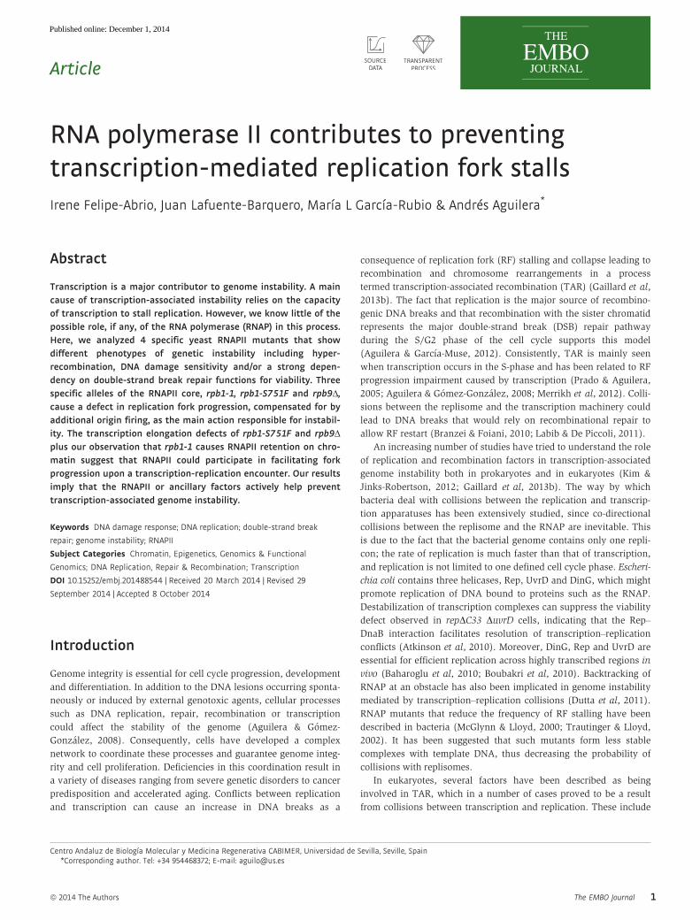
RNAPII mutants show increased DSBs and genetic instability
The need of HR functions for viability of rpb mutants under replica-
tion stress suggests that they may accumulate DSBs. We tested this
possibility by Western blot against the phosphorylated form of
histone H2A (H2A-P), a molecular marker for DSBs, in cells with
and without HU treatment. In rpb1-1 cells, an H2A-P signal was
detected in the absence of HU (Fig 2A), confirming that breaks
occur spontaneously at high frequency. Importantly, this signal was
exacerbated in HU. In the other mutants, the H2A-P signal was
stronger than in the WT in the presence of HU, but not detectable in
its absence. This tendency of all four rpb mutants to accumulate
DSBs was confirmed by analyzing the accumulation of Rad52 foci, a
marker for DSB repair centers (Lisby et al, 2001). RNAPII mutations
increased the percentage of cells with Rad52 foci, and these were
further increased in thermo-sensitive mutants rpb1-1, rpb1-S751F
and rpb9D at 37°C (Fig 2B). Therefore, DNA breaks accumulate in
these RNAPII mutants and are processed into a recombinogenic
intermediate.
Next, we asked whether recombination was enhanced in the
mutants. We used the plasmid-based pTINV system, carrying
inverted repeats of truncated leu2 fragments, and the chromosomal
leu2-k::ADE2-URA3::leu2-k direct-repeat system (Gomez-Gonzalez
et al, 2011b) (Fig 2). A significant increase of Leu+ recombinants
could be seen in rpb1-1 cells with respect to WT in both systems
(Fig 2C and D), whereas the other mutants show recombination
levels similar to WT. We then used a number of plasmid-based
recombination systems to study TAR. These were the L-lacZ and
GL-lacZ systems carrying 0.6-kb leu2 direct repeats flanking lacZ
under the LEU2 or the GAL1 promoter, respectively. Recombination
can be analyzed in these systems under conditions of low (GAL1p in
2% glucose), medium (LEU2p) and high transcription (GAL1p 2%
galactose) (Gomez-Gonzalez et al, 2011b). As can be seen in Fig 2E,
the higher the levels of transcription, the stronger the increase in
recombination in rpb1-S751F. rpb9D showed a clear hyper-recombi-
nation phenotype that increased with transcription, which is partic-
ularly interesting since both mutants have defects in transcription
elongation (Hemming et al, 2000; Strathern et al, 2013). No differ-
ence was observed with the L/GL-lacZ systems in rpb1-1 mutant.
This could be due to a lower efficiency of HR leading to detectable
recombination products, which are different for each assay (Gomez-
Gonzalez et al, 2011b). Interestingly, although the recombinational
behavior of rpb1 mutants and rpb9Δ was heterogeneous, they all
cause a hyper-recombination phenotype that was mainly transcrip-
tion-dependent. In addition, all mutants, with the exception of
rpb2-10, showed a significant increase in plasmid loss when lacZ
was transcribed, as determined with pGAL-LacZ (Fig 2F). In
0 4 8
12 16 20
2.8 5.5
12.0 6.8
2.4
*
Ptet leu2-HOr leu2
Rec
ombi
natio
n fre
quen
cy (x
10-4
)
5.9
15.0
4.8 3.8
6.6
0 4 8
12 16 20
WT
rpb1-1
rpb2-10 rpb9
rpb1-S751F
*
WT
rpb1-1
rpb2-10 rpb9
rpb1-S751F
H2A-P
Loading control
-HU + HU
WT
rpb1-1
rpb1-S751F rpb2-10
rpb9
3.5
8.5
13.0 14.4 13.5
2.5
17.0 15.6
13.1
18.5
0
5
10
15
20
25
rpb1-S751F
S/G
2 ce
lls w
ith R
ad52
foci
(%
)
* *
* *
* * *
WT
rpb1-1
rpb2-10 rpb9
RAD52-YFP 30° C37° C
A B
C
F
DADE2 URA3 leu2-K leu2-K
Rec
ombi
natio
n fre
quen
cy (x
10-5
)
WT
rpb1-1
rpb1-S751F rpb2-10
rpb9
*
E
6 8
36 19 5 16 12
138
34 49
18 27
198
27
120
0
50
100
150
200
250
rpb1-S751F
Rec
ombi
natio
n fre
quen
cy (x
10-4
) Transcription
Medium High
Low
*
*
WT
rpb1-1
rpb2-10 rpb9
lacZ leu2 leu2PLEU2
PGAL
3.9
7.4
17.5
6.4
18.7
0
5
10
15
20
25
rpb1-S751F
* *
Pla
smid
loss
(%)
WT
rpb1-1
rpb2-10 rpb9
*
LacZ GAL1p
URA3 CEN
L-lacZ
GL-lacZ
Figure 2. Genetic instability in RNAPII mutants.
A Western blot against the phosphorylated form of histone H2A in thepresence of 100 mM HU in asynchronously growing WT and RNAPIImutant cells. Ponceau staining is shown as a loading control.
B Rad52 foci formation at 30 and 37°C.C Recombination frequencies between inverted repeats in WT and RNAPII
mutants measured with the plasmid-born pTINV system. Recombinantswere selected as Leu+.
D Recombination frequencies using the direct-repeat chromosomal systemleu2-k::ADE2-URA3::leu2-k in WT and RNAPII mutants. Deletion events weredetected and quantified in media containing 5-fluorotic acid (FOA) andcould be directly visualized as red sectors.
E Recombination frequencies between plasmid-born direct repeats in WTand RNAPII mutants. The frequency of Leu+ recombination was determinedin the systems L-lacZ and GL-lacZ. Recombination frequencies are plottedas a function of the transcription levels. Low transcription refers to theGL-lacZ systems in strains cultured in 2% glucose; medium refers to L-lacZin 2% glucose, and high to GL-lacZ in 2% galactose.
F Mitotic stability of centromeric plasmid pGAL-lacZ in RNAPII mutants.Stability was analyzed in galactose after 23 generations. A small diagram ofeach system (not drawn to scale) is shown.
Data information: statistically significant differences with respect to WTaccording to Student’s t-test (P < 0.05).Source data are available online for this figure.
ª 2014 The Authors The EMBO Journal
Irene Felipe-Abrio et al RNAPII helps prevent genome instability The EMBO Journal
3
Published online: December 1, 2014

summary, despite the heterogeneity of phenotypes, genetic instability
increased in rpb1-1, rpb1-S751F and rpb9Δ, whereas rpb2-10 was
poorly or not affected.
Among the mutants studied, viability of rpb1-1 and rpb1-S751F
shows the strongest requirements for HR functions, in particular
under replication stress. Then, we analyzed the effect of mre11Δ,which abrogates the early steps of DSB repair, on the genome insta-
bility phenotype of rpb1-1. Inverted-repeat recombination increased
in the rpb1-1 mre11D double mutant above the single mutants,
whereas Rad52 foci accumulated at lower levels with respect to the
single mre11Δ cells (Fig 3A and B). This suggests that unrepaired
DNA breaks are channeled into a single-strand annealing pathway
of recombination in the absence of an active MRX complex, avoid-
ing Rad52 foci accumulation. As expected, this pathway was less
effective provided that the Rad52-dependent HR pathway was also
impaired. Consistently, rpb1-1 mre11D double mutants were highly
sensitive to HU (Fig 3C).
These results are consistent with the idea that rpb1 mutants
generate more DNA breaks in a transcription-dependent manner. So
we wondered whether this was also dependent on R-loops as it
occurs in the hpr1Δ mutant, in which R-loops are reduced by RNH1
overexpression (Huertas & Aguilera, 2003), but this was not the
case. As can be seen in Fig 3D, rpb1-1 and rpb1-S751F levels of
Rad52-foci were not reduced by RNH1 overexpression.
DNA replication is impaired in rpb1 and rpb9 mutants
Next, we analyzed whether DNA replication was affected in these
mutants by different means. First, we synchronized cells in G1
with a-factor and monitored growth at different times after G1
release under replicative stress. A clear arrest in G1/early S-phase
was observed in rpb1-1, rpb1-S751F and rpb9Δ, whereas only a
slight delay was observed in rpb2-10 (Fig 4A). In the absence of
HU, a similar tendency of S-phase delay was observed (Supple-
mentary Fig S2).
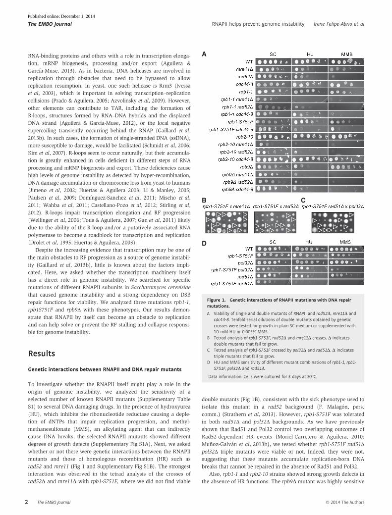
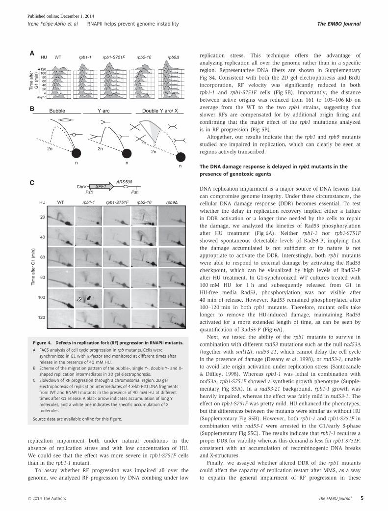
Consequently, we directly analyzed the effect of the rpb muta-
tions in RF progression by 2D gel electrophoresis. A scheme of the
migration pattern of replication intermediates is shown (Fig 4B).
Replication through the SPF1 (YEL031w) gene of chromosome V
lying close to the early replication origin ARS508 (Gomez-Gonzalez
et al, 2009) in G1-synchronized cells released into S-phase under
low replication stress (40 mM HU) revealed a clear delay in RF
progression in rpb1-1, rpb1-S751F and rpb9Δ cells. Y-arcs persisted
much longer in the mutants than in WT cells (Fig 4C). In addition,
replication initiation was significantly delayed; whereas the Y-arc is
fully visible in WT cells after 20 min, it was absent in the three
mutants. However, the rpb2-10 mutant was not affected either in
initiation or in RF progression, since the 2D electrophoresis profile
was the same as that of WT (Fig 4C). The results, consistent with
the FACS analysis, evidence that RF progression is impaired in the
mutants showing genome instability. Interestingly, 2D gel electro-
phoresis of rpb1-1 reveals an accumulation of asymmetric X mole-
cules not observed either in WT cells or in the other mutants tested.
These structures could in principle be a consequence of two conver-
gent forks unable to terminate properly, but this seems unlikely
given the distance of the fork coming from the other side. The other
possibility was that formation of X-structures depended on HR
factors such as Rad51. FACS analysis revealed that the double-
mutant rpb1-1 rad51Δ shows a profile similar to that of rpb1-1, indi-
cating that rpb1-1 was epistatic to rad51Δ (Supplementary Fig S3A).
The 2D gel electrophoresis confirmed this epistatic relationship
and revealed that the X-structures disappear in the double-mutant
rpb1-1 rad51Δ. Therefore, the replication defect observed in rpb1-1
leads to the accumulation of Rad51-dependent recombination inter-
mediates (Supplementary Fig S3B).
Next, we assayed whether this was general to other regions all
over the genome, for which we focused the rest of the study on
the rpb1 alleles, which altered the largest and essential subunit of
RNAPII. First, we analyzed the kinetics of bromodeoxy-uridine
(BrdU) incorporation using conveniently modified strains harboring
the human TK gene via chromatin immunoprecipitation (ChIP)
using anti-BrdU antibody. Two regions were analyzed in which
transcription of the ORF was convergent to replication originated
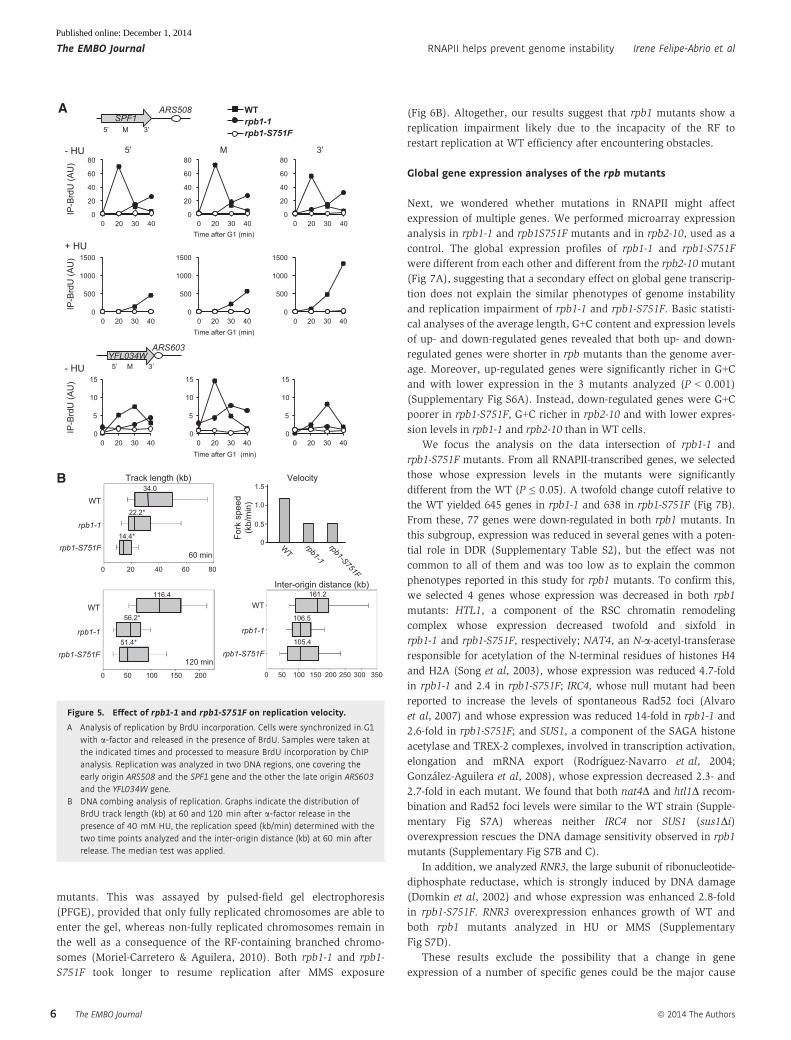
from its closest ARS. As can be seen in Fig 5A, BrdU incorporation
not only was observed at later times than in WT cells, suggesting
a delayed initiation as observed by 2D electrophoresis, but the
speed of incorporation was slower, as can be deduced from a
much lower slope of the incorporation profile of the SPF1 gene.
This reduction in the speed was also observed at YFL034W,
located near a late replication origin (Fig 5A). We could detect
10
100
1000
Via
bilit
y (%
)
0 2 4 7 21
5.9 15.0
12.5
54.1
0
20
40
60
80
Rec
ombi
natio
n fre
quen
cy (x
10-4
) Ptet leu2-HOr leu2
* *
*
WT
rpb1-1 mre11
rpb1-1 mre11
A
C
B
3.5 8.5
46.4
30.7
0
20
40
60
2 foci 1 focus
S/G
2 ce
lls w
ith R
ad52
foci
(%)
WT
rpb1-1 mre11
rpb1-1 mre11
RAD52-YFP
*
*
*
3.0
4.7 6.6
10.6
2.8 3.2
8.0 10.2
0
3
6
9
12 +RNH1 - RNH1
S/G
2 ce
lls w
ith R
ad52
foci
(%
)
*
WT
hpr1
rpb1-1
rpb1-S751F
D
10
100
1000
0 2 4 7 21 Time (h)
WT rpb1-1 rpb1-1 mre11
mre11
Via
bilit
y af
ter H
U tr
eatm
ent (
%)
Figure 3. Requirement of HR functions for viability of rpb1 mutants.
A Recombination frequencies measured with the pTINV system in WT, rpb1-1,mre11D and rpb1-1 mre11D.
B Rad52 focus formation in asynchronously growing cells at 30°C.C Viability after different times in 10 mM HU. The same cultures without HU
were used as a control. The average values and standard deviations fromthree independent experiments are shown.
D Rad52 focus formation in asynchronously growing cells expressing normalor high levels of RNH1.
Data information: *P < 0.05 (Student’s t-test). Other details as in Fig 2.
The EMBO Journal ª 2014 The Authors
The EMBO Journal RNAPII helps prevent genome instability Irene Felipe-Abrio et al
4
Published online: December 1, 2014
replication impairment both under natural conditions in the
absence of replication stress and with low concentration of HU.
We could see that the effect was more severe in rpb1-S751F cells
than in the rpb1-1 mutant.
To assay whether RF progression was impaired all over the
genome, we analyzed RF progression by DNA combing under low
replication stress. This technique offers the advantage of
analyzing replication all over the genome rather than in a specific
region. Representative DNA fibers are shown in Supplementary
Fig S4. Consistent with both the 2D gel electrophoresis and BrdU
incorporation, RF velocity was significantly reduced in both
rpb1-1 and rpb1-S751F cells (Fig 5B). Importantly, the distance
between active origins was reduced from 161 to 105–106 kb on
average from the WT to the two rpb1 strains, suggesting that
slower RFs are compensated for by additional origin firing and
confirming that the major effect of the rpb1 mutations analyzed
is in RF progression (Fig 5B).
Altogether, our results indicate that the rpb1 and rpb9 mutants
studied are impaired in replication, which can clearly be seen at
regions actively transcribed.
The DNA damage response is delayed in rpb1 mutants in thepresence of genotoxic agents
DNA replication impairment is a major source of DNA lesions that
can compromise genome integrity. Under these circumstances, the
cellular DNA damage response (DDR) becomes essential. To test
whether the delay in replication recovery implied either a failure
in DDR activation or a longer time needed by the cells to repair
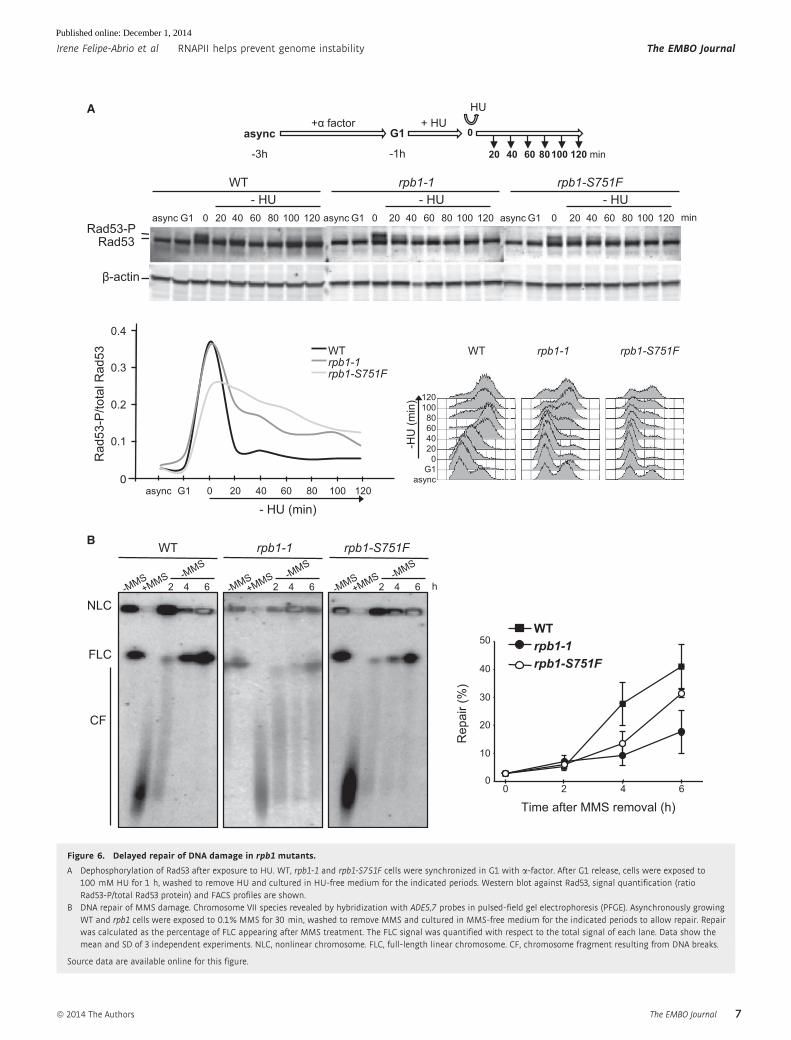
the damage, we analyzed the kinetics of Rad53 phosphorylation
after HU treatment (Fig 6A). Neither rpb1-1 nor rpb1-S751F
showed spontaneous detectable levels of Rad53-P, implying that
the damage accumulated is not sufficient or its nature is not
appropriate to activate the DDR. Interestingly, both rpb1 mutants
were able to respond to external damage by activating the Rad53
checkpoint, which can be visualized by high levels of Rad53-P
after HU treatment. In G1-synchronized WT cultures treated with
100 mM HU for 1 h and subsequently released from G1 in
HU-free media Rad53, phosphorylation was not visible after
40 min of release. However, Rad53 remained phosphorylated after
100–120 min in both rpb1 mutants. Therefore, mutant cells take
longer to remove the HU-induced damage, maintaining Rad53
activated for a more extended length of time, as can be seen by
quantification of Rad53-P (Fig 6A).
Next, we tested the ability of the rpb1 mutants to survive in
combination with different rad53 mutations such as the null rad53Δ(together with sml1Δ), rad53-21, which cannot delay the cell cycle
in the presence of damage (Desany et al, 1998), or rad53-1, unable
to avoid late origin activation under replication stress (Santocanale
& Diffley, 1998). Whereas rpb1-1 was lethal in combination with
rad53Δ, rpb1-S751F showed a synthetic growth phenotype (Supple-
mentary Fig S5A). In a rad53-21 background, rpb1-1 growth was
heavily impaired, whereas the effect was fairly mild in rad53-1. The
effect on rpb1-S751F was pretty mild. HU enhanced the phenotypes,
but the differences between the mutants were similar as without HU
(Supplementary Fig S5B). However, both rpb1-1 and rpb1-S751F in
combination with rad53-1 were arrested in the G1/early S-phase
(Supplementary Fig S5C). The results indicate that rpb1-1 requires a
proper DDR for viability whereas this demand is less for rpb1-S751F,
consistent with an accumulation of recombinogenic DNA breaks
and X-structures.
Finally, we assayed whether altered DDR of the rpb1 mutants
could affect the capacity of replication restart after MMS, as a way
to explain the general impairment of RF progression in these
20
40
60
80
100
120
Tim
e af
ter G
1 (m
in)
rpb1-1 rpb2-10 WT rpb9rpb1-S751F
async
20 40 60 80
100 120
HU
Tim
e af
ter
G1
(min
)
A
B
0
0
0
0
0
0
SPF1 ARS508
ChrV PstI PstI
rpb1-1 rpb2-10 WT rpb9 rpb1-S751F HU
C
n
2n
n
2n
n
2n
Y arc Bubble Double Y arc/ X
Figure 4. Defects in replication fork (RF) progression in RNAPII mutants.
A FACS analysis of cell cycle progression in rpb mutants. Cells weresynchronized in G1 with a-factor and monitored at different times afterrelease in the presence of 40 mM HU.
B Scheme of the migration pattern of the bubble-, single Y-, double Y- and X-shaped replication intermediates in 2D gel electrophoresis.
C Slowdown of RF progression through a chromosomal region. 2D gelelectrophoresis of replication intermediates of 4.3-kb PstI DNA fragmentsfrom WT and RNAPII mutants in the presence of 40 mM HU at differenttimes after G1 release. A black arrow indicates accumulation of long Ymolecules, and a white one indicates the specific accumulation of Xmolecules.
Source data are available online for this figure.
ª 2014 The Authors The EMBO Journal
Irene Felipe-Abrio et al RNAPII helps prevent genome instability The EMBO Journal
5
Published online: December 1, 2014
mutants. This was assayed by pulsed-field gel electrophoresis
(PFGE), provided that only fully replicated chromosomes are able to
enter the gel, whereas non-fully replicated chromosomes remain in
the well as a consequence of the RF-containing branched chromo-
somes (Moriel-Carretero & Aguilera, 2010). Both rpb1-1 and rpb1-
S751F took longer to resume replication after MMS exposure
(Fig 6B). Altogether, our results suggest that rpb1 mutants show a
replication impairment likely due to the incapacity of the RF to
restart replication at WT efficiency after encountering obstacles.
Global gene expression analyses of the rpb mutants
Next, we wondered whether mutations in RNAPII might affect
expression of multiple genes. We performed microarray expression
analysis in rpb1-1 and rpb1S751F mutants and in rpb2-10, used as a
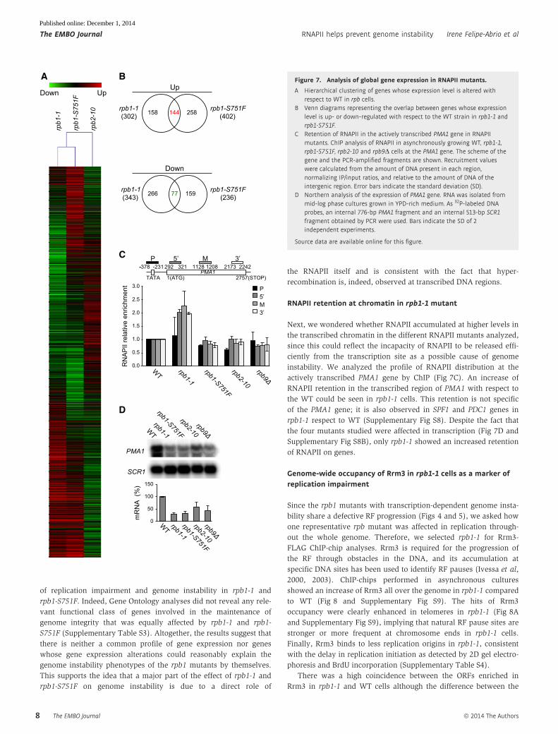
control. The global expression profiles of rpb1-1 and rpb1-S751F
were different from each other and different from the rpb2-10mutant
(Fig 7A), suggesting that a secondary effect on global gene transcrip-
tion does not explain the similar phenotypes of genome instability
and replication impairment of rpb1-1 and rpb1-S751F. Basic statisti-
cal analyses of the average length, G+C content and expression levels
of up- and down-regulated genes revealed that both up- and down-
regulated genes were shorter in rpb mutants than the genome aver-
age. Moreover, up-regulated genes were significantly richer in G+C
and with lower expression in the 3 mutants analyzed (P < 0.001)
(Supplementary Fig S6A). Instead, down-regulated genes were G+C
poorer in rpb1-S751F, G+C richer in rpb2-10 and with lower expres-
sion levels in rpb1-1 and rpb2-10 than in WT cells.
We focus the analysis on the data intersection of rpb1-1 and
rpb1-S751F mutants. From all RNAPII-transcribed genes, we selected
those whose expression levels in the mutants were significantly
different from the WT (P ≤ 0.05). A twofold change cutoff relative to
the WT yielded 645 genes in rpb1-1 and 638 in rpb1-S751F (Fig 7B).
From these, 77 genes were down-regulated in both rpb1 mutants. In
this subgroup, expression was reduced in several genes with a poten-
tial role in DDR (Supplementary Table S2), but the effect was not
common to all of them and was too low as to explain the common
phenotypes reported in this study for rpb1 mutants. To confirm this,
we selected 4 genes whose expression was decreased in both rpb1
mutants: HTL1, a component of the RSC chromatin remodeling
complex whose expression decreased twofold and sixfold in
rpb1-1 and rpb1-S751F, respectively; NAT4, an N-a-acetyl-transferaseresponsible for acetylation of the N-terminal residues of histones H4
and H2A (Song et al, 2003), whose expression was reduced 4.7-fold
in rpb1-1 and 2.4 in rpb1-S751F; IRC4, whose null mutant had been
reported to increase the levels of spontaneous Rad52 foci (Alvaro
et al, 2007) and whose expression was reduced 14-fold in rpb1-1 and
2.6-fold in rpb1-S751F; and SUS1, a component of the SAGA histone
acetylase and TREX-2 complexes, involved in transcription activation,
elongation and mRNA export (Rodrıguez-Navarro et al, 2004;
Gonzalez-Aguilera et al, 2008), whose expression decreased 2.3- and
2.7-fold in each mutant. We found that both nat4D and htl1D recom-
bination and Rad52 foci levels were similar to the WT strain (Supple-
mentary Fig S7A) whereas neither IRC4 nor SUS1 (sus1Di)overexpression rescues the DNA damage sensitivity observed in rpb1
mutants (Supplementary Fig S7B and C).
In addition, we analyzed RNR3, the large subunit of ribonucleotide-
diphosphate reductase, which is strongly induced by DNA damage
(Domkin et al, 2002) and whose expression was enhanced 2.8-fold
in rpb1-S751F. RNR3 overexpression enhances growth of WT and
both rpb1 mutants analyzed in HU or MMS (Supplementary
Fig S7D).
These results exclude the possibility that a change in gene
expression of a number of specific genes could be the major cause
A
0
500
1000
1500
0 20 30 40
0
20
40
60
80
0 20 30 40
IP-B
rdU
(AU
) IP
-Brd
U (A
U)
0
20
40
60
80
0 20 30 40
0
500
1000
1500
0 20 30 40
M
SPF1 ARS508
Time after G1 (min)
+ HU
- HU
Time after G1 (min)
0
500
1000
1500
0 20 30 40
0
20
40
60
80
0 20 30 40
WTrpb1-1rpb1-S751F
B
WT
rpb1-1 rpb1-S751F
Fork
spe
ed
(kb/
min
)
0
0.5
1.5
1.0
Velocity
YFL034W ARS603
0
5
10
15
0 20 30 40
IP-B
rdU
(AU
)
0
5
10
15
0 20 30 40 0
5
10
15
0 20 30 40 Time after G1 (min)
- HU
WT
rpb1-1
rpb1-S751F
0 50 100 150 200 250 300 350
Inter-origin distance (kb) 161.2
106.5
105.4
0 50 100 150 200
WT
rpb1-1
rpb1-S751F 120 min
116.4
56.2*
51.4*
0 20 40 60 80
Track length (kb)
WT
rpb1-1
rpb1-S751F 60 min
14.4*
22.2*
34.0
Figure 5. Effect of rpb1-1 and rpb1-S751F on replication velocity.
A Analysis of replication by BrdU incorporation. Cells were synchronized in G1with a-factor and released in the presence of BrdU. Samples were taken atthe indicated times and processed to measure BrdU incorporation by ChIPanalysis. Replication was analyzed in two DNA regions, one covering theearly origin ARS508 and the SPF1 gene and the other the late origin ARS603and the YFL034W gene.
B DNA combing analysis of replication. Graphs indicate the distribution ofBrdU track length (kb) at 60 and 120 min after a-factor release in thepresence of 40 mM HU, the replication speed (kb/min) determined with thetwo time points analyzed and the inter-origin distance (kb) at 60 min afterrelease. The median test was applied.
The EMBO Journal ª 2014 The Authors
The EMBO Journal RNAPII helps prevent genome instability Irene Felipe-Abrio et al
6
Published online: December 1, 2014
-actin
A
asyncG1
20 40 60 80
100 120
0
-HU
(min
)
async G1 0 20 40 60 80 100 120
WT rpb1-1 rpb1-S751F
Rad53 Rad53-P
async G1 0 20 40 60 80 100 120 async G1 0 20 40 60 80 100 120
- HU - HU - HU min
async
-3h
+ HU 0
+ factor G1
-1h 40 80 20 60 100 120 min
HU
WT rpb1-1 rpb1-S751F
NLC
FLC
CF
B
h-MMS 2 4 6 +MMS -MMS
-MMS 2 4 6 +MMS -MMS
-MMS 2 4 6 +MMS -MMS
0 2 4 6
Time after MMS removal (h)
10
20
30
40
50
0
Rep
air (
%)
WTrpb1-1rpb1-S751F
0
WTrpb1-1rpb1-S751F
0.4
0.3
0.2
0.1
async G1 0 20 40 60 80 100 120
- HU (min)
Rad
53-P
/tota
l Rad
53 WT rpb1-1 rpb1-S751F
Figure 6. Delayed repair of DNA damage in rpb1 mutants.
A Dephosphorylation of Rad53 after exposure to HU. WT, rpb1-1 and rpb1-S751F cells were synchronized in G1 with a-factor. After G1 release, cells were exposed to100 mM HU for 1 h, washed to remove HU and cultured in HU-free medium for the indicated periods. Western blot against Rad53, signal quantification (ratioRad53-P/total Rad53 protein) and FACS profiles are shown.
B DNA repair of MMS damage. Chromosome VII species revealed by hybridization with ADE5,7 probes in pulsed-field gel electrophoresis (PFGE). Asynchronously growingWT and rpb1 cells were exposed to 0.1% MMS for 30 min, washed to remove MMS and cultured in MMS-free medium for the indicated periods to allow repair. Repairwas calculated as the percentage of FLC appearing after MMS treatment. The FLC signal was quantified with respect to the total signal of each lane. Data show themean and SD of 3 independent experiments. NLC, nonlinear chromosome. FLC, full-length linear chromosome. CF, chromosome fragment resulting from DNA breaks.
Source data are available online for this figure.
ª 2014 The Authors The EMBO Journal
Irene Felipe-Abrio et al RNAPII helps prevent genome instability The EMBO Journal
7
Published online: December 1, 2014
of replication impairment and genome instability in rpb1-1 and
rpb1-S751F. Indeed, Gene Ontology analyses did not reveal any rele-
vant functional class of genes involved in the maintenance of
genome integrity that was equally affected by rpb1-1 and rpb1-
S751F (Supplementary Table S3). Altogether, the results suggest that
there is neither a common profile of gene expression nor genes
whose gene expression alterations could reasonably explain the
genome instability phenotypes of the rpb1 mutants by themselves.
This supports the idea that a major part of the effect of rpb1-1 and
rpb1-S751F on genome instability is due to a direct role of
the RNAPII itself and is consistent with the fact that hyper-
recombination is, indeed, observed at transcribed DNA regions.
RNAPII retention at chromatin in rpb1-1 mutant
Next, we wondered whether RNAPII accumulated at higher levels in
the transcribed chromatin in the different RNAPII mutants analyzed,
since this could reflect the incapacity of RNAPII to be released effi-
ciently from the transcription site as a possible cause of genome
instability. We analyzed the profile of RNAPII distribution at the
actively transcribed PMA1 gene by ChIP (Fig 7C). An increase of
RNAPII retention in the transcribed region of PMA1 with respect to
the WT could be seen in rpb1-1 cells. This retention is not specific
of the PMA1 gene; it is also observed in SPF1 and PDC1 genes in
rpb1-1 respect to WT (Supplementary Fig S8). Despite the fact that
the four mutants studied were affected in transcription (Fig 7D and
Supplementary Fig S8B), only rpb1-1 showed an increased retention
of RNAPII on genes.
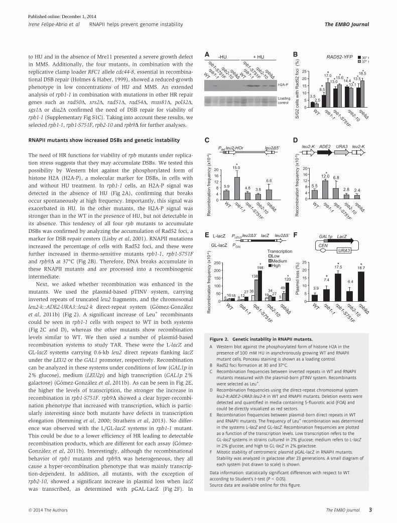
Genome-wide occupancy of Rrm3 in rpb1-1 cells as a marker ofreplication impairment
Since the rpb1 mutants with transcription-dependent genome insta-
bility share a defective RF progression (Figs 4 and 5), we asked how
one representative rpb mutant was affected in replication through-
out the whole genome. Therefore, we selected rpb1-1 for Rrm3-
FLAG ChIP-chip analyses. Rrm3 is required for the progression of
the RF through obstacles in the DNA, and its accumulation at
specific DNA sites has been used to identify RF pauses (Ivessa et al,
2000, 2003). ChIP-chips performed in asynchronous cultures
showed an increase of Rrm3 all over the genome in rpb1-1 compared
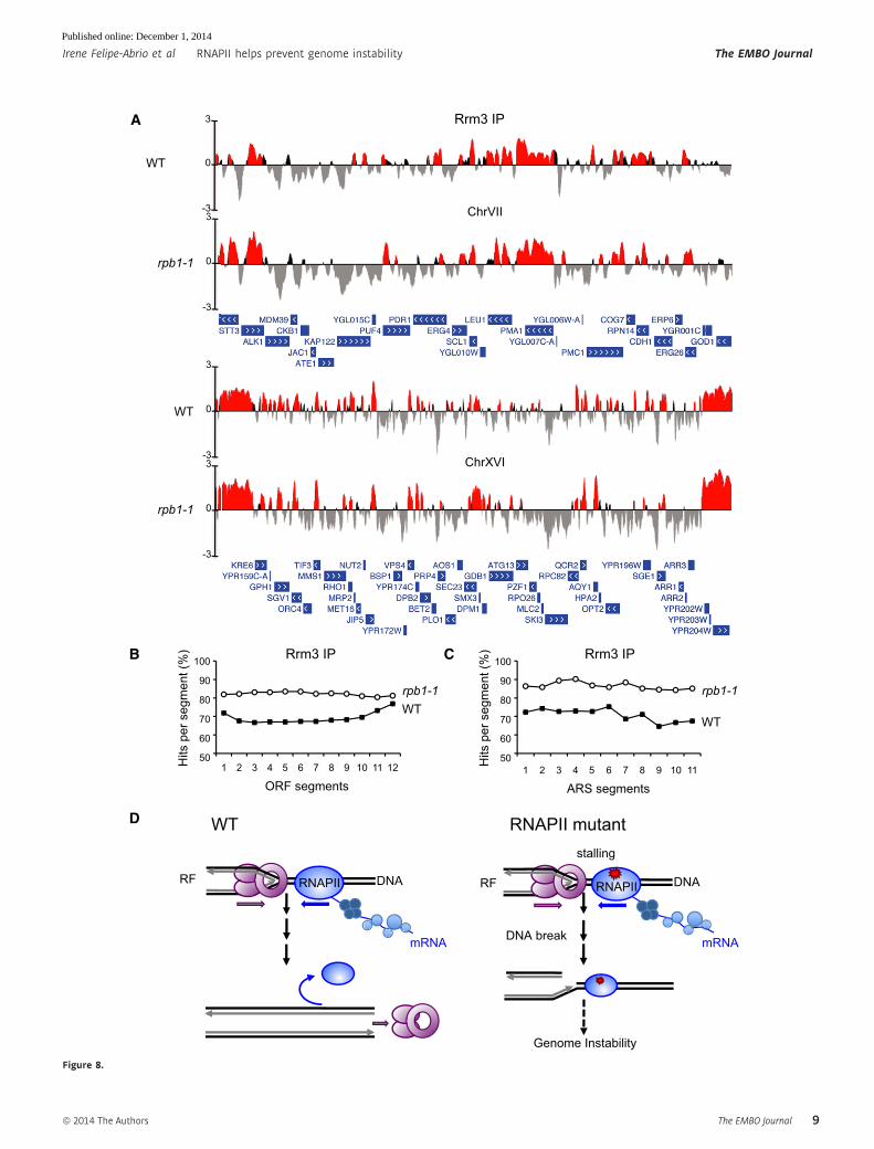
to WT (Fig 8 and Supplementary Fig S9). The hits of Rrm3
occupancy were clearly enhanced in telomeres in rpb1-1 (Fig 8A
and Supplementary Fig S9), implying that natural RF pause sites are
stronger or more frequent at chromosome ends in rpb1-1 cells.
Finally, Rrm3 binds to less replication origins in rpb1-1, consistent
with the delay in replication initiation as detected by 2D gel electro-
phoresis and BrdU incorporation (Supplementary Table S4).
There was a high coincidence between the ORFs enriched in
Rrm3 in rpb1-1 and WT cells although the difference between the
A
C
D
PMA1
SCR1
WT
rpb1-1 rpb1-S751F
rpb2-10 rpb9
0
50
100
150
WT
rpb1-1 rpb1-S751Frpb2-10 rpb9
mR
NA
(%
)
PMA1 1(ATG) 2757(STOP)
292 321 1128 1208 2173 2242 M
TATA
-378 -231 P
RN
AP
II re
lativ
e en
richm
ent 3.0
2.5
2.0
1.0
1.5
0.5
0.0
M
P
WT
rpb1-1
rpb1-S751Frpb2-10 rpb9
144 258 158 rpb1-1(302)
rpb1-S751F(402)
Up
77 159 266 rpb1-1(343)
rpb1-S751F(236)
Down
B
Down Up
rpb1
-1
rpb1
-S75
1F
rpb2
-10
Figure 7. Analysis of global gene expression in RNAPII mutants.
A Hierarchical clustering of genes whose expression level is altered withrespect to WT in rpb cells.
B Venn diagrams representing the overlap between genes whose expressionlevel is up- or down-regulated with respect to the WT strain in rpb1-1 andrpb1-S751F.
C Retention of RNAPII in the actively transcribed PMA1 gene in RNAPIImutants. ChIP analysis of RNAPII in asynchronously growing WT, rpb1-1,rpb1-S751F, rpb2-10 and rpb9D cells at the PMA1 gene. The scheme of thegene and the PCR-amplified fragments are shown. Recruitment valueswere calculated from the amount of DNA present in each region,normalizing IP/input ratios, and relative to the amount of DNA of theintergenic region. Error bars indicate the standard deviation (SD).
D Northern analysis of the expression of PMA1 gene. RNA was isolated frommid-log phase cultures grown in YPD-rich medium. As 32P-labeled DNAprobes, an internal 776-bp PMA1 fragment and an internal 513-bp SCR1fragment obtained by PCR were used. Bars indicate the SD of 2independent experiments.
Source data are available online for this figure.
The EMBO Journal ª 2014 The Authors
The EMBO Journal RNAPII helps prevent genome instability Irene Felipe-Abrio et al
8
Published online: December 1, 2014
A
B
WT
D
1 2 3 4 5 6 7 8 9 10 11 12
ORF segments
100
90
80
70
60
50 Hits
per
seg
men
t (%
)
Hits
per
seg
men
t (%
) Rrm3 IP
1 2 3 4 5 6 7 8 9 10 11
ARS segments
WT rpb1-1
100
90
80
70
60
50
Rrm3 IP C
mRNA
DNA RNAPII RF
WT
Genome Instability
DNA RNAPII
mRNA DNA break
RNAPII mutant
RF
stalling
WT
rpb1-1
rpb1-1
3
0
-3
33
3
0
-3 ChrVII
WT
3
0
-3 ChrXVI
rpb1-1
3
0
-3
Rrm3 IP
Figure 8.
ª 2014 The Authors The EMBO Journal
Irene Felipe-Abrio et al RNAPII helps prevent genome instability The EMBO Journal
9
Published online: December 1, 2014
two strains is significant (Supplementary Table S4). The subset of
ORFs with high Rrm3 occupancy in rpb1-1 is richer in G+C content
and shorter in length than the average WT cells (P < 0.001) (Supple-
mentary Fig S10A), which are coincident with the features of genes
with higher mRNA levels in yeast (Marin et al, 2003). Rrm3 accu-
mulates all over the length of genes in both WT and rpb1-1 cells.
The binding profiles show a tendency to increase toward the 30-end(Supplementary Fig S10B), similar to transcription-dependent
hyper-recombinant mutants defective in mRNP biogenesis (Gomez-
Gonzalez et al, 2011a).
In addition, we took advantage of microarray expression data
and ChIP-chip analyses to study the correlation between replication
impairment and gene expression. As can be seen in Supplementary
Fig S11A, Rrm3 enrichment correlates with gene expression levels,
even though these refer to asynchronous cultures and not to S-phase
transcription, supporting the idea that replication obstacles occur
preferentially at highly transcribed genes. This correlation is still
more evident when we compare genes up- or down-regulated in
rpb1-1 (Supplementary Fig S11B).
Next, we analyzed the results taking into account the average
of the significant signals in ORFs and ARSs all over the genome.
For this, we divided the sequence of each ORF of the yeast
genome into 10 equivalent segments (segments 2–10) plus two
additional segments of the same size upstream (50) (segment 1)
and downstream (30) of each ORF (segment 12). In a similar
way, each ARS sequence was divided into 11 equivalent
segments. Then, we calculated the average signal log2 ratio for
Rrm3 hits mapping on each segment of all ORFs or ARSs,
respectively, and these values were plotted. Interestingly, we see
an increment of Rrm3 retention throughout ORFs and ARSs in
rpb1-1 respect to WT (Fig 8B and C). Since a major occupancy
of Rrm3 at ORFs may be a consequence of transcription–replication
collisions, our data are consistent with the idea that RNAPII itself
participates in preventing such collisions.
Discussion
We provide genetic evidence that specific RNAPII mutants, rpb1-1,
rpb1-S71F and rbp9Δ, show an increase in genetic instability as
detected by hyper-recombination, DNA damage sensitivity, plasmid
loss and/or a dependency on DSB repair functions for viability.
These RNAPII mutants display RF progression impairment that
could be compensated by additional origin firing. DNA damages
accumulate in these mutants under replicative stress that activate the
Rad53-mediated checkpoint, but that are repaired with considerable
delay. Genome-wide Rrm3 occupancy analysis and experiments
showing that the rpb1-1 mutant specifically causes a major retention
of RNAPII at the site of transcription suggest that RNAPII can partic-
ipate actively in facilitating the progression of colliding RFs, presum-
ably by contributing to its own release from the site. Altogether, the
data indicate that RNAPII contributes to maintain genome stability
by distinct manners and without involving high R-loop accumula-
tion.
rpb transcription elongation mutants have a differential effecton genome integrity
From the two specific mutations of the largest RNAPII subunit Rpb1
that cause genome instability, the rpb1-1 mutation (G4622A) maps
in the H region of Rpb1, a highly conserved amino acid region,
which is important for the selection of the transcription start site
(Nonet et al, 1987; Scafe et al, 1990). The rpb1-1 mutation is
temperature sensitive and inactivates RNAPII in vivo, causing a
quick shutdown of mRNA synthesis after a shift to the non-
permissive temperature (Nonet et al, 1987; Schroeder et al, 2000),
even though the mutant form of RNAPII could still be seen at the
transcription sites (Kim et al, 2010). No appreciable decrease of
Rpb1 levels has been detected after this temperature shift (Tardiff
et al, 2007). Also, suppressors of rpb1-1 map in the conserved
segment I of RPB2, suggesting an interaction between region H of
RPB1 and region I of RPB2 (Martin et al, 1990).
rpb1-S751F maps in the F region, involved in transcription elon-
gation (Braberg et al, 2013). The mutant Rpb1-S751F protein,
instead, undergoes elevated transcriptional slippage (Strathern et al,
2013). Transcriptional slippage occurs when RNAPs exhibit higher
inherent error rates during elongation usually due to misincorpora-
tion of the wrong nucleotide in combination with failure of the
intrinsic correction mechanisms like that mediated by the exoribo-
nuclease activity of RNAPII. Indeed, rpb1-S751F has reduced tran-
scription levels compared to WT. The effect of the Rpb1-S751F
bulky substitution on elongation is consistent with the close proxim-
ity of Ser751 to the active center of RNAPII (Strathern et al, 2013).
Both rpb1-1 and rpb1-S751F are MMS sensitive, but the sensitivity of
the latter is clearly stronger.
The rpb2-10 mutation maps in the H region linked to nucleotide
binding. The mutant is sensitive to 6-azauracil (6-AU) and impaired
in transcription elongation (Lennon et al, 1998), but genetic stability
is poorly affected. Interestingly, rpb2-4 also maps in the same region
and causes sensitivity to 6AU, but its defect in transcription elonga-
tion is weaker (Powell & Reines, 1996) whereas rpb2-7, which maps
in the A region, is the most sensitive to 6AU and does not present
Figure 8. Replication–transcription relationship.
A Analysis of Rrm3 recruitment in WT and rpb1-1. Rrm3 IP histogram bars in the y-axis show the average signal log2 ratio of loci enriched in the immunoprecipitatedfraction along the indicated regions in the x-axis. When the ratio fulfills the P-value criteria (P < 0.01) it is shown in red.
B Composite profile of Rrm3 occupancy across the average ORF plotted as Rrm3 percentage of significant ChIP hits per segment. Each gene was divided into 10equivalent segments from the start and end coordinates (segments 2–10). Two additional segments of the same size were considered upstream (50) and downstream(30) of each ORF (segments 1 and 12, respectively). The average signal log2 ratio for Rrm3 significant hits that map on each segment was plotted.
C Composite profile of Rrm3 occupancy across the average ARS plotted as Rrm3 percentage of significant ChIP hits per segment. Each ARS was divided into 11equivalent segments from the start and end coordinates. The average signal log2 ratio for Rrm3 significant hits that map on each segment was plotted.
D Model for a role of RNAPII as a source of genetic instability. Conflicts between transcription and replication machineries affect replication progression causing DSBs athigh levels under exogenous replicative stress, which demands the HR machinery for the repair and restart of RFs. RNAPII would have a function facilitating RFprogression upon a transcription–replication encounter. This function would be affected in the rpb1 and rpb9 mutants analyzed here, causing RF progressionimpairment and genome instability.
◀
The EMBO Journal ª 2014 The Authors
The EMBO Journal RNAPII helps prevent genome instability Irene Felipe-Abrio et al
10
Published online: December 1, 2014
transcription elongation impairment either (Powell & Reines, 1996).
None of these mutants have a clear genome instability phenotype.
The differential DDR phenotypes of the mutants suggest that,
although transcription elongation impairment can be associated
with genome instability, there is no strict correlation with the DDR
defect. Indeed, analysis of transcription factor mutants affecting
elongation is consistent with this conclusion (Rondon et al, 2003;
Rondon et al, 2004; Luna et al, 2005). Despite the different mutants
affected in transcription elongation that cause genome instability,
our data suggest that the elongation defect is not sufficient to trigger
genome instability.
The Rpb9 subunit is not essential for mRNA synthesis in yeast.
During transcription initiation, Rpb9 modulates the selection of the
transcription start site, recruiting TFIIE and interacting with TFIIF.
During the elongation process, Rpb9, together with TFIIS, is
required to stimulate the nascent transcript cleavage activity
intrinsic to RNAPII (Van Mullem et al, 2002). The rpb9D mutant
shows a strong sensitivity to 6-AU or mycophenolic acid (MPA) that
can be suppressed overexpressing TFIIS (Hemming et al, 2000) and
a transcription elongation defect (Tous et al, 2011). Rpb9 is also
involved in transcription-coupled repair (TCR), which has led to the
suggestion that the transcription elongation function of Rpb9 likely
plays a role in backtracking the RNAPII stalled at a lesion, by coor-
dinating RNAPII with TFIIS (Li et al, 2006). In addition, Rpb9 func-
tions in ubiquitylation and degradation of RNAPII in response to
UV-induced DNA damage (Chen et al, 2007). Although the TCR
defect of rpb9D might in principle contributes to its genetic instabil-
ity, the fact that rad26D TCR-defective mutants do not show hyper-
recombination (Gonzalez-Barrera et al, 2002) argues against such a
possibility. Also, rpb9D is highly sensitive to HU, whereas this is not
the case for rad26Δ strains (Gaillard et al, 2009).
Replication fork progression impairment as the cause ofgenome instability
Our data reveal that the rpb1-1, rpb1-S751F and rpb9Δ mutations
increase genome instability, whereas this is not the case for rpb2-10.
Interestingly, 2D gel electrophoresis shows that in rpb1-1, rpb1-
S751F and rpb9D, long Y-shaped molecules accumulated toward the
end of the descending simple Y-arc as a result of the movement of
the RFs away from the restriction fragment containing ARS508
(Fig 4). This is consistent with a slowdown of RFs and supports that
replication failures occurring in the three mutants analyzed are
responsible for genome instability. The same phenotype was previ-
ously described in hpr1D mutants (Gomez-Gonzalez et al, 2009),
implying that under replicative and transcription stress cells respond
with the pausing or stalling of replication. Consistently, recombino-
genic intermediates can be detected in rpb1-1 in 2D gels, whereas
no RF progression impairment was observed in rpb2-10 mutants
(Fig 4).
We believe that the rpb1 and rpb9 mutations analyzed induce
instability by creating transcription intermediates that impair RF
progression (Aguilera & Garcıa-Muse, 2012). The need of the recom-
binational DSB repair functions MRX, Rad52 and Rfc1 for proper
viability of rpb1-1 and rpb1S751F mutants is consistent with the idea
that the replication impairment leads to DSBs that need to be
repaired via homologous recombination; otherwise, cells die. Such
breaks promote two different types of recombination events that
allow the restart of RFs, one Rad51- and the other Pol32-dependent,
consistent with previous results in rad3-102 (Moriel-Carretero &
Aguilera, 2010) and histone deacetylase mutants (Munoz-Galvan
et al, 2013a) that also impair RF progression causing genome insta-
bility.
Replication origins fire more often in rpb1-1 and rpb1-S751F
mutants than in WT cells, as determined by DNA combing (Fig 5).
This may be a mechanism to counteract the slower RF progression
in these mutants.
RNAPII function contributes to the prevention of transcription–replication conflicts
The molecular mechanisms by which a defective RNAPII or tran-
scription elongation could lead to RF progression impairment and
genome instability seem to be multiple. A lower capacity of the
RNAPII to be released from the DNA after a premature transcription
termination due to an elongation failure or after finding an obstacle,
such as a DNA lesion, can increase the probability of a collision
with the RF or its collapse. This explanation would fit for rpb1-1, in
which RNAPII accumulates at higher levels than in WT cells at the
ORFs analyzed, but not for rpb1S751F and rpb9Δ, which show the
same RNAPII occupancy as WT cells. Therefore, an elongation fail-
ure, regardless of whether or not linked to a less efficient release of
the RNAPII from the DNA, can increase genome instability. In this
sense, analysis of the Rrm3 occupancy along the genome of rpb1-1
showing no large increase in the number of hits with respect to WT
cells suggests that the mutant form of the RNAPII does not enhance
the probability of collisions or RF stalling. It is possible that the
main effect of the mutated RNAPII is a reduced ability to resolve RF
stalls, increasing the probability of their collapse. This could explain
why Rrm3 accumulates preferentially in transcribed DNA regions
(Fig 8) as occurs in WT cells (Azvolinsky et al, 2009) and that RF
progression in RNAPII mutants is strongly impaired as determined
by 2D electrophoresis, BrdU incorporation and DNA combing. This
effect differs from that of other transcription mutants with a clear
transcription-dependent genome instability phenotype as is the case
of THO mutants, impaired in transcription elongation and RNA
export, in which Rrm3 is accumulated at higher levels and at more
ORFs than in WT cells (Gomez-Gonzalez et al, 2011a). Indeed,
transcription-associated genome instability can often be associated
with the accumulation of R-loops (Aguilera & Garcıa-Muse, 2012) as
it is the case of THO mutants (Huertas & Aguilera, 2003). However,
since RNase H1 overexpression does not suppress genome instabil-
ity in rpb1 mutations, we rule out that an increase in R-loops is a
major determinant of RF progression impairment observed in the
mutants. Indeed, the lack of effect of null mutations or overexpres-
sion of genes deregulated in rpb1-1 and rpb1S751S strains (HTL1,
NAT4, IRC4, SUS1 and RNR3) on DDR and genome instability is
consistent with the idea that major effect of the rpb mutations is
direct. In this sense, it is worth noting that RNR3 overexpression
diminished the sensitivity of the rpb mutants to replicative stress, as
occurs in wild-type cells, consistent with the general conclusion that
these mutations generate transcription-mediated RF progression fail-
ures as a source of instability.
Our results suggest that the RNAPII helps in minimizing the
effects of natural transcription–replication collisions on genome
integrity. This could occur by either facilitating the release of the
ª 2014 The Authors The EMBO Journal
Irene Felipe-Abrio et al RNAPII helps prevent genome instability The EMBO Journal
11
Published online: December 1, 2014
RNAPII from chromatin after a putative collision with the RF or
by preventing RF collapse, a function that could be defective in
rpb1-1 mutants. The high RNAPII retention at ORFs could
increase the probability of replication collisions. In addition, since
selected elongation factors associate with RNAPII in a transcription-
dependent manner in rpb1-1 mutants (Tardiff et al, 2007), rpb1-1
could also affect recruitment or turnover of factors involved in
transcription or chromatin remodeling that could promote
conflicts between the transcription and replication machineries. In
this sense, it is possible that specific chromatin modifications
associated with RNAPII elongation (Selth et al, 2010) or the topo-
logical constrains serve as a signal to prevent the replisome from
entering a transcribed region and colliding with the RNAPII, or
that a defective elongation causes a TCR failure leading to DNA
lesions that perturb replication and can become DSBs (Belotser-
kovskii et al, 2013; Gaillard & Aguilera, 2013a). Deregulation of
gene gating to the nuclear pore can also contribute to the instabil-
ity of transcribed DNA (Bermejo et al, 2012). However, our iden-
tification of different RNAPII mutants with impact on RF
progression indicates that eukaryotic RNAPII has evolved to mini-
mize the possible impact of transcription–replication conflicts in
genome instability, with important implications for the genetic
origin of cancer.
Materials and Methods
Strains and plasmids
They are described in Supplementary Tables S5 and S6.
All experiments were performed at 30°C with few exceptions
indicated in figure legends.
Analysis of recombination, Rad52-YFP foci and plasmid loss
Recombination frequencies were obtained as the median values of
three fluctuation tests performed with six independent yeast
colonies one from each transformant analyzed. The frequency of
plasmid loss was calculated as the median frequency of six indepen-
dent cultures grown on non-selective rich medium for 23 genera-
tions as previously described (Chavez & Aguilera, 1997).
Spontaneous Rad52-YFP foci from S-G2 mid-log-phase cells bearing
the plasmid pWJ1344 were visualized with a Leica DC 350F micro-
scope (Lisby et al, 2001).
Western blot
Phosphorylated H2A and b-actin were detected by Western blot
of TCA-extracted proteins separated in 15% PAGE using the
ab15083 (Abcam) and ab8226 (Abcam) antibodies, respectively.
For signal detection, SuperSignal West Pico system was used
(Pierce). Detection of Rad53 and b-actin was accomplished by
Western blot analysis of TCA-precipitated proteins separated in
4–20% Criterion TGX gradient PAGE (Bio-Rad). Antibodies sc-20169
(Rad53; Santa Cruz Biotechnology) and ab8224 (b-actin; Abcam)
were used. For quantification, secondary antibodies conjugated to
IRDye 680CW or 800CW (LI-COR) were used; the blot was
scanned in an Odyssey IR scanner and analyzed with Image
Studio 2.0 software (LI-COR). For phosphorylation signal quantifi-
cation, the ratio between Rad53-P versus total Rad53 protein
signal was calculated.
DNA combing
DNA combing was performed as described (Bianco et al, 2012) in
mid-log phase cell TK-containing strains synchronized in G1 with
a-factor and released in the presence of 40 mM HU and 200 lg/ml
BrdU. DNA fibers were extracted in agarose plugs after 60 and
120 min with BrdU as previously described (Duch et al, 2013).
Pulsed-field and 2D gel electrophoresis
PFGE and 2D gel electrophoresis and hybridization were performed
as described (Gomez-Gonzalez et al, 2009; Moriel-Carretero &
Aguilera, 2010).
RNAPII and BrdU chromatin immunoprecipitation
RNAPII ChIPs were performed as described (Gonzalez-Aguilera
et al, 2011) using the monoclonal anti-Rpb1-CTD antibody 8WG16
(Berkeley Antibody Company, Richmond, CA, USA) and Dynabeads
protein A (Invitrogen). The intergenic region at positions 9,716–
9,863 of chromosome V was used as a negative control. BrdU ChIPs
were performed in TK-containing strains synchronized in G1 with
a-factor and released in the presence of 200 lg/ml BrdU (Sigma)
using anti-BrdU antibody (Medical & Biological Laboratories). As
negative control we used the region between coordinates 240,032
and 243,611 of chromosome V. Primers used are indicated in
Supplementary Table S7.
Microarray gene expression analysis
Global gene expression by microarray analysis was performed using
total RNA isolated from yeast cells grown on YPD-rich medium at
30°C to mid-log phase by mechanical disruption using the RNeasy
Midi kit (Qiagen). Microarray data analysis was performed in tripli-
cate using the Affymetrix platform. RNA quality was confirmed with
the Bioanalyzer H (Agilent technology). Synthesis, labeling and
hybridization of cRNA to GeneChip Yeast Genome 2.0 Arrays cover-
ing 5,841 genes of S. cerevisiae were performed following Affyme-
trix protocols. Probe signals were captured and processed with
GeneChip Operating Software 1.4.0.036 (Affymetrix), and the result-
ing CEL files were reprocessed using the Robust Multichip Average
(RMA) normalization (Irizarry et al, 2003). Fold change (log2)
values (M) and their FDR-adjusted P-values were calculated with
LIMMA (linear models for microarray analysis) (Smyth, 2004) using
the affylmGUI interface (Wettenhall et al, 2006). Statistical analysis
was performed using R language and the packages freely available
from the ‘Bioconductor Project’ (http://www.bioconductor.org).
Fold change cutoffs were analyzed at 95% confidence levels (FDR-
adjusted P-values, 0.05). The expression profile of mutants was
compared with that of its isogenic wild-type strain cultured in the
same conditions. Genes showing at least twofold expression change
were considered as altered and selected for further bioinformatics
analysis. The expression data can be accessed at Gene Expression
Omnibus (GS55223). GO annotations were obtained with FatiGO,
The EMBO Journal ª 2014 The Authors
The EMBO Journal RNAPII helps prevent genome instability Irene Felipe-Abrio et al
12
Published online: December 1, 2014
available in Babelomics (http://babelomics.bioinfo.cipf.es). Hits
with P-value < 0.05 were selected.
ChIP-chip experiments
Saccharomyces cerevisiae high-density oligonucleotide tiling micro-
arrays from Affymetrix that allow an analysis of yeast chromo-
somes at a 300-bp resolution, each of the 300-bp region being
covered by at least 60 probes, were used. ChIP-chip of asynchro-
nously growing cells was carried out basically as described
(Bermejo et al, 2009; Gomez-Gonzalez et al, 2011a,b). Data were
analyzed using a modified version of the Affymetrix Tiling Array
Suite software (TAS), which produces a signal and the change
P-value per probe position taking into account the probes local-
ized within a given bandwidth around the inspected probe. The
ChIP-chip data can be accessed at Gene Expression Omnibus
(GS55184). Chromosomal distribution of the signals was
analyzed by binding clusters, defined as ranges within the chro-
mosome under the following conditions: estimated signal (IP/
SUP-binding ratio) positive in the whole range; P-value < 0.01,
minimum run of 100 bp and maximum gap of 250 bp. Results
were visualized with the University of California Santa Cruz
Genome Browser (http://genome.ucsc.edu/). Mapping of binding
clusters into Stanford Genome Database genomic features
(www.yeastgenome.org) was performed using specific Perl
scripts. To visualize the distribution of binding sites along
features, ORFs and ARS were divided into equivalent segments
from the start and end coordinates as previously described
(Gomez-Gonzalez et al, 2011a,b; Santos-Pereira et al, 2013).
Miscellanea
Cell cycle synchronization, flow cytometry analysis (FACSCalibur;
Becton-Dickinson), Northern and other molecular assays were
performed following standard procedures.
Supplementary information for this article is available online:
http://emboj.embopress.org
AcknowledgementsWe would like to thank F. Malagón, S. Rodríguez-Navarro, J. Strathern and R.A.
Young for kindly providing reagents; F. Malagón for pointing us that rpb1S751F
showed a synthetic growth defect with rad52; P. Domínguez (Microscopy Unit,
CABIMER) for technical assistance in confocal microscopy; E. Andújar and
M. Pérez (Genomics Unit, CABIMER) for technical support with microarray and
ChIP-chip experiments; Antonio Marín for providing bioinformatics tools for
analysis of genome-wide data; U. Galindo for technical assistance; and
D. Haun for style supervision. Research was funded by grants from the Spanish
Ministry of Economy and Competitiveness (Consolider 2010 CSD2007-0015
and BFU2010-16372), the Junta de Andalucía (CVI4567) and the European
Union (FEDER). IF-A and JL-B were recipient of predoctoral training grants
from the Spanish Ministry of Economy and Competitiveness and the Instituto
Carlos III, respectively.
Author contributionsIF-A performed most of the experiments; JL-B and MLG-R performed part of
the characterization of rpb1-S751F. IF-A and AA designed the experiments and
wrote the paper.
Conflict of interestThe authors declare that they have no conflict of interest.
References
Aguilera A, Gómez-González B (2008) Genome instability: a mechanistic view
of its causes and consequences. Nat Rev Genet 9: 204 – 217
Aguilera A, García-Muse T (2012) R loops: from transcription byproducts to
threats to genome stability. Mol Cell 46: 115 – 124
Aguilera A, García-Muse T (2013) Causes of genome instability. Annu Rev
Genet 47: 1 – 32
Alvaro D, Lisby M, Rothstein R (2007) Genome-wide analysis of Rad52 foci
reveals diverse mechanisms impacting recombination. PLoS Genet 3:
e228
Atkinson J, Gupta MK, Rudolph CJ, Bell H, Lloyd RG, McGlynn P (2010)
Localization of an accessory helicase at the replisome is critical in
sustaining efficient genome duplication. Nucleic Acids Res 39: 949 – 957
Azvolinsky A, Giresi PG, Lieb JD, Zakian VA (2009) Highly transcribed RNA
polymerase II genes are impediments to replication fork progression in
Saccharomyces cerevisiae. Mol Cell 34: 722 – 734
Baharoglu Z, Lestini R, Duigou S, Michel B (2010) RNA polymerase mutations
that facilitate replication progression in the rep uvrD recF mutant lacking
two accessory replicative helicases. Mol Microbiol 77: 324 – 336
Belotserkovskii BP, Neil AJ, Saleh SS, Shin JH, Mirkin SM, Hanawalt PC
(2013) Transcription blockage by homopurine DNA sequences: role of
sequence composition and single-strand breaks. Nucleic Acids Res 41:
1817 – 1828
Bermejo R, Capra T, Gonzalez-Huici V, Fachinetti D, Cocito A, Natoli G, Katou
Y, Mori H, Kurokawa K, Shirahige K, Foiani M (2009) Genome-organizing
factors Top2 and Hmo1 prevent chromosome fragility at sites of S phase
transcription. Cell 138: 870 – 884
Bermejo R, Kumar A, Foiani M (2012) Preserving the genome by regulating
chromatin association with the nuclear envelope. Trends Cell Biol 22:
465 –473
Bianco JN, Poli J, Saksouk J, Bacal J, Silva MJ, Yoshida K, Lin YL, Tourriere H,
Lengronne A, Pasero P (2012) Analysis of DNA replication profiles in
budding yeast and mammalian cells using DNA combing. Methods 57:
149 – 157
Boubakri H, de Septenville AL, Viguera E, Michel B (2010) The helicases DinG,
Rep and UvrD cooperate to promote replication across transcription units
in vivo. EMBO J 29: 145 – 157
Braberg H, Jin H, Moehle EA, Chan YA, Wang S, Shales M, Benschop JJ,
Morris JH, Qiu C, Hu F, Tang LK, Fraser JS, Holstege FC, Hieter P,
Guthrie C, Kaplan CD, Krogan NJ (2013) From structure to systems:
high-resolution, quantitative genetic analysis of RNA polymerase II. Cell
154: 775 – 788
Branzei D, Foiani M (2010) Maintaining genome stability at the replication
fork. Nat Rev Genet 11: 208 – 219
Castellano-Pozo M, García-Muse T, Aguilera A (2012) R-loops cause
replication impairment and genome instability during meiosis. EMBO Rep
13: 923 – 929
Chavez S, Aguilera A (1997) The yeast HPR1 gene has a functional role in
transcriptional elongation that uncovers a novel source of genome
instability. Genes Dev 11: 3459 – 3470
Chen X, Ruggiero C, Li S (2007) Yeast Rpb9 plays an important role in
ubiquitylation and degradation of Rpb1 in response to UV-induced DNA
damage. Mol Cell Biol 27: 4617 – 4625
ª 2014 The Authors The EMBO Journal
Irene Felipe-Abrio et al RNAPII helps prevent genome instability The EMBO Journal
13
Published online: December 1, 2014
Desany BA, Alcasabas AA, Bachant JB, Elledge SJ (1998) Recovery from DNA
replicational stress is the essential function of the S-phase checkpoint
pathway. Genes Dev 12: 2956 – 2970
Domínguez-Sanchez MS, Barroso S, Gómez-González B, Luna R, Aguilera A
(2011) Genome instability and transcription elongation impairment in
human cells depleted of THO/TREX. PLoS Genet 7: e1002386
Domkin V, Thelander L, Chabes A (2002) Yeast DNA damage-inducible Rnr3
has a very low catalytic activity strongly stimulated after the formation of
a cross-talking Rnr1/Rnr3 complex. J Biol Chem 277: 18574 – 18578
Drolet M, Phoenix P, Menzel R, Masse E, Liu LF, Crouch RJ (1995)
Overexpression of RNase H partially complements the growth defect of an
Escherichia coli delta topA mutant: R-loop formation is a major problem in
the absence of DNA topoisomerase I. Proc Natl Acad Sci USA 92:
3526 – 3530
Duch A, Felipe-Abrio I, Barroso S, Yaakov G, Garcia-Rubio M, Aguilera A, de
Nadal E, Posas F (2013) Coordinated control of replication and
transcription by a SAPK protects genomic integrity. Nature 493: 116 – 119
Dutta D, Shatalin K, Epshtein V, Gottesman ME, Nudler E (2011) Linking
RNA polymerase backtracking to genome instability in E. coli. Cell 146:
533 – 543
Gaillard H, Tous C, Botet J, González-Aguilera C, Quintero MJ, Viladevall L,
García-Rubio ML, Rodríguez-Gil A, Marin A, Arino J, Revuelta JL, Chavez S,
Aguilera A (2009) Genome-wide analysis of factors affecting transcription
elongation and DNA repair: a new role for PAF and Ccr4-not in
transcription-coupled repair. PLoS Genet 5: e1000364
Gaillard H, Aguilera A (2013a) Transcription coupled repair at the interface
between transcription elongation and mRNP biogenesis. Biochim Biophys
Acta 1829: 141 – 150
Gaillard H, Herrera-Moyano E, Aguilera A (2013b) Transcription-associated
genome instability. Chem Rev 113: 8638 – 8661
Gan W, Guan Z, Liu J, Gui T, Shen K, Manley JL, Li X (2011) R-loop-mediated
genomic instability is caused by impairment of replication fork
progression. Genes Dev 25: 2041 – 2056
Gómez-González B, Felipe-Abrio I, Aguilera A (2009) The S-phase checkpoint
is required to respond to R-loops accumulated in THO mutants. Mol Cell
Biol 29: 5203 – 5213
Gómez-González B, García-Rubio M, Bermejo R, Gaillard H, Shirahige K,
Marin A, Foiani M, Aguilera A (2011a) Genome-wide function of THO/TREX
in active genes prevents R-loop-dependent replication obstacles. EMBO J
30: 3106 – 3119
Gómez-González B, Ruiz JF, Aguilera A (2011b) Genetic and molecular
analysis of mitotic recombination in Saccharomyces cerevisiae. Methods
Mol Biol 745: 151 – 172
Gonzalez-Aguilera C, Tous C, Babiano R, de la Cruz J, Luna R, Aguilera A
(2011) Nab2 functions in the metabolism of RNA driven by polymerases II
and III. Mol Biol Cell 22: 2729 – 2740
González-Aguilera C, Tous R, Gómez-González B, Huertas P, Luna R, Aguilera
A (2008) The THP1-SAC3-SUS1-CDC31 complex works in transcription
elongation-mRNA export preventing RNA-mediated genome instability.
Mol Biol Cell 19: 4310 – 4318
González-Barrera S, Prado F, Verhage R, Brouwer J, Aguilera A (2002)
Defective nucleotide excision repair in yeast hpr1 and tho2 mutants.
Nucleic Acids Res 30: 2193 – 2201
Hemming SA, Jansma DB, Macgregor PF, Goryachev A, Friesen JD, Edwards
AM (2000) RNA polymerase II subunit Rpb9 regulates transcription
elongation in vivo. J Biol Chem 275: 35506 – 35511
Holmes AM, Haber JE (1999) Double-strand break repair in yeast requires
both leading and lagging strand DNA polymerases. Cell 96: 415 –424
Huertas P, Aguilera A (2003) Cotranscriptionally formed DNA:RNA hybrids
mediate transcription elongation impairment and transcription-associated
recombination. Mol Cell 12: 711 – 721
Irizarry RA, Hobbs B, Collin F, Beazer-Barclay YD, Antonellis KJ, Scherf U,
Speed TP (2003) Exploration, normalization, and summaries of high
density oligonucleotide array probe level data. Biostatistics 4: 249 – 264
Ivessa AS, Zhou JQ, Zakian VA (2000) The Saccharomyces Pif1p DNA helicase
and the highly related Rrm3p have opposite effects on replication fork
progression in ribosomal DNA. Cell 100: 479 – 489
Ivessa AS, Lenzmeier BA, Bessler JB, Goudsouzian LK, Schnakenberg SL, Zakian
VA (2003) The Saccharomyces cerevisiae helicase Rrm3p facilitates
replication past nonhistone protein-DNA complexes. Mol Cell 12:
1525 – 1536
Kim N, Abdulovic AL, Gealy R, Lippert MJ, Jinks-Robertson S (2007)
Transcription-associated mutagenesis in yeast is directly proportional to
the level of gene expression and influenced by the direction of DNA
replication. DNA Repair (Amst) 6: 1285 – 1296
Kim TS, Liu CL, Yassour M, Holik J, Friedman N, Buratowski S, Rando OJ
(2010) RNA polymerase mapping during stress responses reveals
widespread nonproductive transcription in yeast. Genome Biol 11: R75
Kim N, Jinks-Robertson S (2012) Transcription as a source of genome
instability. Nat Rev Genet 13: 204 – 214
Labib K, De Piccoli G (2011) Surviving chromosome replication: the many
roles of the S-phase checkpoint pathway. Philos Trans R Soc Lond B Biol Sci
366: 3554 – 3561
Lennon JC 3rd, Wind M, Saunders L, Hock MB, Reines D (1998) Mutations in
RNA polymerase II and elongation factor SII severely reduce mRNA levels
in Saccharomyces cerevisiae. Mol Cell Biol 18: 5771 – 5779
Li X, Manley JL (2005) Inactivation of the SR protein splicing factor ASF/SF2
results in genomic instability. Cell 122: 365 – 378
Li S, Ding B, Chen R, Ruggiero C, Chen X (2006) Evidence that the
transcription elongation function of Rpb9 is involved in
transcription-coupled DNA repair in Saccharomyces cerevisiae. Mol Cell Biol
26: 9430 – 9441
Lisby M, Rothstein R, Mortensen UH (2001) Rad52 forms DNA repair and
recombination centers during S phase. Proc Natl Acad Sci USA 98:
8276 – 8282
Luna R, Jimeno S, Marin M, Huertas P, García-Rubio M, Aguilera A (2005)
Interdependence between transcription and mRNP processing and export,
and its impact on genetic stability. Mol Cell 18: 711 – 722
Marin A, Gallardo M, Kato Y, Shirahige K, Gutierrez G, Ohta K, Aguilera A
(2003) Relationship between G+C content, ORF-length and mRNA
concentration in Saccharomyces cerevisiae. Yeast 20: 703 – 711
Martin C, Okamura S, Young R (1990) Genetic exploration of interactive
domains in RNA polymerase II subunits. Mol Cell Biol 10: 1908 – 1914
McGlynn P, Lloyd RG (2000) Modulation of RNA polymerase by (p)ppGpp
reveals a RecG-dependent mechanism for replication fork progression. Cell
101: 35 – 45
Merrikh H, Zhang Y, Grossman AD, Wang JD (2012) Replication-transcription
conflicts in bacteria. Nat Rev Microbiol 10: 449 – 458
Mischo HE, Gómez-González B, Grzechnik P, Rondón AG, Wei W, Steinmetz L,
Aguilera A, Proudfoot NJ (2011) Yeast Sen1 helicase protects the genome
from transcription-associated instability. Mol Cell 41: 21 – 32
Moriel-Carretero M, Aguilera A (2010) A postincision-deficient TFIIH causes
replication fork breakage and uncovers alternative Rad51- or
Pol32-mediated restart mechanisms. Mol Cell 37: 690 – 701
Muñoz-Galván S, Jimeno S, Rothstein R, Aguilera A (2013a) Histone
H3K56 acetylation, Rad52, and non-DNA repair factors control
The EMBO Journal ª 2014 The Authors
The EMBO Journal RNAPII helps prevent genome instability Irene Felipe-Abrio et al
14
Published online: December 1, 2014
double-strand break repair choice with the sister chromatid. PLoS Genet
9: e1003237
Muñoz-Galván S, Lopez-Saavedra A, Jackson SP, Huertas P, Cortes-Ledesma F,
Aguilera A (2013b) Competing roles of DNA end resection and
non-homologous end joining functions in the repair of replication-born
double-strand breaks by sister-chromatid recombination. Nucleic Acids Res
41: 1669 – 1683
Nonet M, Scafe C, Sexton J, Young R (1987) Eucaryotic RNA polymerase
conditional mutant that rapidly ceases mRNA synthesis. Mol Cell Biol 7:
1602 – 1611
Paulsen RD, Soni DV, Wollman R, Hahn AT, Yee MC, Guan A, Hesley JA,
Miller SC, Cromwell EF, Solow-Cordero DE, Meyer T, Cimprich KA
(2009) A genome-wide siRNA screen reveals diverse cellular processes
and pathways that mediate genome stability. Mol Cell 35:
228 – 239
Powell W, Reines D (1996) Mutations in the second largest subunit of RNA
polymerase II cause 6-azauracil sensitivity in yeast and increased
transcriptional arrest in vitro. J Biol Chem 271: 6866 – 6873
Prado F, Aguilera A (2005) Impairment of replication fork progression
mediates RNA polII transcription-associated recombination. EMBO J 24:
1267 – 1276
Rodríguez-Navarro S, Fischer T, Luo MJ, Antúnez O, Brettschneider S, Lechner
J, Pérez-Ortín JE, Reed R, Hurt E (2004) Sus1, a functional component of
the SAGA histone acetylase complex and the nuclear pore-associated
mRNA export machinery. Cell 116: 75 – 86
Rondón AG, Gallardo M, García-Rubio M, Aguilera A (2004) Molecular
evidence indicating that the yeast PAF complex is required for
transcription elongation. EMBO Rep 5: 47 – 53
Rondon AG, García-Rubio M, González-Barrera S, Aguilera A (2003) Molecular
evidence for a positive role of Spt4 in transcription elongation. EMBO J 22:
612 – 620
Santocanale C, Diffley JF (1998) A Mec1- and Rad53-dependent checkpoint
controls late-firing origins of DNA replication. Nature 395: 615 – 618
Santos-Pereira JM, Herrero AB, Garcia-Rubio ML, Marin A, Moreno S, Aguilera A
(2013) The Npl3 hnRNP prevents R-loop-mediated transcription-replication
conflicts and genome instability. Genes Dev 27: 2445 – 2458
Scafe C, Martin C, Nonet M, Podos S, Okamura S, Young RA (1990)
Conditional mutations occur predominantly in highly conserved residues
of RNA polymerase II subunits. Mol Cell Biol 10: 1270 – 1275
Schmidt KH, Reimers JM, Wright BE (2006) The effect of promoter strength,
supercoiling and secondary structure on mutation rates in Escherichia coli.
Mol Microbiol 60: 1251 – 1261
Schroeder SC, Schwer B, Shuman S, Bentley D (2000) Dynamic association of
capping enzymes with transcribing RNA polymerase II. Genes Dev 14:
2435 – 2440
Selth LA, Sigurdsson S, Svejstrup JQ (2010) Transcript Elongation by RNA
Polymerase II. Annu Rev Biochem 79: 271 – 293
Smyth GK (2004) Linear models and empirical bayes methods for assessing
differential expression in microarray experiments. Stat Appl Genet Mol Biol
3: Article 3
Song OK, Wang X, Waterborg JH, Sternglanz R (2003) An
Nalpha-acetyltransferase responsible for acetylation of the N-terminal
residues of histones H4 and H2A. J Biol Chem 278: 38190 – 38112
Stirling PC, Chan YA, Minaker SW, Aristizabal MJ, Barrett I, Sipahimalani P,
Kobor MS, Hieter P (2012) R-loop-mediated genome instability in mRNA
cleavage and polyadenylation mutants. Genes Dev 26: 163 – 175
Strathern J, Malagon F, Irvin J, Gotte D, Shafer B, Kireeva M, Lubkowska L,
Jin DJ, Kashlev M (2013) The fidelity of transcription: RPB1 (RPO21)
mutations that increase transcriptional slippage in S. cerevisiae. J Biol
Chem 288: 2689 – 2699
Tardiff DF, Abruzzi KC, Rosbash M (2007) Protein characterization of
Saccharomyces cerevisiae RNA polymerase II after in vivo cross-linking.
Proc Natl Acad Sci USA 104: 19948 – 19953
Tous C, Aguilera A (2007) Impairment of transcription elongation by R-loops
in vitro. Biochem Biophys Res Comm 360: 428 – 432
Tous C, Rondón AG, García-Rubio M, González-Aguilera C, Luna R, Aguilera A
(2011) A novel assay identifies transcript elongation roles for the Nup84
complex and RNA processing factors. EMBO J 30: 1953 – 1964
Trautinger BW, Lloyd RG (2002) Modulation of DNA repair by mutations
flanking the DNA channel through RNA polymerase. EMBO J 21: 6944 – 6953
Van Mullem V, Wery M, Werner M, Vandenhaute J, Thuriaux P (2002) The
Rpb9 subunit of RNA polymerase II binds transcription factor TFIIE and
interferes with the SAGA and elongator histone acetyltransferases. J Biol
Chem 277: 10220 – 10225
Wahba L, Amon JD, Koshland D, Vuica-Ross M (2011) RNase H and
multiple RNA biogenesis factors cooperate to prevent RNA:DNA
hybrids from generating genome instability. Mol Cell 44:
978 – 988
Wellinger RE, Prado F, Aguilera A (2006) Replication fork progression is
impaired by transcription in hyperrecombinant yeast cells lacking a
functional THO complex. Mol Cell Biol 26: 3327 – 3334
Wettenhall JM, Simpson KM, Satterley K, Smyth GK (2006) affylmGUI: a
graphical user interface for linear modeling of single channel microarray
data. Bioinformatics 22: 897 – 899
ª 2014 The Authors The EMBO Journal
Irene Felipe-Abrio et al RNAPII helps prevent genome instability The EMBO Journal
15
Published online: December 1, 2014