RNA Binding Proteins Accumulate at the Postsynaptic Density with ...
11
Cellular/Molecular RNA Binding Proteins Accumulate at the Postsynaptic Density with Synaptic Activity Guoan Zhang, 1 Thomas A. Neubert, 1 and Bryen A. Jordan 2 1 Kimmel Center for Biology and Medicine at the Skirball Institute and Department of Pharmacology, New York University School of Medicine, New York, New York, 10016, and 2 Dominick P. Purpura Department of Neuroscience, Albert Einstein College of Medicine, Bronx, New York 10461 Neuronal activity elicits changes in synaptic composition that play an important role in experience-dependent plasticity (Choquet and Triller, 2003; Lisman and Raghavachari, 2006; Bourne and Harris, 2008; Holtmaat and Svoboda, 2009). We used a modified version of stable isotope labeling by amino acids in cell culture to identify activity-dependent modifications in the composition of postsynaptic densities (PSDs) isolated from rat primary neuronal cultures. We found that synaptic activity altered 2% of the PSD proteome, which included an increase in diverse RNA binding proteins (RNABPs). Indeed, 12 of the 37 identified proteins whose levels changed with synaptic activity were RNABPs and included the heterogeneous nuclear ribonucleoproteins (hnRNPs) G, A2/B1, M, and D. Knockdown of hnRNPs M and G using shRNAs resulted in altered numbers of dendritic spines, suggesting a crucial role for these proteins in spine density. Synaptic activity also resulted in a concomitant increase in dendritic and synaptic poly(A) mRNA. However, this increase was not affected by knockdown of hnRNPs M or G. Our results suggest that hnRNP proteins regulate dendritic spine density and may play a role in synaptodendritic mRNA metabolism. Introduction The postsynaptic density (PSD) is a protein complex specialized in receiving and transducing information at neuronal synapses (Banker et al., 1974; Blomberg et al., 1977; Kennedy, 1993; Ziff, 1997). Proteomic analyses of the PSD have shown that it contains diverse downstream signaling components including regulators of cell polarity, protein translation and degradation, phosphory- lation, G-protein signaling, and nuclear function (Walsh and Ku- ruc, 1992; Walikonis et al., 2000; Vinade et al., 2003; Jordan et al., 2004, 2007; Li et al., 2004; Peng et al., 2004; Yoshimura et al., 2004; Collins et al., 2006; Dosemeci et al., 2006; Jordan and Ziff, 2008; Jordan and Kreutz, 2009). Among PSD components identified by mass spectrometry are members of the RNA binding proteins (RNABP) family (Jordan et al., 2004, 2006). RNABPs regulate all aspects of RNA metabolism, including mRNA transport, transla- tion, and decay. The presence of RNABPs at synapses is consis- tent with current hypotheses regarding the role of selective mRNA transport and translation in the synapse-specific regula- tion of dendritic spine morphology and function (Martin and Zukin, 2006; Richter and Klann, 2009). Changes in PSD protein composition are intimately linked with short- and long-term regulation of synaptic function. For example, stable increases in AMPA receptors associated with the PSD result in long-term potentiation (LTP) (Malenka, 2003; Malenka and Bear, 2004), which is a physiologically relevant in- crease in synaptic efficacy and a model for learning and memory (Milner et al., 1998; Miller and Mayford, 1999). However, little is known about the dynamics of PSD composition given the tech- nical challenges associated with proteomic quantitation methods as applied to neurons. To overcome these obstacles, we used a variation of stable isotope labeling by amino acids in cell culture (SILAC) (Ong et al., 2002) that corrects for incomplete stable isotopic labeling of nondividing primary neurons (Zhang et al., 2011) to quantitatively investigate PSD protein dynamics in an unbiased manner. We compared PSDs isolated from cultured rat cortical primary neurons stimulated using a modified chemical LTP inducing protocol (cLTP) to PSDs isolated from neurons inhibited with tetrodotoxin (TTX) and found that synaptic activ- ity resulted in increased abundance of diverse RNABPs, including heterogeneous nuclear ribonucleoproteins (hnRNPs) A, A2/B1, A3, M, D, G, and L. In fact, 12 of the 37 proteins whose expression were altered in the PSD were RNABPs, suggesting that neuronal activity regulates synaptic RNA metabolism. We found that knockdown of hnRNPM and hnRNPG, but not hnRNPA2/B1 or hnRNPD, resulted in marked changes in dendritic spine density. These results suggest a crucial role for hnRNP proteins in synap- tic function and provide support for the notion that local regula- tion of RNA metabolism underlies the synapse-specific and activity-dependent regulation of dendritic spine morphology. Materials and Methods Lentiviral vectors. We used pTRIP vectors to generate lentiviral shRNA vectors for knockdown following methods previously described (Janas et al., 2006). pTRIP contains an H1 promoter derived from pSUPER vec- tors to drive shRNA expression, and a GFP construct driven by an elon- Received May 17, 2011; revised Nov. 9, 2011; accepted Nov. 13, 2011. Author contributions: G.Z., T.A.N., and B.A.J. designed research; G.Z. and B.A.J. performed research; G.Z., T.A.N., and B.A.J. analyzed data; G.Z. and B.A.J. wrote the paper. This work was supported by grants from the NIH (K01-MH073759 and R01-AG039521 to B.A.J; P30 NS050276 and S10 RR017990 to T.A.N.). We thank Matthew Klein and Jaafar Tindi for their critical review of this manuscript. The authors declare no financial conflicts of interest. Correspondence should be addressed to Bryen A. Jordan, Dominick P. Purpura Department of Neuroscience, Albert Einstein College of Medicine, 1300 Morris Park Avenue, Rose Kennedy Center, Room 825, Bronx, NY 10461. E-mail: [email protected]. DOI:10.1523/JNEUROSCI.2463-11.2012 Copyright © 2012 the authors 0270-6474/12/320599-11$15.00/0 The Journal of Neuroscience, January 11, 2012 • 32(2):599 – 609 • 599
Transcript of RNA Binding Proteins Accumulate at the Postsynaptic Density with ...

Cellular/Molecular
RNA Binding Proteins Accumulate at the PostsynapticDensity with Synaptic Activity
Guoan Zhang,1 Thomas A. Neubert,1 and Bryen A. Jordan2
1Kimmel Center for Biology and Medicine at the Skirball Institute and Department of Pharmacology, New York University School of Medicine, New York,New York, 10016, and 2Dominick P. Purpura Department of Neuroscience, Albert Einstein College of Medicine, Bronx, New York 10461
Neuronal activity elicits changes in synaptic composition that play an important role in experience-dependent plasticity (Choquet andTriller, 2003; Lisman and Raghavachari, 2006; Bourne and Harris, 2008; Holtmaat and Svoboda, 2009). We used a modified version ofstable isotope labeling by amino acids in cell culture to identify activity-dependent modifications in the composition of postsynapticdensities (PSDs) isolated from rat primary neuronal cultures. We found that synaptic activity altered �2% of the PSD proteome, whichincluded an increase in diverse RNA binding proteins (RNABPs). Indeed, 12 of the 37 identified proteins whose levels changed withsynaptic activity were RNABPs and included the heterogeneous nuclear ribonucleoproteins (hnRNPs) G, A2/B1, M, and D. Knockdown ofhnRNPs M and G using shRNAs resulted in altered numbers of dendritic spines, suggesting a crucial role for these proteins in spinedensity. Synaptic activity also resulted in a concomitant increase in dendritic and synaptic poly(A) mRNA. However, this increase was notaffected by knockdown of hnRNPs M or G. Our results suggest that hnRNP proteins regulate dendritic spine density and may play a rolein synaptodendritic mRNA metabolism.
IntroductionThe postsynaptic density (PSD) is a protein complex specializedin receiving and transducing information at neuronal synapses(Banker et al., 1974; Blomberg et al., 1977; Kennedy, 1993; Ziff,1997). Proteomic analyses of the PSD have shown that it containsdiverse downstream signaling components including regulatorsof cell polarity, protein translation and degradation, phosphory-lation, G-protein signaling, and nuclear function (Walsh and Ku-ruc, 1992; Walikonis et al., 2000; Vinade et al., 2003; Jordan et al.,2004, 2007; Li et al., 2004; Peng et al., 2004; Yoshimura et al., 2004;Collins et al., 2006; Dosemeci et al., 2006; Jordan and Ziff, 2008;Jordan and Kreutz, 2009). Among PSD components identified bymass spectrometry are members of the RNA binding proteins(RNABP) family (Jordan et al., 2004, 2006). RNABPs regulate allaspects of RNA metabolism, including mRNA transport, transla-tion, and decay. The presence of RNABPs at synapses is consis-tent with current hypotheses regarding the role of selectivemRNA transport and translation in the synapse-specific regula-tion of dendritic spine morphology and function (Martin andZukin, 2006; Richter and Klann, 2009).
Changes in PSD protein composition are intimately linkedwith short- and long-term regulation of synaptic function. For
example, stable increases in AMPA receptors associated with thePSD result in long-term potentiation (LTP) (Malenka, 2003;Malenka and Bear, 2004), which is a physiologically relevant in-crease in synaptic efficacy and a model for learning and memory(Milner et al., 1998; Miller and Mayford, 1999). However, little isknown about the dynamics of PSD composition given the tech-nical challenges associated with proteomic quantitation methodsas applied to neurons. To overcome these obstacles, we used avariation of stable isotope labeling by amino acids in cell culture(SILAC) (Ong et al., 2002) that corrects for incomplete stableisotopic labeling of nondividing primary neurons (Zhang et al.,2011) to quantitatively investigate PSD protein dynamics in anunbiased manner. We compared PSDs isolated from cultured ratcortical primary neurons stimulated using a modified chemicalLTP inducing protocol (cLTP) to PSDs isolated from neuronsinhibited with tetrodotoxin (TTX) and found that synaptic activ-ity resulted in increased abundance of diverse RNABPs, includingheterogeneous nuclear ribonucleoproteins (hnRNPs) A, A2/B1,A3, M, D, G, and L. In fact, 12 of the 37 proteins whose expressionwere altered in the PSD were RNABPs, suggesting that neuronalactivity regulates synaptic RNA metabolism. We found thatknockdown of hnRNPM and hnRNPG, but not hnRNPA2/B1 orhnRNPD, resulted in marked changes in dendritic spine density.These results suggest a crucial role for hnRNP proteins in synap-tic function and provide support for the notion that local regula-tion of RNA metabolism underlies the synapse-specific andactivity-dependent regulation of dendritic spine morphology.
Materials and MethodsLentiviral vectors. We used pTRIP vectors to generate lentiviral shRNAvectors for knockdown following methods previously described (Janas etal., 2006). pTRIP contains an H1 promoter derived from pSUPER vec-tors to drive shRNA expression, and a GFP construct driven by an elon-
Received May 17, 2011; revised Nov. 9, 2011; accepted Nov. 13, 2011.Author contributions: G.Z., T.A.N., and B.A.J. designed research; G.Z. and B.A.J. performed research; G.Z., T.A.N.,
and B.A.J. analyzed data; G.Z. and B.A.J. wrote the paper.This work was supported by grants from the NIH (K01-MH073759 and R01-AG039521 to B.A.J; P30 NS050276
and S10 RR017990 to T.A.N.). We thank Matthew Klein and Jaafar Tindi for their critical review of this manuscript.The authors declare no financial conflicts of interest.Correspondence should be addressed to Bryen A. Jordan, Dominick P. Purpura Department of Neuroscience,
Albert Einstein College of Medicine, 1300 Morris Park Avenue, Rose Kennedy Center, Room 825, Bronx, NY 10461.E-mail: [email protected].
DOI:10.1523/JNEUROSCI.2463-11.2012Copyright © 2012 the authors 0270-6474/12/320599-11$15.00/0
The Journal of Neuroscience, January 11, 2012 • 32(2):599 – 609 • 599

gation factor 1� promoter to identify transduced neurons. At least threeshRNA sequences for each rat-derived sequence of hnRNPD, hnRNPG,hnRNPA2/B1, and hnRNPM were selected using the SFOLD online soft-ware prediction algorithms (Wadsworth Center, Albany, NY) and tested.We ended up using shRNAs targeting the rat coding sequences of hn-RNPD [base pair 385: 5�GATCCTATCACAGGGCGAT (sh1); base pair509: 5�CCAAAGCCATGAAAACAAA (sh2)], hnRNPG [base pair 200:5�-GAGATATGAATGGAAAGTC (sh1); base pair 700: 5�CCACCAA-GAGATTATACTT (sh2)], hnRNPM (base pair 1992: 5�ATGGAAGAT-GCTAAAGGACAA (sh1); base pair 87: 5�AGAACGACCUACUC-AGAAU (sh2)],and hnRNPA2/B1 [base pair 84: 5AAGCTTTGAAAC-CACAGAAGA (sh1); base pair 864: 5�CAACTATGGAGGAGGAAAT(sh2)]. Viruses were generated by triple transfection of pTRIP-shRNA,pCMV-dR8.2, and pMD2.G (which provide structural viral proteins)into 293FT cells (Invitrogen). The media from these cells were harvestedand concentrated. We determined viral titers by transducing primaryneuronal cortical cultures plated on 24-well plates with increasingamounts of lentivirus-containing 293FT-derived media.
Cell cultures, neuronal stimulation. Rats were killed using CO2 in com-pliance with the Albert Einstein College of Medicine Institutional AnimalCare and Use Committee. Primary neuronal cultures from E19 embryosof either sex were prepared as described previously (Osten et al., 2000).Cells at DIV 14 –21 were treated with either bicuculline (20 �M) or TTX(500 nM). For cLTP-Gly-treated cells, neurons were washed once in arti-ficial CSF (aCSF) without Mg 2� (in mM: 140 NaCl, 5 KCl, 26 NaHCO3,1.25 NaH2PO4, 1.8 CaCl2, 33 glucose, 25 HEPES, 0.001 strychnine) andthen incubated in aCSF � 200 �M glycine for 5 min followed by 15 minincubation in aCSF without glycine (with 2.5 mM Mg 2�). cLTP-cAMP-treated cells were washed once with aCSF and then incubated withaCSF � Rolipram (100 nM), forskolin (50 �M), and picrotoxin (100 �M)for 30 min. BDNF-treated cells were treated with 50 ng/ml BDNF for 30min and cLTP�BDNF was cLTP-cAMP treated in the presence of BDNF(50 ng/ml) for 30 min.
Neuronal imaging, ELISAs, Western blots, and fluorescent in situ hybrid-izations. All immunocytochemistry (ICC) was performed using standardconditions. Briefly, cells were fixed using 4% paraformaldehyde (PFA)for 5 min and permeabilized using 0.2% Triton X-100 for 5 min. Anti-bodies used were as follows: hnRNPD (ab61193; 1:1000; Abcam), hn-RNPG (NBP1-67923; 1:500; Novus), hnRNPM (NBP1-70822; 1:500;Novus), hnRNPA2/B1 (sc-32316; 1:200; Santa Cruz Biotechnology),PSD-95 (K28/43; 1:2000; University of California Davis/NIH NeuroMabFacility), MAP2 (CPCA-MAP2; 1:5000; EnCor Bio), fragile X mentalretardation protein (FMRP; 7G1-1-s; 0.5 �gs/ml), and SV2 (0.5 �gs/ml;Developmental Studies Hybridoma Bank). Western blots were per-formed using standard conditions; primary antibodies (above) were in-cubated overnight at 4°C with rocking and used at the sameconcentration as used for ICC. For ELISA assays, 7 � 10 4 rat primarycortical neurons were plated in 24-well plates. At DIV 14 –17, the cellswere treated as described, fixed with PFA, and immunostained withGluR1 Ab targeting the extracellular domain (PC246; Calbiochem).Neurons were then washed, incubated in HRP-labeled secondary anti-body, and incubated with o-phenylenediamine for the colorimetric as-say. After the substrate was collected and measured, the cells were lysed inSDS-PAGE buffer, boiled, and subjected to SDS-PAGE for protein nor-malization. Fluorescent in situ hybridizations (FISH) with ICC was per-formed by fixing and permeabilizing primary hippocampal neurons asdescribed above, washing once with wash buffer (10% formamide, 2�SSC), and incubating neurons with 0.5 ngs/�l Oligo dT(50) probe cou-pled to Cy3 (Genelink) in hybridization buffer (10% dextran sulfate, 1�g/�l E. coli tRNA, 2 mM vanadyl ribonucleoside complex, 0.02% acety-lated BSA, and 10% formamide) for 2 h at 37°C. Cells were then washedtwice (30 min per wash) with wash buffer, then washed once with DEPC-treated PBS, blocked with 1% acetylated BSA/PBS for 1 h, and incubatedwith primary and secondary antibodies as per standard conditions forICC. Quantitative analysis of ICC and FISH experiments were performedblind for the identity of the viruses used for transduction.
SILAC and isolation of PSDs from neuronal cultures. For the SILACassays, cortical neurons were grown in 15 cm plates at 9 � 10 6 cells/plateusing the following modified Neurobasal medium: Neurobasal medium
lacking Arg and Lys (Specialty Media; Invitrogen) was supplementedwith either L-Arg:HCl (U-13C6) and L-Lysine:2HCl (4,4,5,5-D4) (me-dium) or L-Arg:HCl (U-13C6; U-15N4) and L-Lys:2HCl (U-13C6;U-15N2) (heavy). All isotope-labeled amino acids were from CambridgeIsotope Laboratories. Lys and Arg were supplemented at 146 and 84 mg/l,respectively (as in regular unlabeled Neurobasal medium). One half ofthe media was changed every 3– 4 d. To obtain a relatively pure neuronalpopulation, we added 0.2 �M cytosine arabinoside (AraC) at DIV 5 toinhibit glial growth. PSDs were enriched according to the methods ofCohen and Carlin (Cohen et al., 1977; Carlin et al., 1980) using the longmethod (Jordan et al., 2004). Briefly, at DIV 15, heavy- and medium-labeled neuronal cultures were scraped off and mixed in ice-cold Solu-tion A (0.32 M sucrose, 1 mM NaHCO3, 1 mM MgCl2, 0.5 mM CaCl2)containing protease and phosphatase inhibitors. Cells were Dounce ho-mogenized in 4 ml of Solution A and diluted to 10% weight/volume. Thisinitial homogenate represents the equivalent of the total lysate (WB)fraction shown in Figure 5, A and B (right). Following centrifugation at1400 g for 10 min, the supernatant fluid was saved and the pellet reho-mogenized to 10% weight/volume in Solution A and centrifuged at 710 gfor 10 min. Supernatants were pooled and subjected to a second centrif-ugation at 710 g for 10 min. The resulting supernatant was then centri-fuged at 30,000 g for 15 min to collect the P2 pellet. The P2 pellet wasresuspended in Solution B (0.32 M sucrose, 1 mM NaHCO3) and layeredon top of a 0.85 M sucrose, 1 M sucrose, and 1.2 M sucrose step sucrosegradient and centrifuged at 82,500 g for 2 h. The synaptosomes (Synfraction; Fig. 5A) were collected at the 1/1.2 M sucrose layer interface andpelleted down at 45,000 g for 30 min after sixfold dilution with solutionB. The pellet was resuspended in pure ice-cold water (with protease andphosphatase inhibitors), incubated at 4°C for 30 min to induce hypo-osmotic shock, and repelleted. The lysed synaptosomes were then resus-pended in Solution B and placed on top of a second 0.85, 1.0, 1.2 M
sucrose step gradient. The enriched synaptosomes at the 1–1.2 M inter-face were diluted with Solution B and an equal volume of 1% TritonX-100 and 0.32 M sucrose; 12 mM Tris, pH 8.1 was added. Synaptosomeswere lysed at 4°C for 15 min, centrifuged at 32,800 g for 25 min, and thepellet resuspended in Solution B. The suspension was layered on a finalsucrose step gradient of 1 M sucrose, 1.5 M sucrose, and 2 M sucrose andcentrifuged at 200,000 g for 2 h. The PSD fraction was collected at the 1.5M and 2 M sucrose interface and diluted with Solution B. An equal volumeof 1% Triton X-100, 150 mM KCl was added to obtain the two-TritonPSD fraction. These enriched PSDs represent the PSD fraction in Figure5, A and B, and were collected by centrifugation at 200,000 g for 20 minand lysed in a 2% SDS-Tris buffer with brief sonication.
SDS-PAGE and LC-MS/MS. Enriched PSD proteins were separated by10% SDS-PAGE and Coomassie stained. Gel lanes were cut into �20bands. Gel bands were cut into small pieces and destained in 25 mM
NH4HCO3/50% acetonitrile, dehydrated with acetonitrile, and dried(Shevchenko et al., 1996). The gel pieces were then rehydrated with 12.5ng/�l trypsin solution in 25 mM NH4HCO3 and incubated overnight at37°C. Peptides were extracted twice with 5% formic acid/50% acetoni-trile followed by a final extraction with acetonitrile. Samples were con-centrated by vacuum centrifugation to dryness and redissolved with 2%acetonitrile in 0.1% formic acid before further analysis. For all LC-MS/MS analysis, an LTQ-Orbitrap hybrid mass spectrometer (ThermoFisher Scientific) equipped with a nanoelectrospray ionization source(Jamie Hill Instrument Services) was used. A nanoLC system (EksigentTechnologies) equipped with a self-packed 750 �m � 12 cm reversephase column (Reprosil C18, 3 �m; Dr. Maisch HPLC) was coupled to themass spectrometer. Peptides were eluted by a gradient of 3–40% acetonitrilein 0.1% formic acid over 110 min at a flow rate of 300 nl/min. Mass spectrawere acquired in data-dependent mode with one 60,000 resolution MS sur-vey scan by the Orbitrap and up to eight MS/MS scans in the LTQ for themost intense peaks selected from each survey scan. Automatic gain controltarget value was set to 1,000,000 for Orbitrap survey scans and 5000 for LTQMS/MS scans. Survey scans were acquired in profile mode and MS/MS scanswere acquired in centroid mode.
Peptide identification and quantitation. The raw mass spectra files wereprocessed using MaxQuant software (version 1.0.13.8) (Cox and Mann,2008). Protein and peptide identification was performed using the Mas-
600 • J. Neurosci., January 11, 2012 • 32(2):599 – 609 Zhang et al. • Activity-Dependent Changes in the PSD

cot search engine (version 2.2.1; Matrix Science) by searching the con-catenated forward and reverse International Protein Index proteindatabase (version 3.52) containing 55,303 mouse and 39,906 rat proteinsequences and 175 commonly observed contaminants (in total, 190,768sequences including reverse and contaminant sequences). The minimumrequired peptide length was 6 a.a., and trypsin cleavage specificity wasapplied with up to two missed cleavages allowed. Variable modificationsincluded oxidation (M) and N-acetylation of protein N termini. Initially,the maximum allowed mass deviations for precursor and fragment ionswere set to 20 ppm and 0.5 Da, respectively. The results of the databasesearch were further processed and statistically evaluated by MaxQuant.For protein and peptide identification, the initial maximum protein andpeptide false discovery rates were set to 1%. To further improve confi-dence in protein identification and quantitation, the following filterswere applied to the MaxQuant output: (1) at least two peptides from eachprotein, (2) �30% SILAC label incorporation, (3) protein was quantifiedby at least three SILAC ratio measurements from peptides in both forwardand reverse experiments, (4) variability of peptide ratio measurements of theprotein was �100% in both forward and reverse experiments, and (5) totalpeptide signal intensity of the protein was �1,000,000. After filtering, therewere 1637 protein identifications matched to the forward protein databaseand three matched to the reverse database. Thus, the false discovery ratewas estimated to be 0.2%. Proteins were considered changing if theirsignificance B values were �0.05 in both forward and reverse experi-ments. Significance B values are p values for detection of significantoutlier protein ratios calculated on the protein subsets obtained by in-tensity binning (Cox and Mann, 2008).
ResultsCell-surface and synaptic GluR1-containing AMPA receptor lev-els are often used as general markers of synaptic activity andplasticity (Lu et al., 2001; Malenka, 2003). We first tested howdifferent neuronal activation protocols altered the abundance ofcell-surface and synaptic GluR1 receptors in cortical neuronalcultures. While the protocols tested [bicuculline (Bic) (Ehlers,2003), ChemLTP using glycine (cLTP-Gly) (Lu et al., 2001),ChemLTP using forskolin and rolipram (cLTP-cAMP) (Boehmet al., 2006; Oh et al., 2006), and BDNF (Kang and Schuman,1996; Ji et al., 2010)] (Fig. 1A) have been previously shown toelicit LTP and morphological changes in hippocampal primarycultures and hippocampal slices, to our knowledge they have notbeen used on primary cortical cultures. We used cortical neuronsbecause we required large quantities of neurons to obtain suffi-cient material for PSD purification. We therefore performedELISA assays on cortical neurons plated at high density andfound that cLTP-cAMP in the presence of BDNF (cLTP�BDNF)resulted in the largest increase in cell surface GluR1 (Fig. 1A). Todetermine whether these changes were synaptic, we quantifiedthe colocalization of GluR1 with the synaptic marker PSD-95 (byfluorescence imaging) and found that cLTP�BDNF drivesGluR1 into synapses, which is indicative of synaptic potentiation(Fig. 1B). We obtained similar results in primary hippocampalneurons (data not shown).
We therefore chose to compare PSDs isolated from neuronstreated with cLTP�BDNF to neurons treated with TTX, specu-lating that these conditions would maximize the changes in PSDcomposition. To perform the SILAC experiment, two popula-tions of primary cortical neurons were grown in either Neuro-basal A medium lacking Arg and Lys and supplemented withArg6 (containing six C 13 atoms) and Lys4 (containing four deu-terium atoms) (medium) or Neurobasal A medium lacking Argand Lys and supplemented with Arg10 (containing six C 13 andfour N 15 atoms) and Lys8 (containing six C 13 and two N 15 at-oms) (heavy). These two sets of labeling amino acids are incor-porated into the neuronal proteome at the same rate, which
allows for a comparison between the two labeled groups and thuseliminates the need to correct for unlabeled peptides (Zhang etal., 2011). At day 15 in culture, we treated all neurons with cyclo-heximide (100 �M) for 1 h and then stimulated heavy neuronsusing cLTP�BDNF and inhibited medium neurons with 1 �M
TTX. After 30 min, the media were replaced and neurons wereallowed to recover for 15 min (cycloheximide was present at everystep). This represented the forward experiment. We also per-formed a separate reverse experiment in which medium neuronswere treated with cLTP�BDNF and heavy neurons were inhib-ited with TTX. The reverse experiment is a biological replicatethat should reveal changes opposite to those found in the forwardexperiment (Fig. 2). After treatment, PSDs were isolated from theneuronal cultures and analyzed by mass spectrometry (see Mate-rials and Methods, above).
Figure 1. cLTP�BDNF stimulation results in an increase in synaptic GluR1 receptors. A,Quantitation of ELISA results for surface GluR1 levels in response to different neuronal stimula-tion paradigms and normalized using total GluR1 levels. No changes in total GluR1 levels wereobserved (data not shown). N � 4 biological replicates in triplicate technical measurements.*p � 0.005, t tests comparing each treatment to untreated cells [Control (Cont)]. B,cLTP�BDNF stimulation results in an increase of GluR1 receptors (green) at synapses labeled byPSD-95 (red) in rat primary cortical neurons (DIV 21). Dendrites are labeled using MAP2 anti-bodies (blue). Quantification of these results using ImageJ (Mander’s coefficients) shows GluR1/PSD95 colocalization was 56.6 � 5.2% in response to TTX-mediated inhibition and increased to75.5 � 7.4% in response to cLTP�BDNF. N � 3 biological replicates, 250 –300 puncta from 12to 15 neurons were measured in 10 �m patches 20 �m away from the cell body; p � 0.05, ttest comparing cLTP�BDNF with TTX values.
Zhang et al. • Activity-Dependent Changes in the PSD J. Neurosci., January 11, 2012 • 32(2):599 – 609 • 601

Figure 3 shows an example of how SI-LAC ratios were measured by MS in theforward and reverse experiments. Consis-tent with previous studies (Spellman et al.,2008; Zhang et al., 2011), the average labelincorporation level was �90% after 15 din SILAC culture, allowing for quantita-tion of almost all identified proteins.From the two biological SILAC replicates,1637 proteins were identified in purifiedPSD fractions that were quantified in bothforward and reverse experiments. The es-timated false discovery rate for proteinidentification was 0.2%. This list of pro-teins included 69% of the 466 proteinslisted in a consensus PSD protein data-base, which was based on the results ofdifferent proteomic studies of the PSD(Collins et al., 2006; Jordan et al., 2006).Gene ontology analysis using the DAVIDfunctional annotation tool 6.7 (Huang etal., 2009) revealed that our SILAC PSD listcontained 55 proteins with synapse anno-tation for GO cellular component, while theconsensus PSD list analyzed previously con-tained 44 (Collins et al., 2006). This suggeststhat our PSD analysis was more compre-hensive than previous proteomic studies.SILAC quantitation showed that most of theprotein ratios (i.e., the ratio of the abun-dance of PSD proteins in stimulated andinhibited neurons) are clustered tightlyaround 1 (Fig. 4), indicating that our SILACscreen exhibited a high accuracy of quanti-tation, as incorrectly identified peptide sig-nal pairs would have ratios deviatingsignificantly from 1.
Stringent criteria were used to identify proteins whoseabundance was significantly changed upon treatment (see Ma-terials and Methods, above). After applying this filter to the1637 identified proteins, we found that 37 proteins (�2% ofthe PSD proteome) displayed significant changes in responseto cLTP�BDNF (Table 1, Fig. 4). Among the identifiedchanges, we found a large decrease in PSD-associated SynGAPand Homer1, as well as changes in cytoskeletal regulators suchas actin-binding LIM protein 1 and ArgBP2 (Table 1). Surpris-ingly, 12 of the 37 (�30%) identified proteins with alteredexpression were RNABPs, with the largest component beingmembers of the hnRNPs. Less than 8% of all proteins identified atthe PSD were RNABPs, suggesting a substantial enrichment ofthese proteins among the activity-dependent changes identifiedin the PSD. We found that hnRNPs A1, A2/B1, A3, G, L, M, andD showed increased abundance at synaptic junctions in the for-ward experiments and decreased abundance in the reverse exper-iment, as expected. Some of these proteins, such as hnRNPA2/B1,have been previously identified in dendrites (Hoek et al., 1998;Munro et al., 1999; Kanai et al., 2004). However, since most ofthese RNABPs have been previously implicated in nuclear func-tions (Venables et al., 2000; Dobi et al., 2006; Jain and Gavis,2008; Lee et al., 2008), we selected four of these to verify theirsynaptic distribution. Western blots showed that, unlike classicalsynaptic markers such as SynGAP and Homer1, these hnRNPswere present but not enriched in PSD fractions (Fig. 5A). West-
ern blots for postsynaptic (PSD95) and presynaptic (SV2) mark-ers confirmed that we had isolated an enriched PSD fraction (Fig.5A). To confirm our SILAC results, we performed Western blotsfor several proteins in PSD fractions isolated from cultured cor-tical neurons stimulated with cLTP�BDNF or inhibited withTTX (Fig. 5B). These immunoblots confirmed the SILAC pre-dicted results (Fig. 5B, arrows), showing that hnRNPs M, D, G,and A2/B1 accumulated in PSDs after cLTP�BDNF treatment,while SynGAP and homer1 were decreased at PSDs in response tothis stimulation (Fig. 5B,C). No changes in abundance were de-tected for PSD95 or FMRP (FMRP was barely detectable in thePSD fraction; Fig. 5B,C). Using immunocytochemical analysis,we found that hnRNPs G, M, D, and A2/B1 were present in thenucleus (data not shown), as well as in neuronal dendrites anddendritic spines where they colocalized with PSD95 (Fig. 5D).Moreover, using image quantitation, we found that cLTP�BDNF resulted in an increase in the colocalization of hnRNPs M,A2/B1, G, and D with PSD-95, which further confirmed our bio-chemical analysis and SILAC results (Fig. 5E).
Translation of dendritically localized mRNAs (Miyashiro et al.,1994; Eberwine et al., 2002; Zhong et al., 2006) plays an importantrole in synaptogenesis (Lyles et al., 2006) and dendritic spine mor-phology (Engert and Bonhoeffer, 1999; Maletic-Savatic et al., 1999;Okamoto et al., 2002; Fukazawa et al., 2003; Matsuzaki et al., 2004;Tanaka et al., 2008; Holtmaat and Svoboda, 2009). We thereforespeculated that these RNABPs might regulate the metabolism ofRNAs that control synapse morphology. To explore this possibility,
Figure 2. SILAC methodology. Neurons were grown in Neurobasal A containing either Arg6/Lys4 amino acids (medium) orArg10/Lys8 amino acids (heavy) to label the neuronal proteome. Forward and reverse experiments are replicates intended toreveal reciprocal changes to ensure that labeling by heavy or medium amino acids did not affect the experimental outcome. After15 d in culture, cells were treated with cLTP�BDNF or TTX. The PSDs were isolated, solubilized, and the proteins fractionated bySDS-PAGE. After tryptic digestion, peptides were extracted from gel slices and analyzed by LC-MS/MS. The activity-dependentchanges in PSD protein composition were determined by comparing the intensity of adjacent heavy- and medium-labeled peptidepeaks. Peptides were then identified by MS/MS and subsequent bioinformatic analysis.
602 • J. Neurosci., January 11, 2012 • 32(2):599 – 609 Zhang et al. • Activity-Dependent Changes in the PSD

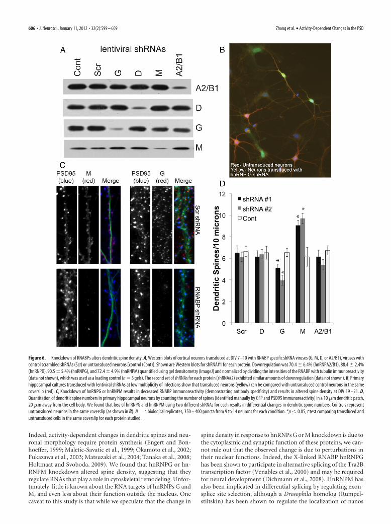
we generated lentiviral shRNA particles using pTRIP vectors to per-form knockdown experiments (Janas et al., 2006). We generated atleast two different shRNA viruses for each RNABP (shRNA#1 andshRNA#2; see Materials and Methods, above). We found that trans-duction of rat primary hippocampal neurons at DIV 7–10 usingthese viruses (incubation, 5–7 d) resulted in �70% specific down-regulation of these proteins compared with a scrambled shRNA se-quence or untransduced cells (Fig. 6A). There was no effect of theseshRNAs on cell viability. RNABP knockdown using shRNAs led to a
loss of their immunoreactivity in immuno-cytochemistry experiments on primary hip-pocampal neurons, confirming the specificityof the antibodies used (Fig. 6C). To determinetheeffectsof lossofthesehnRNPsondendriticspine density, we transduced rat primaryhippocampal neurons at DIV 14 with lenti-viral shRNAs at a low multiplicity of infec-tion (�1) to be able to identify transducedand untransduced cells in the same coverslip(Fig. 6B). Five days later (DIV 19), we fixedand immunostained neurons for GFP andPSD-95 (to identify dendritic spines). Wefound that there was a significant decrease inthe number of dendritic spines in neuronslacking hnRNPG but a significant increasein dendritic spines in neurons lacking hn-RNPM (Fig. 6C,D). No changes were ob-served in neurons lacking hnRNPs D or A2/B1. These results suggest that hnRNPG andhnRNPM play a crucial role in maintainingnormal spine density.
To determine whether the synaptic up-regulation of RNABPs resulted in a con-comitant upregulation of synaptic RNA, wetreated DIV 17–21 hippocampal neuronswith cLTP�BDNF or TTX and imagedpoly(A) mRNA using FISH using an oli-go(dT) probe (Fig. 7A). We found thatcLTP�BDNF resulted in a substantial in-crease in poly(A) mRNA overall and in den-drites (Fig. 7A). To determine whether thismRNA was synaptic, we quantified colocal-ization of oligo(dT)-Cy3 reactive puncta
with PSD-95 immunoreactivity (Fig. 7B). We found that cLTP�BDNF resulted in an increase in the extent of their colocalization(Fig. 7C, Cont), suggesting that poly(A) mRNAs are delivered intosynapses in response to the same treatment that resulted in synapticupregulation of hnRNPs. To determine whether hnRNPG and hn-RNPM played a role in this process, we knocked down their expres-sion using lentiviral shRNAs. However, loss of these proteins had noeffect on the cLTP-dependent upregulation of mRNA into synapses
Figure 3. Representative MS spectra showing SILAC quantitation of protein abundance changes induced by cLTP�BDNF treatment. The data show the MS analysis of a tryptic peptide(LLSDISTALR) from the protein SynGAP, which had decreased abundance in PSDs after cLTP�BDNF treatment. The peptide ratio is calculated by comparing the intensities of the medium signal tothe heavy signal. The light signal, which represents the unlabeled fraction of the protein, was not used for protein quantitation. In the forward and reverse experiments, the ratio change is in oppositedirections because the SILAC labeling was flipped in these two experiments.
Figure 4. Identification of proteins whose abundance were significantly changed upon LTP treatment. The histogram on topshows protein ratios in the forward experiment. The histogram on the right shows protein ratios in the reverse experiment. Proteinratios from the two replicate experiments are shown in the central scatter plot. All ratios are presented as cLTP�BDNF/TTX.Proteins with consistent significant ratio changes are marked with a plus sign (�) ( p value �0.05; Significance B; MaxQuant).
Zhang et al. • Activity-Dependent Changes in the PSD J. Neurosci., January 11, 2012 • 32(2):599 – 609 • 603

(Fig. 7C, Scr, G, M). Moreover, loss of these proteins had no effect onthe activity-dependent upregulation of poly(A) mRNA at dendrites(data not shown). These results suggest that hnRNPG and hnRNPMdo not play a role in activity-dependent mRNA transport.
DiscussionHere we identified changes in PSD composition induced bycLTP�BDNF treatment using a modified version of SILAC.SILAC is an efficient method for precisely measuring the rel-ative abundance of proteins in cell samples. One advantage ofSILAC is that it allows for combining differentially labeledprotein samples early in the sample preparation, thus mini-mizing variations in quantitation caused by parallel samplehandling. This is important for this study since the cellularfractionation required for PSD purification from neuronal cellcultures is complex and could introduce substantial variabilityif the samples were processed separately. Standard SILAC re-quires complete metabolic labeling of proteins and is therefore
difficult to apply to postmitotic cells such as primary neurons. Toovercome this limitation, we have recently shown that by usingtwo different sets of heavy amino acids for labeling, straightfor-ward SILAC quantitation can be performed using partially la-beled cells because the two cell populations are always equallylabeled (Zhang et al., 2011). This enabled us to take full advantageof the high precision of SILAC to measure changes in the abun-dance of PSD components after cLTP induction.
In this study, we found that several RNABPs, including hn-RNPs A1, A2/B1, A3, G, L, M, DEAD-box 17 and DEAD-box 9,demonstrated increased abundance at synapses in response tosynaptic activity. RNABPs regulate all aspects of RNA metabo-lism by binding to and incorporating them into specialized ribo-nucleoparticles (Sossin and DesGroseillers, 2006). To date, a fewRNABPs, including Staufen, CPEB, FMRP, ZBP1 (Martin andZukin, 2006), and hnRNP A2/B1 (Hoek et al., 1998; Munro et al.,1999), have been implicated in neuronal mRNA transport andtranslation. While we identified FMRP, Staufen, and Pur � and � in
Table 1. Proteins of the PSD that displayed significant changes in abundance between the cLTP�BDNF treated and the TTX treated neurons; proteins have been groupedinto categories based on their reported functions
Protein ID Protein name Gene name Forward cLTP/TTX Reverse TTX/cLTP
RNA bindingIPI00553777 Heterogeneous nuclear ribonucleoprotein A1 Hnrnpa1 1.94 0.59IPI00867934 Heterogeneous nuclear ribonucleoprotein A2/B1 Hnrnpa2b1 1.84 0.55IPI00382376 Heterogeneous nuclear ribonucleoprotein A3 Hnrnpa3 1.59 0.63IPI00124979 Heterogeneous nuclear ribonucleoprotein G Hnrnpg 1.93 0.36IPI00765134 Heterogeneous nuclear ribonucleoprotein L Hnrnpl 1.47 0.67IPI00876603 Heterogeneous nuclear ribonucleoprotein M Hnrnpm 1.33 0.70IPI00755892 Heterogeneous nuclear ribonucleoprotein D Hnrpdl 1.30 0.71IPI00828741 HnRNP-associated with lethal yellow C130057N11Rik 1.56 0.59IPI00653307 DEAD box polypeptide 17 Ddx17 1.76 0.48IPI00366249 ATP-dependent RNA helicase A/ DEAD box 9 Dhx9 1.52 0.74IPI00400578 Heterochromatin protein 1-binding protein 3 Hp1bp3 1.36 0.68IPI00117063 RNA-binding protein FUS Fus 1.50 0.56
CytoskeletalIPI00199101 Serine/threonine-protein kinase MRCK alpha Cdc42bpa 0.64 1.39IPI00368911 CTTNBP2 N-terminal-like protein Cttnbp2 nl 1.35 0.67IPI00844646 Echinoderm microtubule-associated protein-like 1 Eml1 0.77 1.42IPI00474002 Stathmin-2 Stmn2 0.66 1.45IPI00309223 Stathmin-3 Stmn3 0.68 1.60IPI00125328 Dematin Epb49 0.43 1.68IPI00364284 LIM and calponin homology domains-containing 1 Limch1 0.71 1.45
GAPsIPI00558902 ADP-ribosylation factor GTPase-activating protein 2 Arfgap2 0.76 1.36IPI00339023 Ras GTPase-activating protein SynGAP Syngap1 0.29 2.73IPI00469012 TBC1 domain family member 10B Tbc1d10b 1.47 0.65IPI00758248 IQ motif and SEC7 domain-containing protein 2 Iqsec2 1.43 0.78
SynapticIPI00556925 Protein bassoon Bsn 1.55 0.67IPI00420725 Calcium/calmodulin-dep. protein kinase II, alpha Camk2a 0.58 1.42IPI00649778 Calcium/calmodulin-dep. protein kinase II, beta Camk2b 0.67 1.27IPI00196730 Calcium/calmodulin-dep. protein kinase II, gamma Camk2g 0.67 1.25IPI00204418 ERC protein 2 Erc2 1.32 0.72IPI00209336 Sorbin and SH3 domain-containing protein 2 Sorbs2 0.52 1.76IPI00358533 Actin-binding LIM protein 1 Ablim1 1.45 0.77IPI00210570 Homer protein homolog 1 Homer1 0.58 1.42
KinasesIPI00196987 Beta-adrenergic receptor kinase 1 Adrbk1 1.83 0.64
OtherIPI00198101 Uncharacterized protein C530008M17Rik 0.74 1.39IPI00190176 Golgin subfamily A member 3 Golga3 0.70 1.44IPI00407835 Ubiquitin-associated protein 2-like Ubap2l 0.60 1.35IPI00230394 Lamin-B1 Lmnb1 1.70 0.25IPI00652934 Core histone macro-H2A0.2 H2afy2 1.62 0.39
604 • J. Neurosci., January 11, 2012 • 32(2):599 – 609 Zhang et al. • Activity-Dependent Changes in the PSD

this study, the levels of these proteins wereeither not altered in response to synaptic ac-tivity, or their level of change fell below ourstatistical cutoff (Fig. 5B,C). This could sug-gest that they either do not participate inactivity-dependent processes, or that theirfunction does not rely on changes in theirabundance at synapses. Since we have in-cluded cycloheximide throughout the stimu-lation paradigms, the synaptic upregulation ofthe identified proteins could reflect an in-crease in the trafficking of these proteinsinto synapses. However, this change couldalso result from other cellular processes suchas protein turnover via ubiquitination.Overall, our results suggest that members ofthe hnRNP class of RNABPs may play animportant role in dendritic RNA metabo-lism and are worthy of further scientificresearch.
Interestingly, 10 of the 12 RNABPsidentified have been previously shown tobe components of RNA granules/trans-port particles in neurons (Kanai et al.,2004; Elvira et al., 2006). This suggeststhat synaptic activity results in the trans-location of these ribonucleoparticles intosynapses. Indeed, we find an increase inmRNA at synaptic sites in response to thesame synaptic stimulation paradigm usedin our proteomic screen (Fig. 7). Our re-sults are consistent with previous findingsshowing that synaptic activity results inthe trafficking of diverse mRNAs intodendrites and dendritic spines, includingtranscripts of BDNF, TrkB (Tongiorgi etal., 1997), CaMKII� (Thomas et al., 1994;Rook et al., 2000; Håvik et al., 2003),ZBP1, Beta actin (Tiruchinapalli et al.,2003), and Arc (Link et al., 1995; Lyford etal., 1995; Schuman et al., 2006). However,hnRNPG or hnRNPM knockdown didnot affect the cLTP-induced increase insynaptic mRNA. This suggests that eitherthese proteins do not participate inmRNA transport, or that there are redun-dant pathways. Alternatively, given thenumber of RNABPs identified in thisstudy, it is possible that the individualcontribution of each RNABP to the totaltransport of mRNA into dendrites is notdetectable.
The control of RNA distribution, sta-bility, and translation is a fundamentalmechanism for the local regulation of syn-aptic morphology and function. Transla-tion of dendritically localized mRNAs(Miyashiro et al., 1994; Eberwine et al.,2002; Zhong et al., 2006) plays an impor-tant role in synaptogenesis (Lyles et al.,2006), synaptic plasticity (Miniaci et al.,2008; Wang et al., 2009), and regenerationafter nerve injury (Weragoda et al., 2004).
Figure 5. Distribution of RNABPs and other proteins in neurons and verification of SILAC results. A, Total lysate [10 �g; Western blot(WB)], synaptosomes (Syn), or PSDs purified from rat brains were separated by SDS-PAGE and immunoblotted using antibodies against theindicated proteins. The presynaptic and postsynaptic markers SV2 and PSD95, respectively, were used to evaluate the quality of the PSDfractions. B,Ratcorticalneurons(DIV18)werepretreatedwithcycloheximide(1h)andthentreatedwithcLTP�BDNF(�)orTTX()andPSDs were isolated. Cycloheximide was present throughout the treatment. Western blots confirm the SILAC results obtained (arrows).Right, Western blots of total lysates confirm that activity did not induce any overall changes in the total levels of these proteins. C, Quanti-tation of Western blots in B by gel densitometry (ImageJ) and normalizing using Western blots against tubulin (data not shown). N � 3gels,*p�0.05; ttestcomparingcLTP�BDNFtoTTXforeachprotein.D,Primaryhippocampalneurons(DIV21)wereimmunostainedwithhnRNPM, hnRNPG, hnRNPA2/B1, or hnRNPD antibodies (green) and costained for the synaptic marker PSD95 (red) showing the synapticdistribution of these proteins. E, cLTP�BDNF resulted in increased colocalization between the RNABPs and PSD-95, as determined usingICC and image quantitation (Mander’s coefficient, ImageJ). N � 3 biological replicates, 250 –300 puncta taken from 10 �m patches thatwere 20 �m from the cell body; 8 –11 neurons per replicate. *p � 0.05, t test between cLTP�BDNF and TTX for each protein.
Zhang et al. • Activity-Dependent Changes in the PSD J. Neurosci., January 11, 2012 • 32(2):599 – 609 • 605
Indeed, activity-dependent changes in dendritic spines and neu-ronal morphology require protein synthesis (Engert and Bon-hoeffer, 1999; Maletic-Savatic et al., 1999; Okamoto et al., 2002;Fukazawa et al., 2003; Matsuzaki et al., 2004; Tanaka et al., 2008;Holtmaat and Svoboda, 2009). We found that hnRNPG or hn-RNPM knockdown altered spine density, suggesting that theyregulate RNAs that play a role in cytoskeletal remodeling. Unfor-tunately, little is known about the RNA targets of hnRNPs G andM, and even less about their function outside the nucleus. Onecaveat to this study is that while we speculate that the change in
spine density in response to hnRNPs G or M knockdown is due tothe cytoplasmic and synaptic function of these proteins, we can-not rule out that the observed change is due to perturbations intheir nuclear functions. Indeed, the X-linked RNABP hnRNPGhas been shown to participate in alternative splicing of the Tra2Btranscription factor (Venables et al., 2000) and may be requiredfor neural development (Dichmann et al., 2008). HnRNPM hasalso been implicated in differential splicing by regulating exon-splice site selection, although a Drosophila homolog (Rumpel-stiltskin) has been shown to regulate the localization of nanos
Figure 6. Knockdown of RNABPs alters dendritic spine density. A, Western blots of cortical neurons transduced at DIV 7–10 with RNABP specific shRNA viruses (G, M, D, or A2/B1), viruses withcontrol scrambled shRNAs (Scr) or untransduced neurons [control (Cont)]. Shown are Western blots for shRNA#1 for each protein. Downregulation was 70.4 � 6.4% (hnRNPA2/B1), 88.4 � 2.4%(hnRNPD), 90.5 � 5.4% (hnRNPG), and 72.4 � 4.9% (hnRNPM) quantified using gel densitometry (ImageJ) and normalized by dividing the intensities of the RNABP with tubulin immunoreactivity(data not shown), which was used as a loading control (n � 3 gels). The second set of shRNAs for each protein (shRNA#2) exhibited similar amounts of downregulation (data not shown). B, Primaryhippocampal cultures transduced with lentiviral shRNAs at low multiplicity of infections show that transduced neurons (yellow) can be compared with untransduced control neurons in the samecoverslip (red). C, Knockdown of hnRNPG or hnRNPM results in decreased RNABP immunoreactivity (demonstrating antibody specificity) and results in altered spine density at DIV 19 –21. D,Quantitation of dendritic spine numbers in primary hippocampal neurons by counting the number of spines (identified manually by GFP and PSD95 immunoreactivity) in a 10 �m dendritic patch,20 �m away from the cell body. We found that loss of hnRNPG and hnRNPM using two different shRNAs for each results in differential changes in dendritic spine numbers. Controls representuntransduced neurons in the same coverslip (as shown in B). N � 4 biological replicates, 350 – 400 puncta from 9 to 14 neurons for each condition. *p � 0.05, t test comparing transduced anduntransduced cells in the same coverslip for each protein studied.
606 • J. Neurosci., January 11, 2012 • 32(2):599 – 609 Zhang et al. • Activity-Dependent Changes in the PSD

mRNA and play a role in anterior/poste-rior patterning (Jain and Gavis, 2008).However, hnRNP A2/B1 (A2) has beenimplicated in RNA transport as a compo-nent of RNA transport particles in brain(Kanai et al., 2004) and plays a role in thetrafficking of myelin basic protein mRNA(Hoek et al., 1998; Munro et al., 1999). Wedid not see any changes in spine density inresponse to knockdown of hnRNPs A2/B1or D. Our SILAC results and verificationof their synaptic distribution by immuno-cytochemistry suggest that these hnRNPsplay a role in synaptic RNA metabolism.One interesting possibility is that thesehnRNPs regulate RNA splicing outsidethe nucleus, as suggested by the growingevidence of intron-containing RNAs inneuronal dendrites (Bell et al., 2008, 2010;Buckley et al., 2011).
In this study, we initially expected tosee changes in proteins traditionally as-sociated with synaptic transmission, in-cluding glutamate receptors and theirscaffolding molecules. For example, wefound an activity-dependent upregulationof GluR1 at the PSD (25%) and a decrease ofNMDA receptors NR1 (�19%) and NR2B(�13%) in the forward experiment that werereciprocated in our reverse experiment. How-ever, these results did not pass our statisticalfilter. Therefore, we cannot rule out thepossibility that our statistical criteria re-moved proteins displaying biologicallymeaningful changes after cLTP stimulation.This points to the difficulties in characteriz-ing the dynamics of PSD composition usingquantitative proteomics and identifies ourlimits of detection.
There is considerable interest in themechanisms that underlie local regulationof protein abundance at synapses giventheir importance in synaptic function anddysfunction. Neuronal stimulation hasbeen shown to target mRNAs selectivelyinto activated spines (Steward et al., 1998;Steward and Worley, 2001; Moga et al.,2004), with translation occurring specifi-cally at activated synapses (Wang et al.,2009). Improper targeting of mRNAs intodendrites results in unstable synaptic po-tentiation and deficits in spatial memorytasks in mice (Miller et al., 2002; Tzingou-nis and Nicoll, 2006). In humans, dys-regulation of RNA distribution is theprimary cause of several neurodegenera-tive disorders including myotonic dystro-phy type 2; spinal cerebellar ataxias types8, 10 and 12; and fragile X tremor ataxiasyndrome (Ranum and Cooper, 2006).Some of these disorders result from ex-panded trinucleotide repeats at noncod-ing regions of RNA transcripts that cause
Figure 7. cLTP�BDNF stimulation results in increased dendritic and synaptic poly(A) mRNA in rat primary hippocampalneurons (DIV 17–21). A, cLTP�BDNF results in increased poly(A) mRNA labeled by FISH using oligo(dT)(50)-Cy3 probe staining incell body and dendrites. Dendrites are marked using MAP2 immunostaining (blue). B, cLTP�BDNF results in increased poly(A)mRNA in dendrites and dendritic spines identified using PSD95 immunostaining (arrows). C, Quantitation of imaging results(ImageJ Mander’s coefficients) shows cLTP�BNDF results in an increase in mRNA/PSD95 colocalization [control (cont)]. There wasno statistical difference in the cLTP-induced increase in synaptic mRNA between RNABP knockdowns (G, M) or control neurons[scrambled shRNA (Scr), untransduced neurons (Cont)]. N � 250 –370 puncta from at least eight neurons in three biologicalreplicates. *p � 0.005 for the statistically significant increase in synaptic poly(A) mRNA at in response to cLTP�BDNF comparedwith TTX for each condition.
Zhang et al. • Activity-Dependent Changes in the PSD J. Neurosci., January 11, 2012 • 32(2):599 – 609 • 607

their toxic accumulation at subnuclear structures (Ranum andCooper, 2006). Other disorders, such as fragile X syndrome, re-sult from null mutations of RNABPs involved in RNA transport/translation (Ranum and Cooper, 2006). Our results showing thatRNABPs are upregulated at synapses in response to cLTP induc-tion are consistent with these previous studies highlighting theimportance of local RNA metabolism during plasticity and sug-gest that hnRNPs provide a layer of local regulation duringplasticity-related events.
NotesSupplemental material for this article is available at http://saturn.med.nyu.edu/files/mylab/neubert/Supplementary_table_1.xlsx. Complete list ofproteins identified in isolated PSD fractions from the SILAC experiments.This material has not been peer reviewed.
ReferencesBanker G, Churchill L, Cotman CW (1974) Proteins of the postsynaptic
density. J Cell Biol 63:456 – 465.Bell TJ, Miyashiro KY, Sul JY, McCullough R, Buckley PT, Jochems J, Meaney
DF, Haydon P, Cantor C, Parsons TD, Eberwine J (2008) CytoplasmicBK(Ca) channel intron-containing mRNAs contribute to the intrinsicexcitability of hippocampal neurons. Proc Natl Acad Sci U S A 105:1901–1906.
Bell TJ, Miyashiro KY, Sul JY, Buckley PT, Lee MT, McCullough R, JochemsJ, Kim J, Cantor CR, Parsons TD, Eberwine J (2010) Intron retentionfacilitates splice variant diversity in calcium-activated big potassiumchannel populations. Proc Natl Acad Sci U S A 107:21152–21157.
Blomberg F, Cohen RS, Siekevitz P (1977) The structure of postsynapticdensities isolated from dog cerebral cortex. II. Characterization and ar-rangement of some of the major proteins within the structure. J Cell Biol74:204 –225.
Boehm J, Kang MG, Johnson RC, Esteban J, Huganir RL, Malinow R (2006)Synaptic incorporation of AMPA receptors during LTP is controlled by aPKC phosphorylation site on GluR1. Neuron 51:213–225.
Bourne JN, Harris KM (2008) Balancing structure and function at hip-pocampal dendritic spines. Annu Rev Neurosci 31:47– 67.
Buckley PT, Lee MT, Sul JY, Miyashiro KY, Bell TJ, Fisher SA, Kim J, EberwineJ (2011) Cytoplasmic intron sequence-retaining transcripts can be den-dritically targeted via ID element retrotransposons. Neuron 69:877– 884.
Carlin RK, Grab DJ, Cohen RS, Siekevitz P (1980) Isolation and character-ization of postsynaptic densities from various brain regions: enrichmentof different types of postsynaptic densities. J Cell Biol 86:831– 845.
Choquet D, Triller A (2003) The role of receptor diffusion in the organiza-tion of the postsynaptic membrane. Nat Rev Neurosci 4:251–265.
Cohen RS, Blomberg F, Berzins K, Siekevitz P (1977) The structure of post-synaptic densities isolated from dog cerebral cortex. I. Overall morphol-ogy and protein composition. J Cell Biol 74:181–203.
Collins MO, Husi H, Yu L, Brandon JM, Anderson CN, Blackstock WP,Choudhary JS, Grant SG (2006) Molecular characterization and com-parison of the components and multiprotein complexes in the postsyn-aptic proteome. J Neurochem 97 [Suppl 1]:16 –23.
Cox J, Mann M (2008) MaxQuant enables high peptide identification rates,individualized p.p.b.-range mass accuracies and proteome-wide proteinquantification. Nat Biotechnol 26:1367–1372.
Dichmann DS, Fletcher RB, Harland RM (2008) Expression cloning in Xe-nopus identifies RNA-binding proteins as regulators of embryogenesisand Rbmx as necessary for neural and muscle development. Dev Dyn237:1755–1766.
Dobi A, Szemes M, Lee C, Palkovits M, Lim F, Gyorgy A, Mahan MA, AgostonDV (2006) AUF1 is expressed in the developing brain, binds to AT-richdouble-stranded DNA, and regulates enkephalin gene expression. J BiolChem 281:28889 –28900.
Dosemeci A, Tao-Cheng JH, Vinade L, Jaffe H (2006) Preparation of post-synaptic density fraction from hippocampal slices and proteomic analysis.Biochem Biophys Res Commun 339:687– 694.
Eberwine J, Belt B, Kacharmina JE, Miyashiro K (2002) Analysis of subcel-lularly localized mRNAs using in situ hybridization, mRNA amplifica-tion, and expression profiling. Neurochem Res 27:1065–1077.
Ehlers MD (2003) Activity level controls postsynaptic composition and sig-naling via the ubiquitin-proteasome system. Nat Neurosci 6:231–242.
Elvira G, Wasiak S, Blandford V, Tong XK, Serrano A, Fan X, del RayoSanchez-Carbente M, Servant F, Bell AW, Boismenu D, Lacaille JC,McPherson PS, DesGroseillers L, Sossin WS (2006) Characterization ofan RNA granule from developing brain. Mol Cell Proteomics 5:635– 651.
Engert F, Bonhoeffer T (1999) Dendritic spine changes associated with hip-pocampal long-term synaptic plasticity. Nature 399:66 –70.
Fukazawa Y, Saitoh Y, Ozawa F, Ohta Y, Mizuno K, Inokuchi K (2003)Hippocampal LTP is accompanied by enhanced F-actin content withinthe dendritic spine that is essential for late LTP maintenance in vivo.Neuron 38:447– 460.
Håvik B, Røkke H, Bårdsen K, Davanger S, Bramham CR (2003) Bursts ofhigh-frequency stimulation trigger rapid delivery of pre-existing alpha-CaMKII mRNA to synapses: a mechanism in dendritic protein synthesisduring long-term potentiation in adult awake rats. Eur J Neurosci17:2679 –2689.
Hoek KS, Kidd GJ, Carson JH, Smith R (1998) hnRNP A2 selectively bindsthe cytoplasmic transport sequence of myelin basic protein mRNA. Bio-chemistry 37:7021–7029.
Holtmaat A, Svoboda K (2009) Experience-dependent structural synapticplasticity in the mammalian brain. Nat Rev Neurosci 10:647– 658.
Huang DW, Sherman BT, Lempicki RA (2009) Systematic and integrativeanalysis of large gene lists using DAVID bioinformatics resources. NatProtoc 4:44 –57.
Jain RA, Gavis ER (2008) The drosophila hnRNP M homolog Rumpel-stiltskin regulates nanos mRNA localization. Development 135:973–982.
Janas J, Skowronski J, Van Aelst L (2006) Lentiviral delivery of RNAi inhippocampal neurons. Methods Enzymol 406:593– 605.
Ji Y, Lu Y, Yang F, Shen W, Tang TT, Feng L, Duan S, Lu B (2010) Acute andgradual increases in BDNF concentration elicit distinct signaling andfunctions in neurons. Nat Neurosci 13:302–309.
Jordan BA, Kreutz MR (2009) Nucleocytoplasmic protein shuttling: the di-rect route in synapse-to-nucleus signaling. Trends Neurosci 32:392– 401.
Jordan BA, Ziff EB (2008) To the nucleus with proteomics. In: Transcrip-tional regulation by neuronal activity (Dudek SM, ed.), pp 27–51. NewYork: Springer.
Jordan BA, Fernholz BD, Boussac M, Xu C, Grigorean G, Ziff EB, Neubert TA(2004) Identification and verification of novel rodent postsynaptic den-sity proteins. Mol Cell Proteomics 3:857– 871.
Jordan BA, Fernholz BD, Neubert TA, Ziff EB (2006) The dynamic synapse:molecular methods in ionotropic receptor biology. Boca Raton: CRC/Taylor and Francis.
Jordan BA, Fernholz BD, Khatri L, Ziff EB (2007) Activity-dependentAIDA-1 nuclear signaling regulates nucleolar numbers and protein syn-thesis in neurons. Nat Neurosci 10:427– 435.
Kanai Y, Dohmae N, Hirokawa N (2004) Kinesin transports RNA: isolationand characterization of an RNA-transporting granule. Neuron43:513–525.
Kang H, Schuman EM (1996) A requirement for local protein synthesis inneurotrophin-induced hippocampal synaptic plasticity. Science273:1402–1406.
Kennedy MB (1993) The postsynaptic density. Curr Opin Neurobiol3:732–737.
Lee C, Gyorgy A, Maric D, Sadri N, Schneider RJ, Barker JL, Lawson M,Agoston DV (2008) Members of the NuRD chromatin remodelingcomplex interact with AUF1 in developing cortical neurons. Cereb Cortex18:2909 –2919.
Li KW, Hornshaw MP, Van Der Schors RC, Watson R, Tate S, Casetta B,Jimenez CR, Gouwenberg Y, Gundelfinger ED, Smalla KH, Smit AB(2004) Proteomics analysis of rat brain postsynaptic density: implica-tions of the diverse protein functional groups for the integration of syn-aptic physiology. J Biol Chem 279:987–1002.
Link W, Konietzko U, Kauselmann G, Krug M, Schwanke B, Frey U, Kuhl D(1995) Somatodendritic expression of an immediate early gene is regu-lated by synaptic activity. Proc Natl Acad Sci U S A 92:5734 –5738.
Lisman J, Raghavachari S (2006) A unified model of the presynaptic andpostsynaptic changes during LTP at CA1 synapses. Sci STKE 2006, re11.
Lu W, Man H, Ju W, Trimble WS, MacDonald JF, Wang YT (2001) Activa-tion of synaptic NMDA receptors induces membrane insertion of newAMPA receptors and LTP in cultured hippocampal neurons. Neuron29:243–254.
Lyford GL, Yamagata K, Kaufmann WE, Barnes CA, Sanders LK, CopelandNG, Gilbert DJ, Jenkins NA, Lanahan AA, Worley PF (1995) Arc, a
608 • J. Neurosci., January 11, 2012 • 32(2):599 – 609 Zhang et al. • Activity-Dependent Changes in the PSD
growth factor and activity-regulated gene, encodes a novel cytoskeleton-associated protein that is enriched in neuronal dendrites. Neuron14:433– 445.
Lyles V, Zhao Y, Martin KC (2006) Synapse formation and mRNA localiza-tion in cultured Aplysia neurons. Neuron 49:349 –356.
Malenka RC (2003) Synaptic plasticity and AMPA receptor trafficking. AnnN Y Acad Sci 1003:1–11.
Malenka RC, Bear MF (2004) LTP and LTD: an embarrassment of riches.Neuron 44:5–21.
Maletic-Savatic M, Malinow R, Svoboda K (1999) Rapid dendritic morpho-genesis in CA1 hippocampal dendrites induced by synaptic activity. Sci-ence 283:1923–1927.
Martin KC, Zukin RS (2006) RNA trafficking and local protein synthesis indendrites: an overview. J Neurosci 26:7131–7134.
Matsuzaki M, Honkura N, Ellis-Davies GC, Kasai H (2004) Structural basisof long-term potentiation in single dendritic spines. Nature 429:761–766.
Miller S, Mayford M (1999) Cellular and molecular mechanisms of mem-ory: the LTP connection. Curr Opin Genet Dev 9:333–337.
Miller S, Yasuda M, Coats JK, Jones Y, Martone ME, Mayford M (2002)Disruption of dendritic translation of CaMKIIalpha impairs stabilizationof synaptic plasticity and memory consolidation. Neuron 36:507–519.
Milner B, Squire LR, Kandel ER (1998) Cognitive neuroscience and thestudy of memory. Neuron 20:445– 468.
Miniaci MC, Kim JH, Puthanveettil SV, Si K, Zhu H, Kandel ER, Bailey CH(2008) Sustained CPEB-dependent local protein synthesis is required tostabilize synaptic growth for persistence of long-term facilitation in Aply-sia. Neuron 59:1024 –1036.
Miyashiro K, Dichter M, Eberwine J (1994) On the nature and differentialdistribution of mRNAs in hippocampal neurites: implications for neuro-nal functioning. Proc Natl Acad Sci U S A 91:10800 –10804.
Moga DE, Calhoun ME, Chowdhury A, Worley P, Morrison JH, Shapiro ML(2004) Activity-regulated cytoskeletal-associated protein is localized torecently activated excitatory synapses. Neuroscience 125:7–11.
Munro TP, Magee RJ, Kidd GJ, Carson JH, Barbarese E, Smith LM, Smith R(1999) Mutational analysis of a heterogeneous nuclear ribonucleopro-tein A2 response element for RNA trafficking. J Biol Chem 274:34389 –34395.
Oh MC, Derkach VA, Guire ES, Soderling TR (2006) Extrasynaptic mem-brane trafficking regulated by GluR1 serine 845 phosphorylation primesAMPA receptors for long-term potentiation. J Biol Chem 281:752–758.
Okamoto S, Li Z, Ju C, Scholzke MN, Mathews E, Cui J, Salvesen GS, Bossy-Wetzel E, Lipton SA (2002) Dominant-interfering forms of MEF2 gen-erated by caspase cleavage contribute to NMDA-induced neuronalapoptosis. Proc Natl Acad Sci U S A 99:3974 –3979.
Ong SE, Blagoev B, Kratchmarova I, Kristensen DB, Steen H, Pandey A, MannM (2002) Stable isotope labeling by amino acids in cell culture, SILAC,as a simple and accurate approach to expression proteomics. Mol CellProteomics 1:376 –386.
Osten P, Khatri L, Perez JL, Kohr G, Giese G, Daly C, Schulz TW, Wensky A,Lee LM, Ziff EB (2000) Mutagenesis reveals a role for ABP/GRIP bind-ing to GluR2 in synaptic surface accumulation of the AMPA receptor.Neuron 27:313–325.
Peng J, Kim MJ, Cheng D, Duong DM, Gygi SP, Sheng M (2004) Semi-quantitative proteomic analysis of rat forebrain postsynaptic density frac-tions by mass spectrometry. J Biol Chem 279:21003–21011.
Ranum LP, Cooper TA (2006) RNA-mediated neuromuscular disorders.Annu Rev Neurosci 29:259 –277.
Richter JD, Klann E (2009) Making synaptic plasticity and memory last:mechanisms of translational regulation. Genes Dev 23:1–11.
Rook MS, Lu M, Kosik KS (2000) CaMKIIalpha 3� untranslated region-directed mRNA translocation in living neurons: visualization by GFPlinkage. J Neurosci 20:6385– 6393.
Schuman EM, Dynes JL, Steward O (2006) Synaptic regulation of transla-tion of dendritic mRNAs. J Neurosci 26:7143–7146.
Shevchenko A, Wilm M, Vorm O, Mann M (1996) Mass spectrometric se-quencing of proteins silver-stained polyacrylamide gels. Anal Chem68:850 – 858.
Sossin WS, DesGroseillers L (2006) Intracellular trafficking of RNA in neu-rons. Traffic 7:1581–1589.
Spellman DS, Deinhardt K, Darie CC, Chao MV, Neubert TA (2008) Stableisotopic labeling by amino acids in cultured primary neurons: applicationto brain-derived neurotrophic factor-dependent phosphotyrosine-associated signaling. Mol Cell Proteomics 7:1067–1076.
Steward O, Worley PF (2001) Selective targeting of newly synthesized ArcmRNA to active synapses requires NMDA receptor activation. Neuron30:227–240.
Steward O, Wallace CS, Lyford GL, Worley PF (1998) Synaptic activationcauses the mRNA for the IEG Arc to localize selectively near activatedpostsynaptic sites on dendrites. Neuron 21:741–751.
Tanaka J, Horiike Y, Matsuzaki M, Miyazaki T, Ellis-Davies GC, Kasai H(2008) Protein synthesis and neurotrophin-dependent structural plastic-ity of single dendritic spines. Science 319:1683–1687.
Thomas KL, Laroche S, Errington ML, Bliss TV, Hunt SP (1994) Spatial andtemporal changes in signal transduction pathways during LTP. Neuron13:737–745.
Tiruchinapalli DM, Oleynikov Y, Kelic S, Shenoy SM, Hartley A, Stanton PK,Singer RH, Bassell GJ (2003) Activity-dependent trafficking and dy-namic localization of zipcode binding protein 1 and beta-actin mRNA indendrites and spines of hippocampal neurons. J Neurosci 23:3251–3261.
Tongiorgi E, Righi M, Cattaneo A (1997) Activity-dependent dendritic tar-geting of BDNF and TrkB mRNAs in hippocampal neurons. J Neurosci17:9492–9505.
Tzingounis AV, Nicoll RA (2006) Arc/Arg3.1: linking gene expression tosynaptic plasticity and memory. Neuron 52:403– 407.
Venables JP, Elliott DJ, Makarova OV, Makarov EM, Cooke HJ, Eperon IC(2000) RBMY, a probable human spermatogenesis factor, and other hn-RNP G proteins interact with Tra2beta and affect splicing. Hum MolGenet 9:685– 694.
Vinade L, Chang M, Schlief ML, Petersen JD, Reese TS, Tao-Cheng JH, Dose-meci A (2003) Affinity purification of PSD-95-containing postsynapticcomplexes. J Neurochem 87:1255–1261.
Walikonis RS, Jensen ON, Mann M, Provance DW Jr, Mercer JA, KennedyMB (2000) Identification of proteins in the postsynaptic density frac-tion by mass spectrometry. J Neurosci 20:4069 – 4080.
Walsh MJ, Kuruc N (1992) The postsynaptic density: constituent and asso-ciated proteins characterized by electrophoresis, immunoblotting, andpeptide sequencing. J Neurochem 59:667– 678.
Wang DO, Kim SM, Zhao Y, Hwang H, Miura SK, Sossin WS, Martin KC(2009) Synapse- and stimulus-specific local translation during long-term neuronal plasticity. Science 324:1536 –1540.
Weragoda RM, Ferrer E, Walters ET (2004) Memory-like alterations in Ap-lysia axons after nerve injury or localized depolarization. J Neurosci24:10393–10401.
Yoshimura Y, Yamauchi Y, Shinkawa T, Taoka M, Donai H, Takahashi N,Isobe T, Yamauchi T (2004) Molecular constituents of the postsynapticdensity fraction revealed by proteomic analysis using multidimensionalliquid chromatography-tandem mass spectrometry. J Neurochem88:759 –768.
Zhang G, Deinhardt K, Chao MV, Neubert TA (2011) Study ofneurotrophin-3 signaling in primary cultured neurons using multiplexstable isotope labeling with amino acids in cell culture. J Proteome Res10:2546 –2554.
Zhong J, Zhang T, Bloch LM (2006) Dendritic mRNAs encode diversifiedfunctionalities in hippocampal pyramidal neurons. BMC Neurosci 7:17.
Ziff EB (1997) Enlightening the postsynaptic density. Neuron 19:1163–1174.
Zhang et al. • Activity-Dependent Changes in the PSD J. Neurosci., January 11, 2012 • 32(2):599 – 609 • 609