rn Department of Veterans Affairs
12
rn Department of Veterans Affairs Journal of Rehabilitation Research and Development Vol. 29 No. 2,1992 Pages 1-12 Artificial nerve graft using glycolide trimethylene carbonate as a nerve conduit filled with collagen compared to sutured autograft in a rat model D ; Jose A. Padilla, BA; Khoi D. Nguyen, MD; James Siedman; Huu N. Pham, MD Division of Plastic and Reconstructive Surgery, NC-104, Hoover Pavilion, Stanford University Medical Center, Palo Alto, CA 94305; Rehabilitation Research and Development Engineering Center, Department of Rterans AJljcairs Medical Center, Palo Alto, CA 94304 Abstract-A study was conducted to compare the regeneration of rat peroneal nerves across 0.5 cm gaps repaired with artifi- cial nerve grafts (ANG) versus sutured autografts (SAG). The ANG model is composed of a synthetic biodegradable passive conduit made of glycolide trimethylene carbonate (GTMC) filled with a collagen matrix (predominately Type I collagen, derived from calf skin, and with the telopeptide ends left intact). Axonal regeneration was studied in 11 long-term animals (two at 6 months and nine at 9 months). The nerves were studied by qualititative and quantitative histological, electrophysiological, and functional assays. Axonal regeneration with the ANG was equal to SAGSas measured by axonal diameters, physiological, and functional methods, although the SAG demonstrated statisti- cally higher axonal counts. Key words: artificial nerve graji, autograji, axonal regenera- tion, biomaterials, collagen matrix, traumatic newe injury. INTRODUCTION In some traumatic nerve injuries, nerve loss results in a significant gap between the cut ends of the nerve that cannot be closed by an end-to-end repair (1,2,3,4). The present treatment of choice is the use of an autograft to bridge the gap (3,4). The major limitation of the autograft Address all correspondence and requests for reprints to: Dr. J.M. Rosen, Dartmouth-Hitchcock Clinic, Plastic Surgery, 1 Medical Center Drive, Leba- non. NH 03756. is the requirement of a donor nerve. Homografts and heterografts have been evaluated as alternatives to auto- grafts, but have been found to be immunologically unac- ceptable (5,6). Although it may be possible to suppress the immune system during regeneration through a homograft (7), one must weigh this with the risks of immunosup- pression. Therefore, the development of an artificial nerve graft (ANG) is one method to solve both problems of avail- ability and rejection by the immune system. Alternatives to the autografts that have been tried include homografts, heterografts, and artificial substitutes for nerve grafts (1,3,6,8,9). Recently, clinical studies of artificial nerve grafts have included polyglycolic acid (PGA) tubes (7) and veins (lo), either implanted empty and subsequently filled with autologous serum, or filled before implantation with auto- logous blood products. Several groups have been experimentally attempting to develop an ideal medium for axonal regeneration (11,12,13,14). Various macromolecules, including laminin, fibronectin, heparan sulfate, and collagen, have been analyzed and shown to have properties important for regeneration (11,15,16,17,18,19,20).Our current interest lies in the role of collagen on axonal regeneration in the artifi- cial nerve graft. Important advantages of using collagen over other biomaterials include the relative ability to manipulate its biochemistry and microstructure (21). We have previously studied Zydermm Collagen Implant (ZCI) (composed of Type I with some Type I11 collagen)
Transcript of rn Department of Veterans Affairs

rn Department of Veterans Affairs
Journal of Rehabilitation Research and Development Vol. 29 No. 2,1992 Pages 1-12
Artificial nerve graft using glycolide trimethylene carbonate as a nerve conduit filled with collagen compared to sutured autograft in a rat model D
; Jose A. Padilla, BA; Khoi D. Nguyen, MD; James Siedman; Huu N. Pham, MD Division of Plastic and Reconstructive Surgery, NC-104, Hoover Pavilion, Stanford University Medical Center, Palo Alto, CA 94305; Rehabilitation Research and Development Engineering Center, Department of Rterans AJljcairs Medical Center, Palo Alto, CA 94304
Abstract-A study was conducted to compare the regeneration of rat peroneal nerves across 0.5 cm gaps repaired with artifi- cial nerve grafts (ANG) versus sutured autografts (SAG). The ANG model is composed of a synthetic biodegradable passive conduit made of glycolide trimethylene carbonate (GTMC) filled with a collagen matrix (predominately Type I collagen, derived from calf skin, and with the telopeptide ends left intact). Axonal regeneration was studied in 11 long-term animals (two at 6 months and nine at 9 months). The nerves were studied by qualititative and quantitative histological, electrophysiological, and functional assays. Axonal regeneration with the ANG was equal to SAGS as measured by axonal diameters, physiological, and functional methods, although the SAG demonstrated statisti- cally higher axonal counts.
Key words: artificial nerve graji, autograji, axonal regenera- tion, biomaterials, collagen matrix, traumatic newe injury.
INTRODUCTION
In some traumatic nerve injuries, nerve loss results in a significant gap between the cut ends of the nerve that cannot be closed by an end-to-end repair (1,2,3,4). The present treatment of choice is the use of an autograft to bridge the gap (3,4). The major limitation of the autograft
Address all correspondence and requests for reprints to: Dr. J.M. Rosen, Dartmouth-Hitchcock Clinic, Plastic Surgery, 1 Medical Center Drive, Leba- non. NH 03756.
is the requirement of a donor nerve. Homografts and heterografts have been evaluated as alternatives to auto- grafts, but have been found to be immunologically unac- ceptable (5,6). Although it may be possible to suppress the immune system during regeneration through a homograft (7), one must weigh this with the risks of immunosup- pression. Therefore, the development of an artificial nerve graft (ANG) is one method to solve both problems of avail- ability and rejection by the immune system. Alternatives to the autografts that have been tried include homografts, heterografts, and artificial substitutes for nerve grafts (1,3,6,8,9). Recently, clinical studies of artificial nerve grafts have included polyglycolic acid (PGA) tubes (7) and veins (lo), either implanted empty and subsequently filled with autologous serum, or filled before implantation with auto- logous blood products.
Several groups have been experimentally attempting to develop an ideal medium for axonal regeneration (11,12,13,14). Various macromolecules, including laminin, fibronectin, heparan sulfate, and collagen, have been analyzed and shown to have properties important for regeneration (11,15,16,17,18,19,20). Our current interest lies in the role of collagen on axonal regeneration in the artifi- cial nerve graft. Important advantages of using collagen over other biomaterials include the relative ability to manipulate its biochemistry and microstructure (21).
We have previously studied Zydermm Collagen Implant (ZCI) (composed of Type I with some Type I11 collagen)
Jim
Text Box
DOI:10.1682/JRRD.1992.04.0001

2
Journal of Rehabilitation Research and Development Vol. 29 No. 2 SF
as an extracellular matrix for nerve regeneration (22) because of its hypoantigenicity and history of use in vivo (23). This collagen has been treated to remove the telopep- tide ends from the triple-helix to reduce its antigenicity (24). Previous studies by us and others (25) have shown that nerves regenerate through this matrix; however, regeneration was shown to be inferior compared to sutured autografts (SAG) (22). This may result from the manufac- turing process producing particles or clumps of collagen fibers up to 250 pm in dimeter (24). These pafiicles prob- ably have a higher concentration than the average of 3.5 percent collagen. The final structure of Zyderm may render this collagen less than ideal as a cellular regeneration matrix.
In this study, we therefore chose to use a collagen with a structure more suitable for nerve regeneration. This Type I collagen was used in a gel form which results in a loosely woven matrix that provides spaces for regeneration; although this collagen retains its telo- peptide ends, the overall antigenicity of collagen is low. The telopeptides play an important role in collagen fiber reassembly (21). This affects the precipitation kinetics, microstructure, and behavior of the collagen in multiple ways that may be advantageous for a nerve regeneration extracellular matrix.
The evaluated artificial nerve grafts were composed of resorbable tubes filled with a growth medium composed of collagen as a possible alternative to an autograft. Insted of PGA tubes, tubes of glycolide trimethylene carbonate (GTMC) were used because it is less reactive, possesses a controllable resorption time, and has better handling characteristics than PGA. The resorption time of GTMC can be made greater than that of PCrA by adding methy- lene groups, making the strucmre more hydrophobic and more resistant to hydrolysis. In addition, the methylene moiety makes GTMC more flexible, unlike PGA which is brittle; therefore, GTMC is less likely to fragment when the grafts are flexed by the movement of the animal. Finally, GTMC is pliable enough to receive sutures, which facili- tates its use surgically.
The interest in finding an ideal medium for axonal growth and passive devices for bridging gaps resulting from resected nerves has spanned over a century (5,8). These passive devices have included a wide range of biological and nonbiological materials (8). Early artificial nerve grafts provided a tube filled with blood or some plasma product to span the nerve gap (26). In a few cases, only threads were used to span the gap with no surrounding tube (27). The early attempts were uniformly unsuccessful, both experimentally and clinically.
In 1944, m i s s reviewed prior studies of artificial nerve grab (5). His experiments demonstrated that tubes filled with blood with or without threads as guides would suc- cessfully act as afiificial nerve grafts (6). Expehents using autogenous veins as passive conduits for nerve regenera- tion found them to be acceplable, but far from being ideal a~if ic id nerve grafts (10,28). Wrkers ushg pseudosynovial or mesothelial tubes have reporled moderate success (11,16,29,30,31,32), while others experimenting with vari- ous synthetic resorbable materials also had moderate suc- cess (733) . Although in some of these studies autologous blood products filled the conduits, none of these studies were conducted to determine the most appropriate matrix material with which to fill the tube as a growth medium for axonal regeneration.
The natural extracellular matrix is composed of mul- tiple proteins, including laminin, fibronectin, and colla- gen (21). The extracellular matrix exerts a strong influence over cellular regeneration, and in particular, nerve regener- ation (34,35,36,37). Cell activity is regulated by contact with the collagenous extracellular matrix, not only for pur- poses of morphogenesis, but also for maintenance and adaptation of the differentiated state (38,39). There is ample evidence that cells can physically respond to the orienta- tion of the substrate on which they rest (40,41,42,43).
Although only a part of the extracellular matrix, collagen is endowed with many unique properties. Cells respond to the chemical nature of collagen by interacting with atlachment sites. They also respond to the porosiq (cross-linking affects this porosity, along with strength, stiffness, and persistence in vivo) of the collagen, its antigenicity, its orienlation, its mobilization of enzymes and other subsbnces (44,459. These parameters can be manipulated to potentially yield enhanced nerve regener- ation. Collagen is only mildly immunoreactive (21), due in par% to masking of potential antigenic determinants by the helical structure, so that further decreasing anti- genicity may not be all that important for our purposes. Finally, recent experimenb have shown that silicone tubes filled with collagen resulted in successful bridging of nerve gaps (46).
There may be a number of reasons why collagen in the form of Zyderm may not be ideal for nerve regenera- tion. The physical form of Zyderm apparently tends to restrict migration of cells to narrow channels, at least until the dense particles or blocks have been degraded (23). Cell penetration must occur around these dense blocks through areas of least resistance (i.e., largely along planes of low fiber density). A sufficiently high collagen concentration, even if entirely homogenous, ilnpedes cellular immigra-
ROSEN et al. Artificial Nerve Graft
tion in general (21). This is also probably true for nerve regeneration and may explain our limited success in our first afiificial nerve graft experiments using Zyderm.
Our second study yielded more favorable results (47). We constructed an ANG composed of a synthetic bio- degradable passive conduit made of PGA filled with a col- lagen extracellular matrix (predominately Type 1 collagen, derived from calf skin, and with the telopeptide ends left intact). This ANC was used in the repair of 0.5 cm gaps in the rat peroneal nerve. Statistical analysis yielded equal results between the sutured autograft and our ANG as measured by qualitative and quantilative histology, phys- iology, and functional (toe spread) analysis.
After finding a matrix which would supporl axonal regeneration, we sought out a conduit makLenaI which would be more applicable to our needs. Although PCA was effective as a biomaterid for the repair of short gaps, longer gaps would necessitate the use of biomakrials with different characteristics. The biomaterial would need to be less reactive, possess a controllable resorption time, and have better handling characteristics than PGA. Our search yielded GTMC. The resovtion time of GTMC can be made greater than that of Pe;A by manipulating its stmcture (GTMC: Davis and Ceck, Pearl River, NU). GTMC con- duits can maintain their tube-like structure for upwrds of 3 months (unpublished data).* In addition, C6IMG is flex- ible, unlike PGA which is brittle; therefore, GTMC is less likely to fragment when the grafts are flexed by the move- ment of the animal. GTMC is pliable enough to receive sutures which faciliQtes its use surgically. We are currently working on creating a porous GTMC tube, which other investigators have shown plays a signficant role in nerve repair using tubes.
MATERIALS AND METHODS
Eleven Sprague-Dawley white male rats were used to compare artificial nerve grafts to sutured autograh (Rgure 1). All animals survived the postoperative period.
Adult rats weighing approximakly 250 grams each were deeply anesthetized with intraperitoneal sodium pen- tobarbital 5 mg1100 gm. The peroneal nerve was used as a test site. The peroneal nerve is a single fascicle (0.6 mm diameter) which arises from the bifurcation of the sciatic nerve. All surgical dissecdons and repairs were done using an operative microscope with magnification of 10 to 25 x .
* Personal communication. Conversation with R.F. Vallentini, 1990.
ARTIFICIAL
Figure 1. Drawing of the rat model showing the surgical repair of the peroneal nerves with the artificial nerve graft (ANG) and sutured autograft (SAG). The SAG is on the left, and the ANG is on the right.
In each animal, both peroneal nerves were exposed and a 0.5 cm-long segment was removed from the distal portion of each nerve 5 mm distal to the sciatic bifurca- tion. One randomly selected side in each animal was repaired using an autograft from the opposite peroneal nerve excised segment. This was sutured using 10-0 nylon on a straight 70 rnicrometer diameter needle, requir- ing two sutures per end of nerve. The sutures were placed through the perineurium only.
The other side in each animal was repaired using an artificial nerve graft (Figure 2). The artificial nerve grafts were cornposed of C;TMC tubes slightly larger in diameter than the nerves, 10 mm long, and filled with a liquid collagen (Collagen [C3511]: Sigma, St. Louis, MO) that subsequently gelled in the tube. The tube was split longi- tudinally and the nerve was pulled into the tube both prox- imally and distally for a distance of 2.5 mm. A blood clot was then allowed to form around both ends of the GTMC tube to hold the two nerve stumps secure within the tube without the need for sutures. Eleven animals were evalu- ated after long-tern regeneration from 6 to 9 months by qualitalive and quantitative histology, physiology, and func- tional assays.
Qualibtive histology provided an assessment of the organization of the proximal and distal repair site. It also provided an assessment of reaction to the surgical materi- als (nylon suture, GTMC tubes, and the collagen within the tubes). The qualitative histological evaluations were used only as guides to the events which occurred at the

Journal of Rehabilitation Research and Development Vol. 29 No. 2 Spring 1992
ARTIFICIAL NERVE GRAFT
GLYCOLIDE TRIMETHYLENE CARBONATE TUBE WITH COLLAGEN BRIDGE
Figure 2. Drawing of the artificial nerve graft model. The top drawing represents the glycolide trimethylene carbonate (GTMC) tube with the collagen bridge filling the 5 mm gap. The middle drawing represents the prox- imal and distal ends of the transected nerve. The lower two drawings show the two ends of the nerve being placed with the W M C tube.
ment); and, failure (no axonal alignment). The other half of the sections were stained with standard hematoxylin- eosin stain to evaluate tissue inflammatory reaction. This evaluation assigned the following subjective qualitative assessments: no reaction (0 WBCIHPF*), minimal (0-50 WBCIHPF*), moderate (50-100 WBCIHPF*), and extreme ( > 100 WBC IHPF*).
Quantitative histology was used to determine the axon diameter histogram (Figure 3) and the average diameter of the myelinated axon population just distal to the graft site (48). The average diameter provides a measure of axonal regeneration which is difficult to deter- mine from axon counts because of branching (49). Nerve biopsies 5 mm distal to the repair site were taken at the termination of the study. The biopsies were fixed in cacodylate buffered 3 percent glutaraldehyde for 24 hours, then in 1 percent osmium tetroxide for 1 hour. The fixed biopsies were embedded in Epon and cut into 1 micrometer sections with an ultramicrotome, using a glass knife. They then were mounted on a microscope slide and stained with phenylene diamine. These sections were used to determine the count and diameter of regenerated myelinated axons.
Axonal cross-sectional area determination and counts were accomplished using a MacintoshB H computer system linked to a DEST Scanner. Each black and white photomicrograph (400 x) of sections distal to the repair
HISTOLOGICAL EVALUATION
AXON DIAMETER HISTOGRAM
repair site. The organization of the repair site was ranked on a 5-point scale from fail to poor to fair to good to ex- cellent. The reaction was ranked using a scale ranging from no reaction, to minimal reaction, to moderate reaction, to o extreme reaction (48). Because of the presence of sutures in the SAG specimens, and occasional remnants of GTMC in the ANG specimens, it was not possible to do the evalu- ation in a completely blind method. All histologic evalua- tions were done by one histologist to avoid inter-tester bias.
\ Nerve repair site biopsies were fixed in 3 percent cacodylate buffered glutaraldehyde for a minimum of 24 Q REPAIR
hours. The site of repair was embedded in paraffin. Lon- DISTAL
gitudinal sections were cut serially at 6 to 8 micrometers. AXON D I A M E T E R
Half of the sections were stained with Bodian stain (silver stain) to determine the organization of the regenerated Qure 3.
Diagram of the preparation of the fiber diameter histogram (FDH). (1) axons. The following subjective qualitative assessments cmss-section is just distal to and a histological slide is pre-
were given: good (70 pared; (2) the histological section is photographed and the axons are percent to 99 percent alignment); fair (40 percent to 69 manually traced; and, (3) a computer system is used to convert the traced percent alignment); poor (10 percent to 39 percent align- axons into histograms.
ROSEN et al. Artificial Nerve Graft
SCIATIC B I F U R C A T I O N
7 z I:.. T I B I A L N E R V E
Figure 4. Diagram of the electrophysiological study. The neuroscope triggers the stimulator to stimulate the nerve either just proximal or just distal to the repair site. The resultant signal is recorded at the most proximal site available (i.e., as the nerve exits the spinal cord at the sciatic notch) and sent to the neuroscope for analysis. The proximal nerve is crushed to yield a monophasic signal.
represenkd approximately 15 percent of the cross-sectional area of the peroneal nerve. Three photomicrographs of each nerve section were used for this purpose. Tracings of these photographs exclusive of unmyelinated axons and blood vessels were digitized and a computer program then determined the area within the myelinated axons from an algorithm. The diameter was then determined from this area. The axonal count was subsequently deter- mined by s u m i n g the number of axons in the percentage of nerve counted and calculating the total number for the whole nerve.
Quantitative physiology was used to measure the popu- lation of regenerated axons. The compound action potential was used to physiologically evaluate axon regener- ation across the repair site in vivo (Figure 4) (50). The area under the monophasic compound action potential was determined by electronically computing the integrated mean compound action potential (IMCAP) (51). The area under
the monophasic compound action potential provides a measure of the proporlion (population) of myelinated axons with distal connections (49), that is, regenerated axons.
As in the original nerve repair, the test animals were given appropriate anesthesia, and all surgery was perfomed under magnification. The repair site was exposed surgi- cally, and the area was bathed in mineral oil maintained at 35 degrees Centigrade. Two sites were used to stimu- late the nerve, one just distal to the repair site, and one 5 mm proximal. These sites were dissected free of con- nective tissue and placed on platinum wire stimulating elec- trodes approximately 12 m apart, without disturbing the repair site (Figure 4). A baseline monophasic recording of the compound action potential (CAP) was obtained by severing and crushing the nerve at the sciatic notch ('just distal to exit from the spinal cord) and placing it on plati- num recording electrodes. The recording site was approx- imately 3.5 cm from the proximal stimulating electrodes. The IMCAP produced by stimulation via the proximal stimulating electrodes was first recorded to provide a meas- ure of the total population of myelinated axons just prox- imal to the repair region. The IMCAP to stimulation via the distal electrodes was then recorded as a measure of the population of myelinated axons that had reconnected across the repair site. The distal IMCAP divided by the proximal IMCAP gives a measure of the ratio of the total myelinated proximal axons having distal connections (49). The ratio is normalized to the total proximal myelinated axon population and expressed as a percentage in Table 1.
A Siegen Neuroscope (Mountain View? CA) was used to trigger a Grass S88 Stimulator (Quincy, MA) with two Model 478A stimulus isolation units. Stimulus parameters were as follows: a constant current with a pulse width of 0.05 msec, amplitude 20 percent above maximal nerve response, and a repetition rate of 2/sec. The nerve signal was recorded and analyzed with the Siegen Neuroscope. The neuroscope allows the storage and retrieval of data; permanent records are made through an attached Hewlett- Packard inkjet prlnter (Palo Alto, CA). The signal was aver- aged by recording 50 runs to reduce background noise. The bandwidth was set from 10 Hz to 3 kHz. The maxi- mum pulse amplitude was set at 20 percent above the value needed to stimulate the total population of myelinated axons as measured by the IMCAP algorithm.
De Medinaceli, and more recently, Bain have various indices of functional recovery in Walking Track Analysis (52,53). From our examination of these studies, we have begun to employ a Toe Spread Analysis with measurements of first to fifih toe spread (Figure 5). We have found that this parameter shows significant change

- - -- - - - -
Journal of Rehabilitation Research and Development Vol. 29 No. 2 Spring 1992
Table 1. Artificial nerve graft (GTMC with collagen type Ic) vs. sutured autograft
Histology P1.lysiology Functional Qualitative Quantitative Quantitative Analysis
Animal Type of Months Reaction Organization Axon Count MFD IMCAP Toe Spread Number Repair Post-Op at Graft at Graft ( P I mm
ANG
SAG
SAG
ANG
SAG
ANG
ANG
SAG
SAG
ANG
ANG
SAG
SAG
ANG
ANG
SAG
SAG
ANG
SAG
ANG
ANG
SAG
MIN
NONE
NONE
NONE
NONE
NONE
NI A
NONE
NONE
NONE
MOD
NONE
NONE
NONE
NONE
NONE
NONE
MOD
NONE
NONE
NONE
NONE
GOOD
FAIR
FAIR
FAIR
FAIR
GOOD
FAIR
FAIR
FAIR
FAIR
FAIR
FAIR
FAIR
GOOD
POOR
FAIR
FAIR
POOR
FAIR
FAIR
FAIR
FAIR
72.6
79.1
83.5
90.2
78.5
75.5
87.7
80.2
94.3
81.7
90.1
82.6
83.2
53.9
95.0
85.7
70.5
NIA
91.2
89.1
93.7
75.4
21.0
19.5
NI A
NI A
21.5
19.5
21.0
19.5
20.5
21.0
NIA
NIA
20.0
21.0
NI A
NIA
20.5
20.0
20.5
21.0
NI A
NIA
Key ORGANIZATION [excellent, good, fair, poor, or fall1 IMCAP = Integrated Mean Compound A ~ t i o n Potentla1 REACTION [none, minimal, moderate (mod), or extreme] ANG = Artificial herve Graft MFD = Mean F~ber Didmeter SAG = Sutured AutoGratt
-- - - - - - - - - - - - - - - - - - - - - - - - - - - - - - - - - - - - -- -- - - - - -- - - - - - - - - - - - - - - - - - - - - - - -
following peroneal nerve injury (unpublished data) and is statistically compared for significance by the Student's easily measured. t-test (5435).
The technique consists of dipping the hind feet of the animal into X-ray paper developer. The animal is then placed into a rectangular box with X-ray film lining the RESULTS floor, and the animal is allowed to walk the length of the box. Measurements of first to fifth toe spread and print Eleven animals were evaluated at 6 or 9 months post- length are easily obtained from the resultant foot prints. operation by qualitative histology and quantitative histol-
The nerve graft repairs were matched pairs and were ogy and physiology (Table 1).

Plexiglass Walking Box
Film 1
!'* Toe Spread . @
* ' 1 9 "
V P r ' @ 4 -4 fi 1st to 5th
Figure 5. Diagram of the rat walking system. On the top, the rat is walking across X-ray film in a Plexiglass walking box. On the right, X-ray film with four footprints is shown. On the bottom, a print with toe spread parameter (first to fifth toe spread) is delineated.
Qualitative histology Organization at the graft site. The grafts were evalu-
ated for organization at the repair site by determining the number of axons that lay outside the graft on cross-sections (Figure 6). In addition, the orientation of the fibers was also used in the determination of the organization of the fascicle. As matched pairs, the SAG were better than the ANG in two cases, while the ANG were better than the SAG in three cases. In six cases, the organization within the ANG and SAG were indistinguishable. Using the Stu- dent's t-test, no significant difference was found between the two graft types with respect to organization at the graft site (p > 0.05).
Reaction at the gra$ site. The grafts were evaluated for cellular reaction at the repair site in cross-sections stained with H & E. In the majority of cases, minimal or no cellular reaction was found to both grafts (ANG and SAG). No evidence of the original collagen in the ANG
Figure 6 (A, B, C, D). (Right) Photomicrographs of cross-sections of nerve repair at graft sites in Rat 996 at 9 months. (Stain: Bodian and Aniline blue.)
ROSEN e t al. Arlificial Nerve Graft
B. ANG at 750x. 1
Journal of Rehabilitation Research and Development Vol. 29 No. 2 Spring 1992
was noted at 9 months (i.e., no clumping of large parti- cles). There was a moderate reaction in three of the ANG, but in none of the SAG. The other eight matched pairs demonstrated no difference. Using the Student's t-test, no significant difference was found between the two graft types with respect to reaction at the graft site (p >0.05).
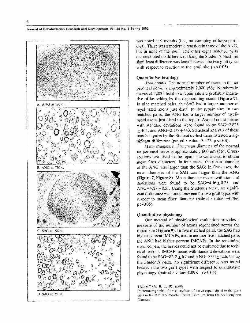
Quantitative histology Axon counts. The normal number of axons in the rat
peroneal nerve is approximately 2,000 (56). Numbers in excess of 2,000 distal to a repair site are probably indica- tive of branching by the regenerating axons (Figure 7). In nine matched pairs, the SAG had a larger number of myelinated axons just distal to the repair site; in two matched pairs, the ANG had a larger number of myeli- nated axons just distal to the repair. Axonal count means with standard deviations were found to be SAG=2,825 f 464, and ANG=2,177 f 443. Statistical analysis of these matched pairs by the Student's t-test demonstrated a sig- nificant difference (paired t value-3.477, p<0.01).
Mean diameters. The mean diameter of the normal rat peroneal nerve is approximately 600 pm (56). Cross- sections just distal to the repair site were used to obtain mean fiber diameters. In four cases, the mean diameter of the ANG was larger than the SAG; in five cases, the mean diameter of the SAG was larger than the ANG (Figure 7, Figure 8). Mean diameter means with standard deviations were found to be SAG=4.16 + 0.23, and ANG=4.27 k0.51. Using the Student's t-test, no signifi- cant difference was found between the two graft types with respect to mean fiber diameter (paired t value=-0.766, p > 0.05).
Quantitative physiology Our method of physiological evaluation provides a
measure of the number of axons regenerated across the repair site (Figure 9). In five matched pairs, the SAG had higher percent IMCAPs, and in another five matched pairs the ANG had higher percent IMCAPs. In the remaining matched pair, the nerves could not be evaluated due to tech- nical reasons. IMCAP means with standard deviations were found to be SAG=82.2 f 6.7 and ANG=83.0 f 12.6. Using the Student's t-test, no significant difference was found between the two graft types with respect to quantitative physiology (paired t value=0.098, p > 0.05).
Figure 7 (A, B, C, D). (Lep) Photomicrographs of cross-sections of nerve repair distal to the graft sites in Rat 996 at 9 months. (Stain: Osmium Tetra OxideiPhenylene Diamine).

DIAMETER DIAMETER
Figure 8A. Figure 88. Fiber diameter histogram of SAG in Rat 996. The vertical axis is the Fiber diameter histogram of ANG in Rat 996. The vertical axis is the number of myelinated axons. The horizontal axis is the outside diameter number of myelinated axons. The horizontal axis is the outside diameter in micrometers. Axons 1 micrometer and smaller were not included. in micrometers. Axons 1 micrometer and smaller were not included.
ROSEN et al. Artificial Nerve Graft
3 5 0 4 - . . ' - . - , . ' - ' . ' . C
Functional evaluation no significant difference was found between the two graft During the course of this study, a new method of func- types with respect to toe spread analysis (p >0.05).
tional evaluation (toe spread analysis) of the repair of the rat peroneal nerve was integrated into our existing methods. Therefore, a subset of the animals (7 animals) in this study DISCUSSION also had this functional evaluation (Figure 5). In five animals, the SAG yielded a better toe spread pattern than Indications for nerve grafting vary from gaps of 1 cm the ANG; in two animals, the ANG yielded a better toe to greater than 5 cm or more before a graft would replace spread pattern than the SAG. Using the Student's t-test, an end-to-end repair under tension (2,3,4). Where a graft
300-
2 msec
RAT 996 - ANG
Figure 9A. 1 Figure 9B. Compound action potentials in Rat 996 at the proximal and distal repair Compound action potentials in Rat 996 at the proximal and distal repair sites of the SAG. The vertical scale is in microvolts. The horizontal sites of the ANG. The vertical scale is in microvolts. The horizontal scale is in milliseconds. The distal CAP for the ANG is delayed and scale is in milliseconds. The distal CAP for the ANG is delayed and spread out compared to the distal CAP for the SAG. spread out compared to the distal CAP for the SAG.

Journal of Rehabilitation Research and Development Vol. 29 No. 2 Spring 1992
is indicated, autografts are the preferred method at the present time. The autograft fulfills three major require- ments for an ideal nerve graft: 1) it acts as a passive con- duit for axonal regeneration; 2) it is a natural substitute which is immunologically acceptable; and, 3) it is vas- cularized by the recipient bed as a free graft.
Based on our thought that collagen might be an ideal material for the extracellular matrix of an artificial nerve graft, we tested a commercially available collagen product, Zyderm, in our first artificial nerve graft experiments (22). Zyderm has been clinically used in a large population of patients as a material for repair of subcutaneous defects (23,57). Because of its low antigenicity and easy availa- bility, we studied this compound as a potential extracellu- lar matrix for our first ANG (22). The collagen acted as an effective matrix for regeneration as shown by histolog- ical evaluation; however, the electrophysiological evalua- tions demonstrated statistically inferior results as compared to the sutured autograft (22). Table 2 summarizes the electrophysiological findings of three of our studies inves-
axons. are similar in diameter between the ANG and the SAG. However, the SAGS have a statistically significantly higher axonal count than the ANGs. Physiological and functional results demonstrated no statistically significant difference between the ANG and SAG. In spite of the differ- ences in axonal counts, the overall conclusion of the study is that the two repair types result in similar outcomes.
One must bear in mind, however, that the experimental gaps were only 5 mm long. Further work needs to be done to determine how well collagen will work over longer, more clinically relevant distances. This will be determined in nonhuman primates to allow for a more clinically appropri- ate model. Furthermore, although collagen alone may not be sufficient to allow regeneration across long gaps, col- lagen is an ideal matrix for the addition of both cellular and noncellular components necessary for the repair of longer gaps.
ACKNOWLEDGMENTS
tigating the use of collagen as a matrix for nerve regener- The authors would like to thank Jo Ann Kipp, Nancy Kelly,
ation. Note the dramatic improvement in outcome when and Kadlcik for their assislance with the lustological preps- switching from 'yderm to ' with the t e l o ~ e ~ - ration and evaluation of specimens. This study is supported by tides intact. Note also the consistency in the results using the Department of Veterans Affairs Research and Development SAG repair. service, Washington, DC. Assistance was also provided by a
The current study has demonstrated that collagen is Career Development Award to Dr. Huu N. Pham. a viable extracellular matrix for regeneration comparable to the gold standard SAG (7). Histologically, it was shown that there is minimal long-term reaction to the collagen matrix; moreover, the distal populations of regenerated
Table 2. A comparison of electrophysiological results from three separate studies.
Study IMCAP ANG Composed of
Reference 38 ANG: 32 + 12 PGA with Zyderm
SAG: 73 + 7
Reference 38b ANG: 82 + 13 PGA with Collagen
SAG: 77 +12
Current study ANG: 83 + 13 GTMC with Collagen
SAG: 82+7
All studies consisted of 5 mm repair sites. IMCAP = Integrated Mean Compound Action Potential ANG = Artificial Nerve Graft PGA = Polyglycolic Acid SAG = Sutured AutoGrdft GTMC = Glycolide Trimethylene Carbonate
-
REFERENCES
1. Seddon HJ. The use of autogenous grafts for repair of large gaps in peripheral nerves. Br J Surg 1947;35:151.
2. Seddon HJ. Surgical disorders of peripheral nerves (2nd ed.). Baltimore: Williams & Wilkins, 1972.
3. Sunderland S. Nerves and nerve injuries (2nd ed.). Edinburgh: Churchill Livingston, 1978.
4. Terzis JK. Principles and techniques of peripheral nerve surgery. In: Daniel RK, editor. Reconstructive microsurgery. Boston: Little Brown & Co., 1977.
5. Weiss PA. The technology of nerve regeneration: a review. Suture- less tubulation and delayed methods of nerve repair. J Neurosurg 1944;l: 315-65.
6. Weiss PA, Taylor AC. Guides for nerve regeneration across gaps. J Neurosurg 1946;3:375-89.
7. Mackinnon S, Dellon L. Surgery of the peripheral nerve. New York: Thieme Medical Publishers, Inc., 1988.
8. Fields R , Le Beau J, Longo F, Ellisman M. Nerve regenera- tion through artificial tubular implants. Prog Neurobiog 1989;33:87-134.
9. Weiss PA. Sutureless reunion of severed nerves with cuffs of tan- talum. J Neurosurg 1944;1:219.
10. Chiu DT, Janecka I, Krizek TJ, Wolff M, Lovelace RE. Autogenous vein graft as a conduit for nerve regeneration. Surgery 1982 ;91(2) :226.
11
ROSEN et al. Afiificial Nerve Graft
Lundborg G, Dahlin LB, Danielson N, et al. Nerve regenera- Lundborg 6 , Hansson HA. Nerve regeneration through prefom~ed pseudosynovial tubes. J Hand Surg 1980:5:35. MacKinnon S, Dellon AL, Hudson AR. Hunter DA. Nerve regeneration through a pseudosynovial sheath in a primate model. Plast Reconstr Surg 1985;74:833. Kline DG, Hayes GJ. The use of a resorbable wrapper for peripheral nerve repair. J Neurosurg 1964;21:737. Barnes MJ. Biochemistry of collagens from mineralized tissues. In: Hard tissue growth, repair and remineralization. Ciba Foun- dation Symposium 11; new series, 1973: 247. Eyre DR. Collagen: molecular diversity in the body's protein scaffold. Science 1980207: 1315. Grobstein C. Development role of intercellular matrix: retro- spective and prospective. In: Slavkin HC. Greulich RC, editors. Extracellular matrix influences on gene expression. New York: Academic Press, 1975. Reddi AH. Collagen and cell differentiation. In: Ramachandran GN, Reddi AH, editors: Biochemistry of collagen, Chapter 9. New York: Plenum Press, 1976. Bissell MJ, Hall HG, Parry G. How does the extracellular matrix direct gene expression? J Theor Biol 1982;99:31. Yamada K. Cell surface interactions with extracellular materi- als. Ann Rev Biochem 1983;52:761. Bellows CG, Melcher AH, Bhargava U, Aubin JE. Fibroblasts contracting three-dimensional collagen gels exhibit ultrastructure consistent with either contraction or protein secretion. J Ultra- struct Res 1982;78: 178. Dunn GA, Heath JP. A new hypothesis of contact guidance in tissue cells. Exp Cell Res 1976;lOl:l. Ormerod EJ, Rudland PS. Mammary gland morphogenesis in vitro: formation of branched tubules in collagen gels by a cloned rat mammary cell line. Dev Biol 1982;91:360. Weiss PA, Taylor AC. Further experimental evidence against neurotropism in nerve regeneration. J Exp Zoo1 1944;95:233. Chvapil M. Collagen sponge: theory and practice of medical applications. J Biomed Mater Res 1977;11:721. Chvapil M. Reconstituted collagen. In: Viidik A, Juust J, editors. Biology of collagen, chap. 22. London: Academic Press, 1980. Madison R, et al. Entubulation repair with protein additives increases the maximum nerve gap distance successfully bridged with tubular prostheses. Brain Res 1988;447:325-34. Rosen JM, Padilla JA, Nguyen KD, Padilla MA. Sabelman EE, Pham HN. Artificial nerve graft using collagen as an extracellu- lar matrix for nerve repair compared to sutured autograft in a rat model. Ann Plast Surg 1990;25(5):375-87. Rosen JM, Hentz V, Kaplan E. Fascicular tubulization: a cellu- lar approach to peripheral nerve repair. Ann Plast Surg 1983;11:397. Rosen JM, Jewett DL. Physiologic methods of evaluating experi- mental nerve repair. In: Jewett DL, McCarroll HR Jr, editors. Nerve repair and regeneration: its clinical and experimental basis. St. Louis: C.V. Mosby, 1980: 150-62. Terzis JK, Dykes RW, Hakstian RW. Electrophysiologieal record- ings in peripheral nerve surgery: a review. J Hand Surg 1976;1:52. Rosen JM, Kaplan E, Jewett D, Daniels J. Fascicular suture- less and suture repair of the peripheral nerves. Orthop Rev 1979;8:85. Bain J, Mackinnon S, Hunter D. Functional evaluation of com- plete sciatic, peroneal, and posterior tibia1 nerve lesions in the rat. Plast Reconstr Surg 1989;83(1):129-38.
tion across an extended gap: a neurobiological review of nerve repair and the possible involvement of neuronotrophic factors. J Hand Surg 1982;7:580. Shine D, Harcourt P, Sidman R. Cultured peripheral nervous sys- tem cells support peripheral nerve regeneration through tubes in the absence of distal nerve stump. J Neurosci Res 1985;14:393-401. Williams L, Longo F, Powell H. Spatial-temporal progress of peripheral nerve regeneration within a silicone chamber: parameters for a bioassay. J Comp Neurol 1983;218:460-70. Williams L, Varon S. Modification of fiber matrix formation in situ enhances nerve regeneration in silicone chambers. J Comp Neurol 1985;231:209-20. Bunge R, Bunge M. Evidence that contact with connective tis- sue matrix is required for normal interaction between Schwann cells and nerve fibers. J Cell Biol 1978;78:935. Lundborg 6 , Dahlin LB, Danielsen NP, et al. Reorganization and orientation of regenerating nerve fibers, perineurium, and epineuriumn preformed mesothelial tubes-an experimental study on the sciatic nerve of rats. J Neurosci Res 1981;6:265. Madison R, Da Silva CF, Dikkes P, et al. Increased rate of peripheral nerve regeneration using bioresorbable nerve guides and a laminin-containing gel. Exp Neurol 1985;88:767-72. Manthorpe M, Engvall E, Ruoslahti Eea. Laminin promotes neuritic regeneration from cultured peripheral and central neurons. J Cell Biol 1983;97:1882-90. Rogers SL, Letourneau PC, Palm SC, et al. Neurite extension by peripheral and central nervous system neurons in response to substratum bound fibronectin and laminin. Dev Biol 1983: 98:212-20. Sanes JR. Laminin for axonal guidance? Nature 1985;315:714-5. Sabelman EE. Biology, biotechnology and biocompatibility of collagen. In: Williams D, editor. Biocompatibility of tissue ana- logs. Boca Raton, FL: CRC Press, 1985. Rosen JM, Pham H, Abraham G, Harold L, Hentz V. Artificial nerve graft compared to autograft in a rat model. J Rehabil Res Dev 1989;26(1):1-14. Stegman SJ, Chu S, Bensch K, Armstrong R. A light and elec- tron microscopic evaluation of Zyderme collagen and ZyplastB implants in aging human facial skin: a pilot study. Arch Der- matol 1987;123:1644-9. Knapp T, Luck E, Daniels J. Behavior of solubilized collagen as a bioimplant. J Surg Res 1977;23:96. De la Torre JC. Catecholamine fiber regeneration across a col- lagen bioimplant after spinal cord transection. Brain Res Bul 1982;9:545-52. Edinger L. Ueher die vereinigung getrennter nerven. Grund- saetzliches und miatteilung eines nerven verfahrens. Munch Med Wschr 1910;63:225. Stopford JSB. The treatment of large defects in peripheral nerve injuries. Lancet 1920;2:16. Chiu DTW, Lovelace RE, Yu L, et al. Comparative electro- physiologic evaluation of nerve grafts and vein grafts as nerve conduits. Surg Forum 1983;34:601. Lundborg G. Regeneration of peripheral nerves: a biological and surgical problem. Scand J Plast Reconstr Surg Suppl 1982;19:38. Lundhorg G, Hansson HA. Regeneration of peripheral nerve through a preformed tissue space. Preliminary observations on the reorganization of regenerating nerve fibers and perineurium. Brain Res 1979;178:573.

- -
Journal of Rehabilitation Research and Development Vol. 29 No. 2 Spring 1992
53. De Medinaceli L, Freed W, Wyatt R. An index of the functional 56. Tamaki K. The effect of unilateral section on the peroneal nerve condition of rat sciatic nerve based on measurements made from of the albino rat on the number of myelinated fibers in the intact walking tracks. Exp Neuroi 1982;77:634-43. nerve on the opposite side. Anat Rec 1933;56:219.
54. Brown BW, Hollander M. Statistics: a biomedical introduction. 57. DeLustro F, Condell RA, Nguyen MA, McPherson JM. A com- New York: John Wiley & Sons, 1977. parative study of the biologic and immunologic response to med-
55. Langley R. Practical statistics. New York: Dover Publica- ical devices derived from dermal collagen. J Biomed Mater Res tions, 1971. 1986;20:109-20.