Risk of Orthostatic Intolerance During Re-exposure to GravityI. Risk of Orthostatic Intolerance...
89
1 Evidence Report: Risk of Orthostatic Intolerance During Re-exposure to Gravity Human Research Program Human Health Countermeasures Element Approved for Public Release: date TBD National Aeronautics and Space Administration Lyndon B. Johnson Space Center Houston, Texas https://ntrs.nasa.gov/search.jsp?R=20150007319 2020-02-22T21:15:36+00:00Z
Transcript of Risk of Orthostatic Intolerance During Re-exposure to GravityI. Risk of Orthostatic Intolerance...

1
Evidence Report:
Risk of Orthostatic Intolerance During Re-exposure to Gravity
Human Research Program Human Health Countermeasures Element
Approved for Public Release: date TBD
National Aeronautics and Space Administration Lyndon B. Johnson Space Center Houston, Texas
https://ntrs.nasa.gov/search.jsp?R=20150007319 2020-02-22T21:15:36+00:00Z

2
CURRENT CONTRIBUTING AUTHORS:
Michael B. Stenger, PhD, Wyle Johnson Space Center
Steven H. Platts, PhD, NASA Johnson Space Center
Stuart MC Lee, MS, Wyle Johnson Space Center
Christian M. Westby, PhD, USRA Johnson Space Center
Tiffany R. Phillips, MS, Wyle Johnson Space Center
Natalia M. Arzeno, MEng, Wyle Johnson Space Center
Smith Johnston, MD, NASA Johnson Space Center
Lealem Mulugeta, MS, USRA Johnson Space Center
PREVIOUS CONTRIBUTING AUTHORS:
Steven H. Platts, PhD, NASA Johnson Space Center
Michael B. Stenger, PhD, Wyle Johnson Space Center
Tiffany R. Phillips, BS, Wyle Johnson Space Center
Natalia M. Arzeno, MEng, Wyle Johnson Space Center
Richard Summers, MD, PhD, University of Mississippi Medical Center, Jackson, Mississippi

3
Table of Contents I. Risk of Orthostatic Intolerance During Re‐Exposure to Gravity .............................................. 5
II. Executive Summary ............................................................................................................. 5
III. Introduction ......................................................................................................................... 5
IV. Evidence .............................................................................................................................. 8
A. Orthostatic Intolerance after Space Flight .......................................................................... 8
B. Fluid shifts and Plasma Volume ......................................................................................... 20
C. Cardiac Atrophy ................................................................................................................. 22
D. Autonomic function ........................................................................................................... 25
E. Sex differences .................................................................................................................. 28
F. Ground ............................................................................................................................... 30
1. Hypovolemia .................................................................................................................. 32
2. Bed rest .......................................................................................................................... 33
G. Animal models ............................................................................................................... 35
H. Computational Modeling and Simulation ..................................................................... 37
1. Mechanisms of Orthostatic Intolerance Inferred from Models and Simulations ......... 37
2. Utility of Computational Modeling for Future Mission Planning .................................. 51
3. Limitations and Gaps ..................................................................................................... 51
I. Partial‐Earth Gravity .......................................................................................................... 52
V. Countermeasures .................................................................................................................. 54
A. Fluid Load .......................................................................................................................... 54
B. Artificial Gravity ................................................................................................................. 55
C. Lower Body Negative Pressure .......................................................................................... 57
D. Pharmacotherapy .............................................................................................................. 60
1. Fludrocortisone ............................................................................................................. 60
2. Midodrine ...................................................................................................................... 60
3. Octreotide...................................................................................................................... 62
E. Compression Garments ..................................................................................................... 63
VI. Risk in Context of Exploration Mission Operational Scenarios ......................................... 66
VII. Research Gaps: .................................................................................................................. 67
4
VIII. Conclusions .................................................................................................................... 67
IX. References ......................................................................................................................... 69
X. Team ...................................................................................................................................... 88

5
I. Risk of Orthostatic Intolerance During Re-Exposure to Gravity Post-flight orthostatic intolerance, the inability to maintain blood pressure while in an upright position, is an established, spaceflight-related medical problem. Countermeasures have been identified and implemented with some success (exercise, fluid loading, compression garments). Completion of these efforts is essential for determining what preventive measures should be used to combat orthostatic intolerance during future mission profiles.
II. Executive Summary Post-spaceflight orthostatic intolerance remains a significant concern to NASA. In Space Shuttle missions, astronauts wore anti-gravity suits and liquid cooling garments to protect against orthostatic intolerance during re-entry and landing, but in-flight exercise and the end-of-mission fluid loading failed to protect ~30% of Shuttle astronauts when these garments were not worn. The severity of the problem appears to be increased after long-duration space flight. Five of six US astronauts could not complete a 10-minutes upright-posture tilt testing (1) on landing day following 4-5 month stays aboard the Mir space station (1). The majority of these astronauts had experienced no problems of orthostatic intolerance following their shorter Shuttle flights. More recently, four of six US astronauts could not complete a tilt test on landing day following ~6 month stays on the International Space Station (2). Similar observations were made in the Soviet and Russian space programs, such that some cosmonauts wear the Russian compression garments (Kentavr) up to 4 days after landing (3). Future exploration missions, such as those to Mars or Near Earth Objects, will be long duration, and astronauts will be landing on planetary bodies with no ground-support teams. The occurrence of severe orthostatic hypotension could threaten the astronauts’ health and safety and success of the mission.
III. Introduction Human evolution has been driven by the environment in which we exist. One major component of the environment that has influenced the development of the cardiovascular system is gravity. For our purposes, the human body is essentially a column of water and the hydrostatic forces that act on this column, due to our upright posture and bipedal locomotion, have led to a very complex system of controls to maintain blood flow to the brain. Removing humans from the effects of gravity, as well as returning them to Earth-gravity from microgravity, presents the body with significant challenges to this control system. It is well documented that the cardiovascular system is affected by spaceflight. However, the mechanisms behind the changes in cardiovascular function due to spaceflight are still not completely understood. One of the most important changes negatively impacting flight operations and crew safety is postflight orthostatic intolerance. Astronauts who have orthostatic intolerance are unable to maintain arterial

6
pressure and cerebral perfusion during upright posture, and may experience presyncope or, ultimately, syncope. This may impair their ability to egress the vehicle after landing. This problem affects about 20-30% of crewmembers that fly short duration missions (4-18 days) (4–6) and 83% of astronauts that fly long duration missions (1) when subjected to a stand or tilt test. Anecdotal reports, one documented by live media coverage, confirm that some astronauts have difficulty with everyday activities such as press conferences, showering, using the restroom or ambulating after a meal. The number affected in this way is much more difficult to accurately document as they are not reported in the literature.
Figure 1. Diagram of the effects of exposure to microgravity on orthostatic intolerance. Taken from Pavy-Le Traon et al. (7)
The etiology of orthostatic intolerance is complicated and multifactorial, as shown in Figure 1. While the decrease in plasma volume, secondary to the headward fluid shift that occurs in space, is an important initiating event in the etiology of orthostatic intolerance, it is the downstream effects and the physiological responses (or lack thereof) that may lead to orthostatic intolerance. This is highlighted by the fact that while all crewmembers that have been tested are hypovolemic on landing day, only a fraction of them develop orthostatic intolerance during stand/tilt testing.

7
One physiological mechanism that has been shown to contribute to post-spaceflight orthostatic intolerance is dysfunction of the sympathetic nervous system (8), with or without failure of the renin-angiotensin-aldosterone system (9). These two control systems are activated with postural changes to the upright position. As central blood volume pools in the lower extremities, aortic-carotid baroreceptors are stimulated by low blood pressure (BP), and cardiopulmonary baroreceptors are stimulated by low blood volume. The baroreflex response via the aortic-carotid pathway is to stimulate the sympathetic nervous system to release norepinephrine, which causes systemic vasoconstriction and increases cardiac contractility, thereby maintaining blood pressure. The baroreflex response via the cardiopulmonary pathway is to stimulate the renin-angiotensin-aldosterone system which causes sodium and water reabsorption to maintain central blood volume and blood pressure. If the sympathetic nervous system and/or renin-angiotensin-aldosterone system are inhibited, orthostatic intolerance may occur. Another possible mechanism for post-spaceflight orthostatic hypotension is cardiac atrophy and the resulting decrease in stroke volume (SV), as has been shown in multiple bed rest studies and a flight study (10,11). Stroke volume is easily altered by mechanical and hydrostatic effects and serves as the primary stimulus to baroreflex regulation of arterial pressure during an orthostatic stress as part of the “triple product” of blood pressure control: BP = HR (heart rate) × SV × TPR (total peripheral resistance) (12). Orthostatic hypotension will ensue if the fall in stroke volume is of sufficient magnitude to overwhelm normal compensatory mechanisms or if the reflex increase in HR and/or TPR is impaired by disease states or by a specific adaptation of the autonomic nervous system (13). After adaptation to real or simulated microgravity, virtually all individuals studied have an excessive fall in stroke volume in the upright position (4,14). Although there are conflicting data regarding changes in baroreflex regulation of heart rate and vascular resistance that may limit the compensatory response to orthostasis (15–23), it may be this excessive fall in stroke volume that is the critical factor of microgravity induced orthostatic hypotension.
While orthostatic intolerance is perhaps the most comprehensively studied cardiovascular effect of spaceflight, the mechanisms are not well understood. Enough is known to allow for the implementation of some countermeasures, yet none of these countermeasures alone has been completely successful at eliminating spaceflight-induced orthostatic intolerance following spaceflight. The combination of multiple countermeasures (fluid loading, re-entry compression garments and post-landing compression garments) and immediate access to medical care has been successful at controlling this risk for short duration flights. Once the post-landing garments have been validated following long duration flights, it is likely that this risk will also be controlled.

8
IV. Evidence
A. Orthostatic Intolerance after Space Flight
The Mercury (1961-1963) and Gemini (1965-1966) missions opened the door for exploration of the physiological effects of spaceflight in humans. Post-spaceflight orthostatic intolerance became evident when the pilot of Mercury-Atlas 9 became hypotensive during an upright 70º tilt test after only 34 hours of flight. Thereafter, tilt testing was performed before and after spaceflight throughout the end of the Gemini Program. The results of the postflight tests consistently yielded increased heart rate, decreased pulse pressure and increased fluid pooling in the lower extremities for up to 50 hours after splashdown, confirming a decrease in orthostatic tolerance after spaceflight in missions of 3-14 days (24).
Based on the cardiovascular changes observed during the Mercury and Gemini missions, testing was extended during the Apollo Program (1968-1972) to achieve a more comprehensive understanding of the physiological effects of spaceflight. However, spacecraft constraints, astronaut schedules and primary mission objectives did not allow for extensive testing, and only those tests considered most important were performed. Because of the easier instrumentation, control of different levels of stresses and potential for future inflight use, lower body negative pressure (LBNP) was implemented (protocol in Figure 2) as a test for orthostatic intolerance (24). However, postflight quarantine protocols exercised on Apollo 10-14 prevented the use of LBNP; and thus stand tests, which had been validated in Apollo 9, were performed after Apollo 10 and 11, whereas no tests of orthostatic intolerance were performed on Apollo 12-14 (24).

9
Figure 2. LBNP protocol as used during the Apollo program for testing orthostatic tolerance. (24)
The change in atmosphere composition and increased mobility in the spacecraft in the Apollo missions were predicted to reduce post-spaceflight orthostatic intolerance compared to the Mercury program; however, orthostatic intolerance remained prevalent. The Apollo 16 and 17 missions introduced countermeasures for orthostatic intolerance in the form of anti-hypotensive garments (24). Though the countermeasure appeared to provide moderate protection against orthostatic hypotension, testing in Apollo 16 was plagued with problems. The countermeasure for this flight was Jobst compression stockings with a pressure of 40-45 mmHg at the ankle and linearly decreasing pressure to the waist at 10 mmHg. The tight space inside the spacecraft prevented the crewmembers from donning the stockings inflight, such that the stockings were only worn for a stand test after the LBNP orthostatic tolerance test. Additionally, the stockings could not be fitted accurately for postflight testing due to the unquantifiable decrease in leg circumference. Finally, postflight testing was performed with ambient temperatures 10ºC higher than preflight testing, augmenting the orthostatic stresses. Conversely, the countermeasure and testing conditions in Apollo 17 successfully prevented heart rate changes during LBNP. In this mission, the orthostatic test was performed in the air-conditioned Skylab Mobile Laboratory, and the anti-hypotension suit was an inflatable suit which applied lower body positive pressure from the ankles to the waist. The crewmember donned the garment before reentry, inflated it after

10
splashdown while still in the spacecraft and did not deflate the suit until ten minutes had elapsed in the passive stand test (Figure 3). Though the crewmember did not exhibit the typical change in heart rate during LBNP, it should be noted that he also did not follow the trend in cardiothoracic ratio change and postflight limb volume changes (24).
Figure 3. Anti-hypotensive suit protocol followed by 1 crewmember on Apollo 17(24).
Of the twenty-one Apollo astronauts (twenty-four total flights) that performed pre- and post-spaceflight orthostatic tolerance tests, thirteen exhibited an increased heart rate when at rest postflight. This heart rate returned to preflight values by the third examination, two to three days after splashdown. During immediate postflight orthostatic evaluations, astronauts exhibited an increase in heart rate and a decrease in stroke volume and systolic and pulse pressures that were greater than those responses before flight. These increases in heart rate were not correlated with mission durations of 8-14 days. Additionally, body weight, calf circumference and cardiothoracic ratio were all decreased immediately postflight. These measurements had not returned to their preflight values by the third postflight examination, suggesting the changes were not entirely due to fluid loss (24). The findings of the Apollo Program aided the understanding of cardiovascular changes in spaceflight in preparation for longer duration spaceflight in the Skylab missions.
Skylab missions (1973-1974) began the era of long duration spaceflight, where each mission set the record for amount of time spent in space (28, 59 and 84 days) by astronauts. The larger spacecraft and longer duration of the missions allowed the Skylab program to assess the effects of spaceflight on more physiological parameters. However, the high cost of extensive hardware prohibited implementation of many inflight measurements that we need today (25). The use of lower body negative pressure was extended from the Apollo Program, where LBNP was used as an orthostatic tolerance test pre- and post-spaceflight, to include in-flight testing as well. Inflight LBNP revealed the existence of orthostatic intolerance after 4 to 6 days of flight(26). Crewmen experienced a greater stress during inflight LBNP than preflight LBNP(25), which is illustrated

11
by their greater increases in heart rate and leg volume and greater decreases in systolic blood pressure (26). Inflight LBNP also served as an indicator of the degree of postflight orthostatic intolerance, information that aided crew health care after long duration missions.
Research from Gemini and Apollo suggested a decrease in cardiac function accompanying spaceflight, raising concerns about potential detrimental effects of long duration spaceflight on the cardiovascular system. Postflight clinical data suggested there might be an impediment to venous return as well as a myocardial effect causing decreased cardiovascular function (25). Two of the three astronauts in Skylab 4 had decreased stroke volume and cardiac output upon their return to Earth, yet the rapid recovery of cardiac volume and mass to preflight values led to the conclusion that 84 days in space is not a long-enough time to produce irreversible cardiac dysfunction (27). The cardiovascular studies that were performed on Skylab provided information about hemodynamic changes that will be valuable for future short and long duration spaceflights, including space station habitation.

12
Figure 4. Changes in heart rate and blood pressure during spaceflight (28).
The Extended Duration Orbiter Medical Project (EDOMP; 1989-1995) aimed to define the effects of short duration spaceflight in a more-controlled environment aboard the Space Shuttle Orbiter with a larger subject pool, understand the changes in cardiovascular physiology, and develop appropriate countermeasures to prevent detrimental effects of spaceflight (28). Descriptive changes on the cardiovascular system were determined in several studies, in which 24-hour Holter monitoring, blood pressure recordings and two-dimensional echocardiography were determined in flight, and heart rate and blood pressures were determined during launch and

13
reentry. Inflight heart rate and systolic and diastolic blood pressure were decreased when compared to the preflight values, as can be seen in Figure 4. Upon reentry, these values increased past their preflight baseline, reaching maximal values at peak gravity (28). Such reentry measurements are no longer performed. During crewmember standing after touchdown, both systolic and diastolic pressures significantly decreased from the seated value, and the decrease in diastolic pressure was greater in the crewmembers who did not inflate their g-suits. Systolic pressure and heart rate returned to preflight values within an hour of landing, whereas all other spaceflight-induced cardiovascular changes were reversed within a week after landing.
Four mechanistic studies were performed to explore the etiology of post-spaceflight orthostatic hypotension, concentrating on changes in autonomic control (28). The first three studies concluded post-spaceflight cardiovascular responses were characterized by decreased orthostatic tolerance, increased low-frequency R-R spectral power, decreased carotid baroreceptor response, and altered blood pressure and heart rate responses to Valsalva maneuvers. Catecholamine analyses revealed norepinephrine and epinephrine levels were increased when he astronauts were both resting and standing postflight (Figure 5). Three days after landing, the astronauts’ norepinephrine levels when they were standing remained increased, while their epinephrine levels had returned to preflight values. The fourth mechanistic study delved into the differences between postflight presyncopal and non-presyncopal crewmembers, and found those in the non-presyncopal group had significantly greater norepinephrine response upon standing, leading to greater peripheral vascular resistance. Analysis of preflight data yielded normal cardiovascular measures in both groups, yet the presyncopal group was characterized by significantly lower diastolic blood pressure and lower systolic blood pressure and peripheral vascular resistance when they were supine. However, it should be noted that plasma volume losses were not significantly different between the presyncopal and non-presyncopal groups (28).

14
Figure 5. Supine (n=23-24) and standing (n=15-16) catecholamine analysis pre- and post-spaceflight ((28).
The last goal of the EDOMP, evaluating countermeasures to increase postflight plasma volume, consisted of four studies implementing different LBNP protocols, salt and fluid loading, and fludrocortisone (28). The first protocol applied lower body negative pressure in a step-wise fashion ranging from 0 to -60 mmHg in 5-minute intervals (ramp). The treatment (soak) consisted of a ramp to -50 mmHg, followed by a decompression at -30 mmHg for approximately 3.5 hours with a fluid and salt load during the first hour. The pre- and post-soak ramps were compared, and results showed the heart rate response post-soak was significantly less than that pre-soak, suggesting the soak treatment was effective for the first 24 hours. The second LBNP protocol required crewmembers to perform a soak within the 24 hours before landing. Upon landing, diastolic pressures and heart rate when the astronauts were seated and standing were lower in the LBNP group than in crewmembers who did not perform the soak. However, testing conducted one to three hours after landing showed no significant differences in heart rate or blood pressures during a stand test as well as no significant differences in plasma volume losses. The third countermeasure study (which was instituted well before EDOMP), a mandatory fluid and

15
salt load before reentry (6), did not allow for any conclusions due to a lack of control of fluid ingestion inflight and postflight before testing. The last countermeasure, fludrocortisone, proved unsuccessful since it was not well-tolerated by the crewmembers and did not result in any differences in plasma volume or orthostatic tolerance (28). The implemented countermeasures in the EDOMP were not successful in preventing post-spaceflight orthostatic intolerance. However, the knowledge gained about spaceflight-induced cardiovascular changes and differences between orthostatic tolerance groups has provided a base for development of future pharmacological and mechanical countermeasures.
Since EDOMP, investigators continue to report orthostatic intolerance following spaceflight.
Figure 6. Hemodynamic responses to standing (5 finishers, 7-9 non finishers) before and after spaceflight (14).
Buckey et al (14) showed these effects following three Spacelab missions (Figure 6). They found an increase in heart rate, decrease in systolic pressure and a decrease in stroke volume during a post-spaceflight five-minute stand test; all of these are hallmarks of orthostatic intolerance. Other studies, utilizing a ten-minute stand/tilt test have shown similar results (Figure 7) as well as a decrease in standing time following short duration spaceflight (1,4–6,29). These studies report orthostatic hypotension that results in presyncope (light headedness, nausea, tunnel vision, or a systolic pressure below 70 mmHg) in 20-30% of returning crewmembers.

16
Figure 7. Summary survival analysis of Shuttle, MIR and ISS crewmembers.
The data for long duration crewmembers is more limited, but suggests a more severe spaceflight effect. The incidence of post-spaceflight orthostatic hypotension among US astronauts increased to greater than 80% on landing day following long duration (~ 6 months) spaceflights; 5 of 6 astronauts could not complete a 10-min tilt test after Mir Space Station missions (1). The survival analysis in Figure 7 shows this difference where the 50% survival is much lower as is the total failure rate at 10 minutes (compared to short duration spaceflight). It is interesting to note that this figure also shows that even long duration crew have recovered sufficiently to pass a 10-minute tilt test following only one day of recovery.
0 2 4 6 8 10
Time Standing
Surv
ival
0.0
0.2
0.4
0.6
0.8
1.0
Shuttle n=44
Long Duration R+0 n=10
Long Duration R+1 n=7
0 2 4 6 8 10
Time Standing
Surv
ival
0.0
0.2
0.4
0.6
0.8
1.0
Shuttle n=44
Long Duration R+0 n=10
Long Duration R+1 n=7

17
Figure 8. Effects of spaceflight on a single crewmember. Left panels show the blood pressure responses to an 80-degree head-up tilt. Right panels show the norepinephrine released during the tilt. This crewmember completed the tilt after short duration spaceflight with normal norepinephrine response, while he failed the tilt test after ~2 minutes following long duration spaceflight and did not increase norepinephrine release upon exposure to tilt.
Figure 8 shows the tilt responses of a single astronaut to both a short duration flight (top) and a long duration Mir flight (bottom). These data show a normal tilt response following a shuttle flight with no indications of orthostatic intolerance and a normal norepinephrine increase to tilt. The bottom panel, however, shows that following long duration spaceflight the crewmember could not complete more than two minutes of tilt before hypotension caused the test to be terminated. This crewmember also failed to mount any adrenergic response to tilt following this long duration spaceflight.
An important point that must be made is that these survival analyses under report the true rate of orthostatic intolerance on landing day, because crewmembers who are very ill on landing day are either not tested (and are thus not included in these calculation) or testing is delayed until the crewmember is sufficiently well to participate in testing (see detailed discussion below). Thus the true figures for presyncope following short duration spaceflight and long duration spaceflight are, in reality, higher than the reported incidences of 20-30% and 83%, respectively.
More recently, a review of tilt tests results from US astronauts after International Space Station and Space Shuttle missions also revealed a higher incidence of presyncope after long-duration missions compared to short-duration flights (2). On landing day, 4 of 6 (66%) ISS astronauts were unable to complete a 10-min 80° head-up tilt test compared to 13 of 66 (20%)

18
Shuttle astronauts. However, the rates of presyncope quickly decline after just one day after return from space; 8 of an additional 9 ISS astronauts completed the tilt test on R+1. On R+3, 13 of 15 (87%) of the ISS and 19 of 19 (100%) of the Shuttle astronauts completed the 10-min test. However, statistical modeling of cardiovascular parameters associated with orthostatic hypotension, specifically stroke volume and diastolic blood pressure, revealed that recovery from long-duration space flight is prolonged compared to recovery following short-duration Space Shuttle missions.
Limitations of Orthostatic Intolerance Reporting from Space Flight
Early in the space flight program, almost all astronauts participated in some form of orthostatic tolerance testing, but in more recent years this has not been the case. The incidence of orthostatic intolerance in Space Shuttle, Mir, and ISS astronauts has been gleaned primarily from scientific publications that specifically tested individuals or groups of astronauts in a controlled manner. Due to the nature of these research projects, not all astronauts have participated, and therefore the results may not be entirely representative of the population of astronauts, cosmonauts, and other space flight participants. Additionally, the perceived requirement for orthostatic intolerance testing has been diminished as the risk for orthostatic intolerance has been considered to be controlled with countermeasures (fluid loading, compression garments, cooling garments) during re-entry and the immediate post-flight period. Further, in some cases, data from astronauts who could not complete a test after landing was excluded or not reported because hemodynamic data were not available. Thus, the incidence of orthostatic intolerance among all astronauts, cosmonauts, and space flight participants may be under reported.
Orthostatic tilt tolerance tests (OTT, 80° head-up tilt for 10 min or until presyncope) were implemented in the NASA program as a “standard measure” for Shuttle astronauts beginning in January of 1997 (STS-81) and in ISS astronauts beginning in October of 2000 (Expedition 1). In Shuttle astronauts, participation in these tests was required for first-time flyers and for individuals with previous orthostatic intolerance issues in earlier flights, at the discretion of the flight surgeon. Testing was planned for landing day for Shuttle astronauts and for landing day or the first day of recovery for ISS astronauts (depending upon landing location and crew accessibility). Additional testing also was planned during the recovery period but could be waived by the crew surgeon if this was not medically indicated. The OTT was terminated as a standard measure in Shuttle astronauts after STS-124 (June 2008) and in ISS astronauts after Expedition 16 (November 2007). Preliminary results have been previously reported (2) but details of the testing protocols and results can be found in an upcoming manuscript to be published in
19
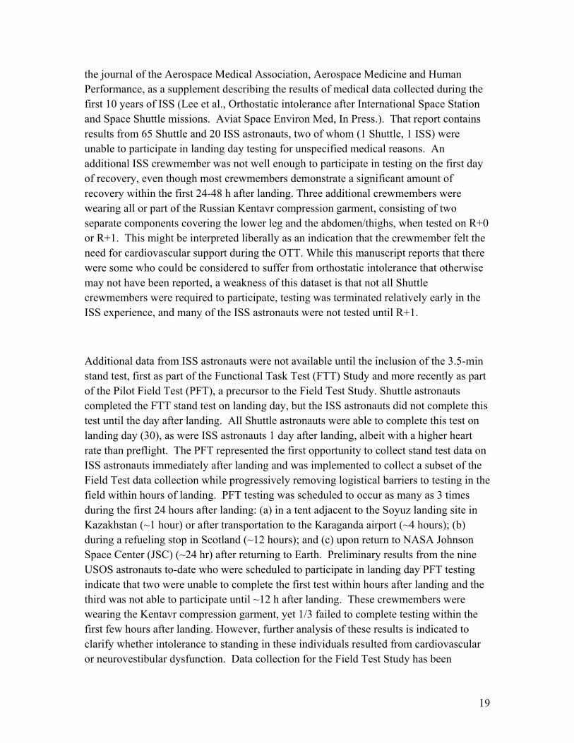
the journal of the Aerospace Medical Association, Aerospace Medicine and Human Performance, as a supplement describing the results of medical data collected during the first 10 years of ISS (Lee et al., Orthostatic intolerance after International Space Station and Space Shuttle missions. Aviat Space Environ Med, In Press.). That report contains results from 65 Shuttle and 20 ISS astronauts, two of whom (1 Shuttle, 1 ISS) were unable to participate in landing day testing for unspecified medical reasons. An additional ISS crewmember was not well enough to participate in testing on the first day of recovery, even though most crewmembers demonstrate a significant amount of recovery within the first 24-48 h after landing. Three additional crewmembers were wearing all or part of the Russian Kentavr compression garment, consisting of two separate components covering the lower leg and the abdomen/thighs, when tested on R+0 or R+1. This might be interpreted liberally as an indication that the crewmember felt the need for cardiovascular support during the OTT. While this manuscript reports that there were some who could be considered to suffer from orthostatic intolerance that otherwise may not have been reported, a weakness of this dataset is that not all Shuttle crewmembers were required to participate, testing was terminated relatively early in the ISS experience, and many of the ISS astronauts were not tested until R+1.
Additional data from ISS astronauts were not available until the inclusion of the 3.5-min stand test, first as part of the Functional Task Test (FTT) Study and more recently as part of the Pilot Field Test (PFT), a precursor to the Field Test Study. Shuttle astronauts completed the FTT stand test on landing day, but the ISS astronauts did not complete this test until the day after landing. All Shuttle astronauts were able to complete this test on landing day (30), as were ISS astronauts 1 day after landing, albeit with a higher heart rate than preflight. The PFT represented the first opportunity to collect stand test data on ISS astronauts immediately after landing and was implemented to collect a subset of the Field Test data collection while progressively removing logistical barriers to testing in the field within hours of landing. PFT testing was scheduled to occur as many as 3 times during the first 24 hours after landing: (a) in a tent adjacent to the Soyuz landing site in Kazakhstan (~1 hour) or after transportation to the Karaganda airport (~4 hours); (b) during a refueling stop in Scotland (~12 hours); and (c) upon return to NASA Johnson Space Center (JSC) (~24 hr) after returning to Earth. Preliminary results from the nine USOS astronauts to-date who were scheduled to participate in landing day PFT testing indicate that two were unable to complete the first test within hours after landing and the third was not able to participate until ~12 h after landing. These crewmembers were wearing the Kentavr compression garment, yet 1/3 failed to complete testing within the first few hours after landing. However, further analysis of these results is indicated to clarify whether intolerance to standing in these individuals resulted from cardiovascular or neurovestibular dysfunction. Data collection for the Field Test Study has been

20
initiated with the crew of the one-year mission and should yield more information, including the efficacy of the new graded compression garment (GCG).
B. Fluid shifts and Plasma Volume
Figure 9. Illustration of the changes in center of mass during spaceflight, from (31).
When astronauts enter microgravity, a cephalad fluid shift occurs which invokes a reflex-mediated hypovolemia. One of the first physiological changes noted during the Apollo program was the decrease in plasma volume, exhibited by the decrease in weight of the crewmen (32). One third of the average five percent weight loss was regained within 24 hours postflight, suggesting this fractional change was due to a loss of fluid. The remainder of the body weight loss was attributed to tissue loss, which is characterized by a longer recovery time(32). Serum and urine samples were analyzed for endocrine and electrolyte changes from pre- to post-spaceflight in order to better understand the etiology of the plasma volume losses of 4.4% upon return to Earth. The cephalad fluid shift and consequent fluid loss were thought to occur during the first two days of spaceflight, as seen in bed rest subjects. The smaller plasma volume loss in spaceflight was attributed to an elevated urinary aldosterone level upon landing. Although the time course of the plasma volume losses was unknown due to the lack of inflight measurements, the degree of plasma volume loss was independent of the duration of the Apollo mission (32). Inflight anthropometric measurements during Skylab allowed for the determination of time course and magnitude of fluid shifts. Photographs of the crewmen illustrated the commonly noted puffy faces and “chicken legs” exhibited during spaceflight as well as postural changes (31). Fluid shifts were further measured by anthropometric techniques and the determination of the center of mass.
The effect of the cephalad fluid shift characteristic of spaceflight on the center of gravity is illustrated in Figure 9. As in earlier missions, plasma volume losses were reported, but to a higher degree than in the Apollo program, with average losses of 8.4%, 13.1% and 15.9% for

21
Skylab 2, 3 and 4 (33). The time course of recovery from fluid losses can be seen in Figure 10. Blood volume analysis also showed a postflight decrease in red cell mass, which did not begin to reconstitute until at least 30 days postflight; this delay is suggestive of an inhibition of bone marrow (33).
Figure 10. Plasma volume losses following three Skylab missions (33).
In 1985, consuming fluid and salt prior to landing (fluid loading) became a medical requirement, thus any data on plasma volume acquired after this date do not capture the true landing day plasma volume deficit. In spite of the fluid loading, astronauts return from space with plasma volume deficits ranging from 5 to 19% (4–6,14,34). Additional confounding factors to accurate measurement of spaceflight-induced plasma volume loss include ad lib water ingestion following landing and IV fluid therapy that is given to the more severely affected crewmembers.
The mechanism of the plasma volume loss has been a matter of some debate (35). There have been limited inflight studies of plasma volume. One study shows a decrease in total body water during flight, suggesting but not proving a dieresis (36). A second study shows a decrease in plasma volume, but an increase in intracellular fluid, suggesting “3rd spacing” and not a diuresis (34); however, postflight studies from the Apollo (32) and EDOMP (37) programs do not show an increase in the intracellular fluid compartment. These disparate flight data reinforce the need for further flight studies (which are in progress as of this revision)..
Similar plasma volume losses (4 – 17%) have been replicated using 6º head-down tilt bed rest as an analog to spaceflight (32,39). Most of the loss occurs within the first week, and plasma volume remains stable for the duration of bed rest. Recent bed rest studies have shown a markedly increased urine excretion upon bed rest (38,39). Further study into this effect during spaceflight is needed and is considered a research gap.

22
C. Cardiac Atrophy
Cardiac muscle is responsive to changes in volume loading conditions. Decreased cardiac work during bed rest due to plasma volume loss, reduced LVEDV, and reduced physical activity results initially in smaller cardiac volumes and later remodeling to a smaller cardiac muscle mass. Assumption of the head-down position increases stroke volume with a concurrent decrease in heart rate, with the highest stroke volumes achieved ~30 minutes after the start of bed rest. Thereafter, resting stroke volume decreases with the lowest value obtained by ~48 h and remains depressed throughout the end of bed rest (Figure 11) (10). Though head-down bed rest initially
Figure 11. Stroke volume, heart rate, and total peripheral resistance measured during 14 d of 6°
head-down bed rest (10).

23
results in a cephalic shift of ~1 liter of blood from the legs to the upper torso, this is counteracted by humoral responses leading to diuresis and attainment of hemostasis at a level approximately half way between supine and upright about 24 to 48 h after the start of bed rest (10).
After 2 weeks of 6 head-down tilt bed rest, cardiac mass and LVEDV measured by 2-d echocardiography was decreased by 5% (p<0.10) and 7% (p<0.05), respectively (10). This was concomitant with a 24% decrease in LBNP tolerance measured using a ramp protocol. Longer duration bed rest results in further cardiac atrophy. LV mass is reduced in men by 4.7, 8.0, and 15.6% after 2, 6, and 12 wk, respectively, of horizontal bed rest (11). Sixty days of 6° head-down bed rest resulted in decreased LV mass of 9.5% in women, which lies along the same line describing the relation between changes in LV mass and bed rest duration in men (40). Importantly, reduced ventricular volume precedes cardiac muscle atrophy. For example, LVEDV was decreased by 2 weeks of bed rest, but LV mass was not reduced (as measured by MRI) until 6 weeks (Figure 12). Thus, the LV mass-volume ratio is initially reduced, followed by cardiac remodeling in response to decreased wall stress (11). Similar results have been obtained from measurements of the right ventricle, although the changes ventricular volume and mass perhaps are less dramatic (11,40).
Figure 12. Change in LVEDV and LVM as a function of bed rest duration. Bed rest results in a comparatively rapid and more profound decrease in LVEDV by a decrease in LVM. Data from (10,11,40–42)
The decrease in cardiac mass is concomitant with a reduced stroke volume at any given filling pressure. In the supine resting condition, after two weeks of bed rest pulmonary capillary wedge pressure (PCWP) and LVEDV are decreased by 18% and 7%, respectively (10). Starling Curves, constructed from PCWP and SV measures during LBNP and with rapid warm saline infusion, indicated a change in the cardiac distensibility, particularly at lower filling pressures (Figure 13). Pre- to post-bed rest, PCWP was not changed during -15 mmHg and -30 mmHg of LBNP, yet SV was reduced. Surprisingly, the relation between LVEDV and SV was unchanged, suggesting that this duration of bed rest had no effect on cardiac contractility (same SV per
Bed Rest Duration (d)
0 20 40 60 80 100
Cha
nge
in L
VE
DV
(%
)
-25
-20
-15
-10
-5
0
Bed Rest Duration (d)
0 20 40 60 80 100
Cha
nge
in L
eft V
entr
icul
ar M
ass
(%)
-25
-20
-15
-10
-5
0

24
LVEDV); lower SV after bed rest resulted from a decrease in diastolic volume. Thus, there is a leftward but parallel shift in the filling pressure-volume relation after bed rest. In total, bed rest-induced changes in cardiac distensibility leads to lower LVEDV and reduced SV at filling pressures representative of low levels of orthostatic stress. The decreased distensibility is reflected in a decrease in left ventricular untwisting during diastole (42). Assuming that reduced cardiac distensibility continues to parallel the progression of cardiac atrophy with longer bed rest durations, a progressive decrease in diastolic function would be one contributing factor to the decreased stroke volume during orthostatic stress and increasing frequency of orthostatic intolerance (11).
Figure 13. Starling Curves constructed from PCWP and SV measured at rest, during LBNP (-15 and -30 mmHg) and with rapid warm saline infusion (15 and 30 ml/kg) before and after 2 weeks of bed rest (10).
That these changes in diastolic cardiac performance result from cardiac remodeling, rather than just bed rest-induced hypovolemia, is supported by the work of Perhonen et al. (41). Subjects were studied before and after 18 day of bed rest as well as before and during furosemide-induced hypovolemia to compare changes in diastolic cardiac performance within the same subjects. Though bed rest and furosemide resulted in similar losses of PV (-15% vs. -14%), LVEDV during supine rest was decreased by almost three times as much after bed rest as by hypvolemia (-20 vs. -7%) while PCWP at rest decreased by only ~50% more (-21 vs. -31%) after bed rest than during hypovolemia. Further, both bed rest and hypovolemia resulted in changes in the linear portion of the Starling Curves (PCWP-SV, from baseline rest through LBNP at -30 mmHg), but the slope of the relation was increased approximately twice as much after bed rest as during hypovolemia. Additionally, while bed rest resulted in a leftward shift of the pressure-volume relation (PCWP-LVDV), there was no change in this relation during hypvolemia. While the reduction LBNP tolerance after bed rest only tended to be more than during hypovolemia (-27 vs -18%), the combination of bed rest-induced cardiac remodeling and plasma volume loss resulted in more dramatic changes in diastolic cardiac function than hypovolemia alone.

25
Thus, it is not surprising that attempts to restore blood and plasma volume status by fluid loading during space flight (43) have failed to fully prevent orthostatic intolerance. Even restoring filling pressures in the face of decreased cardiac distensibility will not prevent the reduction in stroke volume (10,41). Conversely, Shibata et al. (44) report that maintaining diastolic function alone through supine cycle exercise fails to prevent post-bed rest orthostatic intolerance. The results of their work suggests that a combination of fluid loading (through intravenous catheter) and exercise are necessary. Similar results were obtained by Hastings et al. (45) in subjects who combined rowing and resistive exercise throughout bed rest with two days of fluid loading accomplished through fludrocortisone and increased salt intake.
D. Autonomic function
Figure 14. Comparison of plasma volume losses between short duration spaceflight and long duration spaceflight (1).
It has been shown, however, that postflight orthostatic hypotension and presyncope are not dependent on the degree of postflight hypovolemia alone (5,6). Figure 14 shows that plasma volume losses are similar between long duration and short duration crewmembers. However, long duration crewmembers experience a higher rate of presyncope than short duration crewmembers . Also, in a recent study, Waters et al (6) reported on two groups of male short duration astronauts. One group had a 7.1% plasma volume loss on landing day and did not become presyncopal during tilt testing; whereas, the other group also had a 7.1% plasma volume loss, but did become presyncopal. The difference between groups was that the non-presyncopal group had hyper-adrenergic responses to tilt and the presyncopal group did not (Figure 15).
Postflight data measuring muscle sympathetic nerve activity in six non-presyncopal male astronauts (46) also shows that sympathetic responses in these crew members are appropriate These data are supportive of the norepinephrine spillover studies mentioned above. Unfortunately, there were no presyncopal subjects in this study and the postflight sympathetic dysfunction in that group of astronauts could not be duplicated

26
.
Figure 15. Plasma norepinephrine responses in women (n= 4; black bars), presyncopal men (n= 6; light gray bars), and nonpresyncopal men (n =22; dark gray bars) when tested preflight (left), on
landing day (right) (6).
Furthermore, astronauts who experienced both short and then long duration spaceflight were more likely to have a hypo-adrenergic response and become presyncopal during tilt testing after the long duration flight despite similar plasma volume losses in both flights (1). Thus, it is not the plasma volume loss alone that causes presyncope, but the lack of an adequate physiological response to the hypovolemia.
Heart rate variability (HRV) and blood pressure variability provide a means to noninvasively approximate autonomic activity through analysis of fluctuations in the R-R interval and SBP data. In particular, the high frequency component of the R-R interval series (HF-RR), in absolute or normalized units, is an index of parasympathetic modulation (47–49), and the low frequency component of the SBP (LF-SBP) approximates sympathetic activity (47,50–52). The interpretation of the low frequency to high frequency ratio of the R-R interval (LF/HF-RR) is more controversial. The LF/HF-RR is often used as a marker of sympathovagal balance (47,49,50,53). However, this interpretation has been challenged by studies that question the concept and construction of the index (54), suggest variation in the LF/HF-RR is partially due to changes in baroreflex function (55), or conclude that spectral HRV is mainly a measure of parasympathetic activity (56).
Most studies examining HRV in bed rest have found a decrease in HF-RR during bed rest or immediately afterwards (57–60). This bed rest-induced decrease in HF-RR has additionally been evident in bed rest as short as 20 hours with a furosemide infusion (61), during a controlled breathing protocol (22,23), during LBNP (62), and during vasoactive drug infusions (63). Despite some finding no difference in HF-RR with bed rest (64,65), it is well accepted that the HRV results indicate diminished vagal modulation after bed rest. The majority of studies that examined LF/HF-RR as either an index of sympathovagal balance or an indicator of sympathetic activity, found LF/HF-RR increased with bed rest (22,23,57,59,61,62,66). However, this increase in sympathovagal balance is not as explicit as the decrease in vagal modulation, since other studies

27
did not find LF/HF-RR to change with bed rest (60,65,67). It should be noted that (65) only studied a 24-hour bed rest, such that autonomic changes that become significant after the first 24 hours would not be detected. Approximation of sympathetic activity by blood pressure variability has yielded incongruent results, with increase (52,61,63), no change (22,59,64), and decrease (23,58) in LF-SBP occurring after bed rests of various durations.
Other measures of HRV do not have established links to autonomic activity, but can quantify overall variability or complexity. When studied throughout bed rest, these measures indicate that the total variability and complexity of heart rate decrease with bed rest. The total power in power spectral analysis of R-R interval quantifies the overall variability, and has been shown to decrease in bed rests ranging from 28 to 90 days (57,60,62,64). Heart rate complexity cannot be quantified by typical spectral analysis techniques such as methods based on the Fast Fourier Transform. Instead, coarse grain spectral analysis (CGSA) (68) can be used to extract the harmonic and fractal components of HRV and characterize the complexity of HRV by the exponent of the 1/f noise in the signal. This exponent corresponds to the slope of the linear fit of the fractal component plotted in a log-power vs. log-frequency scale. The spectral exponent has been found to increase with bed rest (57,69), indicating a steeper power drop-off at higher frequencies and a decrease in heart rate complexity. Heart rate complexity has also been shown to decrease with bed rest when measured by approximate entropy, which quantifies the predictability of fluctuations in heart rate (67).
In summary, the majority of the studies indicate that bed rest causes autonomic dysfunction by diminishing vagal modulation, increasing sympathovagal balance, and decreasing the overall variability and complexity of heart rate. The effect of bed rest on the approximation of sympathetic activity by blood pressure spectral analysis is not clear due to conflicting results.
Analysis of the heart rate and blood pressure series can further yield information on baroreflex sensitivity. Baroreflex sensitivity has been broadly shown to decrease with bed rest, as measured by examining the maximum slope of the baroreflex curve (15), beat-to-beat sequences of R-R interval and SBP (21,58–60,62–64,66), or the transfer function between the R-R interval and SBP (22,23,58,70). Iwasaki et al. attributed the decrease in baroreflex sensitivity after a 14-day bed rest to hypovolemia, due to a reversal of the baroreflex change after plasma volume restoration. However, Arzeno et al. (63) found that the decrease in baroreflex sensitivity after a 60-day bed rest was not due to decreased plasma volume.

28
83% 20%
80% 25%
All
Ast
r ona
u ts
No n
- Pilo
ts
Women Men
presyncopal non-presyncopal
Figure 16. Gender-specific orthostatic response to spaceflight. Women (6 astronauts, 5 non-pilots) are much more likely to become presyncopal than their male cohorts (30 astronauts, 12 non-pilots), even when pilots, a self-selecting, highly trained subset, are removed from the analysis.
The bed rest-induced autonomic and baroreflex dysfunction can contribute to post-bed rest orthostatic intolerance, and pre-bed rest indices of autonomic function may help identify those at higher risk of post-bed rest orthostatic intolerance. In particular, Convertino et al. (15) found presyncopal subjects suffered a larger decrease in baroreflex sensitivity during bed rest, and Pavy-Le Traon et al. (62) found presyncopal subjects to have a smaller increase in LF/HF-RR during LBNP.
E. Sex differences
The vast majority of astronauts have been male and, consequently, any conclusions drawn regarding the physiological responses to spaceflight are male-biased. NASA has recognized that there are some significant differences in how men and women respond to spaceflight (71), including sex differences in the effects of spaceflight on cardiovascular responses to orthostatic stress (6). Historically, greater than 80% of female crewmembers become presyncopal during a postflight tilt test (6) compared to about 20% for men (Figure 16).
As in spaceflight, a very low number of female subjects are studied during bed rest. This is traditionally done to reduce the variability in the data and to eliminate the scheduling issues related to the menstrual cycle. However, women have also been found to be more susceptible to orthostatic intolerance than men after head-down tilt bed rest (72). This gender difference, which is often seen even before bed rest, is illustrated in the survival analysis published by Grenon et al.
(Figure 1717).
The gender difference in propensity to orthostatic intolerance, observed during both spaceflight and bed rest, leads to an important consideration for
Figure 17. Presyncopal survival (15 females, 14 males) following head‐down tilt
bed rest. From Grenon et al [3].

29
countermeasure development, as a single countermeasure is not likely to be equally effective for both genders. This hypothesis has been confirmed by Grenon et al. (72) when they showed that midodrine was less effective in preventing orthostatic intolerance in women than men following simulated microgravity.
The incidence of orthostatic intolerance has been shown by several investigators to be higher in women than in men (6,73–77), and, though the etiology has not been exactly determined, many studies have identified potential contributory factors that differ between the sexes. Waters et al. (6) nicely summarizes some of the possible reasons sex differences may cause the disparity in orthostatic tolerance. Women have greater heart rate responses than men during mental stress (78), standing (74,79) , infusions of pressor agents (80), and cold pressor tests (81). It also has been shown that estrogen replacement therapy in postmenopausal women reduces muscle sympathetic nerve activity (82,83).
These differences in response to stress can be partially due to a dissemblance in autonomic regulation between the genders. Women are often characterized by attenuated sympathetic response and enhanced parasympathetic reactivity, particularly during orthostatic stress (72,76,84–88). These gender differences in autonomic dominance have been observed during tilt and stand tests (72,76,85,86,89), lower body negative pressure (LBNP) (88), autonomic blockade (84), and vasoactive drug infusions (63). Differing brain activity patterns during LBNP (90) and task performance (87) can provide some insight into the distinctions in autonomic response, but more studies are necessary to exactly determine the cause of these gender differences. The autonomic control differences between the genders are preserved throughout bed rest, with women also experiencing a decrease in baroreflex sensitivity within a shorter time-frame than men (63).
In addition, differences in vascular resistance and reactivity can contribute to the gender difference in orthostatic intolerance. Women have smaller increases in vascular resistance than men in response to LBNP (77,91), standing (92), cold pressor and facial cooling tests (93), and mental stress (94). Women may also have lower orthostatic tolerance because of increased splanchnic blood flow compared to men (75,95) due to attenuated splanchnic constriction in women in the upright posture (96). The attenuated vascular constriction in women has also been observed as attenuated brachial and femoral vasoconstrictor responses to increases in transmural pressure (97). There could be several factors that contribute to the women’s low vascular resistance, the most important of which is probably estrogen. Several studies in humans demonstrate an augmentation of endothelium-dependent vasodilation with estrogen (98–102). Low peripheral vascular resistance is considered one of the main drivers for post-spaceflight orthostatic intolerance (6). Arterial stiffness is another potential contributor to post-spaceflight orthostatic intolerance. An increase in arterial stiffness after spaceflight has been linked to orthostatic tolerance in men (103). However, despite men experiencing increases in arterial stiffness after spaceflight and bed rest, no significant increase in arterial stiffness has been observed in women (104).
30
The aforementioned differences between the genders focus on the autonomic response to orthostatic stress and changes in heart rate and vessel reactivity. However, decreased cardiac filling also contributes to the higher incidence of orthostatic intolerance in women. Fu et al. (73) showed that women had lower tolerance to LBNP, most likely due to a steeper Frank-Starling relationship. They found that women had larger decreases in stroke volume in response to decrements in cardiac filling pressure compared to men and suggested that this smaller and stiffer left ventricle is the primary reason for the propensity of women to have decreased orthostatic tolerance.
F. Ground
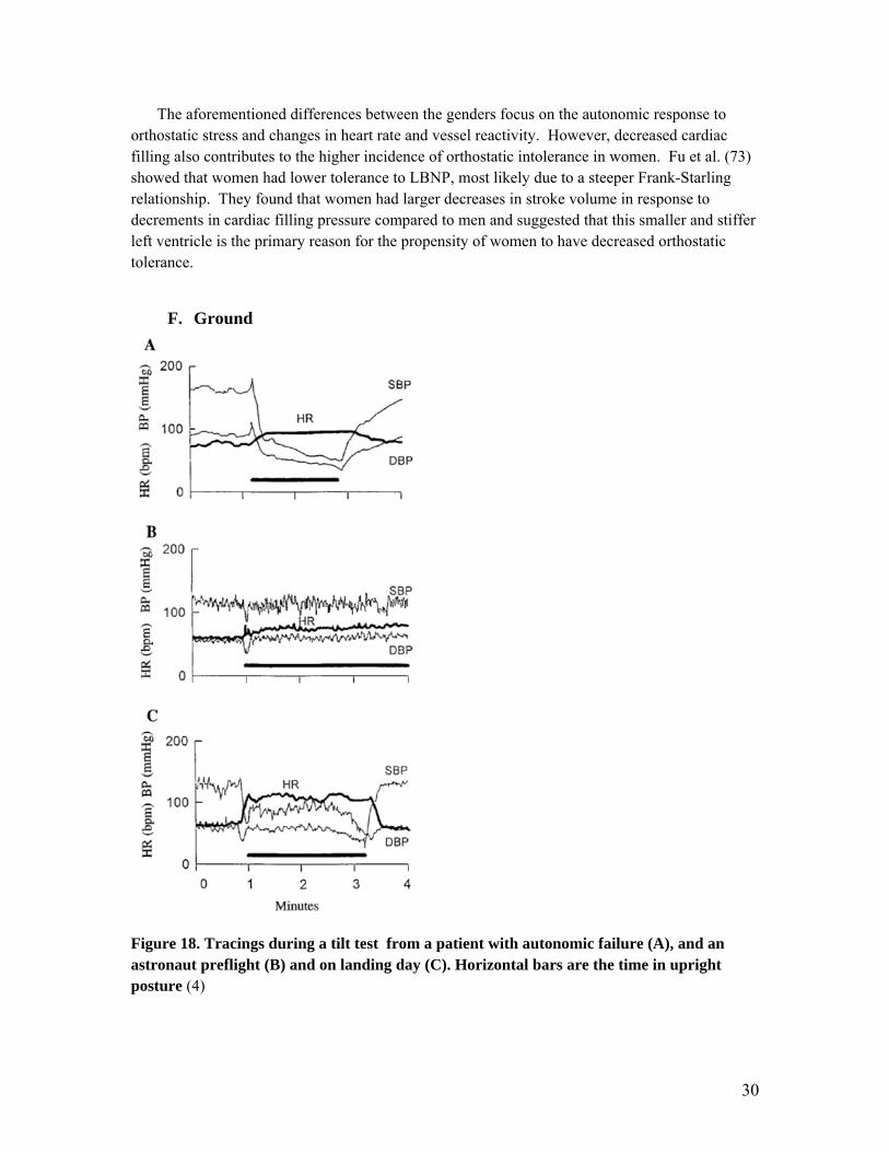
Figure 18. Tracings during a tilt test from a patient with autonomic failure (A), and an astronaut preflight (B) and on landing day (C). Horizontal bars are the time in upright posture (4)

31
In 2004, slightly over 164,000 patients were hospitalized in the United States with a diagnosis of orthostatic hypotension (105). Causes of these hospitalizations ranged from simple volume depletion to autonomic failure. Previous work has shown that the pattern of post-spaceflight orthostatic intolerance is similar to that seen in patients with autonomic failure (Figure 18) (4). In fact the countermeasure midodrine was proposed due to its use for this purpose. Studies that involve ill subjects tend to make extrapolation to the astronaut corps difficult, thus a model that includes otherwise healthy individuals is preferable. Figure 18 shows the similarities between clinical orthostatic hypotension in a patient with adrenergic failure and post-spaceflight orthostatic hypotension. Before spaceflight, the astronaut exhibits normal responses to standing: blood pressure is stable and heart rate increases slightly. This crewmember had no symptoms of orthostatic hypotension, had increased norepinephrine release by 236 pg.ml and completed the full stand test. Following spaceflight, however, the same crewmember exhibited classic signs of orthostatic intolerance during the stand test (Figure 18C). Systolic blood pressure decreased when the astronaut stood and heart rate increased markedly, without any increase in norepinephrine release. After ~ 2 minutes of standing, systolic pressure decreased below the termination threshold and the test was stopped. This pattern of orthostatic intolerance is amazingly similar to that of the adrenergic failure patient shown in Figure 18A. These similarities suggest that clinical research into adrenergic failure would be extremely useful in developing countermeasures to spaceflight-induced orthostatic intolerance.
Similarly, current pharmacological treatments for adrenergic failure may have application to spaceflight-induced orthostatic intolerance. Indeed, midodrine is FDA approved for orthostatic hypotension due to adrenergic failure and is discussed in depth under the countermeasure section. Such research done at NASA may also benefit the larger clinical community. Again using midodrine as an example, we uncovered a drug interaction between midodrine and promethazine that was previously unpublished (106). Healthy test subjects who received midodrine and promethazine together experienced a higher incidence of akathisia than controls or subjects with either drug alone. Anecdotal reports from emergency room physicians report similar symptoms in patients with diabetic neuropathy who present with nausea and are given promethazine while being treated for hypotension with midodrine. In the future, the knowledge of this interaction can help avoid unnecessary patient distress and hospital admissions in clinical practice.

32
1. Hypovolemia
Figure 19. Norepinephrine responses of presyncopal (n=8) and nonpresyncopal (n=9) test subjects during normovolemia and hypovolemia tilt tests
Laboratory models of hypotension may illuminate the phenomenon in astronauts. Several investigators have used pharmaceuticals to induce a plasma volume loss similar to that of spaceflight. Kimmerly and Shoemaker used three days of spironolactone administration to induce a 15.5 ±1.7% decrease in plasma volume (107,108). While this model was useful for their purposes, spironolactone is known to have vasomotor effects, which complicate interpretation of studies involving integrated cardiovascular responses. Fu et al. used a single dose of Lasix®, which decreased plasma volume by ~13% to study the effects of acute hypovolemia on orthostatic tolerance (109). Their results showed that orthostatic tolerance, as induced by LBNP, was markedly decreased in women, but not men, during hypovolemia; but they did not find any differences in norepinephrine responses between genders or between normovolemia and hypovolemia. Iwasaki et al. also used a single dose of Lasix® and found that the effects on cardiac filling pressures, stroke volume and high-frequency baroreflex sensitivity were similar between hypovolemia and two weeks of head-down tilt bed rest; however, they also found that vasomotor function differed between the two protocols. Finally, Meck et al. have used a single Lasix® (furosemide) infusion (0.5 mg/kg) followed by 36 hours of a very low sodium diet (10 mEq/day). This protocol induces a plasma volume loss similar to that after spaceflight (4–6,14). Those who became presyncopal after spaceflight and during hypovolemia exhibited the same etiology, a failure to release the extra amount of norepinephrine necessary to maintain standing arterial pressure when hypovolemic (Figure 19). The differences in these studies may be due to the hypovolemia protocol (acute vs. chronic) or in the orthostatic stimulus (tilt vs. LBNP). Regardless of these differences, pharmacologically-induced hypovolemia has been shown to reproduce the plasma volume losses seen following spaceflight. While obviously useful for some

33
mechanistic studies and countermeasure development, this model is limited in that the disuse (deconditioning) component of spaceflight (and bed rest) is not replicated.
2. Bed rest Bed rest studies, particularly those at 6° head-down tilt, are traditionally used as the best
ground-based analog to spaceflight. An excellent review by Pavy-Le Traon et al. describes these similarities (7), including changes in plasma volume and orthostatic tolerance that occur after only a few days of head-down tilt bed rest (Table 1).
Table 1. Comparison of spaceflight and head down tilt bed rest (7).
A summary of bed rest studies showing changes in physiological measurements that contribute to orthostatic intolerance can be found in Table 2. All bed rest studies listed here, except one, report plasma volume losses in excess of 8%. With the exception of the Shoemaker study (110), stroke volume was shown to decrease and total peripheral resistance to increase. Heart rate was less consistent, although the majority of studies report increases in heart rate at rest and following an orthostatic challenge. These findings are very similar to those seen following spaceflight
Most of the recent bed rest studies have focused on elucidating the mechanisms of orthostatic hypotension. These mechanisms include cardiac atrophy, sympathetic dysfunction, arterial and venous alteration, etc. Numerous publications have shown a cardiac atrophy following bed rest (10,11,23). Levine and co-workers found that 14 days of head-down tilt bed rest results in a smaller, stiffer left ventricle, leading to a decrease in stroke volume (10). This decrease in ventricular volume and stroke volume is similar to that found by Arbeille, et al. (111) and others and is thought to be due to the decrease in myocardial workload that is experienced in bed rest as well as spaceflight. These investigators conclude that the decrease in stroke volume is a primary contributor to orthostatic intolerance.

34
Table 2. Summary of bed rest studies showing orthostatic tolerance. From Waters et al. (112).
Many studies have shown that there is a disruption in the way the autonomic nervous system regulates the cardiovascular system following bed rest. Eckberg and Fritsch (18) and Convertino (15) showed decreases in baroreflex gain following short duration bed rest, which indicates a dysfunction in the carotid baroreflex. Muscle sympathetic nerve activity (MSNA) has been studied as an indicator of the signal sent from the nervous system to the blood vessels (sympathetic tone); however, there have been conflicting results from this research. Kamiya et al. (113) studied male subjects after 120 days of head-down tilt bed rest. During a graded tilt test (30 and 60 degrees), MSNA was measured in the tibial nerve. Resting MSNA and heart rate were higher following bed rest and baroreflex slopes for MSNA were steeper during tilt following bed rest, but there were no presyncopal subjects following this prolonged bed rest. The authors concluded that the augmented MSNA response increased vasomotor tone and prevented presyncope. In a follow-up study, these same authors studied 22 male volunteers before and after 14 days of bed rest (114). In this study, 10 subjects became presyncopal during post-bed rest tilt testing. In the hypotensive subjects, MSNA was lower throughout the tilt and was suppressed during the last minute of tilt. This pattern was not seen in the subjects who were able to complete the tilt test. These subjects responded similarly to their previous study. These data directly support the data that show a decreased norepinephrine response during postflight tilt testing. Pawelczyk et al. (115) also measured MSNA following bed rest. In this study LBNP was used as an orthostatic stress. They found that MSNA was increased during LBNP following bed rest; however, this response was appropriate given the changes in stroke volume and cardiac filling pressure and thus reflex control of MSNA was not altered. These data highlight the difficulty in comparing bed rest studies.
Finally, vascular function, whether arterial or venous, has been shown to be modified after bed rest. In the review paper by Pavy-Le Traon(7), the authors stress that the inability to sufficiently increase peripheral resistance is an important factor in the etiology of post-spaceflight and post-bed rest orthostatic intolerance. This points not only to the importance of the sympathetic nervous system, but also the vasculature. Lower limb arterial resistance has been

35
shown to decrease during head-down tilt bed rest as well as spaceflight, but carotid artery resistance did not change.
Nitric oxide (NO) has been hypothesized to contribute to orthostatic intolerance through its effects on the vascular smooth muscle Bonnin et al. (116) showed that flow-dependent dilation of the brachial artery was increased following seven days of bed rest and that this increase was negatively correlated to post-bed rest orthostatic tolerance. There was no change in the response to nitroglycerin, implying an endothelium-dependent (for example, NO) effect. Bleeker et al. (117) did a similar study in the femoral artery following horizontal bed rest. They found augmented arterial dilation in response to flow and nitroglycerin, implying an endothelium independent mechanism, likely an increased sensitivity to NO in the vascular smooth muscle. It is known that different vascular beds respond differently to the same stimuli, which may explain these differences.
Taken as a whole, these studies may seem disparate, but upon careful examination they all point to a decreased venous return as critical to the development of orthostatic intolerance. Similar mechanisms are likely at play during spaceflight and help inform future countermeasure development.
Limitations in the current literature are highlighted in Table 2, the most obvious of which is the lack of standardization in the bed rest protocols. The number of days in bed rest varies from 7 to 42 days, and a standardized protocol in use in the current NASA bed rest project includes 90 days of bed rest.
G. Animal models
Studies designed to examine the effects of the spaceflight environment on human physiology have benefited greatly from the use of non-human models. Indeed, considerable advances in knowledge have been gained from investigations employing animals. In addition to elucidating the physiological consequences of weightlessness, animal research has proven to be a cost effective platform for elucidating biological mechanisms responsible for such adaptations and toward countermeasure development. The hind-limb suspension rodent (HLS) model in particular has been widely used to study microgravity alterations in skeletal muscle (118–120) and bone/calcium regulation (121–124). The use of the HLS model may not be valid for all aspects of spaceflight research and, as with all models certain limitations exist when extending results to explain human physiological adaptations. Nonetheless, many of the biological and physiological changes in these animals appear similar to those in humans exposed to weightlessness. For example, this model reproduces the cephalad fluid shift that occurs during spaceflight which is central to the cardiovascular deconditioning hypothesis underlying post-spaceflight orthostatic intolerance (125–127). Given the physiologic similarities between the HLS and actual human spaceflight adaptations, there has been increased interest in its use to explain the mechanisms of cardiovascular deconditioning as they relate to orthostatic intolerance.

36
Cardiovascular Control Considerable evidence exists which supports the hypothesis that while hypovolemia is likely
the primary initiator of the cardiovascular dysfunction which occurs with spaceflight, it is not the only cause that underlies the cardiovascular impairment in orthostatic intolerance. The importance of interplay between components of the cardiovascular system should not be overlooked, however a significant strength of the animal research performed to date is the ability to examine individual components of the cardiovascular system control.
Vascular Regulation. Many of the theories underlying the orthostatic intolerance in astronauts point to a significant vascular component which includes differential remodeling of blood vessels in the upper compared to lower body and which may accompanied by alterations in endothelial dependent and independent control of vascular function. A succinct summary of many of the vascular adaptations that occur in animals exposed to spaceflight and HLS has previously been published and the reader is referred to this paper (128). The primary goal of this section is to review some of the more recently published data that have advanced not only our understanding of mechanisms of orthostatic intolerance but may reveal additional insight into areas that have overlapping vascular etiologies.
It is widely accepted that the inability to elevate peripheral vascular resistance following spaceflight explains, in part, the orthostatic hypotension experienced by many astronauts. Earlier research indicate that lower body peripheral resistance vessels have a larger cross-sectional area (increased capacitance) (103) and a diminished ability to constrict following exposure to simulated microgravity (129). Furthermore, in arteries from non-posture related muscles, the lower constrictor function is related to a blunted response to vasoconstrictor stimuli (130,131) and reduced myogenic tone (132). Current countermeasures, such as the Russian Kentaver and the NASA anit-gravity suit, have been developed to reduce the pooling of blood in lower limbs and thus help elevate peripheral resistance upon return to Earth’s gravity. While a great deal of research has been focused on understanding and overcoming the cardiovascular deconditioning effects of the vasculature in the lower body, recent evidence suggests that changes in cerebral vascular conductance may also contribute to the incidence of orthostatic intolerance in astronauts.
Cerebral Blood Flow. Often overlooked, but critically important to orthostasis is the maintenance of cerebral circulation. In general, cerebral blood flow is well protected against systemic changes in pressure and flow without compromising the high demands of brain tissue. Unfortunately, few spaceflight studies have been designed to examine the role of cerebral blood flow and cerebral autoregulation in humans. Much of the data which does exist argues against changes cerebral autoregulation and do not provide evidence for changes in vascular function. For example, Fritsch-Yelle et al. (4) measured blood flow velocity in the middle cerebral artery using ultrasound but did not find a difference in tilt response between presyncopal and nonpresyncopal astronauts. In a similar study, measures of cerebral blood flow, velocities, and beat-to-beat changes in arterial pressure were collected before and after 16 days in space. The authors of this study maintained that static autoregulation was not impaired and dynamic regulation was actually improved (133). Greaves et al. reported similar findings in subjects following 60 days of HDT bed rest (134).

37
While the human data appear undisputable, evidence from several ground-based studies using
the rat hind-limb tail suspension model suggests that the reduction in cerebral blood flow is associated with a decrease in autoregulation and that it is due to alterations in cerebral arterial structure and function. Indeed, data from Geary et al. (135) and Wilkerson et al. (136,137) indicate that myogenic tone and vascular resistance is increased, and that the difference in tone (suspended versus control rats) is related to changes in nitric-oxide-mediated vasodilation. Interestingly, the overall functional consequence of increased tone appears to lead to reduced blood flow and the stimulus does not appear to be an increase in arterial pressure but rather increases in transmural pressure caused by the elevation in the extravascular pressure in the cranium (136).
H. Computational Modeling and Simulation
1. Mechanisms of Orthostatic Intolerance Inferred from Models and Simulations
Computational modeling has been leveraged by NASA since the 1970s for simulating post-
flight orthostatic stress, as well as in spaceflight analog conditions including head-down tilt (HDT) bed rest, lower-body negative pressure (LBNP), head-up tilt (HUT) and water immersion experiments (138–147). Most of these models were lumped parameter systems models based on the Guyton circulation model (148) or the Croston cardiovascular control model for acute simulations (149). The models that were derived from the Guyton model were used for simulating time-averaged chronic adaptation of the cardiovascular system, including fluid balance and fluid regulation, to various perturbations such as HDT. The models derived from the Croston model, on the other hand, were used for simulating beat-by-beat short-term responses of the cardiovascular system. All of the computational models were mostly qualitatively validated against experimental data acquired from spaceflight, LBNP, HUT, head-HDT bed rest and water immersion studies. The validation studies typically focused on overall response of the cardiovascular system with respect heart rate (HR), cardiac output (CO), stroke volume (SV), mean arterial pressure (MAP) and blood volume (BV).
One particular simulation study performed by NASA (150) used a model developed by Croston and Fitzjerrel (1974) (151) which was designed for simulating dynamic response of the cardiovascular system to LBNP and HUT on a beat-by-beat basis. Given that the LBNP portion of the model was developed for a supine subject, it was most suited for microgravity simulations. Therefore, the LBNP simulation capabilities were modified for microgravity analysis. However, the LBNP simulation was very sensitive to changes in total BV, which was not directly measured during flight. Consequently, in-flight BV levels had to be calculated or estimated through various methods.

38
The model was also limited by the fact that it was designed to simulate short term response (minutes) and it represented the cardiovascular system as a closed loop system. To overcome these limitations, White et al. (1983) integrated the LBNP model with the Guyton model so that the Guyton model re-initialized the LBNP model by controlling the fluid intake, fluid output and volume of red blood cells at the end of each short-term LBNP run. The performance of the modified model was validated against a number of flight studies, summarized in Figure 20 and Error! Reference source not found.3.
As a demonstration of how models can be used for hypothesis testing, the authors performed two postulated analyses for long term effects of spaceflight on vasculature. The first postulated condition was based on clinical findings that suggested prolonged partial collapse of the leg veins due to cephalic shift of blood may result in re-toning of vasculature to accommodate the reduced BV in the legs and to maintain near normal venous pressure. This hypothesis was formulated within the model by shifting leg compliance curves as a function of leg BV changes with time. In conjunction with the first hypothesis, it was also postulated that the eventual reduction in total BV would be equal to the amount that migrates from the legs to the upper body. This hypothesis was represented in the model by setting the history of the leg BV reduction equal to the history of the total BV reduction. The simulation was then executed in accordance to a Skylab in-flight LBNP protocol used on one crewmember on mission day 52. A comparison of the simulation results with the Skylab data showed similar trends (Figure 21), with the strongest correlation existing for the hypothesis regarding vascular re-toning in the lower limbs as a consequence of BV reduction in the legs (upper plot in Figure 21).
Table 3: Validation comparison of modified LBNP model with available experimental data (reproduced from White et al. (150))
Changes from rest
LBNP Model
Data Source Apollo crew
preflight av. 12 subjects
Hovey (1972)
Musgrave paper (1971)
Wolthuis paper (1970)
Skylab crew preflight av. 6
subjects Johnson (1972)
Increase in heart rate (heats/min)
-30 7.3 4.4 8.0 11.89
-40 9.8 8.9 -50 15.1 14.6 10.5
Decrease in -30 12.0 18.62
Figure 20: LBNP simulation results compared with pre-flight crew average data for Apollo and Skylab (from White et al. (150)).
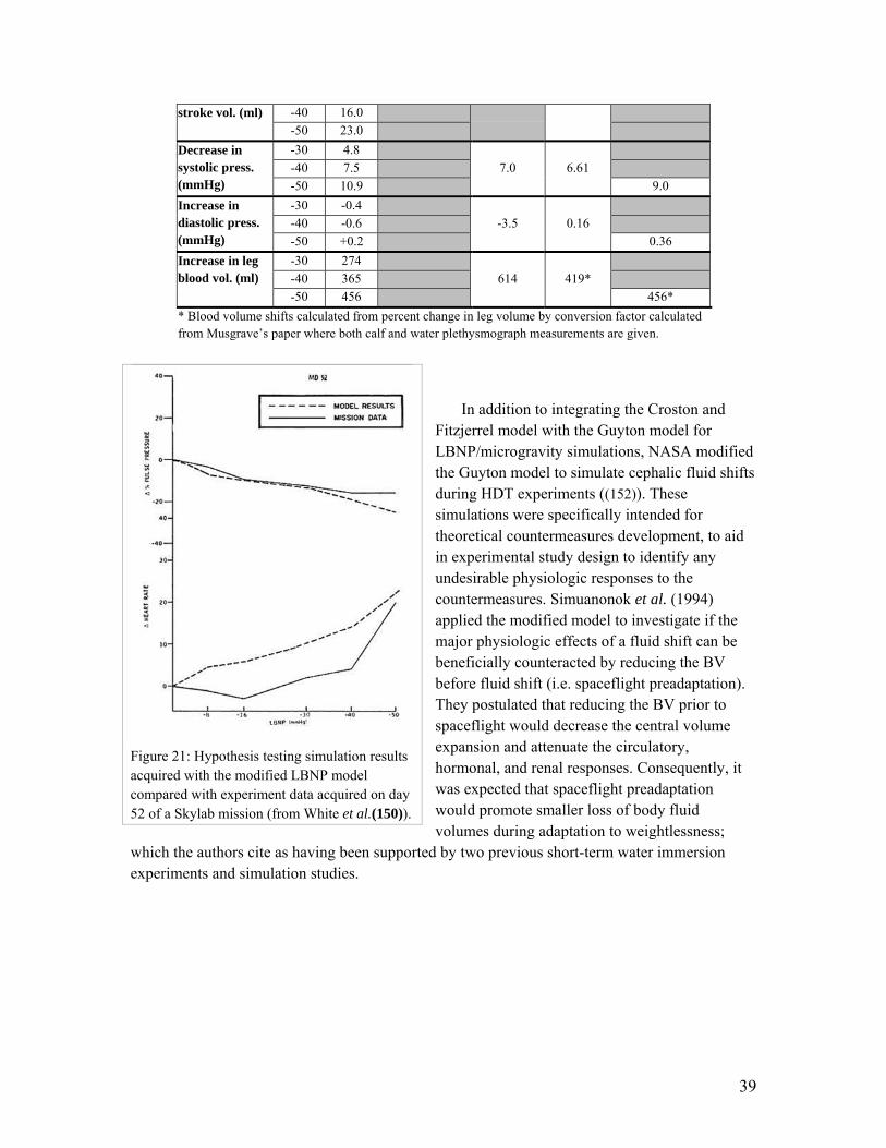
39
stroke vol. (ml) -40 16.0 -50 23.0
Decrease in systolic press. (mmHg)
-30 4.8 7.0 6.61
-40 7.5 -50 10.9 9.0
Increase in diastolic press. (mmHg)
-30 -0.4 -3.5 0.16
-40 -0.6 -50 +0.2 0.36
Increase in leg blood vol. (ml)
-30 274 614 419*
-40 365 -50 456 456*
* Blood volume shifts calculated from percent change in leg volume by conversion factor calculated from Musgrave’s paper where both calf and water plethysmograph measurements are given.
In addition to integrating the Croston and
Fitzjerrel model with the Guyton model for LBNP/microgravity simulations, NASA modified the Guyton model to simulate cephalic fluid shifts during HDT experiments ((152)). These simulations were specifically intended for theoretical countermeasures development, to aid in experimental study design to identify any undesirable physiologic responses to the countermeasures. Simuanonok et al. (1994) applied the modified model to investigate if the major physiologic effects of a fluid shift can be beneficially counteracted by reducing the BV before fluid shift (i.e. spaceflight preadaptation). They postulated that reducing the BV prior to spaceflight would decrease the central volume expansion and attenuate the circulatory, hormonal, and renal responses. Consequently, it was expected that spaceflight preadaptation would promote smaller loss of body fluid volumes during adaptation to weightlessness;
which the authors cite as having been supported by two previous short-term water immersion experiments and simulation studies.
Figure 21: Hypothesis testing simulation results acquired with the modified LBNP model compared with experiment data acquired on day 52 of a Skylab mission (from White et al.(150)).

40
In support of this study, the Guyton model was enhanced with an improved approach for erythropoiesis so that BV would reach a stable equilibrium at a reasonably reduced volume, and normal hematocrit after prolonged HDT. A reasonable BV reduction was assumed to be 10-15% from the initial BV. Furthermore, the model parameters were adjusted to match the decline in red cell mass with experimental data. After the appropriate modifications were made, the model was validated for change in hematocrit to acute BV reduction by simulating a hemorrhage of 15% of BV in 18 minutes. Qualitative comparison of the simulation results with corresponding experimental data from two human subjects showed a good match (Figure 22).
Following the above validation test of the model, a six-degree HDT bed rest simulation was executed without preadaptation until a new equilibrium of reduced BV and normal hematocrit was attained. The total BV change calculated by the model was 11%. This value was then used as the volume to reduce BV in order to test the circulation preadaptation hypothesis.
The circulatory preadaptation hypothesis was tested in a second simulation by first reducing the virtual subject’s BV by 11% at the rate of 41.67 ml/min (the same rate used for the validation study) in a supine position. Following blood removal, the virtual subject was set to a 30 minutes supine rest without tilt; which was also done for the non-preadapted HDT control simulation. Following a 30 minute supine rest, a 70-day six-degree HDT simulation was executed.
The results of the preadapted 70-day HDT simulation suggested that blood reduction prior to HDT may be an effective way to dampen the physiologic responses to fluid shifts and to reduce body fluid losses during HDT. More specifically, BV, production of extracellular volume and total body water after adaptation are higher than without preadaptation for the first 20 to 30 days of bed rest. However, after the first 20 to 30 days, all body fluid volumes in the preadapted condition decrease to similar levels of the non-preadapted conditions. Additionally, the preadapted HDT simulation showed increased plasma rennin activity and angiotensin levels that deviated from homeostasis more than was observed for HDT alone.
Simuanonok et al. (1994) further support the findings of their simulation by providing anecdotal reports of astronauts purposely dehydrating themselves before launch to shed some excess fluid volume before flight. They reported one case of a physician astronaut who slept in head-down tilt for 10 days or more prior to his five spaceflight missions to preadapt his body to fluid shifts. The crewmember reported that they never suffered head congestion, headaches, facial edema or space sickness. It was also reported that cosmonauts routinely sleep in HDT for about a
Figure 22: Validation of 15% hemorrhage simulation against experimental data for two subjects (from Simanonok et al Error! Bookmark not defined.(152)).

41
week prior to spaceflight for the same reasons. However, the effect of sleeping in HDT on the crewmembers’ BV was not studied or reported.
In this light, Simuanonok et al. (1994) suggest that pre-flight preadaptation of the circulation system to weightlessness may be an effective countermeasure for the undesired effects of fluid shift. However, they do stress that such methods must be rigorously tested in ground studies to ensure that other risks are not introduced in the process. One safe method may be preadaptation via HDT, mild diuretics, dietary modifications, and/or ordinary blood donation a week or so prior to launch. Using such methods to preadapt the circulation system to fluid shifts may help reduce some of the body fluid losses that compromise orthostatic tolerance when astronauts return to a gravity environment. However, the simulation results also suggest that using body fluid reduction as a way of preadapting the circulation system may only be effective for missions that do not exceed one month. Therefore, it may not be useful for mitigating OI after long duration missions such as ISS, or exploration missions to Moon or Mars.
In all the various modeling and simulation activities that NASA has pursued for understanding orthostatic stress resulting from spaceflight missions and spaceflight analog studies, there has been only one instance where finite element method was used to represent the cardiovascular system (153). Unlike lumped parameter models, finite element models allow for high spatial resolution analysis of the biomechanical response and flow dynamics of the cardiovascular system. In the study conducted by Sud et al. (1993), the finite element model was constructed to investigate how LBNP affects the distribution of blood flow in the cardiovascular system. The geometrical and physical data of the various arterial and venous segments were compiled from well establish literature sources (153).
When they simulated LBNP of 3999 Pa and 7998 Pa (30 and 60 mmHg respectively), the flow rates, conductance and pressure drops did not change in the vessels that supply blood to the upper portion of the systemic circulation. However, changes were observed in the vessels of the lower portion of the circulation system. When considering the flow rates in the various limbs and organs, the flow rates were unaltered in the in the head, upper extremities, kidneys, spleen and liver; which was expected to be the case. Following the LBNP analysis, the authors investigated the effect of applying varying negative pressures at different segments of the lower body. The results suggested the application of negative pressure at various segments of the lower body may produce similar changes when the orders of magnitude of negative pressures are near equivalent values.
Many of the results presented by Sud et al. lack validation and there are several limitations to their modeling methodology. The authors caution that the boundary conditions considered for the model do not take into account compensatory mechanisms which in practice are preset in the cardiovascular system and are highly complex with unknown quantitative measures. The analysis was conducted without cardiovascular controls such as HR, venous tone and cardiac contractility. Conductance of the various anterior cardiac vein networks were also not obtained from physical or geometrical data, but were estimated indirectly. Moreover, blood was modeled as a homogeneous Newtonian fluid; which is expected to contribute to the error since blood is a non-homogeneous fluid containing suspended particulate matter.

42
The finite element model presented by Sud et al. seems to be the first, and perhaps the last instance, that NASA has attempted a detailed investigation of the fluid flow and fluid distribution in the cardiovascular system during orthostatic stress. Given that finite element math models use first principles, they can, with acceptable accuracy, be used to calculate properties of physiologic parameters for which direct measured values may be lacking. Such parameters include compliance, resistance, capacitance, pressure and flow rates. These values can then be used to improve or validate time resolved models similar to those presented by Guyton and Croston for systems level analysis to investigate the mechanisms of OI and countermeasures development.
More recently the works of Summers and others, in collaboration with NASA, have substantially expanded on the work of Guyton and applied it to spaceflight research (154–158). Over the years, several versions of the model have been used for simulating changes in vital signs and hemodynamics similar to those observed in astronauts during spaceflight (156). It has also been used to simulate adaptive compensatory changes produced as fluid shifts from dependent areas, such as diuresis with loss of plasma volume and resetting of the baroreceptors while effective central volumes and cardiac output are maintained. However, most of the validation carried out for the different versions of the model and simulations have been qualitative in nature largely based on subject matter expert evaluations for clinical significance. Figure 23 shows an example of simulation results with one of the earlier versions of the model known as QCP (Quantitative Cardiovascular Physiology).

43

44
Figure 23: Hemodynamic changes simulated by in microgravity (top panel). The bottom panel depicts the longitudinal changes over several days in fluid distribution and vital signs in a human exposed to microgravity. The first panel shows both general and specific fluid compartments. In the bar graphs section, the single line represents the point where the individual was before the exposure while the solid bar shows their current status after being in microgravity for that amount of time. Also represented are the rates of fluid fluxes at that point in time.
One particular area the model was used to analyze is the relative contracture of the
extracellular fluid compartments and change in capacitance of the veins in the lower extremities secondary to this volume loss. The compliance (pressure-volume relationship) of these veins is determined by adrenergic tone, surrounding muscle tone, and external compressive forces of the interstitial fluids
Simulated exposure to microgravity shifts the pressure-volume curve secondary to the loss of fluid from the interstitium. During spaceflight this compliance change has little impact on hemodynamics due to the low pressure requirements necessary to drive venous return. Upon return into Earth’s gravity, the model predicted a sequestering of blood in these now lower-compliance vessels with a resulting OI occurring when the astronaut stands (
Figure 24).

45
Compensatory mechanisms counteract the fall in blood pressure in most individuals, and the effects are noted to be transient (Figure 25). While varying the cardiac function and baroreceptor sensitivity can potentiate this intolerance, the change in capacitance of the lower extremity veins resulting from a loss of external fluid forces in the dehydrated extracellular compartment was the initiating mechanism associated with post-flight orthostasis.
A

46
Figure 24: Shows the changes in venous compliance of the lower extremity veins during exposure to microgravity over several days (A) and then upon return to the earth environment (B). The curves show the relationship between fluid volume and pressure within the veins (compliance). The dark lines or curves in A are the current state after exposure to microgravity and the lighter line represents the initial condition before exposure. With graph B, the dark lines shows the curve change several minutes after reentry while the lighter curve shows the state of the compliance before reentry.
B

47
Figure 25: Hemodynamic transients and compensations upon reentry into gravity. The first panel demonstrates the vital signs of a returning astronaut during a tilt test after reentry. This individual (male anatomy) is one who is able to compensate for the orthostatic stress and recover his blood pressure after standing.
In a more recent analysis conducted by Summers et al.(158), the model suggested that post-flight orthostasis is accentuated in women due to their inherent lower center of gravity (COG) by about 15% and proportionately larger mass in the lower extremities in comparison to men. When this simple anatomic assumption is incorporated into the simulation without any other complex physiologic or hormonal changes, the orthostasis was more pronounced and overwhelmed all the counter-regulatory interactions as demonstrated by recurring falls in blood pressure upon repeated attempts to stand erect (Figure 26).

48
(a)
(b)
Figure 26: Hemodynamic transients and compensations upon reentry into gravity. (a) The orthostatic mean arterial blood pressure (MAP) response in man when assuming upright posture upon reentry to earth’s gravity after one month in the space environment. The drop in blood pressure is quickly corrected by compensatory physiologic regulatory mechanisms. (b) The “woman model” demonstrates a similar orthostatic blood pressure response when assuming upright posture after reentry but with a failure of the compensatory physiologic responses to correct the hypotension. (from Summers et al. (158) )
The findings reported by Melchior et al. (1994) (159) partially support the above results relating COG as a possible mechanism gender differentiation in the propensity of OI by showing through simulation and experimental results that decrease in lower body capacity diminished blood pooling to the lower body during LBNP. The validity of the hypothesized mechanisms reported by Summers et al. (2010) can be evaluate further by assessing the relationship of COG and OI propensity within the male astronaut population, as well as for the female astronaut population. Moreover, a more thorough sensitivity study of the roles the different hypothesized mechanism (e.g. hormonal changes, COG, baroreflex deconditioning) would help to further assess of COG has a substantial impact as it has been suggested by Summers and colleagues.
Building on the works completed by NASA, Guyton, Croston and others; Heldt et al. (2002) (160) also developed a 12-compartment lumped parameter model that has been successfully adapted for studying the hemodynamic response of the cardiovascular system to short-term and steady-state orthostatic stress. This includes dynamic change in intravascular volume and arterial and cardiovascular baroreflex responses such as those typical of microgravity exposure. Specific attention was also give to assessing highly credible compliance values in each compartment and the use of environmental pressures, independently assessed at each compartment, and has produced a capable simulation environment. The model has been well validated for normal hemodynamics, as well as for predicting transient response to HUT, and longer-term adaptation to LBNP. Some of the validation results are presented in the Figure 27 and Figure 28 below.

49
Figure 27: Validation of the Heldt et al. (2002) 12-compartment model for predicting transient response of (a) mean arterial pressure, (b) mean HR, and (c) mean stroke volume to head-up tilt. The solid lines represent simulation results and the discrete points with error bars represent experimental data measured across a range of tilt angles.(from Heldt et al. (160) )
Figure 28: Validation of the Heldt et al. (2002) 12-compartment model for predicting transient response of (a) mean arterial pressure, (b) mean HR, and (c) mean stroke volume to LBNP. The solid lines represent simulation results and the discrete points with error bars are experimental data measured at 20, 40 and 60 mmHg of LBNP. (from Heldt et al.(160) )

50
Heldt et al. also demonstrated the utility of their model for hypothesis testing by applying it to an illustrative case study to predict the factors that contribute to the overall responses of post-flight HR resulting from OI. In doing so, they simulated the HR recording of a single astronaut at 120 days prior to flight by tuning the combined HR gain, combining peripheral resistance gain and combining venous gain within physiologic limits. Using the simulated HR response curves as a baseline, the simulation was rerun using a different BV, combining parasympathetic and sympathetic HR gain, combining peripheral resistance gain, and combining venous tone. The end result was a simulation of the post-flight HR shown in Figure, which correlates well with the actual measured post-flight HR of the crewmember who had increased HR typical of a crewmember with OI. Moreover, although BV has the largest impact on the magnitude of the HR response, none of the individual parameter were sufficiently adjustable to within physiologic limits to reproduce the same results. This suggests that overall responses of post-flight HR resulting from OI are likely attributable to multiple factors such as BV, venous tone and peripheral resistance.
Mitsis et al. (2006) (161) studied the effects of orthostatic stress due under LBNP conditions on cerebral hemodynamics using a lumped-parameter model. Unlike other lumped parameter models, however, the modeling scheme used by Mitsis et al. employs non-linear memory behavior observed of organs and vessels in the circulatory system that causes them to behave like nonlinear capacitors. It also captures spontaneous fluctuations of the beat-to-beat MAP, mean cerebral blood flow velocity (MCBFV) in the middle cerebral artery, as well as breath-by-breath end-tidal CO2 concentration (PETCO2). Data collected during a previous study on 10 subjects (5 male and 5 female) for beat-to-beat MAP, MCBFV and breath-to-breath PETCO2 measured continuously under resting and LBNP tests to presyncope were used to validate the model. The average LBNP withstood by the subjects before syncope was -50 mmHg.
The model was used to study the dynamic effects of MAP and PETCO2 changes, as well as their nonlinear interactions on MCBFV variations at frequencies ranging 0.005 to 0.30 Hz. The simulations showed that the linear and nonlinear MAP-MCBFV interaction increased substantially above -30 mmHg LBNP in the very low frequency ranges (below 0.04 Hz), implying impaired dynamic autoregulation. In contrast, the PETCO2-MCBFV interaction was reduced during LBNP at all frequencies, thus suggesting attenuated cerebral vasomotor reactivity under dynamic conditions. The authors also speculate that the observed changes may be due to progressively reduced cerebral vascular reserve to compensate for the increasingly unstable systemic circulation during orthostatic stress that may ultimately lead to cerebral hypoperfusion and syncope.
Figure 29: Simulation result of the post-flight HR (dashed line) showed a good correlation with measured post-flight HR (solid line) of the crewmember (from Heldt et al.(160)).

51
2. Utility of Computational Modeling for Future Mission Planning As demonstrated by the information presented above, models and simulations can be useful
for hypothesis testing, gaining insight into countermeasures efficacy to mitigate OI and improving experimental design. In a similar way, computational models can beneficially augment research activities for hypothesis testing to gain insight on how environmental and physiologic factors such as partial gravity, radiation, hypoxic environments, biochemical, hormonal and regulatory mechanism may play a role in the risk of OI and crew performance impairment during extended missions on the Moon and Mars (162).
3. Limitations and Gaps Most of the modeling work that has been done involves the hydrodynamic and biomechanical
responses of the cardiovascular system. There has been very little work done in simulating biochemical, hormonal and regulatory mechanisms that play a role the response of the cardiovascular system to orthostatic stress. The modeling works that have included such capabilities (e.g. Guyton model) have not been well validated. To incorporate such capabilities in future models, it may be appropriate to leverage works from the biotechnology industry where substantial strides have been made in modeling biochemical, hormonal and regulatory processes for drug discovery and clinical trials.
Most of the cardiovascular models developed for spaceflight simulations of orthostatic stress are for acute conditions such as tilt table and LBNP. To date, only models derived from the Guyton model have been able to simulate chronic cases. However, the Guyton model is also limited by the fact that it based on a lumped approach that gives no spatial resolution with regards to anatomical changes to key organs such as the heart. In addition, it does not simulate cardiovascular physiology on a beat-by-beat basis or breath-by-breath basis. Instead it computes all physiologic parameters based on time averaged scale. Additionally, simulating chronic cardiovascular or circulatory performance using lumped models are limited by the fact that minimal empirical data from human studies are available to accurately capture compliance/performance changes of the heart and the arteries over a course of a spaceflight mission. Without the correct compliances (i.e. pressure -volume behavior) for the system, the outputs of the lumped network models are prone to substantial uncertainty. These limitations can be overcome by using spatially resolved finite element and finite volume fluid-structural-tissue models of the heart and arteries that can predict these structural changes from first principal physics and physiology.
Rigorous verification, validation, uncertainty quantification (VV&UQ), and code documentation are greatly lacking in the current state of practice in spaceflight physiology research. In order to use computational models with greater confidence and credibility for countermeasures development and hypothesis testing, they should be vetted using formal quantitative VV&UQ methods, as well as clear documentation practices. This is especially important when the models are applied in areas where clinical data are lacking.

52
I. Partial-Earth Gravity
It is not known if lunar gravity will be sufficient to protect crewmembers from the detrimental effects seen during exposure to microgravity. This is a significant research gap. No studies of orthostatic tolerance have been made in a partial gravity environment, thus the only available data are from bed rest studies. While 6º head-down tilt has been used as an analog for the deconditioning associated with microgravity, 10º head-up tilt has been proposed as an analog of lunar gravity. By using a 10º head-up tilt, the resultant force along the spinal axis of the body is 1/6 that normally seen on Earth (Figure 30).
Figure 30. Representation of the bed that is proposed for lunar gravity simulation testing. This bed allows for weight bearing on the feet and some exercise while minimizing friction.
Few studies have been published in which head-up tilt bed rest was used as a lunar gravity analog (163–167). The duration of these studies ranged from hours (163–165) to up to six days at 10º (167) or 11º (166) head-up tilt. Two of these studies simulated the trip to the Moon by using four days of 6º head-down tilt bed rest before and after the lunar-analog portion of the studies; however, plasma volume was not measured while subjects were in the head-up tilt portion of the study. Pavy-Le Traon reports an average plasma volume loss of 11%, but the measurement was taken after four days of head-down tilt (167). This does not accurately represent the plasma volume during a lunar stay, since the initial plasma volume loss during the analog trip to the Moon and any changes during the lunar-analog portion remain unknown. In addition, the lack of diet monitoring could have affected the results of the plasma volume analysis.
Louisy found that venous capacity and emptying time increased significantly during a simulated microgravity transit to the Moon and returned to control values on the first day of simulated lunar gravity (166), indicating that lunar gravity may reverse some of the detrimental effects of short-term microgravity. However, not only did venous capacity significantly increase on the first day of the simulated return transit, the increase in venous emptying time was larger

53
than during the simulated transit to the Moon (166). These data suggest the two microgravity periods might be characterized by different magnitudes and time courses of change in cardiovascular parameters. Additionally, the adaptation of venous distensibility to 1/6 G could not be determined due to the short duration of the study.
Additional studies examining the effects of 10° head-up tilt (163–165) focused on the differences between various tilt angles in a supine-tilt-stand protocol where subjects were tilted for only six hours. The initial change in cardiovascular parameters characterized by the six hours of tilt did not establish any trends, indicating a transient period without predictive value for a longer-duration study. These protocols consisted of a supine rest period followed by tilt, such that the conclusions cannot be applied to tilt changes from 5° head-down to 10° head-up and vice versa. While results from previous studies suggest that exposure to lunar gravity may be protective of the microgravity transit period, it should be noted that previous studies of lunar analogs are not high fidelity models of what a lunar habitation mission will entail. Firstly, the longest lunar-analog portion of these studies lasted only six days, while the eventual lunar outpost missions will be on the order of months in duration. Secondly, subjects in previous studies did not experience ground reaction forces via a footplate at the end of a bed, but rather relied on friction to maintain position while in a head-up tilt position. The muscle contractions experienced from ground reaction forces contribute to venous return and fluid homeostasis; therefore, their absence would increase plasma volume losses. Thirdly, the subjects only exercised in one study (167), and for only 40 minutes a day, whereas astronauts will spend a majority of their day performing tasks that will exercise multiple physiological systems. Moderate exercise may, or may not, be protective of any deconditioning effects of simply lying in a 10º head-up position. Furthermore, while one echocardiographic study has been published on the effects of six hours of 10º head-up tilt (165), there are no reports in the literature regarding cardiac function during 10º head-up tilt lasting more than six hours. Finally, the existing knowledge base is composed solely of data from male subjects. There is ample evidence that women are more severely affected by the deconditioning effects of microgravity (4,6), and therefore, must be included in any partial gravity deconditioning study.
Mars exploration mission scenarios present a number of challenges for the cardiovascular system. Several transit/stay scenarios have been proposed. Crewmembers will face, at a minimum, a 180 day transit to Mars, a significant stay on the Martian surface of 545 days and a return transit of 180 days. A second possible scenario involves much longer transit times and a much shorter Martian stay (131 day transit/40day Martian stay/308 day return transit).
It is extremely difficult to assess the risks of either scenario given the limited data that are available. That being said, Mir and International Space Station (ISS) data suggest that the majority of crewmembers (80%) exhibit orthostatic hypotension upon landing after 180 days of flight; however, these crewmembers recover quickly. It is not known to what extent 3/8 G will induce orthostatic intolerance. It is important to note that the impact of this level of orthostatic intolerance is likely to be more serious than on Earth due to the lack of medical and support staff/infrastructure on Mars. There are no data that suggest whether a stay on the Martian surface will protect or, more accurately, contribute to recovery of crewmembers. Some believe that 3/8 G will be closer to 1 G than microgravity, but there simply is no evidence for this. The shape of the
54
cardiovascular deconditioning curve between microgravity and 1 G is completely unknown and any discussion is, at this point, purely academic.
The return to Earth gravity will likely be the greatest challenge, from the cardiovascular standpoint, ever faced during the manned space program. Crewmembers are likely to exhibit an extremely high rate of orthostatic intolerance with adrenergic dysfunction and significant cardiac atrophy.
An isolation experiment called Mars 500 (520 day duration) was completed in November, 2011. The mission was designed to simulate the transit to and from Mars, as well as some habitation on the Martian surface. Some cardiovascular measurements were conducted including ECG, BP, and blood markers. The results from this study have not been published yet, but may help determine the baseline cardiovascular effects we may see during a Mars mission that are due solely to the isolation of deep space travel.
This gap is most in need of ground-based research to evaluate various analogs for 3/8 G and simulate various mission scenarios before any predictions can be made. The Integrated Cardiovascular Study planned for ISS will be critical for identifying the risks for cardiac structure and function. Computer modeling will play a pivotal role in guiding future research. Perhaps the most useful information will be gathered on long duration lunar missions.
V. Countermeasures
A number of countermeasures to post-spaceflight orthostatic intolerance have been tested with varying degrees of success.
A. Fluid Load
All astronauts returning from space are required to ingest a “fluid load” of broth or salt tablets and water. The efficacy of this countermeasure following Shuttle flights was evaluated by Bungo et al. (43). These investigators measured heart rate and blood pressure during a passive stand test before and after spaceflight (54 to 194 hours). Both variables, as well as a cardiovascular index of deconditioning, were significantly improved, but not totally restored, when astronauts used the fluid loading countermeasure compared to control flights where fluid loading was not used (Figure 31). Unfortunately, plasma volume was not measured during this study, so it is not known what the direct effect of the countermeasure is on plasma volume. Several studies have shown that there is still a significant plasma volume loss, even with the fluid load, which is now a medical requirement.

55
Figure 31. Effect of reentry Fluid loading on heart rate (43).
The fluid loading protocol currently used by cosmonauts and astronauts returning from ISS differs significantly from the protocol previously used by Shuttle astronauts. Both cosmonauts and astronauts increase salt intake with meals over the last several days of the mission. Between 18 and 20 ml/kg body weight of sodium chloride-water solution or dry salt (0.18-0.20 g/kg body weight) with water (18 -20 ml/kg body weight) are consumed 3-4 times in the last 12-20 hours before landing (168). Russian cosmonauts also attempt to increase fluid levels by consuming fluids during Chibis sessions at the end of the mission The water-salt additive normally taken with food before Chibis LBNP sessions consists of 0.9 g of sodium chloride and 300 ml of water (169). Kozlovskaya et al.(169) report that cosmonauts who participate in the this form of end-of-mission fluid loading better tolerate the workloads associated with the final phase of the space flight mission and the post-flight reconditioning program. We are unaware of any studies which specifically demonstrate the efficacy of the fluid loading countermeasure. Recent post-flight plasma volume data collected as part of NASA’s Functional Task Test Study are confounded by the timing of the measurement (~1 day after landing) and by the common practice of intravenous administration of fluids soon after landing.
B. Artificial Gravity
Artificial gravity (AG) via short radius centrifugation (SRC) has been suggested as a multi-system countermeasure to spaceflight deconditioning. While the idea of artificial gravity to prevent orthostatic intolerance is not new(170), the ideal combination of AG magnitude,

56
frequency and duration needed to prevent cardiovascular deconditioning is yet to be determined. In a head-down bed rest study, two hours a day of passive standing was sufficient to prevent post-bed rest hypotension, although four hours a day of passive standing was required to prevent plasma volume losses (171). Hastreiter and Young determined that 1.5 Gz (at the feet) on a short radius centrifuge was required to evoke calf blood flows that were similar to those when the subject was standing (172). In a non-human primate model, Korolkov et al. showed that SRC AG was successful in preventing extracellular fluid loss and orthostatic hypotension resulting from bed rest. Furthermore, they determined that 1.2 Gz, three times a week was more effective than higher Gz levels, administered four to five times a week (173). Iwasaki et al. determined that daily 1 hour exposures to 2 Gz (at the heart) was sufficient to prevent the adverse effects of 6° head-down tilt bed rest on baroreflex function and plasma volume (174,175). Finally, combining exercise with centrifugation at 0.8, 1.2 and 1.6 Gz (at the feet) was previously shown to be effective in maintaining orthostatic tolerance after return from 3 to 28 days of simulated microgravity (176).
The utility of artificial gravity as a whole body countermeasure has gained momentum in the last several years. Three weeks of daily short radius centrifugation (1 Gz at the heart, 2.5 Gz at the feet) via the NASA Ames Research Center’s Human Performance Centrifuge was recently shown to improve orthostatic tolerance in both ambulatory men and women (177,178). These Ames studies utilized Gz stress in an oscillatory fashion – 2 minutes with 1 Gz at the feet followed by 2 minutes with 2.5 Gz at the feet, repeated for up to 45 minutes a day. At NASA Johnson Space Center, a similar study was conducted using a constant 2.5 Gz at the feet for up to an hour on 6° head-down tilt bed rested subjects. This two week study evaluated the effectiveness of artificial gravity in 8 treatment subjects compared to 7 control subjects, and showed the utility of artificial gravity in preventing bed rest-induced orthostatic intolerance and decrements in aerobic capacity (179). However, this study only enrolled men as women were
unable to tolerate an hour of centrifugation at the intended study magnitudes (180). Additional
centrifuge studies have recently been completed in China (181) and Europe (182) examining cardiovascular parameters, yet these experiments did not address orthostatic intolerance.
Before artificial gravity can be implemented as a viable countermeasure to orthostatic intolerance, the dose-response curve needs to be fully understood. The most efficient duration, magnitude and type of artificial gravity has yet to be elucidated. The NASA Human Research Program convened an international working group in February 2014 to begin discussing the potential role of artificial gravity in future space exploration.

57
C. Lower Body Negative Pressure
Figure 32. Heart rate response in bed rest subjects (n=10) before and after a 4 hour “soak” of LBNP. From (183)
Lower body negative pressure (LBNP) is another means of producing a head-to-foot Gz force to provide an orthostatic challenge (184–186). By encasing the lower body in a rigid container, venous return can be modulated by varying the level of vacuum. This can be used to simulate standing on Earth or other gravity environments, depending on the magnitude of the LBNP. Application of LBNP as a countermeasure during spaceflight and bed rest (Figure 32) has been used with varying degrees of success in preventing orthostatic intolerance (184–189). LBNP applied after exercise during LBNP has been shown to be effective in attenuating post-bed rest orthostatic intolerance (190). The duration and frequency of LBNP required to make it an efficient countermeasure, however, is not operationally feasible.
Lower Body Negative Pressure and Exercise
In a series of bed rest investigations of increasing durations, a team of investigators led by Drs. Alan Hargens and Suzanne Schneider sought to determine whether the combination of lower body negative pressure and exercise (LBNPex) would be an effective countermeasure to bed rest-induced deconditioning, including orthostatic intolerance, and by extension an appropriate countermeasure system for space flight (191). The investigator team hypothesized that this countermeasure could allow astronauts to perform treadmill exercise with axial and ground reaction forces (for protection of bone and muscle) similar to or greater than that experienced during walking and running in normal gravity while simulating the hydrostatic pressure gradients normally experienced by the cardiovascular system. Although most exercise performed during bed rest in the supine position fails to prevent orthostatic intolerance (192), LBNPex might preserve plasma volume (193) and stimulate the cardiovascular reflexes used to promote venous return and maintain cerebral circulation.

58
In the initial study, 24 male subjects completed 5 d of 6° head-down tilt bed rest, with subjects divided into three groups. Control subjects performed no exercise during bed rest while the remaining subjects performed daily intense bouts of either exercise upright on a standard treadmill or supine treadmill exercise within the LBNPex device (194). The exercise protocol used by both countermeasure groups consisted of 30 min of interval-style exercise, with prescribed intensities up to 90% of pre-bed rest peak oxygen consumption. The exercise protocol previously had been shown to maintain peak oxygen consumption (195) and plasma volume (193) during 30 d of bed rest when subjects exercised twice per day on a supine ergometer without LBNP. At the completion of the exercise protocol during this 5-d bed rest, subjects remained upright or with their legs positioned against the treadmill belt while LBNP was applied (190). Before and immediately after 5 d of bed rest all subjects participated in head-up tilt test for 30 min or until presyncope. After bed rest, tilt tolerance time decreased by ~18% in the control group while tolerance time tended to increase in the LBNPex group (~9%) (190,196).
Orthostatic intolerance, as assessed by cumulative stress index calculated from total time and negative pressures achieved during a LBNP ramp protocol, was not maintained in a subsequent 15-d bed rest study (189). Seven men performed two 15-d bed rest exposures in a cross-over design, in which they participated in a random order once as a control and once as an exercise countermeasures subject. Unfortunately, the countermeasure protocol was ineffective in this group of subjects, even though it had been performed each day of bed rest as it had been in the previous study. Although the sub-tolerance cardiovascular responses to LBNP were maintained in the countermeasure group and not in the control group, orthostatic tolerance was decreased in both groups (countermeasure: -41%, control: -36%). Two potentially important differences in the countermeasure protocol existed which may have influenced these results. First, the intensity of the exercise was reduced, but the duration lengthened, to accommodate the limitations on prescribed exercise intensities that was applied by NASA at the time for astronauts. The peak prescribed exercise intensity during this bed rest was 80% of pre-bed rest VO2pk, and the protocol was extended from 30 to 40 min. Second, the subjects in this bed rest study were not exposed to the 5-min of post-bed rest LBNP as in the previous 5-d bed rest study. This additional orthostatic challenge, when the subjects were vasodilated from the previous exercise and thermal stress, may have been a key component to the success of the countermeasure in the prior study (190).
Subsequent 30-d bed rest studies in male and female twins (16 men, 14 women) confirmed the potential efficacy of this countermeasure against space flight-induced orthostatic intolerance (190). In this unique study design, one twin was randomly assigned to the control group and one was assigned to the countermeasure group. Control subjects performed no countermeasures throughout the duration of bed rest, while the countermeasure subjects performed the same exercise prescription as in the previous 15-d bed rest (189) but with the 5 min of post-exercise LBNP used in the 5-d bed rest (196). Tolerance to a combined tilt and LBNP ramp test was not fully protected in the countermeasure group, but the 13% decrease was almost 1/3 of the decrease in orthostatic tolerance seen in control subjects (-34%). Similar to previous observations in this series of studies, the cardiovascular response to the sub-tolerance tilt and LBNP was maintained

59
from pre- to post-bed rest in the countermeasure group but was exaggerated in the control group. These positive results following a longer duration bed rest study suggest that the repeated bouts of the post-exercise orthostatic challenge is an important aspect of the countermeasure effectiveness (197),
Most recently, the combination of the LBNP and exercise countermeasure was tested in women during 60 days of bed rest (198). This bed rest study was different from previous ones in this series of investigations in several important ways. First, the number of LBNP and exercise sessions was reduced from 6 d/wk to only 3 d/wk. A resistive exercise protocol previously shown to maintain muscle strength and mass also was performed 3 d/wk, alternating days with the aerobic exercise sessions so that the two countermeasures were never performed on the same day. Second, although the exercise portion of the LBNP and exercise protocol was maintained from previous studies (199,200), the duration of the post-exercise LBNP was extended to 10 minutes. It was assumed that a greater orthostatic challenge might be necessary to protect against bed rest-induced orthostatic challenge after this longer duration bed rest (198), particularly in women who are be more susceptible to space flight-induced orthostatic intolerance (6). Despite this modification to the post-exercise LBNP protocol, the countermeasures performed during bed rest only partially protected against orthostatic intolerance. Tolerance, measured by time to presyncope during a combined tilt and LBNP protocol, was reduced by 35% in the countermeasure subjects but by 50% in this control subjects. Similar to previous studies from this series, sub-tolerance cardiovascular responses to the orthostatic challenge were less in the countermeasure group than in the control group (198). It was likely that the partial protection against bed-rest induced orthostatic intolerance resulted primarily from the LBNP and exercise protocol since the resistive exercise protocol alone is not effective after 90 d of bed rest (201), but the contributions of the respective countermeasures was not specifically elucidated in this study.
In conclusion, the results from this series of studies suggest that LBNP during and after exercise is a potential countermeasure for protection against post-bed rest, and by extension, post-space flight orthostatic tolerance. Mechanisms for the protection against post-bed rest orthostatic tolerance after bed rest might include from protection of plasma volume (194,199,202) and left ventricular mass and function (10,40). The exclusion of the post-exercise LBNP in the one study in which post-bed rest orthostatic tolerance and sub-tolerance cardiovascular responses were not maintained suggests the importance of the post-exercise orthostatic challenge. Further refinement of the exercise and post-exercise protocols may elucidate the respective roles of the different portions of the countermeasure and result in a more efficacious countermeasure for longer duration bed rest and space flight missions. It should be noted that in these studies it was occasionally necessary to interrupt post-exercise LBNP exposure due to the occurrence of presyncopal symptoms. This occurred more frequently at the start of bed rest and more frequently in women (190,198). The investigator team hypothesized that, although they never allowed the situation to progress to the level of presyncope and the subjects always were closely monitored, should a subject become unconscious when unmonitored, they would be pulled into the LBNP chamber to the point that the waist seal would disconnect from the chamber and the negative pressure would be released.
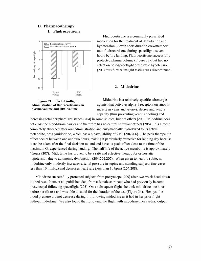
60
D. Pharmacotherapy 1. Fludrocortisone
Fludrocortisone is a commonly prescribed medication for the treatment of dehydration and hypotension. Seven short duration crewmembers took fludrocortisone during spaceflight, seven hours before landing. Fludrocortisone successfully protected plasma volume (Figure 33), but had no effect on post-spaceflight orthostatic hypotension (203) thus further inflight testing was discontinued.
2. Midodrine
Midodrine is a relatively specific adrenergic agonist that activates alpha-1 receptors on smooth muscle in veins and arteries, decreasing venous capacity (thus preventing venous pooling) and
increasing total peripheral resistance (204) in some studies, but not others (205). Midodrine does
not cross the blood-brain barrier and therefore has no central stimulant effects (206). It is almost completely absorbed after oral administration and enzymatically hydrolyzed to its active metabolite, desglymidodrine, which has a bioavailability of 93% (204,206). The peak therapeutic effect occurs between one and two hours, making it particularly attractive for landing day because it can be taken after the final decision to land and have its peak effect close to the time of the maximum Gz experienced during landing. The half-life of the active metabolite is approximately 4 hours (207). Midodrine has proven to be a safe and effective therapy for orthostatic
hypotension due to autonomic dysfunction (204,206,207). When given to healthy subjects, midodrine only modestly increases arterial pressure in supine and standing subjects (increases less than 10 mmHg) and decreases heart rate (less than 10 bpm) (204,208).
Midodrine successfully protected subjects from presyncope (209) after two-week head-down tilt bed rest. Platts et al. published data from a female astronaut who had previously become presyncopal following spaceflight (205). On a subsequent flight she took midodrine one hour before her tilt test and was able to stand for the duration of the test (Figure 34). Her systolic blood pressure did not decrease during tilt following midodrine as it had in her prior flight without midodrine. We also found that following the flight with midodrine, her cardiac output
Figure 33. Effect of in-flight administration of fludrocortisones on plasma volume and RBC volume.

61
did not decrease as was seen after her earlier flight.
Figure 34. Midodrine successfully protected systolic pressure, cardiac output and vascular resistance during tilt testing (n=1). First flight was the control flight and the second flight was the midodrine trial flight. Open circles are before flight and closed circles are post-flight (~ 2 hours after landing) (205).
Interestingly, total peripheral resistance did not increase following midodrine; in fact, midodrine prevented the reflex increase in resistance that was seen in response to tilt following her first flight. This implies a venous mechanism (since a pronounced increased in arterial tone would increase resistance) for midodrine. The lack of increased resistance in this subject is different from other reports of midodrine. We have additional preliminary data for five non-presyncopal astronauts in which resistance also did not increase following midodrine. This may reflect differences between subjects that are normovolemic and those that are hypovolemic. Midodrine was the first cardiovascular countermeasure to follow the progression from clinical treatment to bed rest testing and finally to spaceflight evaluation. there is a significant drawback to midodrine. A double-blind, ground-based study in healthy test subjects revealed an increased akathisia response when promethazine was given with midodrine (106). This reponse is likely due to the fact that both drugs are metabolized by the cytochrome p450 isozyme 2D6 and in some individuals saturation of the isozyme may lead to higher plasma levels than are typically seen (210). Additionally, midodrine has the potential to prolong the QT interval. Since long duration spaceflight (as well as several medications onboard the ISS) also prolongs QT, it was felt that the benefits of using midodrine did not outweigh the risks, thus the countermeasure studies of midodrine were discontinued.
Car
diac
Out
put
(l/m
in)
0
2
4
6
Vas
cula
r R
esis
tanc
e(m
mH
g/l/
min
)
0
10
20
30
40
1st FlightS
ysto
lic
Pre
ssur
e(m
mH
g)
60
80
100
120
2nd Flight
Supine Upright Supine Upright
Car
diac
Out
put
(l/m
in)
0
2
4
6
Vas
cula
r R
esis
tanc
e(m
mH
g/l/
min
)
0
10
20
30
40
1st FlightS
ysto
lic
Pre
ssur
e(m
mH
g)
60
80
100
120
2nd Flight
Supine Upright Supine Upright
Car
diac
Out
put
(l/m
in)
Car
diac
Out
put
(l/m
in)
0
2
4
6
Vas
cula
r R
esis
tanc
e(m
mH
g/l/
min
)V
ascu
lar
Res
ista
nce
(mm
Hg/
l/m
in)
0
10
20
30
40
1st FlightS
ysto
lic
Pre
ssur
e(m
mH
g)S
ysto
lic
Pre
ssur
e(m
mH
g)
60
80
100
120
2nd Flight
Supine Upright Supine Upright
Car
diac
Out
put
(l/m
in)
0
2
4
6
Vas
cula
r R
esis
tanc
e(m
mH
g/l/
min
)
0
10
20
30
40
1st FlightS
ysto
lic
Pre
ssur
e(m
mH
g)
60
80
100
120
2nd Flight
Supine Upright Supine Upright
Car
diac
Out
put
(l/m
in)
Car
diac
Out
put
(l/m
in)
0
2
4
6
Vas
cula
r R
esis
tanc
e(m
mH
g/l/
min
)V
ascu
lar
Res
ista
nce
(mm
Hg/
l/m
in)
0
10
20
30
40
1st FlightS
ysto
lic
Pre
ssur
e(m
mH
g)S
ysto
lic
Pre
ssur
e(m
mH
g)
60
80
100
120
2nd Flight
Supine Upright Supine Upright
Car
diac
Out
put
(l/m
in)
Car
diac
Out
put
(l/m
in)
0
2
4
6
Vas
cula
r R
esis
tanc
e(m
mH
g/l/
min
)V
ascu
lar
Res
ista
nce
(mm
Hg/
l/m
in)
0
10
20
30
40
1st FlightS
ysto
lic
Pre
ssur
e(m
mH
g)S
ysto
lic
Pre
ssur
e(m
mH
g)
60
80
100
120
2nd Flight
Supine Upright Supine Upright
Car
diac
Out
put
(l/m
in)
Car
diac
Out
put
(l/m
in)
0
2
4
6
Vas
cula
r R
esis
tanc
e(m
mH
g/l/
min
)V
ascu
lar
Res
ista
nce
(mm
Hg/
l/m
in)
0
10
20
30
40
1st FlightS
ysto
lic
Pre
ssur
e(m
mH
g)S
ysto
lic
Pre
ssur
e(m
mH
g)
60
80
100
120
2nd Flight
Supine Upright Supine Upright
Sys
toli
c P
ress
ure
(mm
Hg)
Sys
toli
c P
ress
ure
(mm
Hg)
60
80
100
120
2nd Flight
Supine Upright Supine Upright

62
3. Octreotide
Octreotide is a synthetic peptide (octomer) that is an analog of the naturally occurring hormone, somatostatin. It is an FDA approved drug and is used for the treatment of acromegaly, various cancers and hypotension in patients with autonomic dysfunction (211,212). Octreotide causes a pronounced increase in splanchnic and peripheral resistance and a decrease in splanchnic and peripheral blood flow (211). This effect is thought to be via a direct effect on the vasculature and not as a result of a gastrointestinal endocrine release, as it is present even in the absence of changes in gastrointestinal hormone levels (211) and at least 3 somatostatin receptor subtypes
have been localized in human blood vessels (213). It has also been postulated that octreotide increases venous tone since it produces an increase in cardiac output, possibly through an increase in venous return (211). This makes it an especially interesting potential countermeasure for spaceflight.
Octreotide has been clinically tested for its ability to prevent post-prandial hypotension and orthostatic hypotension in subjects with autonomic failure (211,212,214). Octreotide was found
to be superior to dihydroergotamine for constricting the splanchnic vasculature (211), and it did not exhibit the variability (induced by differences in feeding status) that dihydroergotamine did. Similarly, octreotide was shown to be superior to midodrine in preventing both post-prandial and orthostatic hypotension in 16 patients with autonomic neuropathy (212). Since midodrine is currently the preferred pharmacological countermeasure for post-spaceflight orthostatic intolerance, it is essential to compare the two before octreotide is proposed for use in flight. Octreotide has been evaluated in the prevention of orthostatic hypotension in healthy females (95) with very promising results. Recently, in a double blind, placebo controlled crossover study, Jarvis et al showed that octreotide improved orthostatic tolerance to a 45 minute tilt test (215). This benefit was seen in both sexes. The main drawback of this countermeasure is that it must be slowly infused or given subcutaneously. This is necessary because direct bolus injection may lead to a reflex bradycardia and syncope, thus defeating the purpose of the countermeasure.

63
E. Compression Garments
Both the American and Russian space programs utilize compression garments during reentry.
Figure 35. Survival analysis of tilt test standing times before and after American anti-gravity suit (left) and the Russian Kentavr suit (right).
Testing of these garments, using a hypovolemia model to mimic landing day plasma volume and orthostatic tolerance, has shown that both garments are 100% effective in preventing presyncope during a 15 minute tilt test (Figure 35).
Table 4 Comparisons between control subjects and subjects wearing compression garments
(Kentavr, AGS) in the supine and standing positions(216).
Time (min)
Su
rviv
al
No Countermeasure (n= 16)
P = 0.038
0 3 6 9 12 15
0.0
0.2
0.4
0.6
0.8
1.0
AGS (n=9)
P = 0.038
No Countermeasure (n= 16)
Kentavr
P = 0.028
Time (min)0 3 6 9 12 15
Su
rviv
al
0.0
0.2
0.4
0.6
0.8
1.0
No Countermeasure (n= 16)
Kentavr (n=10)
P = 0.028
Time (min)
Su
rviv
al
No Countermeasure (n= 16)
P = 0.038
0 3 6 9 12 15
0.0
0.2
0.4
0.6
0.8
1.0
AGS (n=9)
P = 0.038
No Countermeasure (n= 16)
Kentavr
P = 0.028
Time (min)0 3 6 9 12 15
Su
rviv
al
0.0
0.2
0.4
0.6
0.8
1.0
No Countermeasure (n= 16)
Kentavr (n=10)
P = 0.028
Control: Mean ± SEM Presyncopal (n=7) Nonpresyncopal (n=9) Supine Standing Delta P Supine Standing Delta P
Systolic Pressure, mmHg 112.3 ± 4.8 80.4 ± 7.0 -31.8 ± 7.3 *†‡ 122.7 ± 4.9 118.6 ± 6.0 -4.1 ± 4.4 ‡ Diastolic Pressure, mmHg 66.4 ± 1.5 60.6 ± 5.9 -5.8 ± 5.6 69.6 ± 3.2 72.9 ± 4.6 3.3 ± 2.9
Heart Rate, beats/min 57.6 ± 2.9 87.9 ± 6.2 30.3 ± 4.6 72.2 ± 4.8 115.6 ± 8.0 43.3 ± 5.9 ?§ Stroke Volume, mL 64.7 ± 5.7 24.3 ± 2.6 -40.5 ± 5.6 ¶ 58.3 ± 5.5 25.4 ± 3.5 -32.9 ± 4.1
Cardiac Output, L/min 3.8 ± 0.4 2.1 ± 0.2 -1.7 ± 0.3 4.3 ± 0.5 3.0 ± 0.4 -1.3 ± 0.3 Total Peripheral Resistance,
mmHg/L/min 23.6 ± 3.0 32.7 ± 2.0 9.2 ± 2.6 22.5 ± 2.6 34.4 ± 4.1 11.9 ± 2.5
Countermeasure: Mean ± SEM Kentavr (n=10) Anti-g Suit (n=9) Supine Standing Delta P Supine Standing Delta P Systolic Pressure, mmHg 110.9 ± 3.8 112.7 ± 4.8 1.8 ± 3.3 † 117.8 ± 4.6 125.6 ± 5.8 7.8 ± 4.2 * Diastolic Pressure, mmHg 69.0 ± 2.8 69.9 ± 3.3 0.9 ± 3.0 74.7 ± 3.3 75.6 ± 2.9 0.9 ± 2.1 Heart Rate, beats/min 61.6 ± 3.0 77.7 ± 3.7 16.1 ± 1.7 § 65.0 ± 2.2 80.2 ± 3.0 15.2 ± 2.6 ? Stroke Volume, mL 60.6 ± 4.7 34.6 ± 2.0 -26.0 ± 3.8 55.8 ± 2.9 34.7 ±2.7 -21.1 ± 2.8 ¶ Cardiac Output, L/min 3.7 ± 0.2 2.7 ± 0.1 -1.0 ± 0.2 3.7 ± 0.2 2.8 ± 0.2 -0.9 ± 0.1 Total Peripheral Resistance, mmHg/L/min
22.7 ± 1.4 32.0 ± 1.8 9.2 ± 2.1 24.7 ± 1.6 34.8 ± 2.9 10.0 ± 2.2
One-Way ANOVA with pairwise comparisons by Holm-Sidek method. *P < 0.001, Presyncopal Delta vs. AGS Delta; †P < 0.001, Kentavr Delta vs. Presyncopal Delta; ‡P < 0.001, Nonpresyncopal Delta vs. Presyncopal Delta; ?P < 0.001, Nonpresyncopal Delta vs. AGS Delta; §P < 0.001, Nonpresyncopal Delta vs. Kentavr Delta; ¶ P = 0.003, Nonpresyncopal Delta vs. AGS Delta.

64
At termination of the tilt test, the mean systolic blood pressure of presyncopal control subjects was 31.8 ± 7.3 mmHg lower than baseline conditions (Table 4). In contrast, the Kentavr subjects’ mean systolic blood pressure was 1.8 ± 3.3 mmHg higher than baseline, and the AGS subjects’ mean systolic blood pressure was 7.8 ± 4.2 mmHg higher than baseline. The decrease in systolic blood pressure of presyncopal control subjects was statistically significant as compared to the other groups (P < 0.001). However, the difference in systolic blood pressure between Kentavr, AGS and non-presyncopal control subjects was not statistically significant.
The Kentavr and AGS reduced tachycardia experienced by control subjects during the tilt test (Table 4). Presycopal control subjects’ heart rate increased 30.3 ± 4.6 beats/min during the tilt test, while non-presyncopal control subjects’ increased 43.3 ± 5.9 beats/min. In contrast, Kentavr subjects’ heart rate increased 16.1 ± 1.7 beats/min, and AGS subjects’ heart rate increased 15.2 ± 2.6 beats/min. The heart rate of non-presyncopal control subjects was significantly higher than those wearing either type of anti-gravity suit (P < 0.001). The heart rates of Kentavr and AGS subjects were not significantly different.
Figure 36. Anti-gravity suits. The US antigravity suit (left) contains bladders which are pressurized to produce compression. The Russian antigravity suit (Kentavr) utilizes a compressive material and lacings to produce compression.
The anti-gravity suits (Figure 36) also helped to maintain stroke volume as compared to control subjects (Table 4). Stroke volume decreased by 40.5 ± 5.6 ml in presyncopal control subjects and by 32.9 ± 4.1 ml in non-presyncopal control subjects. Stroke volume only decreased by 21.1± 2.8 ml in AGS subjects and by 26 ± 3.8 ml in Kentavr subjects. The difference in stroke volume between AGS and presyncopal subjects was statistically significant (P = 0.003). In addition, the stroke volume of Kentavr and AGS subjects was significantly higher than non-presyncopal control subjects over the duration of the tilt test (P < 0.001). There was no significant difference in stroke volume between Kentavr and AGS subjects (P = 0.267). Based on

65
the testing conditions in this study, there were no significant differences between the effectiveness of the Kentavr and the AGS in preventing orthostatic intolerance.
Both suits are mechanical countermeasures that provide compression of capacitance vessels, thereby promoting venous return. However, there are some operational differences between the suits. One advantage of the AGS is that the pressure can easily be adjusted by the crewmember in increments of 0.5 psid (25.9 mmHg); the recommended pressure for re-entry is 1.5 psid (77.7 mmHg). A disadvantage of the AGS is that it must be connected to a pressure source to maintain compression. Once it is disconnected so that astronauts can egress the vehicle, the suit deflates as the subject moves. Furthermore, there is evidence that an inflated AGS more than doubles the metabolic cost of ambulation and may impede an astronauts ability to safely egress a vehicle in the event of an emergency (217). In addition, some crewmembers find the high pressure over the lower abdomen uncomfortable. The Kentavr is a non-inflatable elastic garment (nominal compression ~30 mmHg) that maintains protection after egress. In fact, cosmonauts often continue to wear the Kentavr for several days after landing. However, one disadvantage of the Kentavr is that uncovered areas of the body (for example, knees, feet and groin) may swell uncomfortably if the garment is worn for an extended period of time. It also requires extensive crew time and effort to don and adjust the Kentavr.
Neither garment is ideal; thus, recent laboratory research at the Johnson Space Center has been focused on developing a compression garment for exploration class missions that improves upon the beneficial features of the AGS and Kentavr. Because the Kentavr (~30 mmHg) was effective at only half the compression applied by the AGS (~75 mmHg), the AGS was evaluated at “one click”, or only ~26 mmHg (218). In this study, orthostatic responses were evaluated in hypovolemic test subjects during control conditions (no garment), while utilizing the AGS at the lowest operational setting (~26 mmHg), and while utilizing commercially available thigh-high compression garments that applied an average pressure of 25 mmHg from the ankle to the top of the leg. As early as the Apollo era, gradient compression stockings were evaluated as a potential countermeasure to post-space flight orthostatic intolerance (24). However, they were difficult to don due to the extremely limited space in the Apollo capsule and concerns with reductions in leg girth brought into question the effectiveness of commercially available, off-the-shelf garments. In the Platts, study, neither the one-click AGS nor the thigh-high compression garments were as effective as the three-click AGS or Kentavr in preventing signs and symptoms of orthostatic intolerance. In a separate evaluation of these thigh high stockings compared to the AGS, it was found that the stockings did not increase the metabolic cost of walking compared to normal athletic attire, unlike the AGS (219).
Stenger et al evaluated a variation of thigh-high garments that were custom fitted to 5 Space Shuttle crew members (220). Leg circumference was measured by a trained medical professional every 1.5 inches from the feet to the top of the leg to allow for individually customized fit. These garments provided 55 mmHg of compression at the ankle, decreasing linearly to 18 mmHg at the knee and to 6 mmHg at the top of the leg. They provided some protection to orthostatic intolerance after a short duration Space Shuttle mission, but the lack of abdominal compression may have limited their efficacy (221). Despite evidence that leg-only compression can ameliorate
vasovagal syncope (222), it is generally accepted that leg-only compression is insufficient (223)

66
and that abdominal compression alone is superior to leg only compression, although compression of both the legs and abdomen may be the ideal configuration (221,224). Therefore, the JSC Cardiovascular Team recently developed a three piece gradient compression garment (GCG) consisting of thigh-high stockings with overlapping biker-style shorts that provide 55 mmHg at the ankle decreasing to 35 mmHg at the knee, 18 mmHg at the top of the leg, and 16 mmHg over the abdomen. The GCG has zippers over the calf (area of highest compression) and along the sides of the biker shorts to aid in donning. The GCG prevented the tachycardia and drop in stroke volume during orthostasis normally seen after space flight, and were comfortable to wear in a group of seven astronauts tested after 4 different Space Shuttle missions (225). This same GCG was tested after two weeks of 6° head-down tilt bed rest and completely prevented signs of orthostatic intolerance in 15 test subjects exposed to 15 minutes of 80 head-up tilt (226). While utilizing these compression garments, blood pressure, heart rate and stroke volume responses to head-up tilt were not different after two weeks of bed rest deconditioning. Furthermore, wearing some form of compression garment for 3 days of post-bed rest recovery did not diminish the rate of recovery compared to control subjects. The final step of GCG evaluation will occur in the upcoming Field Test study, in which the garment will be tested after long duration (~6 months) ISS missions.
VI. Risk in Context of Exploration Mission Operational Scenarios
The principal risk of orthostatic intolerance is the inability of a crewmember to complete mission tasks that require extended periods of standing immediately upon landing. For lunar habitation missions, it has not been established what the long term effects of exposure to 1/6 G have been; thus, it is unknown whether orthostatic intolerance increases over time or if the lunar environment is protective against orthostatic intolerance. Even less is known about missions to intercept an asteroid. Much depends on duration of mission, distance from Earth and radiation exposure. Missions to Mars (potentially 30 months long) are the least well understood. While a few cosmonauts have lived in low earth orbit for ~ one year, we have no experience with missions which involve the extreme exposure to the spaceflight environment that will occur on Mars missions.
A secondary risk is the inability of crewmembers to effectively emergency egress from the vehicle in the event of an off-nominal landing. This may be particularly true for long duration crewmembers. Flight surgeon landing debriefs with first-time flyers on the Space Shuttle documented emergency shuttle egress difficulty. 202 medical debriefs were reviewed across 134 crew members. 55% crew members could egress with no difficulty while 42% could egress with some level of difficulty. 13% crew members could not egress using the overhead hatch. However, 3% could not egress the side hatch and were unable to exit completely. Of 41 Commanders and Pilots (1 female, 40 males), 50% could egress with no difficulty and 50% could egress with difficulty. 11% could not egress via the overhead hatch, but all were able to egress using the side hatch. Of 93 Mission Specialists and Payload Specialists (20 females, 73 males), 46% could egress with no difficulty and 50% could egress with difficulty. 14% could not egress using the overhead hatch. 4% of these crew members were unable to egress via the side hatch

67
and therefore unable to exit. It is not known what fraction of the perceived difficulties were due to orthostatic intolerance or neurovestibular issues as that information was not part of the mission debriefs.
Furthermore, post-mission crew health can be impacted by orthostatic intolerance. Several instances of post-spaceflight orthostatic intolerance have been documented, though not published. For example, one crewmember twice became presyncopal at a podium during a postflight press conference and there have been anecdotal reports of crewmembers becoming presyncopal during postflight showers, meals and social events.
These risks can largely be controlled by strict use of a suite of countermeasures including fluid loading, reentry compression garments, post-landing compression garments, immediate access to medical care and engineering the vehicle to prevent reentry and landing in upright positions. Even with these countermeasures, there is enough uncertainty with return from a Mars mission to complete the validation of compression garments following long duration missions. Assuming a positive outcome, we can consider the risk as controlled.
VII. Research Gaps:
CV 3 Orthostatic intolerance is still a potential hazard.
This remains an issue for long duration flights. It is an egress issue and a mission performance issue. It is not known if exposure to 1/6 G and 3/8 G will cause orthostatic intolerance or will have mitigating/protective effects on orthostatic intolerance upon return to 1 G. This gap is highly dependent of DRM and mission duration
CV4 Is 1/6 G exposure protective of 1 G orthostatic tolerance?
It is unknown if long-term exposure to 1/6 G will protect the human body from the deconditioning seen during microgravity. This gap cannot be addressed until crew members actually return to the moon, thus this gap is considered inactive.
VIII. Conclusions
Post-spaceflight orthostatic intolerance is prominent in astronauts after long duration spaceflight and, though at a lesser degree, is present after short duration spaceflight. Its convoluted etiology has prevented the implementation of a fully successful countermeasure although the combined use of multiple countermeasures such as exercise, fluid loading and compression garments appears to be effective. Plasma volume losses, female gender and cardiovascular deconditioning increase the risk for orthostatic intolerance, where the main risk is thought to be a hypoadrenergic response to the upright posture after spaceflight. Ground-based simulated microgravity studies and computer simulations provide additional information on the

68
time course of cardiovascular deconditioning and causes of orthostatic intolerance. The main concern of post-spaceflight orthostatic intolerance is in the case of a non-nominal landing, where egress from the spacecraft would be impeded. In addition, it is unknown if long-term exposure to partial gravity environments, such as the Moon or Mars, is protective against the cardiovascular effects of microgravity or if orthostatic intolerance will remain a risk in such missions.

69
IX. References
1. Meck JV, Reyes CJ, Perez SA, Goldberger AL, Ziegler MG. Marked exacerbation of orthostatic intolerance after long- vs. short-duration spaceflight in veteran astronauts. Psychosom Med. 2001 Dec;63(6):865–73.
2. Lee SMC, Feiveson AH, Stenger MB, Stein SP, Platts SH. Orthostatic hypotension after long-duration space flight: NASA’s experiences from the International Space Station. Med Sci Sports Exerc. 2012;44(5 Supplement):S24.
3. Garshnek V. Long-duration Soviet manned space flight: the development and implementation of post-flight recovery measures. In: Lorr DB, Garshnek V, Cadoux C, editors. Working in Orbit and Beyond: the Challenges for Space Medicine. San Diego, CA: Univelt, American Astronautical Society; 1989. p. 127–32.
4. Fritsch-Yelle JM, Whitson PA, Bondar RL, Brown TE. Subnormal norepinephrine release relates to presyncope in astronauts after spaceflight. J Appl Physiol Bethesda Md 1985. 1996 Nov;81(5):2134–41.
5. Meck JV, Waters WW, Ziegler MG, deBlock HF, Mills PJ, Robertson D, et al. Mechanisms of postspaceflight orthostatic hypotension: low alpha1-adrenergic receptor responses before flight and central autonomic dysregulation postflight. Am J Physiol Heart Circ Physiol. 2004 Apr;286(4):H1486–95.
6. Waters WW, Ziegler MG, Meck JV. Postspaceflight orthostatic hypotension occurs mostly in women and is predicted by low vascular resistance. J Appl Physiol Bethesda Md 1985. 2002 Feb;92(2):586–94.
7. Pavy-Le Traon A, Heer M, Narici MV, Rittweger J, Vernikos J. From space to Earth: advances in human physiology from 20 years of bed rest studies (1986-2006). Eur J Appl Physiol. 2007 Sep;101(2):143–94.
8. Goldstein DS, Pechnik S, Holmes C, Eldadah B, Sharabi Y. Association between supine hypertension and orthostatic hypotension in autonomic failure. Hypertension. 2003 Aug;42(2):136–42.
9. Jacob G, Robertson D, Mosqueda-Garcia R, Ertl AC, Robertson RM, Biaggioni I. Hypovolemia in syncope and orthostatic intolerance role of the renin-angiotensin system. Am J Med. 1997 Aug;103(2):128–33.
10. Levine BD, Zuckerman JH, Pawelczyk JA. Cardiac atrophy after bed-rest deconditioning: a nonneural mechanism for orthostatic intolerance. Circulation. 1997 Jul 15;96(2):517–25.

70
11. Perhonen MA, Franco F, Lane LD, Buckey JC, Blomqvist CG, Zerwekh JE, et al. Cardiac atrophy after bed rest and spaceflight. J Appl Physiol Bethesda Md 1985. 2001 Aug;91(2):645–53.
12. Levine BD, Buckey JC, Fritsch JM, Yancy CW Jr, Watenpaugh DE, Snell PG, et al. Physical fitness and cardiovascular regulation: mechanisms of orthostatic intolerance. J Appl Physiol Bethesda Md 1985. 1991 Jan;70(1):112–22.
13. Robertson D, Convertino VA, Vernikos J. The sympathetic nervous system and the physiologic consequences of spaceflight: a hypothesis. Am J Med Sci. 1994 Aug;308(2):126–32.
14. Buckey JC, Lane LD, Levine BD, Watenpaugh DE, Wright SJ, Moore WE, et al. Orthostatic intolerance after spaceflight. J Appl Physiol. 1996 Jul 1;81(1):7–18.
15. Convertino VA, Doerr DF, Eckberg DL, Fritsch JM, Vernikos-Danellis J. Head-down bed rest impairs vagal baroreflex responses and provokes orthostatic hypotension. J Appl Physiol. 1990 Apr 1;68(4):1458–64.
16. Convertino VA, Doerr DF, Ludwig DA, Vernikos J. Effect of simulated microgravity on cardiopulmonary baroreflex control of forearm vascular resistance. Am J Physiol - Regul Integr Comp Physiol. 1994 Jun 1;266(6):R1962–9.
17. Crandall CG, Engelke KA, Convertino VA, Raven PB. Aortic baroreflex control of heart rate after 15 days of simulated microgravity exposure. J Appl Physiol. 1994 Nov 1;77(5):2134–9.
18. Eckberg DL, Fritsch JM. Influence of ten-day head-down bedrest on human carotid baroreceptor-cardiac reflex function. Acta Physiol Scand Suppl. 1992;604:69–76.
19. Fritsch JM, Charles JB, Bennett BS, Jones MM, Eckberg DL. Short-duration spaceflight impairs human carotid baroreceptor-cardiac reflex responses. J Appl Physiol Bethesda Md 1985. 1992 Aug;73(2):664–71.
20. Fritsch-Yelle JM, Charles JB, Jones MM, Beightol LA, Eckberg DL. Spaceflight alters autonomic regulation of arterial pressure in humans. J Appl Physiol Bethesda Md 1985. 1994 Oct;77(4):1776–83.
21. Hughson RL, Maillet A, Gharib C, Fortrat JO, Yamamoto Y, Pavy-Letraon A, et al. Reduced spontaneous baroreflex response slope during lower body negative pressure after 28 days of head-down bed rest. J Appl Physiol Bethesda Md 1985. 1994 Jul;77(1):69–77.
22. Iwasaki K-I, Zhang R, Perhonen MA, Zuckerman JH, Levine BD. Reduced baroreflex control of heart period after bed rest is normalized by acute plasma

71
volume restoration. Am J Physiol Regul Integr Comp Physiol. 2004 Nov;287(5):R1256–62.
23. Iwasaki KI, Zhang R, Zuckerman JH, Pawelczyk JA, Levine BD. Effect of head-down-tilt bed rest and hypovolemia on dynamic regulation of heart rate and blood pressure. Am J Physiol Regul Integr Comp Physiol. 2000 Dec;279(6):R2189–99.
24. Hoffler GW, Johnson RL. Apollo flight crew cardiovascular evaluations. Biomedical results of Apollo. Washington, D.C.: Scientific and Technical Information Office, National Aeronautics and Space Administration; 1975. p. 227–64.
25. Johnson RL, Bergman SA Jr. Evaluation of the Electromechanical Properties of the Cardiovascular System After Prolonged Weightlessness. Biomedical results from Skylab. Washington, D.C.: Scientific and Technical Office National Aeronautics and Space Administration; 1977. p. 351–65.
26. Johnson RL, Hoffler GW, Nicogossian AE, Bergman SA Jr, Jackson MM. Lower Body Negative Pressure: Third Manned Skylab Mission. Biomedical results from Skylab. Washington, D.C.: Scientific and Technical Information Office, National Aeronautics and Space Administration : [for sale by the Supt. of Docs., U.S. Govt. Print. Off.]; 1977. p. 284–312.
27. Henry W, Epstein SE, Griffith JM, Goldstein RE, Redwood DR. Effect of Prolonged Space Flight on Cardiac Function and Dimensions. Biomedical results from Skylab. Washington, D.C.: Scientific and Technical Information Office, National Aeronautics and Space Administration : for sale by the Supt. of Docs., U.S. Govt. Print. Off.; 1977. p. 366–71.
28. Charles JB, Fritsch-Yelle JM, Whitson PA, Wood ML, Brown TE, Fortner GW. Cardiovascular Deconditioning. Extended Duration Orbiter Medical Project: Final Report (1989-1995). National Aeronautics and Space Administration; 1999. p. 1–1 – 1–19.
29. Martin DS, Meck JV. Presyncopal/non-presyncopal outcomes of post spaceflight stand tests are consistent from flight to flight. Aviat Space Environ Med. 2004 Jan;75(1):65–7.
30. Arzeno NM, Stenger MB, Bloomberg JJ, Platts SH. Spaceflight-induced cardiovascular changes and recovery during NASA’s Functional Task Test. Acta Astronaut. 2013;92(1):10–4.
31. Thornton WE, Hoffler GW, Rummel J. Anthropometric Changes and Fluid Shifts. Biomedical results from Skylab. Scientific and Technical Information Office, National Aeronautics and Space Administration: [for sale by the Supt. of Docs., U.S. Govt. Print. Off.]; 1976. p. 330–9.

72
32. Leach C, Alexander W, Johnson P. Endocrine, Electrolyte, and Fluid Volume Changes Associated with Apollo Missions. Biomedical results of Apollo. Scientific and Technical Information Office, National Aeronautics and Space Administration: for sale by the Supt. of Docs., U.S. Govt. Print. Off.; 1975. p. 163–84.
33. Johnson P, Driscoll T, LeBlanc A. Blood Volume Changes. Biomedical results from Skylab. Scientific and Technical Information Office, National Aeronautics and Space Administration: [for sale by the Supt. of Docs., U.S. Govt. Print. Off.]; 1977.
34. Leach CS, Alfrey CP, Suki WN, Leonard JI, Rambaut PC, Inners LD, et al. Regulation of body fluid compartments during short-term spaceflight. J Appl Physiol Bethesda Md 1985. 1996 Jul;81(1):105–16.
35. Norsk P. Cardiovascular and fluid volume control in humans in space. Curr Pharm Biotechnol. 2005 Aug;6(4):325–30.
36. Leach CS, Inners LD, Charles JB. Changes in total body water during spaceflight. J Clin Pharmacol. 1991 Oct;31(10):1001–6.
37. Greenisen MC, Hayes JC, Siconolfi S, Moore AD. Functional Performance Evaluation. Extended Duration Orbiter Medical Project: Final Report (1989-1995). National Aeronautics and Space Administration; 1999. p. 3–1 – 3–24.
38. Meck J. Multisystem Responses to Long-Duration Bed Rest: Overview. Aviat Space Environ Med.
39. Trappe T, Trappe S, Lee G, Widrick J, Fitts R, Costill D. Cardiorespiratory responses to physical work during and following 17 days of bed rest and spaceflight. J Appl Physiol Bethesda Md 1985. 2006 Mar;100(3):951–7.
40. Dorfman TA, Levine BD, Tillery T, Peshock RM, Hastings JL, Schneider SM, et al. Cardiac atrophy in women following bed rest. J Appl Physiol. 2007 Jul 1;103(1):8–16.
41. Perhonen MA, Zuckerman JH, Levine BD. Deterioration of left ventricular chamber performance after bed rest : “cardiovascular deconditioning” or hypovolemia? Circulation. 2001 Apr 10;103(14):1851–7.
42. Dorfman TA, Rosen BD, Perhonen MA, Tillery T, McColl R, Peshock RM, et al. Diastolic suction is impaired by bed rest: MRI tagging studies of diastolic untwisting. J Appl Physiol Bethesda Md 1985. 2008 Apr;104(4):1037–44.
43. Bungo MW, Charles JB, Johnson PC Jr. Cardiovascular deconditioning during space flight and the use of saline as a countermeasure to orthostatic intolerance. Aviat Space Environ Med. 1985 Oct;56(10):985–90.

73
44. Shibata S, Perhonen M, Levine BD. Supine cycling plus volume loading prevent cardiovascular deconditioning during bed rest. J Appl Physiol Bethesda Md 1985. 2010 May;108(5):1177–86.
45. Hastings JL, Krainski F, Snell PG, Pacini EL, Jain M, Bhella PS, et al. Effect of rowing ergometry and oral volume loading on cardiovascular structure and function during bed rest. J Appl Physiol Bethesda Md 1985. 2012 May;112(10):1735–43.
46. Levine BD, Pawelczyk JA, Ertl AC, Cox JF, Zuckerman JH, Diedrich A, et al. Human muscle sympathetic neural and haemodynamic responses to tilt following spaceflight. J Physiol. 2002 Jan 1;538(Pt 1):331–40.
47. Pagani M, Lombardi F, Guzzetti S, Rimoldi O, Furlan R, Pizzinelli P, et al. Power spectral analysis of heart rate and arterial pressure variabilities as a marker of sympatho-vagal interaction in man and conscious dog. Circ Res. 1986 Aug;59(2):178–93.
48. Pomeranz B, Macaulay RJ, Caudill MA, Kutz I, Adam D, Gordon D, et al. Assessment of autonomic function in humans by heart rate spectral analysis. Am J Physiol. 1985 Jan;248(1 Pt 2):H151–3.
49. Task Force of the European Society of Cardiology the North American Society of Pacing. Heart Rate Variability Standards of Measurement, Physiological Interpretation, and Clinical Use. Circulation. 1996 Mar 1;93(5):1043–65.
50. Malliani A, Pagani M, Lombardi F, Cerutti S. Cardiovascular neural regulation explored in the frequency domain. Circulation. 1991 Aug 1;84(2):482–92.
51. Preiss G, Polosa C. Patterns of sympathetic neuron activity associated with Mayer waves. Am J Physiol. 1974 Mar;226(3):724–30.
52. Tanaka K, Nishimura N, Sato M, Kanikowska D, Shimizu Y, Inukai Y, et al. Arterial pressure oscillation and muscle sympathetic nerve activity after 20days of head-down bed rest. Auton Neurosci Basic Clin. 2013 Mar 29;
53. Montano N, Ruscone TG, Porta A, Lombardi F, Pagani M, Malliani A. Power spectrum analysis of heart rate variability to assess the changes in sympathovagal balance during graded orthostatic tilt. Circulation. 1994 Oct;90(4):1826–31.
54. Eckberg DL. Sympathovagal Balance A Critical Appraisal. Circulation. 1997 Nov 4;96(9):3224–32.
55. Goldstein DS, Bentho O, Park M-Y, Sharabi Y. Low-frequency power of heart rate variability is not a measure of cardiac sympathetic tone but may be a measure of modulation of cardiac autonomic outflows by baroreflexes. Exp Physiol. 2011 Dec;96(12):1255–61.

74
56. Reyes Del Paso GA, Langewitz W, Mulder LJM, van Roon A, Duschek S. The utility of low frequency heart rate variability as an index of sympathetic cardiac tone: A review with emphasis on a reanalysis of previous studies. Psychophysiology. 2013 May;50(5):477–87.
57. Hughson RL, Yamamoto Y, Blaber AP, Maillet A, Fortrat JO, Pavy-LeTraon A, et al. Effect of 28-day head-down bed rest with countermeasures on heart rate variability during LBNP. Aviat Space Environ Med. 1994 Apr;65(4):293–300.
58. Hirayanagi K, Iwase S, Kamiya A, Sasaki T, Mano T, Yajima K. Functional changes in autonomic nervous system and baroreceptor reflex induced by 14 days of 6 degrees head-down bed rest. Eur J Appl Physiol. 2004 Jun;92(1-2):160–7.
59. Hirayanagi K, Kamiya A, Iwase S, Mano T, Sasaki T, Oinuma M, et al. Autonomic cardiovascular changes during and after 14 days of head-down bed rest. Auton Neurosci. 2004 Feb 27;110(2):121–8.
60. Sigaudo D, Fortrat JO, Allevard AM, Maillet A, Cottet-Emard JM, Vouillarmet A, et al. Changes in the sympathetic nervous system induced by 42 days of head-down bed rest. Am J Physiol. 1998 Jun;274(6 Pt 2):H1875–84.
61. Patwardhan AR, Evans JM, Berk M, Grande KJ, Charles JB, Knapp CF. Spectral indices of cardiovascular adaptations to short-term simulated microgravity exposure. Integr Physiol Behav Sci Off J Pavlov Soc. 1995 Sep;30(3):201–14.
62. Pavy-Le Traon A, Sigaudo D, Vasseur P, Maillet A, Fortrat JO, Hughson RL, et al. Cardiovascular responses to orthostatic tests after a 42-day head-down bed-rest. Eur J Appl Physiol. 1998;77(1-2):50–9.
63. Arzeno NM, Stenger MB, Lee SMC, Ploutz-Snyder R, Platts SH. Sex differences in blood pressure control during 6° head-down tilt bed rest. Am J Physiol Heart Circ Physiol. 2013 Apr 15;304(8):H1114–23.
64. Ferretti G, Iellamo F, Pizzinelli P, Kenfack MA, Lador F, Lucini D, et al. Prolonged head down bed rest-induced inactivity impairs tonic autonomic regulation while sparing oscillatory cardiovascular rhythms in healthy humans. J Hypertens. 2009 Mar;27(3):551–61.
65. Shoemaker JK, Usselman CW, Rothwell A, Wong SW. Altered cortical activation patterns associated with baroreflex unloading following 24 hours of physical deconditioning. Exp Physiol [Internet]. 2012 May 21 [cited 2013 Apr 24]; Available from: http://ep.physoc.org/content/early/2012/05/18/expphysiol.2012.065557
66. Coupé M, Yuan M, Demiot C, Bai YQ, Jiang SZ, Li YZ, et al. Low-magnitude whole body vibration with resistive exercise as a countermeasure against cardiovascular deconditioning after 60 days of head-down bed rest. Am J Physiol Regul Integr Comp Physiol. 2011 Dec;301(6):R1748–54.

75
67. Goldberger AL, Mietus JE, Rigney DR, Wood ML, Fortney SM. Effects of head-down bed rest on complex heart rate variability: response to LBNP testing. J Appl Physiol Bethesda Md 1985. 1994 Dec;77(6):2863–9.
68. Yamamoto Y, Hughson RL. Coarse-graining spectral analysis: new method for studying heart rate variability. J Appl Physiol Bethesda Md 1985. 1991 Sep;71(3):1143–50.
69. Fortrat JO, Sigaudo D, Hughson RL, Maillet A, Yamamoto Y, Gharib C. Effect of prolonged head-down bed rest on complex cardiovascular dynamics. Auton Neurosci Basic Clin. 2001 Jan 14;86(3):192–201.
70. Custaud M-A, de Souza Neto EP, Abry P, Flandrin P, Millet C, Duvareille M, et al. Orthostatic tolerance and spontaneous baroreflex sensitivity in men versus women after 7 days of head-down bed rest. Auton Neurosci Basic Clin. 2002 Sep 30;100(1-2):66–76.
71. Harm DL, Jennings RT, Meck JV, Powell MR, Putcha L, Sams CP, et al. Invited review: gender issues related to spaceflight: a NASA perspective. J Appl Physiol Bethesda Md 1985. 2001 Nov;91(5):2374–83.
72. Grenon SM, Xiao X, Hurwitz S, Sheynberg N, Kim C, Seely EW, et al. Why is orthostatic tolerance lower in women than in men? Renal and cardiovascular responses to simulated microgravity and the role of midodrine. J Investig Med Off Publ Am Fed Clin Res. 2006 May;54(4):180–90.
73. Fu Q, Arbab-Zadeh A, Perhonen MA, Zhang R, Zuckerman JH, Levine BD. Hemodynamics of orthostatic intolerance: implications for gender differences. Am J Physiol Heart Circ Physiol. 2004 Jan;286(1):H449–57.
74. Gotshall RW, Tsai PF, Frey MA. Gender-based differences in the cardiovascular response to standing. Aviat Space Environ Med. 1991 Sep;62(9 Pt 1):855–9.
75. Montgomery LD, Kirk PJ, Payne PA, Gerber RL, Newton SD, Williams BA. Cardiovascular responses of men and women to lower body negative pressure. Aviat Space Environ Med. 1977 Feb;48(2):138–45.
76. Shoemaker JK, Hogeman CS, Khan M, Kimmerly DS, Sinoway LI. Gender affects sympathetic and hemodynamic response to postural stress. Am J Physiol - Heart Circ Physiol. 2001 Nov 1;281(5):H2028–35.
77. White DD, Gotshall RW, Tucker A. Women have lower tolerance to lower body negative pressure than men. J Appl Physiol Bethesda Md 1985. 1996 Apr;80(4):1138–43.
78. Collins A, Frankenhaeuser M. Stress responses in male and female engineering students. J Human Stress. 1978 Jun;4(2):43–8.

76
79. Schondorf R, Low PA. Gender related differences in the cardiovascular responses to upright tilt in normal subjects. Clin Auton Res Off J Clin Auton Res Soc. 1992 Jun;2(3):183–7.
80. Abdel-Rahman AR, Merrill RH, Wooles WR. Gender-related differences in the baroreceptor reflex control of heart rate in normotensive humans. J Appl Physiol. 1994 Aug 1;77(2):606–13.
81. Girdler SS, Hinderliter AL, Light KC. Peripheral adrenergic receptor contributions to cardiovascular reactivity: influence of race and gender. J Psychosom Res. 1993;37(2):177–93.
82. Hunt BE, Taylor JA, Hamner JW, Gagnon M, Lipsitz LA. Estrogen replacement therapy improves baroreflex regulation of vascular sympathetic outflow in postmenopausal women. Circulation. 2001 Jun 19;103(24):2909–14.
83. Vongpatanasin W, Tuncel M, Mansour Y, Arbique D, Victor RG. Transdermal estrogen replacement therapy decreases sympathetic activity in postmenopausal women. Circulation. 2001 Jun 19;103(24):2903–8.
84. Evans JM, Ziegler MG, Patwardhan AR, Ott JB, Kim CS, Leonelli FM, et al. Gender differences in autonomic cardiovascular regulation: spectral, hormonal, and hemodynamic indexes. J Appl Physiol. 2001 Dec 1;91(6):2611–8.
85. Barantke M, Krauss T, Ortak J, Lieb W, Reppel M, Burgdorf C, et al. Effects of gender and aging on differential autonomic responses to orthostatic maneuvers. J Cardiovasc Electrophysiol. 2008 Dec;19(12):1296–303.
86. Cheng Y-C, Vyas A, Hymen E, Perlmuter LC. Gender differences in orthostatic hypotension. Am J Med Sci. 2011 Sep;342(3):221–5.
87. Nugent AC, Bain EE, Thayer JF, Sollers JJ, Drevets WC. Sex differences in the neural correlates of autonomic arousal: a pilot PET study. Int J Psychophysiol Off J Int Organ Psychophysiol. 2011 Jun;80(3):182–91.
88. Convertino VA. Gender differences in autonomic functions associated with blood pressure regulation. Am J Physiol. 1998 Dec;275(6 Pt 2):R1909–20.
89. Barnett SR, Morin RJ, Kiely DK, Gagnon M, Azhar G, Knight EL, et al. Effects of Age and Gender on Autonomic Control of Blood Pressure Dynamics. Hypertension. 1999 May 1;33(5):1195–200.
90. Kimmerly DS, Wong S, Menon R, Shoemaker JK. Forebrain neural patterns associated with sex differences in autonomic and cardiovascular function during baroreceptor unloading. Am J Physiol Regul Integr Comp Physiol. 2007 Feb;292(2):R715–22.

77
91. Frey MA, Hoffler GW. Association of sex and age with responses to lower-body negative pressure. J Appl Physiol Bethesda Md 1985. 1988 Oct;65(4):1752–6.
92. Frey MA, Tomaselli CM, Hoffler WG. Cardiovascular responses to postural changes: differences with age for women and men. J Clin Pharmacol. 1994 May;34(5):394–402.
93. Kilgour RD, Carvalho J. Gender differences in cardiovascular responses to the cold hand pressor test and facial cooling. Can J Physiol Pharmacol. 1994 Oct;72(10):1193–9.
94. McAdoo WG, Weinberger MH, Miller JZ, Fineberg NS, Grim CE. Race and gender influence hemodynamic responses to psychological and physical stimuli. J Hypertens. 1990 Oct;8(10):961–7.
95. Jarvis SS, Florian JP, Curren MJ, Pawelczyk JA. Sex Differences in Splanchnic Hemodynamics during 70 degrees Head-Up Tilt. Med Sci Sports Exerc. 2006;38(11):S4.
96. Jarvis SS, Florian JP, Curren MJ, Pawelczyk JA. Sex differences in vasoconstrictor reserve during 70 deg head-up tilt. Exp Physiol. 2010 Jan;95(1):184–93.
97. Lott MEJ, Hogeman C, Herr M, Bhagat M, Sinoway LI. Sex differences in limb vasoconstriction responses to increases in transmural pressures. Am J Physiol - Heart Circ Physiol. 2009 Jan;296(1):H186–94.
98. Arora S, Veves A, Caballaro AE, Smakowski P, LoGerfo FW. Estrogen improves endothelial function. J Vasc Surg. 1998 Jun;27(6):1141–7.
99. Gilligan DM, Badar DM, Panza JA, Quyyumi AA, Cannon RO 3rd. Effects of estrogen replacement therapy on peripheral vasomotor function in postmenopausal women. Am J Cardiol. 1995 Feb 1;75(4):264–8.
100. Guetta V, Quyyumi AA, Prasad A, Panza JA, Waclawiw M, Cannon RO 3rd. The role of nitric oxide in coronary vascular effects of estrogen in postmenopausal women. Circulation. 1997 Nov 4;96(9):2795–801.
101. Lieberman EH, Gerhard MD, Uehata A, Walsh BW, Selwyn AP, Ganz P, et al. Estrogen improves endothelium-dependent, flow-mediated vasodilation in postmenopausal women. Ann Intern Med. 1994 Dec 15;121(12):936–41.
102. Tagawa H, Shimokawa H, Tagawa T, Kuroiwa-Matsumoto M, Hirooka Y, Takeshita A. Short-term estrogen augments both nitric oxide-mediated and non-nitric oxide-mediated endothelium-dependent forearm vasodilation in postmenopausal women. J Cardiovasc Pharmacol. 1997 Oct;30(4):481–8.

78
103. Tuday EC, Meck JV, Nyhan D, Shoukas AA, Berkowitz DE. Microgravity-induced changes in aortic stiffness and their role in orthostatic intolerance. J Appl Physiol Bethesda Md 1985. 2007 Mar;102(3):853–8.
104. Tuday EC, Platts SH, Nyhan D, Shoukas AA, Berkowitz DE. A Retrospective Analysis on Gender Differences in the Arterial Stiffness Response to Microgravity Exposure. Gravitational Space Biol [Internet]. 2011 Apr 10 [cited 2013 Apr 9];25(1). Available from: http://gravitationalandspacebiology.org/index.php/journal/article/view/533
105. Shibao C, Grijalva CG, Raj SR, Biaggioni I, Griffin MR. Orthostatic hypotension-related hospitalizations in the United States. Am J Med. 2007 Nov;120(11):975–80.
106. Platts SH, Shi S-J, Meck JV. Akathisia with combined use of midodrine and promethazine. JAMA J Am Med Assoc. 2006 May 3;295(17):2000–1.
107. Kimmerly DS, Shoemaker JK. Hypovolemia and neurovascular control during orthostatic stress. Am J Physiol Heart Circ Physiol. 2002 Feb;282(2):H645–55.
108. Kimmerly DS, Shoemaker JK. Hypovolemia and MSNA discharge patterns: assessing and interpreting sympathetic responses. Am J Physiol Heart Circ Physiol. 2003 Apr;284(4):H1198–204.
109. Fu Q, Witkowski S, Okazaki K, Levine BD. Effects of gender and hypovolemia on sympathetic neural responses to orthostatic stress. Am J Physiol Regul Integr Comp Physiol. 2005 Jul;289(1):R109–16.
110. Shoemaker JK, Hogeman CS, Sinoway LI. Contributions of MSNA and stroke volume to orthostatic intolerance following bed rest. Am J Physiol. 1999 Oct;277(4 Pt 2):R1084–90.
111. Arbeille P, Fomina G, Roumy J, Alferova I, Tobal N, Herault S. Adaptation of the left heart, cerebral and femoral arteries, and jugular and femoral veins during short- and long-term head-down tilt and spaceflights. Eur J Appl Physiol. 2001 Dec;86(2):157–68.
112. Waters WW, Platts SH, Mitchell BM, Whitson PA, Meck JV. Plasma volume restoration with salt tablets and water after bed rest prevents orthostatic hypotension and changes in supine hemodynamic and endocrine variables. Am J Physiol Heart Circ Physiol. 2005 Feb;288(2):H839–47.
113. Kamiya A, Iwase S, Kitazawa H, Mano T, Vinogradova OL, Kharchenko IB. Baroreflex control of muscle sympathetic nerve activity after 120 days of 6 degrees head-down bed rest. Am J Physiol Regul Integr Comp Physiol. 2000 Feb;278(2):R445–52.
79
114. Kamiya A, Michikami D, Fu Q, Iwase S, Hayano J, Kawada T, et al. Pathophysiology of orthostatic hypotension after bed rest: paradoxical sympathetic withdrawal. Am J Physiol Heart Circ Physiol. 2003 Sep;285(3):H1158–67.
115. Pawelczyk JA, Zuckerman JH, Blomqvist CG, Levine BD. Regulation of muscle sympathetic nerve activity after bed rest deconditioning. Am J Physiol Heart Circ Physiol. 2001 May;280(5):H2230–9.
116. Bonnin P, Ben Driss A, Benessiano J, Maillet A, Pavy le Traon A, Levy BI. Enhanced flow-dependent vasodilatation after bed rest, a possible mechanism for orthostatic intolerance in humans. Eur J Appl Physiol. 2001 Sep;85(5):420–6.
117. Bleeker MWP, Groot PCED, Pawelczyk JA, Hopman MTE, Levine BD. Effects of 18 days of bed rest on leg and arm venous properties. J Appl Physiol. 2004 Mar 1;96(3):840–7.
118. Fujino H, Ishihara A, Murakami S, Yasuhara T, Kondo H, Mohri S, et al. Protective effects of exercise preconditioning on hindlimb unloading-induced atrophy of rat soleus muscle. Acta Physiol Oxf Engl. 2009 Sep;197(1):65–74.
119. Ferreira R, Vitorino R, Neuparth MJ, Appell H-J, Duarte JA, Amado F. Proteolysis activation and proteome alterations in murine skeletal muscle submitted to 1 week of hindlimb suspension. Eur J Appl Physiol. 2009 Nov 1;107(5):553–63.
120. Shimano MM, Volpon JB. Biomechanics and structural adaptations of the rat femur after hindlimb suspension and treadmill running. Braz J Med Biol Res Rev Bras Pesqui Médicas E Biológicas Soc Bras Biofísica Al. 2009 Apr;42(4):330–8.
121. Allen MR, Hogan HA, Bloomfield SA. Differential bone and muscle recovery following hindlimb unloading in skeletally mature male rats. J Musculoskelet Neuronal Interact. 2006 Sep;6(3):217–25.
122. Allen MR, Bloomfield SA. Hindlimb unloading has a greater effect on cortical compared with cancellous bone in mature female rats. J Appl Physiol Bethesda Md 1985. 2003 Feb;94(2):642–50.
123. Bloomfield S., Allen M., Hogan H., Delp M. Site- and compartment-specific changes in bone with hindlimb unloading in mature adult rats. Bone. 2002 Jul;31(1):149–57.
124. Hefferan TE, Evans GL, Lotinun S, Zhang M, Morey-Holton E, Turner RT. Effect of gender on bone turnover in adult rats during simulated weightlessness. J Appl Physiol Bethesda Md 1985. 2003 Nov;95(5):1775–80.
125. Deavers DR, Musacchia XJ, Meininger GA. Model for antiorthostatic hypokinesia: head-down tilt effects on water and salt excretion. J Appl Physiol. 1980 Oct 1;49(4):576–82.

80
126. Hargens AR, Steskal J, Johansson C, Tipton CM. Tissue fluid shift, forelimb loading, and tail tension in tail-suspended rats. Physiologist. 1984;27.
127. Provost SB, Tucker BJ. Effect of 14 day head-down tilt on renal function and vascular and extracellular fluid volumes in the conscious rat. The Physiologist. 1992 Feb;35(1 Suppl):S105–6.
128. Zhang LF. Vascular adaptation to microgravity: what have we learned? J Appl Physiol Bethesda Md 1985. 2001 Dec;91(6):2415–30.
129. Delp MD, Colleran PN, Wilkerson MK, McCurdy MR, Muller-Delp J. Structural and functional remodeling of skeletal muscle microvasculature is induced by simulated microgravity. Am J Physiol - Heart Circ Physiol. 2000 Jun 1;278(6):H1866–73.
130. Delp MD, Holder-Binkley T, Laughlin MH, Hasser EM. Vasoconstrictor properties of rat aorta are diminished by hindlimb unweighting. J Appl Physiol Bethesda Md 1985. 1993 Dec;75(6):2620–8.
131. Behnke BJ, Zawieja DC, Gashev AA, Ray CA, Delp MD. Diminished mesenteric vaso- and venoconstriction and elevated plasma ANP and BNP with simulated microgravity. J Appl Physiol Bethesda Md 1985. 2008 May;104(5):1273–80.
132. Purdy RE, Duckles SP, Krause DN, Rubera KM, Sara D. Effect of simulated microgravity on vascular contractility. J Appl Physiol. 1998 Oct 1;85(4):1307–15.
133. Iwasaki K, Levine BD, Zhang R, Zuckerman JH, Pawelczyk JA, Diedrich A, et al. Human cerebral autoregulation before, during and after spaceflight. J Physiol. 2007 Mar 15;579(Pt 3):799–810.
134. Greaves DK, Arbeille P, Hughson RL. WISE 2005: altered cerebrovascular autoregulation after 60 day head-down bed rest. J Gravitational Physiol J Int Soc Gravitational Physiol. 2007 Jul;14(1):P61–2.
135. Geary GG, Krause DN, Purdy RE, Duckles SP. Simulated microgravity increases myogenic tone in rat cerebral arteries. J Appl Physiol Bethesda Md 1985. 1998 Nov;85(5):1615–21.
136. Wilkerson MK, Colleran PN, Delp MD. Acute and chronic head-down tail suspension diminishes cerebral perfusion in rats. Am J Physiol - Heart Circ Physiol. 2002 Jan 1;282(1):H328–34.
137. Wilkerson MK, Lesniewski LA, Golding EM, Bryan RM, Amin A, Wilson E, et al. Simulated microgravity enhances cerebral artery vasoconstriction and vascular resistance through endothelial nitric oxide mechanism. Am J Physiol - Heart Circ Physiol. 2005 Apr 1;288(4):H1652–61.
81
138. Mitchell BA, Giese RP. A Cardiovascular System Model for Lower-Body Negative Pressure Response [Internet]. Houston, TX: NASA; 1971 Sep. Report No.: N72-12015. Available from: http://www.ntis.gov/search/product.aspx?ABBR=N7212015
139. Fitzjerrell DG. Study Report on Modification of the Long Term Circulatory Model for the Simulation of Bed Rest [Internet]. Houston, TX: NASA; 1977 Jul. Report No.: 782-LPS-7011. Available from: http://www.ntis.gov/search/product.aspx?ABBR=N7925727
140. Leonard JI. Integrated Analysis of the Physiological Effects of Space Flight: Executive Summary [Internet]. Houston, TX: NASA; 1985. Report No.: 2114-MED-5009. Available from: http://www.ntis.gov/search/product.aspx?ABBR=N8531796
141. Leonard JI. Description, Validation, and Modification of the Guyton Model for Space-Flight Applications [Internet]. Houston, TX: NASA; 1985 Apr. Report No.: 2114-MED-5006. Available from: http://www.ntis.gov/search/product.aspx?ABBR=N8526122
142. Leonard JI, Leach CS, Rummel JA. Computer simulations of postural change, water immersion and bedrest: an integrative approach for understanding the spaceflight response. The Physiologist. 1979 Dec;22(6):S31–2.
143. Leonard JI, Srinivasan R. Predictions of Cardiovascular Responses during STS Reentry Using Mathematical Models [Internet]. Houston, TX: NASA; 1984 Sep. Report No.: 2114-MED-4002. Available from: http://www.ntis.gov/search/product.aspx?ABBR=N8526119
144. Srinivasan RS. Simulation of a G-Tolerance Curve Using the Pulsatile Cardiovascular Model [Internet]. Houston, TX: NASA; 1985 Jan. Report No.: 2114-MED-4007. Available from: http://www.ntis.gov/search/product.aspx?ABBR=N8526120
145. Leonard JI. Development of an Hypothesis for Simulating Anti-Orthostatic Bed Rest [Internet]. Houston, TX: NASA; 1978 Oct. Report No.: 741-LSP-8023. Available from: http://www.ntis.gov/search/product.aspx?ABBR=N7925717
146. Leonard JI. Analysis of Head-down Tilt as an Analog of Weightlessness Using a Mathematical Simulation Model [Internet]. Houston, TX: NASA; 1984 Sep. Report No.: 2144-MED-4003. Available from: http://www.ntis.gov/search/product.aspx?ABBR=N8526118
147. Leonard JI. Computer Simulation Studies in Fluid and Calcium Regulation and Orthostatic Intolerance [Internet]. Houston, TX: NASA; 1985 May. Report No.: N85-26117/0. Available from: http://www.ntis.gov/search/product.aspx?ABBR=N8526117

82
148. Guyton AC, Coleman TG, Granger HJ. Circulation: overall regulation. Annu Rev Physiol. 1972;34:13–46.
149. Croston RC. A cardiovascular control system simulation for exericse [Ph.D.]. [Houston, TX]: University of Houston; 1972.
150. White RJ, Croston RC, Fitzjerrell DG. Cardiovascular modelling: simulating the human response to exercise, lower body negative pressure, zero gravity and clinical conditions. Adv Cardiovasc Phys. 1983;5(Part I):195–229.
151. R. C. Croston DGF. Cardiovascular model for the simulation of exercise, lower body negative pressure, and tilt experiments. Proceedings of the Fifth Annual Pittsburgh Conference on Mathematical Modeling and Simulation. Pittsburgh, PS: Instrument Society of America; 1974. p. 301–7.
152. Simanonok KE, Srinivasan RS, Myrick EE, Blomkalns AL, Charles JB. A comprehensive Guyton model analysis of physiologic responses to preadapting the blood volume as a countermeasure to fluid shifts. J Clin Pharmacol. 1994 May;34(5):440–53.
153. Sud VK, Srinivasan R, Charles JB, Bungo MW. Effects of lower-body negative pressure on blood flow with applications to the human cardiovascular system. Med Biol Eng Comput. 1993 Nov;31(6):569–75.
154. Summers RL, Coleman TG. Computer systems analysis of the cardiovascular mechanisms of reentry orthostasis in astronauts. Comput Cardiol. 2002;29:521–4.
155. Summers RL, Coleman TG, Meck JV. Development of the Digital Astronaut Program for the analysis of the 3 mechanisms of physiologic adaptation to microgravity: Validation of the cardiovascular system module. Acta Astronaut. 2008;
156. Summers RL, Martin DS, Meck JV, Coleman TG. Computer systems analysis of spaceflight induced changes in left ventricular mass. Comput Biol Med. 2007 Mar;37(3):358–63.
157. Summers RL, Ward KR, Witten T, Convertino VA, Ryan KL, Coleman TG, et al. Validation of a computational platform for the analysis of the physiologic mechanisms of a human experimental model of hemorrhage. Resuscitation. 2009 Dec;80(12):1405–10.
158. Summers RL, Platts S, Myers JG, Coleman TG. Theoretical analysis of the mechanisms of a gender differentiation in the propensity for orthostatic intolerance after spaceflight. Theor Biol Med Model. 2010 Mar 18;7(1):8.
159. Melchior FM, Srinivasan RS, Thullier PH, Clère JM. Simulation of cardiovascular response to lower body negative pressure from 0 to -40 mmHg. J Appl Physiol Bethesda Md 1985. 1994 Aug;77(2):630–40.

83
160. Heldt T, Shim EB, Kamm RD, Mark RG. Computational modeling of cardiovascular response to orthostatic stress. J Appl Physiol. 2002 Mar 1;92(3):1239–54.
161. Mitsis GD, Zhang R, Levine BD, Marmarelis VZ. Cerebral hemodynamics during orthostatic stress assessed by nonlinear modeling. J Appl Physiol Bethesda Md 1985. 2006 Jul;101(1):354–66.
162. Summers RL, Coleman TG. The Digital Astronaut: Theoretical Conception of Physiologic Adaptations to the Mars Environment. J Cosmol. 2010;12:3807–16.
163. Lathers CM, Diamandis PH, Riddle JM, Mukai C, Elton KF, Bungo MW, et al. Acute and intermediate cardiovascular responses to zero gravity and to fractional gravity levels induced by head-down or head-up tilt. J Clin Pharmacol. 1990 Jun;30(6):494–523.
164. Lathers CM, Diamandis PH, Riddle JM, Mukai C, Elton KF, Bungo MW, et al. Orthostatic function during a stand test before and after head-up or head-down bedrest. J Clin Pharmacol. 1991 Oct;31(10):893–903.
165. Lathers CM, Riddle JM, Mulvagh SL, Mukai C, Diamandis PH, Dussack LG, et al. Echocardiograms during six hours of bedrest at head-down and head-up tilt and during space flight. J Clin Pharmacol. 1993 Jun;33(6):535–43.
166. Louisy F, Guezennec CY, Güell A. Leg vein hemodynamics during bedrests simulating lunar trip. J Gravitational Physiol J Int Soc Gravitational Physiol. 1994 May;1(1):P100–1.
167. Pavy-Le Traon A, Allevard AM, Fortrat JO, Vasseur P, Gauquelin G, Guell A, et al. Cardiovascular and hormonal changes induced by a simulation of a lunar mission. Aviat Space Environ Med. 1997 Sep;68(9):829–37.
168. Kozlovskaya IB, Grigoriev AI, Stepantzov VI. Countermeasure of the negative effects of weightlessness on physical systems in long-term space flights. Acta Astronaut. 1995 Dec;36(8-12):661–8.
169. Kozlovskaya I, Pestov I, Egorov A. The system of preventive measures in long space flights. Hum Physiol. 2010;36(7):773–9.
170. White P, Nyberg J, Finney L, White W. Influence of periodic centrifugation on cardiovascular functions of man during bed rest. San Monica, CA: Douglas Aircraft, co, Inc.; 1966. Report No.: Report DAC-59286.
171. Vernikos J. Artificial gravity intermittent centrifugation as a space flight countermeasure. J Gravitational Physiol J Int Soc Gravitational Physiol. 1997 Jul;4(2):P13–6.

84
172. Hastreiter D, Young LR. Effects of a gravity gradient on human cardiovascular responses. J Gravitational Physiol J Int Soc Gravitational Physiol. 1997 Jul;4(2):P23–6.
173. Korolkov VI, Kozlovskaya IB, Kotovskaya AR, Krotov VP, Vil-Viliams IF, Lobachik VI. Efficacy of periodic centrifugation of primates during 4-week head-down tilt. Acta Astronaut. 2001 Nov;49(3-10):237–42.
174. Iwasaki KI, Sasaki T, Hirayanagi K, Yajima K. Usefulness of daily +2Gz load as a countermeasure against physiological problems during weightlessness. Acta Astronaut. 2001 Nov;49(3-10):227–35.
175. Katayama K, Sato K, Akima H, Ishida K, Takada H, Watanabe Y, et al. Acceleration with exercise during head-down bed rest preserves upright exercise responses. Aviat Space Environ Med. 2004 Dec;75(12):1029–35.
176. Vil-Viliams IF. Principle approaches to selection of the short-arm centrifuge regimens for extended space flight. Acta Astronaut. 1994 Jul;33:221–9.
177. Evans JM, Stenger MB, Moore FB, Hinghofer-Szalkay H, Rössler A, Patwardhan AR, et al. Centrifuge training increases presyncopal orthostatic tolerance in ambulatory men. Aviat Space Environ Med. 2004 Oct;75(10):850–8.
178. Stenger MB, Evans JM, Patwardhan AR, Moore FB, Hinghofer-Szalkay H, Rössler A, et al. Artificial gravity training improves orthostatic tolerance in ambulatory men and women. Acta Astronaut. 2007 Feb;60(4-7):267–72.
179. Stenger MB, Evans JM, Knapp CF, Lee SMC, Phillips TR, Perez SA, et al. Artificial gravity training reduces bed rest-induced cardiovascular deconditioning. Eur J Appl Physiol. 2012 Feb;112(2):605–16.
180. Fong KJ, Arya M, Paloski WH. Gender differences in cardiovascular tolerance to short radius centrifugation. J Gravitational Physiol J Int Soc Gravitational Physiol. 2007 Jul;14(1):P15–9.
181. Yao Y-J, Zhu Y-S, Yang C-B, Zhou X-D, Sun X-Q. Artificial gravity with ergometric exercise can prevent enhancement of popliteal vein compliance due to 4-day head-down bed rest. Eur J Appl Physiol. 2012 Apr;112(4):1295–305.
182. Migeotte P-F, Pattyn N, Vanspauwen R, Neyt X, Acheroy M, Van de Heyning P, et al. Respiratory sinus arrhythmia on the ESA-short-arm human centrifuge. IEEE Eng Med Biol Mag Q Mag Eng Med Biol Soc. 2009 Dec;28(6):86–91.
183. Fortney SM. Development of lower body negative pressure as a countermeasure for orthostatic intolerance. J Clin Pharmacol. 1991 Oct;31(10):888–92.
184. Charles JB, Lathers CM. Summary of lower body negative pressure experiments during space flight. J Clin Pharmacol. 1994 Jun;34(6):571–83.
85
185. Guell A, Braak L, Pavy Le Traon A, Gharib C. Cardiovascular deconditioning during weightlessness simulation and the use of lower body negative pressure as a countermeasure to orthostatic intolerance. Acta Astronaut. 1990 Sep;21(9):667–72.
186. Guell A, Cornac A, Faurat MM, Gauquelin G, Pavy-Le Traon A, Gharib C l. Lower body negative pressure as a countermeasure against orthostatic intolerance for long term space flight. Acta Astronaut. 1992 Jul;27:103–7.
187. Arbeille P, Gauquelin G, Pottier JM, Pourcelot L, Güell A, Gharib C. Results of a 4-week head-down tilt with and without LBNP countermeasure: II. Cardiac and peripheral hemodynamics--comparison with a 25-day spaceflight. Aviat Space Environ Med. 1992 Jan;63(1):9–13.
188. Hyatt KH, West DA. Reversal of bedrest-induced orthostatic intolerance by lower body negative pressure and saline. Aviat Space Environ Med. 1977 Feb;48(2):120–4.
189. Schneider SM, Watenpaugh DE, Lee SMC, Ertl AC, Williams WJ, Ballard RE, et al. Lower-body negative-pressure exercise and bed-rest-mediated orthostatic intolerance. Med Sci Sports Exerc. 2002 Sep;34(9):1446–53.
190. Watenpaugh DE, O’Leary DD, Schneider SM, Lee SMC, Macias BR, Tanaka K, et al. Lower body negative pressure exercise plus brief postexercise lower body negative pressure improve post-bed rest orthostatic tolerance. J Appl Physiol Bethesda Md 1985. 2007 Dec;103(6):1964–72.
191. Lee SMC, Moore AD, Everett ME, Stenger MB, Platts SH. Aerobic exercise deconditioning and countermeasures during bed rest. Aviat Space Environ Med. 2010 Jan;81(1):52–63.
192. Greenleaf JE, Wade CE, Leftheriotis G. Orthostatic responses following 30-day bed rest deconditioning with isotonic and isokinetic exercise training. Aviat Space Environ Med. 1989 Jun;60(6):537–42.
193. Greenleaf JE, Vernikos J, Wade CE, Barnes PR. Effect of leg exercise training on vascular volumes during 30 days of 6 degrees head-down bed rest. J Appl Physiol Bethesda Md 1985. 1992 May;72(5):1887–94.
194. Lee SM, Bennett BS, Hargens AR, Watenpaugh DE, Ballard RE, Murthy G, et al. Upright exercise or supine lower body negative pressure exercise maintains exercise responses after bed rest. Med Sci Sports Exerc. 1997 Jul;29(7):892–900.
195. Greenleaf JE, Bernauer EM, Ertl AC, Trowbridge TS, Wade CE. Work capacity during 30 days of bed rest with isotonic and isokinetic exercise training. J Appl Physiol Bethesda Md 1985. 1989 Nov;67(5):1820–6.

86
196. Watenpaugh DE, Fortney SM, Ballard RE, Lee SMC, Bennett BS, Murthy G, et al. Lower body negative pressure exercise during bed rest maintains orthostatic tolerance. FASEB J. 1994;8(4):A261.
197. Lightfoot JT, Febles S, Fortney SM. Adaptation to repeated presyncopal lower body negative pressure exposures. Aviat Space Environ Med. 1989 Jan;60(1):17–22.
198. Guinet P, Schneider SM, Macias BR, Watenpaugh DE, Hughson RL, Le Traon AP, et al. WISE-2005: effect of aerobic and resistive exercises on orthostatic tolerance during 60 days bed rest in women. Eur J Appl Physiol. 2009 May;106(2):217–27.
199. Watenpaugh DE, Ballard RE, Schneider SM, Lee SM, Ertl AC, William JM, et al. Supine lower body negative pressure exercise during bed rest maintains upright exercise capacity. J Appl Physiol Bethesda Md 1985. 2000 Jul;89(1):218–27.
200. Schneider SM, Lee SMC, Macias BR, Watenpaugh DE, Hargens AR. WISE-2005: exercise and nutrition countermeasures for upright VO2pk during bed rest. Med Sci Sports Exerc. 2009 Dec;41(12):2165–76.
201. Belin de Chantemele E, Blanc S, Pellet N, Duvareille M, Ferretti G, Gauquelin-Koch G, et al. Does resistance exercise prevent body fluid changes after a 90-day bed rest? Eur J Appl Physiol. 2004 Aug 1;92(4-5):555–64.
202. Pavy-Le Traon A, Heer M, Narici MV, Rittweger J, Vernikos J. From space to Earth: advances in human physiology from 20 years of bed rest studies (1986-2006). Eur J Appl Physiol. 2007 Sep;101(2):143–94.
203. Shi S-J, South DA, Meck JV. Fludrocortisone does not prevent orthostatic hypotension in astronauts after spaceflight. Aviat Space Environ Med. 2004 Mar;75(3):235–9.
204. McTavish D, Goa KL. Midodrine. A review of its pharmacological properties and therapeutic use in orthostatic hypotension and secondary hypotensive disorders. Drugs. 1989 Nov;38(5):757–77.
205. Platts SH, Ziegler MG, Waters WW, Mitchell BM, Meck JV. Midodrine prescribed to improve recurrent post-spaceflight orthostatic hypotension. Aviat Space Environ Med. 2004 Jun;75(6):554–6.
206. Low PA, Gilden JL, Freeman R, Sheng KN, McElligott MA. Efficacy of midodrine vs placebo in neurogenic orthostatic hypotension. A randomized, double-blind multicenter study. Midodrine Study Group. JAMA J Am Med Assoc. 1997 Apr 2;277(13):1046–51.

87
207. Wright RA, Kaufmann HC, Perera R, Opfer-Gehrking TL, McElligott MA, Sheng KN, et al. A double-blind, dose-response study of midodrine in neurogenic orthostatic hypotension. Neurology. 1998 Jul;51(1):120–4.
208. Ehringer H. [Study of human peripheral hemodynamics after i.v. infusion of a small dosage of dl-1-(2’,5’-dimethoxyphenyl)-2-glycinamidoethanol-(1) hydrochloride (=st 1085)]. Int Z Für Klin Pharmakol Ther Toxikol Int J Clin Pharmacol Ther Toxicol. 1971 Jun;4(4):415–20.
209. Ramsdell CD, Mullen TJ, Sundby GH, Rostoft S, Sheynberg N, Aljuri N, et al. Midodrine prevents orthostatic intolerance associated with simulated spaceflight. J Appl Physiol Bethesda Md 1985. 2001 Jun;90(6):2245–8.
210. Akimoto M, Iida I, Itoga H, Miyata A, Kawahara S, Kohno Y. The in vitro metabolism of desglymidodrine, an active metabolite of prodrug midodrine by human liver microsomes. Eur J Drug Metab Pharmacokinet. 2004 Sep;29(3):179–86.
211. Hoeldtke RD, Davis KM, Joseph J, Gonzales R, Panidis IP, Friedman AC. Hemodynamic effects of octreotide in patients with autonomic neuropathy. Circulation. 1991 Jul;84(1):168–76.
212. Hoeldtke RD, Horvath GG, Bryner KD, Hobbs GR. Treatment of orthostatic hypotension with midodrine and octreotide. J Clin Endocrinol Metab. 1998 Feb;83(2):339–43.
213. Curtis SB, Hewitt J, Yakubovitz S, Anzarut A, Hsiang YN, Buchan AMJ. Somatostatin receptor subtype expression and function in human vascular tissue. Am J Physiol - Heart Circ Physiol. 2000 Jun 1;278(6):H1815–22.
214. Hoeldtke RD, Dworkin GE, Gaspar SR, Israel BC. Sympathotonic orthostatic hypotension: a report of four cases. Neurology. 1989 Jan;39(1):34–40.
215. Jarvis SS, Florian JP, Curren MJ, Pawelczyk JA. A somatostatin analog improves tilt table tolerance by decreasing splanchnic vascular conductance. J Appl Physiol Bethesda Md 1985. 2012 May;112(9):1504–11.
216. Tuxhorn J, Waters W, Ribeiro L, Fortner GW. Evaluation and Comparison of the Kenavr and the Anti-G Suit as Countermeasures to Orthostatic Intolerance. 2006;
217. Bishop PA, Lee SM, Conza NE, Clapp LL, Moore AD, Williams WJ, et al. Carbon dioxide accumulation, walking performance, and metabolic cost in the NASA launch and entry suit. Aviat Space Environ Med. 1999 Jul;70(7):656–65.
218. Platts SH, Tuxhorn JA, Ribeiro LC, Stenger MB, Lee SMC, Meck JV. Compression garments as countermeasures to orthostatic intolerance. Aviat Space Environ Med. 2009 May;80(5):437–42.

88
219. Lee SMC, Guined JR, Brown AK, Stenger MB, Platts SH. Metabolic consequences of garments worn to protect against post-spaceflight orthostatic intolerance. Aviat Space Environ Med. 2011 Jun;82(6):648–53.
220. Stenger MB, Brown AK, Lee SMC, Locke JP, Platts SH. Gradient compression garments as a countermeasure to post-spaceflight orthostatic intolerance. Aviat Space Environ Med. 2010 Sep;81(9):883–7.
221. Denq JC, Opfer-Gehrking TL, Giuliani M, Felten J, Convertino VA, Low PA. Efficacy of compression of different capacitance beds in the amelioration of orthostatic hypotension. Clin Auton Res Off J Clin Auton Res Soc. 1997 Dec;7(6):321–6.
222. Dos Santos RQ, Smidt L, Suzigan BH, De Souza LV, Barbisan JN. Efficacy of lower limb compression in the management of vasovagal syncope--randomized, crossover study. Pacing Clin Electrophysiol PACE. 2013 Apr;36(4):451–5.
223. Protheroe CL, Dikareva A, Menon C, Claydon VE. Are compression stockings an effective treatment for orthostatic presyncope? PloS One. 2011;6(12):e28193.
224. Smit AAJ, Wieling W, Fujimura J, Denq JC, Opfer-Gehrking TL, Akarriou M, et al. Use of lower abdominal compression to combat orthostatic hypotension in patients with autonomic dysfunction. Clin Auton Res Off J Clin Auton Res Soc. 2004 Jun;14(3):167–75.
225. Stenger MB, Lee SMC, Westby CM, Ribeiro LC, Phillips TR, Martin DS, et al. Abdomen-High Elastic Gradient Compression Garments During Post-Spaceflight Stand Tests. Aviat Space Environ Med. May;84(5):459–66.
226. Stenger MB, Lee SMC, Ribeiro LC, Phillips TR, Ploutz-Snyder RJ, Willig MC, et al. Gradient compression garments protect against orthostatic intolerance during recovery from bed rest. Eur J Appl Physiol. 2014 Mar;114(3):597–608.
X. Team
The present document
Michael B. Stenger, PhD: Cardiovascular Discipline Lead & BC Technical Lead, Wyle ST&E Johnson Space Center
Steven H. Platts, PhD: Head, Cardiovascular Laboratory, NASA, Johnson Space Center
Stuart MC Lee, MS: Science Lead, Wyle ST&E, Johnson Space Center
Christian M. Westby, PhD: Scientist, USRA, Johnson Space Center

89
Tiffany R. Phillips, MS: Research Specialist, Wyle ST&E, Johnson Space Center
Natalia M. Arzeno, MEng: Biomedical Engineer, Wyle ST&E, Johnson Space Center
Smith Johnston, MD: NASA, Johnson Space Center
Lealem Mulugeta, MS: Project Scientist, Digital Astronaut, USRA, Johnson Space Center
The 2008 document
Steven H. Platts, Ph.D.: Head, Cardiovascular Laboratory and Discipline Team Lead, NASA, Johnson Space Center
Michael B. Stenger, Ph.D: Cardiovascular Engineer, Wyle Labs, Johnson Space Center
Tiffany R. Phillips, BS: Research Specialist, Wyle Labs, Johnson Space Center
Natalia M. Arzeno, MEng: Biomedical Engineer, Wyle Labs, Johnson Space Center
Richard Summers, MD, Ph.D: Professor Department of Physiology & Biophysics and Division of Emergency Medicine University of Mississippi Medical Center, Jackson, Mississippi