Rift Valley Fever, Sudan, 2007 and 2010
8
To elucidate whether Rift Valley fever virus (RVFV) di- versity in Sudan resulted from multiple introductions or from acquired changes over time from 1 introduction event, we generated complete genome sequences from RVFV strains detected during the 2007 and 2010 outbreaks. Phyloge- netic analyses of small, medium, and large RNA segment sequences indicated several genetic RVFV variants were circulating in Sudan, which all grouped into Kenya-1 or Kenya-2 sublineages from the 2006–2008 eastern Africa epizootic. Bayesian analysis of sequence differences esti- mated that diversity among the 2007 and 2010 Sudan RVFV variants shared a most recent common ancestor circa 1996. The data suggest multiple introductions of RVFV into Sudan as part of sweeping epizootics from eastern Africa. The se- quences indicate recent movement of RVFV and support the need for surveillance to recognize when and where RVFV circulates between epidemics, which can make data from prediction tools easier to interpret and preventive mea- sures easier to direct toward high-risk areas. R ift Valley fever (RVF) is a mosquito-borne viral dis- ease that typically occurs in various areas of sub-Sa- haran Africa, where virus activity varies from a low-level enzootic cycle to explosive outbreaks covering large areas. Periodically, Rift Valley fever virus (RVFV) spreads to other areas, including northward into Egypt in 1977 and eastward across the Red Sea into Saudi Arabia and Yemen in 2000 (1–7). How RVFV travels is unclear but probably involves movement of infected livestock or mosquitoes. Flooding and filling of shallow depressions (damboes) during unusual weather events create ideal conditions for emergence of RVFV-infected mosquitoes. The primary vec- tor is Aedes spp. floodwater mosquitoes, which transmit the virus transovarially, so infected mosquito eggs lay dormant, ready to hatch when surface water levels rise. The infected mosquitoes feed on livestock, causing high viremia, and pro- vide a way for RVFV to 1) infect secondary vector mosquito species (e.g., Culex spp. mosquitoes), which can further transmit the virus to other animals and humans, and 2) infect humans who contact infected animal tissues and blood (8,9). RVF in livestock can devastate agricultural communi- ties; it causes almost 100% mortality rates among young animals and high abortion rates among livestock. Most RVF in humans is asymptomatic or a mild febrile illness; only 1%–2% of cases progress to more severe disease, such as acute hepatitis, encephalitis, retinitis, and/or a hemor- rhagic syndrome; case-fatality rates among hospitalized patients reach 10%–20% (10–12). When outbreaks cover a wide geographic area, hundreds of thousands of livestock are affected, leading to tens of thousands of human infec- tions and hundreds of hospitalizations. The first reported outbreak of RVF in Sudan occurred in 1973 in sheep and cattle in White Nile State; shortly thereafter, RVFV was isolated from a herd of cattle in northern Khartoum (2,3,13). Serologic surveys have de- tected RVFV antibodies in domestic livestock (14,15) and in humans from different Sudanese states, including Nile, Khartoum, Kassala, El Gezira, Sennar, and White Nile (16–18). A recent seroepidemiologic survey reported a high prevalence of RVFV IgG among febrile patients admitted to New Halfa Hospital in Kassala State (19). New Halfa is an extensively irrigated agricultural area ≈500 km east of Khartoum. Although the presence of IgG does not indicate recent infection, it does suggest considerable circulation of RVFV in the area at some time in the past. In late fall and early winter 2007, a large outbreak of RVF, characterized by abortion storms among domestic livestock and febrile hemorrhagic illness in humans, was re- ported in several Sudanese states (20). Estimates suggested ≈75,000 human infections, similar to the number estimated to have occurred in Kenya around this time (21). The clinical Rift Valley Fever, Sudan, 2007 and 2010 Imadeldin E. Aradaib, Bobbie R. Erickson, Rehab M. Elageb, Marina L. Khristova, Serena A. Carroll, Isam M. Elkhidir, Mubarak E. Karsany, AbdelRahim E. Karrar, Mustafa I. Elbashir, and Stuart T. Nichol 246 Emerging Infectious Diseases • www.cdc.gov/eid • Vol. 19, No. 2, February 2013 Author affiliations: University of Khartoum, Khartoum, Republic of the Sudan (I.E. Aradaib, I.M. Elkhidir, A.E. Karrar, M.I. Elbashir); Centers for Disease Control and Prevention, Atlanta, Georgia, USA (B.R. Erickson, M.L. Khristova, S.A. Carroll, S.T. Nichol); and Fed- eral Ministry of Health, Khartoum (R.M. Elageb, M.E. Karsany) DOI: http://dx.doi.org/10.3201/eid1902.120834 RESEARCH
Transcript of Rift Valley Fever, Sudan, 2007 and 2010

To elucidate whether Rift Valley fever virus (RVFV) di-versity in Sudan resulted from multiple introductions or from acquired changes over time from 1 introduction event, we generated complete genome sequences from RVFV strains detected during the 2007 and 2010 outbreaks. Phyloge-netic analyses of small, medium, and large RNA segment sequences indicated several genetic RVFV variants were circulating in Sudan, which all grouped into Kenya-1 or Kenya-2 sublineages from the 2006–2008 eastern Africa epizootic. Bayesian analysis of sequence differences esti-mated that diversity among the 2007 and 2010 Sudan RVFV variants shared a most recent common ancestor circa 1996. The data suggest multiple introductions of RVFV into Sudan as part of sweeping epizootics from eastern Africa. The se-quences indicate recent movement of RVFV and support the need for surveillance to recognize when and where RVFV circulates between epidemics, which can make data from prediction tools easier to interpret and preventive mea-sures easier to direct toward high-risk areas.
R ift Valley fever (RVF) is a mosquito-borne viral dis-ease that typically occurs in various areas of sub-Sa-
haran Africa, where virus activity varies from a low-level enzootic cycle to explosive outbreaks covering large areas. Periodically, Rift Valley fever virus (RVFV) spreads to other areas, including northward into Egypt in 1977 and eastward across the Red Sea into Saudi Arabia and Yemen in 2000 (1–7). How RVFV travels is unclear but probably involves movement of infected livestock or mosquitoes.
Flooding and filling of shallow depressions (damboes) during unusual weather events create ideal conditions for emergence of RVFV-infected mosquitoes. The primary vec-
tor is Aedes spp. floodwater mosquitoes, which transmit the virus transovarially, so infected mosquito eggs lay dormant, ready to hatch when surface water levels rise. The infected mosquitoes feed on livestock, causing high viremia, and pro-vide a way for RVFV to 1) infect secondary vector mosquito species (e.g., Culex spp. mosquitoes), which can further transmit the virus to other animals and humans, and 2) infect humans who contact infected animal tissues and blood (8,9).
RVF in livestock can devastate agricultural communi-ties; it causes almost 100% mortality rates among young animals and high abortion rates among livestock. Most RVF in humans is asymptomatic or a mild febrile illness; only 1%–2% of cases progress to more severe disease, such as acute hepatitis, encephalitis, retinitis, and/or a hemor-rhagic syndrome; case-fatality rates among hospitalized patients reach 10%–20% (10–12). When outbreaks cover a wide geographic area, hundreds of thousands of livestock are affected, leading to tens of thousands of human infec-tions and hundreds of hospitalizations.
The first reported outbreak of RVF in Sudan occurred in 1973 in sheep and cattle in White Nile State; shortly thereafter, RVFV was isolated from a herd of cattle in northern Khartoum (2,3,13). Serologic surveys have de-tected RVFV antibodies in domestic livestock (14,15) and in humans from different Sudanese states, including Nile, Khartoum, Kassala, El Gezira, Sennar, and White Nile (16–18). A recent seroepidemiologic survey reported a high prevalence of RVFV IgG among febrile patients admitted to New Halfa Hospital in Kassala State (19). New Halfa is an extensively irrigated agricultural area ≈500 km east of Khartoum. Although the presence of IgG does not indicate recent infection, it does suggest considerable circulation of RVFV in the area at some time in the past.
In late fall and early winter 2007, a large outbreak of RVF, characterized by abortion storms among domestic livestock and febrile hemorrhagic illness in humans, was re-ported in several Sudanese states (20). Estimates suggested ≈75,000 human infections, similar to the number estimated to have occurred in Kenya around this time (21). The clinical
Rift Valley Fever, Sudan, 2007 and 2010
Imadeldin E. Aradaib, Bobbie R. Erickson, Rehab M. Elageb, Marina L. Khristova, Serena A. Carroll, Isam M. Elkhidir, Mubarak E. Karsany, AbdelRahim E. Karrar, Mustafa I. Elbashir, and Stuart T. Nichol
246 Emerging Infectious Diseases • www.cdc.gov/eid • Vol. 19, No. 2, February 2013
Author affiliations: University of Khartoum, Khartoum, Republic of the Sudan (I.E. Aradaib, I.M. Elkhidir, A.E. Karrar, M.I. Elbashir); Centers for Disease Control and Prevention, Atlanta, Georgia, USA (B.R. Erickson, M.L. Khristova, S.A. Carroll, S.T. Nichol); and Fed-eral Ministry of Health, Khartoum (R.M. Elageb, M.E. Karsany)
DOI: http://dx.doi.org/10.3201/eid1902.120834
RESEARCH
Rift Valley Fever, Sudan
descriptions of severe RVF cases from the 2007 outbreak are similar to those reported from earlier outbreaks (22–24). Several human RVF cases also were reported in 2010 from the El Gezira State after an increase in abortions among ewes and does; however, the outbreak appears to have been limited geographically, and little information is available about the outbreak (I.E. Aradaib, pers. comm.)
The large, well-documented RVF outbreak in Kenya and Tanzania in 2006–2007, with subsequent spread to Madagascar by 2008 (1,8,25–30), was characterized as a steady expansion of many circulating strains of RVFV from 2 sublineages (Kenya-1 and Kenya-2) rather than the intro-duction and spread of a single strain (5,25,26,31). Howev-er, to our knowledge, no information is available about the genetic lineages of RVFV strains circulating in Sudan dur-ing this time and in subsequent years. The purpose of this study, therefore, was to generate whole-genome sequences of RVFV associated with the 2007 and 2010 RVFV out-breaks in Sudan. Comparison of these sequences with those derived from known strains from eastern Africa and Egypt may provide insight into the origins of Sudan RVFV strains and whether recent outbreaks in Sudan resulted from single or multiple virus introductions.
Materials and Methods
Case DefinitionsA suspected RVF case was defined as fever with or
without hemorrhagic or neurologic signs, jaundice, or reti-nitis, in a person who had a history of contact with infected animals (meat, body fluid, aborted fetus). For the 2007 out-break, the case definition was restricted to illness in per-sons seeking care during October–December 2007; for the 2010 outbreak, the definition was restricted to persons from the El Gezira State. A confirmed RVF case was defined as laboratory-confirmed acute or recent RVFV infection by positive RVFV IgM detection and/or positive reverse tran-scription PCR (RT-PCR).
Specimen Collection and PreparationSerum was collected from a total of 256 suspected RVF
case-patients. During October–December 2007, a total of 156 samples were collected from different Sudanese states: Nile, Khartoum, Kassala, El Gezira, White Nile, Sennar, and Upper Nile (now in the Republic of South Sudan; Fig-ure 1). The remaining 100 samples were collected in Octo-ber 2010 from the El Gezira State in central Sudan (Figure 1). After informed consent was obtained, a blood specimen was collected, and each participant was interviewed. A standardized questionnaire was used to collect information about demographic characteristics, such as age, sex, state of residence, and sample collection date for the 2007 suspected case-patients.
Blood samples were allowed to clot, and serum was separated and sent to the National Medical Health Labora-tory (Khartoum, Sudan) for serologic diagnostic screening. RNA was extracted from serum by using a QiaAmp viral RNA Mini Kit (QIAGEN, Hilden, Germany) and sent to the Molecular Biology Laboratory, University of Khar-toum, for conventional RT-PCR amplification. Aliquots of selected serum samples in Trizol reagent buffer at a 1:5 ratio were shipped to the Centers for Disease Control and Prevention (CDC; Atlanta, GA, USA) for whole-genome sequence analysis and subsequent phylogenetic studies.
Serologic Diagnostic TestsVirus isolation was not attempted because of a lack of
a biosafety level 3 facility at the University of Khartoum and Ministry of Health. Therefore, virus identification de-pended solely on serologic testing and conventional RT-PCR amplification results. In Sudan, serum was screened for RVFV IgM by using the ELISA kit from Biologic Diag-nostic Supplies Ltd (Johannesburg, South Africa) follow-ing the manufacturer’s instructions.
RVFV Molecular Diagnostic TestsSelection of RVFV primers was based on a highly
conserved fragment of the small (S) RNA segment of
Emerging Infectious Diseases • www.cdc.gov/eid • Vol. 19, No. 2, February 2013 247
Figure 1. Sudan and South Sudan. States with confirmed Rift Valley fever cases are in boldface. Light gray indicates Sudan; dark gray indicates South Sudan. The Nile, White Nile, and Blue Nile Rivers are depicted in white, and other bodies of water were removed for clarity.
RESEARCH
the Smithburn RVFV strain (GenBank accession no. GQ862371) and multiple published RVFV sequences and by using BioEdit software (www.mbio.ncsu.edu/bioedit/bioedit.html). A forward primer RVF1 (5′-AAG CCA TAT CCT GGC CTC TT-3′) and a reverse primer RVF2 (5′-TCC AGT TGT TTC TCC CCA TC-3′) were used to amplify a 390-bp primary PCR product.
RVFV RNA was amplified with conventional RT-PCR by using a SuperScript III One-Step RT-PCR System with Platinum Taq High Fidelity (Invitrogen, Carlsbad, CA, USA), as described (32). Crimean-Congo hemorrhagic fever virus and dengue virus RNA were used as negative-control templates. Thermal profiles were performed on a Techne PHC-2 thermal cycler (Techne, Princeton, NJ, USA); reactions were incubated for 30 min at 50°C, fol-lowed by 40 cycles of 95°C for 1 min, 56°C for 30 sec, and 72°C for 45 sec, and a final incubation at 72°C for 10 min.
After amplification, the 390-bp PCR products were purified by using the QIAquick PCR Purification Kit (QIA-GEN, Hilden, Germany) and shipped to a commercial com-pany (Seqlab, Göttingen, Germany) for partial sequencing. Sequences were edited by using BioEdit software, and BLAST (http://blast.ncbi.nlm.nih.gov) was used to confirm the identity of the generated sequences.
In addition to the RT-PCR described above, the con-ventional RT-PCR described by Aradaib et al. (33) and TaqMan-based real-time RT-PCR described by Drosten et al. (34) also were used. Samples were negative for den-gue virus, Crimean-Congo hemorrhagic fever virus, and flaviviruses.
RVFV Whole-Genome SequencingTwelve serum samples from the 2007 outbreak and 18
serum samples from the 2010 RVF outbreak, all positive for RVFV by conventional RT-PCR, were sent to CDC for complete S, medium (M), and large (L) segment am-plification and sequencing as described (25,26,31). Serum samples were sent in Trizol reagent; after a chloroform extraction, RNA was extracted by using the RNeasy Mini Kit (QIAGEN, Valencia, CA, USA). The SuperScript III One-Step RT-PCR System with Platinum Taq High Fidel-ity (Invitrogen) was used in accordance with the manufac-turer’s instructions by using segment-specific primers (31). The samples were cycled as follows: 51°C (S, M) or 56°C (L) for 30 min; 94°C for 2 min; 40 cycles at 94°C for 15 sec, 56°C (S) or 46°C (M, L) for 30 sec, 68°C for 2 min (S) or 5 min (M, L); and a final extension at 68°C for 5 min.
RT-PCR products were purified with ExoSAP-IT (USB Corporation, Cleveland, OH, USA) before sequenc-ing by using BigDye Terminator version 3.1 (Applied Biosystems, Carlsbad, CA, USA) and the ABI 3730XL automated DNA sequencer (Applied Biosystems). For se-quence gaps, specific internal primers were used to amplify
smaller products for additional sequencing. Thirteen com-plete S segments (GenBank accession nos. JQ820472–82, JQ840745–6), 5 complete M segments (GenBank acces-sion nos. JQ820487–91), and 4 complete L segments (Gen-Bank accession nos. JQ820483–6) were generated for phy-logenetic analysis.
Phylogenetic AnalysisSeaView software (http://pbil.univ-lyon1.fr/software/
seaview3.html) was used to align each complete S, M, and L RVFV segment sequence obtained from this study with those available in GenBank as of December 2011. Bayesian coalescent analyses were performed by using the BEAST and Tracer software packages (35). The relaxed uncorrelat-ed exponential clock (36) and Bayesian Skyline population size models were chosen for the S, M, and L segments on the basis of recent analyses conducted by Carroll et al. (25). Runs consisted of 100 million to 240 million generations to ensure effective sample sizes of at least 200. Maximum clade credibility trees were summarized with TreeAnnota-tor and were depicted by using FigTree (35).
Statistical AnalysisData were analyzed by using Statistical Package for
Social Sciences (SPSS; IBM, Armonk, NY, USA) ver-sion 17 for Windows (Microsoft, Redmond, WA, USA). Chi-square tests were used to compare ELISA and RT-PCR data, association between RVFV infection and time period, and sex. According to data distribution, mean age was compared with RVFV infection by using the Mann-Whitney U test.
Results
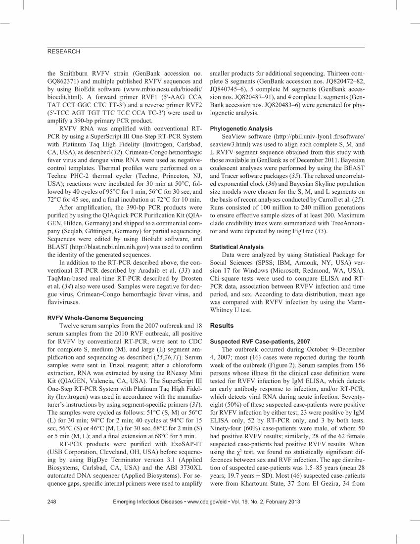
Suspected RVF Case-patients, 2007The outbreak occurred during October 9–December
4, 2007; most (16) cases were reported during the fourth week of the outbreak (Figure 2). Serum samples from 156 persons whose illness fit the clinical case definition were tested for RVFV infection by IgM ELISA, which detects an early antibody response to infection, and/or RT-PCR, which detects viral RNA during acute infection. Seventy-eight (50%) of these suspected case-patients were positive for RVFV infection by either test; 23 were positive by IgM ELISA only, 52 by RT-PCR only, and 3 by both tests. Ninety-four (60%) case-patients were male, of whom 50 had positive RVFV results; similarly, 28 of the 62 female suspected case-patients had positive RVFV results. When using the χ2 test, we found no statistically significant dif-ferences between sex and RVF infection. The age distribu-tion of suspected case-patients was 1.5–85 years (mean 28 years; 19.7 years ± SD). Most (46) suspected case-patients were from Khartoum State, 37 from El Gezira, 34 from
248 Emerging Infectious Diseases • www.cdc.gov/eid • Vol. 19, No. 2, February 2013
Rift Valley Fever, Sudan
Kassala, 24 from White Nile, 6 from Sennar, 6 from Upper Nile, and 3 from Nile State (Figure 1).
Suspected RVF Case-patients, 2010The 2010 outbreak was restricted to a rural area in El
Gezira State in central Sudan (Figure 1). One hundred se-rum samples were collected from suspected case-patients. Suspected RVFV samples were tested solely by RT-PCR, and 18 samples were positive.
Whole-Genome SequencingSerum from patients with positive test results for
RVFV by conventional RT-PCR was subjected to whole-genome sequencing. Twelve samples from 2007 and 18 samples from 2010 were used to attempt generation of complete S, M, and L segment sequences (Table). From the 2007 samples, 6 complete S segments, 1 complete M segment, and 1 complete L segment were amplified and se-quenced; 7 S, 4 M, and 3 L segments were obtained from the 2010 samples. Of these sequences, S segments from samples 2V, 1, and SP from 2007 were identical. S seg-ments from samples 7 and 85 from 2010 also were identi-
cal, and the M segment of these samples contained a single base change. Sample 4 from 2010 and sample 77 from 2007 also shared an identical S segment sequence. The generated sequences represent Khartoum, El Gezira, and White Nile States (Table, Figure 1).
Phylogenetic AnalysisWhen conducting the phylogenetic analysis to com-
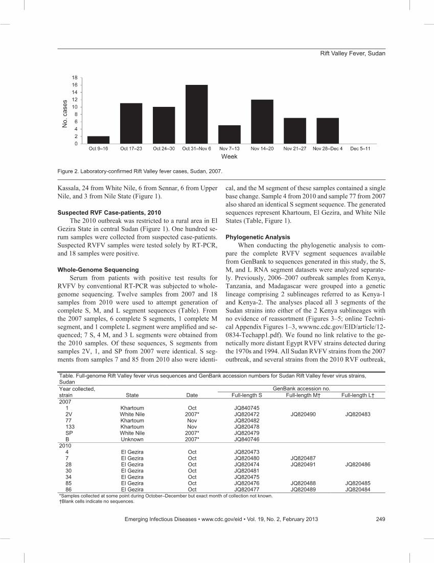
pare the complete RVFV segment sequences available from GenBank to sequences generated in this study, the S, M, and L RNA segment datasets were analyzed separate-ly. Previously, 2006–2007 outbreak samples from Kenya, Tanzania, and Madagascar were grouped into a genetic lineage comprising 2 sublineages referred to as Kenya-1 and Kenya-2. The analyses placed all 3 segments of the Sudan strains into either of the 2 Kenya sublineages with no evidence of reassortment (Figures 3–5; online Techni-cal Appendix Figures 1–3, wwwnc.cdc.gov/EID/article/12-0834-Techapp1.pdf). We found no link relative to the ge-netically more distant Egypt RVFV strains detected during the 1970s and 1994. All Sudan RVFV strains from the 2007 outbreak, and several strains from the 2010 RVF outbreak,
Emerging Infectious Diseases • www.cdc.gov/eid • Vol. 19, No. 2, February 2013 249
Figure 2. Laboratory-confirmed Rift Valley fever cases, Sudan, 2007.
Table. Full-genome Rift Valley fever virus sequences and GenBank accession numbers for Sudan Rift Valley fever virus strains, Sudan Year collected, strain State Date
GenBank accession no. Full-length S Full-length M† Full-length L†
2007 1 Khartoum Oct JQ840745 2V White Nile 2007* JQ820472 JQ820490 JQ820483 77 Khartoum Nov JQ820482 133 Khartoum Nov JQ820478 SP White Nile 2007* JQ820479 B Unknown 2007* JQ840746 2010 4 El Gezira Oct JQ820473 7 El Gezira Oct JQ820480 JQ820487 28 El Gezira Oct JQ820474 JQ820491 JQ820486 30 El Gezira Oct JQ820481 34 El Gezira Oct JQ820475 85 El Gezira Oct JQ820476 JQ820488 JQ820485 86 El Gezira Oct JQ820477 JQ820489 JQ820484 *Samples collected at some point during October–December but exact month of collection not known. †Blank cells indicate no sequences.
RESEARCH
were embedded into the Kenya-2 sublineage; however, 2 strains from 2010 were included in the Kenya-1 sublineage. Of the 7 states where suspected RVF cases were distrib-uted, whole-genome sequences were generated from case-patients from Khartoum, White Nile, and El Gezira. The Khartoum strains grouped with strains from El Gezira and White Nile States (Figures 3–5).
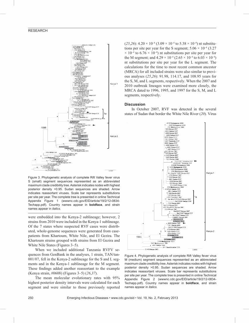
When we included additional Tanzania RVFV se-quences from GenBank in the analyses, 1 strain, TAN/tan-001/07, fell in the Kenya-2 sublineage for the S and L seg-ments and in the Kenya-1 sublineage for the M segment. These findings added another reassortant to the example (Kenya strain, #0608) (Figures 3–5) (26,37).
The mean molecular evolutionary rates with 95% highest posterior density intervals were calculated for each segment and were similar to those previously reported
(25,26): 4.20 × 10⁻⁴ (3.09 × 10⁻⁴ to 5.38 × 10⁻⁴) nt substitu-tions per site per year for the S segment; 5.06 × 10⁻⁴ (3.27 × 10⁻⁴ to 6.76 × 10⁻⁴) nt substitutions per site per year for the M segment; and 4.29 × 10⁻⁴ (2.65 × 10⁻⁴ to 6.03 × 10⁻⁴) nt substitutions per site per year for the L segment. The calculations for the time to most recent common ancestor (MRCA) for all included strains were also similar to previ-ous analyses (25,26): 91.98, 114.17, and 108.95 years for the S, M, and L segments, respectively. When the 2007 and 2010 outbreak lineages were examined more closely, the MRCA dated to 1996, 1995, and 1997 for the S, M, and L segments, respectively.
Discussion In October 2007, RVF was detected in the several
states of Sudan that border the White Nile River (20). Virus
250 Emerging Infectious Diseases • www.cdc.gov/eid • Vol. 19, No. 2, February 2013
Figure 3. Phylogenetic analysis of complete Rift Valley fever virus S (small) segment sequences represented as an abbreviated maximum clade credibility tree. Asterisk indicates nodes with highest posterior density >0.95. Sudan sequences are shaded. Arrow indicates reassortant viruses. Scale bar represents substitutions per site per year. The complete tree is presented in online Technical Appendix Figure 1 (wwwnc.cdc.gov/EID/article/19/2/12-0834-Techapp.pdf). Country names appear in boldface, and strain names appear in italics.
Figure 4. Phylogenetic analysis of complete Rift Valley fever virus M (medium) segment sequences represented as an abbreviated maximum clade credibility tree. Asterisk indicates nodes with highest posterior density >0.95. Sudan sequences are shaded. Arrow indicates reassortant viruses. Scale bar represents substitutions per site per year. The complete tree is presented in online Technical Appendix Figure 2 (wwwnc.cdc.gov/EID/article/19/2/12-0834-Techapp.pdf). Country names appear in boldface, and strain names appear in italics.
Rift Valley Fever, Sudan
activity was detected in 7 states: Nile, Khartoum, Kassala, El Gezira, White Nile, Sennar, and Upper Nile; large numbers of human infections occurred during the relatively short pe-riod of 2 months (Figure 1). The substantial RVF outbreak was first detected in Kenya and Tanzania in late 2006 to ear-ly 2007 after a season of heavy rainfall (21). RVFV activity continued for several years and covered a large geographic area, including Sudan in 2007, and South Africa and Mada-gascar in 2008 (1,20,25,38,39).
Although substantial rainfall events were most likely the major cause of spread and maintenance of the RVF outbreak (21,40), the contribution of irrigation projects is less well understood. Several recent studies have examined the effects of irrigation and agricultural practices on mos-quito populations (41–44 in online Technical Appendix). However, the effect of these agricultural processes on RVF
outbreaks and maintenance during interepidemic periods has not been directly studied. The large RVF outbreak that occurred in the regions surrounding the Senegal River dur-ing 1987–1988 was thought to be linked to completion of the Maka-Diama dam built in 1986 and the extensive irriga-tion system developed at this time (45 in online Technical Appendix). The potential effect of irrigation on RVF should be considered, since new industries in the Sudan are chang-ing the landscape in several RVF-affected states. El Gezira and, to a lesser degree, Sennar, White Nile, and Khartoum States, have vast tracts of irrigated land. Khartoum State has a growing agricultural industry along the Blue Nile River, particularly in Hilat Kuku, Khartoum North, which was the focus of the 1977 Sudan RVF outbreak (15). Dur-ing the end of November 2007, the El Gezira authorities instituted an extensive insecticide spraying program and the federal government restricted trade of livestock and as-sociated products in the state, which may have contributed to the subsequent decline in suspected cases. Future studies would be useful for determining appropriate vector control strategies in irrigated areas.
The epidemiologic data for the 156 suspected RVF cases of 2007 indicated that more male than female case-patients fit the case definition for RVF; however, the per-centage of confirmed cases was equivalent for both sexes. The mean age of suspected case-patients was 28 years, con-sistent with reports from Kenya and Tanzania, where per-sons 20–30 years old were most affected (27,29). Young adult men in these affected regions are generally more ex-posed than women to potentially infected mosquitoes dur-ing agricultural work or direct contact with viremic live-stock and potentially infected livestock by-products, such as aborted fetal material and raw milk products.
Suspected case-patients were from the 7 states listed previously; most were from Khartoum, Kassala, El Gezira, and White Nile (Figure 1). Rainfall or the new irrigation schemes mentioned previously might have influenced case distribution. Although the overall percentage of confirmed cases was 50%, the percentage of confirmed cases in each state ranged from 15% to 84% (data not shown). The varia-tion also could result from different interpretations of the case definition or increases in other febrile illnesses affect-ed by weather conditions similar to those affecting RVF (e.g., an increase in disease vectors). The possibility of re-porting bias also exists because patients can be referred to medical centers in neighboring states.
In 2010, RVF cases were again detected in El Gezira State. The outbreak was first characterized by abortions in ewes and does and followed by infections in persons with histories of contact with aborted fetal material (I.E. Ara-daib, pers. comm.). Unfortunately, detailed information about the 100 suspected case-patients tested was not avail-able for analysis.
Emerging Infectious Diseases • www.cdc.gov/eid • Vol. 19, No. 2, February 2013 251
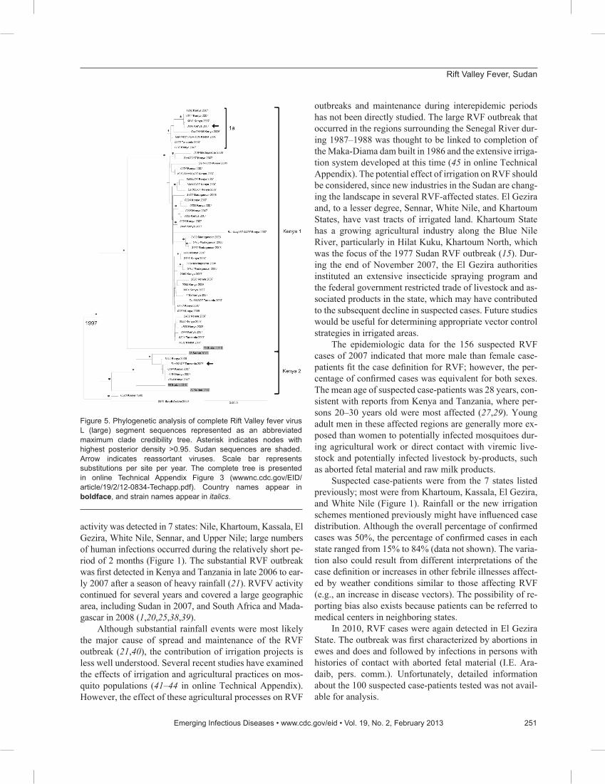
Figure 5. Phylogenetic analysis of complete Rift Valley fever virus L (large) segment sequences represented as an abbreviated maximum clade credibility tree. Asterisk indicates nodes with highest posterior density >0.95. Sudan sequences are shaded. Arrow indicates reassortant viruses. Scale bar represents substitutions per site per year. The complete tree is presented in online Technical Appendix Figure 3 (wwwnc.cdc.gov/EID/article/19/2/12-0834-Techapp.pdf). Country names appear in boldface, and strain names appear in italics.
RESEARCH
RVFV-positive samples from several states of Sudan were selected for full-genome analysis to determine the relationship of the strains circulating in Sudan to other known strains identified globally, especially those from the 2006–2007 outbreaks in Kenya and Tanzania and from Egypt during the 1970s and in 1994. A total of 13 com-plete S segment, 5 complete M segment, and 4 complete L segment sequences were obtained. Phylogenetic analysis of these sequences identified several RVFV variants cir-culating during the Sudan outbreaks and placed them all in the large lineage containing the Kenya-1 and Kenya-2 sublineages, which defined the eastern Africa outbreak in 2006–2008 (26). No genetic relationship was found rela-tive to the earlier Egypt strains. Previously, only RVFV strains identified in Kenya were embedded in the Kenya-2 sublineage; however, as more sequences become available, this sublineage clearly has also become widely geographi-cally distributed (25,26). Bayesian analysis was used to help elucidate whether RVFV diversity in Sudan resulted from multiple introductions or from acquired changes over time from a single introduction event. Several observations indicate that multiple introductions of RVFV occurred as part of its spread from eastern Africa since the 1996–1997 RVF outbreak (46 in online Technical Appendix). The date to the MRCA for the overall Kenya lineage is circa 1996, and the MRCA for the 2007 and 2010 Sudan sequences also dates to 1996 instead of 2007. The closest MRCA between the 2007 and 2010 sequences is 5 years (2005). Dating of the MRCA of the overall Kenya lineage concurs with the MRCA of previous studies, which supports the ro-bustness of the models chosen for analysis, even consider-ing the limitations of sample size, collection methods, and environmental factors (25,26). Identical or nearly identical sequences were identified for different states and years, Khartoum in 2007 and El Gezira in 2010, as well as Khar-toum and West Nile in 2007. These sequences indicate re-cent movement of the virus in this region and support the necessity and utility of surveillance systems for recogniz-ing when and where a large epidemic is imminent. Under-standing where the virus is circulating during interepidemic periods can make it easier to interpret data from prediction tools (21) and focus preventive measures, such as vector control, livestock vaccination, and education campaigns, on high-risk areas.
The significance of detecting an additional M segment reassortant remains unclear. Reassortment seems to be a relatively rare event because only 2 reassortants were de-tected for the 54 complete genome sequences (S, M, and L) from the 2006–2007 RVF outbreak. However, it does sup-port cocirculation of both Kenya sublineages in the same geographic location.
The addition of RVFV sequences from Sudan en-hances our understanding of the expansion and, to some
degree, maintenance of the virus during a large epidemic and the interepidemic period that follows. The ability to se-quence entire viral genomes relatively quickly should lead to rapid progress in understanding the detailed ecology of RVFV. Ongoing surveillance and RVFV characterization also should help determine the pattern of virus maintenance between epizootic events. As prediction tools become more accurate and available, these data will provide public health authorities an opportunity to anticipate and prepare for RVF outbreaks.
AcknowledgmentsThis study was made possible by the invaluable assistance
provided by the medical staff from the different states of Sudan. We thank Abdall M. Fadl Elmoula for technical assistance, Craig Manning for his assistance in creating the Sudan map, and Tanya Klimova for her editorial assistance.
This study received partial financial support from the Direc-torate of Scientific Research and Cultural relations, University of Khartoum, and CDC.
Dr Aradaib is a professor of molecular medicine at the mo-lecular biology laboratory, Department of Clinical Medicine, Faculty of Veterinary Medicine, University of Khartoum. His re-search interests focus on the study of epizootics including viral hemorrhagic fevers.
References
1. Andriamandimby SF, Randrianarivo-Solofoniaina AE, Jeanmaire EM, Ravololomanana L, Razafimanantosoa LT, Rakotojoelinan-drasana T, et al. Rift Valley fever during rainy seasons, Madagascar, 2008 and 2009. Emerg Infect Dis. 2010;16:963–70. http://dx.doi.org/10.3201/eid1606.091266
2. Eisa M, Obeid HMA, El Sawi ASA. Rift Valley fever in the Sudan. I. Results on field investigations of the first epizootic in Kosti District, 1973. Bull Anim Health Prod Afr. 1977;25:343–7.
3. Eisa M, Obeid HMA. Rift Valley fever in the Sudan. II. Isolation and identification of the virus from a recent epizootic in Kosti District, 1973. Bull Anim Health Prod Afr. 1977;25:349–55.
4. Sall AA. Zanotto PMdeA, Vialat P, Sene OK, Bouloy M. Ori-gin of 1997–98 Rift Valley fever outbreak in east Africa. Lancet. 1998;352:1596–7. http://dx.doi.org/10.1016/S0140-6736(05)61043-4
5. Shoemaker T, Boulianne C, Vincent MJ, Pezzanite L, Al-Qahtani MM, Al-Mazrou Y, et al. Genetic analysis of viruses associated with emergence of Rift Valley fever in Saudi Arabia and Yemen, 2000–1. Emerg Infect Dis. 2002;8:1415–20. http://dx.doi.org/10.3201/eid0812.020195
6. Meegan JM, Hoogstraal H, Moussa MI. An epizootic of Rift Val-ley fever in Egypt in 1977. Vet Rec. 1979;105:124–5. http://dx.doi.org/10.1136/vr.105.6.124
7. Meegan JM. Rift Valley fever in Egypt: an overview of the epizoot-ics in 1977 and 1978. Contributions to Epidemiology and Biostatis-tics. 1981;3:100–13.
8. Sang R, Kioko E, Lutomiah J, Warigia M, Ochieng C, O’Guinn M, et al. Rift Valley fever virus epidemic in Kenya, 2006/2007: the entomologic investigations. Am J Trop Med Hyg. 2010;83(Sup-pl):28–37. http://dx.doi.org/10.4269/ajtmh.2010.09-0319
252 Emerging Infectious Diseases • www.cdc.gov/eid • Vol. 19, No. 2, February 2013
Rift Valley Fever, Sudan
9. Turell MJ, Linthicum KJ, Patrican LA, Davies FG, Kairo A, Bailey CL. Vector competence of selected African mosquito (Diptera: Culicidae) species for Rift Valley fever virus. J Med Entomol. 2008;45:102–8. http://dx.doi.org/10.1603/0022-2585(2008) 45[102:VCOSAM]2.0.CO;2
10. McIntosh BM, Russell D, dos Santos I, Gear JH. Rift Valley fever in humans in South Africa. S Afr Med J. 1980;58:803–6.
11. Meegan JM, Watten RH, Laughlin LW. Clinical experience with Rift Valley fever in humans during the 1977 Egyptian epizootic. Contri-butions to Epidemiology and Biostatistics. 1981;3:114–23.
12. Madani TA, Al-Mazrou YY, Al-Jeffri MH, Mishkhas AA, Al-Rabeah AM, Turkistani AM, et al. Rift Valley fever epidemic in Saudi Ara-bia: epidemiological, clinical, and laboratory characteristics. Clin Infect Dis. 2003;37:1084–92. http://dx.doi.org/10.1086/378747
13. Eisa M, Kheir el-Sid ED, Shomein AM, Meegan JM. An outbreak of Rift Valley fever in the Sudan–1976. Trans R Soc Trop Med Hyg. 1980;74:417–9. http://dx.doi.org/10.1016/0035-9203(80)90122-4
14. Davies FG. Rift Valley fever in the Sudan. Trans R Soc Trop Med Hyg. 1990;84:141. http://dx.doi.org/10.1016/0035-9203 (90)90410-G
15. Eisa M. Preliminary survey of domestic animals of the Sudan for precipitating antibodies to Rift Valley fever virus. J Hyg (Lond). 1984;93:629–37. http://dx.doi.org/10.1017/S0022172400065207
16. McCarthy MC, Haberberger RL, Salib AW, Soliman BA, El-Tigani A, Khalid IO, et al. Evaluation of arthropod-borne viruses and other infectious disease pathogens as the causes of febrile illnesses in the Khartoum Province of Sudan. J Med Virol. 1996;48:141–6. http://dx.doi.org/10.1002/(SICI)1096-9071(199602)48:2<141::AID-JMV4>3.0.CO;2-9
17. Saleh AS, Mohammed KA, Hassan MM, Bucci TJ, Meegan JM. Antibodies to Rift Valley fever virus in the human population of Sudan. Trans R Soc Trop Med Hyg. 1981;75:129–30. http://dx.doi.org/10.1016/0035-9203(81)90039-0
18. Woodruff PW, Morrill JC, Burans JP, Hyams KC, Woody JN. A study of viral and rickettsial exposure and causes of fever in Juba, southern Sudan. Trans R Soc Trop Med Hyg. 1988;82:761–6. http://dx.doi.org/10.1016/0035-9203(88)90229-5
19. Hassanain AM, Noureldien W, Karsany MS, Saeed ES, Ara-daib IE, Adam I. Rift Valley fever among febrile patients at New Halfa hospital, eastern Sudan. Virol J. 2010;7:97. http://dx.doi.org/10.1186/1743-422X-7-97
20. World Health Organization. Global alert and response. Rift Valley fever in Sudan, update November 7. 2007 [2012 May 1]. http://www.who.int/csr/don/2007_11_05/en/index.html
21. Anyamba A, Linthicum KJ, Small J, Britch SC, Pak E, de La Rocque S, et al. Prediction, assessment of the Rift Valley fever activity in east and southern Africa 2006–2008 and possible vector control strategies. Am J Trop Med Hyg. 2010;83(Suppl):43–51. http://dx.doi.org/10.4269/ajtmh.2010.09-0289
22. Adam AA, Karsany MS, Adam I. Manifestations of severe Rift Val-ley fever in Sudan. Int J Infect Dis. 2010;14:e179–80. http://dx.doi.org/10.1016/j.ijid.2009.03.029
23. Anyangu AS, Gould LH, Sharif SK, Nguku PM, Omolo JO, Mu-tonga D, et al. Risk factors for severe Rift Valley fever infection in Kenya, 2007. Am J Trop Med Hyg. 2010;83(Suppl):14–21. http://dx.doi.org/10.4269/ajtmh.2010.09-0293
24. Madani TA, Al-Mazrou YY, Al-Jeffri MH, Mishkhas AA, Al-Rabeah AM, Turkistani AM, et al. Rift Valley fever epidemic in Saudi Ara-bia: epidemiological, clinical, and laboratory characteristics. Clin Infect Dis. 2003;37:1084–92. http://dx.doi.org/10.1086/378747
25. Carroll SA, Reynes JM, Khristova ML, Andriamandimby SF, Rollin PE, Nichol ST. Genetic evidence for Rift Valley fever outbreaks in Madagascar resulting from virus introductions from the east African mainland rather than enzootic maintenance. J Virol. 2011;85:6162–7. http://dx.doi.org/10.1128/JVI.00335-11
26. Bird BH, Githinji JWK, Macharia JM, Kasiiti JL, Muriithi RM, Gacheru SG, et al. Multiple virus lineages sharing recent common an-cestry were associated with a large Rift Valley fever outbreak among
livestock in Kenya during 2006–2007. J Virol. 2008;82:11152–66. http://dx.doi.org/10.1128/JVI.01519-08
27. Mohamed M, Mosha F, Mghamba J, Zaki SR, Shieh WJ, Paweska J, et al. Epidemiologic and clinical aspects of a Rift Valley fever outbreak in humans in Tanzania, 2007. Am J Trop Med Hyg. 2010;83(Sup-pl):22–7. http://dx.doi.org/10.4269/ajtmh.2010.09-0318
28. Munyua P, Murithi RM, Wainwright S, Githinji J, Hightower A, Mutonga D, et al. Rift Valley fever outbreak in livestock in Kenya, 2006–2007. Am J Trop Med Hyg. 2010;83(Suppl):58–64. http://dx.doi.org/10.4269/ajtmh.2010.09-0292
29. Nguku PM, Sharif SK, Mutonga D, Amwayi S, Omolo J, Moham-med O, et al. An investigation of a major outbreak of Rift Valley fever in Kenya: 2006–2007. Am J Trop Med Hyg. 2010;83(Sup-pl):5–13. http://dx.doi.org/10.4269/ajtmh.2010.09-0288
30. Rich KM, Wanyoike F. An assessment of the regional and national socio-economic impacts of the 2007 Rift Valley fever outbreak in Kenya. Am J Trop Med Hyg. 2010;83(Suppl):52–7. http://dx.doi.org/10.4269/ajtmh.2010.09-0291
31. Bird BH, Khristova ML, Rollin PE, Ksiazek TG, Nichol ST. Com-plete genome analysis of 33 ecologically and biologically diverse Rift Valley fever virus strains reveals widespread virus movement and low genetic diversity due to recent common ancestry. J Virol. 2007;81:2805–16. http://dx.doi.org/10.1128/JVI.02095-06
32. Aradaib IE, Erickson BR, Karsany MS, Khristova ML, Elageb RM, Mohamed MEH, et al. Multiple Crimean-Congo hemorrhagic fever virus strains are associated with disease outbreaks in Sudan, 2008–2009. PLoS Negl Trop Dis. 2011;5:e1159. http://dx.doi.org/10.1371/journal.pntd.0001159
33. Aradaib IE, Elageb RM, Abdalla TM, Karrar AE, Karsani MA, Musa HA, et al. Improved diagnosis of Rift Valley fever in clini-cal samples using conventional semi-nested reverse transcriptase-PCR. International Journal of Molecular Medicine and Advanced Science. 2008;4:77–81.
34. Drosten C, Göttig S, Schilling S, Asper M, Panning M, Schmitz H, et al. Rapid detection and quantification of RNA of Ebola and Marburg viruses, Lassa virus, Crimean-Congo hemorrhagic fever virus, Rift Valley fever virus, dengue virus, and yellow fever virus by real-time reverse transcription–PCR. J Clin Microbiol. 2002;40:2323–30. http://dx.doi.org/10.1128/JCM.40.7.2323-2330.2002
35. Drummond AJ, Rambaut A. BEAST: Bayesian Evolutionary Analy-sis by Sampling Trees. BMC Evol Biol. 2007;7:214. http://dx.doi.org/10.1186/1471-2148-7-214
36. Drummond AJ, Ho SYW, Phillips MJ, Rambaut A. Relaxed phylo-genetics and dating with confidence. PLoS Biol. 2006;4:e88. http://dx.doi.org/10.1371/journal.pbio.0040088
37. Nderitu L, Lee JS, Omolo J, Omulo S, O’Guinn ML, Hightower A, et al. Sequential Rift Valley fever outbreaks in eastern Africa caused by multiple lineages of the virus. J Infect Dis. 2011;203:655–65. http://dx.doi.org/10.1093/infdis/jiq004
38. Archer BN, Weyer J, Paweska J, Nkosi D, Leman P, Tint KS, et al. Outbreak of Rift Valley fever affecting veterinarians and farmers in South Africa, 2008. S Afr Med J. 2011;101:263–6.
39. World Health Organization. Together for better health. A report on WHO collaborative programmes with the Government of Sudan and partners 2006–2007. WHO Sudan biennial report 2006–2007. Ge-neva: The Organization; 2008.
40. Food Security and Technical Secretariat, Southern Sudan Center for Census, Statistics and Evaluation, Government of Southern Sudan. Southern Sudan: agrometeorology update, October 2010 [cited 2012 Dec 4]. http://www.fao.org/fileadmin/user_upload/sifsia/docs/Ag-romet_Bulletine_October_2010_FSTS.pdf
Address for correspondence: Stuart T. Nichol, Centers for Disease Control and Prevention, 1600 Clifton Rd NE, Mailstop G14, Atlanta, GA 30033, USA; email: [email protected]
Emerging Infectious Diseases • www.cdc.gov/eid • Vol. 19, No. 2, February 2013 253