RIF-1 Murine Fibrosarcoma: Inhibited by Hypophysectomy and ... · Michael Pollak, Lady Davis...
5
Metastatic Behavior of the RIF-1 Murine Fibrosarcoma: Inhibited by Hypophysectomy and Partially Restored by Growth Hormone Replacement Ato Sekyi-Otu, Robert Bell, Irene Andrulis, Michael Pollak* Background: We recently demon- strated that hypophysectomy pro- foundly inhibits metastatic behavior in the MGH-OGS murine osteosarcoma model and speculated that this effect is related at least in part to ablation of the growth hormone (GH)—insulin-like growth factor I (IGF-I) axis. Purpose: In this study, we determined whether the administration of GH to animals rendered GH and IGF-I deficient by hypophysectomy attenuates the in- hibitory effects of hypophysectomy on metastatic behavior. Methods: Meta- static behavior was assayed by count- ing visible metastases in lungs 3 weeks after tail vein injection of RIF-I fibrosarcoma cells to control mice (n = 29), hypophysectomized mice (n = 19), and hypophysectomized mice admin- istered 0.05 u.g/g body weight recom- binant human GH twice daily (n = 21). Results: Twenty of 21 hypophysec- tomized mice receiving GH, eight of 19 hypophysectomized mice not receiving GH, and 26 of 29 controls had grossly visible pulmonary metastases 3 weeks after intravenous injection of 5 x 10 5 cells; mean numbers ± SD of gross metastases were 38.4 ± 11J, 6.4 ± 2.2, and 13.1 ± 2.8 in the three groups, respectively. The presence (P<.005, chi- square test) and number (P = .0003, Mann-Whitney U test) of metastases were significantly reduced in hypophy- sectomized hosts compared with con- trol hosts and were significantly higher in hypophysectomized, GH-replaced hosts compared with hypophysec- tomized hosts (P<.001, chi-square test; P = .011, Mann-Whitney U test), while the difference in presence and extent of metastases between control and hypo- physectomized, GH-replaced hosts was not statistically significant. Conclu- sions: These data support the hypoth- esis that the status of the host with respect to GH and/or GH-dependent factors such as IGF-I influences the metastatic behavior of certain neo- plasms. Implications: Our results raise the possibility that compounds that reduce GH output or interfere with GH action, such as somatostatin ana- logues, GH antagonists, IGF antagon- ists, and GH-releasing hormone antagonists, may suppress metastatic behavior of certain neoplasms. [J Natl Cancer Inst 86:628-632,1994] The insulin-like growth factor (IGF) mitogenic signal transduction system in- cludes ligands, receptors, and binding proteins. The ligands IGF-I and IGF-II have structural homology to insulin, sug- gesting evolutionary derivation from a parent gene {12)- IGF-I is a potent mitogen for a variety of cell types (2-6) and is known to mediate much of the ac- tion of growth hormone (GH) on somatic growth (7). Although the gene encoding IGF-I is expressed in many tissues, most circulating IGF-I is produced by the liver, where transcription is regulated by a complex hypothalamic-pituitary-hepatic axis (8J?). GH-releasing hormone re- leased from the hypothalamus stimulates pituitary-GH release, while somatostatin inhibits GH release. Hepatocytes syn- thesize and release IGF-I in response to GH. Finally, the bioavailability of IGF-I is influenced by the presence of the various IGF-bindmg proteins, which modulate binding interactions between IGFs and their receptors (70). Data consistent with a role for IGF-I in the behavior of certain sarcomas include the demonstration of IGF-I receptors on both sarcoma cell lines (11,12) and primary tumors (11-13), the autocrine production of ligands for this receptor by a subset of primary sarcomas (13), the demonstration of inhibition of in vitro sarcoma proliferation by antisense oligo- nucleotides against the IGF-I receptor (14), and the demonstration that hypo- physectomy, which is associated with a variety of hormonal changes including ablation of GH production and GH-de- pendent IGF-I gene expression, inhibits local growth rate of the MGH-OGS experi- mental osteosarcoma by approximately 50%, and inhibits metastatic behavior in this model by close to 100% (15). In vitro studies of MGH-OGS cells documented that these cells are growth- stimulated by IGF-I, but not by GH or other pituitary factors, which is consistent with the hypothesis that the inhibitory ef- fects of hypophysectomy are related, at least in part, to the reduction in IGF-I gene expression and in serum levels that follow this ablative surgical procedure (15). In the present study, we sought to determine directly if the GH-IGF-I axis influences the metastatic behavior of an experimental sarcoma by assessing me- tastases in intact, hypophysectomized, and GH-replaced hosts Materials and Methods Control and surgically hypophysectomized C3H male mice were obtained from Charles River Laboratories (Montreal, Quebec, Canada) Hypo- physectomy was performed at 6-7 weeks and hypophysectomized and control animals were both obtained at 8 weeks of age. Both hypophysec- tomized and control mice were fed ad libitum and chmatized to a cycle of 12 hours of light and 12 hours of dark for at least 1 week prior to any ex- perimental manipulations All protocols adhered to approved standards as determined by the Animal Care Committees of the University of Toronto and of McGill University, Montreal Previous experiments evaluating the effect of hypophysectomy in munne sarcoma behavior used the MGH-OGS model The length of time necessary for the development of metastases using this system made it impractical for GH replacement The RIF-1 radiation-induced fibrosarcoma was therefore ob- tained from Dr R Hill (University of Toronto) for these purposes In vitro growth and in vivo behavior of RIF-1 cells have been described previously {16- 18) Cell cultures of RIF-1 were grown in oc-mini- mum essential medium and 10% fetal calf serum (GIBCO BRL, Grand Island, N Y ) . *Affiliattons of authors A. Sekyi-Otu, R Bell, Samuel Lunenfeld Research Institute, and Division of Orthopaedic Surgery, Mount Sinai Hospital and University of Toronto, Toronto, Ontario, Canada I Andrulis, Samuel Lunenfeld Research Institute, and Departments of Molecular and Medical Genetics and Pathology, Mount Sinai Hospital and University of Toronto Michael Pollak, Lady Davis Research Institute of the Jewish General Hospital, McGill University, Montreal, Quebec, Canada. Correspondence to. Robert S Bell, M D., Department of Orthopaedic Surgery. Mount Sinai Hospital, 600 University Ave, Rm 476, Toronto, 0NM5G 1X5, Canada. See "Notes" section following "References." 628 REPORTS Journal of the National Cancer Institute. Vol. 86, No. 8, April 20, 1994 at McGill University Libraries on December 9, 2011 http://jnci.oxfordjournals.org/ Downloaded from
Transcript of RIF-1 Murine Fibrosarcoma: Inhibited by Hypophysectomy and ... · Michael Pollak, Lady Davis...

Metastatic Behavior of theRIF-1 Murine Fibrosarcoma:Inhibited by Hypophysectomyand Partially Restored byGrowth Hormone Replacement
Ato Sekyi-Otu, Robert Bell, IreneAndrulis, Michael Pollak*
Background: We recently demon-strated that hypophysectomy pro-foundly inhibits metastatic behavior inthe MGH-OGS murine osteosarcomamodel and speculated that this effect isrelated at least in part to ablation ofthe growth hormone (GH)—insulin-likegrowth factor I (IGF-I) axis. Purpose:In this study, we determined whetherthe administration of GH to animalsrendered GH and IGF-I deficient byhypophysectomy attenuates the in-hibitory effects of hypophysectomy onmetastatic behavior. Methods: Meta-static behavior was assayed by count-ing visible metastases in lungs 3 weeksafter tail vein injection of RIF-Ifibrosarcoma cells to control mice (n =29), hypophysectomized mice (n = 19),and hypophysectomized mice admin-istered 0.05 u.g/g body weight recom-binant human GH twice daily (n = 21).Results: Twenty of 21 hypophysec-tomized mice receiving GH, eight of 19hypophysectomized mice not receivingGH, and 26 of 29 controls had grosslyvisible pulmonary metastases 3 weeksafter intravenous injection of 5 x 10⁵
cells; mean numbers ± SD of grossmetastases were 38.4 ± 11J, 6.4 ± 2.2,and 13.1 ± 2.8 in the three groups,respectively. The presence (P<.005, chi-square test) and number (P = .0003,Mann-Whitney U test) of metastaseswere significantly reduced in hypophy-sectomized hosts compared with con-trol hosts and were significantly higherin hypophysectomized, GH-replacedhosts compared with hypophysec-tomized hosts (P<.001, chi-square test;P = .011, Mann-Whitney U test), whilethe difference in presence and extent ofmetastases between control and hypo-
physectomized, GH-replaced hosts wasnot statistically significant. Conclu-sions: These data support the hypoth-esis that the status of the host withrespect to GH and/or GH-dependentfactors such as IGF-I influences themetastatic behavior of certain neo-plasms. Implications: Our results raisethe possibility that compounds thatreduce GH output or interfere withGH action, such as somatostatin ana-logues, GH antagonists, IGF antagon-ists, and GH-releasing hormoneantagonists, may suppress metastaticbehavior of certain neoplasms. [J NatlCancer Inst 86:628-632,1994]
The insulin-like growth factor (IGF)mitogenic signal transduction system in-cludes ligands, receptors, and bindingproteins. The ligands IGF-I and IGF-IIhave structural homology to insulin, sug-gesting evolutionary derivation from aparent gene {12)- IGF-I is a potentmitogen for a variety of cell types (2-6)and is known to mediate much of the ac-tion of growth hormone (GH) on somaticgrowth (7). Although the gene encodingIGF-I is expressed in many tissues, mostcirculating IGF-I is produced by the liver,where transcription is regulated by acomplex hypothalamic-pituitary-hepaticaxis (8J?). GH-releasing hormone re-leased from the hypothalamus stimulatespituitary-GH release, while somatostatininhibits GH release. Hepatocytes syn-thesize and release IGF-I in response toGH. Finally, the bioavailability of IGF-Iis influenced by the presence of thevarious IGF-bindmg proteins, whichmodulate binding interactions betweenIGFs and their receptors (70).
Data consistent with a role for IGF-I inthe behavior of certain sarcomas includethe demonstration of IGF-I receptors onboth sarcoma cell lines (11,12) andprimary tumors (11-13), the autocrineproduction of ligands for this receptor bya subset of primary sarcomas (13), thedemonstration of inhibition of in vitrosarcoma proliferation by antisense oligo-nucleotides against the IGF-I receptor(14), and the demonstration that hypo-physectomy, which is associated with avariety of hormonal changes includingablation of GH production and GH-de-pendent IGF-I gene expression, inhibits
local growth rate of the MGH-OGS experi-mental osteosarcoma by approximately50%, and inhibits metastatic behavior inthis model by close to 100% (15).
In vitro studies of MGH-OGS cellsdocumented that these cells are growth-stimulated by IGF-I, but not by GH orother pituitary factors, which is consistentwith the hypothesis that the inhibitory ef-fects of hypophysectomy are related, atleast in part, to the reduction in IGF-Igene expression and in serum levels thatfollow this ablative surgical procedure(15). In the present study, we sought todetermine directly if the GH-IGF-I axisinfluences the metastatic behavior of anexperimental sarcoma by assessing me-tastases in intact, hypophysectomized,and GH-replaced hosts
Materials and Methods
Control and surgically hypophysectomized C3Hmale mice were obtained from Charles RiverLaboratories (Montreal, Quebec, Canada) Hypo-physectomy was performed at 6-7 weeks andhypophysectomized and control animals were bothobtained at 8 weeks of age. Both hypophysec-tomized and control mice were fed ad libitum andchmatized to a cycle of 12 hours of light and 12hours of dark for at least 1 week prior to any ex-perimental manipulations All protocols adhered toapproved standards as determined by the AnimalCare Committees of the University of Toronto andof McGill University, Montreal
Previous experiments evaluating the effect ofhypophysectomy in munne sarcoma behavior usedthe MGH-OGS model The length of time necessaryfor the development of metastases using this systemmade it impractical for GH replacement The RIF-1radiation-induced fibrosarcoma was therefore ob-tained from Dr R Hill (University of Toronto) forthese purposes In vitro growth and in vivo behaviorof RIF-1 cells have been described previously {16-18) Cell cultures of RIF-1 were grown in oc-mini-mum essential medium and 10% fetal calf serum(GIBCO BRL, Grand Island, N Y ) .
*Affiliattons of authors A. Sekyi-Otu, R Bell,Samuel Lunenfeld Research Institute, and Divisionof Orthopaedic Surgery, Mount Sinai Hospital andUniversity of Toronto, Toronto, Ontario, Canada
I Andrulis, Samuel Lunenfeld Research Institute,and Departments of Molecular and MedicalGenetics and Pathology, Mount Sinai Hospital andUniversity of Toronto
Michael Pollak, Lady Davis Research Institute ofthe Jewish General Hospital, McGill University,Montreal, Quebec, Canada.
Correspondence to. Robert S Bell, M D.,Department of Orthopaedic Surgery. Mount SinaiHospital, 600 University Ave, Rm 476, Toronto,0NM5G 1X5, Canada.
See "Notes" section following "References."
628 REPORTS Journal of the National Cancer Institute. Vol. 86, No. 8, April 20, 1994
at McG
ill University L
ibraries on Decem
ber 9, 2011http://jnci.oxfordjournals.org/
Dow
nloaded from
rlim
Rectangle
rlim
Rectangle

In Vitro Assays
Binding of recombinant IGF-I (rIGF-I; Amer-sham Corp.. Arlington Heights, III.) and affinitylabeling of IGF-I receptors were performed on plas-ma membrane-enriched subcellular fractions ofthese murine tumors prepared by differential cen-trifugation (19). Percent specific binding of cellmembrane preparations to '^-labeled IGF-I wasperformed in the presence and absence of unlabeledIGF-I, as described previously (11,15). Human pla-centa served as a positive control tissue.
RIF-1 tumors were plated in 35-mm dishes for invitro proliferation assays. Thymidine-incorporationexperiments were performed following addition ofrIGF-I (Amersham Corp.) in quadruplicate, asdescribed previously (15).
Preparation of RIF-1 Cell Suspensionsand Assay of Local Growth In Vivo
RIF-1 tumors were maintained in vitro and invivo in source mice. Source tumors from four F2generation RIF-1 sources were harvested for eachRIF-1 experiment described below. An injectablecell suspension of RIF-1 cells from each harvest wasprepared under sterile conditions as follows. Firstthe source tumors were minced with scissors into aslurry and rinsed with phosphate-buffered saline(PBS) citrate. Using a cell scraper, the slurry wasthen passed once through a cell mesh. The sievedmixture was incubated at 37 'C with pancreaticDNA'ase and trypsin (Sigma Chemical Co., St.Louis, Mo.) and passed through a second sieve. Themixture was then centrifuged at 3000 rpm for 7minutes at room temperature, and the resulting cellpellet was rcsuspended in 10% fetal calf serum. Theconcentration of RIF-1 cells in the cell suspension wasdetermined manually by using a hemocytometer.
To evaluate the effect of hypophysectomy onlocal tumor growth, 2 x 10* RIF-1 cells preparedfrom a single cell suspension were injected in-tramuscularly into the right gastrocnemius muscle of19 hypophysectomized and 20 control mice. Resul-tant tumor volumes were obtained three times week-ly by measuring three orthogonal diameters (R, x R2
x Rj) with 0.05-mm precision calipers and multiply-ing the product by pi/620. Time for tumors to grow to1000 mm3 was recorded.
Experimental Metastases Assay andGH Replacement in RIF-1 Tumors
To evaluate the effect of hypophysectomy onRIF-1 metastases, hypophysectomized and controlmice were randomly assigned to four experimentalgroups. Mice in hypophysectomized groups (n = 12and n = 10) and control groups (n = 17 and n = 14)received either 1 x 10⁵ or 5 x 10⁵ RIF-1 cellsprepared from a single cell suspension and injectedinto the lateral tail vein, as previously described byKorycka and Hill (17) and Ling et al. (18). The finalinoculum volume given was 0.25 mL in a tuberculinsyringe. Mice were observed for 3 weeks followingthis single inoculation dose and then killed; lungswere harvested at the time of death and stained inBouin's solution for 2-3 days before metastaticcolonies were counted.
To evaluate the effect of hypophysectomy andGH replacement on RIF-1 metastases, hypophysec-
tomized mice were allocated into replacement andplacebo groups. A dose of 0.05 ng human GH(HuGH) per gram of body weight given twice dailyhas been shown to normalize skeletal growth ratesin hypophysectomized mice (21-24). The adequacyof hypophysectomy and GH replacement was as-sessed by comparing weekly mouse weights ingroups. Treatment mice (hypophysectomy + HuGH;n = 21) received 0.05 u.g of HuGH per gram of bodyweight twice daily by subcutaneous administration;hypophysectomized control mice (hypophysectomy+ normal saline; n = 19) received equivalent dosesof normal saline subcutaneously. Normal controlmice (control + normal saline; n = 29) also receivednormal saline placebo. Seven days after initiation ofdaily injections, mice received 5 x 10̂ RIF-1 cellsby lateral tail vein injection. Lungs were harvested 3weeks following the tumor inoculum. This experi-ment was performed in duplicate, using two inde-pendent sets of RIF-1 sources to prepare single cellsuspensions for tail vein injections.
Statistical Analyses
Unpaired Student's t tests were used to analyzethe average time for tumors in control and hypo-physectomized groups to grow to 1000 mm . Chi-square statistics were used to compare presence orabsence of gross metastases between experimentalgroups; nonparametric KruskaJ—WaJlis and Mann-Whitney V tests were used to compare the numberof metastases in RIF-1 experimental groups, whereappropriate. Analysis of variance (ANOVA) wasused to evaluate intergroup differences in mouseweights in replacement experiments.
Results
In Vitro Assays
Preliminary in vitro studies were per-formed on the murine tumors to quan-titate levels of cell membrane IGF-Ireceptors and to determine the effect ofIGF-I and GH on in vitro cell prolifera-tion. Trie results of affinity-bindingstudies using l25I-labeled IGF-I areshown in Fig. 1. Compared with ourknown positive controls for the IGF-Ireceptor (human placenta; 19.9% specificbinding of l25I-IabeIed IGF-I), RIF-1tumors showed 9.0% specific binding.RIF-1 tumor proliferation in vitro wasstimulated to 140% ± 8% of control in thepresence of 1 x 10"9 M rHuIGF-I (P<.05),as determined by thymidine incorporationand cell number assays. Recombinanthuman GH (rHuGH) had no mitogenicactivity for RIF cells in these assays.
Effect of Hypophysectomy on RIF-1Local Growth
The local growth of the RIF-1 tumorswas significantly slower when implantedin hypophysectomized hosts. The time
2 3 4
I P205-
116-
Fig. 1. Affinity labeling of IGF-I receptors of RIF-1sarcoma. Affinity labeling experiments using I25I-labeled IGF-I were performed on plasma mem-brane-enriched subcellular fractions of RIF-1 rumors(lanes 1 and 2) and human placenta (lanes 3 and 4)prepared as described previously (//). Lanes 1 and 3were incubated in the absence of excess unlabeledIGF-I, and lanes 2 and 4 were incubated in thepresence of excess unlabeled IGF-I.
(means ± SD) required for tumors toreach 1000 mm3 averaged 23 ± 4 days inhypophysectomized mice compared withonly 15 ± 2 days in control mice (f<.001;unpaired t test).
Effect of Hypophysectomy onMetastases in RIF-1 Tumors
The effect of hypophysectomy on thefrequency distribution of RIF-1 metas-tases following injection of 1 x 10⁵ or 5 x10⁵ cells demonstrated significant dif-ferences between groups (P<.00\4).There was no difference in the presence(P>.05) and number (P>.05) of metas-tases observed between hypophysec-tomized and control mice 3 weeksfollowing injections with 1 x 10⁵ cells.However, following an inoculation doseof 5 x 10⁵ RIF-1 cells, a significant dif-ference was observed in number ofmetastases (P = .001) between hypophy-sectomized and control groups (Fig. 2).
Effect of HuGH Replacement
Analysis of mouse weights byANOVA demonstrated that mice random-ly assigned to hypophysectomized treat-ment (hypophysectomy + HuGH) andcontrol (hypophysectomy + normalsaline) showed no significant variation insize prior to commencement of the ex-periment (P>.05); however, both groupsof hypophysectomized mice were signifi-cantly smaller than their intact controlcounterparts (P - .0001). Hypophy-
Journal of the National Cancer Institute, Vol. 86, No. 8, April 20, 1994 REPORTS 629
at McG
ill University L
ibraries on Decem
ber 9, 2011http://jnci.oxfordjournals.org/
Dow
nloaded from
rlim
Rectangle
rlim
Rectangle

sectomized mice receiving twice-daily in-jections of HuGH (hypophysectomy +HuGH) gained significantly more weightthan hypophysectomized mice receivingplacebo (hypophysectomy + normalsaline) (P-c.0001). The amount of weightgained by the hypophysectomized re-placement group during the course of theexperiment was equivalent to that of thecontrol group.
The effect of exogenous HuGH re-placement in hypophysectomized mice onthe metastatic potential of the RIF-1tumor is illustrated in Fig. 3. Almost allof the control and HuGH replacementanimals demonstrated visible metastases(26 of 29 controls and 20 of 21 replace-ment mice) compared with fewer than50% of hypophysectomized animals(eight of 19; P<.00\). The mean number± SD of gross metastases observed in thethree groups also differed significantly(38.4 ± 11.3, 13.1 ± 2.8, and 6.4 ± 2.2,respectively; P = .0006). In paired com-parisons of groups, a significant dif-ference in presence (P<.005) and number(P = .0003) of metastases was observedbetween control mice treated with normalsaline and hypophysectomized micetreated with normal saline; hypophysec-tomized mice treated with HuGH andhypophysectomized mice treated withnormal saline also demonstrated sig-nificant differences in presence (/><.001)and number (P = .011) of metastases.Using nonparametric analysis, the dif-ference between the number of metas-tases in hypophysectomized mice treatedwith HuGH and control mice treated withnormal saline did not reach statistical sig-nificance (P = .08). Similar results wereobserved when this experiment wasrepeated.
Discussion
Previous in vitro studies (11-13) haveidentified human osteosarcoma and soft-tissue sarcoma cell lines that exhibit IGF-I receptors on their cell membranes andthat also respond mitogenically to IGF-Iin culture. We were therefore interestedin identifying murine tumor models fromwhich in vivo experiments might be ex-trapolated to the human clinical disease.The murine MGH-OGS osteosarcoma(20), which parallels the human disease,has been shown to be IGF-I responsive in
60
n n n1-5 6-10 11-15 16-20 21-25 26-30 >30
# Metastases/mouse
Fig. 2. Metastatic growthof RIF-1 in hypophysec-tomized (solid bars) andcontrol (open bars) mice.Assay of RIF-1 metas-tases demonstrated thatpresence and number ofmetastases were signif-icantly different betweenhypophysectomized andcontrol mice inoculatedwith 5 x 10⁵ cells (/><.001by Mann-Whitney U test;P>.05 by chi-square test).
60
50
30
S 20
a.10
1-5 6-10 11-15 16-20 21-25 26-30 >30
# Metastases/mouse
Fig. 3. Effect of GH re-placement on RIF-1 me-tastases. Control micetreated with normal saline(open bars) and hypophy-sectomized mice treatedwith normal saline (solidbars) differed in presence(/><.005) and number (P =.0003) of metastases. Sim-ilarly, hypophysectomizedmice treated with HuGH(cross-hatched bars) andhypophysectomized micetreated with normal salinediffered in presence (P<O5)and number (P = .0021) ofmetastases. Hypophysec-tomized mice treated withHuGH and control micetreated with normal salinedemonstrated no differencein metastases.
vitro; in vivo, both the local and metas-tatic growth of this tumor were inhibitedby hypophysectomy (75). We postulatedthat these observations in the MGH-OGSosteosarcoma tumor are due to sequelaeof GH reduction. We have thereforetested the effect of GH replacement onthe metastatic behavior of an experimen-tal sarcoma in which metastatic behavioris inhibited by hypophysectomy. TheRIF-1, rather than the MGH-OGS ex-perimental system, was selected for theseexperiments because the long latencyperiod of the former model made GH re-placement impractical.
Evaluation of the effect of hypophysec-tomy on RIF-1 metastases demonstrated adifference in the presence of and thenumber of metastases between groups ofhypophysectomized and control mice in-
oculated with doses of 5 x 10⁵ cells butnot with 1 x 10⁵ cells. Other studies (17)have shown that tumor load is importantin the development of metastases in thismodel, and adequate tumor inoculum ap-pears necessary to demonstrate the effectof hypophysectomy.
While the results of the experimentsreported here are consistent with thehypothesis that certain tumors are de-pendent on the GH-IGF-I axis, theprecise molecular mechanisms underlyingour observations remain to be deter-mined. IGF-I bioactivity in the microen-vironment of neoplastic cells is a functionnot only of IGF-I concentration but alsoof the concentration of specific IGF-bind-ing proteins that modulate responsivenessto IGFs. In addition to the changes inserum IGF-I level that are induced by
630 REPORTS Journal of the National Cancer Institute. Vol. 86, No. 8, April 20, 1994
at McG
ill University L
ibraries on Decem
ber 9, 2011http://jnci.oxfordjournals.org/
Dow
nloaded from
rlim
Rectangle
rlim
Rectangle

hypophysectomy with or without GH re-placement, these interventions affect theconcentrations of serum IGF-binding pro-teins and may also affect local expressionof IGFs and IGF-binding proteins inneoplastic tissue and/or target organs formetastasis.
The profound in vivo effect of hypo-physectomy on both local and metastaticgrowth in RIF-1 tumors and the ability todecrease the effect of hypophysectomy onmetastasis by GH replacement has notbeen reported previously. These observa-tions suggest a novel approach to themanagement of human tumors that maybe dependent on the GH-IGF-I axis invivo for optimum cell proliferation and/ormetastatic behavior. A variety of ap-proaches for pharmacologically ablatingthe GH-IGF-I axis have been proposed(25). Somatostatin analogues have beenobserved to reduce IGF-I levels both inexperimental systems and in clinical tri-als, but the degree of suppression fallsshort of that achieved by hypophysec-tomy (25). Recent findings (26) suggestthat there may be synergy betweentamoxifen and somatostatin analogues inreducing IGF-I gene expression. Otherapproaches include GH antagonists (27),GH-releasing hormone antagonists (28),and pharmacologic interference withbinding of IGF-I to its receptor by meansof peptide IGF-I receptor antagonists(29), antibodies (30), or agents such assuramin, which have been observed to in-hibit sarcoma proliferation in vitro (31)and in vivo (52). Another novel thera-peutic approach would involve the use ofGH or IGF-I as synchronization-recruit-ment agents prior to the administration ofcytotoxic drugs (33 J4).
It remains to be determined if stimula-tion of metastatic behavior of neoplasmsby GH and/or by GH-dependent factors isa rare or a common phenomenon. Whiledata obtained with both the MGH-OGS(75) and the RIF-1 systems are consistentwith a stimulatory role of GH-dependentfactors, no significant effect of hypophy-sectomy on local growth was seen in themurine SCC-7 squamous cell carcinomamodel that has low IGF-I receptor expres-sion (data not shown). In addition topreclinical investigation of the activity ofthe therapeutic approaches listed above, itwill be necessary to investigate expres-sion of genes for the IGF-I receptor, as
well as the IGF-binding proteins, whichmay allow prediction of in vivo respon-siveness to GH-dependent factors in bothexperimental tumors and clinical tumorspecimens. To gain insight into themechanisms by which changes in the hor-monal milieu affect metastatic behavior,it also will be of interest to identify geneswhose expression in neoplastic tissue ismodulated by hypophysectomy and GHreplacement.
References(1) Ullrich A, Gray A, Tam AW, et al Insulin-like
growth factor I receptor primary structurecomparison with insulin receptor suggestsstructural determinants that define functionalspecificity EMBOJ 5 2503-2512, 1986
(2) Massague J, Czech MP The subumt structureof two distinct receptors for insulin-likegrowth factors I and II and their relationshipto the insulin receptor J Biol Chem257 5038-5045, 1982
(J) Froesch ER, Schmid C, Schwander J, et alActions of insulin-like growth factors AnnuRev Physiol 47 443-467, 1985
(4) Ewton DZ, Flonni JR Effects of thesomatomedins and insulin on myoblast dif-ferentiation in vitro Dev Biol 86 31-39, 1981
(5) Salmon WD Jr, DuVall MR A serum fractionwith sulfation factor activity stimulates in vitroincorporation of leucine and sulfate intoprotein-polysacchande complexes, undine intoRNA and thymidine into DNA of costal car-tilage from hypophysectomized rats En-docrinology 86 721-727,1970
(6) Rechler MM, Nissley SP Insulin-like growthfactors In Peptide Growth Factors and TheirReceptors (Spora MB, Roberts AB, eds) Ber-lin. Springer-Verlag, 1990, pp 263-280
(7) Schlechter NL, Russel SM, Spencer EM, et alEvidence suggesting that the direct growth-promoting effect of growth hormone on car-tilage in vivo is mediated by local productionof somatomedin Proc Natl Acad Sci U S A83 7932-7934,1986
(S) Mathews LS, Norstedt G, Palmiter RDRegulation of insulin-like growth factor I geneexpression by growth hormone Proc NatlAcad Sci U S A 83 9343-9347, 1986
(9) IsakssonOG, EdenS, JanssonJO Mode of ac-tion of pituitary growth hormone on targetcells Annu Rev Physiol 47 483^99, 1985
(10) Rechler MM, Brown AL Insulin-like growthfactor-binding proteins gene structure and ex-pression Growth Regul 2 55-68, 1992
(//) Pollak MN, Polychronakos C, Rjchard M: In-suhnlike growth factor I a potent mitogen forhuman osteogemc sarcoma J Natl Cancer Inst82:301-305,1990
(12) Cullen KJ, Yee D, Rosen N Insulinlikegrowth factors in human malignancy. CancerInvest 9:443-454, 1991
(13) Sekyi-Otu A, Bell R, Andrulis I, et al In-suhn-like growth factor 1 (IGF-1) respon-sivity of human sarcomas- correlation ofIGF-1-receptor gene expression measured byRT-PCR with IGF-1 binding capacity. ProcASCO 12 121, 1993
(14) Hirschfield S, Kappel CC, Helman LJ: Anti-sense oligonucleotide directed against type 1receptor for IGF-1 inhibits proliferation of os-
teosarcoma (OS) cells in culture Proc ASCO12 416, 1993
(15) Pollak M, Sem AW, Richard M, et al Inhibi-tion of metastatic behavior of munne osteosar-coma by hypophysectomy J Natl Cancer Inst84 966-971.1992
(16) Twemyman PR, Brown JM, Gray JW, et al- Anew mouse tumor model system (RIF-1) forcomparison of end-point studies J Natl CancerInst 64 595-604, 1980
(17) Korycka BM, Hill RP Dynamic hetero-geneity, experimental metastasis studies withRIF-1 fibrosarcoma Clin Exp Metastasis7 107-116,1989
(/8) Ling V. Chambers AF, Hams JF, et al Quan-titative genetic analysis of tumor progressionCancer Metastasis Rev 4 173-192, 1985
(19) Pollak MN, Perdue JF, Margolese RG.Presence of somatomedin receptors on primaryhuman breast and colon carcinomas CancerLett 38 223-230, 1987
(20) Choi CH, Sedlacek RS, Suit HD Radiation-in-duced osteogemc sarcoma of C3H mouse ef-fects of Corynebactermm panum and WBI onits natural history and response to irradiationEur J Cancer 15 433^42, 1979
(21) Thomgren KG, Hansson LI Effect ofthyroxine and growth hormone on longitudinalbone growth in the hypophysectomized rat.Acta Endocronol (Copenh)74 24-40, 1973
(22) Isgaard J, Carlsson L, Isaksson GP, et al Pul-satile intravenous growth hormone (GH) in-fusion to hypophysectomized rats increasesinsulin-like growth factor I messengernbonucleic acid in skeletal tissues more effec-tively than continuous GH infusion En-docrinology 123 2605-2610,1988
(23) Schoenle E, Zapf J, Humbel RE, et al Insulin-like growth factor I stimulates growth inhypophysectomized rats Nature 296 252-253,1982
(24) Eigenmann JE, Patterson DF, Froesch ERBody size parallels insulin-like growth factor 1levels but not growth hormone secretorycapacity Acta Endocnnol (Copenh) 106 448-453, 1984
(25) Pollak MN, Polychronakos C, Guyda HSomatostatin analogue SMS 201-995 reducesserum IGF-I levels in patients with neoplasmspotentially dependent on IGF-I AnlicancerRes 9 889-891, 1989
(26) Hung H, Pollak M Synergistic reduction ofexpression of the igf-1 gene by the combina-tion of somatostatin and tamoxifen ProcASCO 12 121, 1993
(27) Cunningham BC, Wells JA Rational design ofreceptor-specific variants of human growthhormone Proc Natl Acad Sci U S A 88 3407-3411, 1991
(28) Lumpkin MD, Mulroney SE, Haramati A In-hibition of pulsatile growth hormone (GH)secretion and somatic growth in immature ratswith a synthetic GH-releasing factor an-tagonist Endocrinology 124 1154-1159,1989
(29) Pietrzkowski Z, Wemicke D, Porcu P, et alInhibition of cellular proliferation by peptideanalogues of insulin-like growth factor I Can-cer Res 52.6447-6451, 1992
(30) Arteaga CL, Kitten U , Coronado EB, et alBlockade of the type I somatomedin receptorinhibits growth of human breast cancer cellsin athymic mice. J Clm Invest 84 1418-1423,1989
(31) Pollak M, Richard M- Suramin blockade ofinsulinlike growth factor I-stimulated pro-liferation of human osteosarcoma cells [pub-lished erratum appears in J Natl Cancer Inst
Journal of the National Cancer Institute, Vol. 86, No 8, April 20, 1994 REPORTS 631
at McG
ill University L
ibraries on Decem
ber 9, 2011http://jnci.oxfordjournals.org/
Dow
nloaded from
rlim
Rectangle
rlim
Rectangle
82 1510, 1990]. J Natl Cancer Inst 82 1349-1352.1990
(32) Walz TM, Abdiu A, Wingren S, et al Suramininhibits growth of human osteosarcomaxenografts in nude mice Cancer Res 51 3585-3589.1991
(33) Drewinko B, Patchen M, Yang LY, et al-Differential killing efficacy of twenty anti-tumor drugs on proliferating and nonpro-liferating human tumor cells. Cancer Res41 2328-2333, 1981
(34) Conte PF, Gardin G, Pronzato P, et al. In vivomanipulation of human breast cancer growthby estrogens and growth hormone- kinetic and
clinical results J Steroid Biochem Mol Biol371103-1108,1990
Notes
Supported by grants from the National Cancer In-stitute of Canada.
We thank Genentech, Inc (South San Francisco,Calif.) for the gift of recombinant human growthhormone
Manuscript received September 1, 1993, revisedJanuary 5, 1994, accepted January 31, 1994
Make Tracks...to your nearest mailbox and send for tbe
latest copy of the free Consumer InformationCatalog. It lists about 200 free or tow-costgovernment publications. Just send your Dameand address taConsumer Information CenterDepartment MT, Pueblo, Colorado 81009
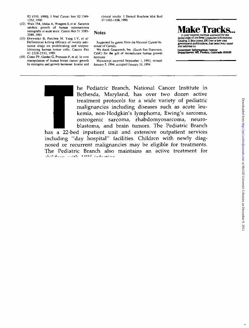
he Pediatric Branch, National Cancer Institute inBethesda, Maryland, has over two dozen activetreatment protocols for a wide variety of pediatricmalignancies including diseases such as acute leu-kemia, non-Hodgkin's lymphoma, Ewing's sarcoma,osteogenic sarcoma, rhabdomyosarcoma, neuro-blastoma, and brain tumors. The Pediatric Branch
has a 22-bed inpatient unit and extensive outpatient servicesincluding "day hospital" facilities. Children with newly diag-nosed or recurrent malignancies may be eligible for treatments.The Pediatric Branch also maintains an active treatment forchildren with HTV infection.
Emphasis is placed on maintaining close communication andcooperation with referring physicians. For more information onpediatric referrals, please call collect 301-402-0696 (cancer) or301-402-1391 (HIV disease).
NATIONAL CANCER INSTITUTE
PEDIATRIC BRANCHA Public Service Announcement Courtesy of this Publication
632 REPORTS Journal of the National Cancer Institute, Vol. 86, No. 8, Apnl 20, 1994
at McG
ill University L
ibraries on Decem
ber 9, 2011http://jnci.oxfordjournals.org/
Dow
nloaded from
rlim
Rectangle
rlim
Rectangle
rlim
Rectangle
rlim
Rectangle