Rhythm and EKG Interpretation Presented for Nursing Cindy ... and... · • If no response, may try...
68
Avera eCARE® © 2018 Rhythm and EKG Interpretation Presented for Nursing Cindy Pirrung, BSN-RN, CEN, CPEN Avera eCARE Clinical Nurse Educator
Transcript of Rhythm and EKG Interpretation Presented for Nursing Cindy ... and... · • If no response, may try...

Avera eCARE® © 2018
Rhythm and EKG InterpretationPresented for Nursing
Cindy Pirrung, BSN-RN, CEN, CPEN Avera eCARE Clinical Nurse Educator
Avera eCARE® © 2018
EKG Basics

Avera eCARE® © 2018
Assess Your Patient
• Are they having chest pain?• Are they short of breath?• How does their skin look? (Pale, normal for race,
dry, diaphoretic, etc.)• Can you palpate peripheral pulses?
• Nurse.org
Avera eCARE® © 2018
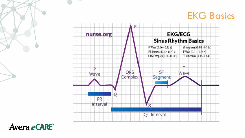
Sinus Rhythms – Normal Sinus Rhythm
60-100bpm
EKG Academy, 2018

Avera eCARE® © 2018
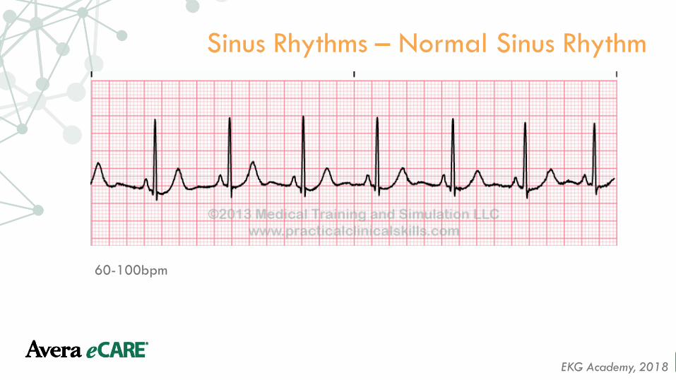
Sinus Bradycardia
<60 bpm and regular
EKG Academy, 2018
Avera eCARE® © 2018
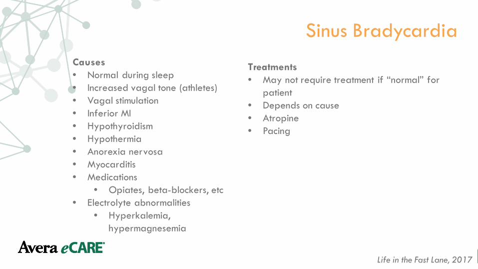
Sinus Bradycardia
Life in the Fast Lane, 2017
Causes• Normal during sleep • Increased vagal tone (athletes)• Vagal stimulation • Inferior MI • Hypothyroidism • Hypothermia • Anorexia nervosa • Myocarditis • Medications
• Opiates, beta-blockers, etc • Electrolyte abnormalities
• Hyperkalemia, hypermagnesemia
Treatments• May not require treatment if “normal” for
patient• Depends on cause • Atropine • Pacing

Avera eCARE® © 2018
Sinus Tachycardia
EKG Academy, 2018
>100 bpm and regular

Avera eCARE® © 2018
Sinus Tachycardia
Life in the Fast Lane, 2017
Causes• Exercise • Pain, Anxiety • Hypoxia • Sepsis • Pulmonary Embolism (PE)• Hyperthyroidism • Medications • Beta-agonists, antihistamines, etc • Caffeine • Marijuana
Treatments • Medications to slow heart rate • Depends on cause

Avera eCARE® © 2018
Sinus Arrhythmia
EKG Academy, 2017
60-100 bpm and irregular

Avera eCARE® © 2018
Sinus Arrhythmia
• Normal physiological phenomenon • Commonly seen in young, healthy persons • Incidence decreases with age • Inspiration increases the heart rate by decreasing vagal tone • With expiration, vagal tone is restored which decreases heart
rate
Life in the Fast Lane, 2017

Avera eCARE® © 2018
Atrial Rhythms – Atrial Fibrillation
ACLS Medical Training, 2018; EKG Academy, 2018
Irregular, rate may be slow, normal or fast
Avera eCARE® © 2018
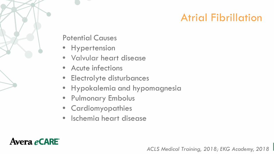
Atrial Fibrillation
ACLS Medical Training, 2018; EKG Academy, 2018
Potential Causes• Hypertension • Valvular heart disease • Acute infections • Electrolyte disturbances • Hypokalemia and hypomagnesia • Pulmonary Embolus • Cardiomyopathies • Ischemia heart disease
Avera eCARE® © 2018
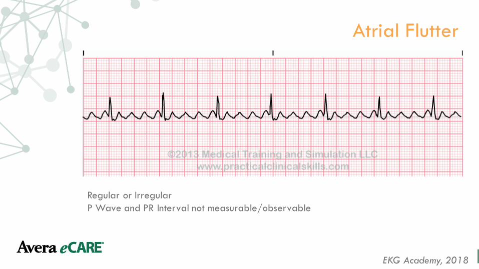
Atrial Flutter
EKG Academy, 2018
Regular or Irregular P Wave and PR Interval not measurable/observable

Avera eCARE® © 2018
Atrial Flutter
Life in the Fast Lane, 2017
A type of supraventricular tachycardiaTreatment:• May try vagal maneuvers• May try Adenosine
• Usually will not convert with these treatments
Avera eCARE® © 2018
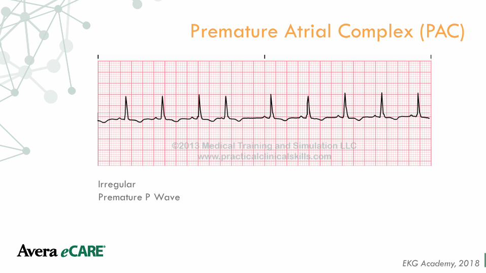
Premature Atrial Complex (PAC)
EKG Academy, 2018
Irregular Premature P Wave

Avera eCARE® © 2018
Premature Atrial Complex (PAC)
Life in the Fast Lane, 2017
• Normal electrophysiological phenomenon not usually requiring investigation or treatment
• Frequent PACs may cause palpitations and a sense of the heart “skipping a beat”• Potential causes:
• Anxiety• Digoxin toxicity• Excess caffeine• Medications• Low potassium/magnesium• Myocardial ischemia

Avera eCARE® © 2018
Supraventricular Tachycardia
EKG Academy, 2018
Regular Fast Rate (150-250 bpm

Avera eCARE® © 2018
Supraventricular Tachycardia
Life in the Fast Lane, 2017
• May respond to vagal maneuvers• If no response, may try Adenosine
• Large bore IV in antecubital• Elevate patient’s arm• Three-way stopcock• Administer fast and flush quickly as Adenosine has quick half-life• First dose: 6mg then may try 12mg if no conversion
• Cardioversion is rarely required

Avera eCARE® © 2018
Wolff-Parkinson-White Syndrome
EKG Academy, 2018
Regular Short PR Interval, Wide QRS

Avera eCARE® © 2018
Ventricular Rhythms – Idioventricular Rhythm
EKG Academy, 2018
Regular rhythm with slow rate (20-40 bpm) Absent P Wave and unmeasurable PR interval
Avera eCARE® © 2018
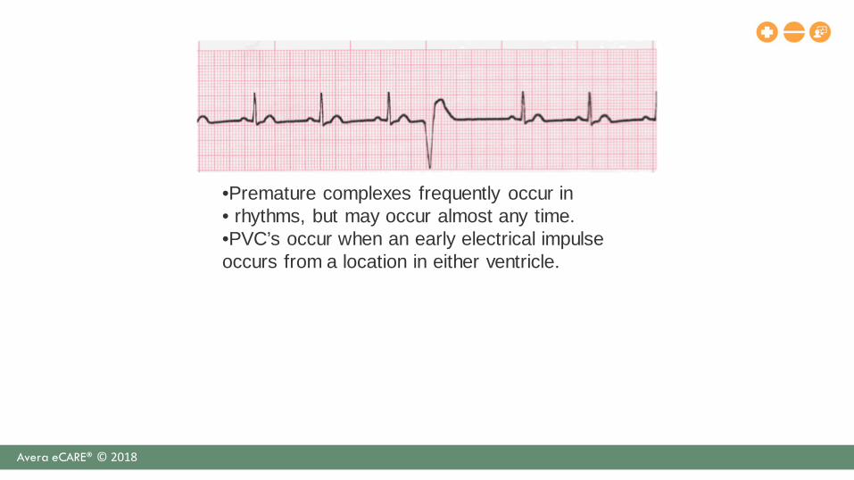
•Premature complexes frequently occur in • rhythms, but may occur almost any time.•PVC’s occur when an early electrical impulse occurs from a location in either ventricle.
Avera eCARE® © 2018
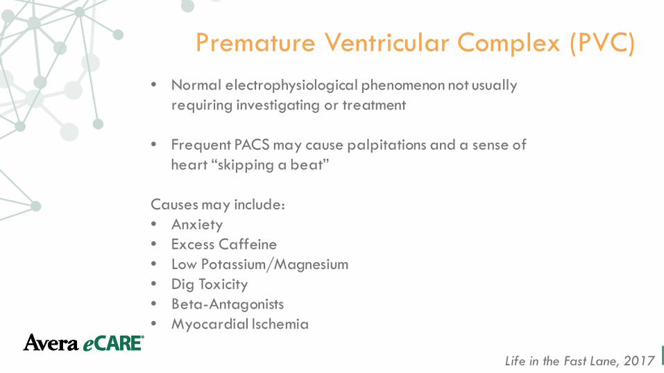
Premature Ventricular Complex (PVC)
Life in the Fast Lane, 2017
• Normal electrophysiological phenomenon not usually requiring investigating or treatment
• Frequent PACS may cause palpitations and a sense of heart “skipping a beat”
Causes may include: • Anxiety • Excess Caffeine • Low Potassium/Magnesium• Dig Toxicity • Beta-Antagonists • Myocardial Ischemia
Avera eCARE® © 2018
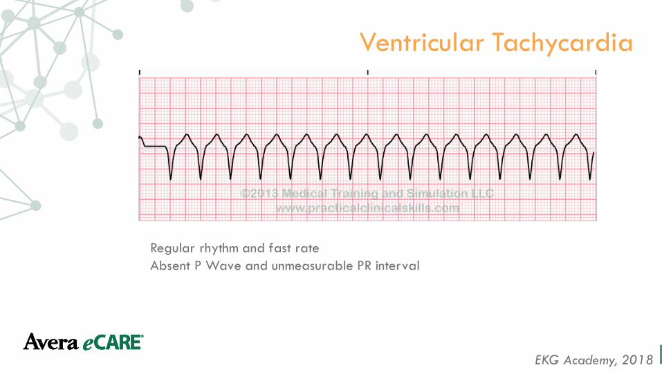
Ventricular Tachycardia
EKG Academy, 2018
Regular rhythm and fast rateAbsent P Wave and unmeasurable PR interval

Avera eCARE® © 2018
Ventricular Tachycardia
Life in the Fast Lane, 2017
May be hemodynamically stableHemodynamically Unstable: • Hypotension• Chest pain• Heart failure• Decrease LOC
• May impair cardiac output due to hypotension and acute cardiac failure through loss of atrial kick
• Decreased cardiac output may decrease myocardial perfusion leading to vfib
Prompt recognition and initiation of treatment is required in all cases of vtach!
Avera eCARE® © 2018
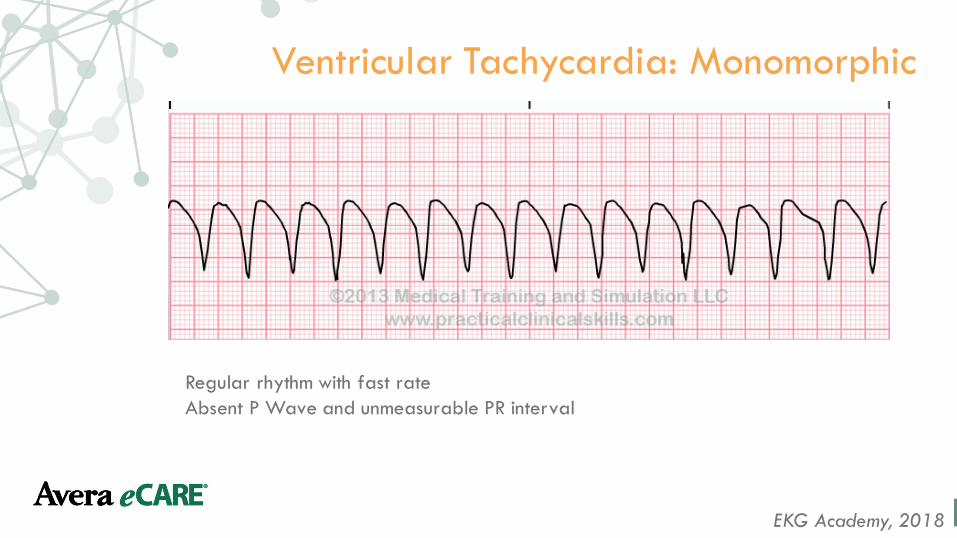
Ventricular Tachycardia: Monomorphic
EKG Academy, 2018
Regular rhythm with fast rate Absent P Wave and unmeasurable PR interval
Avera eCARE® © 2018
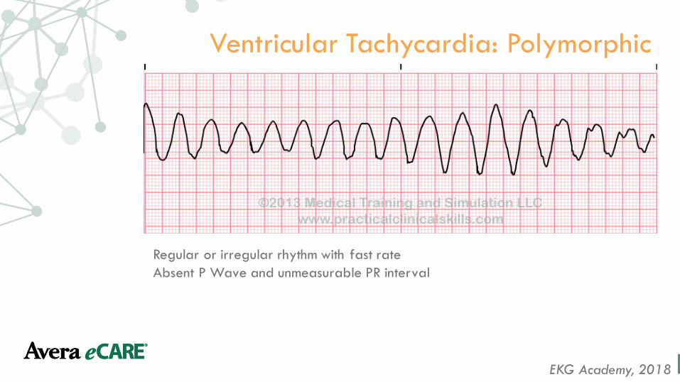
Ventricular Tachycardia: Polymorphic
EKG Academy, 2018
Regular or irregular rhythm with fast rate Absent P Wave and unmeasurable PR interval
Avera eCARE® © 2018
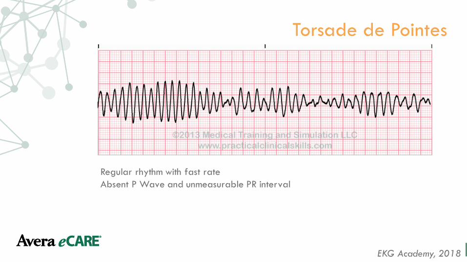
Torsade de Pointes
EKG Academy, 2018
Regular rhythm with fast rate Absent P Wave and unmeasurable PR interval
Avera eCARE® © 2018
Torsade de Pointes
Life in the Fast Lane, 2017
Potential Causes: • Drugs/Poison • Electrolyte abnormalities such as
hypokalemia • Medical conditions
• May degenerate to ventricular fibrillation
Avera eCARE® © 2018
Atrioventricular Rhythms

Avera eCARE® © 2018
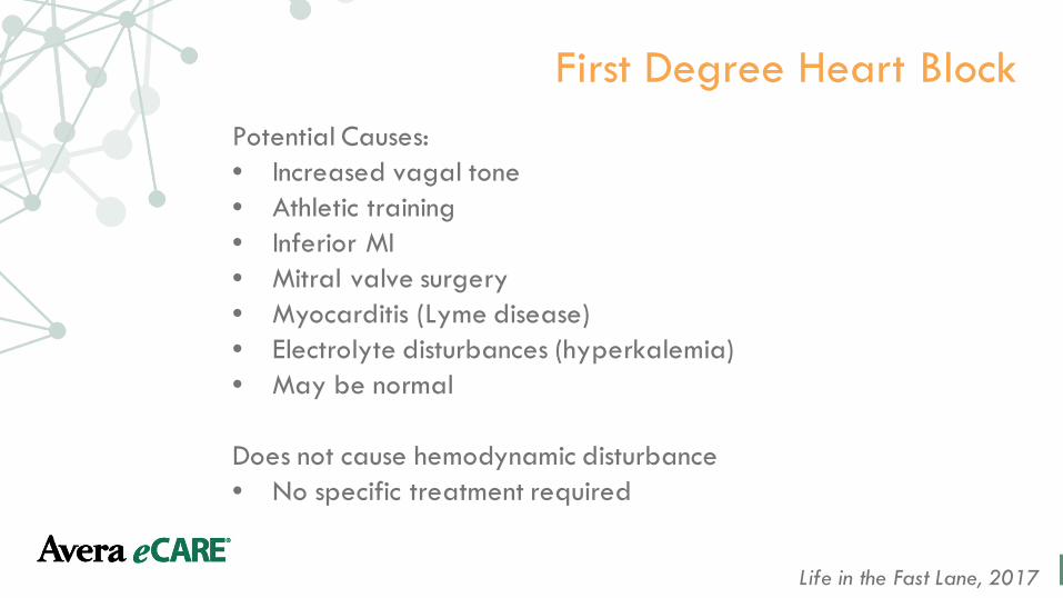
First Degree Heart Block
Regular rhythm with prolonged PR Interval
ACLS Medical Training, 2018
Avera eCARE® © 2018
First Degree Heart Block Potential Causes: • Increased vagal tone • Athletic training • Inferior MI • Mitral valve surgery • Myocarditis (Lyme disease) • Electrolyte disturbances (hyperkalemia) • May be normal
Does not cause hemodynamic disturbance • No specific treatment required
Life in the Fast Lane, 2017

Avera eCARE® © 2018
Second Degree Heart Block Type 1- Wenckebach
EKG Academy, 2018
Irregular rhythm with progressively longer PR interval until a QRS complex is missed, then cycle repeats

Avera eCARE® © 2018
Second Degree Heart Block Type 1- Wenckebach
Life in the Fast Lane, 2017
Potential Causes: • Medications – beta-blockers, calcium channel blockers, digoxin, Amiodarone• Increase vagal tone (athletes) • Inferior MI • Myocarditis • Following cardiac surgery (mitral valve repair, tetralogy of Fallot repair)
• Asymptomatic patients do not require treatment • Symptomatic patients usually respond to Atropine • Permanent pacing is rarely required
Avera eCARE® © 2018
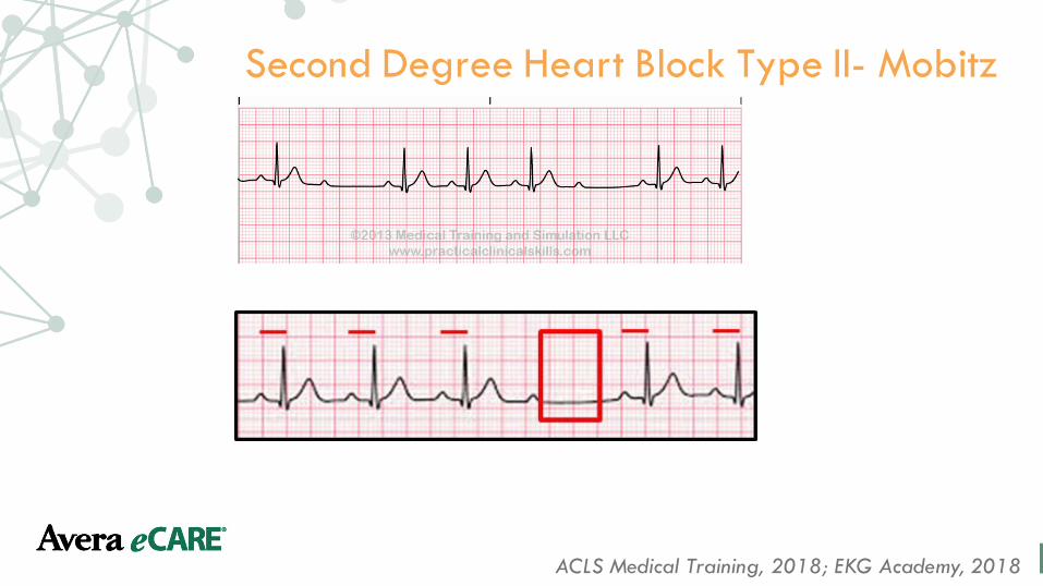
Second Degree Heart Block Type II- Mobitz
ACLS Medical Training, 2018; EKG Academy, 2018

Avera eCARE® © 2018
Avera eCARE® © 2018
Third Degree (Complete) Heart Block
Life in the Fast Lane, 2017
Potential Causes: • Inferior MI • AV node blocking drugs: calcium channel blockers, beta-blockers,
Digoxin• Risk for sudden cardiac death • May require temporary pacing and insertion of permanent
pacemaker
Avera eCARE® © 2018
Bundle Branch Block
EKG Academy, 2018
• Right Bundle branch block Next EKG:• Electrical Conduction problem• Sometimes treatment not required.• Possible pacemaker if symptomatic• Left Bundle branch block• Treat as MI until proven they have had it

Avera eCARE® © 2018
Right Bundle block Branch

Avera eCARE® © 2018
Left Bundle branch block
• See next EKG
Avera eCARE® © 2018
Avera eCARE® © 2018
Electrolyte Imbalances
Avera eCARE® © 2018
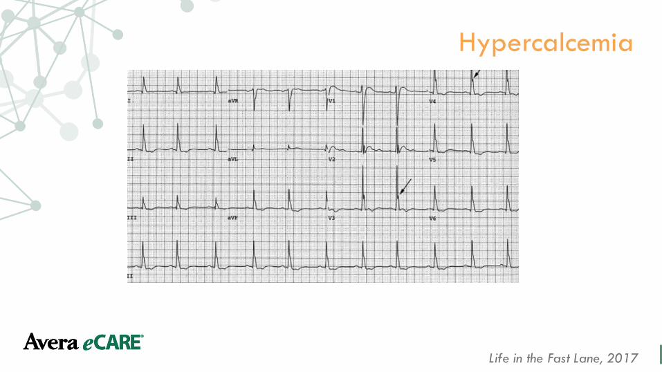
Hypercalcemia
Life in the Fast Lane, 2017

Avera eCARE® © 2018
Hypocalcemia
Life in the Fast Lane, 2017
Avera eCARE® © 2018
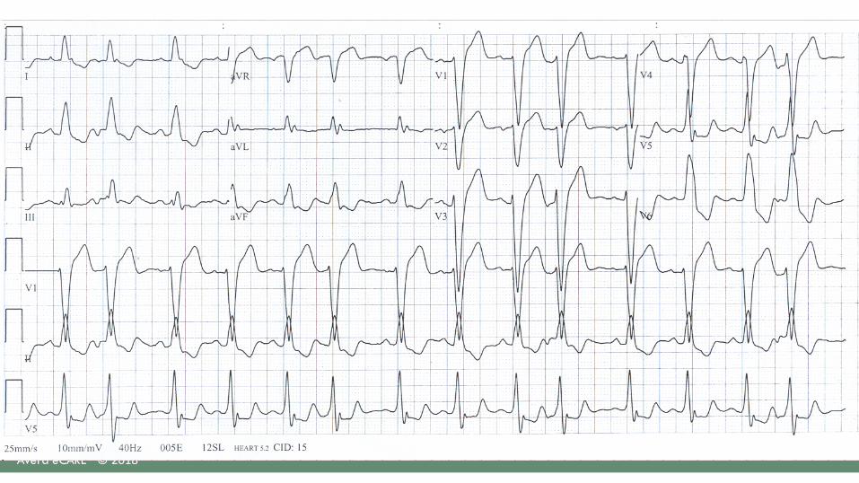
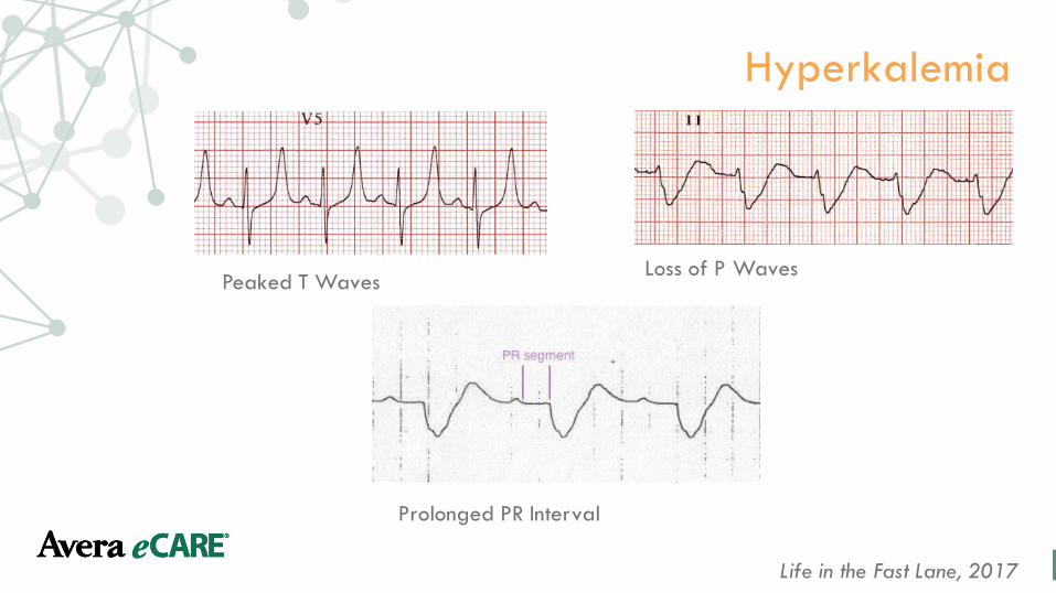
Hyperkalemia
Life in the Fast Lane, 2017
Peaked T Waves Loss of P Waves
Prolonged PR Interval
Avera eCARE® © 2018
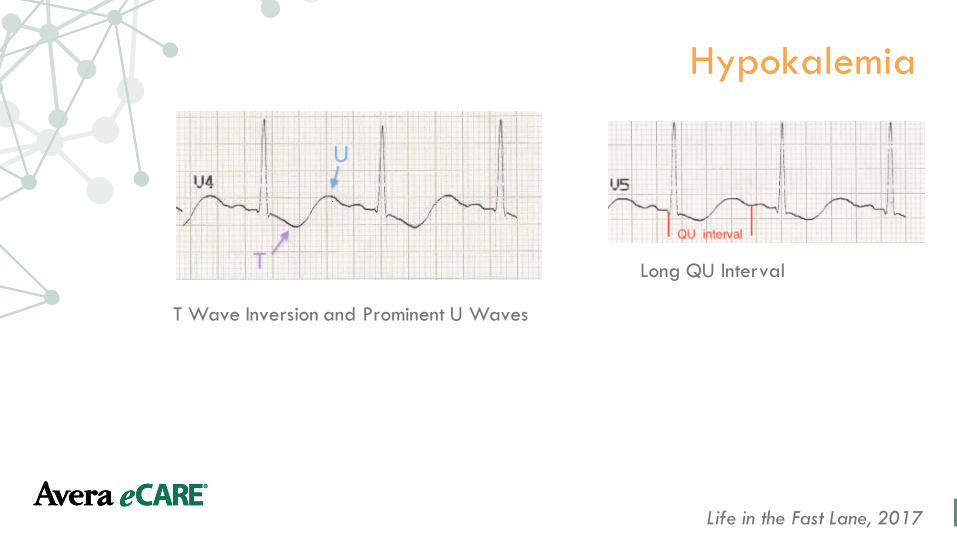
Hypokalemia
Life in the Fast Lane, 2017
T Wave Inversion and Prominent U Waves
Long QU Interval
Avera eCARE® © 2018
Pacemaker Rhythms
Avera eCARE® © 2018
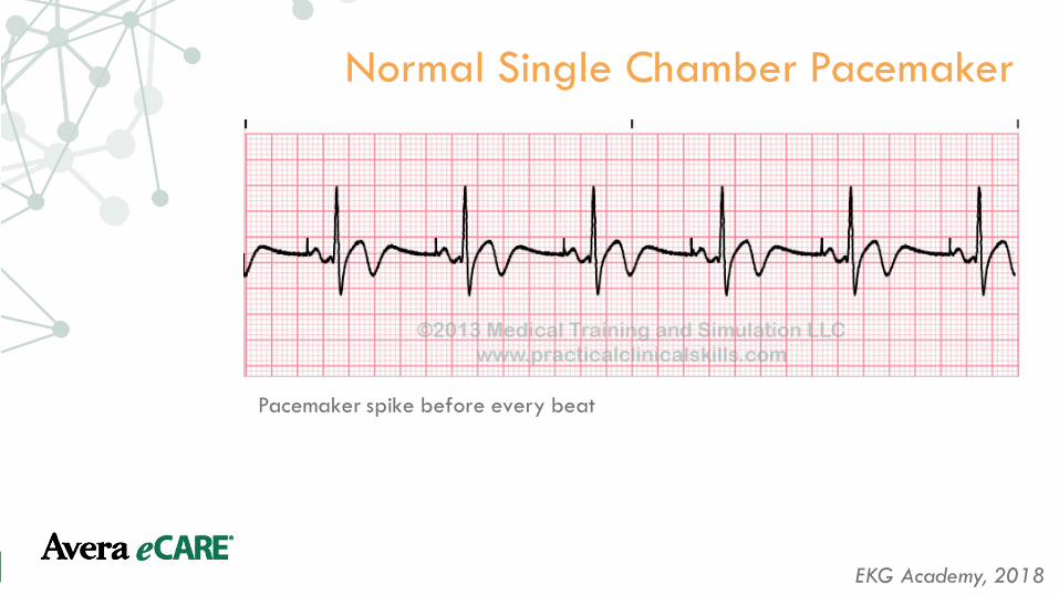
Normal Single Chamber Pacemaker
Pacemaker spike before every beat
EKG Academy, 2018
Avera eCARE® © 2018
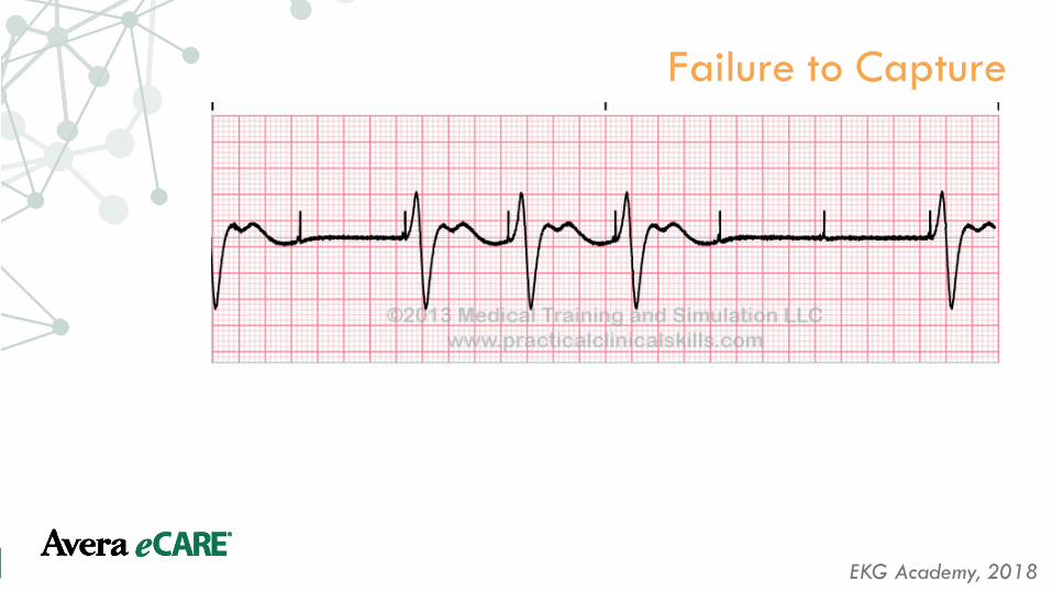
Failure to Capture
EKG Academy, 2018

Avera eCARE® © 2018
Failure to Pace
EKG Academy, 2018
Avera eCARE® © 2018
Myocardial Infarctions

Avera eCARE® © 2018
Background
• Troponin is more specific to cardiac injury and remains elevated longer than other cardiac markers– With contemporary Troponin assays, CKMB and
Myoglobin are not useful for diagnosis of Acute Coronary Syndromes

Avera eCARE® © 2018
Troponin T
• Cardiac Troponin is the preferred marker for myocardial necrosis – A Troponin level should be drawn when the patient is first
assessed– Troponin T levels rise 4 to 6 hours after the onset of chest pain
and peak at 48 hours– Troponin T levels remain elevated and plateau for another 2-5
days, typically returning to normal (reference range) levels 7-14 days after onset of infarction

Avera eCARE® © 2018
Troponin
• Troponin is primarily elevated due to myocardial injury– However, Troponin can be elevated in:
• Unstable angina• Cardiac contusions/Myocardial stunning• Cardiac transplant• CABG surgery• HF and other conditions that may damage the
myocardium
Avera eCARE® © 2018
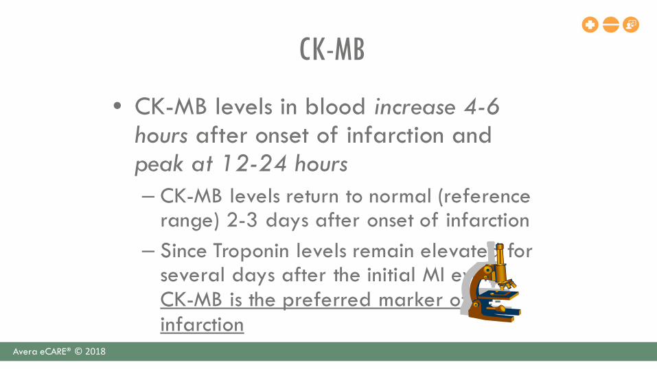
CK-MB
• CK-MB levels in blood increase 4-6 hours after onset of infarction and peak at 12-24 hours– CK-MB levels return to normal (reference
range) 2-3 days after onset of infarction– Since Troponin levels remain elevated for
several days after the initial MI event, CK-MB is the preferred marker of re-infarction

Avera eCARE® © 2018
Anterior MI
Healio, 2018

Avera eCARE® © 2018
Lateral MI
Healio, 2018

Avera eCARE® © 2018
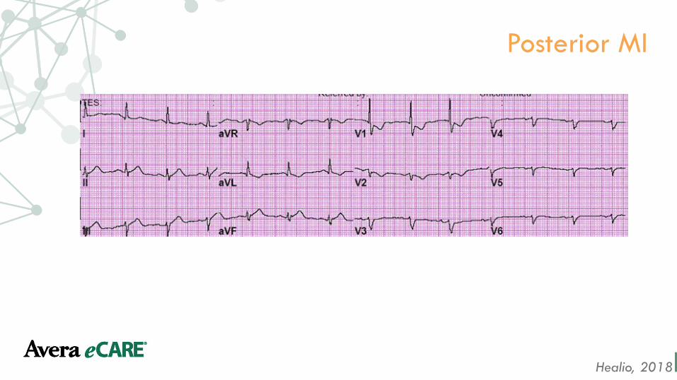
Posterior MI
Life in the Fast Lane, 2017
• Account for 15-20% of STEMIs• Usually occur with an inferior or lateral infarction• Lack of obvious ST elevation (depressed, instead!)
• This causes the diagnosis to be missed
Avera eCARE® © 2018
Posterior MI
Healio, 2018
Avera eCARE® © 2018
Pulseless Rhythms

Avera eCARE® © 2018
Asystole
Chest compressions and epinephrine!
ACLS Medical Training, 2018
Avera eCARE® © 2018
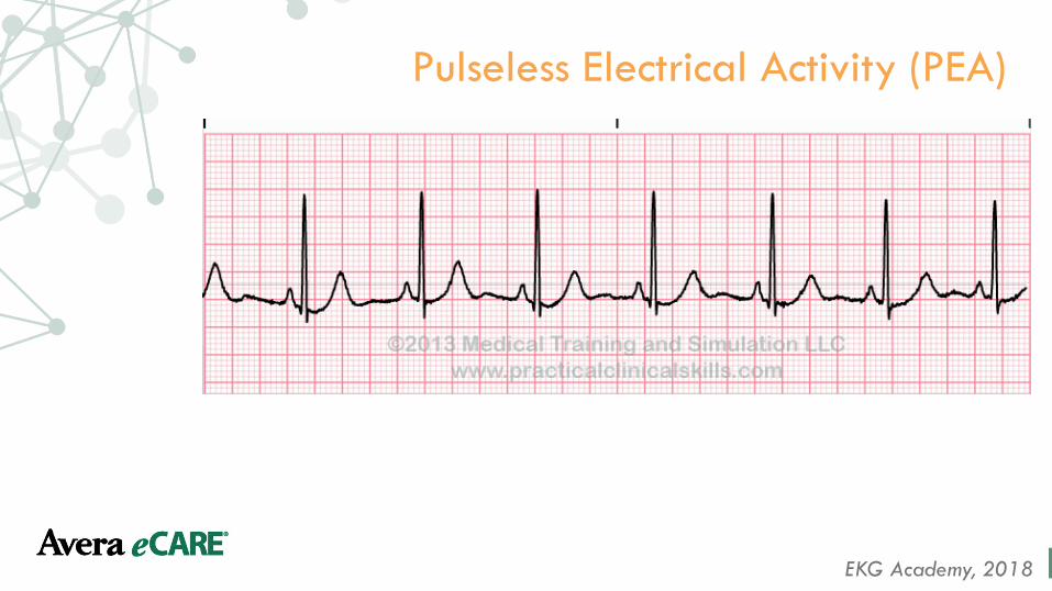
Pulseless Electrical Activity (PEA)
EKG Academy, 2018
Avera eCARE® © 2018
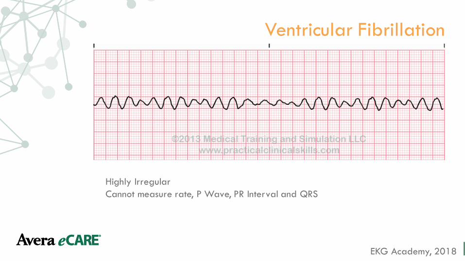
Ventricular Fibrillation
EKG Academy, 2018
Highly Irregular Cannot measure rate, P Wave, PR Interval and QRS
Avera eCARE® © 2018
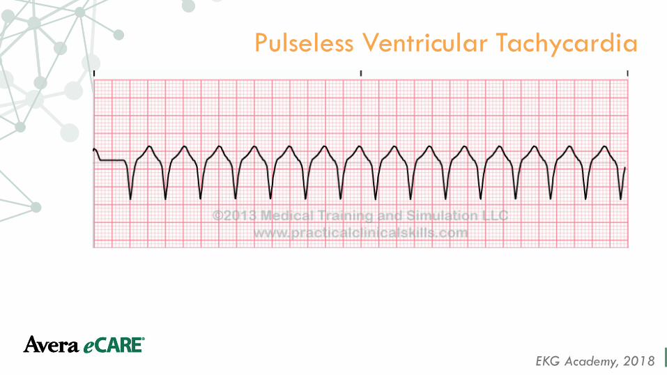
Pulseless Ventricular Tachycardia
EKG Academy, 2018
Avera eCARE® © 2018
Demonstration
Transcutaneous Pacing
Cardioversion
Defibrillation
Avera eCARE® © 2018
Demonstration – Transcutaneous Pacing
• Assess your patient! • For severe/unstable bradycardia • Does your patient have poor perfusion?
• If so, do not delay pacing! • May also administer Atropine 0.5mg IV
• May be repeated every 3 to 5 minutes • Maximum dose 3mg or six total doses
Avera eCARE® © 2018
Demonstration – Cardioversion
• Synchronized: a LOW energy shock• Press the “sync” button • Look for “carrots”• Complete a Time Out with your team • Make sure everyone is clear • Then press SHOCK • Used for:
• Unstable atrial fib, atrial flutter, atrial tachy, SVT• Unsynchronized cardioversion = defibrillation
ACLS Medical Training, 2018
Avera eCARE® © 2018
Demonstration – Defibrillation
• Used for pulseless ventricular tachycardia and ventricular fibrillation • May shock at 200J• Complete a Time Out prior to shock • Make sure everyone is clear
ACLS Medical Training, 2018
Avera eCARE® © 2018
ReferencesACLS Medical Training. (2018). Rhythm recognition. Retrieved from https://www.aclsmedicaltraining.com/rhythm-recognition/
EKG Academy. (2018). EKG academy. Retrieved from https://ekg.academy
Life in the Fast Lane. (2017). ECG clinical interpretation. Retrieved from https://lifeinthefastlane.com/ecg-library/basics/diagnosis/
Nurse.org. (2017). How to read an electrocardiogram (EKG/ECG). Retrieved from https://nurse.org/articles/how-to-read-an-ECG-or-EKG-electrocardiogram/