· Revista Mexicana de FITOPATOLOGÍA Mexican Journal of Phytopathology Sociedad Mexicana de...
167
REVISTA MEXICANA DE FITOPATOLOGÍA MEXICAN JOURNAL OF PHYTOPATHOLOGY ISSN-2007-8080 VOLUMEN 37, NÚMERO 2, Mayo 2019 Órgano Internacional de Difusión de la Sociedad Mexicana de Fitopatología, A.C. Fully Bilingual
Transcript of · Revista Mexicana de FITOPATOLOGÍA Mexican Journal of Phytopathology Sociedad Mexicana de...
REVISTA MEXICANA DE
FITOPATOLOGÍAMEXICAN JOURNAL OF PHYTOPATHOLOGY
ISSN-2007-8080
VOLUMEN 37, NÚMERO 2, Mayo 2019
Órgano Internacional de Difusión de la Sociedad Mexicana de Fitopatología, A.C.
Fully Bilingual

Revista Mexicana de FITOPATOLOGÍAMexican Journal of PhytopathologySociedad Mexicana de Fitopatología, A. C.
Volumen 37, Número 2, 2019Fully Bilingual
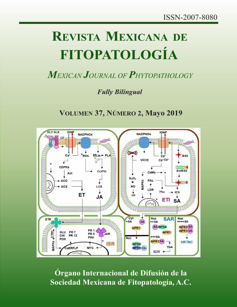
Portada: Inducción de resistencia sistémica, inmunidad activada por efectores y resistencia sistémica adquirida en plantas de Capsicum.
Original: Dr. Ismael Fernando Chávez Díaz y Dra. Emma Zavaleta Mejía, pág. 265.
Editor en Jefe * Editor in ChiefDr. Gustavo Mora Aguilera, COLPOS
Editora Técnica * Technical EditorDra. Norma Ávila Alistac, UAM-Xochimilco
Composición Web y App Movil * Web and App Composition M.C. Eduardo Guzmán Hernández, LANREF, COLPOS Ing. Oscar Eder Flores Colorado, LANREF, COLPOS Ing. Edgar Padilla Ramírez, LANREF, COLPOS
Editoras(es) Adjuntos * Senior EditorsDra. Sylvia Patricia Fernández Pavía, UMSNHDra. Graciela Dolores Ávila Quezada, UACHDra. Silvia Bautista Baños, IPNDra. Irasema del Carmen Vargas Arispuro, CIAD
Comité Editorial Internacional * International Editorial Advisory BoardDr. Rodrigo Valverde, LSU, USA.Dr. Sami Jorge Micheref, Universidad Federal Rural de Pernambuco, Br.Dr. Miguel Dita Rodríguez, EMBRAPA, Br.Dr. Vicente Febres, University of Florida, USA
Editoras(es) Asociados * Associate Editors Dra. María Liliana Flores López, UAAAN Dr. Carlos de León García de Alba, COLPOS Dra. Evangelina Esmeralda Quiñones Aguilar, CIATEJ Dr. Vicente J. Febres, UF, USA Dr. Raúl Allende Molar, Universidad Veracruzana Dr. David Espinosa Victoria, COLPOS Dr. Loreto Robles Hernández, UACH Dr. Alberto Uc Varguez, CIATEJ Dr. Ariel Guzmán Franco, COLPOS Dr. Ángel Rebollar Alviter, UACh Dr. Misael Martínez Bolaños, INIFAP Dr. David Heriberto Noriega Cantú, INIFAP Dr. Luciano Martínez Bolaños, UACh Dra. Guadalupe Valdovinos Ponce, COLPOS Dr. José Armando Carrillo Fasio, CIAD Dr. Rodrigo A. Valverde, LSU, USA Dra. María Alejandra Gutiérrez Espinosa, COLPOS Dr. Hernán García Ruíz, UNL, USA
Artículos Científicos * Scientific Articles
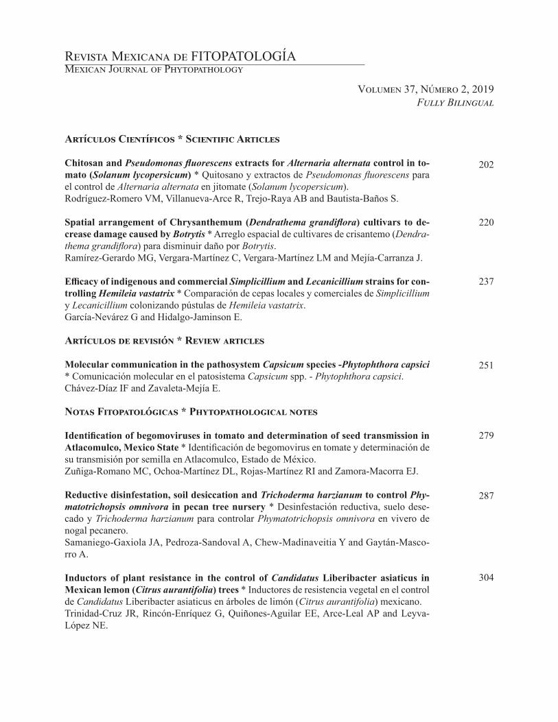
Chitosan and Pseudomonas fluorescens extracts for Alternaria alternata control in to-mato (Solanum lycopersicum) * Quitosano y extractos de Pseudomonas fluorescens para el control de Alternaria alternata en jitomate (Solanum lycopersicum). Rodríguez-Romero VM, Villanueva-Arce R, Trejo-Raya AB and Bautista-Baños S.
Spatial arrangement of Chrysanthemum (Dendrathema grandiflora) cultivars to de-crease damage caused by Botrytis * Arreglo espacial de cultivares de crisantemo (Dendra-thema grandiflora) para disminuir daño por Botrytis.Ramírez-Gerardo MG, Vergara-Martínez C, Vergara-Martínez LM and Mejía-Carranza J.
Efficacy of indigenous and commercial Simplicillium and Lecanicillium strains for con-trolling Hemileia vastatrix * Comparación de cepas locales y comerciales de Simplicillium y Lecanicillium colonizando pústulas de Hemileia vastatrix.García-Nevárez G and Hidalgo-Jaminson E.
Artículos de revisión * Review articles
Molecular communication in the pathosystem Capsicum species -Phytophthora capsici * Comunicación molecular en el patosistema Capsicum spp. - Phytophthora capsici.Chávez-Díaz IF and Zavaleta-Mejía E.
Notas Fitopatológicas * Phytopathological notes
Identification of begomoviruses in tomato and determination of seed transmission in Atlacomulco, Mexico State * Identificación de begomovirus en tomate y determinación de su transmisión por semilla en Atlacomulco, Estado de México.Zuñiga-Romano MC, Ochoa-Martínez DL, Rojas-Martínez RI and Zamora-Macorra EJ.
Reductive disinfestation, soil desiccation and Trichoderma harzianum to control Phy-matotrichopsis omnivora in pecan tree nursery * Desinfestación reductiva, suelo dese-cado y Trichoderma harzianum para controlar Phymatotrichopsis omnivora en vivero de nogal pecanero.Samaniego-Gaxiola JA, Pedroza-Sandoval A, Chew-Madinaveitia Y and Gaytán-Masco-rro A.
Inductors of plant resistance in the control of Candidatus Liberibacter asiaticus in Mexican lemon (Citrus aurantifolia) trees * Inductores de resistencia vegetal en el control de Candidatus Liberibacter asiaticus en árboles de limón (Citrus aurantifolia) mexicano.Trinidad-Cruz JR, Rincón-Enríquez G, Quiñones-Aguilar EE, Arce-Leal AP and Leyva-López NE.
Revista Mexicana de FITOPATOLOGÍAMexican Journal of Phytopathology
Volumen 37, Número 2, 2019Fully Bilingual
220
237
251
279
287
202
304
Revista Mexicana de FITOPATOLOGÍAMexican Journal of Phytopathology
Volumen 37, Número 2, 2019Fully Bilingual
318
330
345
357
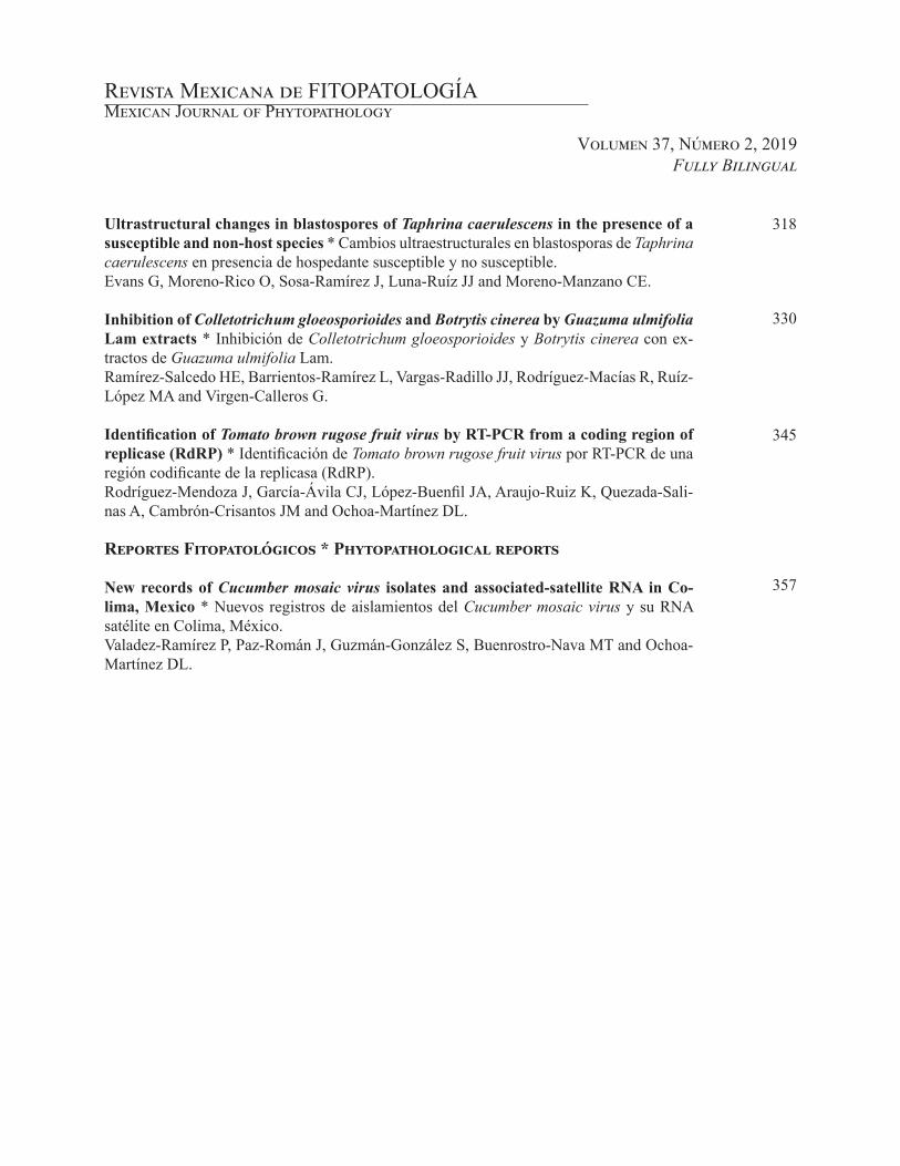
Ultrastructural changes in blastospores of Taphrina caerulescens in the presence of a susceptible and non-host species * Cambios ultraestructurales en blastosporas de Taphrina caerulescens en presencia de hospedante susceptible y no susceptible.Evans G, Moreno-Rico O, Sosa-Ramírez J, Luna-Ruíz JJ and Moreno-Manzano CE.
Inhibition of Colletotrichum gloeosporioides and Botrytis cinerea by Guazuma ulmifolia Lam extracts * Inhibición de Colletotrichum gloeosporioides y Botrytis cinerea con ex-tractos de Guazuma ulmifolia Lam.Ramírez-Salcedo HE, Barrientos-Ramírez L, Vargas-Radillo JJ, Rodríguez-Macías R, Ruíz-López MA and Virgen-Calleros G.
Identification of Tomato brown rugose fruit virus by RT-PCR from a coding region of replicase (RdRP) * Identificación de Tomato brown rugose fruit virus por RT-PCR de una región codificante de la replicasa (RdRP).Rodríguez-Mendoza J, García-Ávila CJ, López-Buenfil JA, Araujo-Ruiz K, Quezada-Sali-nas A, Cambrón-Crisantos JM and Ochoa-Martínez DL.
Reportes Fitopatológicos * Phytopathological reports
New records of Cucumber mosaic virus isolates and associated-satellite RNA in Co-lima, Mexico * Nuevos registros de aislamientos del Cucumber mosaic virus y su RNA satélite en Colima, México.Valadez-Ramírez P, Paz-Román J, Guzmán-González S, Buenrostro-Nava MT and Ochoa-Martínez DL.

202Publicación en línea, mayo 2019
Rodríguez-Romero VM, Villanueva-Arce R, Trejo-Raya AB and Bautista-Baños S. 2019. Chitosan and Pseudo-monas fluorescens extracts for Alternaria alternata con-trol in tomato (Solanum lycopersicum). Mexican Journal of Phytopathology 37(2): 202-219.DOI: 10.18781/R.MEX.FIT.1812-2
Primera publicación DOI: 25 de Febrero, 2019.First DOI publication: February 25, 2019.
Resumen. Alternaria alternata es un hongo que ocasiona daños en el cultivo de jitomate, se carac-teriza por producir manchas negras y marchitez en plantas y frutos; los fungicidas sintéticos son las principales herramientas para controlar este hongo. El objetivo del estudio fue evaluar el efecto de la aplicación de mezclas de quitosano y extractos de Pseudomonas fluorescens en el control de A. alter-nata bajo condiciones in vitro sobre micelio y co-nidios, así como la incidencia y severidad en plantas
Chitosan and Pseudomonas fluorescens extracts for Alternaria alternata control in tomato
(Solanum lycopersicum)
Quitosano y extractos de Pseudomonas fluorescens para el control de Alternaria alternata en jitomate (Solanum lycopersicum)
Víctor Manuel Rodríguez-Romero*, Ramón Villanueva-Arce, Ariadna Berenice Trejo-Raya, Laboratorio de Biotecnología Alimentaria, Unidad Profesional Interdisciplinaria de Biotecnología (UPIBI). Instituto Po-litécnico Nacional (IPN). Avenida Acueducto s/n, Barrio La Laguna Ticomán, Ciudad de México, CP 07340, México; Silvia Bautista-Baños, Departamento de Tecnología Postcosecha, Centro de Desarrollo de Productos Bióticos (CeProBi). IPN. Km 8.5 Carretera Yautepec-Jojutla, San Isidro, Yautepec CP 62731, Morelos, México. *Autor de correspondencia [email protected].
Recibido: 19 de Diciembre, 2018. Aceptado: 10 de Febrero, 2019.
Abstract. Alternaria alternata is a fungus that causes damage to the tomato crop, it is characterized by producing black spots and wilting in plants and fruits; synthetic fungicides are the main tools to control this fungus. The objective of this study was to evaluate the effect of the application of mixtures of chitosan and extracts of Pseudomonas fluorescens in the control in vitro of A. alternata on mycelium and conidia, as well as the incidence and severity in greenhouse tomato plants. The use of the mixture of chitosan 1.5% (w/v) + extract of P. fluorescens 50% (v/v) resulted in 60 and 100% of in vitro inhibition of mycelial growth and conidia germination of A. alternata respectively. In greenhouse, the plants were inoculated with A. alternata, later they were sprayed with the mixture of chitosan 1.5% (w/v) + extract of P. fluorescens 50% (v/v) every 7 days until flowering. The incidence was 100%, while the severity was 51.8 and 38.9% for 7 days and 16.9 and 16.2% for 60 days, respectively. The mixture used is an option for the control of A. alternata.

Publicación en línea, mayo 2019 203
Fully BilingualRevista Mexicana de FITOPATOLOGÍA
Mexican Journal of Phytopathology
de jitomate en invernadero. El uso de la mezcla de quitosano 1.5% (p/v) + extracto de P. fluorescens 50% (v/v) resultó en 60 y 100% de inhibición in vitro de crecimiento micelial y germinación de co-nidios de A. alternata respectivamente. En inver-nadero, las plantas se inocularon con A. alternata, posteriormente se asperjaron con la mezcla de qui-tosano 1.5% (p/v) + extracto de P. fluorescens 50% (v/v) cada 7 días hasta la floración. La incidencia fue del 100 %, mientras que la severidad fue 51.8 y 38.9 para 7 días y 16.9 y 16.2% para 60 días, respectivamente. La mezcla utilizada es una opción para el control de A. alternata.
Palabras clave: actividad antifúngica, control bio-lógico, incidencia, severidad.
México es uno de los diez países a nivel mun-dial con mayor producción de jitomate (Solanum lycopersicum), el cultivo en México satisface la demanda interna y se exporta aproximadamente el 46% de la cosecha (FAO, 2016). Diversos micro-organismos fitopatógenos pueden ocasionar enfer-medades en el cultivo de jitomate, principalmente los hongos (Orberá et al., 2014). Los géneros que ocasionan las mayores pérdidas en pre y poscose-cha son Alternaria, Botrytis, Penicillium, Colleto-trichum y Rhizopus (Petriacq, 2018; Trigos et al., 2008). Las afectaciones que provocan son variadas, desde síntomas poco visibles hasta devastaciones completas de áreas de cultivo (Dean et al., 2012; Strange y Scott, 2005). Los daños por Alternaria son comunes alrededor del mundo, se caracterizan por la aparición de manchas negras y marchitez, que afectan hojas, tallos, flores y frutos (Agrios, 1997; Logrieco et al., 2009). Los fungicidas de ori-gen sintético son las principales herramientas para el control y manejo de enfermedades en plantas; sin embargo, el uso excesivo genera problemas
Key words: antifungal activity, biological control, incidence, severity.
Mexico is one of the ten countries in the world with the highest production of tomato (Solanum lycopersicum). The crop satisfies the internal demand, and approximately 46% of the harvest is exported (FAO, 2016). Diverse phytopathogenic microorganisms may cause diseases in the tomato crop, and particularly fungi (Orberá et al., 2014). The genera which cause the greatest losses in pre- and postharvest are Alternaria, Botrytis, Penicillium, Colletotrichum and Rhizopus (Petriacq, 2018; Trigos et al., 2008). Their effects range from hardly visible symptoms to the devastation of complete plantations (Dean et al., 2012; Strange and Scott, 2005). Damages by Alternaria are common around the world and characteristically display black spots and wilting that affects leaves, stems, flowers and fruits (Agrios, 1997; Logrieco et al., 2009). Synthetic fungicides are the main tools for the control and management of plant diseases; however, excessive use produces important health and environmental problems, which has led to a demand for the management of agrochemical-free crops and foods by consumers (Sánchez-Bayo and Tennekes, 2015).
An alternative for the reduction of agrochemicals is the use of biopolymers such as chitosan, which is an abundant compound in nature, and can be obtained by the partial deacetylation of chitin (main component of the exoskeleton of arthropods) through a thermal alkaline treatment; it has properties of biodegradability and inocuity, as well as antifungal activities, making it an ideal and easily manageable product (Waewthongrak et al., 2015). Diverse authors report that this biopolymer controls diseases caused by phytopathogenic fungi in papaya (Carica papaya), tomato (Physalis

Publicación en línea, mayo 2019 204
Fully BilingualRevista Mexicana de FITOPATOLOGÍAMexican Journal of Phytopathology
importantes en la salud y medio ambiente. Por lo que existe una exigencia de manejo de cultivos y alimentos libres de agroquímicos por parte de los consumidores (Sánchez-Bayo y Tennekes, 2015).
Una alternativa para la reducción de agroquími-cos es el uso de biopolímeros como el quitosano, el cual es un compuesto abundante en la naturale-za, que se obtiene por la desacetilación parcial de la quitina (componente principal del exoesqueleto de artrópodos) a través de un tratamiento térmico alcalino, tiene propiedades de biodegradabilidad, inocuidad y actividad antifúngica que lo hacen un producto ideal y de fácil manejo (Waewthongrak et al., 2015). Diversos autores reportan que este biopolímero controla enfermedades causadas por hongos fitopatógenos en cultivos de papaya (Ca-rica papaya), tomate (Physalis ixocarpa), plátano (Musa × paradisiaca) y otros (Bautista-Baños et al., 2003; De Oliveira et al., 2016; Liu et al., 2007; Maqbool et al., 2010). Además, tiene la capacidad de formar películas o recubrimientos y sirve como matriz para incorporar otros aditivos o componen-tes que pueden añadir o potenciar alguna propiedad (Zargar et al., 2015).
En este sentido, el quitosano es una opción para mezclarse con otros agentes antifúngicos, tales como metabolitos secundarios, como los presentes en los extractos libres de células de Pseudomonas fluorescens. Esta bacteria muestra efectos de con-trol frente a hongos fitopatógenos en los cultivos de col (Brassica oleracea), alfalfa (Medicago sativa), trigo (Triticum aestivum), haba (Vicia faba) y otros (Alemu y Alemu, 2013; Mishra y Arora, 2012; Ya-nes et al., 2012;), mediante la producción de meta-bolitos extracelulares, que pueden recuperarse del medio de cultivo y ejercen un efecto antifúngico sin la necesidad de tener la bacteria presente (Pal y McSpadden, 2006) y abordar las limitaciones del uso de biomasa. El objetivo de este estudio fue evaluar el efecto antifúngico de la combinación de
ixocarpa), banana (Musa × paradisiaca) and other crops (Bautista-Baños et al., 2003; De Oliveira et al., 2016; Liu et al., 2007; Maqbool et al., 2010). It also has the ability to produce films or coatings and it serves as a matrix to incorporate other additives or components that can add or strengthen some properties (Zargar et al., 2015).
In this sense, chitosan is an option for mixing with other antifungal agents, such as secondary metabolites like the ones found in the free extracts in Pseudomonas fluorescens cells. This bacteria displays control effects over phytopathogenic fungi in plantations of cabbage (Brassica oleracea), alfalfa (Medicago sativa), wheat (Triticum aestivum), broad bean (Vicia faba) and others (Alemu and Alemu, 2013; Mishra and Arora, 2012; Yanes et al., 2012;) by producing extracellular metabolites, which can be recovered from the culture medium and have an antifungal effect, without the need of having the bacteria present (Pal and McSpadden, 2006) and tackle the limitations of the use of biomass. The aim of this study was to evaluate the antifungal effect of the combination of chitosan and P. fluorescens extracts on the control in vitro of mycelia and A. alternata conidia, as well as the incidence and severity on tomato plants in greenhouse as an alternative possibility to reduce the use of agrochemicals.
MATERIALS AND METHODS
Biological material. The bacteria Pseudomonas fluorescens was isolated from the bacterial rhizosphere of strawberry stolons (Fragaria sp.) from the location of Ejido de la Finca, Villa Guerrero, State of Mexico (18° 53’ 07” N, 99° 37’ 36” W and an altitude of 1839 masl) during the 2012-2013 production cycle. The bacteria was purified and identified at the level of genus,

Publicación en línea, mayo 2019 205
Fully BilingualRevista Mexicana de FITOPATOLOGÍA
Mexican Journal of Phytopathology
quitosano y extractos de P. fluorescens en el control in vitro de micelio y conidios de A. alternata; así como, la incidencia y severidad en plantas de jito-mate en invernadero, como una posible alternativa para reducir el uso de agroquímicos.
MATERIALES Y MÉTODOS
Material biológico. La bacteria Pseudomonas fluorescens se aisló de la rizosfera microbiana de estolones de fresa (Fragaria sp.) de la localidad del Ejido de la Finca, Villa Guerrero, Estado de Méxi-co (18° 53’ 07” N, 99° 37’ 36” O y altitud de 1839 msnm) durante el ciclo productivo 2012-2013. La bacteria se purificó e identificó a nivel género de acuerdo con la metodología propuesta por Schaad (1988). Se realizó la caracterización molecular por amplificación y secuenciación del rARN 16s y ali-neación con las bases de datos del Banco de Genes del Centro Nacional para la Información Biotecno-lógica (NCBI, por sus siglas en inglés).
El hongo fitopatógeno Alternaria alternata se aisló de frutos de jitomate con síntomas caracte-rísticos de la enfermedad, se identificó y caracteri-zó molecularmente en el Laboratorio de Fisiología Postcosecha del Centro de Desarrollo de Produc-tos Bióticos del Instituto Politécnico Nacional, se cultivó en medio Papa Dextrosa Agar (PDA, BD Bioxon, México) durante 4-7 días e incubó a tem-peratura ambiente (25 ± 2 °C) para los ensayos pos-teriores.
Para los experimentos en invernadero, se utili-zaron plantas de jitomate de la variedad Saladett (Eterno F1) en la etapa vegetativa (de 15 a 25 cm de altura) hasta la floración, obtenidas de siembra directa en pellets de peatmoss y cultivadas en ma-cetas de 5 L aproximadamente, en una mezcla de tierra orgánica-peatmoss-agrolita en proporción 4:1:1, en invernadero (32 °C, 75% HR, aproxima-damente) de febrero a junio de 2017.
following the methodology proposed by Schaad (1988). The molecular characterization was carried out by amplifying and sequencing the rRNA 16s and aligning with the bases of the National Center for Biotechnology Information (NCBI).
The phytopathogenic fungus Alternaria alternata was isolated from tomato fruits with typical symptoms of the disease, then identified and characterized molecularly in the Postharvest Physiology Lab in the Center for the Development of Biotic Products of the National Polytechnic Institute; it was then cultivated a Potato Dextrose Agar medium (PDA, BD Bioxon, Mexico) for 4-7 days and incubated at room temperature (25± 2 °C) for later trials.
Experiments performed in greenhouse were carried out using tomato plants of the Saladett (Eterno F1) variety in stages between vegetative (15 to 25 cm in height) to a flowering states, obtained from direct seeding in peatmoss pellets and grown in pots with an approximate capacity of 5 L, in a mixture of organic soil-peatmoss-agrolite in a proportion of 4:1:1, in greenhouse (32 °C, 75% RH, approximately) between February and June, 2017.
Preparation of chitosan and P. fluorescens extracts. The chitosan solution (Qs) (Sigma Aldrich, USA) was prepared at 3% (w/v) and obtained by dissolving 3.0 g of chitosan of low molecular molecular weight (50-190 kDa, 85% deacetylation) in 100 mL of distilled water, it was adding slowly glacial acetic acid (Fermont, Mexico) at 1% (v/v). The solution was stirred and warmed to 40 °C for 24 h, the pH was adjusted at 5.6 with sodium hydroxide 1 N and sterilized for 15 min at 15 psi. The P. fluorescens (EPf) extract was obtained by cultivating bacteria in a King´s B medium (25±2 °C, 72 h, 120 rpm), centrifuged for 15 min at 10015 xg, and the supernatant was filtered with 0.22 µm sterile membranes (Cole Palmer, USA) to retain the remaining cell biomass.

Publicación en línea, mayo 2019 206
Fully BilingualRevista Mexicana de FITOPATOLOGÍAMexican Journal of Phytopathology
Preparación de quitosano y extractos de P. fluo-rescens. La solución de quitosano (Qs) (Sigma Al-drich, EUA) se preparó al 3% (p/v) y se obtuvo al disolver 3.0 g de quitosano de bajo peso molecular (50-190 kDa, 85% desacetilación) en 100 mL de agua destilada, se añadió lentamente ácido acético glacial (Fermont, México) al 1% (v/v). La solución se agitó y calentó a 40 °C durante 24 h, el pH se ajustó a 5.6 con hidróxido de sodio 1 N y se esterili-zó por 15 min a 15 psi. El extracto de P. fluorescens (EPf) se obtuvo al cultivar la bacteria en medio B de King (25 ± 2 °C, 72 h, 120 rpm), se centrifugó 15 min a 10015 xg, el sobrenadante se filtró con membranas estériles (Cole Palmer, EUA) de 0.22 µm para retener la biomasa celular remanente.
Pruebas de control in vitro de A. alternata: creci-miento micelial y germinación de conidios. Para las pruebas de inhibición del crecimiento micelial se realizó un diseño de tratamientos en arreglo fac-torial, los factores y niveles fueron Qs [0, 0.5, 1.0, y 1.5% (p/v)] y EPf [0, 15, 30 y 50% (v/v)] con seis repeticiones por tratamiento. Se incluyó Cap-tan (0.25% p/v) como control positivo. El pH de todos los tratamientos se ajustó a 5.6. La unidad experimental fue una caja Petri (90 x 15 mm) con medio de cultivo PDA, se depositó y distribuyó de manera uniforme sobre la superficie del medio 0.5 mL de cada uno de los tratamientos y se dejó secar de 5 a 10 min. Después se colocaron en el centro de la caja discos (5 mm de diámetro) de PDA con micelio del hongo e incubaron a temperatura am-biente. El tiempo límite de incubación se determinó cuando el crecimiento micelial del control negati-vo (Qs 0% + Epf 0%) alcanzó el borde de la caja. Para cada unidad experimental se midió y prome-dió el diámetro de la colonia en dos direcciones. El porcentaje de inhibición micelial se calculó según Korsten y Jager (1995) con la fórmula: Inhibición (%) = [(DC - DT) / DC) x 100]; donde DC es el
Control tests in vitro of A. alternata: mycelial growth and conidia germination. For the inhibition trials, was carried out in a treatments design in a factorial arrengement, the factors and levels were Qs [0, 0.5, 1.0, and 1.5% (w/v)] and EPf [0, 15, 30 and 50% (w/v)] with six repetitions per treatment it was included Captan (0.25% w/v) as a positive control. The pH of all treatments was adjusted to 5.6. The experimental unit was a Petri dish (90 x 15 mm) with PDA culture medium; 0.5 mL of each of the treatments was evenly placed and distributed on the surface of the medium, and left to dry for 5 to 10 min. PDA discs (5 mm in diameter) with fungal mycelia were then placed in the center of the dishes and incubated at room temperature. The time limit for incubation was determined when the mycelial growth of the negative control (Qs 0% + Epf 0%) reached the edge of the dish. For each experimental unit was measured and averaged the diameter of culture in two directions. The percentage of mycelial inhibition was calculated following Korsten and Jager (1995) using the formula: Inhibition (%) = [(DC - DT) / DC) x 100], where DC is the diameter of the control culture, and DT is the diameter culture of the treatment.
For the conidia germination trials, it was used the mixture that displayed the most statistically significant mycelial inhibition in vitro (%)*=p≤0.05. Distilled water was included as a negative control, and Captan (0.25% w/v) as a positive control, with 6 repetitions for each treatment. It was placed 0.5 mL of each treatment on the surface of the Petri dishes with PDA, spread it out evenly, left it to dry for 5 to 10 min, inoculated using a conidial suspension (0.1 mL) (106 mL-1) and incubated at 25±2 °C for 4 days, and the percentage of conidia germination was determined.
A. alternata control tests in tomato plants in greenhouse conditions. For the trial in a greenhouse, it was used the mixture that presented

Publicación en línea, mayo 2019 207
Fully BilingualRevista Mexicana de FITOPATOLOGÍA
Mexican Journal of Phytopathology
diámetro del cultivo control, y DT el diámetro del cultivo del tratamiento.
Para la prueba de germinación de conidios se utilizó la mezcla que presentó la mayor inhibi-ción micelial in vitro (%) estadísticamente signi-ficativa *=p ≤ 0.05. Se incluyeron agua destilada como control negativo y Captan (0.25% p/v) como control positivo, con 6 repeticiones para cada tra-tamiento. Se depositó 0.5 mL de cada tratamiento sobre la superficie de las cajas Petri con PDA, se distribuyó de manera uniforme, se dejó secar de 5 a 10 min, se inoculó con una suspensión (0.1 mL) de conidios (106 mL-1) e incubó a 25 ± 2°C durante 4 días y se determinó el porcentaje de germinación de conidios.
Pruebas de control de A. alternata en plantas de jitomate en invernadero. Para la prueba en inver-nadero se utilizó la mezcla que presentó la mayor inhibición micelial in vitro (%) estadísticamente significativa *=p ≤ 0.05. Se incluyeron agua des-tilada como control negativo y Captan (0.25 % p/v) como control positivo, con 15 repeticiones para cada tratamiento. Las plantas se colocaron en cámaras húmedas 24 h antes de la inoculación por aspersión con una suspensión de conidios (106
mL-1). Las plantas inoculadas se incubaron 72 h en cámaras húmedas, después se asperjaron los tratamientos y se llevaron a invernadero (32 °C, 75% HR, aproximadamente). Después de la prime-ra aplicación, los tratamientos se asperjaron cada 7 días hasta la floración y se evaluó la incidencia como la fracción de unidades experimentales por tratamiento con síntomas (tizón de hojas, clorosis, defoliación o marchitez) y la severidad de la enfer-medad como la fracción de hojas dañadas respecto al total de hojas de cada unidad experimental por cada tratamiento (Terna et al., 2016). Las evalua-ciones se realizaron 7, 30 y 60 días después de la inoculación (DDI).
the most the most in vitro mycelial inhibition (%) statistically significant. Distilled water was included as a negative control and Captan (0.25% w/v) as a positive control, with 15 repetitions per treatment. The plants were placed in wet chambers for 24 h before inoculation by spraying a suspension of conidia (106 mL-1). Inoculated plants were incubated for 72 h in wet chambers, and the treatments were later sprayed and taken to a greenhouse (32 °C, 75% HR, approximately). After the first application, the treatments were sprayed every 7 days until flowering. Incidence was evaluated as the fraction of experimental units per treatments with symptoms (leaf smut, chlorosis, defoliation or wilting) and the severity of the disease as the fraction of leaves damaged in regard to the total of leaves of each experimental unit per treatment (Terna et al., 2016). Evaluations were carried out 7, 30 and 60 days after inoculation (DAI).
Data analysis. For the treatments in vitro and in greenhouse, a completely random design was used, and they were analyzed using an ANOVA; the treatments were compared using Tukey’s Test (*=p≤0.05). Before the analysis, the severity data as percentages were transformed with a logarithm [log (x + 1)]. The analyses were carried out using SAS® 9.4.
RESULTS AND DISCUSSION
Biological material. The P. fluorescens bacteria isolated in strawberry stolons was Gram negative; in the specific yeast-dextrose-calcium carbonate (YDC) growth medium, the colonies displayed a creamy-white color after 48 h in incubation; in King B agar (KB), after 48 h of incubation, the colonies produced a yellow pigment that surrounded the

Publicación en línea, mayo 2019 208
Fully BilingualRevista Mexicana de FITOPATOLOGÍAMexican Journal of Phytopathology
Análisis de datos. Para los tratamientos in vitro y en invernadero se utilizó un diseño completamen-te al azar y se analizaron por medio de un ANDE-VA, los tratamientos se compararon con la prueba Tukey (*=p ≤ 0.05). Antes del análisis, los datos de severidad en porcentaje se transformaron con loga-ritmo [log (x + 1)]. Estos análisis se realizaron con SAS® 9.4.
RESULTADOS Y DISCUSIÓN
Material biológico. La bacteria P. fluorescens aislada en estolones de fresa fue Gram negativa, en el medio de crecimiento específico levadura-dextrosa-carbonato de calcio (YDC) las colonias mostraron coloración crema-blanco después de 48 h de incubación; en agar B de King (KB), después de 48 h de incubación, las colonias produjeron un pigmento amarillo que rodeó la colonia y que bajo condiciones de luz ultravioleta fue verde-amarillo fluorescente (Figura 1). Las colonias blancas en YDC pueden corresponder a los géneros Pseudo-monas, Erwinia o Agrobacterium, mientras que la
colony, which was fluorescent greenish-yellow under UV light (Figure 1). The white colonies in YDC may belong to the genera Pseudomonas, Erwinia or Agrobacterium, while pigmentation and fluorescence in King´s B agar are typical of P. fluorescens (Schaad, 1988).
The result of the culture characterization was corroborated using molecular characterization (Table 1) with the analysis of the gene 16s and the alignment of the sequence of base pairs (bp) with those from the NCBI Gene Bank. The DNA had a molecular weight of 425 bp; the sequence was aligned in first, second and third places, respectively with Pseudomonas lutea (Access number NZ_JRMB01000004.1, NZ_JRMB01000003.1 and NZ_JRMB01000002.1, Kwak et al., 2016), with a similarity index of 99% and 467 bp for all sequences; the difference was two nucleotides (NZ_JRMB01000004.1) and three nucleotides (NZ_JRMB01000003.1, NZ_JRMB01000002.1) and maximum scores of 774, 769 and 769, respectively, out of a total score of 774. In seventh place, it aligned with P. fluorescens (access number NR113647, Redondo et al., 2012) with a value
Figura 1. Características culturales de P. fluorescens. Colonias en medio YDC (A), pigmento amarillo en agar King B (B), y fluorescencia bajo luz UV (C).
Figure 1. Cultural characteristics of P. fluorescens. Colonies in YDC medium YDC (A), yellow pigment in King´s B agar (B), and fluorescence under UV light (C).

Publicación en línea, mayo 2019 209
Fully BilingualRevista Mexicana de FITOPATOLOGÍA
Mexican Journal of Phytopathology
pigmentación y fluorescencia en agar B de King es característico de P. fluorescens (Schaad, 1988).
El resultado de la caracterización cultural se co-rroboró con la caracterización molecular (Cuadro 1) mediante el análisis del gen 16s y el alineamien-to de la secuencia de los pares de bases (pb) con las del Banco de Genes del NCBI. El ADN tuvo un peso molecular de 425 pb, la secuencia se ali-neó en primer, segundo y tercer lugar respectiva-mente con Pseudomonas lutea (Número de acceso NZ_JRMB01000004.1, NZ_JRMB01000003.1 y NZ_JRMB01000002.1, Kwak et al., 2016), con un índice de similaridad del 99% y 467 pb para to-das las secuencias, la diferencia fue de dos nucleó-tidos (NZ_JRMB01000004.1) y tres nucleótidos (NZ_JRMB01000003.1, NZ_JRMB01000002.1) y puntuaciones máximas de 774, 769 y 769 res-pectivamente de una puntuación total de 774. En séptimo lugar se alineó con P. fluorescens (Número de acceso NR113647, Redondo et al., 2012) con un valor de 763 y un índice de 99 %, con una di-ferencia de cuatro nucleótidos, ambas secuencias con 425 pb. Las diferencias entre nucleótidos de P. fluorescens y la secuencia de estudio se encuentran distribuidas de manera aleatoria a lo largo de toda la secuencia, a diferencia de P. lutea, donde las di-ferencias se localizan en los mismos codones para las tres secuencias; la diferencia de los nucleótidos y pares de bases entre la secuencia de estudio y P. lutea indica diferencias moleculares que caracte-rizan a cada especie, mientras que las diferencias
Cuadro 1. Secuencia de pares de bases de P. fluorescens aislada en Villa Guerrero, Estado de MéxicoTable 1. Sequence of base pairs of P. fluorescens isolated in Villa Guerrero, State of Mexico.
CCGGGAACGTATTCACCGCGACATTCTGATTCGCGATTACTAGCGATTCCGACTTCACGCAGTCGAGTTGCAGACT-GCGATCCGGACTACGATCGGTTTTATGGGATTAGCTCCACCTCGCGGCTTGGCAACCCTTTGTACCGACCATTGTA-GCACGTGTGTAGCCCAGGCCGTAAGGGCCATGATGACTTGACGTCATCCCCACCTTCCTCCGGTTTGTCACCGG-CAGTCTCCTTAGAGTGCCCACCATAACGTGCTGGTAACTAAGGACAAGGGTTGCGCTCGTTACGGGACTTAACCCA-ACATCTCACGACACGAGCTGACGACAGCCATGCAGCACCTGTCTCAATGTTCCCGAAGGCACCAATCCATCTCTG-GAAAGTTCATTGGATGTCAAGGCCTGGTAAGGTTCTTCGCGTTGCTTC
of 763 and an index of 99 %, with a difference of four nucleotides, both sequences with 425 pb. The differences between P. fluorescens nucleotides and the sequence under study are distributed at random throughout the sequence, unlike P. lutea, where the differences are found in the same codons for the three sequences; the difference of the nucleotides and the base pairs between the sequence under study and P. lutea indicates molecular differences that distinguish such species, whereas the differences between P. fluorescens and the sequence under study may be due to polymorphisms by substitution of base pairs, attributable to a possible mutation; based on the high molecular homology and culture characterization, the biocontrol agent was concluded to be P. fluorescens.
In vitro A. alternata control tests: mycelial growth and conidia germination. The radial growth in the negative control A. alternata in PDA medium covered 100% of the Petri dish after 26 days of incubation. All treatments had a reduction mycelial growth statistically significant (*=p≤0.05) in regard to the negative control, as well as an inhibiting effect (Table 2, Figure 2).
In the treatments containing only Qs, the mycelial growth of A. alternata was inhibited between 9 and 33%, although in other studies the inhibiting effect was observed with concentrations starting at 0.5% of chitosan and higher than those reported in the present paper, with an inhibition of 54.6%

Publicación en línea, mayo 2019 210
Fully BilingualRevista Mexicana de FITOPATOLOGÍAMexican Journal of Phytopathology
entre P. fluorescens y la secuencia de estudio puede deberse a polimorfismos por sustitución de pares de bases, atribuibles a una posible mutación; con base a la alta homología molecular y la caracterización cultural, se concluye que el agente de biocontrol fue P. fluorescens.
Pruebas de control in vitro de A. alternata: cre-cimiento micelial y germinación de conidios. El crecimiento radial en el control negativo de A. al-ternata en medio PDA cubrió el 100% de la caja de Petri después de 26 días de incubación. Todos los tratamientos tuvieron una reducción en el cre-cimiento micelial estadísticamente significativo (*=p≤0.05) con respecto al control negativo, así como un efecto inhibitorio (Cuadro 2, Figura 2).
En los tratamientos que contenían únicamente Qs, el crecimiento micelial de A. alternata se inhi-
Cuadro 2. Crecimiento micelial e inhibición (%) in vitro de las mezclas de quitosano y extractos de P. fluorescens sobre A. alternata.
Table 2. In vitro mycelial growth and inhibition (%) of the mixtures of chitosan and extracts of P. fluore-scens on A. alternata.
Tratamiento (Qs % + EPf %) Crecimiento de A. alternata (mm) Inhibición (%) de A. alternata
Captan (0.25 % p/v) x 24.8 h 70.8 h
Qs 0.0 + EPf 0.0 y 85.0 a 0.0 a
Qs 0.5 + EPf 0.0 77.2 b 9.2 b
Qs 1.0 + EPf 0.0 75.6 b 11.0 b
Qs 1.5 + EPf 0.0 56.8 c 33.2 c
Qs 0.0 + EPf 15 55.1 c 35.2 c
Qs 0.0 + EPf 30 46.3 d 45.5 d
Qs 0.0 + EPf 50 44.4 d e 48.8d e
Qs 0.5 + EPf 15 52.7 c 38.0 c
Qs 0.5 + EPf 30 43.5 d e 48.8 d e
Qs 0.5 + EPf 50 39.6 e f 54.9 e f
Qs 1.0 + EPf 15 43.8 d e 48.5d e
Qs 1.0 + EPf 30 41.6 d e f 51.0 d e f
Qs 1.0 + EPf 50 38.3 f g 56.9 f g
Qs 1.5 + EPf 15 44.4 d 47.8 d
Qs 1.5 + EPf 30 39.6 e f 53.4 e f
Qs 1.5 + EPf 50 33.8 g 60.2 g
x Control positivo. y Control negativo. Medias con distinta letra son estadísticamente diferentes (Tukey *=p≤0.05) / x Positive control. y Negative control. Means with different letters are statistically different (Tukey’s Test *=p≤0.05).
for Fusarium solani f.sp. glycines (Prapagdee et al., 2007) and 100% for Botrytis cinerea (Liu et al., 2007) and Alternata chikunkiana (Meng et al., 2008).
On the other hand, the individual antifungal effect of the EPfs were higher than for Qs in all cases, with a maximum inhibition of 47%; in comparison with other cell extracts, the extracts of Streptomyces griseus reduced the in vitro growth of Fusarium oxysporum f. sp. cubense (Zacky and Ting, 2013) by 33% and Bacillus subtilis extracts inhibited the in vitro mycelial development of Penicillium digitatum by 94% (Waewthongrak et al., 2015).
The mixtures of Qs and EPfs had a higher inhibition in comparison with the individual effects in all cases; the mixture with the highest concentration of both components (Qs 1.5% +

Publicación en línea, mayo 2019 211
Fully BilingualRevista Mexicana de FITOPATOLOGÍA
Mexican Journal of Phytopathology
Figura 2. Efecto de control in vitro de las mezclas de quitosano y extractos de P. fluorescens (Qs % + Epf %) sobre A. alter-nata.
Figure 2. Figure 2. Effect of the control in vitro of the mixtures of chitosan and extracts of P. fluorescens (Qs % + Epf %) on A. alternata.

Publicación en línea, mayo 2019 212
Fully BilingualRevista Mexicana de FITOPATOLOGÍAMexican Journal of Phytopathology
bió entre 9 y 33%, sin embargo, en otros estudios el efecto inhibitorio se observó con concentraciones desde 0.5% de quitosano y mayores a las reporta-das en el presente trabajo, con una inhibición del 54.6% para Fusarium solani f.sp. glycines (Prapag-dee et al., 2007) y del 100% para Botrytis cinerea (Liu et al., 2007) y Alternata chikunkiana (Meng et al., 2008).
Por otro lado, el efecto antifúngico individual de los EPf fue mayor que la del Qs en todos los ca-sos, con una inhibición máxima del 47 %; en com-paración con otros extractos celulares, los extractos de Streptomyces griseus redujeron un 33 % el de-sarrollo in vitro de Fusarium oxysporum f. sp. cu-bense (Zacky y Ting, 2013) y extractos de Bacillus subtilis inhibieron un 94% el desarrollo micelial in vitro de Penicillium digitatum (Waewthongrak et al., 2015).
Las mezclas de Qs y EPf tuvieron mayor inhibi-ción en comparación con los efectos individuales en todos los casos, la mezcla con la mayor concentra-ción de ambos componentes (Qs 1.5 % + EPf 50%) tuvo el mayor efecto de control en el crecimiento micelial de A. alternata; ya se reportó el efecto de la mezcla de quitosano y otros compuestos anti-fúngicos para el control de hongos fitopatógenos, la mezcla de quitosano con cera de abeja y aceite esencial de limón inhibe el 100% el crecimiento micelial de Rhizopus stolonifer (Ramos-García et al., 2012), la mezcla de quitosano con etanol (Qs 0.5% + etanol 20%) redujo hasta 94% el deterioro por B. cinerea en uva (Romanazzi et al., 2007), la mezcla de quitosano con extractos de hojas de pa-paya y chirimoya redujeron el crecimiento micelial de Colletotrichum gloeosporoides hasta 50% con respecto al control (Bautista-Baños et al., 2003).
La mayor inhibición del crecimiento micelial de este estudio (60.2%) fue menor respecto a ensayos anteriores, en donde los agentes antifúngicos se mezclaron con el sustrato (PDA) como un medio
EPf 50%) had the greatest effect of controlling the mycelial growth of A. alternata. The effect of the mixture of chitosan and other antifungal effects for the control of phytopathogenic fungi was reported earlier; the mixture of chitosan with beeswax and essential lemon oil inhibits the mycelial growth of Rhizopus stolonifer (Ramos-García et al., 2012) by 100%, the mixture of chitosan with ethanol (Qs 0.5% + ethanol 20%) reduced by up to 94% the deterioration caused by B. cinerea in grapes (Romanazzi et al., 2007), whereas the mixture of chitosan with papaya and custard apple leaf extracts reduced the mycelial growth of Colletotrichum gloeosporoides by up to 50% in comparison with the control (Bautista-Baños et al., 2003).
The greatest inhibition of mycelial growth in this study (60.2%) was lower than in previous studies, where antifungal agents were mixed with the substrate (PDA) as poisoned medium; for this investigation, it was applied as a cover on the surface of the culture medium to simulate the conditions of in vivo applications, where the biocontrol agents are sprayed on the surface of leaves and fruits (Feliziani et al., 2015; Saavedra et al., 2016). The mixture of Qs 1.5% + EPf 50% was used to evaluate the conidia germination, and had an inhibiting effect of 100%, equal to the effect of the commercial fungicide, in comparison with the negative control, 4 days after applying the treatments (Figure 3).
The control effect of the mixture Qs 1.5% + EPf 50% is related to the antifungal capacity of the components. The polycationic nature of the chitosan interferes with the negative charges of the cell membrane, modifies permeability and causes the leak of intracellular material, as well as the separation between cell membrane and wall of hyphae and conidia (Bautista-Baños et al., 2016; Sánchez-Domínguez et al., 2011). The slimming of hyphae and the loss of cytoplasmic content is due

Publicación en línea, mayo 2019 213
Fully BilingualRevista Mexicana de FITOPATOLOGÍA
Mexican Journal of Phytopathology
envenenado; para este trabajo, se aplicó como recu-brimiento en la superficie del medio de cultivo, que simula las condiciones de aplicaciones in vivo en donde los agentes de biocontrol se asperjan sobre la superficie de hojas y frutos (Feliziani et al., 2015; Saavedra et al., 2016). La mezcla de Qs 1.5% + EPf 50% se utilizó para evaluar la germinación de co-nidios y tuvo un efecto inhibitorio de 100 %, igual al efecto del fungicida comercial, en comparación con el control negativo, después de 4 días de apli-car los tratamientos (Figura 3).
El efecto de control de la mezcla Qs 1.5% + EPf 50%, se relaciona con la capacidad antifúngica de los componentes. La naturaleza policatiónica del quitosano interfiere con las cargas negativas de la membrana celular, modifica la permeabilidad y provoca la fuga del material intracelular, así como la separación entre membrana y pared celular de hifas y conidios (Bautista-Baños et al., 2016; Sán-chez-Domínguez et al., 2011). El adelgazamiento de hifas y perdida del contenido citoplasmático se debe al aumento en la permeabilidad de la membra-na, inducida por la interacción electrostática de los
Figura 3. Germinación de conidios de Alternaria alternata en PDA; control negativo (A), mezcla de Qs 1.5% + EPf 50% (B),
Captan 0.25% p/v (C).
Figure 3. Conidia germination of Alternaria alternata in PDA; negative control (A), mixture of Qs 1.5% + EPf 50% (B),
Captan 0.25% w/v (C).
to the increase in membrane permeability, induced by the electrostatic interaction of the amino groups of the chitosan with the negative charges of the membrane (Palma et al., 2008).
The use of P. fluorescens extracts (Epf) is an interesting option for tackling the limitations of the use of cell biomass. P. fluorescens is commonly used as a biocontrol agent by the application of viable cells in the soil or plants, with variable results due to the lack of long term viability and inability to produce specialized resistance structures (endospores) like other biocontrol genera do (Narayasamy, 2003). In addition, inoculation is inconsistent between fields and between years, due to the variability in colonization, which results in a variable expression of the biocontrol mechanisms as antibiotics (Mark et al., 2006). Another limitation of the use of P. fluorescens is the environmental impact on the native saprophytic populations, with negative effects on the rhizosphere (Couillerot et al., 2008). In addition, there is the possible antibiotic transference (Nwosu, 2001) to other bacteria, it been documented the transference

Publicación en línea, mayo 2019 214
Fully BilingualRevista Mexicana de FITOPATOLOGÍAMexican Journal of Phytopathology
grupos amino del quitosano con las cargas negati-vas de la membrana (Palma et al., 2008).
El uso de extractos de P. fluorescens (Epf) re-presenta una interesante opción para abordar las li-mitaciones del uso de biomasa celular. Comúnmen-te P. fluorescens se usa como agente de biocontrol mediante la aplicación de células viables en el sue-lo o plantas, con resultados variables debido a la falta de viabilidad a largo plazo e incapacidad para producir estructuras especializadas de resistencia (endosporas) como otros géneros de biocontrol (Narayasamy, 2003). Adicionalmente la inocula-ción es inconsistente de un campo de cultivo a otro y de un año a otro, debido a la variabilidad en la colonización que resulta en una expresión variable de los mecanismos de biocontrol como antibióti-cos (Mark et al., 2006). Otra limitante del uso de P. fluorescens es el impacto ecológico en las pobla-ciones saprofitas indígenas, con efectos negativos sobre la rizosfera (Couillerot et al., 2008). Además, existe la posible transferencia a antibióticos (Nwo-su, 2001) a otras bacterias, ya que se ha documen-tado la transferencia de plásmidos entre cepas in-troducidas e indígenas de bacterias del suelo (Daa-ne et al., 1996). El uso de los extractos producidos por P. fluorescens (Epf) de este estudio demuestra el efecto antifúngico in vitro, como una alternativa viable para el control de A. alternata.
En los extractos de P. fluorescens del presente estudio, no se determinaron las identidades de los compuestos antifúngicos, pero el control biológico por P. fluorescens se atribuye a la producción de compuestos extracelulares como fenazinas, fluo-roglucinoles, pirrolidina, pirrolnitrina, lipopépti-dos cíclicos, sideróforos y cianuro de hidrógeno (Gerhardson, 2002; Hass y Defago, 2005). Se ha propuesto que estos compuestos actúan como in-hibidores enzimáticos en el metabolismo de la glucosa, que se difunden a través de la membrana, actúan como agentes reductores, producen com-
of plasmids due to the transference of plasmids between introduced and native strains of soil bacteria (Daane et al., 1996). The use of extracts produced by P. fluorescens (Epf) in this study show the in vitro antifungal effect as a viable alternative for the control of A. alternata.
In the extracts of P. fluorescens in this study, the identities of the antifungal compounds were not determined, yet the biological control by P. fluorescens is attributed to the production of extracellular compounds such as phenazines, fluoroglucinols, pyrrolidine, pyrrolnitrine, cyclic lipopeptides, siderophores and hydrogen cyanide (Gerhardson, 2002; Hass and Defago, 2005). It has been suggested that these compounds act as enzyme inhibitors in the metabolism of glucose, which diffuse through the membrane, act as reduction agents, produce toxic compounds that affect the morphology of hyphae and conidia, and speed up the death process (Chin-A-Woeng et al., 2003; Premachandra et al., 2016).
A. alternata control trials in tomato in greenhouse. The incidence of the disease caused by A. alternata was 100 %. All plants treated showed the characteristic symptoms, such as leaves with dark brown to black circle-shaped spots and wilting. The severity of the disease showed variations throughout the application of the treatments; at the end of the applications we observed a behavior with no statistical difference between Qs 1.5 % + EPf 50 % and Captan (Table 3).
The success of a biological control product depends on the pathogen’s control ability, although reaching high effectiveness levels (95-98%) with the use of one single biocontrol agent is difficult (Guetsky et al., 2002). Nowadays there is a search for the mixtures of biological agents or additives to overcome the variable performance of the

Publicación en línea, mayo 2019 215
Fully BilingualRevista Mexicana de FITOPATOLOGÍA
Mexican Journal of Phytopathology
puestos tóxicos que afectan la morfología de hifas y conidios y aceleran el proceso de muerte (Chin-A-Woeng et al., 2003; Premachandra et al., 2016).
Pruebas de control de A. alternata en plantas de jitomate en invernadero. La incidencia de la enfermedad causada por A. alternata fue 100%, todas las plantas tratadas mostraron los síntomas característicos, como hojas con manchas circulares de color café obscuro a negro y marchitez. La se-veridad de la enfermedad mostró variaciones a lo largo de la aplicación de los tratamientos, al final de las aplicaciones se observó un comportamiento sin diferencia estadística entre Qs 1.5% + EPf 50% y Captan (Cuadro 3).
El éxito de un producto de control biológico, depende de la capacidad de control del patógeno, sin embargo alcanzar niveles de efectividad altos (95-98%), mediante el uso de un único agente de biocontrol resulta difícil (Guetsky et al., 2002). Ac-tualmente se busca el uso de mezclas de agentes de biológicos o aditivos para superar el rendimiento variable de los agentes de biocontrol y aumentar la efectividad del control (Droby, 2006). El uso de mezclas de agentes de biocontrol que exhiben va-rios o distintos mecanismos de acción dará lugar a efectos sinérgicos y en algunos casos antagónicos (Guetsky et al., 2001).
Cuadro 3. Efecto de la aplicación de la mezcla Qs 1.5% + EPf 50% en la severidad en plantas de jitomate en invernadero.Table 3. Effect of the application of the mixture Qs 1.5% + EPf 50% on the severity in tomato plants in the greenhouse.
Tratamiento Severidad (%)7 DDIx 30 DDIx 60 DDIx
Control negativo (agua) 57.4 a 36.0 a 21.3 ªControl positivo (Captan) 38.9 b 21.0 b 16.9 b
Qs1.5 % + EPf50 % 51.8 a 29.8 ab 16.2 b
Medias con distinta letra en cada columna son estadísticamente diferentes (Tukey *=p≤0.05) / Means with different letters in each column are statistical different (Tukey´s Test *=p≤0.05).x DDI (días después de la inoculación) / x DAI (days after inoculation).
biocontrol agents and increase the effectiveness of the control (Droby, 2006). The use of mixtures of biocontrol agents that display various or different action mechanisms will give rise to synergistic, and in some cases, antagonistic effects (Guetsky et al., 2001).
There are diverse reports about the management of diseases caused by fungi in crops of nutritional interest with the application of mixtures of chitosan with extracts of biocontrol microorganisms or their viable cells, yet results are variable. Contrary to this study, Postma et al., (2009) observed that a mixture of extract of Lysobacter enzymogenes and chitosan (1.0%) did not inhibit the disease caused by Pythium aphanidermatum in cucumber plants, but the mixture with viable biomass reduced the incidence of the disease by 74 %. On the other hand, concentrations starting at 0.01 % of chitosan reduce the number of viable cells of the biocontrol agent Cryptococcus laurentii (Ting et al., 2012), therefore a mixture of both agents is not viable.
The combination of chitosan with the extracts of P. fluorecens (Qs + Epf) proved to be effective for the control of A. alternata. The mode of action of the chitosan can be attributed to a direct antimicrobial effect on the pathogen or the induction of resistance of the plant (Bakeer et al., 2016). On the other hand, the main mechanism of action attributable

Publicación en línea, mayo 2019 216
Fully BilingualRevista Mexicana de FITOPATOLOGÍAMexican Journal of Phytopathology
Existen diversos reportes sobre el manejo de las enfermedades causadas por hongos en culti-vos de interés alimentario mediante la aplicación de mezclas de quitosano con extractos de microor-ganismos de biocontrol o células viables de éstos, pero los resultados son variables. Contrario a este estudio, Postma et al., (2009) observaron que una mezcla de extracto de Lysobacter enzymogenes y quitosano (1.0%) no inhibió la enfermedad causada por Pythium aphanidermatum en plantas de pepi-no, pero la mezcla con biomasa viable redujo en 74% de la incidencia de la enfermedad. Por otro lado, concentraciones a partir de 0.01% de quito-sano disminuyen el número de células viables del agente de biocontrol Cryptococcus laurentii (Ting et al., 2012) por lo que una mezcla de ambos agen-tes no es viable.
La combinación del quitosano en combinación con los extractos de P. fluorecens (Q + Epf) demos-tró ser efectiva para el control de A alternata. El modo de acción del quitosano puede ser atribuido a un efecto antimicrobiano directo sobre el patógeno o inducción de resistencia de la planta (Bakeer et al., 2016). Por otro lado, el principal mecanismo de acción atribuible a P. fluorecens es la produc-ción de diferentes tipos de antibióticos (Walsh et al., 2001). En el presente estudio la reducción de la severidad de A. alternata podría atribuirse la com-binación de los distintos mecanismos de acción de ambos agentes.
Los mejores resultados de inhibición de las en-fermedades se obtienen con quitosano en concen-traciones superiores a 0.5% (Miranda, 2016), pero los mecanismos de acción que se ejercen sobre los hongos fitopatógenos también podrían inhibir el crecimiento de algunos microrganismos de bio-control (Xing et al., 2015). Por lo tanto, la mezcla antifúngica de este estudio es una alternativa a este comportamiento, ya que no existe un efecto anta-gónico del quitosano sobre el inóculo o las células
to P. fluorecens is the production of different types of antibiotics (Walsh et al., 2001). In this study, the reduction of the severity of A. alternata may be due to the combination of the different action mechanisms of both agents.
The best results of disease inhibition are obtained with chitosan in concentrations above 0.5% (Miranda, 2016), although the action mechanisms exerted on the phytopathogenic fungi could also inhibit the growth of some biocontrol microorganisms (Xing et al., 2015). Therefore, the antifungal mixture of this study is an alternative to this behavior, since there is no antagonistic effect of chitosan on the inoculum or on the cells of P. fluorescens, and the effect of both antifungal agents is maintained.
CONCLUSIONS
The P. fluorescens extracts, chitosan and the mixture of both agents inhibited mycelial growth and the conidia germination of A. alternata in trials in vitro. In the trials in greenhouse, all plants presented the typical symptoms of the A. alternata fungus. The antifungal effect of the mixtures of chitosan and extracts of P. fluorescens was greater than the control agents used individually. The trials in the greenhouse show that there is no significant difference between the use of the mixture of chitosan and extracts of P. fluorescens and the commercial agrochemical, hence the mixture could be a strategy for the control of phytopathogenic fungi in fruit and vegetable crops.
End of the English version

Publicación en línea, mayo 2019 217
Fully BilingualRevista Mexicana de FITOPATOLOGÍA
Mexican Journal of Phytopathology
de P. fluorescens y se mantiene el efecto de ambos agentes antifúngicos.
CONCLUSIONES
Los extractos de P. fluorescens, el quitosano y la mezcla de ambos agentes, inhibieron el creci-miento micelial y la germinación de conidios de A. alternata en pruebas in vitro. En las pruebas en invernadero todas las plantas mostraron los sínto-mas típicos del hongo A. alternata. El efecto an-tifúngico de las mezclas de quitosano y extractos de P. fluorescens, fue mayor respecto a los agentes de control usados de forma individual. Las pruebas en invernadero muestran que no hay diferencia sig-nificativa entre el uso de la mezcla de quitosano y extractos de P. fluorescens y el agroquímico comer-cial, por lo que la mezcla podría ser una estrategia para el control de hongos fitopatógenos en cultivos hortofrutícolas.
LITERATURA CITADA
Agrios, GN. 1997. Plant Pathology. 4th Edition. Academic Press. San Diego, California, USA. 300-303. https://doi.org/10.1017/S0014479700015507
Alemu F, and Alemu T. 2013. Antifungal activity of secon-dary metabolites of Pseudomonas fluorescens isolates as a biocontrol agent of chocolate spot disease (Bo-trytis fabae) of faba bean in Ethiopia. African Journal of Microbiology Research. 7: 5364-5373. https://doi.org/10.5897/AJMR2013.5899
Bakeer AR, El-Mohamedy RSR, Saied NM, Abd-El-Ka-reem. 2016. Field suppression of Fusarium soil borne diseases of tomato plants by the combined applica-tions of bio agents and chitosan. 3: 1-10. https://doi.org/10.9734/bbj/2016/24985
Bautista-Baños S, Hernández-López M, Bosquez-Molina E, and Wilson CL. 2003. Effects of chitosan and plant ex-tracts on growth of Colletotrichum gloeosporioides, an-thracnose levels and quality of papaya fruit. Crop Pro-tection 22: 1087–1092. https://doi.org/10.1016/S0261-2194(03)00117-0
Bautista-Baños S, Barrera NL, Hernández-López M, and Ro-dríguez-González F. 2016. Morphological and ultrastruc-tural modifications of chitosan-treated fungal phytopatho-
gens. (251-275). In: Bautista-Baños S, Romanazzi G. and Jiménez-Aparicio A. (Eds.). Chitosan in the preservation of agricultural commodities. Academic Press/Elsevier USA 394p. https://doi.org/10.1016/c2014-0-03033-x
Chin A Woeng TFC, Bloemberg GV and Lugtenberg BJJ. 2003. Phenazines and their role in biocontrol by Pseudo-monas bacteria. New Phytologist. 157: 503–523. https://doi.org/10.1046/j.1469-8137.2003.00686.x
Couillerot O, Prigent-Combaret C, Caballero-Mellado J and Moënne-Loccoz. 2008. Pseudomonas fluorescens and closely-related fluorescent pseudomonads as biocontrol agents of soil-borne phytopathogens. Letters in Applied Microbiology. 48: 505-512. https://doi.org/10.1111/j.1472-765x.2009.02566.x
Daane LL, Molina JA, Berry EC and Sadowsky MJ. 1996. In-fluence of earthworm activity on gene transfer from Pseu-domonas fluorescens to indigenous soil bacteria. Applied Environmental Microbiology. 62: 515–521. https://www.ncbi.nlm.nih.gov/pubmed/8593052.
Dean R, Van Kan JAL, Petrorius ZA, Hammond KE, Di Pietro AD, Spanu PD, Rudd JJ, Dickman M. 2012. The top 10 fungal pathogens in molecular plant pathology. Molecu-lar Plant Pathology. 13: 414-430. https://doi.org/10.1111/j.1364-3703.2011.00783.x
De Oliveira ENJ. 2016. Fungal growth control by chitosan and derivates (62-76). In: Sultan S (Eds). Fungal Pathogenici-ty. IntechOpen. USA. https://doi.org/10.5772/63308
Droby, S. 2006. Improving quality and safety of fresh fruit and vegetables after harvest by the use of biocontrol agents and natural materials. Acta Horticulturae. 709:45–51. https://doi.org/10.17660/actahortic.2006.709.5
FAO (Food and Agriculture Organization of the United Na-tions). 2016. FAOSTAT Statistics Database. www.fao.org/faostat (consulta noviembre 2018).
Feliziani E, Landi L, and Romanazzi G. 2015. Preharvest treatments with chitosan and other alternatives to conven-tional fungicides to control postharvest decay of straw-berry. Carbohydrate Polymers. 132: 111–117. https://doi.org/10.1016/j.carbpol.2015.05.078
Gerhardson B. 2002. Biological substitutes for pestici-des. Trends in Biotechnology 20: 338-343. https://doi.org/10.1016/s0167-7799(02)02021-8
Guetsky R, Shtienberg D, Elad Y, Fischer E, Dinoor A. 2001. Combining biocontrol agents to reduce the varia-bility of biological control. The American Phytopatho-logical Society. 91: 1024:1031. https://doi.org/10.1094/phyto.2001.91.7.621
Guetsky R, Shtienberg D, Elad Y, Fischer E, Dinoor A. 2002. Improving biological control by combining bio-control agents each with several mechanisms of disease suppression. Biological Control. 92: 976:985. https://doi.org/10.1094/phyto.2002.92.9.976
Hass D and Defago G. 2005. Biological control of soil-borne pathogens by fluorescent pseudomonads. Nature Reviews Microbiology. 3: 307-319. https://doi.org/10.1038/nrmi-cro1129.
Korsten, L, and Jager EE. 1995. Mode of action of Bacillus subtilis for control of avocado postharvest pathogens. SAAGA Yearbook 18: 124-130. http://agris.fao.org/agris-search/search.do?recordID=ZA9600511
Publicación en línea, mayo 2019 218
Fully BilingualRevista Mexicana de FITOPATOLOGÍAMexican Journal of Phytopathology
Kwak Y, Park GS, Shin, JH. 2016. High quality draft genome sequence of the type strain of Pseudomonas lutea OK2T, a phosphate-solubilizing rhizospheric bacterium. Standards in Genomic Sciences. 11: 1-10. https://doi.org/10.1186/s40793-016-0173-7
Liu J, Tian S, Meng X and Xu Y. 2007. Effects of chitosan on control of postharvest diseases and physiological res-ponses of tomato fruit. Postharvest Biology and Techno-logy. 44: 300–306. https://doi.org/10.1016/j.postharv-bio.2006.12.019
Logrieco A, Moretti A and Solfrizzo M. 2009. Alternaria Toxins and plant diseases: an overview of of origin, oc-currence and risks. World Mycotoxin Journal. 2: 129-140. https://doi.org/10.3920/wmj2009.1145
Maqbool M, Ali A, Ramachandran S, Smith DR and Alder-son PG. 2010. Control of postharvest anthracnose of banana using a new edible composite coating. Crop Pro-tection. 29:1136 – 1141. https://doi.org/10.1016/j.cro-pro.2010.06.005
Mark GL, Morrissey PJ, Higgins P and O´Hara F. 2006. Mole-cular-based strategies to exploit Pseudomonas biocontrol strains for environmental biotechnology applications. Fe-deration of European Microbiological Societies. 56:167-177. https://doi.org/10.1111/j.1574-6941.2006.00056.x
Meng X, Li B, Liu J and Tian S. 2008. Physiological respon-ses and quality attributes of table grape fruit to chitosan preharvest spray and postharvest coating during storage. Food Chemistry. 106: 501–508. https://doi.org/10.1016/j.foodchem.2007.06.012
Miranda CS 2016. Application of Chitosan in Fresh and Mini-mally Processed Fruits and Vegetables. In: Bautista-Baños S, Romanazzi G. and Jiménez-Aparicio A. (Eds.). Chito-san in the preservation of agricultural commodities. Aca-demic Press/Elsevier USA 394p. https://doi.org/10.1016/c2014-0-03033-x
Mishra S and Arora NK. 2012. Management of black rot in cabbage by rhizospheric Pseudomonas species and analy-sis of 2,4- diacetylphloroglucinol by qRT-PCR. Biologi-cal Control 61: 32–39. https://doi.org/10.1016/j.biocon-trol.2011.12.011.
Narayasamy P. 2003. Development of formulations and com-mercialization of biological products. In: Hokkanen MTH (Eds). Biological Management of Diseases. Springer USA 382p. https://doi.org/10.1007/978-94-007-6377-7_2
NCBI (National Center for Biotechnology Information). 2018. Basic Local Alignment Search Tool. https://blast.ncbi.nlm.nih.gov/Blast.cgi (consulta agosto 2018).
Nwosu V. 2001. Antibiotic resistance with particular re-ference to soil microorganisms. Research in Micro-biology. 152:421-430. https://doi.org/10.1016/s0923-2508(01)01215-3
Orberá TM, Serrat MJ, Ortega E. 2014. Potential applications of Bacillus subtilis strain SR/B-16 for the control of phyto-pathogenic fungi in economically relevant crops. Biotec-nología. Aplicada. 31:13-17. http://scielo.sld.cu/pdf/bta/v31n1/bta02114.pdf
Pal KK, and McSpadden BG. 2006. Biological Control of Plant Pathogens. The Plant Health Instructor. 1-25p. https://doi.org/10.1094/PHI-A-2006-1117-02
Palma GJ, H BJ, Salina J and Lopez LLV. 2008. Effect of chi-tosan on hyphal growth and spore germination of plant pathogenic and biocontrol fungi. Journal Applied Micro-biology. 104: 541-553. https://doi.org/10.1111/j.1365-2672.2007.03567.x
Petriacq P, López A and Luna E. 2018. Fruit decay to disea-ses: Can Induced Resistance and Priming Gelp?. Plants. 7: 1-16. https://doi.org/10.3390/plants7040077
Postma J, Stevens L, Wiegers G, Davelaar E, and Nijhuis E. 2009. Biological control of Pythium aphanidermatum in cucumber with a combined application of Lysobac-ter enzymogenes strain 3.1T8 and chitosan. Biological Control 48: 301–309. https://doi.org/10.1016/j.biocon-trol.2008.11.006
Prapagdee B, Kotchadat K Kumsopa A and Visarathanonth N. 2007. The role of chitosan in protection of soybean from sudden death syndrome caused by Fusarium solani f. sp. glycines. Bioresource Technology. 98:1353-1358. https://doi.org/10.1016/j.biortech.2006.05.029.
Premachandra D, Hudek L and Brau L. 2016. Bacterial modes of action for enhancing of plant growth. Journal of Biotech-nology and Biomaterials 6: 1-8. https://doi.org/10.1016/j.biortech.2006.05.029
Ramos-García M, Bosquez-Molina E, Hernández-Romano J, Zavala-Padilla G, Terrés-Rojas E, Alia-Tejacal I, Barrera-Necha L, Hernández-Lopez M, and Bautista-Baños S. 2012. Use of chitosan-based edible coatings in combina-tion with other natural compounds, to control Rhizopus stolonifer and Escherichia coli DH5a in fresh tomatoes. Crop Protection. 38:1-6. https://doi.org/10.1016/j.cro-pro.2012.02.016
Redondo-Nieto M, Barret M, Morrisey JP, Germaine K, Marti-nez-Granero F, Barahona E, Navazo A, Sánchez-Contreras M, Moynihan JA, Giddens SR, Coppoolse ER, Muriel C, Stiekeme WJ, Rainey PB, Dowling DO, Fergal M, and Ri-villa MR. 2012. Genome Sequence of the Biocontrol Stra-in Pseudomonas fluorescens F113. Journal of Bacteriology 1273–1274. https://doi.org/10.1128/JB.06601-11.
Romanazzi G, Karabulut OA and Smilanick JL. 2007. Com-bination of chitosan and ethanol to control postharvest gray mold of table grapes. Postharvest Biology and Te-chnology 45: 134-140. https://doi.org/10.1016/j.postharv-bio.2007.01.004
Saavedra GM, Figueroa NE, Poblete LA, Cherian S and Fi-gueroa CR. 2016. Effects of preharvest applications of me-thyl jasmonate and chitosan on postharvest decay, quality and chemical attributes of Fragaria chiloensis fruit. Food Chemistry 190: 448–453. https://doi.org/10.1016/j.food-chem.2015.05.107
Sánchez-Bayo F and Tennekes HA. 2015. Environmental Risk Assessment of Agrochemicals - A Critical Appraisal of Current Approaches. Toxicity and Hazard of Agrochemi-cals. Chapter 1: 2-37. https://doi.org/10.5772/60739
Sánchez Dominguez D, Ríos MY, Castillo-Ocampo P, Zava-la-Padilla G, Ramos GM, and Bautista BS. 2011. Cyto-logical and biochemical changes induced by chitosan in the pathosystem Alternaria alternata–tomato. Pesticide Biochemistry and Physiology 99:250–255. https://doi.org/10.1016/j.pestbp.2011.01.003

Publicación en línea, mayo 2019 219
Fully BilingualRevista Mexicana de FITOPATOLOGÍA
Mexican Journal of Phytopathology
Schaad NW. 1988. Identification schemes. In: N.W. Scha-ad (Ed.). Laboratory guide of identification of plant pa-thogenic bacteria. 2nd ed. American Phytopathological Society. Press,USA. 1-15. https://doi.org/10.1046/j.1365-3059.2001.00635.x
Strange NR, and Scott PR. 2005. A threat to global food secu-rity. Annual Review of Phytopathology. 43:83-116. https://doi.org/10.1146/annurev.phyto.43.113004.133839
Terna TP, Okoro JK, Bem AA, Okogbaa JI and Waya JI. 2016. Incidence and severity of diseases associated with rain-fed tomatoes in Benue State, Nigeria. 9: 59-65. https://doi.org/10.1016/j.sajb.2017.01.184
Ting Y, Chen Y, Fangxia C, Kuang S, Tao Z, Mahbuba Z, Or-nisa A, Sheng Y, and Xiaodong Z. 2012. Integrated con-trol of blue mold in pear fruit by combined application of chitosan, a biocontrol yeast and calcium chloride. Pos-tharvest Biology and Technology 69: 49-53. https://doi.org/10.1016/j.postharvbio.2012.02.007
Trigos A, Ramírez K, y Salinas A. 2008. Presencia de hon-gos fitopatógenos en frutas y hortalizas y su relación en la seguridad alimentaria. Revista Mexicana de Mi-cología 28:125-129. https://www.redalyc.org/articulo.oa?id=88319381015
Waewthongrak W, Pisuchpen S and Leelasuphakul W. 2015. Effect of Bacillus subtilis and chitosan applications on green mold (Penicilium digitatum Sacc.) decay in citrus fruit. Postharvest Biology and Technology. 99: 44-49. https://doi.org/10.1016/j.postharvbio.2014.07.016
Walsh F, Morrissey P, O´Gara F. 2001. Pseudomonas for bio-control of phytopathogens: from functional genomics to commercial exploitation. Enviromental biotechnology. 12: 289:295. https://doi.org/10.1016/s0958-1669(00)00212-3
Xing K, Zhu X, Peng X and Qin S. 2015. Chitosan antimicro-bial and eliciting properties for pest control in agriculture: a review. Agronomy for Sustainable Development. 35:1–20. https://doi.org/10.1007/s13593-014-0252-3
Yanes ML, De La Fuente L, Altier N and Arias A. 2012. Cha-racterization of native fluorescent Pseudomonas isolates associated with alfalfa roots in Uruguayan agroecosystems. Biological Control. 63: 287–295. https://doi.org/10.1016/j.biocontrol.2012.08.006
Zacky FA and Ting ASY. 2013. Investigating the bioactivi-ty of cells and cell-free extracts of Streptomyces griseus towards Fusarium oxysporum f. sp. cubense race 4. Bio-logical Control 66: 204–208. https://doi.org/10.1016/j.bio-control.2013.06.001
Zargar V, Asghari M and Dashti A. 2015. A review on chitin and chitosan polymers: Structure, chemistry, solubility, derivatives, and applications. ChemBioEng Reviews. 2: 204–226. https://doi.org/10.1002/cben.201400025

220Publicación en línea, mayo 2019
Ramírez-Gerardo MG, Vergara-Martínez C, Vergara-Martínez LM and Mejía-Carranza J. 2019. Spatial arrangement of Chrysanthemum (Dendrathema gran-diflora) cultivars to decrease damage caused by Botrytis. Mexican Journal of Phytopathology 37(2): 220-236.DOI: 10.18781/R.MEX.FIT.1812-4
Primera publicación DOI: 27 de Marzo, 2019.First DOI publication: March 27, 2019.
Resumen. Botrytis cinerea (Teleomorfo: Botryoti-nia fuckeliana) es el agente causal de la pudrición gris en crisantemo (Dendrathema grandiflora), uno de los cultivos más importantes de flor de corte en México. Chena (Ch), cultivar de mayor demanda comercial, es más susceptible a dicho hongo res-pecto de cultivares como Flamingo (F) y Moreliana (M). En ésta investigación, se evaluó la inciden-cia de B. cinerea y la calidad del tallo floral en el cultivar Chena, bajo tres arreglos espaciales: A1, Chena flanqueada por Flamingo (F-Ch-F); A2,
Spatial arrangement of cultivars of Chrysanthemum (Dendrathema grandiflora) to decrease damage by Botrytis
Arreglo espacial de cultivares de crisantemo (Dendrathema grandiflora) para disminuir daño por Botrytis
Marithza Guadalupe Ramírez-Gerardo,* César Vergara-Martínez, Luis Miguel Vergara-Martínez, Di-visión de Ingeniería en Innovación Agrícola Sustentable, TecNM-Tecnológico de Estudios Superiores de Vi-lla Guerrero. Carretera Federal Toluca-Ixtapan de la Sal, La Finca, Villa Guerrero, Estado de México, C.P. 51760, México. Jaime Mejía-Carranza, Centro Universitario Tenancingo. Universidad Autónoma del Estado de México. Carretera Tenancingo-Villa Guerrero Km 1.5, Tenancingo, Estado de México, C.P. 52400 México. *Autor para correspondencia: [email protected]
Recibido: 28 de Diciembre, 2018. Aceptado: 07 de Marzo, 2019.
Abstract. Botrytis cinerea (Teleomorph: Botryotinia fuckeliana) is the causal agent of gray rot in chrysanthemum (Dendrathema grandiflora), one of the most important cut flower crops in Mexico. Chena (Ch), a cultivar with greater commercial demand, is more susceptible to this fungus with respect to cultivars such as Flamingo (F) and Moreliana (M). In this investigation, the incidence of B. cinerea and the quality of the floral stem in the Chena cultivar were evaluated under three spatial arrangements consisting of A1, Chena flanked by Flamingo (F-Ch-F); A2, Chena flanked by Moreliana (M-Ch-M) and A3 only Chena (Ch-Ch-Ch). At the cut of the floral head, B. cinerea was present only in A3 (22 %) and in cuttings life at 16 days, Chena in A3 showed 100 % infection, followed by A1 and A2 with 15 and 0 %, respectively. The quality of Chena’s floral stem (height, diameter of the flowered head, stem thickness) in arrangements A1 and A2 was significantly higher (P≤0.05) compared to A3. The low incidence of B. cinerea in Chena in A1 and A2 indicates that Flamingo

Publicación anticipada en línea, 2019 221
Fully BilingualRevista Mexicana de FITOPATOLOGÍA
Mexican Journal of Phytopathology
Chena flanqueada por Moreliana (M-Ch-M) y A3 solamente Chena (Ch-Ch-Ch). Al corte del capítu-lo floral, B. cinerea se presentó solamente en A3 (22 %) y en vida de florero a los 16 días, Chena en A3 mostró 100 % de infección, seguido de A1 y A2 con 15 y 0 %, respectivamente. La calidad del tallo floral de Chena (altura, diámetro del capítulo, grosor del tallo) en los arreglos A1 y A2 fue signifi-cativamente mayor (P≤0.05) respecto a A3. La baja incidencia de B. cinerea en Chena en A1 y A2 indi-ca que Flamingo y Moreliana actuaron como barre-ras laterales que pueden ser útiles para disminuir la incidencia de la enfermedad y el uso de fungicidas.
Palabras clave: policultivo, flores, Moreliana, Chena, Flamingo.
A nivel mundial, una de las plantas ornamen-tales más comercializadas como flor de corte y en maceta es el crisantemo (Dendrathema grandiflora sinónimo Chrysanthemum morifolium (Xialong et al., 2014; Hanudin y Marwoto, 2017). Localmen-te, el Distrito VI de desarrollo agropecuario de Coatepec Harinas, Méx., es el principal productor (SIAP, 2018) del híbrido complejo D. grandiflora (Anderson, 2007), sin embargo, su producción es afectada por la presencia de hongos fitopatógenos que afectan al cultivo reduciendo su calidad comer-cial y ocasionando un consumo importante de fun-gicidas para su control (Solano-Baez et al., 2013). Particularmente, Botrytis cinerea (Teleomorfo: Bo-tryotinia fuckeliana) que es el agente causal de la pudrición gris conocida comúnmente como Botri-tis, es un hongo que además del crisantemo ataca al menos a 200 especies de plantas en todo el mundo, ya sea en campo o después de la cosecha. Su con-trol frecuentemente es con la aplicación de fungici-das que muchas veces no resultan efectivos debido a la resistencia que ha desarrollado el hongo (Ro-dríguez et al., 2014), lo que incrementa costos de
and Moreliana as lateral barriers may be useful in reducing the disease incidence and the use of fungicides.
Key words: polyculture, flowers, Moreliana, Chena, Flamingo.
Globally, one of the ornamental plants marketed most as cut or potted flowers is the chrysanthemum (Dendrathema grandiflora syn. Chrysanthemum morifolium; Xialong et al., 2014; Hanudin and Marwoto, 2017). Locally, the VI District of Agriculture and Livestock in Coatepec Harinas, Mexico (SIAP, 2018) is the major producer of the D. grandiflora hybrid complex (Anderson, 2007). However, its production is affected by phytopathogenic fungi that reduce its commercial quality and cause high consumption of fungicides for controlling fungi (Solano-Baez et al., 2013). In particular, Botrytis cinerea (Teleomorph: Botryotinia fuckeliana), the causal agent of gray rot, commonly known as Botritis, is a fungus that in addition to attacking chrysanthemum infects at least 200 plant species around the world, both in the field and postharvest. Fungicides are usually applied to control Botritis, but they are not always effective due to the resistance the fungus has developed (Rodríguez et al., 2014), thus increasing production costs (Solano-Baez et al., 2013) and negatively affecting the environment (Ortiz et al., 2013; Shinoyama et al., 2015; Zhao et al., 2016). B. cinerea is difficult to control because of its diverse host inoculum sources and its ability to survive on crop residues (Nuñez-Ríos et al., 2013). Symptoms of the damage caused by B. cinerea on chrysanthemum are light brown spots on the lower part of the ligulated flower heads, as well as on the flower involucre and peduncle (Garces, 1999), which, as in the case of rose crops (Rosa x hybrid), are more evident after cutting (Elad, 1988).

Publicación en línea, mayo 2019 222
Fully BilingualRevista Mexicana de FITOPATOLOGÍAMexican Journal of Phytopathology
producción (Solano-Baez et al., 2013) y un efecto perjudicial para el ambiente (Ortiz et al., 2013; Shi-noyama et al., 2015; Zhao et al., 2016). B. cinerea es difícil de controlar debido a que puede presen-tar diversos hospedantes como fuente de inóculo y sobrevivir en residuos de cultivos (Nuñez-Ríos et al., 2013). El daño por B. cinerea en crisantemo se evidencia por la presencia de manchas de color marrón claro en la parte inferior de las flores ligula-das del capítulo, además del involucro y pedúnculo floral (Garces, 1999) y que, como en el cultivo de rosa (Rosa x hybrida) es más evidente después del corte (Elad, 1988).
Actualmente, se buscan alternativas que per-mitan una producción florícola más amigable a los agrosistemas (Migoya, 2011), que controlen pató-genos y mejoren la calidad de producción, como herramientas convencionales de mejoramiento ge-nético (Chen et al., 2013; Datta y Janakiram, 2015; Liu et al., 2015; Zhang et al., 2018), transforma-ción genética (Noda et al., 2013; Shinoyama et al., 2015), mutación inducida (Nakagawa, 2009; Kaul et al., 2011; Sadhukhan et al., 2015; Patil et al., 2017), biotecnología (Furuta et al., 2004; Hanudin y Marwoto, 2017; Arroyave-Toro et al., 2017), im-plementación de mejoras en la nutrición (Gaytán-Acuña et al., 2006; Dordas, 2008), así como el de-sarrollo de prácticas accesibles a los floricultores que además contribuyan a disminuir los costos de producción. Tal es el caso de optimización del rie-go y maniobras culturales (Zeng et al., 2013), como la organización de diferentes variedades en un mis-mo espacio de acuerdo a su respuesta a diferentes factores como lo es la susceptibilidad a plagas y enfermedades. Estos arreglos cumplen con diferen-tes funciones como el ajuste del microclima (tem-peratura, humedad relativa e intensidad de luz) y barreras físicas o trampas que reducen la presencia de plagas o enfermedades propagadas por el viento o la lluvia (Potts, 1990, Raseduzzaman y Jensen,
Currently, alternatives are being sought that favor a flower production system that is more friendly to the agrosystems (Migoya, 2011), and that control pathogens and improve the quality of production, such as conventional genetic improvement tools (Chen et al., 2013; Datta and Janakiram, 2015; Liu et al., 2015; Zhang et al., 2018), genetic transformation (Noda et al., 2013; Shinoyama et al., 2015), induced mutation (Nakagawa, 2009; Kaul et al., 2011; Sadhukhan et al., 2015; Patil et al., 2017), biotechnology (Furuta et al., 2004; Hanudin and Marwoto, 2017; Arroyave-Toro et al., 2017), implementation of nutrition improvements (Gaytán-Acuña et al., 2006; Dordas, 2008), as well as development of practices that are accessible to flower growers and also help lower production costs. That is the case of irrigation optimization and agronomic operations (Zeng et al., 2013), for example, organizing different varieties within the same space based on their response to different factors such as pest and disease susceptibility. These arrangements play different roles, such as adjusting the microclimate (temperature, relative humidity and light intensity) and being physical barriers or traps that reduce the presence of pests and diseases spread by wind or rain (Potts, 1990, Raseduzzaman and Jensen, 2017; Costa et al. 2019). In several cases, plants of a variety susceptible to a pathogen escape a disease when intercropped with non-susceptible plants because, in this way, they receive a lower amount of inoculum compared to what they receive under monocropped conditions (Agrios, 2005).
Spatial arrangements or strategic plant distribution may provide an opportunity to prevent the presence of pathogens (Potts, 1990; Storkey et al., 2018). Different studies mention plant physical arrangement or organization as a strategy to control weeds (Evers and Bastiaans, 2016), improve crop production (Boyd et al., 2009) and quality

Publicación anticipada en línea, 2019 223
Fully BilingualRevista Mexicana de FITOPATOLOGÍA
Mexican Journal of Phytopathology
2017; Costa et al. 2019). En varios casos, las plan-tas de una variedad susceptible a un patógeno es-capan a la enfermedad debido a que intercalarlas con otras que no lo son, disminuyen la cantidad de inóculo que les llega comparado a condiciones de monocultivo (Agrios, 2005).
En este sentido, el arreglo espacial, o distribu-ción de las plantas de manera estratégica, puede ser una oportunidad para evitar la presencia de pa-tógenos (Potts, 1990; Storkey et al., 2018). Dife-rentes estudios mencionan que el arreglo o dispo-sición física de plantas es una estrategia para con-trolar la presencia de maleza (Evers y Bastiaans, 2016), mejorar la producción (Boyd et al., 2009) y la calidad de cultivos (De Souza-Schlick et al., 2014), así como afectar la morfología y calidad de la planta (Jirmanová et al., 2016). Investigaciones recientes sobre los compuestos orgánicos volátiles (β-farneseno, linalool, β-pineno, β-cariofileno, en-tre otros) generados por cultivares de crisantemo infectados por B. cinerea, proponen la planifica-ción del arreglo de los cultivares en el invernadero como un factor a considerar ya que un cultivar que libera este tipo de compuestos podría ser la protec-ción de otro cultivar diferente (Piesik et al., 2015).
Así, la identificación de variabilidad dentro de una especie, puede ser de importancia tanto en mejoramiento genético como para protección de una variedad a otra, actuando como barrera físi-ca. Tal es el caso de cultivares de crisantemo (D. grandiflora), donde se ha demostrado la presencia de cultivares resistentes al hongo (Puccinia horia-na P. Hennings), agente causal de la roya blanca (Vences-Contreras y Vázquez-García, 2008). En otro estudio, se evaluó la respuesta de diferentes cultivares de crisantemo al daño por este mismo hongo, encontrando que los antecedentes genéticos determinaron el grado de infección (Yusuf et al., 2017). Estudios de Solano-Baez et al. (2013) de-muestran que algunas variedades de crisantemo son
(De Souza-Schlick et al., 2014), and affect plant morphology and quality (Jirmanová et al., 2016). Recent studies on volatile organic compounds (β-farnesene, linalool, β-pinene, β-caryophyllene, among others) produced by chrysanthemum cultivars infected by B. cinerea propose planning cultivar arrangements in the greenhouse as a factor to be considered, since a cultivar that releases this kind of compounds could protect a different cultivar (Piesik et al., 2015).
Therefore, identifying the variability within a species could be important both for genetic improvement and to protect one variety with another that will act as a physical barrier. This is the case of chrysanthemum cultivars (D. grandiflora), where the presence of cultivars resistant to the fungus Puccinia horiana P. Hennings, causal agent of white rust (Vences-Contreras and Vázquez-García, 2008), has been detected. Another study that evaluated the response of different chrysanthemum cultivars to Puccinia horiana P. Hennings found that the infection level was determined by the genetic background (Yusuf et al., 2017). Studies conducted by Solano-Baez et al. (2013) showed that some chrysanthemum varieties are more susceptible to infections by fungi such as Fusarium solani. On the other hand, the use of multilinear cultivars (Li et al., 2013; Guang-yu et al., 2016) grown in polyculture systems and using rustic cultivars could also be an option for reducing the presence of diseases (Mundt, 2002; Gallet et al., 2014). So, arrangements among species, plant density and genotypes with different levels of susceptibility to pathogens can be an alternative to prevent pests and diseases (Prieto et al., 1986; Matsushita et al., 2012; Robert et al., 2018).
Cultivars that seem to be more resistant to B. cinerea infection have been identified through observations during several production cycles in chrysanthemum fields of producers in Coatepec

Publicación en línea, mayo 2019 224
Fully BilingualRevista Mexicana de FITOPATOLOGÍAMexican Journal of Phytopathology
más susceptibles a la infección por hongos como Fusarium solani. Por otra parte, la utilización de cultivares multilíneales (Li et al., 2013; Guang-yu et al., 2016), plantados en policultivo con cultiva-res rústicos han demostrado también ser una opción para reducir la presencia de enfermedades (Mundt, 2002; Gallet et al., 2014). De manera que la dis-posición entre especies, la densidad de plantas y el tipo de genotipos con diferente susceptibilidad a patógenos pueden ser una elección para evitar pla-gas y enfermedades (Prieto et al., 1986; Matsushita et al., 2012; Robert et al., 2018).
En observaciones realizadas durante varios ci-clos de producción de crisantemo con productores de Coatepec Harinas, Méx. se han identificado cul-tivares que parecen ser más resistentes al ataque de B. cinerea. En la presente investigación se partió de la hipótesis de que un arreglo que utiliza distintas variedades de crisantemo en una misma cama de plantación disminuye el daño de B. cinerea en la variedad más susceptible. Así, el objetivo del pre-sente trabajo fue evaluar, en condiciones de cultivo comercial, la incidencia de B. cinerea y el efecto en la calidad del tallo floral del cultivar Chena dis-puesta en tres arreglos espaciales con las varieda-des resistentes Flamingo y Moreliana.
MATERIALES Y MÉTODOS
Sitio de estudio. El estudio se hizo en la comuni-dad de Acuitlapilco, Coatepec Harinas, Méx. (18º 54’ 20” N y 99º 47’ 12” O, a 2104 msnm). Se es-tablecieron dos experimentos independientes, el primero del 01 de junio al 16 octubre de 2016 y el segundo del 20 de marzo al 25 julio de 2017, con una distancia entre ambos experimentos de 300 m. Para conocer las características del suelo de ambos experimentos, se obtuvo una muestra compuesta de suelo a partir de cinco submuestras. Los análisis de
Harinas, Mexico. In this study, we started from the hypothesis that an arrangement in which different chrysanthemum varieties are planted on the same crop bed reduces the damage caused by B. cinerea to the most susceptible variety. Therefore, the objective of the present study was to evaluate the incidence of B. cinerea and its effect on the flower stem quality of the Chena cultivar grown in three spatial arrangements with resistant varieties Flamingo and Moreliana.
MATERIALS AND METHODS
Study site. The study was conducted in the community of Acuitlapilco, Coatepec Harinas, Mexico (18º 54’ 20” N and 99º 47’ 12” W, at 2104 masl). Two independent experiments were established. The first was established from June 01-October 16, 2016, and the second from March 20-July 25, 2017, leaving 300 m between the two experiments. In order to analyze the soil characteristics for both experiments, a composite soil sample from five subsamples was used. Soil analyses were carried out at the Instituto de Investigación y Capacitación Agropecuaria Acuícola y Forestal (ICAMEX). The results indicated that the soil pH was moderately acid (6.5) and suitable for chrysanthemum production (García, 2014). The organic matter content was high (2.5 %); the nitrogen content (0.2 %) indicated that the soils are moderately rich in this element; the potassium content was 326-419 ppm; and the phosphorus content was 120-150 ppm (Rodríguez and Rodríguez, 2011), so there was no lack of these nutrients.
Biological material. For the experiment, the Chena, Flamingo and Moreliana cultivars were used, all of them from the hybrid complex Dendrathem

Publicación anticipada en línea, 2019 225
Fully BilingualRevista Mexicana de FITOPATOLOGÍA
Mexican Journal of Phytopathology
suelos se solicitaron al Instituto de Investigación y Capacitación Agropecuaria Acuícola y Forestal (ICAMEX). Los resultados indicaron que el pH del suelo fue medianamente ácido (6.5), apropiado para la producción de crisantemo (García, 2014). El contenido de materia orgánica fue alto (2.5 %), el contenido de nitrógeno indica que son suelos medianamente ricos con 0.2 %, con un contenido de potasio entre 326 a 419 ppm y un alto contenido de fósforo (120 a 150 ppm) (Rodríguez y Rodrí-guez, 2011), por lo que estos nutrimentos no fueron carentes.
Material biológico. Se emplearon los cultivares Chena, Flamingo y Moreliana, todos del comple-jo híbrido Dendrathem grandiflora Tzevelev, con ciclo de producción de 90-120 días. El cultivar Chena, de capítulo blanco con centro cremoso, es el crisantemo de mayor extensión cultivada en la región florícola del sur del Estado de México, pero es susceptible a B. cinerea. El cultivar Flamingo es de inflorescencia rosa, mientras que el cultivar Mo-reliana es de inflorescencia blanca con centro mo-rado, ambos de demanda comercial similar que se reportan como resistentes a B. cinerea (Figura 1).
Figura 1. Cultivares de crisantemo (Dendrathema grandiflora Tzevelev). (A) Chena, (B) Flamingo, (C) Moreliana.Figure 1. Chrysanthemum cultivars (Dendrathema grandiflora Tzevelev). (A) Chena, (B) Flamingo, (C) Moreliana.
grandiflora Tzevelev, and with a 90-120-day production cycle. Chena, with a white head and a cream-colored center, is the most widely planted cultivar in terms of area in the flower producing region of the southern part of the State of Mexico, but it is susceptible to B. cinerea. The Flamingo cultivar produces pink flowers, while the Moreliana cultivar produces white flowers with a purple center; both have similar market demand and are reported to be resistant to B. cinerea (Figure 1).
Experimental design. The Chena (Ch), Flamingo (F) and Moreliana (M) cultivars were planted in microtunnels, on 9 beds 50 m long and with 10-cm spacing between lines and between plants. The experimental design included blocks with randomly distributed plots. Three spatial arrangements (A) were established: A1=F-Ch-F; A2=M-Ch-M and A3=Ch-Ch-Ch, each with three repetitions. Arrangements A1 and A2 had 16 lines of the Chena cultivar in the middle flanked by four lines of a resistant cultivar (Flamingo or Moreliana) at both ends of the microtunnel, where the fungus is more frequently found due to water accumulation caused by runoff from the plastic cover. In arrangement

Publicación en línea, mayo 2019 226
Fully BilingualRevista Mexicana de FITOPATOLOGÍAMexican Journal of Phytopathology
Diseño experimental. La plantación con los culti-vares Chena (Ch), Flamingo (F) y Moreliana (M) se estableció en microtúnel, en 9 camas de 50 m de largo con 10 cm entre líneas y entre plantas. El diseño experimental fue de bloques con parcelas distribuidas al azar. Se establecieron tres arreglos espaciales (A) que fueron los tratamientos, A1=F-Ch-F; A2=M-Ch-M y A3=Ch-Ch-Ch, cada uno con tres repeticiones. Los arreglos A1 y A2 tuvieron 16 líneas centrales de cultivar Chena flanqueadas en cada orilla del microtúnel por cuatro líneas del cul-tivar resistente (Flamingo o Moreliana), zonas don-de es más frecuente la presencia del hongo debido a la acumulación de agua por escurrimiento de la cubierta plástica. En el arreglo A3 todas las líneas (24) se plantaron con el cultivar Chena, como lo realiza el floricultor.
Prácticas agrícolas y culturales. Las plantas ino-cuas fueron adquiridas de un productor de esque-jes de la comunidad de Zacango, Villa Guerrero, Méx. El riego inicial fue por goteo hasta los 30 días después de la plantación, a partir del cual el riego fue rodado. A los 8 días de la plantación se rea-lizó el despunte del tallo principal para promover el crecimiento de brotes laterales. Posteriormente, se hizo una aplicación inicial del fertilizante NPK, 18-46-00. El cultivo se mantuvo con luz artificial amarilla las 24 h del día durante 20 días después de la poda, la luz se distribuyó en tres líneas a una altura de 1.5 m. Se realizaron deshierbes y escardas manuales durante el ciclo del cultivo. Cuando las plantas tuvieron una altura aproximada de 50 cm se estableció tutoreo con rafia. Al aparecer los botones florales se retiraron los brotes laterales de cada tallo y se dejó solamente el botón central. Se aplicaron agroquímicos comerciales Lucaphos® (diclorvos) (800 mL ha-1) para control de trips (Frankliniella occidentalis Pergande), Abamectina® (avermecti-na B1a y avermectina B1b) (0.5 L ha-1) para araña
A3, all the lines (24) were sown with the Chena cultivar, the same way flower growers do.
Agricultural and agronomic practices. Safe plants were provided by a cutting producer from the community of Zacango, Villa Guerrero, Mexico. Initially drip irrigation was applied up to 30 days after sowing, and furrow irrigation was applied after that. Eight days after sowing, the main stem was trimmed in order to promote side shoot growth. Then, the first NPK (18-46-00) fertilizer was applied. The crop was kept under yellow artificial light 24 h a day during 20 days after pruning, and the light was distributed in three lines 1.5 m high. Manual weeding and hoeing were carried out during the crop cycle. When the plants were approximately 50 cm high, raffia twine was wrapped around them. When flower buds emerged, the side buds were removed from each stem and only the middle bud was left. Commercial agrochemicals Lucaphos® (diclorvos) (800 mL ha-1) were used to control trips (Frankliniella occidentalis Pergande), Abamectina® (avermectin B1a and avermectine B1b) (0.5 L ha-1) for red spider (Tretanychus urticae Koch) and Beleaf® (pridinacarboxamide) (200 g ha-1) for whitefly (Trialeurodes vaporariorum Westwood). All these products were applied once a week during the production cycle, and Mancozeb® (manganese ethylene-bis-dithiocarbamate along with zinc ion) was used to prevent white rust (Puccinia horiana Hennings) (1 kg ha-1) and B. cinerea, at the crop’s initial stage. Conventional chrysanthemum production requires at least four fungicide applications a week, and more combined products, such as Zineb®, Manzate®, Ziran®, can also be used (personal communication with flower producers).
Evaluated variables. Three weeks after transplanting, the plants were measured every
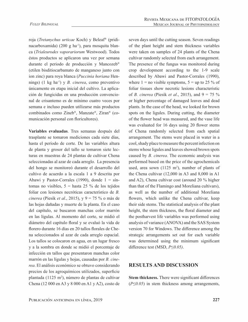
Publicación anticipada en línea, 2019 227
Fully BilingualRevista Mexicana de FITOPATOLOGÍA
Mexican Journal of Phytopathology
roja (Tretanychus urticae Koch) y Beleaf® (pridi-nacarboxamida) (200 g ha-1), para mosquita blan-ca (Trialeurodes vaporariorum Westwood). Todos éstos productos se aplicaron una vez por semana durante el periodo de producción y Mancozeb® (etilen bisditiocarbamato de manganeso junto con ion zinc) para roya blanca (Puccinia horiana Hen-nings) (1 kg ha-1) y B. cinerea, como preventivo únicamente en etapa inicial del cultivo. La aplica-ción de fungicidas en una producción convencio-nal de crisantemo es de mínimo cuatro veces por semana e incluso pueden utilizarse más productos combinados como Zineb®, Manzate®, Ziran® (co-municación personal con floricultores).
Variables evaluadas. Tres semanas después del trasplante se tomaron mediciones cada siete días, hasta el período de corte. De las variables altura de planta y grosor del tallo se tomaron siete lec-turas en muestras de 24 plantas de cultivar Chena seleccionadas al azar de cada arreglo. La presencia del hongo se monitoreó durante el desarrollo del cultivo de acuerdo a la escala 1 a 9 descrita por Abawi y Pastor-Corrales (1990), donde 1 = sín-tomas no visibles, 5 = hasta 25 % de los tejidos foliar con lesiones necróticas característico de B. cinerea (Piesik et al., 2015), y 9 = 75 % o más de las hojas dañadas y muerte de la planta. En el caso del capítulo, se buscaron manchas color marrón en las lígulas. Al momento del corte, se midió el diámetro del capítulo floral y se evaluó la vida de florero durante 16 días en 20 tallos florales de Che-na seleccionados al azar de cada arreglo espacial. Los tallos se colocaron en agua, en un lugar fresco y a la sombra en donde se midió el porcentaje de infección en tallos que presentaron manchas color marrón en las lígulas y hojas, causadas por B. cine-rea. El análisis económico se obtuvo considerando precios de los agroquímicos utilizados, superficie plantada (1125 m2), número de plantas de cultivar Chena (12 000 en A3 y 8 000 en A1 y A2), costo de
seven days until the cutting season. Seven readings of the plant height and stem thickness variables were taken on samples of 24 plants of the Chena cultivar randomly selected from each arrangement. The presence of the fungus was monitored during crop development according to the 1-9 scale described by Abawi and Pastor-Corrales (1990), where 1 = no visible symptoms, 5 = up to 25 % of foliar tissues show necrotic lesions characteristic of B. cinerea (Piesik et al., 2015), and 9 = 75 % or higher percentage of damaged leaves and dead plants. In the case of the head, we looked for brown spots on the ligules. During cutting, the diameter of the flower head was measured, and the vase life was evaluated for 16 days using 20 flower stems of Chena randomly selected from each spatial arrangement. The stems were placed in water in a cool, shady place to measure the percent infection on stems whose ligules and leaves showed brown spots caused by B. cinerea. The economic analysis was performed based on the price of the agrochemicals used, area sown (1125 m2), number of plants of the Chena cultivar (12,000 in A3 and 8,000 in A1 and A2), Chena cultivar cost (around 20 % higher than that of the Flamingo and Moreliana cultivars), as well as the number of additional Moreliana flowers, which unlike the Chena cultivar, keep their side stems. The statistical analysis of the plant height, the stem thickness, the floral diameter and the postharvest life variables was performed using analysis of variance (ANOVA) and the SAS System version 70 for Windows. The difference among the strategic arrangements set out for each variable was determined using the minimum significant difference test (MSD, P≤0.05).
RESULTS AND DISCUSSION
Stem thickness. There were significant differences (P≤0.05) in stem thickness among arrangements,

Publicación en línea, mayo 2019 228
Fully BilingualRevista Mexicana de FITOPATOLOGÍAMexican Journal of Phytopathology
cultivar Chena (alrededor de 20 % más que culti-var Flamingo y Moreliana) así como el número de flores extra de Moreliana, que a diferencia de Che-na, éstas conservan sus tallos laterales. El análisis estadístico de las variables altura de planta, grosor de tallo, diámetro floral y vida pos-cosecha se rea-lizó mediante análisis de varianza (ANOVA) con el programa SAS System versión 7.0 para Windows. La diferencia entre los arreglos estratégicos plan-teados en cada variable evaluada se determinó con la prueba de diferencia mínima significativa (DMS, P≤0.05).
RESULTADOS Y DISCUSIÓN
Grosor del tallo. Hubo diferencias significativas (P≤0.05) entre arreglos en el grosor del tallo, don-de el diámetro osciló entre 0.62 y 0.72 cm (Cuadro 1). El cultivar Chena, en el arreglo tradicional (A3), registró los valores más bajos en todas las medi-ciones respecto a Chena flanqueada con Flamingo (A1) y Moreliana (A2). Así, el cultivar Chena en los arreglos A1 y A2, mostró ventaja en una de las características más importantes en la calidad del cultivo (Flores-Ruvalcaba et al., 2005), en donde el grosor del tallo determina el peso del capítulo floral que puede soportar. El grosor de tallo no es afectado por B. cinerea, ya que este hongo infecta tejidos blandos y viejos (Palmieri y Dardón, 2012; Schumacher, 2017). Sin embargo, se observa que el intercalado de A1 y A2 favorece que plantas de cultivar Chena sean más vigorosas que, aunado a la ausencia de B. cinerea, genera ventajas en la ca-lidad comercial, como lo mencionan Costa et al. (2019), quienes citan que una mayor altura se atri-buye a respuestas morfológicas, fisiológicas y/o de competencia, características que no fueron consi-deradas en este estudio.
where the diameter ranged from 0.62 to 0.72 cm (Table 1). The Chena cultivar in the traditional arrangement (A3) had the lowest values in all measurements compared to Flamingo (A1) and Moreliana (A2). In arrangements A1 and A2, the Chena cultivar had an advantage in one of the most important characteristics of the crop’s quality (Flores-Ruvalcaba et al., 2005), where stem thickness determines the weight of the flower head that the stem can support. The stem thickness is not affected by B. cinerea because the fungus infects old and soft tissues (Palmieri and Dardón, 2012; Schumacher, 2017). However, when A1 and A2 were intercropped, plants of the Chena cultivar were more vigorous, a fact that, along with the absence of B. cinerea, provides commercial quality advantages, as mentioned by Costa et al. (2019), who suggest that the greater height can be attributed to morphological, physiological and/or competition responses, characteristics that were not considered in this study.
Plant height. In arrangement A3, with significant differences (P≤0.05), Chena’s height (64.5 cm) was lower than the height of the same variety in arrangements A1 (70.6 cm) and A2 (69.9 cm), which were combinations of Chena and Moreliana or Flamingo (Table 2). Spatial arrangements usually vary in population density because of the distance between rows and plants, a fact that influences not only the yield components but also the products’ quality (Jirmanová et al., 2016; Coelho et al., 2016). However, in this study, the sowing density was the same in the three arrangements, which suggests that due to possible variations in nutrient demands (Valdez-Aguilar et al., 2015) caused by the strategic distribution of the cultivars, Chena plants were taller in A1 and A2 compared with those in A3. Piesik et al. (2015) mentioned that the

Publicación anticipada en línea, 2019 229
Fully BilingualRevista Mexicana de FITOPATOLOGÍA
Mexican Journal of Phytopathology
Altura de la planta. El arreglo A3, con diferen-cias significativas (P≤0.05), presentó menor altura (64.5 cm) de Chena respecto a la observada en la misma variedad en los arreglos A1 (70.6 cm) y A2 (69.9) que fueron combinados de Chena con More-liana o Flamingo (Cuadro 2). Los arreglos espacia-les generalmente varían en densidad de población por distancia entre surcos y plantas, los cuales in-fluyen no solo en los componentes del rendimiento sino también en la calidad de los productos (Jirma-nová et al., 2016; Coelho et al., 2016). Sin embar-go, en esta investigación, la densidad de plantación fue la misma en los tres arreglos, lo que sugiere que posibles variaciones en la demanda nutricio-nal (Valdez-Aguilar et al. 2015) por la distribución estratégica de los cultivares influyó en una mejor altura de la planta de Chena en A1 y A2 con res-pecto a A3. Piesik et al. (2015) mencionan que la asociación de ciertos cultivares de crisantemo en invernadero es determinante para proteger a alguno de éstos. La variable altura de planta también es de
Cuadro 1. Comparación de medias con diferencia mínima significativa (DMS) del grosor del tallo de cultivar Chena (Ch), en diferentes etapas de creci-miento y en tres arreglos espaciales (A) con Flamingo (F) y Moreliana (M). A1, Chena flanqueada por Flamingo (F-Ch-F); A2, Chena flan-queada por Moreliana (M-Ch-M) y A3, solo Chena (Ch-Ch-Ch).
Table 1. Comparison of means with least significant differences (LSD) of stem thickness of the Chena cultivar (Ch) at different growth stages and three spatial arrangements (A) with Flamingo (F) and Moreliana (M). A1, Chena flanked by Flamingo (F-Ch-F); A2, Chena flanked by More-liana (M-Ch-M) and A3, Chena a (Ch-Ch-Ch).
Arreglo Grosor del tallo (cm) en ddty
30 37 44 51 57
A1 0.46 a 0.52 a 0.55 a 0.61 a 0.70 a A2 0.46 a 0.51 a 0.55 a 0.61 a 0.72 a A3 0.42 b 0.45 b 0.51 b 0.58 b 0.62 bDMS 0.026 0.032 0.024 0.024 0.023
Valores de columna con la misma letra no difieren estadísticamente (DMS, de acuer-do a la prueba de Fisher, P≤0.05); yddt= días después de trasplante / In each column, values with the same letter are not statistically different (LSD=LSD least significant difference, according to Fisher’s test, P≤0.05); yddt= days after transplanting.
association of certain chrysanthemum cultivars in the greenhouse is decisive to protect some of them. The plant height variable is also very important because in the marketing chain, taller stems make it possible to make cuts at their base without affecting quality (Flores-Ruvalcaba et al., 2005). Similarly, taller plants have competitive advantages, such as greater light gathering and faster nutrient assimilation (Zheng and Van Labeke, 2018). The flower head is more exposed to sunlight and avoids the higher relative humidity present in the plant’s low or intermediate parts, a factor that determines spore germination and mycelium growth (Palmieri and Dardón 2012; Schumacher, 2017). In this regard, although the plant’s height was not directly affected by the presence of B. cinerea, it could be an advantage that limits the damage to the flower head.
Flower head diameter. The largest diameter of a flower head and with significant differences
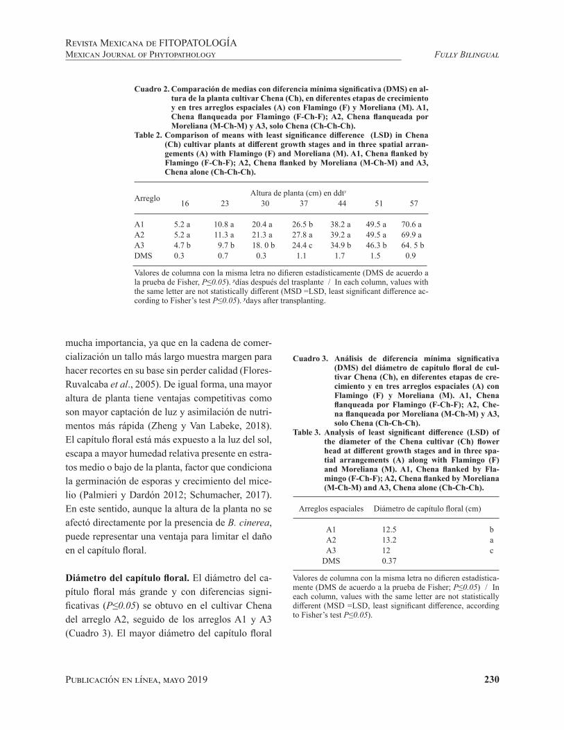
Publicación en línea, mayo 2019 230
Fully BilingualRevista Mexicana de FITOPATOLOGÍAMexican Journal of Phytopathology
mucha importancia, ya que en la cadena de comer-cialización un tallo más largo muestra margen para hacer recortes en su base sin perder calidad (Flores-Ruvalcaba et al., 2005). De igual forma, una mayor altura de planta tiene ventajas competitivas como son mayor captación de luz y asimilación de nutri-mentos más rápida (Zheng y Van Labeke, 2018). El capítulo floral está más expuesto a la luz del sol, escapa a mayor humedad relativa presente en estra-tos medio o bajo de la planta, factor que condiciona la germinación de esporas y crecimiento del mice-lio (Palmieri y Dardón 2012; Schumacher, 2017). En este sentido, aunque la altura de la planta no se afectó directamente por la presencia de B. cinerea, puede representar una ventaja para limitar el daño en el capítulo floral.
Diámetro del capítulo floral. El diámetro del ca-pítulo floral más grande y con diferencias signi-ficativas (P≤0.05) se obtuvo en el cultivar Chena del arreglo A2, seguido de los arreglos A1 y A3 (Cuadro 3). El mayor diámetro del capítulo floral
Cuadro 2. Comparación de medias con diferencia mínima significativa (DMS) en al-tura de la planta cultivar Chena (Ch), en diferentes etapas de crecimiento y en tres arreglos espaciales (A) con Flamingo (F) y Moreliana (M). A1, Chena flanqueada por Flamingo (F-Ch-F); A2, Chena flanqueada por Moreliana (M-Ch-M) y A3, solo Chena (Ch-Ch-Ch).
Table 2. Comparison of means with least significance difference (LSD) in Chena (Ch) cultivar plants at different growth stages and in three spatial arran-gements (A) with Flamingo (F) and Moreliana (M). A1, Chena flanked by Flamingo (F-Ch-F); A2, Chena flanked by Moreliana (M-Ch-M) and A3, Chena alone (Ch-Ch-Ch).
Arreglo Altura de planta (cm) en ddty
16 23 30 37 44 51 57
A1 5.2 a 10.8 a 20.4 a 26.5 b 38.2 a 49.5 a 70.6 aA2 5.2 a 11.3 a 21.3 a 27.8 a 39.2 a 49.5 a 69.9 aA3 4.7 b 9.7 b 18. 0 b 24.4 c 34.9 b 46.3 b 64. 5 bDMS 0.3 0.7 0.3 1.1 1.7 1.5 0.9
Valores de columna con la misma letra no difieren estadísticamente (DMS de acuerdo a la prueba de Fisher, P≤0.05). ydías después del trasplante / In each column, values with the same letter are not statistically different (MSD =LSD, least significant difference ac-cording to Fisher’s test P≤0.05). ydays after transplanting.
Cuadro 3. Análisis de diferencia mínima significativa (DMS) del diámetro de capítulo floral de cul-tivar Chena (Ch), en diferentes etapas de cre-cimiento y en tres arreglos espaciales (A) con Flamingo (F) y Moreliana (M). A1, Chena flanqueada por Flamingo (F-Ch-F); A2, Che-na flanqueada por Moreliana (M-Ch-M) y A3, solo Chena (Ch-Ch-Ch).
Table 3. Analysis of least significant difference (LSD) of the diameter of the Chena cultivar (Ch) flower head at different growth stages and in three spa-tial arrangements (A) along with Flamingo (F) and Moreliana (M). A1, Chena flanked by Fla-mingo (F-Ch-F); A2, Chena flanked by Moreliana (M-Ch-M) and A3, Chena alone (Ch-Ch-Ch).
Arreglos espaciales Diámetro de capítulo floral (cm)
A1 12.5 bA2 13.2 aA3 12 c
DMS 0.37
Valores de columna con la misma letra no difieren estadística-mente (DMS de acuerdo a la prueba de Fisher; P≤0.05) / In each column, values with the same letter are not statistically different (MSD =LSD, least significant difference, according to Fisher’s test P≤0.05).

Publicación anticipada en línea, 2019 231
Fully BilingualRevista Mexicana de FITOPATOLOGÍA
Mexican Journal of Phytopathology
en A2 y A1 representó una ventaja comercial, ya que es una de las características más importantes en la calidad de crisantemo (Flores-Ruvalcaba et al., 2005), lo que indicó que en el arreglo de mono-cultivo con cultivar Chena (A3), no se encontró una ventaja comercial del capítulo floral.
Vida de florero. La medición de vida de florero se detuvo a los 16 días cuando las plantas de cul-tivar Chena en A3, con diferencias significativas (P≤0.05), mostraron 100 % de infección por B. ci-nerea (Cuadro 4). No obstante, se estima que, en esta especie, la vida de florero puede durar hasta 30 días debido a que es una planta que produce poco etileno y es considerada como una especie no cli-matérica (Hidalgo et al., 2011).
La presencia de B. cinerea en el cultivo de cri-santemo no fue evidente en el follaje durante el
Cuadro 4. Comparación de medias con diferencia mínima sig-nificativa (DMS) del porcentaje de plantas infecta-das en vida de florero de cultivar Chena (Ch), en tres arreglos (A) espaciales con Flamingo (F) y Moreliana (M). A1, Chena flanqueada por Flamingo (F-Ch-F); A2, Chena flanqueada por Moreliana (M-Ch-M) y A3, solo Chena (Ch-Ch-Ch).
Table 4. Comparison of means with least significant difference (LSD) of the percentage of vase life of infected plants of the Chena cultivar (Ch) in three spatial arrangements (A) with Flamingo (F) and Moreliana (M). A1, Chena flanked by Flamingo (F-Ch-F); A2, Chena flanked by Moreliana (M-Ch-M) and A3, Chena alone (Ch-Ch-Ch).
Arreglo Incidencia de plantas dañadas en ddcZ
0 4 8 12 16
A1 0.0 b 0.0 b 7.5 b 12.5 b 15.0 bA2 0.0 b 0.0 b 0.0 b 0.0 b 0.0 cA3 22.5 a 22.5 a 50 a 90.0 a 100 aDMS 4.2 5.1 11.0 12.7 6.0
Valores de columna con la misma letra no difieren estadísticamente (DMS de acuerdo a la prueba de Fisher; P≤0.05), Zdías después del corte / In each column, values with the same letter are not statisti-cally different (MSD=LSD, least significant difference, according to Fisher’s test P≤0.05), Zdays after cutting.
(P≤0.05) was produced by the Chena cultivar in arrangement A2, followed by arrangements A1 and A3 (Table 3). The largest diameter of a flower head in A2 and A1 represented a commercial advantage because it is one of the most important characteristics of chrysanthemum quality (Flores-Ruvalcaba et al., 2005). This indicated that in the monocropped arrangement that included the Chena cultivar (A3), the flower head had no commercial advantage.
Vase life. Measurement of vase life was interrupted on day 16, when the Chena plants in A3, with significant differences (P≤0.05), were 100 % infected by B. cinerea (Table 4). However, according to estimations, this species may have a vase life of up to 30 days due to its low production of ethylene and because it is considered a non-climateric species (Hidalgo et al., 2011).
Publicación en línea, mayo 2019 232
Fully BilingualRevista Mexicana de FITOPATOLOGÍAMexican Journal of Phytopathology
desarrollo del cultivo, sin embargo, al momento del corte, las plantas seleccionadas al azar en el arre-glo A3 (cultivo convencional) manifestaron cre-cimiento del hongo (Cuadro 4), con 25 % de las plantas con síntomas visibles en el capítulo floral incluyendo manchas marrón y deshidratación de las lígulas. Al igual que en otras flores como rosa (Rosa x hybrida), la infección por B. cinerea no es visible en el momento del corte, pero está laten-te y se manifiesta en condiciones de humedad du-rante el almacenamiento y transporte (Elad, 1988). Plantas de Chena, en A2, no mostraron infección por B. cinerea durante el período de vida de florero evaluado, lo que indica una ventaja comercial de este arreglo para el cultivar Chena. Estos resultados muestran la importancia de controlar al hongo des-de el desarrollo del cultivo.
Los resultados obtenidos tienen un impacto eco-nómico, ya que en un arreglo convencional la pro-ducción de cultivar Chena involucra la aplicación de fungicidas, como Mancozeb, que debe aplicarse de cuatro veces por semana en período de lluvias con un costo estimado de $2 540.00 MN ha-1 (133 USD). Con los arreglos A1 y A2 se logró obtener control de B. cinerea con una sola aplicación, con gasto de $645.00 MN ha-1 (34 USD), lo que repre-sentó un ahorro del 75 %. Por otra parte, aunque existe diferencia de precio, 20 % más en Chena res-pecto de Moreliana y Flamingo y menor número de estas por m2 en A1 y A2 respecto de A3, se llega a compensar la ganancia ya que un tallo en Morelia-na produce en promedio 5 flores.
El arreglo espacial propuesto permite que cul-tivares que quedan en los laterales (Flamingo o Moreliana), donde comúnmente hay escurrimien-to del agua de lluvia o agua que se condensa en horas tempranas, funcione como una barrera física, lo que evita la presencia de humedad para que se desarrolle el hongo en cultivar Chena, que es más susceptible al ataque de B. cinerea. Otros estudios
The presence of B. cinerea on the chrysanthemum crop was not detected on the foliage during crop development, but when randomly selected plants from arrangement A3 (conventional crop) were cut, we noted fungus growth (Table 4); 25 % of the plants had visible symptoms on the flower head, including brown spots and ligule dehydration. Just as in other flowers, such as rose (Rosa x hybrid), B. cinerea infection is not visible when flowers are cut, but it is latent and appears under moisture conditions during storage and transportation (Elad, 1988). Chena plants in A2 did not show infection by B. cinerea during the vase life period we evaluated, which indicates there is a commercial advantage when using this arrangement for this cultivar. These results show how important it is to control this fungus when the crop starts to develop.
The obtained results have an economic impact because a conventional arrangement to produce the Chena cultivar involves the use of fungicides such as Mancozeb, which must be applied four times a week during the rainy season, at an estimated cost of MX$2540.00 ha-1 (US$133). Using arrangements A1 and A2, we were able to control B. cinerea with a single application at a cost of MX$645.00 ha-1 (34 USD), which represented savings of 75 %. On the other hand, although there is a price difference, 20 % more in Chena compared with Moreliana and Flamingo, and a lower number of them per square meter in A1 and A2 compared with A3, earnings are compensated because one Moreliana stem produces 5 flowers on average.
The proposed spatial arrangement allows (Flamingo or Moreliana) cultivars located on the sides (where there is usually runoff of rain water or water that condenses during early morning hours) may act as a physical barrier, which in turn prevents moisture that favors fungus development on the Chena cultivar, which is more susceptible to B. cinerea infection. Other studies (García-Velasco

Publicación anticipada en línea, 2019 233
Fully BilingualRevista Mexicana de FITOPATOLOGÍA
Mexican Journal of Phytopathology
(García-Velasco et al., 2003; Solano-Baez et al., 2013) muestran que existen cultivares de crisante-mo más resistentes al daño por hongos, por lo que es importante entender el tipo de respuesta fisio-lógica incluyendo la liberación de compuestos or-gánicos volátiles inhibidores de patógenos (Piesik et al., 2015), así como identificar cultivares con potencial de resistencia a patógenos que pudieran ser utilizados como barreras físicas en arreglos es-paciales como el reportado en esta investigación o para programas de fitomejoramiento.
CONCLUSIONES
La incidencia de B. cinerea disminuyó significa-tivamente en plantas cercanas al corte y en vida de florero del capítulo floral del cultivar Chena cuando su cultivo estuvo flanqueado por Flamingo (A1) o Moreliana (A2). De la misma manera, la calidad comercial de los tallos florales de cultivar Chena fue mejor en los arreglos espacial A1 y A2 respecto a A3 que fue monocultivo de Chena.
LITERATURA CITADA
Abawi GS and Pastor-Corrales MA. 1990. Root rots of beans in Latin America and Africa: diagnosis, research methodo-logies, and management strategies. CIAT. Cali, Colombia. 114 p.
Agrios GN. 2005. Plant Pathology. Fifth Edition. Academic Press. New York, USA. 922p
Anderson NO. 2007. Chrysanthemum: Dentrathema x gran-diflora. Pp. 389-439. In: Anderson NO (ed.). Flower bree-ding and genetics. Issues challenges and opportunities for the 21st Century. Springer. USA. 824 p.
Arroyave-Toro JJ, Mosquerac S. and Villegas-Escobara V. 2017. Biocontrol activity of Bacillus EA-CB0015 cells and lipopeptides against postharvest fungal pathogens. Biolo-gical control 114: 195-200. http://dx.doi.org/10.1016/j.biocontrol.2017.08.014
Boyd N, Brennan EB, Smith RF and Yokota R. 2009. Effect of seeding rate and planting arrangement on rye cover crop and weed growth. Agronomy Journal 10:47-51. https://naldc.nal.usda.gov/download/26826/PDF
et al., 2003; Solano-Baez et al., 2013) have shown that there are chrysanthemum cultivars that are more resistant to fungal damage, so it is important to understand the type of physiological response, including the release of volatile organic compounds that inhibit pathogens (Piesik et al., 2015), as well as to identify cultivars with potential resistance to pathogens that could be used as physical barriers in spatial arrangements such as the one used in this study or in improvement programs.
CONCLUSIONS
B. cinerea incidence was significantly lower in plants close to the Chena cultivar during cutting and in terms of vase life, when it was flanked by Flamingo (A1) or Moreliana (A2). Similarly, the commercial quality of the Chena cultivar’s floral stems was better in spatial arrangements A1 and A2 compared with those in A3, in which Chena was monocropped.
End of the English version
Chen X, Sun M, Liang J, Xue H, Qixiang Z. 2013. Genetic diversity of species of Chrysanthemum and related genera and groundcover cultivars assessed by amplified fragment length polymorphic markers. Hortscience 48(5):539–546. http://hortsci.ashspublications.org/content/48/5/539.full.pdf+html
Coelho B, Sandro D and Bosco PJ. 2016. Effect of spatial arrangement on the production components and yield of sunflower. Revista Ceres 63(2):214-222. http://dx.doi.org/10.1590/0034-737X201663020013
Costa CA, Guine´ RPF, Costa DVTA, Correia HE and Nave A. 2019. Pest control in organic farming. Organic Farming. 41-90 DOI: https://doi.org/10.1016/B978-0-12-813272-2.00003-3
Datta SKD, Janakiram T. 2015. Breeding and genetic diversity in Chrysanthemum morifolium in India: A review. Indian Journal of Agricultural Sciences 85:1379-1395. https://www.researchgate.net/publication/289630327_Breeding_and_genetic_diversity_in_Chrysanthemum_morifolium_in_India_A_review

Publicación en línea, mayo 2019 234
Fully BilingualRevista Mexicana de FITOPATOLOGÍAMexican Journal of Phytopathology
De Souza-Schlick GD, Peres R and Dutra M. 2014. Optimi-zing row spacing and plant population arrangement for a new short-height castor genotype in fall-winter. Acta Scientiarum 36(4):475-481. http://www.scielo.br/pdf/asagr/v36n4/12.pdf
Dordas C. 2008. Role of nutrients in controlling plant diseases in sustainable agriculture. A review. Agronomy for Sustai-nable Development 28:33–46. Disponible en línea: https://link.springer.com/article/10.1051/agro:2007051
Elad Y. 1988. Latent infection of Botrytis cinerea in rose flowers and combined chemical and physiological con-trol of the disease. Crop Protection 7:361-366. Dispo-nible en línea: https://www.researchgate.net/publica-tion/248415725_Latent_infection_of_Botrytis_cinerea_in_rose_flowers_and_combined_chemical_physiological_control_of_the_disease
Evers JB and Bastiaans L. 2016. Quantifying the effect of crop spatial arrangement on weed suppression using functio-nal-structural plant modelling. Journal of Plant Research 129:339-351. Disponible en línea: https://www.ncbi.nlm.nih.gov/pmc/articles/PMC4850179/
Flores-Ruvalcaba JS, Becerril-Román AE, González-Hernán-dez VA, Tijerina-Chávez L, Vásquez-Rojas T. 2005. Cre-cimiento vegetativo y floral del crisantemo Dendratema x gradiflorum (Ramat) Kitamura en respuesta a la presión osmótica de la solución nutritiva. Revista Chapingo Serie Horticultura 11(2):241-249. Disponible en línea: https://www.redalyc.org/articulo.oa?id=60911209
Furuta H, Shinoyama H, Nomura Y, Maeda M and Makara K. 2004. Production of intergeneric somatic hybrid of chrysan-themum [Dendranthema × grandiflorum (Ramat.) Kita-mura] and wormwood (Artemisia sieversiana J. F. Ehrh. ex. Willd) with rust (Puccinia horiana Henning) resistance by electrofusion of protoplasts. Plant Science 166:695-702. https://doi.org/10.1016/j.plantsci.2003.11.007
Gallet R, Bonnot F, Milazzo J, Tertois C, Adreit H, Ravigné V, Tharreau D and Fournier E. 2014. The variety mixtu-re strategy assessed in a G x G experiment with rice and the blast fungus Magnaporthe oryzae. Frontier in Genetics 4(312):1-12. https://doi.org/10.3389/fgene.2013.00312
Garcés E. 1999. Consideraciones sobre Botrytis cinerea Pers., agente causal de la pudrición de las flores. Agro-nomía Colombiana 9(2):196-201. Disponible en línea: https://revistas.unal.edu.co/index.php/agrocol/article/view/21168/22139
García A. 2014. Manual del crisantemo. Instituto de Inves-tigación y Capacitación Acuícola y Forestal del Estado de México (ICAMEX), No. De autorización CCE2017 /E/049/14. Disponible en línea: http://webcache.googleu-sercontent.com/search?q=cache:_iY6w0ShnYoJ:icamex.edomex.gob.mx/sites/icamex.edomex.gob.mx/files/files/publicaciones/2014/CRISANTEMO.pdf+&cd=2&hl=es-419&ct=clnk&gl=mx
García-Velasco R, Ochoa-Martínez D, Zavaleta-Mejía E, Mora-Aguilera G y Gaytan-Acuña E. 2003. Etiología de la marchitez del crisantemo (Dendrathema grandiflora Tzevelec) cv. Polaris. Revista Mexicana de Fitopatología 21:75-78. Disponible en línea: https://www.redalyc.org/pdf/612/61221112.pdf
Gaytán-Acuña EA, Ochoa-Martínez D, García-Velasco R, Zavaleta-Mejía E y Mora-Aguilera G. 2006. Producción y calidad comercial de flor de crisantemo. Terra Latinoa-mericana 24:541-548. Disponible en línea: http://www.redalyc.org/pdf/573/57324412.pdf
Guang-yu H, Jie L, Yan S, Yun-yue W, You-yong Z and Bao-rong L. 2016. Intercropping of rice varieties increases the efficiency of blast control through reduced disease occurrence and variability. Journal of Integrative Agri-culture 15(4):795-802. https://doi.org/10.1016/S2095-3119(15)61055-3
Hanudin KB and Marwoto B. 2017. Application of PGPR and antagonist fungi-based biofungicide for white rust disea-se control and its economic analysis in Chrysanthemum production. AGRIVITA Journal of Agricultural Science 39(3):266-278. Disponible en línea: https://agrivita.ub.ac.id/index.php/agrivita/article/view/1326
Hidalgo L, Garzón D y Flores V. 2011. Notas sobre posco-secha de crisantemos en la sabana de Bogotá y sus bases fisiológicas. Pp. 132-142. En: Flórez R., V.J. (Eds.). Avan-ces sobre fisiología de la producción de flores de corte en Colombia. Bogotá: Editorial Universidad Nacional de Co-lombia, Colombia, 105p.
Jirmanová J, Fuksa P, Hakl J, Brant V and Šantrucek J. 2016. Effect of different plant arrangements on maize morpho-logy and forage quality. Agriculture (Poľnohospodárstvo) Agriculture (Poľnohospodárstvo) 62(2): 62−71. https://doi.org/10.1515/agri-2016-0007.
Kaul A, Kumar S, Thakur M and Ghani M. 2011. Gamma ray-induced in vitro mutations in flower color in Dendrathe-ma grandiflora Tzelev. Floriculture and ornamental bio-technlogy 5(1): 71-73. Disponible en línea: http://www.globalsciencebooks.info/Online/GSBOnline/images/2011/FOB_5(1)/FOB_5(1)71-73o.pdf
Li XF, Liu ZX, Lu DB, Liu YZ, Mao XX, Li ZX and Li HJ. 2013. Development and evaluation of multi-genotype varieties of rice derived from MAGIC lines. Euphytica 192:77-86. doi: 10.1007/s10681-013-0879-1
Liu H, Zhang QX, Sun M, Pan HT and Kong ZX. 2015. Deve-lopment of expressed sequence tag-simple sequence repeat markers for Chrysanthemum morifolium and closely rela-ted species. Genetics and Molecular Research 14(3):7578-7586. http://dx.doi.org/10.4238/2015.July.13.1
Matsushita Y, Aoki Y and Sumitomo K. 2012. Selection and inheritance of resistance to Chrysanthemum stunt viroid, Crop Protection 35:1-4. https://doi.org/10.1016/j.cro-pro.2011.12.009
Migoya J. 2011. Plan Nacional de eliminación del consumo de bromuro de metilo en México. SAGARPA. ONUDI. 32 pp. http://apps2.semarnat.gob.mx:8080/sissao/index.html (Consulta octubre 2018).
Mudt CC. 2002. Use de multiline cultivars and culti-vars mixures for disease management. Annual Re-view Phytopathology 40:381–410. Disponible en lí-nea: https://www.annualreviews.org/doi/full/10.1146/annurev.phyto.40.011402.113723?casa_token=N-kjnfz jneRsAAAAA:ooHe6xJ69Vv7oXAUJzdGJ U h 9 d L 4 H N V d 5 2 t i a k O B A L X m Q L l O a 0 g c f 9 -dvoIJFtEyWSvwvn6QdVwNRmNn54w
Publicación anticipada en línea, 2019 235
Fully BilingualRevista Mexicana de FITOPATOLOGÍA
Mexican Journal of Phytopathology
Nakagawa H. 2009. Induced mutations in plant breeding and biological researches in Japan. In: Induced Plant Mutations in the Genomics Era. Q.Y. Shu (ed.). Food and Agriculture Organization of the United Nations, Rome, 2009, 51-58. Disponible en línea: https://www.fnca.mext.go.jp/english/mb/docdb/data/jpn035.pdf (consulta octubre 2018).
Noda N, Aida R, Kishimoto S, Ishiguro K, Fukuchi-Mizutani M, Tanaka Y and Ohmiya A. 2013. Genetic engineering of novel bluer-colored chrysanthemums produced by accumu-lation of delphinidin-based anthocyanins. Plant Cell Phy-siology 492 (54):1684-1695. Disponible en línea: https://academic.oup.com/pcp/article/54/10/1684/1907668
Núñez-Ríos T, Leyva-Mir SG, Rodríguez-Pérez JE y Maris-cal-Amaro ME. 2013. Etiología y control de la necrosis de flores y pudrición de frutos de pepino en Morelos, México. Revista Chapingo Serie Horticultura 19(2):255-266. Disponible en línea: http://www.scielo.org.mx/scielo.php?pid=S1027-152X2013000200010&script=sci_abs-tract
Ortiz I, Ávila-Chávez M, Torres LG. 2013. Plaguicidas en México: usos, riesgos y marco regulatorio. Revista Lati-noamericana de Biotecnología Ambiental y Algal 4(1):26-46. Disponible en línea: https://www.globalsciencejour-nals.com/content/pdf/10.7603%2Fs40682-013-0003-1.pdf
Palmieri M y Dardón E. 2012. Diagnóstico de la situación fi-tosanitaria del crisantemo y otras flores de corte y evalua-ción de planes emergentes de manejo integrado del cultivo en los municipios de San Pedro y San Juan Sacatepéquez. Facultad de Agronomía, Universidad de San Carlos, Gua-temala. 99p. Disponible en línea: http://168.234.106.70/library/images/d/dc/FODECYT_2006.76.pdf
Patil UH, Karale AR, Katwate SM and Patil MS. 2017. Mu-tation breeding in chrysanthemum (Dendranthema gran-diflora T.) Journal of Pharmacognosy and Phytochemistry 6(6):230-232. Disponible en línea: http://www.phytojour-nal.com/archives/2017/vol6issue6/PartD/6-5-393-273.pdf
Piesik D, Miler N, Lemanczykg G and Bocianowski J. 2015. Botrytis cinerea infection in three cultivars of Chrysanthe-mum in ‘Alchimist’ and its mutants: Volatile iduction of pathogen-infection plants. Scientia Horticulturae 193:127-135 http://dx.doi.org/10.1016/j.scienta.2015.06.040
Potts M. 1990. Influence of intercropping in warm climates on pests and diseases of potato, with special reference to their control. Field Crops Research 25:133-144. https://doi.org/10.1016/0378-4290(90)90078-P
Prieto EC, Escobar JA, y Posasa A. 1986. Densidades de siem-bra de dos variedades comerciales de crisantemo Chrysan-themum morifolium Ramat en Piedamod (Cauca). Acta Agronómica 36(3):5667. Disponible en línea: http://bdi-gital.unal.edu.co/18850/1/14788-44467-1-PB.pdf
Raseduzzaman MD and Jensen E. 2017. Does intercropping enhance yield stability in arable crop production? A me-ta-analysis. European Journal of Agronomy. 91: 25-33. https://doi.org/10.1016/j.eja.2017.09.009
Robert C, Garin G, Abichou M, Houlés V, Pradal C and Four-nier C. 2018. Plant architecture and foliar senescence impact the race between wheat growth and Zymosepto-ria tritici epidemics. Annals of Botany 121(5): 975-989. Disponible en línea: https://www.ncbi.nlm.nih.gov/pub-med/29373663
Rodríguez A, Acosta A and Rodríguez, A. 2014. Fungici-de resistance of Botrytis cinerea in tomato greenhouses in the Canary Islands and effectiveness of non-chemical treatments against gray mold. World Journal of Micro-biology and Biotechnology 30(9):2397-2406. Disponi-ble en línea: https://link.springer.com/article/10.1007%2Fs11274-014-1665-5
Rodríguez H y Rodríguez J. (2011). Métodos de análisis de suelos y plantas. Ed. Trillas. México 239p.
Sadhukhan R, Swhathi K, Sarmah D and Mandal T. 2015. Effect of different dosses of gamma rays on survivability and rooting ability in chrysanthemum (Chrysantemum mo-rifoliu Ramat). Journal Crop and Weed 11:62-65. Disponi-ble en línea: http://www.cropandweed.com/archives/2015/vol11issue1/11.pdf
Schumacher J. 2017. How life affects the life of Botrytis. Ge-netics and Biology 106:26-41. http://dx.doi.org/10.1016/j.fgb.2017.06.002
Servicio de Información Agroalimentaria y Pesquera. 2018. Avance de siembras y cosechas. http://infosiap.siap.gob.mx:8080/agricola_siap_gobmx/ResumenDelegacion.do (consulta, noviembre 2018)
Shinoyama H, Mitsuhara I, Ichikawa H, Kato K and Mochizu-ki A. 2015. Transgenic chrysanthemums (Chrysanthemum morifolium Ramat.) carrying both insect and disease re-sistance. Acta Horticulturae 1087:485-497.Disponible en línea: https://www.ishs.org/ishs-article/1087_66
Solano-Baez R, Leyva-Mir SG, Tlapal-Bolaños B y Mariscal-Amaro LA. 2013. Etiología y respuesta de variedades de crisantemo a la pudrición del tallo en el Estado de Méxi-co. Revista Chapingo Serie Horticultura. http://dx.doi.org/10.5154/r.rchsh.2010.06.121
Storkey J, Bruce TJA, Mcmillan VE and Neve P. 2018. The future of sustainable crop protection relies on increased diversdity of cropping systems and landscapes. Pp. 199-209. In: Lemaire G, Carvalho PCF and Recous S. (eds). Agroecosystem Diversity. Academic Press. USA. 478p
Valdez-Aguilar LA, Hernández-Pérez A, Alvarado-Carrillo D, and Cruz-Altunar A. 2015. Diseño de un programa de fertilización para crisantemo en base a extracción de ma-cronutrimentos. Revista Mexicana de Ciencias Agrícolas 12:2263-2276. Disponible en lpinea: http://www.redalyc.org/html/2631/263143809
Vences-Contreras C y Vázquez-García LM. 2008. Inoculación in vitro de la roya blanca (Puccinia Horiana Hennings) en crisantemo (Dendranthema grandiflora Tzvelev). Agro-nomía Mesoamericana 19(1):81-85. Disponible en línea: http://www.mag.go.cr/rev_meso/v19n01_081.pdf
Xialong X, Yafeng S, Jiafu J, Lipping R, Fadi CH, Weimin F, Zhiyong G and Sumei Ch. 2014. Gene expression profiles response to aphid feeding in Chrysanthemum (Chrysante-mum morifolium). BMC Genomics 15:1050. Disponible en línea: https://bmcgenomics.biomedcentral.com/arti-cles/10.1186/1471-2164-15-1050
Yusuf ES, Budiarto K, Djatnika I and Suhardi. 2017. Effects of varieties, cutting health and fungicide application on chrysanthemum white rust. AGRIVITA Journal of Agri-cultural Science 39: 21-30. Dsiponible en línea: https://www.researchgate.net/publication/313147898_Effects_of_Varieties_Cutting_Health_and_Fungicide_Applica-tion_on_Chrysanthemum_White_Rust

Publicación en línea, mayo 2019 236
Fully BilingualRevista Mexicana de FITOPATOLOGÍAMexican Journal of Phytopathology
Zeng J, Sun J, Xu Y, Chen F, Jiang J, Fang W and Chen S. 2013. Variation for resistance to white rust (Puccinia ho-riana) among Ajania and Chrysanthemum species. HortS-cience 48(10)1231-1234. Disponible en línea: http://horts-ci.ashspublications.org/content/48/10/1231.full.pdf
Zhang, M, Huang H, Wang Q and Dai S. 2018. Cross bree-ding new cultivars of early-flowering multiflora chrysan-themum based on mathematical analysis. HortScience 53(4):42-426. Disponible en línea: http://hortsci.ashspu-blications.org/content/53/4/421.abstract
Zhao S., Chen X, Deng S. Dong X, Song A, Yao J, Fang W and Chen F. 2016. The effects of fungicide, soil fumi-gant, bio-organic fertilizer and their combined applica-tion on chrysanthemum fusarium wilt controlling, soil enzyme activities and microbial properties. Molecules 21(4):526. https://doi.org/10.3390/molecules21040526
Zheng L and Van Labeke MC. 2018. Effects of different irra-diation levels of light quality on Chrysanthemum. Scien-tia Horticulturae 233:124-131. https://doi.org/10.1016/j.scienta.2018.01.033

237Publicación en línea, mayo 2019
García-Nevárez G and Hidalgo-Jaminson E. 2019. Efficacy of indigenous and commercial Simplicillium and Lecanicillium strains for controlling Hemileia vastatrix. Mexican Journal of Phytopathology 37(2): 237-250.DOI: 10.18781/R.MEX.FIT.1810-4
Primera publicación DOI: 04 de Abril, 2019.First DOI publication: April 04, 2019.
Resumen. La roya es la enfermedad del café de mayor importancia económica, puede ocasio-nar pérdidas de producción superiores a 50%. La información sobre prácticas de control biológico aplicado para controlar la roya del café en Costa Rica es limitada. El estudio tuvo por objetivo se-leccionar una cepa local de Simplicillium con alto grado de virulencia, así como evaluar la eficiencia de cepas comerciales de micoparásitos destinados al control de H. vastatrix en Costa Rica. El estudio comprendió ocho cepas de hongos, cinco de ellas fueron aisladas de la zona cafetalera de Turrialba e identificadas como Simplicillium lanosoniveum y
Efficacy of indigenous and commercial Simplicillium and Lecanicillium strains for controlling Hemileia vastatrix
Comparación de cepas locales y comerciales de Simplicillium y Lecanicillium colonizando pústulas de Hemileia vastatrix
Gerardo García-Nevárez*, Instituto Nacional de Investigaciones Forestales Agrícolas y Pecuarias. Campo Experimental Delicias. Carretera Delicias-Rosales Km 2.5. Delicias, Chihuahua, México, CP. 33000 México; Eduardo Hidalgo-Jaminson, Centre for Agriculture and Biosciences International, Turrialba, Cartago, Costa Rica, CP. 30501 Costa Rica. *Autor para correspondencia: [email protected]
Recibido: 24 de Octubre, 2018. Aceptado: 11 de Marzo, 2019.
Abstract. The aim of this research was to select an indigenous strain of Simplicillium with a high degree of virulence, as well as to assess the efficacy of commercial strains of mycoparasites for the control of H. vastatrix in Costa Rica. The study included eight strains of fungi, five of them were isolated from the coffee region of Turrialba, which were identified as Simplicillium lanosoniveum and three biopesticides recommended to control coffee rust in Costa Rica. The EC strain showed to be the most effective, by colonizing coffee rust pustules in a shorter time. This strain was compared with commercial formulations after two successive passes in PDA and after been stored on filter paper. The indigenous isolates showed a greater degree of specificity than the rest of the strains. This is the first report pointing out Simplicillium as the main natural enemy of H. vastatrix in Costa Rica.
Key words: indigenous isolates, colonization rate, mycoparasites.
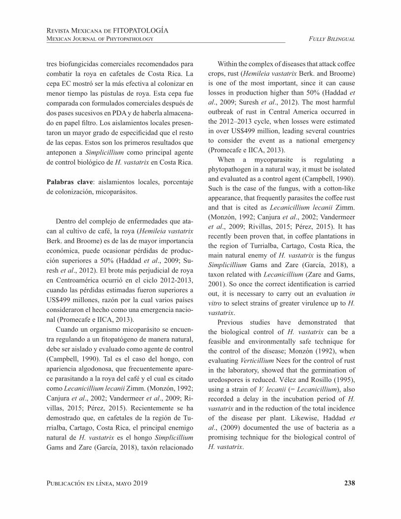
Publicación en línea, mayo 2019 238
Fully BilingualRevista Mexicana de FITOPATOLOGÍAMexican Journal of Phytopathology
tres biofungicidas comerciales recomendados para combatir la roya en cafetales de Costa Rica. La cepa EC mostró ser la más efectiva al colonizar en menor tiempo las pústulas de roya. Esta cepa fue comparada con formulados comerciales después de dos pases sucesivos en PDA y de haberla almacena-do en papel filtro. Los aislamientos locales presen-taron un mayor grado de especificidad que el resto de las cepas. Estos son los primeros resultados que anteponen a Simplicillium como principal agente de control biológico de H. vastatrix en Costa Rica.
Palabras clave: aislamientos locales, porcentaje de colonización, micoparásitos.
Dentro del complejo de enfermedades que ata-can al cultivo de café, la roya (Hemileia vastatrix Berk. and Broome) es de las de mayor importancia económica, puede ocasionar pérdidas de produc-ción superiores a 50% (Haddad et al., 2009; Su-resh et al., 2012). El brote más perjudicial de roya en Centroamérica ocurrió en el ciclo 2012-2013, cuando las pérdidas estimadas fueron superiores a US$499 millones, razón por la cual varios países consideraron el hecho como una emergencia nacio-nal (Promecafe e IICA, 2013).
Cuando un organismo micoparásito se encuen-tra regulando a un fitopatógeno de manera natural, debe ser aislado y evaluado como agente de control (Campbell, 1990). Tal es el caso del hongo, con apariencia algodonosa, que frecuentemente apare-ce parasitando a la roya del café y el cual es citado como Lecanicillium lecanii Zimm. (Monzón, 1992; Canjura et al., 2002; Vandermeer et al., 2009; Ri-villas, 2015; Pérez, 2015). Recientemente se ha demostrado que, en cafetales de la región de Tu-rrialba, Cartago, Costa Rica, el principal enemigo natural de H. vastatrix es el hongo Simplicillium Gams and Zare (García, 2018), taxón relacionado
Within the complex of diseases that attack coffee crops, rust (Hemileia vastatrix Berk. and Broome) is one of the most important, since it can cause losses in production higher than 50% (Haddad et al., 2009; Suresh et al., 2012). The most harmful outbreak of rust in Central America occurred in the 2012–2013 cycle, when losses were estimated in over US$499 million, leading several countries to consider the event as a national emergency (Promecafe e IICA, 2013).
When a mycoparasite is regulating a phytopathogen in a natural way, it must be isolated and evaluated as a control agent (Campbell, 1990). Such is the case of the fungus, with a cotton-like appearance, that frequently parasites the coffee rust and that is cited as Lecanicillium lecanii Zimm. (Monzón, 1992; Canjura et al., 2002; Vandermeer et al., 2009; Rivillas, 2015; Pérez, 2015). It has recently been proven that, in coffee plantations in the region of Turrialba, Cartago, Costa Rica, the main natural enemy of H. vastatrix is the fungus Simplicillium Gams and Zare (García, 2018), a taxon related with Lecanicillium (Zare and Gams, 2001). So once the correct identification is carried out, it is necessary to carry out an evaluation in vitro to select strains of greater virulence up to H. vastatrix.
Previous studies have demonstrated that the biological control of H. vastatrix can be a feasible and environmentally safe technique for the control of the disease; Monzón (1992), when evaluating Verticillium Nees for the control of rust in the laboratory, showed that the germination of uredospores is reduced. Vélez and Rosillo (1995), using a strain of V. lecanii (= Lecanicillium), also recorded a delay in the incubation period of H. vastatrix and in the reduction of the total incidence of the disease per plant. Likewise, Haddad et al., (2009) documented the use of bacteria as a promising technique for the biological control of H. vastatrix.

Publicación en línea, mayo 2019 239
Fully BilingualRevista Mexicana de FITOPATOLOGÍA
Mexican Journal of Phytopathology
con Lecanicillium (Zare y Gams, 2001). Así pues, una vez realizada la correcta identificación es nece-sario la evaluación in vitro para seleccionar cepas de mayor virulencia hacia H. vastatrix.
Estudios previos han demostrado que el control biológico de H. vastatrix puede ser una técnica fac-tible y ambientalmente segura para el control de la enfermedad; Monzón (1992) al evaluar Verticillium Nees para el control de la roya a nivel de labora-torio demostró que la germinación de uredosporas se ve reducida. Vélez y Rosillo (1995), también con aplicaciones de una cepa de V. lecanii (= Le-canicillium) registraron un retraso en el periodo de incubación de H. vastatrix y en la reducción de la incidencia total de la enfermedad por planta. Por su parte, Haddad et al., (2009) documentaron el uso de bacterias como una técnica promisoria para el control biológico de H. vastatrix.
Actualmente, el manejo de la roya en Costa Rica está enfocado en el uso de fungicidas protectantes (a base de cobre) y fungicidas sistémicos pertene-cientes a la familia de los triazoles principalmen-te (los efectos indeseables de estas acciones de manejo son bien conocidos), además de prácticas culturales como la poda, el manejo de la sombra y el uso de variedades resistentes (Avelino et al., 2004; Barquero, 2013). Si bien el uso de variedades resistentes es una práctica promisoria para mitigar la enfermedad, la variedad genética de la roya dis-minuye el tiempo de resistencia inicial (Gouveia et al., 2005). Como contraparte, existen enemigos naturales de H. vastatrix que regulan su inciden-cia y severidad (Guharay et al., 2001); como por ejemplo, los hongos L. lecanii y Cladosporium he-mileiae (=Digitopodium hemileiae Steyaert). Tam-bién es común encontrar larvas de Mycodiplosis Rüb. alimentándose de pústulas de roya (Virginio Filho, 2017). Desafortunadamente la información sobre prácticas de control biológico aplicado para controlar la roya del café en Costa Rica es limitada.
Currently, the control of rust in Costa Rica is focused on the use of protector (copper-based) and systemic fungicides that belong to the family of triazols, mainly (the undesireable effects of these control actions are well-known), along with cultural practices such as pruning, the use of shade and the use of resistant varieties (Avelino et al., 2004; Barquero, 2013). Although the use of resistant varieties is a promising practice for the mitigation of the disease, the genetic variety of rust reduces the time of initial resistance (Gouveia et al., 2005). On the other hand, there are natural enemies of H. vastatrix that regulate its incidence and severity (Guharay et al., 2001), such as the fungi L. lecanii and Cladosporium hemileiae (=Digitopodium hemileiae Steyaert). It is also common to find larvae of Mycodiplosis Rüb. feeding of rust pustules (Virginio Filho, 2017). Unfortunately, the information on applied biological control practices for the control of coffee rust in Costa Rica is limited.
The aim of the study was to isolate and compare local and commercial strains of Simplicillium and Lecanicillium against coffee rust.
MATERIALS AND METHODS
Isolation and identification of local and commercial strains. The study covered eight fungal strains, five of which were isolated from rust pustules in the coffee-growing region of Turrialba, Cartago, Costa Rica, in the locations of San Juan Norte (SJ), Jabillos (JV), Aquiares (AQ), Santa Rosa (SR) and CATIE (EC). A variation of this last strain was also included after having been kept for one month using the filter paper method (Morales, 2008) in order to observe the loss of virulence due to dehydration at the end of the process (codified as EC1). The plots in which the collections were performed are located between 600 and 950 masl;

Publicación en línea, mayo 2019 240
Fully BilingualRevista Mexicana de FITOPATOLOGÍAMexican Journal of Phytopathology
El estudio tuvo por objetivo aislar y comparar cepas locales y comerciales de Simplicillium y Le-canicillium contra la roya del café.
MATERIALES Y MÉTODOS
Aislamiento e identificación de cepas locales y comerciales. El estudio comprendió ocho cepas de hongos, cinco fueron aisladas de pústulas de roya en la zona cafetalera de Turrialba, Cartago, Cos-ta Rica en las localidades de San Juan Norte (SJ), Jabillos (JV), Aquiares (AQ), Santa Rosa (SR) y CATIE (EC); también, se incluyó una variante de esta última cepa después de haber sido conservada durante un mes por el método de papel filtro (Mo-rales, 2008), con la finalidad de observar pérdida de virulencia debida al sometimiento de deshidra-tación al final del proceso (codificada como EC1). Las parcelas donde se hicieron las colectas se en-cuentran entre los 600 a 950 msnm, la temperatura promedio registrada en el área de estudio durante los últimos años ha sido de 22.4 °C y 90.6% de humedad relativa. Todas las parcelas estaban plan-tadas con café arábica variedad Caturra con una densidad de 5000 plantas/ha bajo sombra
Las tres cepas restantes (INA, C1 y C2) corres-ponden a productos recomendados para combatir la roya en cafetales de Costa Rica. INA proviene de un centro de capacitación y transferencia de tec-nología, esta cepa se reproduce de manera artesa-nal utilizando como sustrato arroz. C1 también fue producida con el mismo sustrato, pero a diferencia del anterior, esta contaba con empaque y etiquetado para su venta. C2 es un producto comercial cuya formulación era polvo humectable.
Colonias monospóricas de las diferentes cepas fueron enviadas al Laboratorio de Biología Mo-lecular de la Corporación Bananera Nacional de Costa Rica (Corbana). Para la secuenciación de
the average temperature recorded in the area of study in recent years has been 22.4°C and a relative humidity of 90.6%. All plots were planted with Arabica coffee of the variety Caturra, with a density of 5000 plants/ha under shade.
The three remaining strains (INA, C1 and C2) are products recommended against rust in coffee plantations in Costa Rica. INA comes from a training and technology transfer center; this strain reproduces in a traditional manner, using rice as a substrate. C1 was also produced with the same substrate, but unlike the above, it was packaged and labeled for sale. C2 is a commercial product which was formulated as a wettable powder.
Monospori colonies of the different strains were sent to the Molecular Biology Lab of the Corporación Bananera Nacional de Costa Rica (National Banana Corporation of Costa Rica - Corbana). For the sequencing of the samples, the genetic material was extracted following the protocol described by Kuske et al. (1998) with modifications made by Corbana, followed by an amplification of the ITS region. The primers used for the amplification were ITS1 and ITS4; the corroboration of the amplification was carried out in a 1% agarose gel. The product of the amplification was sent to the company Macrogen Inc. for purifying and sequencing. After generating the sequences, they were edited using the program BioEdit and compared in the database of the NCBI (National Center for Biotechnology Information). The organisms identified for each product are shown in Table 1. The strains are safeguarded in the Microbial Control Laboratory of the Centro Agronómico Tropical de Investigación y Enseñanza (Tropical Agriculture Research and Training Center - CATIE).
Collection of rust pustules and preparation of leaf discs. Initially, leaves of the coffee variety Caturra,

Publicación en línea, mayo 2019 241
Fully BilingualRevista Mexicana de FITOPATOLOGÍA
Mexican Journal of Phytopathology
las muestras se realizó la extracción del material genético según el protocolo descrito por Kuske et al. (1998) con modificaciones hechas por Corbana, seguido de una amplificación de la región ITS. Los iniciadores utilizados para dicha amplificación fue-ron ITS1 e ITS4; la corroboración de la amplifica-ción se hizo en un gel al 1% de agarosa. El producto de la amplificación se envió a la empresa Macrogen Inc. para su purificación y secuenciación. Una vez generadas las secuencias se editaron con el progra-ma BioEdit y se compararon en la base de datos de NCBI (National Center for Biotechnology In-formation). Los organismos identificados para cada producto se enlistan en el Cuadro 1. Las cepas se encuentran resguardadas en el Laboratorio de Con-trol Microbial del Centro Agronómico Tropical de Investigación y Enseñanza (CATIE).
Colecta de pústulas de roya y preparación de discos de hoja. Inicialmente se hicieron colectas de hojas de café variedad Caturra completamente de-sarrolladas y con presencia de pústulas de roya en sus diferentes estados de desarrollo y sin presencia de hongos micoparásitos; posteriormente las hojas fueron llevadas a laboratorio en donde con la ayuda de un sacabocado de 1.8 cm de diámetro, se extra-jeron discos de hoja conteniendo pústulas de roya con lesiones recién esporuladas de color naranja in-tenso (estado intermedio) y lesiones completamen-te desarrolladas con apariencia naranja pálido (es-tado avanzado). Una vez hechos los discos de hoja se colocaron en placas multipozos CELLSTAR(R) de seis cavidades donde previamente a cada cavi-dad se le agregaron 5 mL de agua destilada estéril (ADE) y sobre esta un disco de Foam de 2.5 cm de diámetro. Finalmente, los discos de hoja quedaron sobre los discos de Foam flotando en ADE (Figura 1). Previamente se inocularon con una suspensión de esporas de las diferentes cepas como se describe en el siguiente apartado.
Cuadro 1. Organismos identificados en cepas locales y co-merciales.
Table 1. Organisms identified in local and commercial strains.
Cepa Organismos coincidentes NCBI
EC Simplicillium lanosoniveumAQ Simplicillium lanosoniveumSR Simplicillium lanosoniveum SJ Simplicillium lanosoniveum JV Simplicillium lanosoniveum INA Simplicillium lanosoniveum C1 Lecanicillium attenuatumC2y Lecanicillium sp.
y Identificación hecha en base a las claves taxonómicas pro-puestas por Zare y Gams (2001) / y Identification performed based on the taxonomic codes proposed by Zare and Gams (2001).
fully developed and with rust pustules in different stages of development without microparasitic fungi were collected; these leaves were transferred to a lab, where, using a hole puncher, 1.8 cm in diameter, we extracted leaf discs with rust pustules with deep orange-colored, recently sporulated (intermediate stage) lesions and fully developed lesions colored pale orange (advanced stage). Once the leaf discs were made, they were placed in a CELLSTAR(R) multiwell plate with six cavities, each of which previously contained 5 mL of sterile distilled water (SDW), and on top of this, a Foam disc, 2.5 cm in diameter. Finally, the leaf discs were placed on top of the Foam discs floating on SDW (Figure 1). They were then inoculated with a suspension of spores from the different strains, as described in the next section.
Application method and doses. Petri dishes with 15 day old monospore cultures of the different strains were flooded with SDW, followed by scraping with a Drigalsky spatula until the mycelium was completely separated from the culture medium.
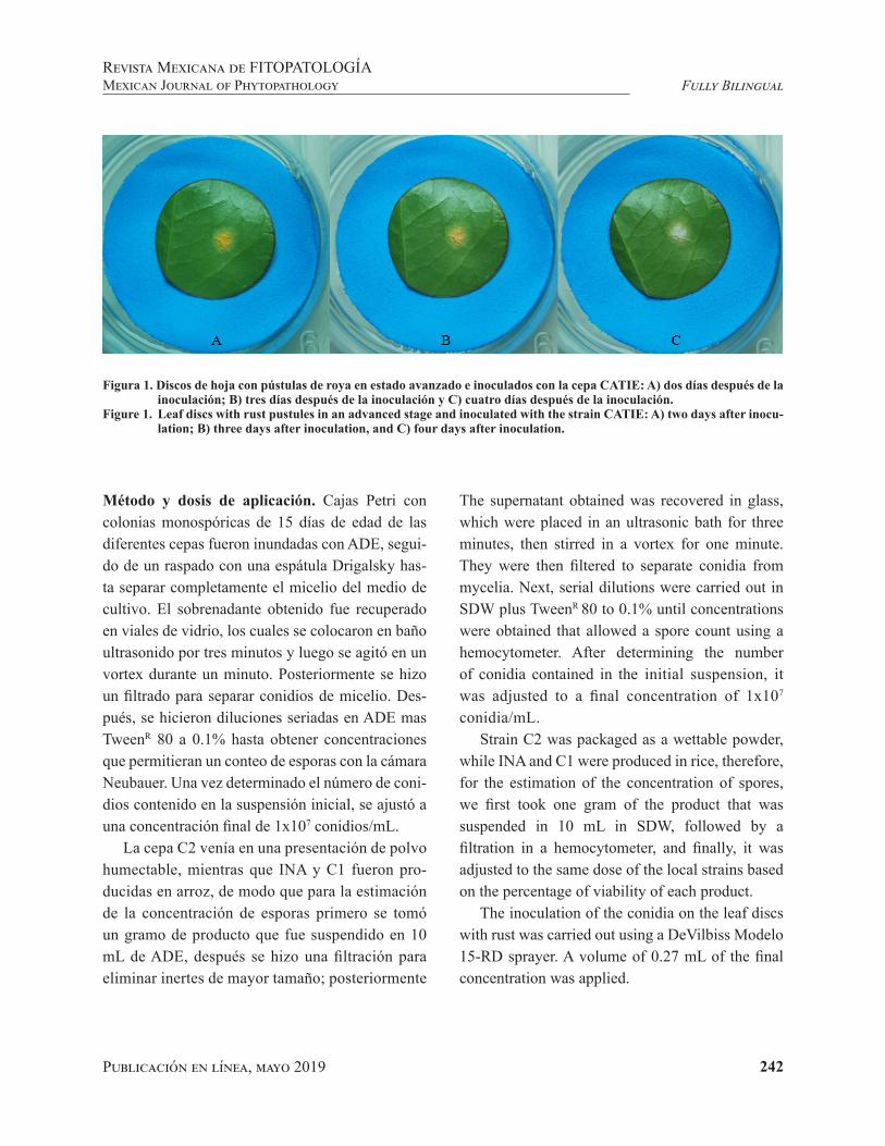
Publicación en línea, mayo 2019 242
Fully BilingualRevista Mexicana de FITOPATOLOGÍAMexican Journal of Phytopathology
Método y dosis de aplicación. Cajas Petri con colonias monospóricas de 15 días de edad de las diferentes cepas fueron inundadas con ADE, segui-do de un raspado con una espátula Drigalsky has-ta separar completamente el micelio del medio de cultivo. El sobrenadante obtenido fue recuperado en viales de vidrio, los cuales se colocaron en baño ultrasonido por tres minutos y luego se agitó en un vortex durante un minuto. Posteriormente se hizo un filtrado para separar conidios de micelio. Des-pués, se hicieron diluciones seriadas en ADE mas TweenR 80 a 0.1% hasta obtener concentraciones que permitieran un conteo de esporas con la cámara Neubauer. Una vez determinado el número de coni-dios contenido en la suspensión inicial, se ajustó a una concentración final de 1x10⁷ conidios/mL.
La cepa C2 venía en una presentación de polvo humectable, mientras que INA y C1 fueron pro-ducidas en arroz, de modo que para la estimación de la concentración de esporas primero se tomó un gramo de producto que fue suspendido en 10 mL de ADE, después se hizo una filtración para eliminar inertes de mayor tamaño; posteriormente
Figura 1. Discos de hoja con pústulas de roya en estado avanzado e inoculados con la cepa CATIE: A) dos días después de la inoculación; B) tres días después de la inoculación y C) cuatro días después de la inoculación.
Figure 1. Leaf discs with rust pustules in an advanced stage and inoculated with the strain CATIE: A) two days after inocu-lation; B) three days after inoculation, and C) four days after inoculation.
The supernatant obtained was recovered in glass, which were placed in an ultrasonic bath for three minutes, then stirred in a vortex for one minute. They were then filtered to separate conidia from mycelia. Next, serial dilutions were carried out in SDW plus TweenR 80 to 0.1% until concentrations were obtained that allowed a spore count using a hemocytometer. After determining the number of conidia contained in the initial suspension, it was adjusted to a final concentration of 1x10⁷ conidia/mL.
Strain C2 was packaged as a wettable powder, while INA and C1 were produced in rice, therefore, for the estimation of the concentration of spores, we first took one gram of the product that was suspended in 10 mL in SDW, followed by a filtration in a hemocytometer, and finally, it was adjusted to the same dose of the local strains based on the percentage of viability of each product.
The inoculation of the conidia on the leaf discs with rust was carried out using a DeVilbiss Modelo 15-RD sprayer. A volume of 0.27 mL of the final concentration was applied.

Publicación en línea, mayo 2019 243
Fully BilingualRevista Mexicana de FITOPATOLOGÍA
Mexican Journal of Phytopathology
se hicieron diluciones seriadas, conteo en cámara Neubauer y por último se ajustó a la misma dosis de las cepas locales en base al porcentaje de viabi-lidad de cada producto.
La inoculación de conidios sobre los discos de hoja con roya fue realizada con la ayuda de un ato-mizador DeVilbiss Modelo 15-RD. Se aplicó un volumen de 0.27 mL de la concentración final.
Una vez hecha la aplicación, se dejó pasar el tiempo necesario para que estos perdieran la hume-dad obtenida durante la inoculación para luego ser puestos en las placas multipozos como se describe en el apartado anterior.
Medición de la variable respuesta. La variable medida fue el porcentaje de colonización de las cepas sobre las pústulas de H. vastatrix. Las me-diciones se hicieron cada 24 horas hasta siete días después de la inoculación. Para la estimación del porcentaje diario de colonización fue necesario to-mar fotografías diarias individuales a cada disco de hoja (Figura 1) para después hacer un procesamien-to con el cambio de colores utilizando el programa ImageJ 1.47v (Rasband, 2016). Inicialmente, se seleccionó y midió el área ocupada por la lesión de roya. Después, se ajustó el umbral de color de manera tal que la coloración blanquecina debida a la colonización de Simplicillium sobre las pús-tulas quedará claramente definida. Finalmente, el porcentaje de colonización se calculó dividiendo el área cubierta por Simplicillium entre el área total de la lesión y el resultado multiplicado por 100.
Diseño y análisis estadístico. El experimento tuvo 10 repeticiones cada una de las cuales consistió de dos placas multipozos. Cada placa contenía seis discos de un mismo estado de desarrollo de la roya (intermedio o avanzado); cinco de estos discos fue-ron inoculados, cada uno con una cepa diferente,
After spraying, it was left to stand until the humidity obtained during inoculation was lost, and then placed in the multiwell plate, as described in the previous section.
Measurement of the response variable. The variable measured was the percentage of colonization of the strains on the pustules of H. vastatrix. Measurements were made every 24 hours until 7 days after inoculation. To estimate the daily percentage of colonization, it was necessary to take daily, individual photographs of each leaf disc (Figure 1) to then process with the change of colors using the program ImageJ 1.47v (Rasband, 2016). Initially, we selected and measured the area covered by the rust lesion. Next, we adjusted the color threshold in such a way that the milky-white color due to the colonization of Simplicillium on the pustules was clearly defined. Finally, the percentage of colonization was calculated by dividing the area covered by Simplicillium by the total area of the lesion and multiplied the result by 100.
Design and statistical analysis. The experiment had 10 replicates, each one consisted of two multiwell plate, which contanied six discs with one stage of development of rust (intermediate or advanced); five of these discs were inoculated, each one with a different strain, in such a way that a sixth disc was used as a control without inoculation. The analysis was carried out using General Linear and Mixed Models using the Infostat statistical package (Di Rienzo et al., 2016). Growth curves were adjusted using mixed, non-linear models; the model that best described the growth curves was chosen using criteria AIC and BIC. Finally, an ANOVA was carried out, along with means separation test (LSD=0.05) for the coefficients Beta (increase in percentage of colonization).

Publicación en línea, mayo 2019 244
Fully BilingualRevista Mexicana de FITOPATOLOGÍAMexican Journal of Phytopathology
de manera tal que un sexto disco fue usado como testigo sin inoculación. El análisis fue hecho me-diante Modelos Lineales Generales y Mixtos con el paquete estadístico Infostat (Di Rienzo et al., 2016). Se ajustaron curvas de crecimiento utilizan-do modelos no lineales mixtos; el modelo que des-cribió mejor las curvas de crecimiento fue elegido utilizando los criterios AIC y BIC. Finalmente, se hizo un ANOVA y prueba de separación de medias (LSD=0.05) para los coeficientes Beta (incremento en porcentaje de colonización).
RESULTADOS
Bioensayos de cepas locales contra H. vasta-trix. El análisis estadístico muestra una diferencia altamente significativa entre las cepas evaluadas (F4,81=44.58, p˂ 0.0001). El avance en porcentaje de colonización de cepas tuvo un comportamiento similar en ambas etapas evaluadas; es decir, no se observó diferencia estadística para estado de desa-rrollo de la roya (F1,81=2.73, p=0.1025). La cepa EC mostró ser la más efectiva, al colonizar en menor tiempo las pústulas de roya, seguida de la cepa SJ; en tercer lugar en grado de efectividad estuvieron las cepas SR y JV, cuyos valores no difirieron es-tadísticamente (LSD=0.05). La cepa AQ mostró un menor desempeño respecto a las demás (Figura 2).
Bioensayos de cepas comerciales contra H. vas-tatrix. Al hacer la comparación de cepas comercia-les y la mejor cepa local seleccionada en el ensayo anterior, se observó una diferencia estadística al-tamente significativa (F4,81=76.55, p˂0.0001). Al igual que en el experimento anterior, la variable evaluada no difirió estadísticamente en los esta-dos de desarrollo de pústulas de roya (F1,81=5.06, p=0.0272. Los porcentajes más altos de coloniza-ción en menor tiempo se dieron en la cepa EC, esta
RESULTS
Bioassay of local strains against H. vastatrix. The statistical analysis shows a highly significant difference between the evaluated strains (F4,81=44.58, p˂ 0.0001). The progress in percentage of colonization of strains had a similar behavior in both stages evaluated; in other words, there was no statistical difference for the stage of rust development (F1,81=2.73, p=0.1025). Strain EC proved to be the most effective, since it colonized the rust pustules in the least amount of time, followed by SJ; the third most effective strains were SR and JV, with values that had no statistical difference (LSD=0.05). Strain AQ proved to have a lower performance rate in comparison to the rest (Figure 2).
Bioassay of commercial strains against H. vastatrix. After comparing the commercial strains and the best local strain selected in the previous trial, a highly significant statistical difference (F4,81=76.55, p˂0.0001) was observed. As in the previous experiment, the evaluated variable did not differ statistically in the stages of development of rust pustules (F1,81=5.06, p=0.0272). The highest percentages of colonization in the lowest times were recorded in strain EC, which, after having been stored with the filter paper method, did not undergo a loss in virulence. In second place in terms of efficacy was INA, and finally, with a very poor performance, were commercial strains C2 and C1 (Figure 3).
DISCUSSION
These are the first results that report the effect of Simplicillium as the main biological control agent for H. vastatrix in Costa Rica. Most publications

Publicación en línea, mayo 2019 245
Fully BilingualRevista Mexicana de FITOPATOLOGÍA
Mexican Journal of Phytopathology
misma cepa (EC1), después de ser almacenada me-diante el método de papel filtro, no sufrió pérdida de virulencia. En segundo lugar de eficacia estuvo INA y, finalmente, y con un desempeño muy pobre las cepas comerciales C2 y C1 (Figura 3).
DISCUSIÓN
Estos son los primeros resultados que anteponen y miden el efecto de Simplicillium como el princi-pal agente de control biológico de H. vastatrix en Costa Rica. La mayoría de publicaciones referidas al control biológico de roya del cafeto son enfo-cadas al uso de Lecanicillium como controlador biológico (Esques et al., 1991; Monzón, 1992; Ri-vas et al., 1996; Canjura et al., 2002; Vandermeer et al., 2009; Jackson et al., 2012; Rivillas, 2015; Pérez, 2015). Esto debido en parte a que algunos estudios relacionados fueron hechos previo a la división del género Verticillium hecha por Zare y
Aquiares Santa Rosa Jabillos San Juan Norte CATIE
1 2 3 4 5 6 7
Días después de la inoculación
0
25
50
75
100
A
BB
C
D**C
olon
izac
ión
(%)
Aquiares Santa Rosa Jabillos San Juan Norte CATIE
Figura 2. Eficacia de cepas locales de Simplicillium spp. colonizando pústulas de H. vastatrix**Medias con misma letra no difieren estadísticamente (LSD=0.05).(AQ: Aquiares; SR: Santa Rosa; JV: Jabillos; SJ: San Juan Norte; EC: CATIE).Figure 2. Efficacy of local strains of Simplicillium spp. colonizing H. Vastatrix pustules **Means with the same letter do not differ statistically (LSD=0.05).(AQ: Aquiares; SR: Santa Rosa; JV: Jabillos; SJ: San Juan Norte; EC: CATIE)
referring to the biological control of rust in coffee crops focus on the use of Lecanicillium as a biological control agent (Esques et al., 1991; Monzón, 1992; Rivas et al., 1996; Canjura et al., 2002; Vandermeer et al., 2009; Jackson et al., 2012; Rivillas, 2015; Pérez, 2015). This is partly to the fact that some related studies were made before the division of the genus Verticillium by Zare and Gams (2001), and in other cases, there is no taxonomic or molecular identification of the isolated strain, and the assumption is made that it is Lecanicillium , due to the morphological characteristics they share (Lim et al., 2014). Studies performed in the same region in which the strain CATIE was isolated mention that Lecanicillium can naturally parasite rust pustules in levels near 9% in coffee plantations under the shade (Pico 2014). On the other hand, Canjura et al. (2002) evaluated different strains of Verticillium isolated in the same region applied on greenhouse plants, and did not find differences in the incidence of rust pustules; these authors recorded a natural

Publicación en línea, mayo 2019 246
Fully BilingualRevista Mexicana de FITOPATOLOGÍAMexican Journal of Phytopathology
Gams (2001) y en otros casos no se cuenta con la identificación taxonómica o molecular de la cepa aislada y se llega a asumir que se trata de Lecanici-llium por las características morfológicas que am-bos géneros comparten (Lim et al., 2014). Estudios hechos en la misma región donde se aisló la cepa CATIE mencionan que Lecanicillium puede llegar a parasitar de manera natural pústulas de roya en niveles cercanos a 9% de manera natural en plan-taciones de café bajo sombra (Pico 2014). Por su parte, Canjura et al., (2002) evaluaron diferentes cepas de Verticillium aisladas en la misma región aplicadas en plantas en invernadero, no encontran-do diferencias en la incidencia de pústulas de roya, estos autores registraron un parasitismo natural de 10.5%. Monzón (1992) encontró que aplicaciones de Verticillium pueden reducir la germinación de uredosporas en 60%. Otros estudios han demostra-do que la presencia de Verticillium psalliotae sobre hojas de café, disminuye la severidad de daño oca-sionado por la roya. También, la germinación de uredosporas fue afectada de manera significativa
Figura 3. Eficacia de cepas comerciales en la colonización de pústulas de H. vastatrix**Medias con misma letra no difieren estadísticamente (LSD=0.05).(C2: comercial; C1: comercial; INA: artesanal; EC1: CATIE almacenada; EC: CATIE)Figure 3. Efficiency of commercial strains in the colonization of H. Vastatrix pustules**Means with the same letter do not differ statistically (LSD=0.05).(C2: commercial; C1: commercial; INA: traditional; EC1: stored CATIE; EC: CATIE)
C2 C1 INA CATIE almacenada CATIE
1 2 3 4 5 6 7Días después de la inoculación
0
25
50
75
100
Colo
niza
ción
(%)
A
A
B
CC**
C2 C1 INA CATIE almacenada CATIE
parasitism of 10.5%. Monzón (1992) found that applying Verticillium can reduce the germination of uredospores by 60%. Other studies have shown that the presence of Verticillium psalliotae on coffee leaves reduces the severity of the damage caused by rust. Likewise, the germination of uredospores was affected significantly when they were put in contact with conidia of the mycoparasite (Mahfud et al., 2006).
The results obtained in this study indicate a higher degree of specificity of the local isolations, from S. Lanosoniveum, in comparison with the commercial strains evaluated. A relevant observation is that the strain INA, obtained from the Instituto Nacional de Aprendizaje (National Training Institute) of Costa Rica, according to the analyses for its identification, belong to the genus Simplicillium, as well as local strains; this strain was the second most efficient after EC and EC1, which only strengthens what was mentioned earlier. This is inferred by strains C1 (L. attenuatum) and C2 (Lecanicillium sp.) being unable to colonize completely, and weren’t

Publicación en línea, mayo 2019 247
Fully BilingualRevista Mexicana de FITOPATOLOGÍA
Mexican Journal of Phytopathology
cuando estás fueron puestas en contacto con coni-dios del micoparásito (Mahfud et al., 2006).
Los resultados aquí obtenidos indican un mayor grado de especificidad de los aislamientos locales, los cuales corresponden a S. lanosoniveum en com-paración de las cepas comerciales evaluadas. Una observación relevante es que la cepa INA, la cual se obtuvo del Instituto Nacional de Aprendizaje de Costa Rica, según los análisis para su identificación corresponde al género Simplicillium, al igual que las cepas locales; esta cepa fue segunda en eficacia después de EC y EC1, lo cual viene a reforzar lo anteriormente dicho. Se infiere lo anterior ya que la cepa C1 (L. attenuatum) y la cepa C2 (Lecani-cillium sp.) no fueron capaces de colonizar com-pletamente, incluso no lograron alcanzar un 50%. Nuestros resultados coinciden con estudios simi-lares hechos en México por Gómez et al., (2017), quienes después de aislar y evaluar el efecto de control de diferentes cepas de hongos micoparási-tos en pústulas de roya, determinaron que el hongo Simplicillium ofrece un 88.86 % de parasitismo en 120 h después de la inoculación, mientras que Le-canicillium en este mismo tiempo alcanzó 68.1 %.
Es importante señalar que las cepas comercia-les C1 y C2 presentaron valores bajos de pureza y viabilidad (datos no presentados) y aún cuando la dosis se ajustó acorde a ello, es probable tam-bién la pérdida de virulencia debida al proceso de producción y formulación, por lo que es necesaria una evaluación constante de la calidad de los bio-fungicidas destinados al manejo de H. vastatrix en Costa Rica. Además de las pruebas de pureza, con-centración y viabilidad, los formuladores de bio-plaguicidas deben contar con pruebas rutinarias de efectividad hacia el organismo objetivo, sobre todo al final del proceso de formulación.
En base a lo expuesto anteriormente, es conve-niente que antes de introducir cepas de micoparási-tos para el manejo de H. vastatrix, se exploren las
even able to cover 50%. Our results coincide with similar studies carried out in Mexico by Gómez et al., (2017), who isolated and evaluated the control effect of different strains of mycoparasitic fungi in rust pustules, and determined that the fungus Simplicillium offers 88.86% of parasitism 120 h after inoculation, whereas Lecanicillium reached 68.1% in the same period of time.
It is important to point out that the commercial strains C1 and C2 presented low values of purity and viability (data not shown), and although the application rate was adjusted according to this, a loss of virulence due to the process of production and formulation is possible, making it crucial to constantly evaluate the quality of biofungicides for the management of H. vastatrix in Costa Rica. Along with the tests of purity, concentration and viability, biopesticide formulators must undergo routine tests of effectiveness towards the goal organism, particularly at the end of the formulation process.
Based on the above, it is convenient that, before strains of mycoparasites are introduced for the management of H. vastatrix, locally present strains are explored, or in this case, products based on Simplicillium be used, and not to assume, as has been the case so far, that Lecanicillium is the main natural regulator of H. vastatrix in Costa Rica, since this assumption can be determinant in the failure of programs of biological control against rust.
Along with this, it is advisable to evaluate fungicides that may have a certain degree of selectivity towards Simplicillium in order to favor the biological control by conservation, or in this case, to opt for strains that are resistant to fungicides commonly used for controlling H. vastartrix in Costa Rica.
In addition to the main objective of the study, it has been proven that the method of conservation using filter paper does not diminish

Publicación en línea, mayo 2019 248
Fully BilingualRevista Mexicana de FITOPATOLOGÍAMexican Journal of Phytopathology
cepas locales presentes, o en su caso se haga uso de productos a base de Simplicillium y no se asu-ma como hasta el momento que Lecanicillium es el principal regulador natural de H. vastatrix en Costa Rica, dicha asunción puede ser un factor determi-nante en el fracaso de los programas de control bio-lógico contra la roya.
Aunado a lo anterior, es aconsejable evaluar fungicidas que puedan presentar cierto grado de selectividad hacia Simplicillium con la finalidad de favorecer el control biológico por conservación, o en su caso seleccionar cepas resistentes a los fun-gicidas comúnmente usados para el combate de H. vastartrix en Costa Rica.
Adicional al principal objetivo del estudio, se demuestra que el método de conservación de papel filtro no demerita la patogenicidad de Simplicillium hacia H. vastatrix, el uso de éste método ha demos-trado ser eficiente en conservar hongos entomopa-tógenos y fitopatógenos por más de cinco años sin afectar su capacidad patogénica (Morales, 2008); aun y cuando la cepa EC tenía solo un mes de alma-cenada al evaluarla, se destaca la capacidad de esta de soportar la desecación final del proceso sin ser afectada, y se recupera fácilmente del papel filtro para de nuevo ser utilizada.
CONCLUSIONES
Los resultados obtenidos demuestran que las cepas locales de S. lanosoniveum son más eficaces que productos comerciales a base de Lecanicillium en colonizar pústulas de roya. Simplicillium presen-ta un alto grado de especificidad hacia H. vastatrix, por lo que para la zona de estudio es recomenda-ble que compañías formuladoras de biofungicidas para el combate de esta enfermedad, se enfoquen en cepas locales de Simplicillium antes de formular productos con otros géneros.
the pathogenicity of Simplicillium towards H. Vastatrix; the use of this method has proven to be efficient in the conservation of entomopathogenic and phytopathogenic fungi for more than five years without affecting its pathogenic capacity (Morales, 2008); although the EC strain had only been stored for one month at the moment of evaluating it, it is worth highlighting its capacity to tolerate the final desiccation at the end of the process without being affected, as well as its rapid recovery from the filter paper to be used.
CONCLUSIONS
The results obtained show that the local S. lanosoniveum strains are more efficient than commercial Lecanicillium – based products for colonizing rust pustules. Simplicillium shows a high degree of specificity towards H. vastatrix, and therefore, for the area of study, it is recommended that companies that manufacture biofungicides or the treatment of this disease focus on local strains of Simplicillium before formulating products with other genera.
AcknowledgementsThe author thanks the CONACYT and INIFAP for the
support provided.
End of the English version
AgradecimientosEl autor agradece al CONACYT e INIFAP por el apoyo
otorgado.

Publicación en línea, mayo 2019 249
Fully BilingualRevista Mexicana de FITOPATOLOGÍA
Mexican Journal of Phytopathology
LITERATURA CITADA
Avelino J, Willocquet L and Savari S. 2004. Effects of crop management patterns on coffee rust epidemics. Plant Pa-thology 53(5): 541–547. https://onlinelibrary.wiley.com/doi/epdf/10.1111/j.1365-3059.2004.01067.x
Barquero MM. 2013. Recomendaciones para el combate de la roya del cafeto (Hemileia vastatrix Berk et Br.). Tercera edición. Instituto del Café en Costa Rica. 63 p.
Campbell R. 1989. Biological control of microbial plant pa-thogens. Cambridge University Press. New York, USA, 218 p.
Canjura SEM, Sánchez GV, Krauss U y Somarriba E. 2002. Reproducción masiva de Verticillium sp. hiperparásito de la roya del café, Hemileia vastatrix. Manejo Integrado de Plagas y Agroecología 66:1319. https://www.researchgate.net/publication/324212242_Reproduccion_masiva_de_Verticillium_sp_hiperparasita_de_la_roya_del_cafe_He-mileia_vastatrix
Di Rienzo JA, Casanoves F, Balzarini MG, González L, Tabla-da M y Robledo CW; InfoStat versión 2016. Universidad Nacional de Córdoba. http://www.infostat. com.ar
Esques A, Mendes MDL and Robbs CF. 1991. Laboratory and field studies on parasitism of Hemileia vastatrix with Verticillium lecanii and V. leptobactrum. Café Cacao Thé. 35(4): 275–282.
García NG. 2018. Evaluación de la calidad y métodos de pro-ducción de bioplaguicidas para el manejo de Hemileia vastatrix en plantaciones de café. Centro Agronómico Tropical de Investigación y Enseñanza. Tesis de doctora-do. Turrialba, Costa Rica. 62 p. http://repositorio.bibliote-caorton.catie.ac.cr/bitstream/handle/11554/8823/Evaluaci on_de_la_calidad.pdf?sequence=1&isAllowed=y
Gómez DCI, Pérez PE, Escamilla PE, Martínez BM, Carrion VGLL y Hernández LTI. 2017. Selección in vitro de mico-parásitos con potencial de control biológico sobre roya del café (Hemileia vastatrix). Revista Mexicana de Fitopatolo-gía 36(1):172–183. http://www.rmf.smf.org.mx/ojs/index.php/RMF/article/view/93/90
Gouveia MMC, Ribeiro A, Várzea VMP and Rodríguez CJ. 2005. Genetic diversity in Hemileia vastatrix base on RAPD markers. Mycologia 97(2):396–404. http://www.academia.edu/5506510/Genetic_diversity_in_Hemileia_vastatrix_based_on_RAPD_markers
Guharay F, Monterroso D and Staver C. 2001. El diseño y manejo de la sombra para la supresión de plagas en cafe-tales de América Central. Agroforestería en las Américas 8(29):22-29. Disponible en línea: https://www.research-gate.net/publication/285079004_El_diseno_y_ manejo_de_la_sombra_para_la_supresion_de_plagas_en_cafeta-les_de_America_Central
Haddad F, Maffia LA, Mizubuti ESG and Teixeira H. 2009. Biological control of coffee rust by antagonistic bacte-ria under field conditions in Brazil. Biological Control 49(2):114-119. https://www.sciencedirect.com/science/article/pii/S1049964409000474
Jackson D, Zemenick K and Huerta G. 2012. Ocurrence in the soil and dispersal of Lecanicillium lecanii a fungal pathogen of the green coffee scale (Coccus viridis) and coffee rust (Hemileia vastatrix). Tropical and Subtropical
Agroecosystems 15(2):389–401. http://www.revista.ccba.uady.mx/ojs/index.php/TSA/article/view/912/741
Kuske CR, Banton KL, Adorada DL, Stark PC, Gil KK and Jackson JP. 1998. Smal – scale DNA sample preparation method for filed PCR detection of microbial cells and spo-res in soil. Applied and Enviromental Microbiology 64(7): 2463–2472.
Lim SY, Lee S; Kong HG and Lee J. 2014. Entomopathogeni-city of Simplicillium lanosoniveum isolated in Korea. My-cobiology 42(4):317-321. https://www.ncbi.nlm.nih.gov/pmc/articles/PMC4298834/
Mahfud MC, Mior AZA, Meon S and Kadir J. 2006. In vitro and in vivo tests for parasitism of Verticillium psalliotae Treschow on Hemileia vastatrix BERK. And BR. Malai-sian Journal of Microbiology. 2(1):46-50. http://mjm.usm.my/uploads/issues/117/research8.pdf
Monzón JA. 1992. Distribución de Verticillium sp. en tres zonas cafetaleras de Nicaragua y evaluación de dos aisla-mientos del hongo como agente de control biológico de la roya (Hemileia vastatrix) del cafeto (Coffea arabica L.). Centro Agronómico Tropical de Investigación y Enseñan-za. Tesis de maestría. Turrialba, Costa Rica. 66 p.
Morales A. 2008. A simple way to preserve fungal cultu-res. Cornell University. https://blog.mycology.cornell.edu/2008/01/10/a-simple-way-to-preserve-fungal-cultu-res/
Pérez, V. L. 2015. La roya del cafeto (Hemileia vastatrix) en Cuba: Evolución al manejo alternativo de la enfermedad. In Memorias del Seminario Científico Internacional “Ma-nejo Agroecológico de la Roya del Café”. FAO. Panamá, Panamá p. 17-19. http://www.fao.org/3/a-i5137s.pdf
Pico RJT. 2014. Efecto de la sombra del café y el manejo sobre la incidencia, severidad, cantidad de inóculo y dis-persión de Hemileia vastatrix en Turrialba, Costa Rica. Thesis Mag. Sc. Turrialba, Costa Rica, CATIE. 65 p. http://repositorio.bibliotecaorton.catie.ac.cr/bitstream/handle/11554/7054/Efecto_de_la_sombra_del_cafe.pdf?sequence=4&isAllowed=y
Promecafé (Programa Cooperativo Regional para el Desarrollo Tecnológico y la Modernización de la Caficultura); IICA (Instituto Interamericano de Cooperación para la Agricul-tura. 2013. La crisis del café en Mesoamérica. Causas y respuestas apropiadas. http://promecafe.net/documents/Publicaciones/la%20roya%20en%20centroamerica.pdf
Rasband WS. 2016. ImageJ. National Institutes of Health, Be-thesda. Maryland, USA. Disponible en línea: https://ima-gej.net/ImageJ
Rivas, S; Leguizamón. J; Ponce, C. 1996. Estudio histológi-co, anatómico y morfológico de Verticillium lecanii y Ta-laromyces wortmannii con Hemileia vastatrix. Cenicafe 47(1):16–31.
Rivillas OCA. 2015. Acciones emprendidas por Colombia en el manejo de la roya del cafeto. en Memorias del Semina-rio Científico Internacional “Manejo Agroecológico de la Roya del Café”. Panamá, Panamá. FAO. p. 11-16. http://www.fao.org/3/a-i5137s.pdf
Suresh N, Ram AS and Shivanna MB. 2012. Coffee leaf rust and disease triangle: A Case Study. International Journal of Food, Agriculture and Veterinary Sciences 2(2):50-55.
Vandermeer J, Perfecto I and Liere H. 2009. Evidence for hyperparasitism of coffee rust (Hemileia vastatrix) by

Publicación en línea, mayo 2019 250
Fully BilingualRevista Mexicana de FITOPATOLOGÍAMexican Journal of Phytopathology
entomogenous fungus, Lecanicillium lecanii, through a complex ecological web. Plant Pathology 58(4):636–641. https://doi.org/10.1111/j.1365-3059.2009.02067.x
Vélez APE y Rosillo GAG. 1995. Evaluación del antagonismo del hongo Verticillium lecanii, sobre Hemileia vastatrix, en condiciones de invernadero y de campo. Cenicafé 46(1): 45-55. http://biblioteca.cenicafe.org/handle/10778/693
Virginio Filho EM. 2017. Cafetales sanos, productivos y am-bientalmente amigables. Guía para trabajo con familias productoras. Centro Agronómico Tropical de Investi-gación y Enseñanza. Turrialba, Costa Rica. 32 p. http://
www.iica.int/sites/default/files/publications/files/2018/BVE18029722e.pdf
Zare R and Gams W. 2001. A revision of Verticillium section Prostrata. IV. The genera Lecanicillium and Simplicillium gen. nov. Nova Hedwigia 73:1-50. https://www.research-gate.net/profile/Rasoul_Zare/publication/279556292_A_revision_of_Verticillium_section_Prostrata_IV_The_genera_Lecanicillium_and_Simplicillium_gen_nov/links/5694de6408ae425c6897b3fd/A-revision-of-Verti-cillium-section-Prostrata-IV-The-genera-Lecanicillium-and-Simplicillium-gen-nov.pdf

251Publicación en línea, mayo 2019
Chávez-Díaz IF and Zavaleta-Mejía E. 2019. Molecular communication in the pathosystem Capsicum species-Phytophthora capsici. Mexican Journal of Phytopathology 37(2): 251-278.DOI: 10.18781/R.MEX.FIT.1901-3
Primera publicación DOI: 28 de Marzo, 2019.First DOI publication: March 28, 2019.
Resumen. Phytophthora capsici es un fitopató-geno que limita la producción de hortalizas en el mundo. Es reconocido como el agente causal de la marchitez del chile, que afecta a los cultivos del género Capsicum causando pérdidas casi totales. El patosistema Capsicum spp. – P. capsici ha sido ampliamente estudiado, pero se encuentra lejos de ser comprendido. Investigaciones sobre diferentes cultivares de chile resistentes al oomiceto, sugieren que la mayoría de los genotipos portan genes de defensa para afrontar al patógeno; sin embargo, la capacidad defensiva difiere en intensidad y velocidad.
Molecular communication in the pathosystem Capsicum species -Phytophthora capsici
Comunicación molecular en el patosistema Capsicum spp. – Phytophthora capsici
Ismael Fernando Chávez-Díaz, Emma Zavaleta-Mejía*, Laboratorio de Fisiología de la Interacción Planta-Patógeno, Especialidad de Fitopatología, Instituto de Fitosanidad, Colegio de Postgraduados, Km 35.5 Carrete-ra México-Texcoco, Montecillo, Estado de México. C.P. 56230. Teléfono: 01 (595) 95-202-65 Extensión 1625. Fax: 01 (595) 95-202- 00 Extensión 1602. Correo electrónico: [email protected] *Autor pora correspon-dencia: [email protected]
Recibido: 17 de Enero, 2019. Aceptado: 05 de Marzo, 2019.
Abstract. Phytophthora capsici is a phytopathogen that limits the production of vegetables worldwide. It is known to be the causal agent of the wilting of chili pepper, which affects the plantations of the genus Capsicum, causing almost complete losses. The pathosystem Capsicum spp. – P. capsici has been widely studied, but it is still far from being understood. Investigations on different chili pepper cultivars resistant to the oomycete suggest that most genotypes carry defense genes to confront the pathogen; however, the defensive capacity differs in intensity and speed. The specific resistance of some Capsicum species to P. capsici seems to be unrelated to R proteins, but rather mediated by a complex molecular dialogue. In some species of Capsicum, the growth regulators play an important part in this dialogue that leads the plant to express the genes related to defense, locally at first, by limiting the progress of the oomycete, and later, systemically, by preventing new points of infection. This revision carries out a critical analysis of the information available on the

Publicación en línea, mayo 2019 252
Fully BilingualRevista Mexicana de FITOPATOLOGÍAMexican Journal of Phytopathology
La Resistencia Especifica de algunas especies de Capsicum a P. capsici parece ser un fenómeno no relacionado a las proteínas R, sino mediado por un complejo diálogo molecular. En algunas especies de Capsicum los reguladores de crecimiento jue-gan un papel importante en este diálogo que lleva a la planta a expresar los genes relacionados con la defensa, primero de forma local, al limitar el avan-ce del oomiceto, y después, de forma sistémica, al prevenir nuevos puntos de infección. En esta revi-sión, se hace un análisis crítico de la información disponible referente a la red de comunicación esta-blecida entre las plantas de chile y P. capsici, mis-ma que define el desenlace de la interacción entre la planta y P. capsici como resistente, tolerante o susceptible.
Palabras clave: Diálogo molecular, inmunidad vegetal, interacción planta-patógeno, genes de de-fensa, reguladores de crecimiento con función de moléculas mensajeras.
En el devenir evolutivo, los patógenos han desa-rrollado mecanismos para obtener nutrientes de las plantas causando enfermedad o la muerte del hos-pedante. En respuesta, las plantas han desarrollado barreras físicas y químicas para dificultar el pro-greso del patógeno. Estas pueden ser barreras pre-existentes, inherentes a la planta, como las cutícu-las cerosas o los alcaloides. Otras, son inducidas y se activan ante la interacción con los fitopatógenos, como el reforzamiento de paredes celulares con lig-nina o la producción de compuestos tóxicos como las fitoalexinas (Muthamilarasan y Pasard, 2013). A través del tiempo, estos mecanismos de ataque y defensa han evolucionado. Algunos patógenos, como los oomicetos, incluso han desarrollado la habilidad de interrumpir, interferir o evadir las res-puestas de defensa de las plantas (Brich et al., 2008;
communication network established between chili plants and P. capsici, which defines the outcome of the interaction between the plant and P. capsici as resistant, tolerant or susceptible.
Key words: Molecular dialogue, plant immunity, plant-pathogen interaction, defense genes, growth regulators with the function of messenger molecules.
In the process of evolution, pathogens have developed mechanisms to obtain nutrients from plants, causing disease or even the death of their hosts. In response, plants have developed physical and chemical barriers to impede the progress of the pathogen. These barriers may be pre-existing, inherent to the plant, such as waxy cuticles or alkaloids. Others are induced and activated by the interaction with phytopathogens, such as the reinforcement of cell walls with lignin or the production of toxic compounds, such as phytoalexins (Muthamilarasan and Prasad, 2013). These attack and defense mechanisms have evolved over time. Some pathogens, such as oomycetes, have even developed the ability to interrupt, interfere with, or evade the defense responses of plants (Brich et al., 2008; Lamour et al., 2012), which makes them devastating. On the other hand, plants have developed R proteins, which provide resistance to specific pathogens, and in their presence, lead to a fast reaction that interrupts the infection (Sanzón-Gómez and Zavaleta-Mejía, 2011; Gururani et al., 2012). The pathosystem formed by Capsicum spp.-Phytotphthora capsici is, in most cases, harmful to the host, which causes the wilting of the chili plant. Experimental evidence suggest the possible absence of R proteins that recognize the oomycete in the Capsicum genus (Smith et al. 1967; Reifschneider et al., 1992; Minamiyama et

Publicación en línea, mayo 2019 253
Fully BilingualRevista Mexicana de FITOPATOLOGÍA
Mexican Journal of Phytopathology
Lamour et al., 2012), lo que los vuelve devastado-res. Por su parte, las plantas han desarrollado las proteínas R, que confieren resistencia a patógenos específicos, y ante su presencia, inducen una rápi-da reacción de hipersensibilidad que interrumpe la infección (Sanzón-Gómez y Zavaleta-Mejía, 2011; Gururani et al., 2012). El patosistema formado por Capsicum spp.-Phytotphthora capsici en la mayo-ría de los casos resulta dañino para el hospedante lo que causa la marchitez del chile. Las evidencias experimentales sugieren la posible ausencia de pro-teínas R que reconozcan al oomiceto en el género Capsicum (Smith et al. 1967; Reifschneider et al., 1992; Minamiyama et al., 2007), no obstante, se observa una marcada resistencia en ciertos genoti-pos (Mongkolporn y Tylor, 2011). También, se ha reconocido que la resistencia de Capsicum a P. cap-sici es de naturaleza multigénica y que los genes de defensa involucrados en la resistencia al oomiceto se encuentran agrupados en el cromosoma 5, pero la intensidad y la velocidad de la respuesta defen-siva varía entre las diferentes variedades (Castro-Rocha et al., 2012; Barchenger et al., 2018). Asi-mismo, los niveles de resistencia a P. capsici son modificados por la presencia de algunos agentes de control biológico (Veloso y Díaz, 2012). El obje-tivo de esta revisión fue hacer un análisis crítico del estado del arte del patosistema en cuestión, que permitió proponer de forma sintetizada mediante esquemas la compleja red de comunicación que de-fine el desenlace de la interacción entre el chile y P. capsici como resistente, tolerante o susceptible.
La solanaceae hospedante: Capsicum spp. El gé-nero Capsicum es nativo de los trópicos de Amé-rica. Su producción y rendimiento son limitados por diversos fitopatógenos como bacterias (Xhan-thomonas vesicatoria Doidge y Pectobacterium carotovorum Jones), hongos (Fusarium oxysporum Scltdl., Rhizoctocnia solani J.G. Kühn y Leveillula
al., 2007); regardless, there is a strong resistance in certain genotypes (Mongkolporn and Tylor, 2011). The resistance of Capsicum to P. capsici has also been known to have a multigenic nature, and that the defense genes involved in the resistance to the oomycete are grouped in chromosome 5, although the intensity and the speed of the defensive response varies with the different varieties (Castro-Rocha et al., 2012; Barchenger et al., 2018). Likewise, the levels of resistance to P. capsici are modified by the presence of some biological control agents (Veloso and Díaz, 2012). The aim of this revision was to carry out a critical analysis of the state of the art of the pathosystem in question, which helped propose, in a synthesized manner, with the use of schemes, the complex communication networks that define the outcome of the interaction between the chili pepper and P. capsici as resistant, tolerant or susceptible.
The host solanaceae: Capsicum spp. The Capsicum genus is native to the tropics of the Americas. Its production and yield are limited by diverse phytopathogens such as bacteria (Xhanthomonas vesicatoria Doidge and Pectobacterium carotovorum Jones), fungi (Fusarium oxysporum Scltdl., Rhizoctocnia solani J.G. Kühn and Leveillula taurica (Lév.) E.S. Salmon), viruses (Pepper mottle virus, Pepper mild mottle virus, Cucumber mosaic virus and Tobacco mosaic Virus), nematodes (Meloidogyne spp. Goeldi and Nacobus spp. Thorne) and oomycetes (Pythium ultimum Trow and P. capsici) (Goldberg, 2001). Particularly, P. capsici has caught the attention of plant pathologists; named as “the destroyer of Capsicum plants,” the oomycete establishes a complex pathosystem by interacting with its host. However, some Capsicum cultivars have shown high levels of natural resistance, and in other cases, some varieties are able to resist the

Publicación en línea, mayo 2019 254
Fully BilingualRevista Mexicana de FITOPATOLOGÍAMexican Journal of Phytopathology
taurica (Lév.) E.S. Salmon), virus (Pepper mottle virus, Pepper mild mottle virus, Cucumber mosaic virus y Tobacco mosaic Virus), nematodos (Me-loidogyne spp. Goeldi y Nacobus spp. Thorne) y oomicetos (Pythium ultimum Trow y P. capsici) (Goldberg, 2001). Particularmente, P. capsici ha llamado la atención de los fitopatólogos; nombra-do como “el destructor de plantas de Capsicum”, el oomiceto establece un patosistema complejo al in-teractuar con su hospedante. Sin embargo, algunos cultivares de Capsicum han mostrado altos niveles naturales de resistencia y, en otros casos, algunas variedades son capaces de resistir el ataque en pre-sencia de microorganismos agentes de control bio-lógico (ACBs) con capacidades antagonistas o de promoción de crecimiento vegetal (Cuadro 1). En ambos casos, el desarrollo o supresión de la enfer-medad está determinada por una red comunicacio-nal mediada por una variedad de moléculas.
Moléculas de Capsicum spp. involucradas en la red comunicacional. Como otras plantas, Capsi-cum spp. presenta tres tipos diferentes de moléculas en la interacción con microorganismos benéficos o patógenos. Las involucradas en el reconocimiento de posibles amenazas, las que llevan el mensaje de alerta y las expresadas como respuestas de defensa.
Moléculas de reconocimiento. Mediante Recep-tores de Reconocimiento de Patrones (PRRs) las plantas detectan y reconocen estímulos físicos, químicos y biológicos del ambiente (Muthamilara-san y Prasad, 2013). Las proteínas receptoras con dominios de repeticiones ricas en leucina y un C-terminal ligado a la membrana (RLPs); y las qui-nasas RLKs, que poseen un dominio extracelular con un N-terminal, un dominio transmembranal y un dominio quinasa C-terminal dentro de la célula son los receptores más descritos. Ambos receptores se localizan en la membrana, perciben moléculas
attack in the presence of biological control agent microorganisms (ACBs) with antagonistic or plant growth promotion abilities (Table 1). In both cases, the development or suppression of the disease is determined by a communication network, mediated by a variety of molecules.
Capsicum spp. molecules involved in the communication network. Just like other plants, Capsicum spp. displays three different types of molecules in the interaction with beneficial or pathogenic microorganisms. Those involved in the recognition of possible threats, those that carry the alert message and those expressed as defense responses.
Recognition molecules. Using Pattern Recognition Receptors (PRRs), plants detect and recognize physical, chemical and biological stimuli from the surroundings (Muthamilarasan and Prasad, 2013). The Receptor-Like proteins with leucine rich repeats domains and a C-terminal bound to the membrane (RLPs); and RLKs, which possess an N-terminal extracellular domain, a transmembrane domain and a C-terminal kinase domain inside the cell are the most widely described receptors. Both receptors are found in the membrane, perceive foreign molecules, and send an alert to the inside of the cell. Thus, PRRs detect Microorganism (MAMPs) and Pathogen (PAMPs) Associated Molecular Patterns such as flagellin domains, exopolysaccharides, products of the Type 3 Secretion System (T3SS) and its peptidic domains, peptidoglycans and molecules related to the quorum sensing such as the N-acyl-homoserin-lactones (AHL) (negative Gram bacteria) or oligopeptides (positive Gram bacteria). These molecular patterns reveal the presence of bacteria in the plant. The presence of fungi is revealed by molecules such as chitin, β-glucans, lypoproteins,

Publicación en línea, mayo 2019 255
Fully BilingualRevista Mexicana de FITOPATOLOGÍA
Mexican Journal of Phytopathology
extrañas y emiten una alerta al interior de la célula. Así, los PRRs detectan los Patrones Moleculares Asociados a Microorganismos (MAMPs) y a Pa-tógenos (PAMPs) tales como: dominios de flage-linas, exopolisacáridos, productos del sistema de secreción tipo 3 (T3SS) y sus dominios peptídicos, peptidoglicanos y moléculas relacionadas al quo-rum sensing como las N-acil-homoserin-lactonas (AHL) (bacterias Gram negativas) u oligopéptidos
specific glycosylated proteins, ergosterol and olygomannosides. In oomycetes such as P. capsici, the domains of PEP-13 of the transglutaminase, PcNPP1 type proteins, elicitins as capsicein and lipids as arachidonic acid, warns its presence to the host plant (Bent and Makey, 2007; Muthamilarasan and Prasad, 2013; Vidhyasekaran, 2014). In 2010, Yi and coworkes described a RLK-type receptor in Capsicum, related to the delay of the Hypersensitive
Cuadro 1. Diversidad de agentes de control biológico usados para proteger a la planta contra la marchitez del chile y sus mecanismos de acción implicados y el efecto resultante de la interacción.
Table 1. Diversity of biological control agents used to protect the plant against the wilting of chili and its action mechanisms implied and the resulting effect of the interaction.
Agente de Control Biológico
Variedad de Chile
Mecanismo Reportado en: Efecto ReferenciaAntagonista Planta
Bacillus amyloliquefaciens, B.
thuringensis
Hibrido SV3198HJ Antibiosis -
Inhibición en germinación de
zoosporas
Ley-López et al., 2018.
Penicillum sp. Anaheim Micoparasitismo y competencia - Desintegración de hifas Jiménez-Camargo
et al., 2018Trichoderma hamatum
y Pseudomonas aeruginosa
Landung Micoparasitismo y competencia -
Reducción de la incidencia de la
enfermedad
Chemeltorit et al., 2017.
Paenibacillus polymixa Supermanitta Antibiosis e ISR POX, PPO, PAL, SOD
Promoción de la salud vegetal Xu y Kim 2016.
Streptomyces plicatus California wonder
Antibiosis, hiperparasitismo -
Reducción de la incidencia de la
enfermedadChen et al., 2016
Fusarium oxysporum - ISR y competencia PR1, CHI, SC Promoción de salud vegetal
Veloso y Díaz, 2012
Trichoderma ovalisporum,
T. hamatum, T. theobromicola, T. stilbohypoxyl, T.
carbbaeum
Bugang Micoparasitismo y antibiosis
CHI, PR4, EAS, SC
Reducción en el desarrollo de la
enfermedadBae et al., 2011
Xylaria poitei Mirasol Competencia - Protección de la planta Ramos et al., 2010Glomus mosseae, G. etunicatum, G. fasciculatum, G.
margarita
Charliston Bagci ISR y competencia PAL, CHI,
GLU, capsidiol
Mayor desarrollo de raíces y resistencia a la
enfermedad
Ozgonen et al., 2009
CHI Quitinasas; EAS Epi-5-Aristoloqueno sintasa; GLU Glucanasas; PAL Fenilalanina amonio liasa; POX Peroxidasas; PPO Polifenoloxidasas; PR1 y PR4 Proteinas relacionadas a la patogénesis; SC Sesquiterpeno cyclasa; SOD Superóxido dismutasa / CHI Chitinases; EAS Epi-5-Aristoloquene synthase; GLU Glucanases; PAL Phenylalanine amonnia lyase; POX Peroxidases; PPO Polyphenoloxidases; PR1 and PR4 Proteins related to pathogenesis; SC Sesquiterpene cyclase; SOD Superoxide dismutase.

Publicación en línea, mayo 2019 256
Fully BilingualRevista Mexicana de FITOPATOLOGÍAMexican Journal of Phytopathology
(bacterias Gram positivas). Estos patrones mo-leculares evidencian la presencia de bacterias en la planta. La presencia de hongos es revelada por moléculas como la quitina, las β-glucanas, las lipo-proteínas, proteínas glicosiladas específicas, el er-gosterol y los oligomanósidos. En oomicetos como P. capsici los dominios de PEP-13 de la transgluta-minasa, proteínas tipo PcNPP1, elicitinas como la capsiceina y lípidos como el ácido araquidónico re-velan su presencia a la planta (Bent y Makey, 2007; Muthamilarasan y Prasad, 2013; Vidhyasekaran, 2014). En el año 2010, Yi junto a su grupo de inves-tigación describieron un receptor del tipo RLK en Capsicum, asociado con el retardo de la Respuesta de Hipersensibilidad (RH). A pesar de la habilidad de los PRRs para detectar la presencia de micro-organismos invasores, algunos fitopatógenos han desarrollado diferentes estrategias para establecer-se y colonizar los tejidos de la planta. Los pató-genos que afectan significativamente la producción agrícola son capaces de bloquear, interferir o evadir las respuestas de defensa de las plantas (Lamour et al., 2012). Por su parte, las plantas han desarrolla-do receptores moleculares especializados como las proteínas R (productos de los genes de resistencia), capaces de desencadenar una respuesta de defensa violenta. Las proteínas R permanecen en el citosol con el fin de reconocer directamente a los efecto-res (modelo receptor-ligando), o bien, pueden es-tar acopladas a la molécula diana para reconocer la acción del efector sobre ésta (modelo guardián) (Brich et al., 2008), y así activar la resistencia me-diada por la RH. Las proteínas R consisten en un dominio con repeticiones ricas en leucina (LRR) y un sitio de unión a nucleótidos (NB) acoplados a un receptor del tipo 1 Toll-Interleucina (TIR-NB-LRR) (Gururani et al., 2012).
Los genes R de Capsicum son bien conocidos: el gen Bs2 confiere resistencia a Xanthomonas campestris (reconoce a AvrB2), CaMi a Meloido-gyne incognita (reconoce diferentes moléculas
Response (HR). Despite the ability of the PRRs to detect the presence of invasive microorganisms, some phytopathogens have developed different strategies to establish themselves and colonize plant tissues. The pathogens that significantly affect agricultural production are able to block, interfere or evade plant defense responses (Lamour et al., 2012). On the other hand, plants have developed specialized molecular receptors such as R proteins (product of resistance genes), capable of triggering a violent defense response. R proteins remain in the cytosol in order to directly recognize the effectors (receptor-ligand model), or it may be bonded to the target molecule to recognize the action of the effector on it (guardian model) (Brich et al., 2008), and therefore activate the resistance mediated by the RH. The R proteins consist of a domain with leucine-rich repeats (LRR) and a nucleotide biding domain (NB) coupled to a 1-Toll-Interleucin receptor (TIR-NB-LRR) (Gururani et al., 2012).
The R genes of Capsicum are well-known: gene Bs2 confers resistance to Xanthomonas campestris (recognizes AvrB2), CaMi recognizes Meloidogyne incognita (it recognizes different molecules related to the plant-nematode interaction), pvr1 and pvr2 recognize the Potato virus Y (they recognize Vpg), and L1, L2 and L3 recognize the Turnip mosaic virus (they recognize a protein of the capsid) (Gururani et al., 2012). Cannon et al. (2002) suggest the possible presence of more R genes distributed in the species of Capsicum. Various authors have concentrated on the search for specific R genes involved in the resistance to P. capsici in resistant lines, such as the serrano Criollo de Morelos 334 (CM334), ACC 2258 and Smith 5 genotypes (Smith et al. 1967; Reifschneider et al., 1992; Minamiyama et al., 2007). Results are contradictory and lines with a stable resistance have not been obtained (Mongkolporn y Tylor, 2011). Based on this information, it is possible to speculate that the phenomenon of resistance is not related to
Publicación en línea, mayo 2019 257
Fully BilingualRevista Mexicana de FITOPATOLOGÍA
Mexican Journal of Phytopathology
relacionadas a la interacción planta-nematodo), pvr1 y pvr2 al Potato virus Y (reconocen a Vpg), y L1, L2 y L3 al Turnip mosaic virus (reconocen una proteína de la capside) (Gururani et al., 2012). Can-non et al. (2002) sugieren la posible presencia de más genes R distribuidos en las especies de Cap-sicum. Varios autores han enfocado sus esfuerzos en la búsqueda de genes R específicos involucra-dos en la resistencia a P. capsici en líneas resisten-tes como el genotipo serrano Criollo de Morelos 334 (CM334), ACC 2258 y Smith 5 (Smith et al. 1967; Reifschneider et al., 1992; Minamiyama et al., 2007). Los resultados son contradictorios y no se han podido obtener líneas con resistencia esta-ble (Mongkolporn y Tylor, 2011). Con base en esta información es posible especular que el fenóme-no de resistencia no se relaciona a la presencia de proteínas R. Otros autores concuerdan en atribuir la resistencia a un origen multigénico; en donde una rápida, intensa y coordinada respuesta, lleva a una RH local acompañada de la sobre expresión de genes como PAL, EAS y genes PR como POX, GLU y CHI principalmente (Fernández-Herrera et al., 2012; Sudisha et al., 2012; Villar-Luna et al., 2015). Las evidencias experimentales sugieren que en general, las plantas de Capsicum son capaces de reconocer al oomiceto P. capsici, y activar las mis-mas respuestas de defensa, sin embargo, solo algu-nos cultivares muestran resistencia (Castro-Rocha et al., 2012).
Moléculas Señal. Si la planta reconoce un riesgo potencial, transmite y amplifica la señal a través de una serie de moléculas diversas. Éstas pueden ser desde especies reactivas de oxígeno (ROs), pro-teínas (proteínas dependientes de Ca+, proteínas quinasas activadas por mitógenos), reguladores de crecimiento que cumplen la función de moléculas señal (ácido jasmónico, etileno, ácido salicílico), hasta receptores complejos e incluso factores de
the presence of R proteins. Other authors agree in attributing the resistance to a multigenic origin, in which a prompt, intense and coordinated response leads to a local RH, along with the overexpression of genes such as PAL, EAS and PR genes such as POX, GLU and CHI, mainly (Fernández-Herrera et al., 2012; Sudisha et al., 2012; Villar-Luna et al., 2015). The experimental evidences suggest that, in general, Capsicum plants are able to recognize the oomycete P. capsici, and activate the same defense responses, althought only some cultivars display resistance (Castro-Rocha et al., 2012).
Signal Molecules. If the plant recognizes a potential risk, it transmits and amplifies the signal through a series of diverse molecules. These may be anywhere from reactive species of oxygen (ROs), proteins (Ca+ dependant proteins , mytogen-activated kinase proteins), growth regulators that function as signal molecules (jasmonic acid, ethylene, calicylic acid), up to complex receptors and even transcription factors (WRKY, CaRFLP, MYC) (Yi, et al., 2010; Vidhyasekaran, 2014).
Response Molecules. Resistance in Capsicum species is far from being fully understood; in some cultivars it depends on the density of the inoculum or the plant’s physiology. However, several researchers have demonstrated the important role of some products of the genes related to defense in chili plants. Among these are the family of genes that codify for phenylalanine ammonia lyase (PAL), involved in the synthesis of phenoles, which are precursors in the process of cell wall reinforcement and defense by toxicity, and is also a key enzyme for the synthesis of salicylic acid (Vidhyasekaran, 2014). Genes such as EAS, involved in the synthesis of the phytoalexin capsidiol and POX, involved in the processes of lignification, contribute to establishing chemical

Publicación en línea, mayo 2019 258
Fully BilingualRevista Mexicana de FITOPATOLOGÍAMexican Journal of Phytopathology
transcripción (WRKY, CaRFLP, MYC) (Yi, et al., 2010; Vidhyasekaran, 2014).
Moléculas Respuesta. La resistencia en especies de Capsicum se encuentra lejos de ser comprendida en su totalidad; en algunos cultivares depende de la densidad del inóculo o del estado fisiológico de la planta. No obstante, varios investigadores han de-mostrado el importante papel de algunos productos de los genes relacionados con la defensa en plan-tas de chile. Entre éstos, la familia de genes que codifican para la fenilalanina amonio liasa (PAL), involucrada en la síntesis de fenoles, los cuales son precursores en procesos de reforzamiento de pare-des celulares y defensa por toxicidad, y también es una enzima clave para la síntesis de ácido salicí-lico (Vidhyasekaran, 2014). Genes como EAS, in-volucrado en la síntesis de la fitoalexina capsidiol y POX, involucrado en los procesos de lignifica-ción, contribuyen a establecer barreras químicas y físicas, respectivamente (Villar-Luna et al., 2015; Villar-Luna et al., 2017). Adicionalmente, los ge-nes de proteínas relacionadas con la patogénesis (PRs) como PR2 (glucanasas) y PR3 (quitinasas), degradan las paredes celulares de los patógenos (Dahiya et al., 2006; Hardham y Shan, 2009). La rápida acumulación de estas moléculas en la planta la vuelve un medio hostil que limita el ciclo bioló-gico del fitopatógeno. Aunque estos genes son los principales involucrados en la defensa, hay muchos otros que participan en la respuesta de la planta y que son regulados positiva o negativamente. Por ejemplo, la familia de genes que codifican para la enzima hidroximetilglutaril-coenzima A reductasa (HMGR), involucrada en desviar la ruta del meva-lonato hacia la síntesis de compuestos isoprenoi-des, como los esteroles (HMG1) que favorecen el desarrollo del oomiceto, o a la producción de fitoa-lexinas sesquiterpenicas, como el capsidiol (HMG2 y HMG3) que limita su desarrollo (Villar-Luna et al., 2017).
and physical barriers, respectively (Villar-Luna et al., 2015; Villar-Luna et al., 2017). In addition, the genes of proteins related to pathogenesis (PRs) such as PR2 (glucanases) and PR3 (chitinases), degrade the cell walls of the pathogens (Dahiya et al., 2006; Hardham and Shan, 2009). The quick accumulation of these molecules in the plant makes it a hostile medium that limits the biological cycle of the pathogen. Although these are the main genes involved in defense, there are many others that participate in the response of the plant and that are regulated positively or negatively. For example, the family of genes that codify for the enzyme hydroxymethylglutaryl-coenzyme A reductase (HMGR), involved in changing the path of the mevolanate towards the synthesis of isoprenoid compounds, such as steroles (HMG1) that favor the development of the oomycete, or the production of sesquiterpenic phytoalexins, such as (HMG2 and HMG3) that limit its development (Villar-Luna et al., 2017).
The pathogen: Phytophthora capsici. The range of P. capsici hosts includes species of Capsicum (C. annuum, C. baccatum, C. chinense, C. frutesens and C. pubescens), cucurbits, eggplants and tomatoes, as well as green beans and broad beans (Lamour et al., 2012). The parasitic success of P. capsici is due to several evolutionary advantages: 1) its mobile, flagellated zoospores equipped with receptors increase their ability to disseminate and search (Bishop et al., 2002); 2) its resistance structures (thick-walled oospores) can survive for up to 4 years in the soil, and are the main source of primary inoculant (French et al., 2007); 3) its ability to break up the cell wall by secreting polygalacturonases, pectin methylesterases and pectate lyases (Feng et al., 2010), as well as the cell membrane by secreting capsicein (Nespoulous et al, 1999); and 4) its hemibiotrophic habits, the ability to feed off living and dead tissue (Hardham and Shan, 2009).

Publicación en línea, mayo 2019 259
Fully BilingualRevista Mexicana de FITOPATOLOGÍA
Mexican Journal of Phytopathology
El patogeno: Phytophthora capsici. El rango de hospedantes de P. capsici incluye a especies de Capsicum (C. annuum, C. baccatum, C. chinense, C. frutesens and C. pubescens), cucurbitáceas, be-renjenas y jitomates, así como ejotes y habas (La-mour et al., 2012). El éxito parasítico de P. cap-sici se debe a varias ventajas evolutivas: 1) sus zoosporas motiles flageladas dotadas de receptores incrementan su capacidad de diseminación y bús-queda (Bishop et al., 2002); 2) sus estructuras de resistencia (oosporas de pared gruesa) son capaces de sobrevivir hasta por 4 años en el suelo, siendo el principal recurso de inóculo primario (French et al., 2007); 3) su capacidad de disgregar la pared ce-lular mediante la secreción de poligalacturonasas, pectinmetil esterasas y pectato liasas (Feng et al., 2010), y la membrana celular mediante la secreción de la capsiceina (Nespoulous et al, 1999); y 4) sus hábitos hemibiótrofos, facultad de alimentarse de tejido vivo y muerto (Hardham y Shan, 2009).
Moléculas de P. capsici involucradas en la inte-racción con Capsicum. P. capsici puede activar o suprimir la inmunidad basal de las plantas a tra-vés de la producción de moléculas especializadas que podrían propiciar un ambiente favorable para su desarrollo y reproducción (Hardham y Shan, 2009); estas moléculas se conocen como PAMPs (Patrones Moleculares Asociados a Patógenos) y efectores (Kamoun, 2006; Bent y Mackey, 2007).
PAMPs. Los PAMPs y MAMPs son moléculas conservadas que contribuyen a la aptitud biológi-ca de los microorganismos (Thomma et al., 2011). También se conocen como elicitores, y son reco-nocidos por los receptores de la célula vegetal ac-tivando la inmunidad de la planta; este evento es conocido como Inmunidad Activada por Patógenos o por Microorganismos (PTI o MTI) (Torto et al., 2009). Los PAMPS de P. capsici que interactúan
Molecules of P. capsici involved in the interaction with Capsicum. P. capsici can activate or suppress the basal immunity of plants by producing specialized molecules that may promote a favorable environment for its development and reproduction (Hardham and Shan, 2009); these molecules are known as PAMPs (Pathogens Associated Molecular Patterns) and effectors (Kamoun, 2006; Bent and Mackey, 2007).
PAMPs. PAMPs and MAMPs are preserved molecules that contribute to the biological fitness of microorganisms (Thomma et al., 2011). They are also known as elicitors, and are recognized by the receptors of the plant cell by activating the immunity of the plant; this event is known as Pathogen-Triggered Immunity or Microorganism-Triggered Immunity (PTI o MTI) (Torto et al., 2009). PAMPS of P. capsici that interact with Capsicum are: Peptide PEP-13, a glycoprotein that activates the synthesis of phytoalexins and of ROs (Tör, 2008). PcPNPP1, that acts as a virulence factor, helps it change its habits from biotrophic to necrotrophic, and induces a programmed cell death (PCD) (Jupe et al., 2013). Capsicein, a compound that encapsulates the ergosterol of the host cell membrane and translocates it to the cells of the pathogen, and also triggers diverse responses (Nespoulous et al, 1999). Arachidonic Acid, that induces an increase in the concentration of ethylene and elicits RH (Bostock et al., 2011).
Effectors. The effect of these molecules depends on the genotype of the plant. In susceptible plants, the effector acts as a factor of virulence, modifying the plant cell structure and function inducing the disease. In resistant plants (with specific R genes), the same effector can activate the immunity of the plant (ETI) acting as a factor of avirulence (Win et al., 2012). In addition, the non-pathogenic

Publicación en línea, mayo 2019 260
Fully BilingualRevista Mexicana de FITOPATOLOGÍAMexican Journal of Phytopathology
con Capsicum son: el Péptido PEP-13, una glico-proteína de pared celular que activa la síntesis de fitoalexinas y de ROs (Tör, 2008). PcPNPP1, que actúa como factor de virulencia, le permite cambiar de hábitos biótrofos a necrótrofos, e induce muer-te celular programada (MCP) (Jupe et al., 2013). Capsiceina, compuesto que encapsula el ergosterol de la membrana de la célula hospedante y lo tras-loca a las células del patógeno, y dispara también diversas respuestas (Nespoulous et al, 1999). Áci-do Araquidónico, que induce el incremento en la concentración de etileno y dispara la RH (Bostock et al., 2011).
Efectores. El efecto de estas moléculas depende del genotipo de la planta. En plantas susceptibles, el efector actúa como un factor de virulencia mo-dificando la estructura y función celular inducien-do enfermedad. En plantas resistentes (con genes R específicos) el mismo efector puede activar la inmunidad de la planta (ETI) actuando como fac-tor de avirulencia (Win et al., 2012). Además, los simbiontes no patogénicos producen efectores para establecer relaciones parasíticas de tipo mutualista o comensalista (Torto et al., 2009). Los efectores producidos por P. capsici son: RXLR, proteínas con un dominio N-terminal altamente conservado y un motivo RXLR dónde X participa en su trans-locación a la célula hospedante; este efector modu-la y suprime las respuestas de defensa durante la fase biotrófica y actúa como potenciador durante la patogénesis. Al menos 400 genes putativos relacio-nados a estas moléculas han sido identificados en P. capsici (Brich et al., 2008; Lamour et al., 2012). Crinklers, contienen un dominio N-terminal, un motivo LXLFLAK altamente conservado y un dominio C-terminal (promotor de la virulencia); actúan sobre proteínas del núcleo posiblemente involucradas en el transporte de ácidos nucleicos. Estas moléculas están asociadas a la necrosis y la
symbionts produce effectors to establish parasitic relationships, whether mutualistic or commensalistic (Torto et al., 2009). The effectors produced by P. capsici are RXLR, proteins with a highly preserved N-terminal domain and an RXLR motive, in which X participates in its translocation to the host cell; this effector modulates and suppresses the defense responses during the biotrophic phases, and acts as an enhancer during pathogenesis. At least 400 putative genes related to these molecules have been identified in P. capsici (Brich et al., 2008; Lamour et al., 2012). Crinklers contain an N-terminal domain, a highly conserved LXLFLAK motive and a C-terminal domain (promoter of virulence); they act on proteins in the nucleus possibly involved in the transportation of nucleic acids. These molecules are associated to necrosis and epinasty; 80 genes have been identified that seem to codify for crinkler-type effectors in P. capsici (Stam et al., 2013). Other effectors, such as the EPIs similar to the glucanase inhibitors described in P. sojae and cystatine-type proteases described in P. infestans (Tian et al., 2016), may also be produced by P. capsici, since the presence of EPI genes has also been found in their genome.
Immunity activated by MAMPs or PAMPs and the induction of systemic resistance in Capsicum spp. The interaction between Capsicum spp. and P. capsici takes place through a dialogue mediated by the molecules mentioned above. The result of this dialogue depends widely on the biological fitness of the organisms (ability to respond to stimuli), environmental factors, and even on multiple specific factors. For example, the agronomic manage, the phenological stage in which the interaction takes place, or even the type of microbial populations present in the pathosystem at that moment. The sum of the particularities under which a pathosystem takes place is directly related with the different outcomes in which the interaction can end.

Publicación en línea, mayo 2019 261
Fully BilingualRevista Mexicana de FITOPATOLOGÍA
Mexican Journal of Phytopathology
epinastia; se han identificado 80 genes que parecen codificar para efectores tipo crinkler en P. capsici (Stam et al., 2013). Otros efectores, como los EPI similares a los inhibidores de glucanasas descritos en P. sojae y proteasas tipo cistatina descritos en P. infestans (Tian et al., 2016), podrían también ser producidos por P. capsici pues se ha detectado la presencia de genes EPI en su genoma.
Inmunidad activada por MAMPs o PAMPs y la inducción de resistencia sistémica en Capsicum spp. La interacción entre Capsicum spp. y P. cap-sici se lleva a cabo a través de un dialogo mediado por las moléculas mencionadas anteriormente. El resultado de este diálogo depende estrechamente de la aptitud biológica de los organismos (capaci-dad de responder a los estímulos), los factores am-bientales, e incluso de múltiples factores particula-res. Por ejemplo: el manejo agronómico, el estado fenológico en el que ocurre la interacción o incluso el tipo de poblaciones microbianas presentes en el patosistema en ese momento. La sumatoria de las particularidades bajo las cuales tiene lugar un patosistema, está directamente relacionada con los diferentes desenlaces en los que pueda culminar la interacción.
El primer desenlace posible es que la inmuni-dad sea activada por MAMPs o por PAMPs (MTI o PTI). La planta se comporta como resistente ante patógenos, endófitos, patógenos no adaptados o atenuados, o como tolerante resultando en infec-ciones asintomáticas. En este proceso, los MAMPs o los PAMPs estimulan a los receptores RLPs y RLKs (incluyendo a CaRLK1). Éstos activan a las proteínas G de la membrana que actúan como un interruptor que “enciende” las bombas de iones (Vidhyasekaran, 2014). Se inicia un flujo iónico a través de la membrana introduciendo H+ y Ca2+
y bombeando Cl- y K+ al exterior (Sanzón-Gómez y Zavaleta-Mejía, 2011). El incremento de iones
The first possible outcome is that the immunity is activated by MAMPs or by PAMPs (MTI or PTI). The plant behaves as resistant against plant pathogens, endophytes, and non-adapted or attenuated pathogens, or as tolerant resulting in asymptomatic infections. In this process, MAMPs or PAMPs stimulate receptors RLPs and RLKs (including CaRLK1). These activate the G proteins of the membrane that act as a switch that “turns on” the ion pumps (Vidhyasekaran, 2014). Thus begins an ionic flow through the membrane introducing H+ and Ca2+ and pumping Cl- and K+
to the outside (Sanzón-Gómez and Zavaleta-Mejía, 2011). The increase of Ca2+ ions in the cytosol activates signaling processes along with NADPH oxidase, increasing the production of ROs (H2O2, O2
- and OH-), and preparing the cell for a possible PCD (Glowacki et al., 2011). In addition, Ca2+
activates proteins such as calmodulins (CaMs), type B calcineurins (CLBs) and calcium-dependant kinases (CDPKs) (Vidhyasekaran, 2014). These proteins phosphorilate the MAP-Ks, carrying the signal downstream to the type WRKY transcription factors that recognize the W boxes (TTGAC[C/T]) of the genes related to defense; then, a reprogramming in the transcription leads to cellular metabolic changes in the infection zone(Jingyuan et al., 2011) (Figure 1).
The Capsicum cells around the area of infection accumulate toxic substances such as phenolic compounds (synthesized in the route of the phenylpropanoids, where the enzyme PAL is key) or products of its oxidation, and phytoalexins such as capsidiol (synthesized by EAS).
There is also an increase in the synthesis of PRs involved in the dissociation of the cell walls of the pathogens (GLU and CHI), and in the reinforcement of cell walls of the host by lignification (POX) (Castro-Rocha et al., 2012). Along with the defense response, the stimulus

Publicación en línea, mayo 2019 262
Fully BilingualRevista Mexicana de FITOPATOLOGÍAMexican Journal of Phytopathology
Ca2+ en el citosol activa procesos de señalización y a la NADPH oxidasa, aumentando la producción de ROs (H2O2, O2
- y OH-), y preparando a la célu-la para una posible MCP (Glowacki et al., 2011). Adicionalmente, el Ca2+ activa proteínas como las calmodulinas (CaMs), calcineurinas tipo B (CLBs) y quinasas dependientes de calcio (CDPKs) (Vid-hyasekaran, 2014). Estas proteínas fosforilan a las MAP-Ks, llevando la señal río abajo a los factores de transcripción tipo WRKY que reconocen las ca-jas W (TTGAC[C/T]) de los genes relacionados a la defensa; entonces una reprogramación en la trans-cripción induce cambios metabólicos en la zona de infección (Jingyuan et al., 2011) (Figura 1).
Las células de Capsicum circundantes al área de infección acumulan substancias toxicas como compuestos fenólicos (sintetizados en la ruta de los fenilpropanoides donde es clave la enzima PAL) o productos de su oxidación, y fitoalexinas como capsidiol (sintetizado por EAS). También, hay un incremento en la síntesis de PRs involucradas en la disociación de las paredes celulares de los patóge-nos (GLU y CHI), y en el reforzamiento de pare-des de las células del hospedante por lignificación (POX) (Castro-Rocha et al., 2012). Además de las respuestas de defensa, el estímulo sobre el recep-tor CaRLK1, especifico de Capsicum,incrementa la expresión de enzimas superóxido dismutasa (SOD), aparentemente para aliviar el estrés oxida-tivo y para retrasar la MCP, permitiendo que otros procesos ocurran. El radical superóxido (H2O2) y el óxido nítrico (NO) son esenciales para activar la RH, pero cuando un patógeno necrotrófico, no adaptado o atenuado, endófitos o ACBs estimu-lan los receptores CaRLK1, éstos incrementan la producción de SOD, disgregando el H2O2 e incre-mentando la concentración del radical de oxígeno libre (O2
-). Al reaccionar el O2- con el NO forma
peroxinitrito (ONOO-), que previene la interac-ción entre NO y H2O2 y retrasa la MCP (Figura 1).
on the receptor CaRLK1, specific to Capsicum, increases the expression of enzymes superoxide dismutase (SOD), apparently to alleviate the oxidative stress and to delay PCD, allowing other processes to take place. The radical superoxide (H2O2) and nitric oxide (NO) are not essential to activate RH, but when a necrotrophic pathogen, neither adapted nor weakened, endophytes or ACBs stimulate the CaRLK1 receptors, these increase the production of SOD, disgregating the H2O2 and increasing the concentration of the free oxygen radical (O2
-). Reacting O2- with the NO
forms peroxynitrite (ONOO-), that prevents the interaction between NO and H2O2 and delays PCD (Figure 1). In addition, ONOO is toxic for some microorganisms, including necrotrophic pathogens (Yi et al., 2010).
Basal defense (MTI/PTI) is effective against some necrotrophic pathogens or invasive microorganisms not related to the plant, although it only covers the area around the infection. Simultaneously to MTI/PTI, signaling events mediated by the plant growth regulators as jasmonic acid (JA) and ethylene (ET) take places to induce systemic resistance (ISR) in Capsicum plant. Naturally, phospholipase A (PLA) releases α-linoleic acid (α-lin) from the membrane, although the damage caused by the entrance of pathogens or ACB causes a greater release of this acid. The presence of α-lin and the uprising concentration of ROs timulate the octadecanoid pathway, in which lypoxygenase (LOX) and other enzymes turn free α-lin into oxylipins (Bertoni, 2012). Out of the oxylipins produced, the most important group is that of JA, which sends the signal to the distant cells, in which a complex receptor (JAR) formed by three molecules (F-box protein COI1, zinc finger protein JAZ and inositol pentakisphosphate) receives the alert. It is still unclear if the signaling process continues downstream via MAP-Ks;

Publicación en línea, mayo 2019 263
Fully BilingualRevista Mexicana de FITOPATOLOGÍA
Mexican Journal of Phytopathology
Figura 1. Imnunidad activada por MAMPs o por PAMPs en plantas de Capsicum (Yi et al., 2010; Glowacki et al., 2011; Jingyuan et al., 2011; Sanzón-Gómez y Zavaleta-Mejía, 2011; Castro-Rocha et al., 2012; Vidhyasekaran, 2014). ApSOD Superóxido dismutasa en el apoplasto; CaMs Calmodulinas; CaRLK Receptor tipo quinasa en Capsicum relacionado al retrazo de la RH; CBLs Calcineurinas tipo B; CDPKs Quinasas Dependientes de Calcio; CHI PR3-Quitinasas; Cpsdl Síntesis de capsidiol; CySOD Superóxido dismustasa en el citosol; EAS 5-epi-aristoloqueno sintasa; GLU PR2-Glucanasas; GP Proteínas G; IONP Bombas de iones; HRD Respuesta de Hipersensibilidad retrasada; LDR Respuestas de defensa locales; Lig Proceso de lignificación de paredes celulares; MAP-Ks Proteínas Quinasas Activadas por Mitógenos; MTI Inmunidad activada por MAMPs; NADPH Nicotinamida adenina dinucleótido fosfato; NADP NADPH reducido; NADPHOx Enzima NADPH oxidasa; NO Óxido Nítrico; ONOO- Peroxinitrito; PAL Fenilalanina Amonio Liasa; Phn Síntesis de fenoles; POX PR9-Peroxidasas; PRRs Receptores reconocedores de patrones; PTI Inmunidad activada por PAMPs; RLK Receptor tipo quínasa; RLP Receptores tipo proteína; ROs Especies Reactivas de Oxígeno; WRKY Factores de transcripción WRKY.
Figure 1. MAMPs or PAMPs triggered immunity in Capsicum plants (Yi et al., 2010; Glowacki et al., 2011; Jingyuan et al., 2011; Sanzón-Gómez y Zavaleta-Mejía, 2011; Castro-Rocha et al., 2012; Vidhyasekaran, 2014). ApSOD Super-oxide dismutase in the apoplast; CaMs Calmodulines; CaRLK Receptor-like kinases in Capsicum related to HR delay; CBLs B type Calcineurines; CDPKs Calcium Dependent Kinases; CHI PR3-Chitinases; Cpsdl Capsidiol Synthesis; CySOD Superoxide dismutase in the cytosol; EAS 5-epi-aristolochene syntase; GLU PR2-Glucanases; GP G Proteins; IONP Ion Pumps; HRD HR Delayed; LDR Local Defense Responses; Lig Cell-wall lignification process; MAP-Ks Mitogen Activated Protein Kinases; MTI MAMPs Triggered Immunity; NADPH Nicotinamide adenine dinucleotide phosphate;NADP reduced NADPH; NADPHOx Enzyme NADPH oxidase; NO Nitric oxide; ONOO- Peroxinitrite; PAL Phenilalanine Ammonia Lyase; Phn Phenoles synthesis; POX PR9-Peroxidases; PRRs Pattern Recognition Receptors; PTI PAMPs Triggered Immunity; RLK Receptor-Like kinase; RLP Receptor-Like protein; ROs Oxygen Reactive Species; WRKY WRKY type transcription factors.

Publicación en línea, mayo 2019 264
Fully BilingualRevista Mexicana de FITOPATOLOGÍAMexican Journal of Phytopathology
Adicionalmente, el ONOO- es tóxico para algunos microorganismos incluyendo patógenos necrotrófi-cos (Yi et al., 2010).
La defensa basal (MTI/PTI) es efectiva contra algunos patógenos necrotróficos o microorganis-mos invasivos no asociados a la planta, pero sólo cubre el área circundante a la infección. Simultá-neamente a la MTI/PTI, toman lugar eventos de señalización mediados por reguladores de creci-miento como el ácido jasmónico (JA) y el etileno (ET) para inducir resistencia sistémica (ISR) en la planta de Capsicum. Naturalmente, la fosfolipasa A (PLA) libera ácido α-linoleico (α-lin) de la mem-brana, pero el daño causado por la entrada de pató-genos o ACB causa una mayor liberación de éste. La presencia de α-lin y la creciente concentración de ROs estimula la ruta octadecanoide, en donde la lipoxigenasa (LOX) y otras enzimas convierten el α-lin libre en oxilipinas (Bertoni, 2012). Entre las oxilipinas producidas, el grupo más importante es el del JA, mismo que trasmite la señal a las células distantes en donde un receptor complejo (JAR) for-mado por tres moléculas (la proteína COI1 con caja F, la proteína con dedos de zinc JAZ e inositol pen-taquisfosfato) recibe la señal de alerta. No es claro aún si el proceso de señalización continua río abajo vía MAP-Ks, sin embargo, la señal llega al núcleo activando una familia de factores de transcripción formados por una proteína básica del tipo hélice-bucle-hélice (factores MYC); éstos a su vez activan la expresión de genes relacionados con la defensa (Vidhyasekaran, 2014). Algunos de los genes ex-presados en solanáceas por la señal del JA corres-ponden a los que codifican para PRs como PR1, PR5 (proteínas tipo taumatina) y PR6 (inhibidores de proteasas) (Sudisha et al., 2012) (Figura 2A). Al mismo tiempo, la vía de señalización del ET toma lugar para completar la activación de la ISR. El incremento en la concentración de ROs activa la transcripción de dos enzimas clave para la síntesis
however, the signal reaches the nucleus activating a family of transcription factors formed by a basic protein of the type helix-loop-helix (MYC factors), which in turn, activate the expression of genes related to the defense (Vidhyasekaran, 2014). Some of the genes expressed in solanaceae by JA signal correspond to those that encode for PRs as PR 1, PR 5 (thaumatin like proteins) and PR 6 (proteinase inhibitors) (Sudisha et al., 2012) (Figure 2A). At the same time, the ET signaling path takes place to complete the activation of the ISR. The increase in the concentration of ROs activates the transcription of two key enzymes for the synthesis of ET: ACC synthase (ACS) and ACC oxidase (ACO); in addition, CDPKs activated by the Ca+ increase the activity of these enzymes, triggering the synthesis of ET.
The growing concentration of ET carries the alert to distant cells, where it is recognized by trans-membrane receptors (ETR) composed of five molecules (ETR1, ETR2, ERS1, ERS2 and EIN4). The ETR molecules interact amongst each other and with CTR1 (with Raf kinase considered as a MAP-K) to activate downstream the signaling via MAP-Ks towards the nucleus (Vidhyasekaran, 2014). Later, the transcription factor CaERLFP receives the signal and recognizes the GCC boxes of the genes of the PRs (Lee et al., 2004), inducing the expression of PRs as PR 2 (GLU), PR 3 (CHI), PR 7 (endoproteinases), PR 9 (POX) and PR 12 (defensins) (Sudisha et al., 2012) (Figure 2A). The activation of the ISR varies in intensity according to the circumstances of the interaction, and does not always result in the systemic activation of the defense responses, but it may result in a higher ability to respond faster and more intensely against the attack by phytopathogens in the surrounding tissues (priming effect) (Conrath, 2009). ISR is effective against a wide range of phytopathogens, yet it is more complex than explained here, since the

Publicación en línea, mayo 2019 265
Fully BilingualRevista Mexicana de FITOPATOLOGÍA
Mexican Journal of Phytopathology
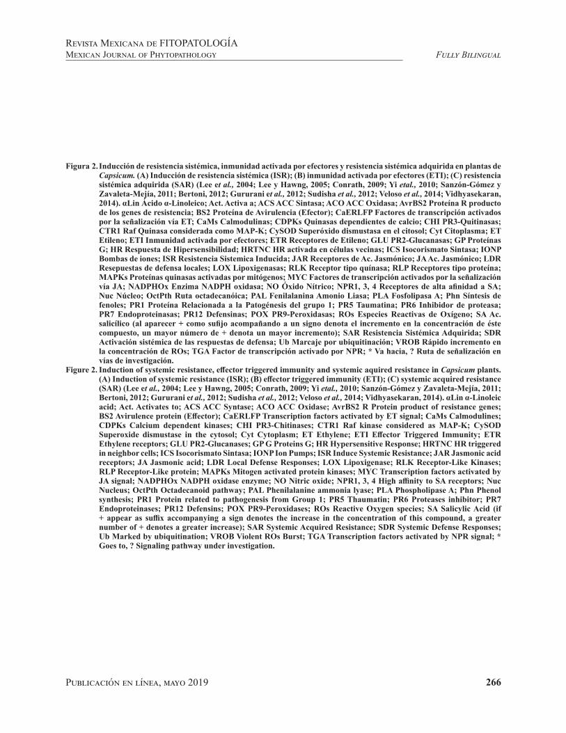
Publicación en línea, mayo 2019 266
Fully BilingualRevista Mexicana de FITOPATOLOGÍAMexican Journal of Phytopathology
Figura 2. Inducción de resistencia sistémica, inmunidad activada por efectores y resistencia sistémica adquirida en plantas de Capsicum. (A) Inducción de resistencia sistémica (ISR); (B) inmunidad activada por efectores (ETI); (C) resistencia sistémica adquirida (SAR) (Lee et al., 2004; Lee y Hawng, 2005; Conrath, 2009; Yi etal., 2010; Sanzón-Gómez y Zavaleta-Mejía, 2011; Bertoni, 2012; Gururani et al., 2012; Sudisha et al., 2012; Veloso et al., 2014; Vidhyasekaran, 2014). αLin Ácido α-Linoleico; Act. Activa a; ACS ACC Sintasa; ACO ACC Oxidasa; AvrBS2 Proteína R producto de los genes de resistencia; BS2 Proteína de Avirulencia (Efector); CaERLFP Factores de transcripción activados por la señalización vía ET; CaMs Calmodulinas; CDPKs Quinasas dependientes de calcio; CHI PR3-Quitinasas; CTR1 Raf Quinasa considerada como MAP-K; CySOD Superóxido dismustasa en el citosol; Cyt Citoplasma; ET Etileno; ETI Inmunidad activada por efectores; ETR Receptores de Etileno; GLU PR2-Glucanasas; GP Proteínas G; HR Respuesta de Hipersensibilidad; HRTNC HR activada en células vecinas; ICS Isocorismato Sintasa; IONP Bombas de iones; ISR Resistencia Sistemica Inducida; JAR Receptores de Ac. Jasmónico; JA Ac. Jasmónico; LDR Resepuestas de defensa locales; LOX Lipoxigenasas; RLK Receptor tipo quínasa; RLP Receptores tipo proteína; MAPKs Proteínas quinasas activadas por mitógenos; MYC Factores de transcripción activados por la señalización vía JA; NADPHOx Enzima NADPH oxidasa; NO Óxido Nítrico; NPR1, 3, 4 Receptores de alta afinidad a SA; Nuc Núcleo; OctPth Ruta octadecanóica; PAL Fenilalanina Amonio Liasa; PLA Fosfolipasa A; Phn Síntesis de fenoles; PR1 Proteína Relacionada a la Patogénesis del grupo 1; PR5 Taumatina; PR6 Inhibidor de proteasa; PR7 Endoproteinasas; PR12 Defensinas; POX PR9-Peroxidasas; ROs Especies Reactivas de Oxígeno; SA Ac. salicílico (al aparecer + como sufijo acompañando a un signo denota el incremento en la concentración de éste compuesto, un mayor número de + denota un mayor incremento); SAR Resistencia Sistémica Adquirida; SDR Activación sistémica de las respuestas de defensa; Ub Marcaje por ubiquitinación; VROB Rápido incremento en la concentración de ROs; TGA Factor de transcripción activado por NPR; * Va hacia, ? Ruta de señalización en vías de investigación.
Figure 2. Induction of systemic resistance, effector triggered immunity and systemic aquired resistance in Capsicum plants. (A) Induction of systemic resistance (ISR); (B) effector triggered immunity (ETI); (C) systemic acquired resistance (SAR) (Lee et al., 2004; Lee y Hawng, 2005; Conrath, 2009; Yi etal., 2010; Sanzón-Gómez y Zavaleta-Mejía, 2011; Bertoni, 2012; Gururani et al., 2012; Sudisha et al., 2012; Veloso et al., 2014; Vidhyasekaran, 2014). αLin α-Linoleic acid; Act. Activates to; ACS ACC Syntase; ACO ACC Oxidase; AvrBS2 R Protein product of resistance genes; BS2 Avirulence protein (Effector); CaERLFP Transcription factors activated by ET signal; CaMs Calmodulines; CDPKs Calcium dependent kinases; CHI PR3-Chitinases; CTR1 Raf kinase considered as MAP-K; CySOD Superoxide dismustase in the cytosol; Cyt Cytoplasm; ET Ethylene; ETI Effector Triggered Immunity; ETR Ethylene receptors; GLU PR2-Glucanases; GP G Proteins G; HR Hypersensitive Response; HRTNC HR triggered in neighbor cells; ICS Isocorismato Sintasa; IONP Ion Pumps; ISR Induce Systemic Resistance; JAR Jasmonic acid receptors; JA Jasmonic acid; LDR Local Defense Responses; LOX Lipoxigenase; RLK Receptor-Like Kinases; RLP Receptor-Like protein; MAPKs Mitogen activated protein kinases; MYC Transcription factors activated by JA signal; NADPHOx NADPH oxidase enzyme; NO Nitric oxide; NPR1, 3, 4 High affinity to SA receptors; Nuc Nucleus; OctPth Octadecanoid pathway; PAL Phenilalanine ammonia lyase; PLA Phospholipase A; Phn Phenol synthesis; PR1 Protein related to pathogenesis from Group 1; PR5 Thaumatin; PR6 Proteases inhibitor; PR7 Endoproteinases; PR12 Defensins; POX PR9-Peroxidases; ROs Reactive Oxygen species; SA Salicylic Acid (if + appear as suffix accompanying a sign denotes the increase in the concentration of this compound, a greater number of + denotes a greater increase); SAR Systemic Acquired Resistance; SDR Systemic Defense Responses; Ub Marked by ubiquitination; VROB Violent ROs Burst; TGA Transcription factors activated by NPR signal; * Goes to, ? Signaling pathway under investigation.

Publicación en línea, mayo 2019 267
Fully BilingualRevista Mexicana de FITOPATOLOGÍA
Mexican Journal of Phytopathology
del ET: la ACC sintasa (ACS) y la ACC oxidasa (ACO); adicionalmente, las CDPKs activadas por el Ca+ incrementan la actividad de estas enzimas, iniciando la síntesis de ET. La creciente concen-tración de ET lleva la alerta a las células distantes, en donde es reconocido por receptores transmem-branales (ETR) compuestos por cinco moléculas (ETR1, ETR2, ERS1, ERS2 y EIN4). Las molé-culas de ETR interactúan entre ellas y con CTR1 (una Raf quinasa considerada como una MAP-K) para activar la señal hacia el núcleo vía MAP-Ks (Vidhyasekaran, 2014). Después, el factor de trans-cripción CaERLFP, recibe la señal y reconoce las cajas GCC de los genes de PRs (Lee et al., 2004), induciendo la expresión de PRs como PR 2 (GLU), PR 3 (CHI), PR 7 (endoproteinasas), PR 9 (POX) y PR 12 (defensinas) (Sudisha et al., 2012) (Figura 2A). La activación de la ISR varía en intensidad de acuerdo a las circunstancias de la interacción, y no siempre resulta en la activación sistémica de las respuestas de defensa, pero puede resultar en una mayor capacidad de responder más rápida e intensamente contra el ataque por fitopatógenos en el tejido circundante (efecto priming) (Conrath, 2009). La ISR es efectiva contra un amplio rango de fitopatógenos, pero es más compleja de lo que aquí se explica, ya que las vías de señalización que participan en su activación, también regulan posi-tiva y negativamente una considerable cantidad de genes que podrían estar involucrados en el proceso de la inducción de resistencia.
Un segundo posible desenlace es el desarrollo de la enfermedad. Factores como un déficit nutri-mental, la ausencia de microorganismos benéficos, el estado fenológico, el potencial de inóculo y el manejo afectan directamente la expresión de la MTI/PTI, la ISR e incluso a los ACBs. La com-binación de condiciones no óptimas para el desa-rrollo de la planta y la presencia de un genotipo patogénico fácilmente adaptable, lleva al desarrollo
signaling paths that participate in their activation also regulate in a positive and negative manner a considerable amount of genes that may be involved in the process of induction of resistance.
A second possible outcome is the development of the disease. Factors such as a nutritional deficiencies, the absence of beneficial microorganisms, the phenological state, the inoculum potential and management practices, all directly affect the expression of MTI/PTI, ISR and even the ACBs. The combination of non-optimum conditions for the plant’s development and the presence of an easily adaptable pathogenic genotype, leads to the development of the disease. In this case, P. capsici enters the tissue directly via the formation of an appressorium, but in both cases, by the secretion of enzymes (Feng et al., 2010; Castro-Rocha et al., 2012). During the biotrophic phase, the hyphae remain in the apoplast and the uptake of nutrients and secretion of virulence factors is carried out with a specialized haustorium invaginated into the cell membrane of the host. In this point, P. capsici secretes molecules such as capsicein to obtain the sterols contained in the host cell membrane (Nespoulous et al, 1999). However, the presence of this and other molecules such as Pep-13 and arachidonic acid (PAMPs), alert the plant and activate PTI. In response, P. capsici releases the effectors RLXLR and Crinklers (CRN) to evade the defense responses. Diverse contributions reveal that in incompatible interactions, these effectors accumulate in the nuclei of the host, blocking the expression of defense genes (Stam et al., 2013). In this way the modulation of the expression of the local genes suppresses the PTI during the biotrophic phase (Kamoun, 2006). This event helps the oomycete feed itself and mature. Later, it produces the protein PcPNPP1, which first changes the behaviour of the phytopathogen from biotrophic to necrotrophic; and finally, when secreted to the

Publicación en línea, mayo 2019 268
Fully BilingualRevista Mexicana de FITOPATOLOGÍAMexican Journal of Phytopathology
de la enfermedad. En este caso, P. capsici entra al tejido directamente o a través de la formación de un apresorio, pero en ambos casos mediante la secre-ción de enzimas (Feng et al., 2010; Castro-Rocha et al., 2012). Durante la fase biotrófica, las hifas permanecen en el apoplasto y la toma de nutrien-tes y secreción de factores de virulencia se lleva a cabo mediante un haustorio especializado invagi-nado en la membrana celular del hospedante. En este punto, P. capsici secreta moléculas como la capsiceina para obtener los esteroles contenidos en la membrana de la célula hospedante (Nespoulous et al, 1999). Sin embargo, la presencia de ésta y otras moléculas como Pep-13 y ácido araquidónico (PAMPs), alertan a la planta y activan la PTI. En respuesta, P. capsici libera los efectores RLXLR y Crinklers (CRN) para evadir las respuestas de de-fensa. Diversas contribuciones revelan que en inte-racciones compatibles estos efectores se acumulan en los núcleos del hospedante bloqueando la expre-sión de genes de defensa (Stam et al., 2013). Así, la modulación de la expresión de los genes locales suprime la PTI durante la fase biotrófica (Kamoun, 2006). Este evento permite al oomiceto alimentarse y madurar. Después, produce la proteína PcPNPP1, la cual primero cambia el comportamiento del fito-patógeno de biotrófico a necrotrófico; y después, al ser secretada al tejido del hospedante, induce la MCP y la necrosis, permitiéndole al patógeno colo-nizar el tejido y causar enfermedad (Lamour et al., 2012; Jupe et al., 2013).
Inmunidad activada por efectores (ETI) y resis-tencia sistémica en Capsicum spp. Para explicar hipoteticamente la resistencia específica a P. cap-sici, es necesario basarse en el conocimiento que se tiene del funcionamiento de las proteínas R ante patógenos biotróficos como Xanthomonas cam-pestris o Meloidogyne incognita en cultivares re-sistentes de Capsicum. Las proteínas R reportadas
host’s tissue, it induces PCD and necrosis, allowing the pathogen to colonize the tissue and cause the disease (Lamour et al., 2012; Jupe et al., 2013).
Effector-triggered immunity (ETI) and systemic resistance in Capsicum spp. In order to hypothetically explain the specific resistance to P. capsici, it is necessary to be based on the knowledge on the way R proteins work when faced with biotrophic pathogens such as Xanthomonas campestris or Meloidogyne incognita in resistant Capsicum cultivars. The R proteins reported for Capsicum correspond to the recognition of bacteria, nematodes and viruses with biotrophic habits; for the moment, no R proteins have been reported for fungal phytopathogens or oomycetes with necrotrophic or hemibiotrophic habits. When the R proteins of Capsicum recognize an effector, they generate a dramatic increase in the concentration of ROs via the signaling of Ca+, which inhibits the local expression of enzymes such as SOD, and it favors a balance between H2O2 / NO, activating the RH (Yi etal., 2010; Gururani et al., 2012). Ca+
also activates the defense responses via MAP-K and the expression of the isochorismate synthase (ISC) via CaMs. The expression of PAL results in the accumulation of phenoles and phytotoxic compounds that accompany the RH and the expression of ISC in the synthesis of SA (Lee and Hawng, 2005; Sanzón-Gómez and Zavaleta-Mejía, 2011). This event characteristically culminates in a fast and intense PCD, and is known as effector-triggered immunity (ETI) (Figure 2B). The growing production of SA leads to its translocation to distant cells, where it is received by three receptors: NPR1 (medium affinity to SA), NPR3 (low affinity) and NPR4 (high affinity). Upon the arrival of the SA to the distant cell, NPR1 migrates from the cytosol to the nucleus, where NPR4 joins it and marks it by ubiquitination for its degradation, although, if
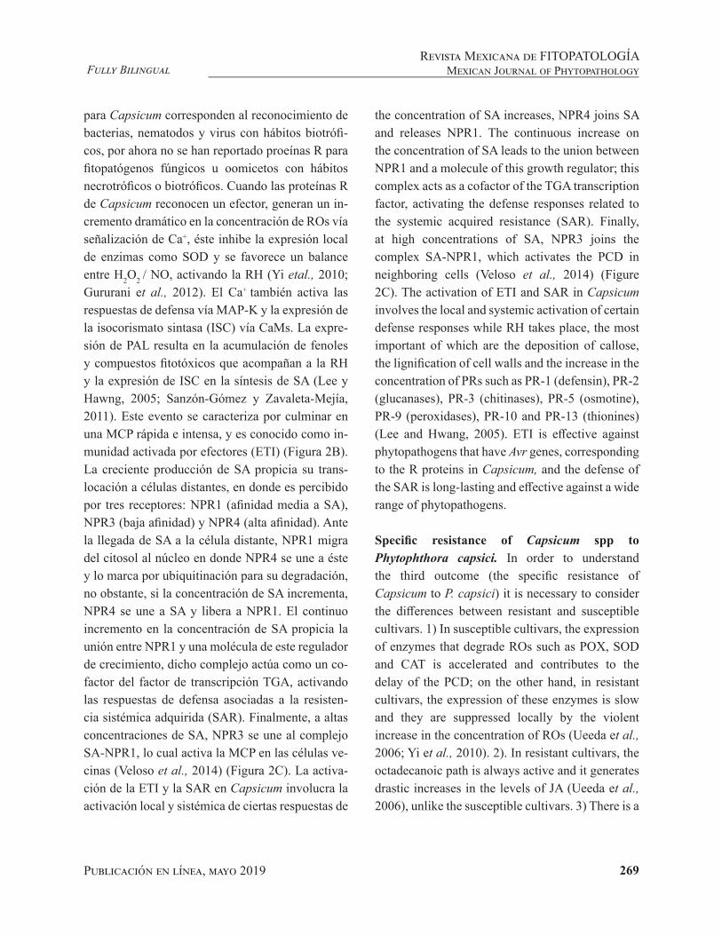
Publicación en línea, mayo 2019 269
Fully BilingualRevista Mexicana de FITOPATOLOGÍA
Mexican Journal of Phytopathology
para Capsicum corresponden al reconocimiento de bacterias, nematodos y virus con hábitos biotrófi-cos, por ahora no se han reportado proeínas R para fitopatógenos fúngicos u oomicetos con hábitos necrotróficos o biotróficos. Cuando las proteínas R de Capsicum reconocen un efector, generan un in-cremento dramático en la concentración de ROs vía señalización de Ca+, éste inhibe la expresión local de enzimas como SOD y se favorece un balance entre H2O2 / NO, activando la RH (Yi etal., 2010; Gururani et al., 2012). El Ca+ también activa las respuestas de defensa vía MAP-K y la expresión de la isocorismato sintasa (ISC) vía CaMs. La expre-sión de PAL resulta en la acumulación de fenoles y compuestos fitotóxicos que acompañan a la RH y la expresión de ISC en la síntesis de SA (Lee y Hawng, 2005; Sanzón-Gómez y Zavaleta-Mejía, 2011). Este evento se caracteriza por culminar en una MCP rápida e intensa, y es conocido como in-munidad activada por efectores (ETI) (Figura 2B). La creciente producción de SA propicia su trans-locación a células distantes, en donde es percibido por tres receptores: NPR1 (afinidad media a SA), NPR3 (baja afinidad) y NPR4 (alta afinidad). Ante la llegada de SA a la célula distante, NPR1 migra del citosol al núcleo en donde NPR4 se une a éste y lo marca por ubiquitinación para su degradación, no obstante, si la concentración de SA incrementa, NPR4 se une a SA y libera a NPR1. El continuo incremento en la concentración de SA propicia la unión entre NPR1 y una molécula de este regulador de crecimiento, dicho complejo actúa como un co-factor del factor de transcripción TGA, activando las respuestas de defensa asociadas a la resisten-cia sistémica adquirida (SAR). Finalmente, a altas concentraciones de SA, NPR3 se une al complejo SA-NPR1, lo cual activa la MCP en las células ve-cinas (Veloso et al., 2014) (Figura 2C). La activa-ción de la ETI y la SAR en Capsicum involucra la activación local y sistémica de ciertas respuestas de
the concentration of SA increases, NPR4 joins SA and releases NPR1. The continuous increase on the concentration of SA leads to the union between NPR1 and a molecule of this growth regulator; this complex acts as a cofactor of the TGA transcription factor, activating the defense responses related to the systemic acquired resistance (SAR). Finally, at high concentrations of SA, NPR3 joins the complex SA-NPR1, which activates the PCD in neighboring cells (Veloso et al., 2014) (Figure 2C). The activation of ETI and SAR in Capsicum involves the local and systemic activation of certain defense responses while RH takes place, the most important of which are the deposition of callose, the lignification of cell walls and the increase in the concentration of PRs such as PR-1 (defensin), PR-2 (glucanases), PR-3 (chitinases), PR-5 (osmotine), PR-9 (peroxidases), PR-10 and PR-13 (thionines) (Lee and Hwang, 2005). ETI is effective against phytopathogens that have Avr genes, corresponding to the R proteins in Capsicum, and the defense of the SAR is long-lasting and effective against a wide range of phytopathogens.
Specific resistance of Capsicum spp to Phytophthora capsici. In order to understand the third outcome (the specific resistance of Capsicum to P. capsici) it is necessary to consider the differences between resistant and susceptible cultivars. 1) In susceptible cultivars, the expression of enzymes that degrade ROs such as POX, SOD and CAT is accelerated and contributes to the delay of the PCD; on the other hand, in resistant cultivars, the expression of these enzymes is slow and they are suppressed locally by the violent increase in the concentration of ROs (Ueeda et al., 2006; Yi et al., 2010). 2). In resistant cultivars, the octadecanoic path is always active and it generates drastic increases in the levels of JA (Ueeda et al., 2006), unlike the susceptible cultivars. 3) There is a

Publicación en línea, mayo 2019 270
Fully BilingualRevista Mexicana de FITOPATOLOGÍAMexican Journal of Phytopathology
defensa mientras ocurre la RH, principalmente la deposición de calosa, la lignificación de paredes ce-lulares y el incremento en la concentración de PRs como PR-1 (defensina), PR-2 (glucanasas), PR-3 (quitinasas), PR-5 (osmotina), PR-9 (peroxidasas), PR-10 y PR-13 (tioninas) (Lee y Hwang, 2005). La ETI es efectiva contra fitopatógenos que tienen los genes Avr correspondientes a las proteínas R en Capsicum, y la defensa de la SAR es duradera y efectiva contra un amplio rango de fitopatógenos.
Resistencia específica de Capsicum spp a Phyto-phthora capsici. Para entender el tercer desenlace (la resistencia específica de Capsicum a P. capsici) es necesario considerar las diferencias consignadas entre cultivares resistentes y susceptibles. 1) En cultivares susceptibles la expresión de enzimas de-gradadoras de ROs como POX, SOD y CAT es ace-lerada y contribuye al retraso de la MCP, contraria-mente, en los cultivares resistentes, la expresión de estas enzimas es lenta y son suprimidas localmente por el violento incremento en la concentración de ROs (Ueeda et al., 2006; Yi et al., 2010). 2). En los cultivares resistentes, la ruta octadecanóica siempre está activa y genera incrementos drásticos en los niveles de JA (Ueeda et al., 2006), a diferencia de los cultivares susceptibles. 3) Hay una correlación entre la concentración de capsidiol, el total de área necrosada y la invasión del oomiceto; en cultivares resistentes: grandes cantidades de capsidiol se con-centran en pequeñas áreas necróticas inhibiendo el progreso de P. capsici; en plantas susceptibles: las cantidades de ésta fitoalexina son menores, las áreas necróticas grandes y hay mayor infección en el tejido (Egea et al., 1996; Villar-Luna et al., 2015). 4) Los cultivares resistentes muestran una mayor expresión de los genes PAL, HMG2, HMG3 y EAS asociado a una mayor actividad de las enzi-mas correspondientes; esto se refleja en una mayor concentración de fenoles tóxicos para el oomiceto
correlation between the concentration of capsidiol, the total of the area with necrosis and the invasion of the oomycete; in resistant cultivars, large amounts of capsidiol concentrate in small necrotic areas, inhibiting the progress of P. capsici; in susceptible plants, the amounts of this phytoalexin are lower, necrotic areas are large, and there is more infection in the tissue (Egea et al., 1996; Villar-Luna et al., 2015). 4) Resistant cultivars display a greater expression of genes PAL, HMG2, HMG3 and EAS associated to a greater activity of the corresponding enzymes; this is reflected in a higher concentration of phenols, toxic for the oomycete and precursors of lignin and synthesis of phytoalexins (López-Martínez et al., 2011; Villar-Luna et al., 2015; Villar-Luna et al., 2017) and SA (Lee and Hawng, 2005).
Based on the above, we propose that the Specific Resistance of Capsicum to P. capsici (RECP) begins with the recognition of PAMPs by receptors RLPs and RLKs, just like PTI, except that in this case, there is a rapid increase in the concentration of ROs that locally inactivates the enzymes that dissociate H2O2. The accumulated H2O2 reacts with the NO activating the RH (between 12 h and 24 h) (Villar-Luna et al., 2009). During this time, the local defense responses become activated, as in the PTI. In resistant cultivars, the octadecanoic and ICS paths generate drastic increases of the growth regulators involved in the signaling (JA-ET o SA). Unlike ISR and SAR, in which signaling paths seem to be antagonistic, in RECP there is a coordinated process in which each growth regulator plays an important part in a given time (Ueeda et al., 2006). Initially, JA reaches a maximum concentration approximately 2 h after recognition, making the surrounding tissue sensitive; the ET reaches the maximum level after 6 to 12 h, inducing the accumulation of PRs in neighboring cells (Kim and Hwang, 2000). Overall, these growth regulators
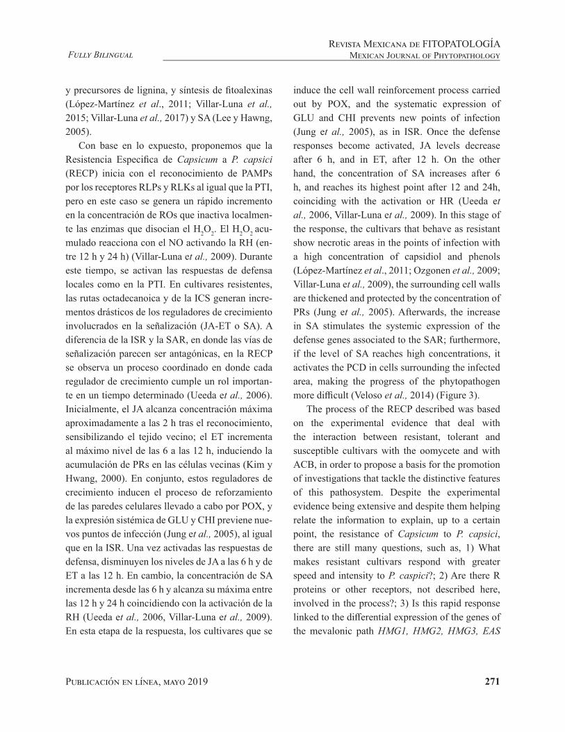
Publicación en línea, mayo 2019 271
Fully BilingualRevista Mexicana de FITOPATOLOGÍA
Mexican Journal of Phytopathology
y precursores de lignina, y síntesis de fitoalexinas (López-Martínez et al., 2011; Villar-Luna et al., 2015; Villar-Luna et al., 2017) y SA (Lee y Hawng, 2005).
Con base en lo expuesto, proponemos que la Resistencia Especifica de Capsicum a P. capsici (RECP) inicia con el reconocimiento de PAMPs por los receptores RLPs y RLKs al igual que la PTI, pero en este caso se genera un rápido incremento en la concentración de ROs que inactiva localmen-te las enzimas que disocian el H2O2. El H2O2 acu-mulado reacciona con el NO activando la RH (en-tre 12 h y 24 h) (Villar-Luna et al., 2009). Durante este tiempo, se activan las respuestas de defensa locales como en la PTI. En cultivares resistentes, las rutas octadecanoica y de la ICS generan incre-mentos drásticos de los reguladores de crecimiento involucrados en la señalización (JA-ET o SA). A diferencia de la ISR y la SAR, en donde las vías de señalización parecen ser antagónicas, en la RECP se observa un proceso coordinado en donde cada regulador de crecimiento cumple un rol importan-te en un tiempo determinado (Ueeda et al., 2006). Inicialmente, el JA alcanza concentración máxima aproximadamente a las 2 h tras el reconocimiento, sensibilizando el tejido vecino; el ET incrementa al máximo nivel de las 6 a las 12 h, induciendo la acumulación de PRs en las células vecinas (Kim y Hwang, 2000). En conjunto, estos reguladores de crecimiento inducen el proceso de reforzamiento de las paredes celulares llevado a cabo por POX, y la expresión sistémica de GLU y CHI previene nue-vos puntos de infección (Jung et al., 2005), al igual que en la ISR. Una vez activadas las respuestas de defensa, disminuyen los niveles de JA a las 6 h y de ET a las 12 h. En cambio, la concentración de SA incrementa desde las 6 h y alcanza su máxima entre las 12 h y 24 h coincidiendo con la activación de la RH (Ueeda et al., 2006, Villar-Luna et al., 2009). En esta etapa de la respuesta, los cultivares que se
induce the cell wall reinforcement process carried out by POX, and the systematic expression of GLU and CHI prevents new points of infection (Jung et al., 2005), as in ISR. Once the defense responses become activated, JA levels decrease after 6 h, and in ET, after 12 h. On the other hand, the concentration of SA increases after 6 h, and reaches its highest point after 12 and 24h, coinciding with the activation or HR (Ueeda et al., 2006, Villar-Luna et al., 2009). In this stage of the response, the cultivars that behave as resistant show necrotic areas in the points of infection with a high concentration of capsidiol and phenols (López-Martínez et al., 2011; Ozgonen et al., 2009; Villar-Luna et al., 2009), the surrounding cell walls are thickened and protected by the concentration of PRs (Jung et al., 2005). Afterwards, the increase in SA stimulates the systemic expression of the defense genes associated to the SAR; furthermore, if the level of SA reaches high concentrations, it activates the PCD in cells surrounding the infected area, making the progress of the phytopathogen more difficult (Veloso et al., 2014) (Figure 3).
The process of the RECP described was based on the experimental evidence that deal with the interaction between resistant, tolerant and susceptible cultivars with the oomycete and with ACB, in order to propose a basis for the promotion of investigations that tackle the distinctive features of this pathosystem. Despite the experimental evidence being extensive and despite them helping relate the information to explain, up to a certain point, the resistance of Capsicum to P. capsici, there are still many questions, such as, 1) What makes resistant cultivars respond with greater speed and intensity to P. caspici?; 2) Are there R proteins or other receptors, not described here, involved in the process?; 3) Is this rapid response linked to the differential expression of the genes of the mevalonic path HMG1, HMG2, HMG3, EAS

Publicación en línea, mayo 2019 272
Fully BilingualRevista Mexicana de FITOPATOLOGÍAMexican Journal of Phytopathology
comportan como resistentes muestran zonas necró-ticas en los puntos de infección con alta concen-tración de capsidiol y fenoles (López-Martínez et al., 2011; Ozgonen et al., 2009; Villar-Luna et al., 2009), las paredes celulares circundantes son en-grosadas y protegidas por la concentración de PRs (Jung et al., 2005). Posteriormente, el incremento de SA estimula la expresión sistémica de los genes de defensa asociados a la SAR, además, si el nivel de SA alcanza altas concentraciones, éste activa la MCP en las células vecinas a la zona infectada difi-cultando el progreso del fitopatógeno (Veloso et al., 2014) (Figura 3).
El proceso de la RECP descrito, se basó en las evidencias experimentales que abordan la interac-ción entre cultivares resistentes, tolerantes y sus-ceptibles con el oomiceto y con ACB, con la fina-lidad de proponer una base que promueva investi-gaciones que aborden las particularidades de este patosistema. A pesar de que las evidencias experi-mentales son bastas y permiten relacionar la infor-mación para explicar hasta cierto punto la resisten-cia de Capsicum a P. capsici, se tienen aún muchas interrogantes, como por ejemplo: 1) ¿Qué hace que los cultivares resistentes respondan con mayor ra-pidez e intensidad a P. caspici?; 2) ¿Hay proteínas R u otros receptores no descritos involucrados en este proceso?; 3) ¿Esta respuesta rápida está ligada a la expresión diferencial de los genes de la ruta mevalonica HMG1, HMG2, HMG3, EAS y a la ac-tividad incrementada de la ruta octadecanoica en cultivares resistentes?; 4) ¿La aplicación de ACBs podría ser una estrategia para incrementar los ni-veles de tolerancia en los cultivares susceptibles? Y de ser así, ¿cuánto tiempo durará la protección?
Consideraciones finales. El análisis crítico del es-tado del arte de este patosistema permitió discer-nir que la resistencia específica de Capsicum a P. capsici es el resultado de un proceso coordinado
and to the increased activity of the octodecaoinc path in resistant cultivars?; 4) Could the application of ACBs be a strategy to increase tolerance levels in susceptible cultivars? And if so, how long will the protection last?
Final considerations. The critical analysis of the state of the art of this pathosystem helped discern that the specific resistance of Capsicum to P. capsici is the result of a process coordinated by growth regulators JA, ET and SA. This entails an exception to the antagonism commonly mentioned between these signaling paths. However, further studies are required that take over the role of each growth regulator in the process of signaling in the defense against this oomycete. Although it is clear that genes such as PAL, HMG, EAS, GLU, CHI and POX are broadly related with the process of defense and resistance of Capsicum to P. capsici, underneath there is a large number of genes that are positively and negatively affected by the paths of signaling of these growth regulators. Because this is a phenomenon with multifactorial causes, the new massive data generation technologies may offer a wider outlook regarding activated and suppressed genes during interaction. It is also important to highlight the implication of these growth regulators in the process of resistance induction by beneficial microorganisms, which are an important component of the soil native microbiota that establish associations with the pathosystem. The microbial diversity plays an important role, since it directly affects the result of the molecular dialogue; first, since it acts as a barrier to complicate or prevent the phytopathogen from establishing on the plant, and secondly, by increasing the defensive ability by activating the basal defense and inducing resistance. Despite the progress made, there are still many questions to be answered. As the comprehension of pathosystems

Publicación en línea, mayo 2019 273
Fully BilingualRevista Mexicana de FITOPATOLOGÍA
Mexican Journal of Phytopathology

Publicación en línea, mayo 2019 274
Fully BilingualRevista Mexicana de FITOPATOLOGÍAMexican Journal of Phytopathology
Figura 3. Propuesta del dialogo molecular que toma lugar en la resistencia específica de Capsicum a Phytophthora capsici. En la parte superior de la figura aparece una línea que marca los tiempos en los cuales se estima que suceden los eventos relacionados con la expresión de resistencia específica de Capsicum a P. capsici. La primera línea corresponde a la vía de señalización del JA. La segunda corresponde a la activación de respuestas de defensa locales activadas en cada punto de infección, la cual sucede de forma similar al proceso de MTI/PTI. La tercera línea corresponde a la vía de señalización del ET. La cuarta línea indica la vía de señalización del SA. La quinta línea corresponde al proceso de activación de la HR local inducida por ROs en materiales resistentes, mientras que la sexta, indica la activación de la HR en las células vecinas inducida por el constante incremento en la concentración de la SA. Con base al crítico análisis del estado del arte del patosistema en cuestión, la presente propuesta sugiere que la resistencia específica de Capsicum a P. capsici es el resultado de la sumatoria de una serie de eventos coordinados en la cual los principales actores son los reguladores de crecimiento involucrados en las vías de señalización (Egea et al., 1996; Kim y Hwang, 2000; Jung et al., 2005; Lee y Hawng, 2005; Bent y Makey, 2007; Conrath, 2009; Ozgonen et al., 2009; Ueeda et al., 2006; Yi et al., 2010; Glowacki et al., 2011; Jingyuan et al., 2011; López-Martínez et al., 2011; Sanzón-Gómez y Zavaleta-Mejía, 2011; Bertoni, 2012; Gururani et al., 2012; Sudisha et al., 2012; Castro-Rocha et al., 2012; Muthamilarasan y Prasad, 2013; Veloso et al., 2014; Villar-Luna et al., 2015; Vidhyasekaran, 2014; Villar-Luna et al., 2009, 2017). αLin Ácido α-Linoleico; ACS ACC Sintasa; ACO ACC Oxidasa; CaMs Calmodulinas; CDPKs Quinasas dependientes de calcio; CHI PR3-Quitinasas; CTR1 Raf Quinasa considerada como MAP-K; Cpsdl Síntesis de capsidiol; CySOD Superóxido dismustasa en el citosol; EAS 5-epi-aristoloqueno sintasa; ET Etileno; ETR Receptores de Etileno; GLU PR2-Glucanasas; GP Proteínas G; HMG 1, 2, 3 familia de genes codificantes para la enzima hidroximetilglutaril-coenzima A reductasa; HR Respuesta de Hipersensibilidad; HRTNC HR activada en células vecinas; ICS Isocorismato Sintasa; IONP Bombas de iones; JAR Receptores de Ac. Jasmónico; JA Ac; Jasmónico; LDR Resepuestas de defensa locales; Lig Proceso de lignificación de paredes celulares; RLK Receptor tipo quínasa; RLP Receptores tipo proteína; MAPKs Proteínas quinasas activadas por mitógenos; NADPHOx Enzima NADPH oxidasa; NO Óxido Nítrico; NPR 1, 3 Receptores de alta afinidad a SA; OctPth Ruta octadecanóica; PAL Fenilalanina Amonio Liasa; PLA Fosfolipasa A; Phn Síntesis de fenoles; PR1 Proteína Relacionada a la Patogénesis del grupo 1; PR5 Taumatina; PR6 Inhibidor de proteasa; PR7 Endoproteinasas; PR12 Defensinas; POX PR9-Peroxidasas; SA Ac. salicílico; VROB Rápido incremento en la concentración de ROs; *, , , Va hacia.
Figure 3. Proposal of the molecular dialogue that takes place in the specific resistance of Capsicum to Phytophthora capsici. In the upper part of the figure appears a line that marks the times in which the events related to specific resistance from Capsicum to P. capsici are estimated to occur. The first line corresponds to the JA signaling pathway. The second correspond to local defense responses activated in each infection point, which occurs in similarly to the MTI/PTI process. Third line correspond to the ET signaling pathway. Fourth line indicates the SA signaling pathway. The fifth line corresponds to the activation of local HR induced by ROs in resistant genotypes, while the sixth indicates the activation of HR in neighbor cells induced by the constant increase of SA concentration. Based on the critical analysis of the state of the art of the pathosystem in question, this proposal suggests that the specific resistance of Capsicum to P. capsici is the result of the summation of a series of coordinated events, in which the main actors are the plant growth regulators involved in the signaling pathways (Egea et al., 1996; Kim y Hwang, 2000; Jung et al., 2005; Lee y Hawng, 2005; Bent y Makey, 2007; Conrath, 2009; Ozgonen et al., 2009; Ueeda et al., 2006; Yi et al., 2010; Glowacki et al., 2011; Jingyuan et al., 2011; López-Martínez et al., 2011; Sanzón-Gómez y Zavaleta-Mejía, 2011; Bertoni, 2012; Gururani et al., 2012; Sudisha et al., 2012; Castro-Rocha et al., 2012; Muthamilarasan y Pasard, 2013; Veloso et al., 2014; Villar-Luna et al., 2015; Vidhyasekaran, 2014; Villar-Luna et al., 2009, 2017). αLin Linoleic acid; ACS ACC Syntase; ACO ACC Oxidase; CaMs Calmodulines; CDPKs Calcium dependent kinases; CHI PR3-Chitinases; CTR1 Raf kinase considered as a MAP-K; Cpsdl Capsidiol synthesis; CySOD Superoxide dismustase in the capsidiol; EAS 5-epi-aristolochene synthase; ET Ethylene; ETR Ethylene receptors; GLU PR2-Glucanases; GP G proteins ; HMG 1, 2, 3 family of genes that codifies hidoxymethylglutaril-coenzyme A reductase; HR Hypersensitive Response; HRTNC HR Triggered in Neighbor Cells; ICS Isochorismate Synthase; IONP Ion Pumps; JAR JA receptors; JA Jasmonic acid; LDR Local Defense Responses; Lig Cell wall lignification process; RLK Receptor-Like Kinases; RLP Receptor-like Protein; MAPKs Mitogen Activated Protein Kinases; NADPHOx NADPH oxidase; NO Nitric oxide; NPR 1, 3 Receptors with high affinity to SA; OctPth Octadecanoid pathway; PAL Phenilalanine Ammonia Lyase; PLA Phospholipase A; Phn Phenols synthesis; PR1 Pathogenesis related protein from Group 1; PR5 Thaumatin; PR6 Protease inhibitor; PR7 Endoproteinases; PR12 Defensins; POX PR9-Peroxidases; SA Salicylic acid; VROB Violent ROs Burst; *, , , Goes to.

Publicación en línea, mayo 2019 275
Fully BilingualRevista Mexicana de FITOPATOLOGÍA
Mexican Journal of Phytopathology
mediado por los reguladores de crecimiento JA, ET y SA. Esto supone una excepción al antagonismo comúnmente mencionado entre estas vías de se-ñalización. No obstante, se requiere de mayores estudios que aborden el rol de cada regulador de crecimiento en el proceso de señalización en la de-fensa contra este oomiceto. Aunque es claro que genes como PAL, HMG, EAS, GLU, CHI y POX están estrechamente relacionados con el proceso de defensa y resistencia de Capsicum hacia P. capsici, en el trasfondo hay un gran número de genes que son afectados positiva y negativamente por las vías de señalización de estos reguladores de crecimien-to. Dado que este es un fenómeno de causa mul-tifactorial, las nuevas tecnologías de generación masiva de datos pueden ofrecer un panorama más amplio en cuanto a los genes activados y suprimi-dos durante la interacción. También, es importante destacar la implicación de estos reguladores de cre-cimiento en el proceso de inducción de resistencia por microorganismos benéficos, los cuales son un componente importante de la microbiota nativa del suelo que se asocia al patosistema. La diversidad microbiana juega un papel importante al afectar di-rectamente el resultado del dialogo molecular; pri-mero, al actuar como una barrera para dificultar o prevenir que el fitopatógeno se establezca sobre la planta, y en segundo lugar, al incrementar la capa-cidad defensiva al activar la defensa basal e indu-cir resistencia. A pesar del avance logrado, hay aún muchas incógnitas por resolver. En la medida en la que se avance a la comprensión de los patosistemas y que la investigación abarque la complejidad de los ecosistemas agrícolas, se podrá reducir gradual-mente el impacto de los fitopatógenos. El control de fitopatógenos no debe dirigirse a su exclusión de los agroecosistemas, sino a emplear tecnologías ecológicas que promuevan el balance de las po-blaciones de los microorganismos del suelo, y que eventualmente se logre un manejo sustentable de fitopatógenos y plagas.
advances, and as investigation covers the complexity of agricultural ecosystems, the impact of phytopathogens can gradually be reduced. The control of plant pathogens should not be directed to their exclusion from the agroecosystems, but rather, to employ ecological technologies that promote the balance of the populations of soil microorganisms, which eventually will allow to achieve a sustainable management of plant pathogens and pests.
AcknowledgementsTo the National Science and Technology Council –
CONACyT for the scholarship granted to the main author.
End of the English version
AgradecimientosAl Consejo Nacional de Ciencia y Tecnología – CONA-
CyT por la beca doctoral otorgada al primer autor.
LITERATURA CITADA
Bae H, Roberts DP, Lim HS, Strem MD, Park SC, Ryu CM, Meinick RL, Bailey BA. (2011). Endophytic Trichoder-ma isolates from tropical environments delay disease on-set and induce resistance against P. capsici in hot pepper using multiple mechanisms. Mol. Plant-Microbe Interac. 24:336-351. Doi: https://doi.org/10.1094/MPMI-09-10-0221
Barchenger DW, Lamour KH, Bosland PW. (2018). Challen-ges and Strategies for Breeding Resistance in Capsicum annuum to the Multifarious Pathogen, Phytophthora cap-sici. Front. Plant Sci. 9:628. Doi: http://journal.frontiersin.org/article/10.3389/fpls.2018.00628/full
Bent AF, Makey D. (2007). Elicitors, Effectors and R ge-nes: The new paradigm and lifetime supply of questions. Annu. Rev. Phytopathol. 45:399-436. Doi: https://doi.org/10.1146/annurev.phyto.45.062806.094427
Bertoni G. (2012). Oxylipins and plant palatability. Plant Cell. 24: 1305. Doi: https://doi.org/10.1105/tpc.112.240412
Bishop HSL, Mounter SA, Laskey J, Morris RO, Elder J, Roop P, Rouse C, Schmidt FJ, English JT. (2002). Phage-Displa-yed peptides as developmental agonists for Phytophthora capsici zoospores. App.Envir. Microbiol. 68:3315-3320. Doi: https://doi.org/10.1128/AEM.68.7.3315-3320.2002

Publicación en línea, mayo 2019 276
Fully BilingualRevista Mexicana de FITOPATOLOGÍAMexican Journal of Phytopathology
Bostock RM, Saychenko C, Lazarus C, Dehesh K. (2011). Ei-cosapolyenoic acids. Novel MAMPs with reciprocal effect on oomycete-plant defense signaling networks. Plant Signal. Behav. 6:531-533. Doi: https://doi.org/10.4161/psb.6.4.14782
Brich PRJ, Boevink PC, Gilroy EM, Hein I, Pritchard L, Whisson SC. (2008). Oomycete RXLR effectors: deli-very, functional redundancy and durable disease resistan-ce. Curr. Opin. Plant Biol. 11:337-379. Doi: https://doi.org/10.1016/j.pbi.2008.04.005
Cannon SB, Zhu H, Baumgarte AM, Spangler R, May G, Cook DR, Young ND. (2002). Diversity, distribution and ancient taxonomic relationship within the TIR and Non-TIR NBS-LRR resistance gene subfamilies. J. Mol. Evol. 54:548-562. Doi: https://doi.org/10.1007/s0023901-0057-2
Castro-Rocha A, Fernández-Pavia SP, Osuna-Avila P. (2012) Chili defense mechanisms in the Capsicum annuum-Phytophthora capsici pathosystem. R.M.F. 30:49-65. En línea: www.scielo.org.mx/scielo.php?script=sci_arttext&pid=S0185-33092012000100005
Chemeltorit PP, Mutaqin KH, Widodo W. (2017). Combining Trichoderma hamatum THSW13 and Pseudomonas aeru-ginosa BJ10-86:a synergistic chili pepper seed treatment for Phytophthora capsici infested soil. Eur. J. Plant. Pa-thol. 147:157-166. Doi: https://doi.org/10.1007/s10658-016-0988-5
Chen YY, Chen PC, Tsay TT. (2016). The biocontrol effica-cy and antibiotic activity of Streptomyces plicatus on the oomycete Phytophthora capsici. Bio. Con. 98:34-42. Doi: https://doi.org/10.1016/j.biocontrol.2016.02.011
Conrath U. (2009). Priming of induced plant defense res-ponses. Adv. Bot. Res. 51:361-395. Doi: https://doi.org/10.1016/S0065-2296(09)51009-9
Dahiya N, Tewari R, Hoondal GS. (2006). Biotechnological aspects of chitinolytic enzymes: a review. Appl. Microbiol. Biotechnol. 71:773-782. Doi: https://doi.org/10.1007/s00253-005-0183-7
Egea C, Alcázar MD, Candela E. (1996). Capsidiol: Its role in the resistance of Capsicum annuum to P. cap-sici. Physiol. Plantarum. 98:737:742. Doi: https://doi.org/10.1111/j.1399-3054.1996.tb06679.x
Feng B, Li P, Wang H, Zhang X. (2010). Functional analy-sis of Pcpme6 fom oomycete plant pathogen Phytophtho-ra capsici. Microb. Pathog. 49:23-31. Doi: https://doi.org/10.1016/j.micpath.2010.03.004
Fernández-Herrera E, Rojas-Martínez RI, Gómez-Rodríguez O, Guevara-Olvera L, Rivas-Dávila ME, Valdez-Moc-tesuma E, Zavaleta-Mejía E. (2012). Genes de defensa, actividad enzimática y contenido de capsidiol en chile CM-334 inoculado con Phytophthora capsici. Intercien-cia. 37:370-377. En línea: http://www.redalyc.org/articulo.oa?id=33922756007
French MRD, Jones JB, Ozores HM, Roberts PD. (2007). Sur-vival of inoculum of Phytophthora capsici in soil trough time under different soil treatment. Plant Dis. 91:593-598. Doi: https://doi.org/10.1094/PDIS-91-5-0593
Glowacki S, Macoiszek VK, Kononowicz AK. (2011). R proteins as fundamentals of plant innate immunity. Cell. Mol. Biol. Letters. 16:1-24. Doi: https://doi.org/10.2478/s11658-010-0024-2
Goldberg NP. (2001) Chile Pepper Diseases Circular 549. Co-llege of Agriculture, Consumer and Environmental Scien-ce. New Mexico State Universiy. En línea: https://aces.nmsu.edu/pubs/_circulars/CR549/welcome.html
Gururani MA, Vankatesh J, Upadhyaya CP, Nookaraju A, Padney SK, Park SW. (2012). Plant disease resistance genes: Current status and future directions. Physiol. Mol. Plant Pathol. 78:51-65. Doi: https://doi.org/10.1016/j.pmpp.2012.01.002
Hardham AR, Shan W. (2009). Cellular and molecular bio-logy of Phytophthora-Plant Interactions. In: Deising H (Ed.) Plant Relationships, 2nd Edition The Mycota V. Edi-tion. Springer-Verlag Berlin. pp. 3-27. Doi: https://doi.org/10.1007/978-3-540-87407-2_1
Jiménez-Camargo A, Valadez-Moctezuma E, Lozoya-Saldaña H. (2018). Antagonism by Penicillum sp. Against Phyto-phthora capsici (Leonian). Rev. Fitotec. Mex. 41:137-148. En línea: https://www.revistafitotecniamexicana.org/documentos/41-2/5a.pdf
Jingyuan Z, Xuexiao Z, Zhenchuan M, Bingyan X. (2011). A novel pepper (Capsicum annuum L.) WRKY Gene CaWRKY30, is involved in pathogen stress responses. J. Plant Biol. 54:329-337. Doi: https://doi.org/10.1007/s12374-011-9171-x
Jung WJ, Jin YL, Kim KY, Park RD, Kim TH. (2005). Chan-ges in pathogenesis-related proteins in pepper plants with regard to biological control of phytopthora blight with Paenibacillus illinoisensis. BioControl. 50:165-178. Doi: https://doi.org/10.1007/s10526-004-0451-y
Jupe J, Stam R, Howden AJM, Morrin JA, Zhang R, Hedley PE, Huitema E. (2013). Phytophthora capsici-tomato inte-raction features dramatic shifts gene expression associated with a hemi-biotrophic lifestyle. Genome Biol. 14:R63. Doi: https://doi.org/10.1186/gb-2013-14-6-r63
Kamoun S. (2006). A catalogue of the effector secreto-me of plant pathogenic oomycetes. Annu. Rev. Phy-siol. 44:41-60. Doi: https://doi.org/10.1146/annurev.phyto.44.070505.143436
Kim BS, Lee JY, Hwang BK. (2000). In vivo control and in vitro antifungal activiry of rhamnolipid B, a glycolipid antibio-tic, against Phytophthora capsici and Colletotrichum orbi-culare. 56:1029-1035. Doi: https://doi.org/10.1002/1526-4998(200012)56:12<1029::AID-PS238>3.0.CO;2-Q
Lamour KH, Stam R, Jupe J, Huitema E. (2012). The oomyce-te broad-host-range pathogen Phytophthora capsici. Mol. Plant Pathol. 13:329-337. Doi: https://doi.org/10.1111/j.1364-3703.2011.00754.x
Lee JH, Hong JP, Oh SK, Lee S, Choi D, Kim WT. (2004). The ethylene-responsive factor like protein 1 (CaERFLP1) of hot pepper (Capsicum annuum L.) interacts in vitro with both GCC and DRE/CRT sequence with different binding affinities possible biological roles of CaERFPL1 in res-ponse to pathogen interactions and high salinity conditions in transgenic tobacco plants. Plant. Mol. Biol. 42:335-344. Doi: https://doi.org/10.1007/s11103-004-0417-6
Lee SC, Hwang BK. (2005). Induction of some defense-rela-ted genes and oxidative burst is required for the establis-hment of systemic acquired resistance in Capsicum an-nuum. Planta. 221: 790-800. Doi: https://doi.org/10.1007/s00425-005-1488-6

Publicación en línea, mayo 2019 277
Fully BilingualRevista Mexicana de FITOPATOLOGÍA
Mexican Journal of Phytopathology
Ley-López N, Márquez-Zequera I, Carrillo-Fasio JA, León-Félix J, Cruz-Lachica I, García-Estrada RS. (2018). Effect of biocontrol and germinative inhibition of Bacillus spp. On zoospores of Phytophthora capsici. RMF. 36:215-232. Doi: http://dx.doi.org/10.18781/R.MEX.FIT.1711-2
López-Martínez NM, Colinas-León T, Peña-Valdivia CB, Salinas-Moreno Y, Fuentes-Montiel P, Biesaga M, Zava-leta-Mejía E. (2011). Alterations in peroxidase activity and phenylpropanoid metabolism induced by Nacobbus aberrans Thorne and Allen in chilli (Capsicum annuum L.) CM334 resistant to Phytophthora capsici Leo. Plant and soil. 338:399-409. Doi: https://doi.org/10.1007/s11104-010-0553-5
Minamiyama Y, Tsuro M, Kubo T, Hirai M. (2005). QTL analysis for recistance to Phytophtthora capsici in pepper using a high density SSR-based map. Breed. Sci. 57:129-134. Doi: https://doi.org/10.1270/jsbbs.57.129
Mongkolporn O, Taylor PWJ. (2011). Capsicum. In: Wild Crop Relatives: Genomics and Breeding Resourses, Vegetables. (Ed) Kole, C. Springer-Verlag Berlin Heidelberg. Pp. 43-57. Doi: https://doi.org/10.1007/978-3-642-20450-0
Muthamilarasan M, Prasad M. (2013). Plant innate immuni-ty: An updated insight into defense mechanism. J. Biosci. 38:1-17. Doi: https://doi.org/10.1007/s12038-013-9302-2
Nespoulous C, Gaudemer O, Huet JC, Pernollet JC. (1999). Characterization of elicitin-like phospholipases isolated from Phytophthora caspici culture filtrate. FEBS let-ters. 452:400-406. Doi: https://doi.org/10.1016/S0014-5793(99)00654-7
Ozgonen H, Yardimci N, Kilic HC. (2009). Induction of phe-nolic compounds and pathogenesis-related proteins by mycorrhizal fungal inoculations against Phytophthora capsici Leonian in pepper. Pak. J. Biol. Sci. 12:1181-1187. Doi: 10.3923/pjbs.2009.1181.1187. En línea: https://scia-lert.net/abstract/?doi=pjbs.2009.1181.1187
Ramos SRU, Gutiérrez SJG, Rodrígez GR, Salcedo MSM, Hernández LCE, Luna OHA, Jiménez BJF, Fraire VS, Almeyda LIH. (2010) Antagonismo de dos ascomicetos contra Phytophthora capsici Leonian, causante de la mar-chitez del chile (Capsicum annuum L.) RMF. 28:75-86. En línea: www.scielo.org.mx/scielo .php?script=sci_arttext&pid=S0185-33092010000200001
Reifschneider FJB, Boiteux LS, Della Vecchia PT. (1992). In-heritance of resisance in peppers to Phytophthora capsici in pepper. Euphytica 62:45-49. https://doi.org/10.1007/BF00036086
Sanzón-Gómez D, Zavaleta-Mejía E. (2011). Respuesta de hi-persensibilidad, una muerte programada para defenderse del ataque por fitopatógeno. RMF. 29:154-164. En línea: www.scielo.org.mx/scielo.php?script=sci_arttext&pid=S018533092011000200007&lng=es&nrm=iso
Stam R, Jupe J, Howden AJM, Morris JA, Bovenik PC, Heldey PE, Huitema E. (2013). Identification and characterization CRN effectors in Phytophthora capsici show modularity and functional diversity. PLoS ONE 8(3):e59517. En lí-nea: www.plosone.org/article/citationList.action?articleURI=info%3Adoi%2F10.1371%2Fjournal.pone.0059517
Sudisha J, Sarathchandra RG, Amrutesh KN, Kumar A, Shetty HS (2012). Pathogenesis Related Proteins in Plant Defense Response. In: Plant Defence: Biological Control. Progress
in Biological Control Vol 12. (Eds) Mérillon J, Ramawat K. ED Springer Dordrecht. Pp. 379-403. Doi: https://doi.org/10.1007/978-94-007-1933-0_17
Thomma BPHJ, Nürnebereger T, Joosten MHAJ. (2011). Of PAMPs and effectors: The blurred PTI-ETI Dichotomy. The Plant Cell. 23:4-15. Doi: https://doi.org/10.1105/tpc.110.082602
Tian MY, Win .J, Song J, van Der HR, van Der KE, Kamoun.S. (2016). A Phytophthora infestans cystatin-like protein targets a novel tomato papain-like apoplastic protease. Plant Physiol. 143:364-377. Doi: https://doi.org/10.1104/pp.106.090050
Tör M. (2008). Tapping into molecular conversation between oomycete plant pathogens and their hosts. Eur. J. Plant Pa-thol. 122:57-69. Doi: https://doi.org/10.1007/s10658-008-9288-z
Torto AT, Collmer CW, Lindeber M, Bird D, Collmer A, Tyler BM. (2009). Common and contrasting themes in host cell-targeted effectors from bacterial, fungal, oomy-cete and nematode plant symbionts described using the Gene Ontology. BMC Microbiol. 9:1-8. Doi: https://doi.org/10.1186/1471-2180-9-S1-S3
Ueeda M, Kubota M, Nishi K. (2006). Contribution of jasmonic acid to resistance against Phytophthora blight in Capsicum annuum cv. SCM334. Physiol. Mol. Plant. Pathol. 67:149-154. Doi: https://doi.org/10.1016/j.pmpp.2005.12.002
Veloso J, Díaz J. (2012). Fusarium oxysporum Fo47 confers protection Pepper plants against Verticillium dahlia and Phytophthora capsici, and induces the expression of de-fence genes. Plant Pathol. 61:281-188. Doi: https://doi.org/10.1111/j.1365-3059.2011.02516.x
Veloso J, García T, Bernal A, Díaz J. (2014). New bricks on the wall of induced resistance: salicylic acid receptors and transgenerational priming. Eur. J. Plant Pathol. 38:685-693. Doi: https://doi.org/10.1007/s10658-013-0350-0
Vidhyasekaran P. (2014). PAMP signaling in plant innate im-munity: Signal perception and transduction. In: Signaling and communication in plants series number 21. Sprin-ger Science+Busines Media Dordrecht. Pp. 17-161. Doi: https://doi.org/10.1007/978-94-007-7426-1
Villar-Luna E, Reyes-Trejo B, Rojas-Martínez RI, Gómez-Rodríguez O, Hernández-Anguiano AM, Zavaleta-Mejía E. (2009). Respuesta hipersensitiva en follaje de chi-le CM-334 resistente a Phytophthora capsici infectado con Nacobus aberrans. Nematropica. 39:143-155. En línea: http://journals.fcla.edu/nematropica/article/viewFi-le/64475/62143
Villar-Luna E, Rojas-Martinez R, Reyes-Trejo B, Gómez-Ro-dríguez O, Zavaleta-Mejía E. (2017). Mevalonate pathway genes expressed in chilli CM334 inoculated with Phyto-phthora capsici and infected by Nacobbus aberrans and Meloidogyne enterolobii. Eur. J. Plant Pathol. 148:867-881. Doi: https://doi.org/10.1007/s10658-016-1142-0
Villar-Luna H, Reyes-Trejo B, Gómez-Rodríguez O, Villar-Luna E, Zavaleta-Mejía E. (2015) Expresión de genes de defensa y acumulación de capsidiol en la interacción compatible CM334/ Nacobus aberrans e incompatible CM334/Meloidogyne incognita. Nematropica 45:9-19. En línea: http://journals.fcla.edu/nematropica/article/view/85048/81977

Publicación en línea, mayo 2019 278
Fully BilingualRevista Mexicana de FITOPATOLOGÍAMexican Journal of Phytopathology
Win J, Chaparro GA, Belhaj K, Saunders DG, Yoshida K, Dong S, Shornack S, Zipfel C, Robatzek S, Hogenhout SA, Kamoun S. (2012). Effector biology of plant-associa-ted organisms: Concepts and perspectives. Cold Spring Harb Symp Quant. Biol. 77:235-47. Doi: http://dx.doi.org/10.1101/sqb.2012.77.015933
Xu S, Kim BS. (2016). Evaluation of Paenibacillus polymyxa strain SC09-21 for biocontrol of Phytophthora blight and growth stimulation in pepper plants. Trop. Plant Pathol. 41:62. Doi: https://doi.org/10.1007/s40858-016-0077-5
Yi SY, Lee DJ, Yeom SI, Yoon J, Kim YH, Kwon SY, Choi D. (2010). A novel pepper (Capsicum annuum) recep-tor-like kinase functions as a negative regulator of plant cell death via accumulation of superoxide anions. New Phytol. 185:701-705. Doi: https://doi.org/10.1111/j.1469-8137.2009.03095.x

279Publicación en línea, mayo 2019
Zuñiga-Romano MC, Ochoa-Martínez DL, Rojas-Mar-tínez RI and Zamora-Macorra EJ. Identification of be-gomoviruses in tomato and determination of seed trans-mission in Atlacomulco, Mexico State. Mexican Journal of Phytopathology 37(2): 279-286.DOI: 10.18781/R.MEX.FIT.1902-2
Primera publicación DOI: 01 de Abril, 2019.First DOI publication: April 01, 2019.
Resumen. En un invernadero de alta tecnología ubicado en Atlacomulco, Estado de México, se han observado incidencias del 60 al 90% de plantas con síntomas típicos de begomovirus. Los objetivos del presente estudio fueron identificar begomovi-rus presentes, conocer si se transmiten por semi-lla, determinar la especie de mosca blanca y si ésta es portadora de los virus. Hojas de plantas adultas con síntomas se analizaron con oligonucleótidos universales para begomovirus. Semillas de frutos de plantas positivas a begomovirus se sembraron en almácigos para obtener plántulas, éstas y parte
Identification of begomoviruses in tomato and determination of seed transmission in Atlacomulco, Mexico State
Identificación de begomovirus en tomate y determinación de su transmisión por semilla en Atlacomulco, Estado de México
Maria del Carmen Zuñiga Romano, Daniel Leobardo Ochoa Martínez*, Reyna Isabel Rojas Martínez, Posgrado en Fitosanidad-Fitopatología. Colegio de Postgraduados, Km 36.5 Carretera México-Texcoco, Mon-tecillo, Texcoco, Estado de México, CP 56230, México; Erika Janet Zamora Macorra, Universidad Autó-noma Chapingo Km 38.5 Carretera México-Texcoco, CP. 56230, Texcoco, Estado de México. * Autor para correspondencia: [email protected]
Recibido: 14 de Febrero, 2019. Aceptado: 22 de Marzo, 2019.
Abstract. In a high-tech greenhouse located at Atlacomulco, Mexico State, incidences from 60 to 90% of plants showing symptoms associated to begomoviruses have been observed. The objectives of this study were to identify the begomoviruses present, to know if they are transmitted by seed, to determine the species of whitefly and if they are carriers of the virus. Leaves of adult symptomatic plants were analyzed with universal primers for be-gomoviruses. Seeds of fruits from positive plants were seeded to obtain seedlings; seeds and seedlings were analyzed by PCR for begomoviruses. Adults whiteflies were analyzed with specific primers to the mitochondrial gene of the cytochrome C oxida-se subunit I and universal primers for begomoviru-ses. Sequences of PCR products from symptomatic plants had 99% of identity with Tomato severe leaf curl virus (ToSLCV). No begomoviruses were de-tected in seed and seedlings. Sequences from the whiteflies present in the greenhouse had an identity of 99% with Bemisia tabaci and they were carriers of ToSLCV. This is the first report of ToSLCV in-fecting tomato grown in greenhouse in Mexico.

Publicación en línea, mayo 2019 280
Fully BilingualRevista Mexicana de FITOPATOLOGÍAMexican Journal of Phytopathology
de las semillas se analizaron por PCR para detectar begomovirus. Adultos de mosca blanca se analiza-ron con oligonucleótidos específicos del gen mito-condrial de la subunidad I del citocromo C oxidasa y con oligonucleótidos universales para begomovi-rus. Las secuencias de los productos obtenidos por PCR de plantas con síntomas tuvieron una simili-tud de 99% con el Tomato severe leaf curl virus (ToSLCV). No se detectaron begomovirus en se-milla y plántula. Las secuencias obtenidas por PCR de mosca blanca en el invernadero tuvieron 99% de similitud con Bemisia tabaci y los individuos analizados fueron portadores del ToSLCV. Este es el primer reporte del ToSLCV asociado a tomate en invernadero en México.
Palabras clave: Tomato severe leaf curl virus, Be-misia tabaci, Solanum lycopersicum.
En México, el tomate (Solanum lycopersicum) es el segundo cultivo en importancia después del chile con una producción de 3.2 millones de to-neladas en 2017 y las enfermedades causadas por begomovirus constituyen una limitante para su producción. Los begomovirus se consideran pató-genos emergentes de cultivos tropicales y subtro-picales, pueden tener una o dos moléculas de ADN monocatenarias circulares (denominadas compo-nentes A y B, respectivamente) de aproximadamente 2.6 kb cada una y están constituidos de dos partícu-las isométricas incompletas unidas. El componen-te B depende del A para la replicación del virus, ambos tienen una región común (CR) de aproxima-damente 200 pb y son necesarios para que ocurra la infección. En África, Asia, Australasia y Euro-pa, la mayoría de los begomovirus son de genoma monopartito y algunos tienen un genoma bipartito, mientras que los nativos del continente Americano son en su mayoría bipartitos. En la naturaleza, los
Key words: Tomato severe leaf curl virus, Bemisia tabaci, Solanum lycopersicum.
In Mexico, the tomato (Solanum lycopersicum) is the second most important crop, after the chili pepper, with a production of 3.2 million tons in 2017, and the diseases caused by begomoviruses can decrease its production. Begomoviruses are considered emerging pathogens from tropical and subtropical crops, they may have one or two circular single-stranded DNA molecules (designed as component A and B, respectively), of approximately 2.6 kb each, and are made up of two joined incomplete isometric particles. Component B depends on A for viral replication, both have a common region (CR) of approximately 200 bp, and they are necessary for cause infection. In Africa, Asia, Australasia and Europe, most begomoviruses have a monopartite genome, and some have a bipartite genome, whereas in America are mostly bipartite. In nature, begomoviruses are not transmitted mechanically, but rather do so eficiently by the complex of species of Bemisia tabaci in a persistent manner. The outbreaks of diseases caused by these viruses in tomato plants have been attributed to the introduction and spreading of diverse biotypes of B. tabaci that are polyphagous and easily colonize the crop (Brown et al., 2015).
Most of the seed-transmitted viruses have an RNA genome. However, some begomoviruses have been reported which can also be transmitted in this way (Ortega et al., 2019). High technology tomato-producing greenhouses allocate almost all of their entire production for export. In greenhouses located in Atlacomulco, Mexico State, the Impatiens necrotic spot virus (INSV), Tobacco mosaic virus (TMV) and Pepino mosaic virus (PepMV) have been found in plants showing mosaics and small necrotic lesions in the first case, as well as

Publicación en línea, mayo 2019 281
Fully BilingualRevista Mexicana de FITOPATOLOGÍA
Mexican Journal of Phytopathology
begomovirus no se transmiten mecánicamente pero lo hacen de manera eficiente de forma persistente por el complejo de especies de Bemisia tabaci. La aparición de brotes de enfermedades ocasionadas por estos virus en tomate se ha atribuido a la intro-ducción y propagación de diversos biotipos de B. tabaci que son polífagos y colonizan fácilmente al cultivo (Brown et al., 2015).
La mayoría de los virus fitopatógenos que se transmiten por semilla poseen genoma de ARN. Sin embargo, recientemente se han reportado algu-nos begomovirus que también pueden transmitirse de este modo (Ortega et al., 2019). Los invernade-ros productores de tomate de alta tecnología desti-nan casi la totalidad de su producción al mercado de exportación. En invernaderos localizados en Atlacomulco, Estado de México, se ha detectado al Impatiens necrotic spot virus (INSV), Tobacco mosaic virus (TMV) y Pepino mosaic virus (Pe-pMV) en plantas con mosaicos y pequeñas lesiones necróticas en el primer caso, así como al Mexican papita viroid (MPVd) en plantas con síntomas de acortamiento de entrenudos y reducción del tama-ño de foliolos en la parte apical. En los últimos meses se han observado incidencias de 60 a 90% de plantas donde, además de mosaicos en ciertos casos, se observan abarquillamientos, moteados, amarillamientos y, en situaciones severas, reduc-ción del tamaño del fruto, síntomas característicos de una infección por begomovirus, ocasionando pérdidas económicas al disminuir el número de fru-tos aptos para su comercialización. Considerando que las plantas con esta nueva sintomatología han sido negativas a INSV, TMV, PepMV y MPVd, el propósito de esta investigación fue identificar a los begomovirus asociados, establecer si se transmiten por semilla, determinar la especie de mosca blanca presente en los invernaderos y confirmar si ésta es portadora del virus.
Se recolectó tejido foliar del estrato superior de plantas de tomate que tenían los síntomas descritos
the Mexican papita viroid (MPVd) in plants with internodal shortening and reductions in the size of leaflets in the apical area. In recent months, incidences have been observed in 60 to 90% of plants, which included, not only mosaics in certain cases, but also leaf curling, spotting, yelowing, and, in severe situations, reduction in the size of the fruit, typical symptoms caused by begomoviruses, causing economic losses due to the reduction in the number of marketable fruits. Considering that plants with this new symptomatology have tested negative for INSV, TMV, PepMV and MPVd, the purpose of this investigation was to identify the related begomoviruses, to establish if they are seed-transmitted, to determine the species of whitefly present in the greenhouses, and confirm if this insect is a virus carrier.
Foliar tissue was collected from the top of tomato plants with the symptoms described above. At the base of the symptomatic plants, Moëricke traps containing water + liquid soap (1000:1 v/v) were placed in order to collect insects in 15 mL Falcon tubes containing 96% ethanol every week for one month. DNA was extracted from plants following the protocol by Dellaporta et al. (1983), from 0.1 g of the collected tissue; for whitefly DNA was extracted from 5 adult individuals with CTAB 2%. In the latter case, the insects were washed four times with distilled water and macerated in a 1.5 mL tube containing 500 µL of CTAB. The tubes were mixed in a vortex for 2 min, incubated at 65 °C in a thermoblock for 15 min, kept for 10 min at room temperature to add 500 µL of chloroform, and then mixed by inverting for 10 min. They were centrifuged at 13,000 rpm for 10 min and 400 µL of the supernatant were placed to a new 1.5 mL tube, then added 200 µL of isopropanol and incubated at -20 °C for 20 min. After being centrifuged at 13,000 rpm for 15 min at 4 °C, the supernatant was poured to add 1 mL of absolute ethanol and centrifuged again at 13,000 rpm for 10 min at
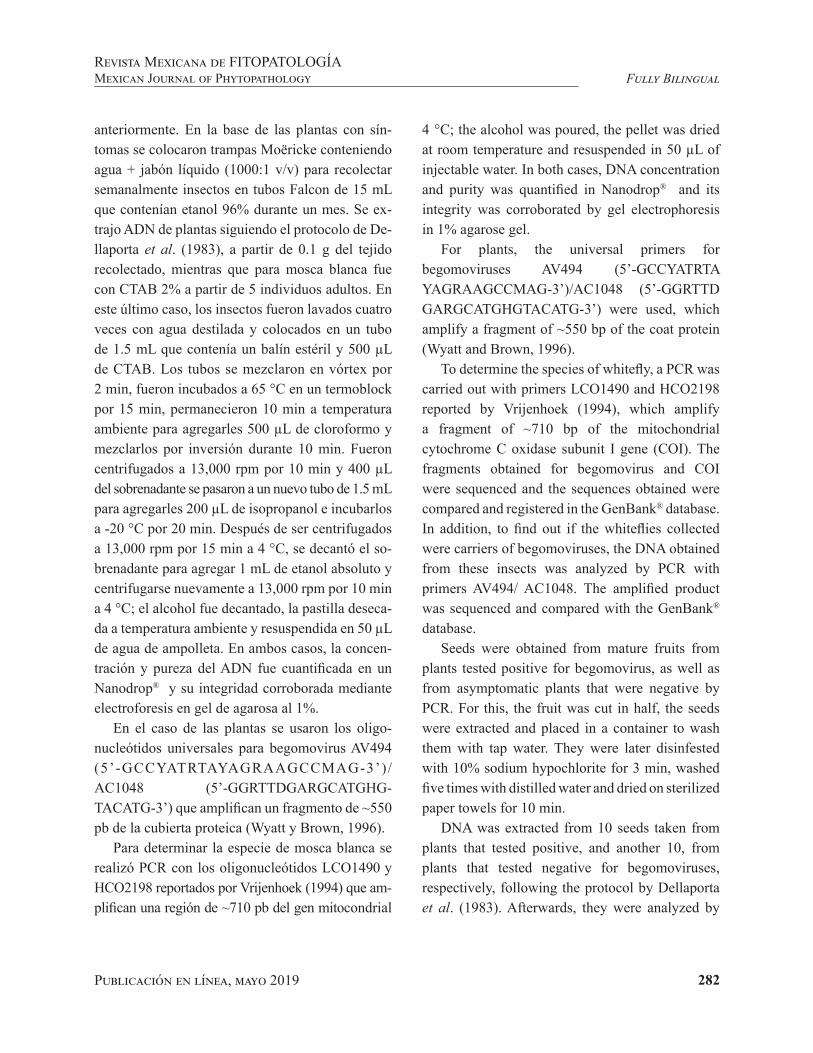
Publicación en línea, mayo 2019 282
Fully BilingualRevista Mexicana de FITOPATOLOGÍAMexican Journal of Phytopathology
anteriormente. En la base de las plantas con sín-tomas se colocaron trampas Moëricke conteniendo agua + jabón líquido (1000:1 v/v) para recolectar semanalmente insectos en tubos Falcon de 15 mL que contenían etanol 96% durante un mes. Se ex-trajo ADN de plantas siguiendo el protocolo de De-llaporta et al. (1983), a partir de 0.1 g del tejido recolectado, mientras que para mosca blanca fue con CTAB 2% a partir de 5 individuos adultos. En este último caso, los insectos fueron lavados cuatro veces con agua destilada y colocados en un tubo de 1.5 mL que contenía un balín estéril y 500 µL de CTAB. Los tubos se mezclaron en vórtex por 2 min, fueron incubados a 65 °C en un termoblock por 15 min, permanecieron 10 min a temperatura ambiente para agregarles 500 µL de cloroformo y mezclarlos por inversión durante 10 min. Fueron centrifugados a 13,000 rpm por 10 min y 400 µL del sobrenadante se pasaron a un nuevo tubo de 1.5 mL para agregarles 200 µL de isopropanol e incubarlos a -20 °C por 20 min. Después de ser centrifugados a 13,000 rpm por 15 min a 4 °C, se decantó el so-brenadante para agregar 1 mL de etanol absoluto y centrifugarse nuevamente a 13,000 rpm por 10 min a 4 °C; el alcohol fue decantado, la pastilla deseca-da a temperatura ambiente y resuspendida en 50 µL de agua de ampolleta. En ambos casos, la concen-tración y pureza del ADN fue cuantificada en un Nanodrop® y su integridad corroborada mediante electroforesis en gel de agarosa al 1%.
En el caso de las plantas se usaron los oligo-nucleótidos universales para begomovirus AV494 (5’-GCCYATRTAYAGRAAGCCMAG-3’)/AC1048 (5’-GGRTTDGARGCATGHG-TACATG-3’) que amplifican un fragmento de ⁓550 pb de la cubierta proteica (Wyatt y Brown, 1996).
Para determinar la especie de mosca blanca se realizó PCR con los oligonucleótidos LCO1490 y HCO2198 reportados por Vrijenhoek (1994) que am-plifican una región de ⁓710 pb del gen mitocondrial
4 °C; the alcohol was poured, the pellet was dried at room temperature and resuspended in 50 µL of injectable water. In both cases, DNA concentration and purity was quantified in Nanodrop® and its integrity was corroborated by gel electrophoresis in 1% agarose gel.
For plants, the universal primers for begomoviruses AV494 (5’-GCCYATRTA YAGRAAGCCMAG-3’)/AC1048 (5’-GGRTTD GARGCATGHGTACATG-3’) were used, which amplify a fragment of ~550 bp of the coat protein (Wyatt and Brown, 1996).
To determine the species of whitefly, a PCR was carried out with primers LCO1490 and HCO2198 reported by Vrijenhoek (1994), which amplify a fragment of ~710 bp of the mitochondrial cytochrome C oxidase subunit I gene (COI). The fragments obtained for begomovirus and COI were sequenced and the sequences obtained were compared and registered in the GenBank® database. In addition, to find out if the whiteflies collected were carriers of begomoviruses, the DNA obtained from these insects was analyzed by PCR with primers AV494/ AC1048. The amplified product was sequenced and compared with the GenBank® database.
Seeds were obtained from mature fruits from plants tested positive for begomovirus, as well as from asymptomatic plants that were negative by PCR. For this, the fruit was cut in half, the seeds were extracted and placed in a container to wash them with tap water. They were later disinfested with 10% sodium hypochlorite for 3 min, washed five times with distilled water and dried on sterilized paper towels for 10 min.
DNA was extracted from 10 seeds taken from plants that tested positive, and another 10, from plants that tested negative for begomoviruses, respectively, following the protocol by Dellaporta et al. (1983). Afterwards, they were analyzed by
Publicación en línea, mayo 2019 283
Fully BilingualRevista Mexicana de FITOPATOLOGÍA
Mexican Journal of Phytopathology
de la subunidad I del citocromo C oxidasa (COI). Los fragmentos amplificados para begomovirus y COI fueron secuenciados y las secuencias obteni-das se compararon y registraron en la base de datos del GenBank®. Adicionalmente, para conocer si las moscas blancas colectadas son portadoras de be-gomovirus, el ADN obtenido de estos insectos se analizó por PCR con los oligonucleótidos AV494/ AC1048. El producto amplificado se secuenció y comparó en la base datos del GenBank®.
Se obtuvieron semillas de frutos maduros de plantas positivas a begomovirus así como de plan-tas asintomáticas que fueron negativas por PCR. Para ello se cortó el fruto por la mitad, se extrajeron las semillas y se colocaron en un colador para la-varlas bajo el chorro de agua de la llave. Posterior-mente se desinfestaron con hipoclorito de sodio al 10% por 3 min, se lavaron de cuatro a cinco veces con agua destilada y se secaron sobre papel absor-bente estéril por 10 min.
Se extrajo ADN a partir de 10 semillas prove-nientes de plantas positivas y 10 negativas a be-gomovirus respectivamente, siguiendo el protoco-lo de Dellaporta et al. (1983). Posteriormente se analizaron por PCR con oligonucleótidos univer-sales para begomovirus AV494/AC1048 (Wyatt y Brown, 1996).
Por otro lado, se sembraron 50 semillas de fru-tos de plantas positivas a begomovirus en charo-las germinadoras con suelo estéril; como testigo se sembraron 50 semillas de frutos obtenidos de plan-tas negativas. En ambos casos las charolas se man-tuvieron en cámara de ambiente controlado a una temperatura promedio de 27 °C y un fotoperiodo de 16 h. Se colectaron hojas verdaderas a los 14, 21 y 28 días después de la siembra y se extrajo ADN (Dellaporta et al. , 1983) para realizar PCR.
Los principales síntomas observados en las plantas colectadas fueron reducción del crecimien-to y abarquillamiento además de mosaico y manchas
PCR with universal primers for begomoviruses AV494/AC1048 (Wyatt and Brown, 1996).
On the other hand, 50 seeds taken from fruits positive for begomovirus were planted in germination trays with sterile soil; as a control 50 seeds taken from fruits of plants that tested negative were used. In both cases, the trays were kept in a growth chamber, at 27 °C and a photoperiod of 16 h. True leaves were collected 14, 21 and 28 days after planting, and DNA was extracted (Dellaporta et al., 1983) to carry out PCR.
The main symptoms observed in the plants collected were reduction in growth and leaf curling, as well as mosaics and chlorotic spots in some cases (Figures 1A-1C), which have been associated to begomovirus infection. Previously, plants were sporadically found with mosaic, distributed at random throughout the greenhouse; some tested positive for Impatiens necrotic spot virus, Tobacco mosaic virus or Pepino mosaic virus and negative for Cucumber mosaic virus and Tomato spotted wilt virus as well as a higher number (~30%) of plants with a shortening of internodes and small leaflets in the top, which tested positive for Mexican papita viroid (data not shown). The presence of a high percentage of plants with a severe reduction in growth and leaf curling suggested the presence of a virus or viroid different to those analyzed previously.
The adult whiteflies collected in Moëricke traps were morphologically similar, and were recovered practically intact (Figure 1D). The expected product for begomovirus in the tomato symptomatic plants was obtained (Figure 1E). The sequences of the products obtained (Accession number MK325267 and MK325268) had an identity of 99% with the Tomato severe leaf curl virus (ToSLCV). In Mexico, the ToSLCV was first reported in tomato crops in Baja California Sur (Holguín et al., 2003), and then in Rioverde, San Luis Potosí and

Publicación en línea, mayo 2019 284
Fully BilingualRevista Mexicana de FITOPATOLOGÍAMexican Journal of Phytopathology
M 1 2 3 4 5 6
550 pb
M 1 2 3 4 5 6
710 pb
cloróticas en algunos casos (Figuras 1A-1C), los cuales se han asociado a begomovirus. Previa-mente se detectó de manera esporádica plantas con mosaico distribuidas aleatoriamente en el inverna-dero, algunas fueron positivas a Impatiens necrotic spot virus, Tobacco mosaic virus o Pepino mosaic virus y negativas a Cucumber mosaic virus y To-mato spotted wilt virus así como un mayor número (~30%) de plantas con acortamiento de entrenudos y foliolos pequeños en la parte apical que resul-taron positivas a Mexican papita viroid (datos no
Xochitepec, Morelos. Only the begomoviruses Chino del tomate virus and Pepper Huasteco virus (currently Pepper huasteco yellow vein virus) had been reported in tomato grown under greenhouse conditions in Mexico.
The expected fragment of ~710 bp from whiteflies analyzed by PCR with primers of the COI gene was obtained (Figure 1F) and its sequence had an identity of 99% with Bemisia tabaci (access numbers MK290856, MK290857, MK290858 and MK290859). B. tabaci is the main vector species
Figura 1. Síntomas en plantas de tomate asociados a begomovirus. A. Reducción severa del crecimiento y reducción de tamaño de hojas terminales. B. Abarquillamiento. C. Manchas cloróticas. D. Adulto de mosca blanca colectado en trampas Moëricke. E) Productos de PCR obtenidos con oligonucleótidos universales para begomovirus AV494/AC1048 (~550 pb) en plantas de tomate recolectadas en invernadero. Carril 1: Control positivo (planta infecta-da con Okra yellow mosaic Mexico virus); Carril 2: Control negativo (agua); Carril 3-4: Plantas con síntomas; Carriles 5-6: Plantas asintomáticas; Carril M: marcador de peso molecular 100 pb (Promega®). F) Productos de PCR obtenidos con oligonucleótidos LCO1490 y HCO2198 (~710 pb) a partir de ADN de mosca blanca. Carril 1: Control positivo (adulto de Bemisia tabaci); Carril 2: Control negativo (agua); Carriles 3-6: Adultos en trampas próximas a plantas con mosaico y abarquillamiento; Carril M: marcador de peso molecular 100 pb (Promega®).
Figure 1. Symptoms in tomato plants associated to begomovirus infection. A. Severe plant growth reduction and reduced size of terminal leaves. B. Leaf curling. C. Chlorotic spots. D. Adult whitefly collected in Moëricke traps. E) PCR products obtained with universal primers for begomoviruses AV494/AC1048 (~550 bp) in tomato plants collected in a greenhouse. Lane 1: Positive control (plant infected with Okra yellow mosaic Mexico virus); Lane 2: Negative control (water); Lanes 3-4: Symptomatic plants; Lane 5-6: Asymptomatic plants; Lane M: 100 bp DNA ladder (Promega®). F) PCR products obtained with primers LCO1490 and HCO2198 (~710 bp) from whiteflies DNA. Lane 1: Positive control (adult of Bemisia tabaci); Lane 2: Negative control (water); Lanes 3-6: Adults collected in traps near plants showing mosaic and leaf curling; Lane M: 100 bp DNA ladder (Promega®).
A B C D
E F

Publicación en línea, mayo 2019 285
Fully BilingualRevista Mexicana de FITOPATOLOGÍA
Mexican Journal of Phytopathology
mostrados). La presencia de un alto porcentaje de plantas con una severa reducción del crecimiento y abarquillamiento sugirieron la presencia de un virus o viroide diferente de los analizados con an-terioridad.
Los adultos colectados de mosca blanca en las trampas Moëricke fueron morfológicamente simi-lares y se recuperaron prácticamente intactos (Fi-gura 1D). Se tuvo amplificación del producto espe-rado para begomovirus en las plantas de tomate con los síntomas indicados (Figura 1E). Las secuencias de los productos obtenidos (No. acceso MK325267 y MK325268) tuvieron una identidad de 99% con el Tomato severe leaf curl virus (ToSLCV). En México, el ToSLCV fue reportado por vez primera en cultivos de tomate en Baja California Sur (Hol-guín et al., 2003), posteriormente en los municipios de Rioverde, San Luis Potosí, y Xochitepec, More-los. En tomate cultivado en invernadero sólo se ha reportado a los begomovirus Chino del tomate vi-rus y Pepper Huasteco virus (actualmente Pepper huasteco yellow vein virus).
En el ADN de las moscas blancas analizadas se obtuvo con los oligonucleótidos del gen COI el fragmento esperado de ~710 pb (Figura 1F) y su se-cuencia tuvo una identidad de 99% con Bemisia ta-baci (Números de acceso MK290856, MK290857, MK290858 y MK290859). B. tabaci es la principal especie vectora de begomovirus a nivel mundial y, por lo general, los síntomas de amarillamiento, en-chinamiento y reducción del crecimiento aunados a la presencia de este insecto vector en cultivos de tomate se consideran como un indicativo de la in-fección por estos fitopatógenos.
En diferentes estudios se menciona que altas poblaciones de B. tabaci están asociadas con infec-ciones de distintas especies de begomovirus en cul-tivos de tomate en México, lo cual contrasta con lo observado en el presente estudio donde el número de moscas blancas fue bajo.
of begomoviruses wordlwide, and generally the symptoms of yellowing, leaf curling and growth reduction associated to the presence of this vector in tomato crops are considered an indication of the infection by these viruses.
In different studies, large populations of B. tabaci are related to infections of several species of begomoviruses in tomato crops in Mexico, which contrasts with observations in the present study, where the number of whiteflies was low.
The adult whitefly amplified the expected fragment for begomoviruses (~550 bp) (data not shown) and its sequence had an identity of 99% with ToSLCV (Accession numbers MK325269, MK325270 and MK325271). This correspondence between the virus found in tomato plants and associated whiteflies has been reported for Tomato golden mottle virus.
No begomoviruses were detected by PCR in seeds from tomato fruits obtained from symptomatic plants, nor in seedlings in the dates of evaluation. Certain begomoviruses which are not transmitted by seed of the crop, but rather in weeds, such as Okra yellow mosaic Mexico virus, which is not transmitted by roselle seed (Hibiscus sabdariffa L.), but it can transmit in Sida collina, S. acuta, S. haenkeana and Malacra fasciata seeds (Ortega et al., 2019). These results, as well as the detection of ToSLCV in whiteflies and the high incidence of symptomatic plants, suggest that the population of B. tabaci found is efficient in the transmission of this begomovirus.
Based on the results obtained, it is the first report of Tomato severe leaf curl virus associated to tomato plants under greenhouse conditions, and its presence in adults of B. tabaci in Atlacomulco, Mexico State.
End of the English version

Publicación en línea, mayo 2019 286
Fully BilingualRevista Mexicana de FITOPATOLOGÍAMexican Journal of Phytopathology
Así mismo, los individuos de mosca blanca am-plificaron el fragmento esperado para begomovi-rus (~550 pb) (datos no mostrados) y su secuencia tuvo una identidad de 99% con ToSLCV (Números de acceso MK325269, MK325270 y MK325271). Esta correspondencia del virus detectado en toma-te y mosca blanca se ha encontrado con el Tomato golden mottle virus.
En el análisis de PCR no se detectó al begomo-virus en semilla de frutos de tomate obtenidos de plantas con síntomas, ni en plántula en las fechas de evaluación. Se sabe de begomovirus que no se transmiten por semilla del hospedante de inte-rés agrícola pero sí lo hacen en maleza, como en el caso del Okra yellow mosaic Mexico virus que no se transmite por semilla de jamaica (Hibiscus sabdariffa L.) pero sí en semilla de Sida collina, S. acuta, S. haenkeana y Malacra fasciata (Ortega et al., 2019). Estos resultados aunados a la detección del ToSLCV en mosca blanca y la alta incidencia de plantas con síntomas sugieren que la población de B. tabaci encontrada es eficiente en la transmi-sión del begomovirus.
Con base en los resultados obtenidos, se repor-ta por vez primera al Tomato severe leaf curl vi-rus asociado a tomate en invernadero así como su presencia en Atlacomulco, Estado de México y su presencia en adultos de B. tabaci.
LITERATURA CITADA
Brown JK, Zerbini MF, Navas CJ, Moriones E, Ramos SR, Silva JCF, Fiallo OE, Briddon RW, Hernández ZC, Idris AM, Malathi VG, Martin DP, Rivera BR, Ueda S and Var-sani A. 2015. Revision of begomovirus taxonomy based on pairwise sequence comparisons. Archives of virolo-gy 160:1593-1619. https://doi.org/10.1007/s00705-015-2398-y
Dellaporta SL, Wood J and Hicks JB. 1983. A plant DNA mini-preparation: version II. Plant Molecular Biology Reporter 1:19-21. https://doi.org/10.1007/BF02712670
Holguín PRJ, Vázquez JR and Rivera BRF. 2003. First report of a geminivirus associated with leaf curl in Baja Cali-fornia Península tomato fields. Plant Disease 87:1397. https://doi.org/10.1094/PDIS.2003.87.11.1397A
Ortega AC, Ochoa MDL, Hernández MJ, Ramírez RS and Gutiérrez GJA. 2019. Evauation of seed transmission of begomoviruses in roselle and roselle-associated weeds. Revista Mexicana de Fitopatología 37(1):135-146. DOI: 10.18781/R.MEX.FIT.1806-3
Vrijenhoek R. 1994. DNA primers for amplification of mi-tochondrial cytochrome c oxidase subunit I from diverse metazoan invertebrates. Molecular Marine Biology Biote-chnology 3:294-299. https://www.mbari.org/wp-content/uploads/2016/01/Folmer_94MMBB.pdf
Wyatt SD and Brown JK.1996. Detection of subgroup III gemi-nivirus isolates in leaf extracts by degenerate primers and polimerasa chain reaction. Phytopathology 86:1288-1293. https://www.apsnet.org/publications/phytopathology/bac-kissues/Documents/1996Articles/Phyto86n12_1288.pdf
287Publicación en línea, mayo 2019
Samaniego-Gaxiola JA, Pedroza-Sandoval A, Chew-Madinaveitia Y and Gaytán-Mascorro A. 2019. Reductive disinfestation, soil desiccation and Trichoderma harzianum to control Phymatotrichopsis omnivora in pecan tree nursery. Mexican Journal of Phytopathology 37(2): 287-303.DOI: 10.18781/R.MEX.FIT.1808-7
Primera publicación DOI: 08 de Abril, 2019.First DOI publication: April 08, 2019.
Resumen. En el laboratorio, suelo adicionado con glucosa 0.5-4.0 mg g-1 e inundado, el pH y el potencial oxidación-reducción (ORP) alcanzaron valores de 6.2 y -250 mV, también observamos hasta 22 mmol L-1 de ácidos grasos volátiles (VAFs). El sobrenadante del suelo se ajustó o no a pH ~ 4, luego los esclerocios de P. omnivora
Reductive disinfestation, soil desiccation and Trichoderma harzianum to control
Phymatotrichopsis omnivora in pecan tree nursery
Desinfestación reductiva, suelo desecado y Trichoderma harzianum para controlar Phymatotrichopsis omnivora en vivero de nogal pecanero
José Alfredo Samaniego-Gaxiola*, Campo Experimental La Laguna INIFAP, Bulevar José Santos Valdez 1200 Pte., Colonia Centro Matamoros, Coahuila, México. C.P. 27440; Aurelio Pedroza-Sandoval, Universi-dad Autónoma Chapingo, Unidad Regional de Zonas Áridas. Carretera Gómez Palacio - Ciudad Juárez Km 40, Bermejillo, Durango. C.P. 35230; Yasmín Chew-Madinaveitia, Arturo Gaytán-Mascorro, Campo Experi-mental La Laguna INIFAP, Bulevar José Santos Valdez 1200 Poniente, Colonia Centro Matamoros, Coahuila, México. C. P. 27440. *Autor de correspondencia: [email protected].
Recibido: 28 de Agosto, 2018. Aceptado: 28 de Marzo, 2019.
Abstract. In the laboratory, flooded soil with glucose, 0.5-4.0 mg g-1, the pH and potential oxidation-reduction (ORP) reached values of 6.2 and -250 mV, also, we observed up to 22 mmol L-1 of volatile fatty acids (VAFs). The supernatant of the soil was adjusted or not to pH ~ 4, then sclerotia of P. omnivora were submerged in it. The sclerotia died only in supernatants at pH ~ 4 that came from soil with 2.0 and 4.0 mg g-1 of glucose added. In the field, there were six treatments: a control or untreated soil (C); 55 t ha-1 of molasses was added and the ground was covered with plastic (M); inoculated soil with Trichoderma harzianum (T); dried soil (DS) and their combinations DS+M, DS+T and DS+M+ T. Where molasses was added, the pH and ORP reached 6.5 and -200 mV. The pH and ORP reached in soils with glucose and molasses are characteristic of soil reductive disinfestation (RSD). In the field, treatments were applied to the
Publicación en línea, mayo 2019 288
Fully BilingualRevista Mexicana de FITOPATOLOGÍAMexican Journal of Phytopathology
se sumergieron en él. Los esclerocios murieron solo en sobrenadantes a pH ~4 que provenían del suelo con 2.0 y 4.0 mg g-1 de glucosa añadida. En el campo, hubo seis tratamientos: un testigo o suelo no tratado (C); se agregaron 55 t ha-1 de melaza y el suelo se cubrió con plástico (M); suelo inoculado con Trichoderma harzianum (T); suelo desecado (DS) y sus combinaciones DS+M, DS+T y DS+M+T. Cuando se añadió melaza, el pH y el ORP alcanzaron 6.5 y -200 mV. El pH y el ORP alcanzados en suelos con glucosa y melaza son característicos de la desinfestación reductiva del suelo (RSD). En el campo, los tratamientos se aplicaron al suelo, con cuatro (repeticiones) parcelas por tratamiento y se sembraron 12 nueces por parcela. Después de tres años, no hubo diferencia en la incidencia y la mortandad de los árboles causada por P. omnivora, pero las raíces fueron invadidas por Trichoderma sp.
Palabras clave: desinfestación reductiva del suelo, antagonismo, control biológico.
En México, el nogal pecanero Carya illinoinen-sis (Wangenh.) Koch es un cultivo establecido en 112,000 ha, cuyo valor fue de 330 millones de dó-lares en 2015 (Centro de Investigación y Asisten-cia en Tecnología y Diseño del Estado de Jalisco, 2018). La enfermedad más importante de las raí-ces del nogal es la pudrición de la raíz causada por el hongo Phymatotrichopsis omnivora (Duggar) Hennebert (Herrera-Perez y Samaniego-Gaxiola, 2002). Un método reciente para el control de pa-tógenos de plantas en el suelo, es la desinfestación reductiva del suelo (RSD), donde se agrega una fuente de carbono (C) como melaza, paja de trigo o compuestos ricos en carbohidratos al suelo, lue-go se satura con agua y se cubre con un plástico (Momma et al., 2006).
soil, with four plots per treatment and 12 pecan tree seeds were planted per plot. After three years, there was no difference in incidence and mortality of trees caused by P. omnivora, but the roots were invaded by Trichoderma sp.
Key words: soil reductive disinfestation, antagonism, biocontrol.
In Mexico, the pecan tree Carya illinoinensis (Wangenh.) Koch is a crop established in 112,000 ha, whose value in generating foreign currency was of 330 million dollars in 2015 (Centro de Investigación y Asistencia en Tecnología y Diseño del Estado de Jalisco, 2018). The most important disease of the pecan tree roots is the cotton root rot root caused by the fungus Phymatotrichopsis omnivora (Duggar) Hennebert (Herrera-Pérez and Samaniego-Gaxiola, 2002). A recent method for the control of soil borne plant pathogens is the reductive soil disinfestation (RSD), where a source of carbon (C) such as molasses, wheat straw or compounds rich in carbohydrates are added to the soil, then it is saturated with water and covered with a plastic (Momma et al., 2006). In the RSD, the added residues are decomposed in an anaerobic condition, the pH of the soil solution becomes acidic, the oxidation-reduction potential or ORP (Eh) reaches negative (reductive) values and volatile fatty acids (VFAs), organic sulfur compounds and metal ions Mn2+ and Fe2+ are generated all toxic to fungi, bacteria and weeds (Hewavitharana et al., 2014; Momma, 2008; Momma et al., 2011). The VAFs are toxic to fungi, bacteria and nematodes (Shrestha et al., 2016) and the toxicity increases in acidified media (Samaniego-Gaxiola and Pedroza-Sandoval, 2013). The survival of sclerotia of P. omnivora was null after being immersed in VAFs solutions, as well as in flooded soil with glucose added

Publicación en línea, mayo 2019 289
Fully BilingualRevista Mexicana de FITOPATOLOGÍA
Mexican Journal of Phytopathology
En la RSD, los residuos agregados se descom-ponen en una condición anaeróbica, el pH de la solución del suelo se acidifica, el potencial oxida-ción-reducción ORP (Eh) alcanza valores negativos (reductores) y ácidos grasos volátiles (VFAs), com-puestos orgánicos de azufre y iones metálicos Mn2+ y Fe2+ se generan, tóxicos para hongos, bacterias, nematodos y malezas (Hewavitharana et al., 2014; Momma, 2008; Momma et al., 2011; Shrestha et al., 2016). Los VFAs aumentan su toxicidad acidi-ficando el medio (Samaniego-Gaxiola y Pedroza-Sandoval, 2013).
La supervivencia de los esclerocios de P. omni-vora fue nula después de sumergirse en soluciones de FAVs, así como en suelo inundado con glucosa agregada (Samaniego-Gaxiola, 1994; Samaniego-Gaxiola y Balagurusamy, 2013). Sin embargo, no se ha registrado si el suelo infestado con P. om-nivora, inundado y agregado con glucosa induce los cambios de la RSD, si los FAVs en la solución de este suelo solo o ajustando su pH pueden matar los esclerocios del hongo. Por otro lado, los escle-rocios de P. omnivora son susceptibles de morir cuando se secan o permanecen en contacto con Trichoderma spp. (Samaniego-Gaxiola 2008; Sa-maniego-Gaxiola et al., 2010). El suelo seco o ino-culado con Trichoderma harzianum Rifai, podría disminuir el inóculo de P. omnivora. Los métodos de control en el suelo: RSD, desecado e inoculando con T. harzianum, podrían combinarse y dar me-jores resultados que individualmente, para prote-ger a al nogal pecanero contra la pudrición texana. Por lo tanto, los objetivos de este trabajo fueron: i) registrar el pH, ORP, producción de FAVs en el suelo agregado con glucosa e inundado permanen-temente; ii) evaluar la supervivencia de los escle-rocios de P. omnivora después de permanecer en el sobrenadante proveniente de este suelo, donde el pH se ajusta o no ~ 4; iii) evaluar la incidencia y muerte nogal pecanero sembrado en suelos tratados
(Samaniego-Gaxiola, 1994; Samaniego-Gaxiola and Balagurusamy, 2013). However, it has not been registered if soil infested with P. omnivora, flooded and added with glucose induce the changes of the RSD, if the VAFs in the solution of this soil alone or adjusting its pH can kill the sclerotia of the fungus. On the other hand, the sclerotia of P. omnivora are susceptible to die when they are dried or remain in contact with Trichoderma spp. (Samaniego-Gaxiola 2008; Samaniego-Gaxiola et al., 2010). Dried or inoculated soil with Trichoderma harzianum Rifai, could decrease the inoculum of P. omnivora. The RSD control methods, dried the soil and inoculating T. harzianum in soil, could be combined and give better results than individually, to protect the pecan tree from cotton root rot. Therefore, the objectives of this work were: i) to register the pH, ORP, production of VAFs in soil added with glucose and flooded permanently; ii) to evaluate the survival of sclerotia of P. omnivora after remaining in the supernatant coming from this soil, where the pH is adjusted or not to ~ 4; iii) to evaluate the incidence and death of the pecan tree sown in soils treated with RSD using molasses (M), inoculated with T. harzianum (T), dried soil (DS) and its combinations DS+T, DS+M and DS+T+M, and iv) record changes in pH and ORP in treated field soils.
Soil was extracted at a depth of 0-5 cm from the Hormiguero orchard located on the Torreón-San Pedro Km 16 road (25° 41’ 16.3” N 103° 19’ 52.4” W). To eight repetitions (glass flasks) of 1L were added 400 g of soil, then 80 ml of a glucose solution of 0.0, 0.5, 2.0 and 4.0 mg g-1, then 250 ml of water (flooded) and finally the flasks were incubated for five days at 28 °C. From the beginning of the incubation period, 20 ml of the supernatant of each flask were extracted daily. This sample was divided into two groups of vials (each with 10 ml), in both groups pH and ORP were measured, with a multi-
Publicación en línea, mayo 2019 290
Fully BilingualRevista Mexicana de FITOPATOLOGÍAMexican Journal of Phytopathology
con RSD utilizando melaza (M), inoculado con T. harzianum (T), suelo desecado (DS) y sus combi-naciones DS+T, DS+M y DS+T+M; y iv) registrar los cambios del pH y ORP en suelos tratados.
El suelo se extrajo de 0-5 cm de profundidad, del huerto Hormiguero ubicado en la carretera To-rreón-San Pedro Km 16 (25° 41’ 16.3” N 103° 19’ 52.4” W). Ocho repeticiones (frascos de vidrio) de 1 l se agregaron 400 g de suelo, luego 80 ml de una solución de glucosa de 0.0, 0.5, 2.0 y 4.0 mg g-1, luego 250 ml de agua (inundación) y finalmente se incubaron los frascos, por cinco días a 28 °C. Desde el comienzo del período de incubación, se extrajeron diariamente 20 ml del sobrenadante de cada frasco. Esta muestra se dividió en dos grupos de viales (cada uno con 10 ml), en ambos grupos se midieron el pH y la ORP, con un multi-parámetro de marca HI 9828 de Hanna Instruments. A un gru-po de viales no se ajustó el pH y el otro sí a pH ~ 4 al añadirle H2SO4; en este último caso, se cuantificó la cantidad de ácido utilizado para ajustar el pH. En ambos grupos de viales, se colocaron 25 esclero-cios de P. omnivora (por vial) durante una hora, y luego se midió el pH y los esclerocios se extrajeron y germinaron. Los esclerocios fueron reproduci-dos, cosechados, manejados y germinados según lo indicado por Samaniego-Gaxiola (2008).
Desde el segundo hasta el quinto día de incuba-ción de los frascos, se tomaron muestras compues-tas del sobrenadante, para cada dosis de glucosa-suelo 0.0, 0.5, 2.0 y 4.0 mg g-1. Cada muestra com-puesta de 10 ml se analizó para ácidos orgánicos, mediante cromatografía líquida de alta presión, en una columna Aminex HPX 87H, 300 x 7,8 mm, con fase móvil: H2SO4 5 mM, condiciones de flujo de 0,5 ml / min, 50 °C y análisis de detección por dis-posición de diodos a 210 nm. La cuantificación de la curva estándar se obtuvo por estándares de refe-rencia. El perfil de cada ácido se verificó mediante el espectro de absorción y el tiempo de retención correspondiente.
parameter HI 9828 brand Hanna Instruments. A group of vials did not have an adjusted pH and the other were added H2SO4 to adjust the pH ~ 4; in the latter case, the amount of acid used to adjust the pH was quantified. In both groups of vials, 25 sclerotia of P. omnivora (per vial) were placed for one hour, and then the pH in the vials was measured. Sclerotia were extracted and germinated in sand. The sclerotia of P. omnivora were reproduced, harvested, managed and germinated as indicated by Samaniego-Gaxiola (2008).
From the second to the fifth day of incubation of the flasks, composite samples of the supernatant were taken, for each dose of glucose-soil 0.0, 0.5, 2.0 and 4.0 mg g-1. Each composite sample of 10 ml was analyzed for organic acids by high pressure liquid chromatography, on an Aminex HPX 87H column, 300 x 7.8 mm, with mobile phase: 5 mM H2SO4, flow conditions of 0.5 mL / min, 50 °C and an analysis of detection by arrangement of diodes at 210 nm. The quantification of the standard curve was obtained by reference standards. The profile of each acid was checked by absorption spectrum and corresponding retention time.
In the Hormiguero orchard, in March of 2012, one control and six randomized treatments were established, each treatment had four repetitions. The repetition was a micro-plot of 3x2 m, where 12 pecan-nut seeds were planted equidistantly. Between plots, towards both ends, a space of four meters of unseeded soil was left. The control treatment (C) consisted in the dry sowing of the walnut and then the application of a 15 cm irrigation sheet, similar irrigation sheet was applied for the rest of the treatments. The desiccated soil (DS) was a treatment where soil was extracted up to 60 cm and dried in the sun to reach a humidity of less than 5%, then the soil was returned to the well and the nuts were sown. The moisture that the soil reached in all treatments (soils with or without desiccation) was determined by taking two samples of 100 g

Publicación en línea, mayo 2019 291
Fully BilingualRevista Mexicana de FITOPATOLOGÍA
Mexican Journal of Phytopathology
En Hormiguero, en marzo de 2012, se estable-cieron un control y seis tratamientos aleatorios, cada tratamiento tuvo cuatro repeticiones. La re-petición fue una microparcela de 3x2 m, donde se sembraron equidistantemente 12 semillas de nuez pecanera. Entre parcelas, hacia ambos extremos, de dejaron cuatro metros de tierra sin sembrar. El tratamiento control (C) consistió en la siembra en seco de la nuez y luego la aplicación de una lámina de irrigación de 15 cm, que fue similar para los tra-tamientos. En el tratamiento suelo desecado (DS) el suelo se extrajo hasta 60 cm y secó al sol para alcanzar una humedad inferior al 5%, luego se de-volvió el suelo al pozo y se sembraron las nueces. La humedad de todos los tratamientos (suelos con o sin desecación) se determinó tomando dos mues-tras de 100 g para cada profundidad y repetición; Todas las muestras se pesaron antes y después de secar durante 48 horas con aire seco a 100 °C para determinar su contenido de humedad (W/W). La RSD se implementó utilizando melazas P/P (M) a una tasa de 33 kg (55 t ha-1), las melazas se disol-vieron en 800 l de agua de riego y se vertieron en la parcela, y luego la superficie se cubrió durante 14 d con plástico negro Polietileno de 600 µm. El ter-cer día después de aplicar las melazas, se vertieron 100 l de una solución de H2SO4 (23 mmol L-1) en el suelo (retirando el plástico). La parcela se cubrió nuevamente con plástico durante 11 d, luego se destapó por dos semanas para permitir la aireación, y después se sembraron las nueces. Un aislado de T. harzianum (T) obtenido de la rizósfera de no-gal pecanero se utilizó para hacer un inóculo agua, arena y paja de trigo, según lo descrito por Lewis y Papavizas (1984), se aplicó 3 kg de inóculo (peso seco) por parcela. Otros tres tratamientos combi-nados fueron DS + M, DS+T y DS+M+T, según el caso, primero se secó el suelo, luego se agregaron las melazas y finalmente se aplicó T. harzianum.
En cada parcela, el tercer día después de apli-car las melazas, ocho tubos de PVC se enterraron
for each depth and repetition; all the samples were weighed before and after drying 48 hours with dry air at 100 °C to determine their moisture content (W/W). The RSD was implemented using molasses (M) at a rate of 33 kg (55 t ha-1), molasses were dissolved in 800 L of irrigation water and poured into the plot, and then the surface of the plot was covered with black plastic polyethylene of 600 µm during 14 d. On the third day after applying the molasses, 100 L of a solution of H2SO4 (23 mmol L-1) was poured to the ground (removing the plastic). The plot was again covered with the plastic during 11 days, then it was uncovered by two weeks to allow aeration, and later the nuts were sown. An isolate of T. harzianum (T) obtained from pecan tree rhizosphere, was used to make an inoculum whose substrate was water, sand and wheat straw as described by Lewis and Papavizas (1984), the amount of inoculum was 3 kg (dry weight) per plot. Three other combined treatments were DS+M, DS+T and DS+M+T, depending on the case, first the soil was dried, then the molasses was added and, finally T. harzianum was applied.
In each plot, on the third day after applying the molasses, eight PVC tubes were vertically buried equidistantly to a depth of 60 cm. On the fourth and seventh days, half of the tubes were extracted, respectively, and cut into segments of 0-20, 20-40 and 40-60 cm, and frozen until the soil analysis. From each soil segment, a 2:1 water-soil extract was made and pH and ORP were measured.
During September to October of each the years 2013-2015, the incidence of cotton root rot and mortality of the pecan tree was evaluated. When the trees showed wilt leaves were considered sick; while dead tress had dried epidermis of branches and stem. Nevertheless, some diseased pecan trees withered and defoliated after October, but their wood was alive and rebounded the following year; at first, one of these trees was considered dead, but their roots were invaded at the same time by

Publicación en línea, mayo 2019 292
Fully BilingualRevista Mexicana de FITOPATOLOGÍAMexican Journal of Phytopathology
equidistantemente a 60 cm de profundidad. En el cuarto y séptimo día, la mitad de los tubos se extra-jeron, respectivamente, se cortaron en segmentos de 0-20, 20-40 y 40-60 cm y congelaron hasta el análisis del suelo; mediante un extracto agua-suelo 2:1 donde se midieron el pH y el ORP.
De septiembre a octubre de cada año 2013-2015, se evaluó la incidencia de la pudrición texana y la mortalidad de los nogales. Cuando los nogales mostraban hojas marchitas se consideró enfermos; mientras que los árboles muertos, tenían seca la epidermis de ramas y tallos. Sin embargo, algunos nogales enfermos se marchitaron y defoliaron, pero su madera estaba viva y se recuperaron al siguiente año; Al principio, uno de estos árboles se consideró muerto, pero sus raíces estaban invadidas al mis-mo tiempo por P. omnivora y Trichoderma sp. Por lo tanto, el resto de los nogales marchitos con su epidermis viva, no fueron removidos y sus raíces tampoco fueron inspeccionadas.
A los datos de pH y ORP del experimento de laboratorio, se aplicó un análisis de varianza de dos vías (efectos del tratamiento) para mediciones repetidas o tiempo de muestreo (rmANOVA) SAS (1999). En el experimento de campo, el análisis de pH y ORP fue un ANOVA para cada tiempo de muestreo, porque provenía de diferentes muestras. La incidencia y la mortalidad de los árboles de pa-cana se analizaron cada año por separado mediante un ANOVA. Cuando los análisis estadísticos fue-ron significativos (P<0,001), la comparación de medias entre tratamientos se realizó con la prueba de rango múltiple de medias de Duncan (P=0,05).
El pH alcanzó hasta 6.2 y se mantuvo en el suelo adicionado con 4.0 mg g-1 de glucosa, las dosis más bajas de glucosa no permitieron un pH tan bajo o solo hasta el segundo día; el suelo sin glucosa man-tuvo un pH alrededor de 8. El error estándar de los valores de pH del sobrenadante después de colocar esclerocios de P. omnivora aumentó con respecto al sobrenadante sin los esclerocios (Figura 1).
P. omnivora and Trichoderma sp. So, the rest of withered pecan trees with their alive epidermis, were not removed and their roots neither were inspected.
A two-way analysis of variance (treatment effects) for repeated measurements or sampling time (rmANOVA) SAS (1999) was applied to the pH and ORP data of the laboratory experiment. In the field experiment, the pH and ORP analysis was an ANOVA for each sampling time, because it came from different samples. The incidence and mortality of pecan trees was analyzed each year separately by an ANOVA. When the statistical analyzes were significant (P<0.001), the mean comparison between treatments was performed with the multiple range test of Duncan means (P=0.05).
A pH of up to 6.2 was reached and maintained in soil amended with 4.0 mg g-1 of glucose, lower doses of glucose did not allow such a low pH or only remained until the second day; the soil without glucose maintained a pH around 8. The standard error of the pH values of the supernatant after placing sclerotia of P. omnivora increased with respect to the supernatant without the sclerotia (Figure 1).
The ORP descended only in soil with glucose as time passed, until reaching values of -50 to - 250 mV, and it remained around 50 mV in the soil without glucose (Figure 2).
The organic acids detected in the supernatants between days two to five from the soils amended with glucose are indicated in Table 1. The total of VAFs detected from the third day of incubation was almost twice (20 against 11 mmol L- 1) in 4.0 versus 2.0 mg g-1 of glucose added in soil; while only <0.2 mmol L-1 was detected from the other organic acids.
The amounts of sulfuric acid to adjust to ~ 4 the pH of the supernatant are shown in Figure 3; only the supernatants obtained from the soils with 2.0 and 4.0 mg g-1 of glucose added exceeded 60 μL and reached near of 180 μL of sulfuric acid.

Publicación en línea, mayo 2019 293
Fully BilingualRevista Mexicana de FITOPATOLOGÍA
Mexican Journal of Phytopathology
El ORP descendió solo en el suelo con glucosa a medida que pasaba el tiempo, hasta alcanzar va-lores de -50 a - 250 mV, y permaneció alrededor de 50 mV en el suelo sin glucosa (Figura 2).
The pH values of the supernatants adjusted to pH ~ 4 and those reached in the supernatants where sclerotia of P. omnivora remained are shown in Figure 4; the pH of these last supernatants had
Figura 1. Izquierda, pH del sobrenadante obtenido del suelo con glucosa agregada e inundada hasta 5 d. Derecha, pH del sobrenadante después de colocar 25 esclerocios de P. omnivora durante una hora. Barra de error estándar n=8, las mismas letras sobre la barra no son significativamente diferentes según la prueba de separación de medios de Duncan (P>0.05).
Figure 1. Left, pH of the supernatant obtained from soil with glucose added and flooded up to 5 d. Right, pH of the superna-tant after placing 25 sclerotia of P. omnivora for one hour. Standard error bar n=8, the same letters above the bar are not significantly different according to the Duncan media separation test (P>0.05).
Figura 2. Potencial oxidación-reducción (mV) del sobrenadante obtenido del suelo con glucosa agregada e inundado hasta 5 d. Barra de error estándar n=8, las mismas letras sobre la barra no son significativamente diferentes, según la prueba de separación de medios de Duncan (P>0.05).
Figure 2. Oxidation-reduction potential (mV) of the supernatant obtained from soil with glucose added and flooded up to 5 d. Standard error bar n=8, the same letters above the bar are not significantly different according to the Duncan media separation test (P>0.05).
8.5
8.0
7.5
7.0Glucose mg g1
pH
0.50.0
2.04.0
0 1 2 3 4 5
b
c
a a
aaaaaa
bb
b
b
b
c
c cd
Time (d)
8.5
8.0
7.5
7.0
Glucose mg g1
pH
0.50.0 2.0
4.0
0 1 2 3 4 5Time (d)
6.5
6.0
6.5
6.0
145
50
45
140
mV
0 1 2 3 4 5Time (d)
235
330
Glucose mg g1
0.50.0
2.04.0
bc
aaaaa
a
bb
b
c cc
d
dd
d

Publicación en línea, mayo 2019 294
Fully BilingualRevista Mexicana de FITOPATOLOGÍAMexican Journal of Phytopathology
Los ácidos orgánicos detectados en los sobre-nadantes entre los días dos y cinco de los suelos adicionados con glucosa se indican en la Cuadro 1. El total de VAFs detectados desde el tercer día de incubación fue casi dos veces (20 contra 11 mmol L-1) en 4.0 versus 2,0 mg g-1 de glucosa agregada en el suelo; y solo <0.2 mmol L-1 fue detectado de otros ácidos orgánicos.
Las cantidades de ácido sulfúrico para ajustar a ~ 4 el pH del sobrenadante se muestran en la Figura 3; solo los sobrenadantes obtenidos de los suelos con 2.0 y 4.0 mg g-1 de glucosa agregada excedie-ron los 60 μL y alcanzaron cerca de 180 μL de áci-do sulfúrico.
Los valores de pH de los sobrenadantes ajusta-dos a pH ~ 4 y los alcanzados en los sobrenadantes donde permanecieron esclerocios de P. omnivora se muestran en la Figura 4; el pH de estos últimos oscilaron alrededor de 4.5 a 2.5, en contraste con el
Cuadro 1. Ácidos grasos volátiles y otros ácidos orgánicos, detectados en sobrenadantes obtenidos del suelo con dosis agregadas de glucosa-suelo de 0.0, 2.0 y 4.0 mg g-1 e inundadas hasta cinco días.
Table 1. Volatile fatty acids and other organic acids, detected in supernatants obtained from soil with 0.0, 2.0 and 4.0 mg g-1 glucose-soil doses added and flooded up to five days.
Acidsz in mmol L-1
Volatile fatty acids Other organic acids Total mmol L-1
Dose mg g-1 Day AA AB AF AC AL AS Volatile Other
0 5 Nd Nd Nd Nd Nd Nd Nd Nd
2 2 4.85 Nd 0.93 0.01 0.02 Nd 5.78 0.033 9.71 Nd Nd Nd 0.01 Nd 9.82 0.014 9.68 0.14 Nd 0.01 Nd Nd 11.00 Nd5 11.15 0.08 Nd Nd 0.01 Nd 11.23 0.01
4 2 6.23 0.07 1.37 0.02 0.09 0.01 7.67 0.113 18.23 Nd 0.09 Nd Nd 0.015 18.31 0.014 20.06 Nd 0.07 0.01 0.04 0.051 20.12 0.105 22.02 Nd 0.09 0.01 0.02 0.038 22.10 0.07
z Ácidos, AA = acético; AB = butírico; AF = fórmico; AC = cítrico; AL = láctico; AS = succínico / z Acids, AA = acetic; AB = butyric; AF = formic; AC = citric; AL = lactic; AS = succinic.Nd = No detectado / Nd = No detected.
values that ranged from about 4.5 to 2.5, in contrast with the pH of the supernatants without sclerotia with pH values of 4.2 to 3.9.
The sclerotia of P. omnivora survived between 90 to 100% in the supernatants where the pH was not adjusted, in contrast with a survival of 0 to 20% of the sclerotia that remained one hour in supernatants obtained from soil with 2.0 and 4.0 mg g-1 of glucose added, two days of flooding and which pH was adjusted ~ 4, (Figure 5). The percentage of moisture in the soils where treatments were applied showed that the humidity ranged from 6.8 to 9.6, from 9.6 to 11.1 and from 10.9 to 11.6% in the depths of 0-20, 20-40 and 40- 60 cm in non-dried soils, respectively; while in dried soils the humidity was 1.7 to 3.9%.
In the treatments where molasses was added to the soil, the average pH ranged from 7 to 6.5, but where the molasses was not added was from 7 to

Publicación en línea, mayo 2019 295
Fully BilingualRevista Mexicana de FITOPATOLOGÍA
Mexican Journal of Phytopathology
pH de los sobrenadantes sin esclerocios fluctuaron de 4.2 a 3.9.
Los esclerocios de P. omnivora sobrevivieron entre 90 y 100% en los sobrenadantes en los que no se ajustó el pH, en contraste, una supervivencia del 0 al 20% de los esclerocios que permanecieron una hora en sobrenadantes obtenidos del suelo con 2.0 y 4.0 mg g-1 de glucosa añadida, con dos días de inundación y cuyo pH se ajustó ~ 4, (Figura 5). El porcentaje de humedad en los suelos no desecados y donde se aplicaron tratamientos osciló de 6.8 a 9.6, de 9.6 a 11.1 y de 10.9 a 11.6% en las profundi-dades de 0-20, 20-40 y 40-60 cm, respectivamente; mientras que en suelos desecados la humedad fue de 1.7 a 3.9%.
En tratamientos donde se agregó melazas al sue-lo, el pH promedio varió de 7 a 6.5, pero donde no se agregó fue de 7 a 7.5. Los rangos promedio de ORP de -20 a -60 mV se observaron en tratamien-tos de suelo sin melaza, pero en suelos con melaza, los rangos de ORP fueron de -120 a -200 mV. Los tratamiento (M) suelos con melaza agregada, el pH mínimo fue de 6.4, pero donde no se agregó la
Figura 3. Ácido sulfúrico utilizado para ajustar el pH (~ 4) del sobrenadante obtenido del suelo con glucosa agregada e inundado hasta 5 d. Barra de error estándar n=8.
Figure 3. Sulfuric acid used to adjust the pH (~ 4) of the supernatant obtained from soil with glucose added and flooded up to 5 d. Standard error bar n=8.
Figura 4. Izquierda, pH ajustado a ~ 4 del sobrenadante obtenido del suelo con glucosa agregada e inundado hasta 5 d. A la derecha, pH del sobrenadante después de colocar 25 esclerocios de P. omnivora durante 1 h.
Figure 4. Left, pH adjusted to ~ 4 of the supernatant obtained from soil with glucose added and flooded up to 5 d. Right, pH of the supernatant after placing 25 sclerotia of P. omnivora for 1 h.
7.5. Average ORP ranges from -20 to -60 mV were observed in soil treatments without molasses, in contrast in soils amended with molasses the ORP ranges were from -120 to -200 mV. The treatments
200
160
120
80H2S
O4 (
uL)
0 1 2 3 4 5Time (d)
40
0
Glucose mg g1
0.50.0
2.04.0
4.5
4.0
3.5
Glucose mg g1
pH
0.50.0
2.04.0
0 1 2 3 4 5Time (d)
3.0
2.5
4.5
4.0
3.5
Glucose mg g1
pH
0.50.0
2.04.0
0 1 2 3 4 5Time (d)
3.0
2.5

Publicación en línea, mayo 2019 296
Fully BilingualRevista Mexicana de FITOPATOLOGÍAMexican Journal of Phytopathology
melaza (C) fue de 7.3; mientras que en tratamientos similares, el ORP mínimo de -60 mV y -200 mV, respectivamente. (Figura 6).
Figura 5. Supervivencia de los esclerocios de P. omnivora en los sobrenadantes, donde el pH no se ajustó (derecha) o donde el pH se ajustó a ~ 4 (izquierda). Ambos sobrenadantes se obtuvieron del suelo con glucosa agregada e inundado hasta 5 d. Barra de error estándar n=8.
Figure 5. Survival of the sclerotia of P. omnivora in the supernatants, where the pH was not adjusted (right) or where the pH was adjusted to ~ 4 (left). Both supernatants were obtained from soil with glucose added and flooded up to 5 d. Standard error bar n=8.
Figura 6. Izquierda, cambios en pH y derecha, cambio de ORP (mV). Ambos cambios después de cuatro o siete días para los tratamientos con melaza (M), con T. harzianum (T), suelo desecado (DS), sin tratamiento o control (C) y combinaciones (DS+T, DS+M y DS+T+M). Barra de error estándar n=4, las mismas letras sobre la barra no son significativamente diferentes según la prueba de separación de medios de Duncan (P>0.05).
Figure 6. Left, changes in pH and right, ORP change (mV). Both changes after four or seven days for the treatments of with Molasses (M), with T. harzianum (T), dried soil (DS), no treatment or control (C) and combinations (DS+T, DS+M and DS+T+M). Standard error bar n = 4, the same letters above the bar are not significantly different according to the Duncan media separation test (P>0.05).
(M) where molasses was added to the soils the minimum pH was 6.4, but where the molasses was not added (C) was 7.3; while in similar treatments,
100
80
40
Surv
ival
(%)
0 1 2 3 4 5Time (d)
20
0
Glucose mg g1
0.50.0
2.04.0
60
100
80
40
Surv
ival
(%)
0 1 2 3 4 5Time (d)
20
0
60
Glucose mg g1
0.50.0
2.04.0
8.0
7.6
6.8
pH
T DS+T DS C DS+M MTreatments
6.4
6.0
a
7.2a a a a a a
a
bb
bbb b
DS+M+T
Days 4 7
0
-60
-180
mV
T DS+TDS C DS+M MTreatments
-240
-300
a
-120
c
e
aa aa
c
b
bb
bb
d
DS+M+T
Days 4 7

Publicación en línea, mayo 2019 297
Fully BilingualRevista Mexicana de FITOPATOLOGÍA
Mexican Journal of Phytopathology
Para cada año, no hubo diferencias significati-vas en incidencia y muerte de los nogales. En 2013, no se detectaron nogales con síntomas de pudrición texana ni muertos. Para 2014, la incidencia de la enfermedad fue de 2 a 48% y para 2015 de 27 a 69%. La mortandad de los nogales varió de 2 a 29 y de 27 a 54%, en los años 2014 y 2015, respecti-vamente. El tratamiento DS+T+M tuvo la menor incidencia y mortandad de nogales, con 0, 2 y 27% para los años 2013, 2014 y 2015, respectivamente.
La RSD puede inducirse con residuos de culti-vos, fertilizantes o materiales orgánicos que pro-porcionen C orgánico para los microorganismos del suelo; sustratos utilizados tan diversos como melaza, glucosa, etanol, salvado y pajas de cerea-les, residuos de canola (Brassica napus), orujo de uva, alfalfa, semillas de mostaza (Brassica juncea), residuos de pasto, excreta líquida de cerdo, entre otros (Conn et al., 2005; Hewavitharana y Mazzo-la, 2016; Shrestha et al., 2016).
Cuando las fuentes de C son residuos de culti-vos o abono verde, las cantidades que se usan ge-neralmente para RSD varían de 20 a 40 t ha-1, lo que permite el control de hongos, bacterias, nema-todos y malezas (Katase et al., 2009; Shrestha et al., 2016; Shennan et al., 2014).
Dos indicadores del proceso de RSD son cam-bios de acidez de pH (<7) y ORP reductivos (-200 mV) (Momma, 2008); estos cambios se observaron en este trabajo, tanto en suelos agregados con glu-cosa como con melazas. En este trabajo, la cantidad de C agregada como glucosa (2.0-4.0 mg g-1) que indujo la muerte de los esclerocios de P. omnivo-ra fue consistente con trabajos previos (Liu et al., 2016; Samaniego-Gaxiola, 1994) y concordó con el C (4 mg C g-1) calculada para el control de otros fitopatógenos en suelo (Shennan et al., 2014). El ácido acético en solución necesario para matar más del 50% de los esclerocios de P. omnivora en un período de tiempo entre 5 y 1 h fue de 17
the minimum ORP of -60 mV and -200 mV, respectively (Figure 6).
For each year, there was no significantly difference for incidence and death of pecan trees. In 2013, neither pecan trees were detected with symptoms of cotton root rot, nor any died. For 2014, the incidence of the disease was 2 to 48% and for 2015 was 27 to 69%. The mortality of pecan trees ranged from 2 to 29 and 27 to 54%, in the years 2014 and 2015, respectively. The DS+T+M treatment had the lowest incidence and mortality of pecan trees, with 0, 2, and 27% for the years 2013, 2014, and 2015, respectively.
The RSD can be induced with crop residues, fertilizers or organic materials that provide a source or input of organic C for soil microorganisms; some substrates are as diverse as molasses, glucose, ethanol, bran and cereal straws, canola residues (Brassica napus), grape marc, alfalfa, mustard seeds (Brassica juncea), grass residues, liquid pig manure, among others (Conn et al., 2005; Hewavitharana and Mazzola, 2016; Shrestha et al., 2016).
When C sources are crop residues or green manure, the amounts that are usually used for RSD range from 20 to 40 t ha-1, which allows the control of fungi, bacteria, nematodes and weeds (Katase et al., 2009; Shrestha et al., 2016; Shennan et al., 2014).
Two indicators of the RSD process are changes in pH towards acidity (<7) and ORP towards reductive (-200 mV) (Momma, 2008); these changes were observed in this work, both in soil added with glucose and with molasses. In this work, the amount of C added as glucose (2.0-4.0 mg g-1) necessary to induce the death of the sclerotia of P. omnivora was consistent with previous works (Liu et al., 2016; Samaniego-Gaxiola, 1994) and according to the amount of C (4 mg C g-1) calculated for the control of other plant pathogens in soil (Shennan

Publicación en línea, mayo 2019 298
Fully BilingualRevista Mexicana de FITOPATOLOGÍAMexican Journal of Phytopathology
a 33 mmol L-1, respectivamente (Samaniego-Gaxiola y Balagurusamy, 2013); en contraste, en este estudio, observamos que el 80% de los esclero-cios murieron en 1 h después de permanecer en los sobrenadantes a pH ~ 4 que contenían de 5,8 a 22 mmol L-1 de VAFs (Cuadro 1 y Figura 5). Esto su-giere que los sobrenadantes pueden contener otros compuestos tóxicos para los esclerocios y que el pH debe ajustarse para que estos compuestos au-menten su toxicidad. Otros compuestos orgánicos como el alil isotiocianato, sulfuros, disulfuros y tri-sulfuros se producen en la RSD utilizando semillas de mostaza, que han demostrado ser antifúngicas y nematicidas (Hewavitharana et al., 2014); así como los iones Fe2 + y Mn2 + son igualmente tóxicos en condiciones RSD para Fusarium oxysporum f. sp. lycopersici (Momma et al., 2011). En concordan-cia con Samaniego-Gaxiola y Pedroza-Sandoval (2013); Tenuta et al. (2002); Tenuta y Lazarovits (2002), la ecuación de Henderson-Hasselbalch predice la toxicidad y las formas químicas de los VAFs, del ácido nitroso y del amoníaco en función del pH. Adicionalmente, la RSD permitió el control de hongos fitopatógenos si los VAFs, pasaban a áci-do nitroso y el amoníaco en un pH≤5 y un pH≥8, respectivamente, Lazarovits (et al., 2005). Lo úl-timo, también concuerda con nuestros resultados, donde los esclerocios solo murieron en los sobre-nadantes donde se ajustó el pH ~ 4.
Tres veces más ácido sulfúrico se necesitó para ajustar el pH ~ 4 de los sobrenadantes con 0.0 y 0.5 que con 2 y 4 mg g-1 de glucosa agregada en el suelo (Figura 3) ello apunta a la formación de compues-tos con la capacidad de amortiguadora de pH, ade-más de los VAFs. Asimismo, los cambios en el pH observados en las soluciones donde permanecieron los esclerocios (Figura 4) sugieren que responden a la modificación del pH de su entorno, previamente consignado (Samaniego-Gaxiola, 2013).
et al., 2014). The acetic acid in solution necessary to kill more than 50% of sclerotia of P. omnivora in a period of time between 5 and 1 h was from 17 to 33 mmol L-1, respectively (Samaniego-Gaxiola and Balagurusamy, 2013); in contrast, in this study, we observed that 80% of the sclerotia died in 1 h after remaining in the supernatants at pH ~ 4 that contained 5.8 to 22 mmol L-1 of VAFs (Table 1 and Figure 5). This suggests that the supernatants may contain other toxic compounds to sclerotia and that the pH needs to be adjusted so that these compounds increase their toxicity. Other organic compounds such as allyl isothiocyanate, sulfides, disulfides and trisulfides are produced with the RSD using mustard seeds, which have been shown to be antifungal and nematicidal (Hewavitharana et al., 2014); as well as the Fe2+ and Mn2+ ions are equally toxic under RSD conditions for Fusarium oxysporum f. sp. lycopersici (Momma et al., 2011). In concordance with Samaniego-Gaxiola and Pedroza-Sandoval (2013); Tenuta et al. (2002); Tenuta and Lazarovits (2002) the Henderson-Hasselbalch equation predicts the toxicity and chemical forms of the VAFs, nitrous acid and ammonia as a function of pH. Additionally, in Lazarovits et al. (2005) the RSD allowed the control of phytopathogenic fungi if the VAFs, nitrous acid and the ammonia were in a pH ≤ 5 and pH ≥ 8, respectively. The latter, is also in concordance with our results, where the sclerotia only died in the supernatants where the pH was adjusted ~ 4.
Three times more sulfuric acid was needed to adjust to the pH ~ 4 of the supernatants with 0.0 and 0.5 than with 2 and 4 mg g-1 of glucose added in soil (Figure 3) which points to the formation of compounds with the ability to buffer pH, in addition to the VAFs. Likewise, the changes in pH observed in the solutions where the sclerotia remained (Figure 4) suggest that they respond to
Publicación en línea, mayo 2019 299
Fully BilingualRevista Mexicana de FITOPATOLOGÍA
Mexican Journal of Phytopathology
En el campo, los suelos con o sin melaza mos-traron diferencias en pH y ORP (Figura 6). Si bien, durante el primer año no hubo síntomas de noga-les enfermos, en los años siguientes observamos lo contrario, posiblemente debido a la colonización de las raíces por P. omnivora fuera del suelo tra-tado y la restauración de una condición favorable para ello. Los nogales del huerto de Hormiguero, durante los tres años de nuestro trabajo de campo, ninguno de los nogales adultos murió, sin embargo, en un vivero alrededor de nuestro lugar de trabajo, 179 (11% de 1504) de nogales murieron por la pu-drición texana ( Samaniego-Gaxiola et al., 2014). Lo que sugiere que Phymatotrichopsis está amplia-mente distribuida en el suelo de Hormiguero, pero el nogal adulto tolera su ataque y sobrevive pero el árbol joven no. Esto explicaría por qué los nogales adultos del de Hormiguero no murieron durante los tres años de este estudio. Aunque, este trabajo no tuvo el alcance para evaluar el papel de Trichoder-ma spp. en suelo de la huerta, la colonización de Trichoderma sobre cordones de P. omnivora y las raíces de los nogales fue evidente; en contraste, la muerte de nogales durante el tercer año fue de 27-54% (Figura 7) muy por encima del 11% mencio-nado, lo que sugiere que ninguno de nuestros trata-mientos tuvo efecto en la protección de los nogales contra P. omnivora.
En La Laguna, México, en el suelo de huertos de nogal, es común encontrar especies de Tricho-derma (Samaniego-Gaxiola y Chew-Madinaveitia, 2007) e incluso en las raíces o en la rizósfera (in-édito). Si bien, no hubo diferencias significativas en ningún año en la incidencia y muerte de los no-gales, los tratamientos con Trichoderma tuvieron la incidencia más baja de la enfermedad (Figura 7). Es posible que T. harzianum utilizado en este tra-bajo u otros Trichoderma nativos, se conviertan en antagonistas de P. omnivora, colonicen permanen-temente el suelo y raíces o los árboles adquieran
the modification of the pH from their environment, which has been previously reported (Samaniego-Gaxiola, 2013).
In the field, soils with or without molasses showed differences in pH and the ORP (Figure 6). Although, during the first year there was no symptom of the disease in the pecan trees, in the following years we observed the opposite, possibly due to the colonization of the roots by P. omnivora outside the treated soils and the restoration of a favorable condition for it. In the pecan trees from the orchard Hormiguero, during the three years of our field work none of the adult pecan trees died, however, in a nursery around our workplace, 179 (11% of 1504) pecan trees died by the cotton root rot (Samaniego-Gaxiola et al., 2014). Which suggests that the Phymatotrichopsis is widely distributed in the soil of Hormiguero orchard, but the adult pecan tree tolerates its attack and survives and the young tree does not. This would explain why the adult trees from the Hormiguero orchard did not die during the three years of this study. Although, this work did not have the scope to assess the role of Trichoderma spp. in orchard soils, the colonization of Trichoderma over P. omnivora strands, and tree roots was evident, in contrast, the pecan tree death of pecan trees for the third year was 27-54% (Figure 7) well above the 11% indicated, which suggests that none of our treatments had effect in protecting the pecan tree from P. omnivora.
In La Laguna, Mexico, in soil of pecan tree orchards, it is common to find species of Trichoderma (Samaniego-Gaxiola y Chew-Madinaveitia, 2007) and even on the roots or in the rhizosphere (unpublished). Although, there was no significantly difference in any year for incidence and death of pecan trees, Trichoderma treatments had the lowest incidence of the disease (Figure 7). It is possible that T. harzianum used in this work or other native Trichoderma, become antagonists

Publicación en línea, mayo 2019 300
Fully BilingualRevista Mexicana de FITOPATOLOGÍAMexican Journal of Phytopathology
resistencia al ataque del fitopatógeno; aunque estas relaciones estaba fuera del alcance de este trabajo y aún se está investigando, fue evidente la presencia de T. harzianum en el suelo y en las raíces del nogal, que inicialmente se consideró muerto (Figura 8).
Shrestha et al. (2013) encontraron que con RSD los Sclerotium rolfsii Sacc. se colonizaron entre 93-94% por Trichoderma spp. nativos del suelo. En consecuencia, los hongos y las bacterias antago-nistas de los hongos fitopatógenos en el suelo po-drían establecerse después de la RSD, lo cual es un beneficio potencial de este método. Esta idea está respaldada por RSD donde se eliminó patógenos y modificó la biota del suelo, con antagonistas de los fitopatógenos y al ciclo de nutrientes benéficos para el crecimiento de los cultivos (Huang et al., 2015). Si bien, el aporte de C utilizado en la RSD y el sub-siguiente establecimiento de cultivos, determinará el tipo de microbiota dominante en el suelo (Huang et al., 2016); es decir, la adición de fuentes de C en la RSD, induce un cambio de microbiota en el suelo,
Figura 7. Incidencia de pudrición texana (izquierda) y mortandad de árboles (derecha) en tratamientos aplicados al suelo de Melaza (M), T. harzianum (T), suelo desecado (DS), sin tratamiento o control (C) y las combinaciones (DS+T, DS+M y DS+M+T). La barra de error estándar n=4, el número sobre la barra indica el porcentaje de incidencia y muerte de los nogales.
Figure 7. Figure 7. Incidence of cotton root rot (left) and tree mortality (right) in the treatments applied to the Molasses soil (M), with T. harzianum (T), dried soil (DS), no treatment or control (C) and the combinations (DS+T, DS+M and DS+M+T). Standard error bar n=4, number above the bar indicates the percentage of incidence and death of pecan trees.
of P. omnivora, permanently colonize the soil and roots or pecan tree acquire resistance to the attack of the phytopathogen; although these relationships were as beyond the scope of this work and it is still under investigation, it was evident the presence of T. harzianum in the soil and on the roots of the pecan tree, which was initially considered as dead (Figure 8).
Shrestha et al. (2013) found that after the RSD, sclerotia of Sclerotium rolfsii Sacc. were colonized between 93-94% by Trichoderma spp. natives of the soil. Consequently, fungi and antagonistic bacteria of phytopathogenic fungi in the soil could be established after the RSD, which is a potential benefit of this method. This idea is supported by studies, where RSD eliminated pathogens and modified soil biota by favoring antagonists of the phytopathogens and the beneficial nutrient cycle for crop growth (Huang et al., 2015). Although, the input of C used in the RSD and subsequent establishment of crops, will determine the type
80
60
20
Inci
denc
e (%
)
T DS+T DSCDS+M MTreatments
0 0
40
2
27
0
10
0
23
3529
5448
2933
54
DS+M+T
Years 2013 2014 2015
0000
4858
6960
45
15
Mor
talit
y (%
)
T DS+TDS CDS+M MTreatments
0 0
30
2
27
0
10
0
19
35
21
48
29
2327
46
DS+M+T
Years 2013 2014 2015
0000
44
52 54

Publicación en línea, mayo 2019 301
Fully BilingualRevista Mexicana de FITOPATOLOGÍA
Mexican Journal of Phytopathology
lo que depende tanto de la fuente de C como de la microbiota nativa del cultivo. Una modalidad de la RSD es usar varios sustratos como fuente de C para el control de un solo patógeno (Phytophthora nicotianae) (Serrano-Pérez et al., 2017).
Según Liu et al. (2018) después de la RSD fue posible reducir las poblaciones de Fusarium oxys-porum f. sp. en el suelo cultivado con sandía (Citru-llus lanatus (Thunb.) Matsum y Nakai), también se redujo significativamente el marchitamiento cau-sado por F. oxysporum; pero las poblaciones del patógeno se restauraron cuando la sandía se sembró en un segundo ciclo, la enfermedad se restableció y se favoreció el aumento de otros patógenos del cultivo como Monosporascus. En nuestro caso, P.
Figura 8. Trichoderma sp. en la raíz del nogal y cordones de P. omnivora y en suelo. A. Cordones en las raíces (flechas blan-cas), las áreas verdes son Trichoderma sp. sobre cordones y raíces. B y C. Trichoderma sp. envolviendo los cordo-nes. D y E. Cordones envueltos por Trichoderma sp. dentro de las grietas del suelo.
Figure 8. Trichoderma sp. on pecan tree root, strands of P. omnivora and in soil. A. Strands on roots (white arrows), green ar-eas are Trichoderma sp. over strands and roots. B and C. Trichoderma sp. wrapping the strands. D and E. Strands wrapped by Trichoderma sp. inside the cracks of the soil.
of dominant microbiota in the soil (Huang et al., 2016); that is, the addition of sources of C in the RSD, induces a change of microbiota in the soil, but this also depends on both the C source and the native microbiota of the crop that will be established. An additional modality of the RSD is to use several substrates as a source of C for the control of a single pathogen (Phytophthora nicotianae) (Serrano-Pérez et al., 2017).
According to Liu et al. (2018) after RSD it was possible to reduce the populations of Fusarium oxysporum f. sp. niveum in soil cultivated with watermelon (Citrullus lanatus (Thunb.) Matsum and Nakai) also, the wilt caused by F. oxysporum was significantly reduced; but the populations

Publicación en línea, mayo 2019 302
Fully BilingualRevista Mexicana de FITOPATOLOGÍAMexican Journal of Phytopathology
omnivora posiblemente recoloniza el suelo a partir del segundo año, donde los cordones fueron evi-dentes en las raíces del nogal.
AgradecimientosAl fondo SAGARPA-CONACYT mediante financiamien-
to a través del proyecto clave 2011-13-175247. Asimismo, a
Daniela Samaniego Castruita por revisar el manuscrito y su
versión en inglés.
LITERATURA CITADACentro de Investigación y Asistencia en Tecnología y Dise-
ño del Estado de Jalisco (Sistema de Centros Públicos de Investigación CONACYT). 2018. La nuez pecanera en México. Disponible en línea: https://centrosconacyt.mx/objeto/nuez-pecanera/
Conn KL, Tenuta M and Lazarovits G. 2005. Liquid swine manure can kill Verticillium dahliae microsclerotia in soil by volatile fatty acid, nitrous acid, and ammonia toxicity. Phytopathology 95(1):28-35. http://dx.doi.org/10.1094/PHYTO-95-0028
Herrera-Pérez T y Samaniego-Gaxiola JA. 2002. Enfermeda-des del nogal. Pp. 177-206. En: Arreola, AA y Reyes JI (eds). Tecnología de Producción del Nogal Pecanero. Ins-tituto Nacional de Investigaciones Forestales, Agrícolas y Pecuarias (INIFAP), Campo Experimental La Laguna. Ma-tamoros, Coahuila, México. 220 p. http://biblioteca.inifap.gob.mx:8080/jspui/bitstream/handle/123456789/1967/Tecnologia%20de%20produccion%20de%20nogal%20pecanero.pdf?sequence=1
Hewavitharana SS, and Mazzola M. 2016. Carbon source-de-pendent effects of anaerobic soil disinfestation on soil mi-crobiome and suppression of Rhizoctonia solani AG-5 and Pratylenchus penetrans. Phytopathology 106(9):1015-1028. http://dx.doi.org/10.1094/PHYTO-12-15-0329-R
Hewavitharana SS, Ruddell D, and Mazzola M. 2014. Car-bon source-dependent antifungal and nematicidal volati-les derived during anaerobic soil disinfestation. European Journal of Plant Pathology 140(1):39-52. http://dx.doi.org/10.1007/s10658-014-0442-5
Huang X, Liu L, Wen T, Zhu R, Zhang J and Cai Z. 2015. Illumina MiSeq investigations on the changes of micro-bial community in the Fusarium oxysporum f. sp. cuben-se infected soil during and after reductive soil disinfes-tation. Microbiological research 181:33-42. https://doi.org/10.1016/j.micres.2015.08.004
Huang X, Liu L, Wen T, Zhang J, Wang F and Cai Z. 2016. Changes in the soil microbial community after reducti-ve soil disinfestation and cucumber seedling cultivation. Applied microbiology and biotechnology 100(12):5581-5593. DOI 10.1007/s00253-016-7362-6
Katase M, Kubo C, Ushio S, Ootsuka E, Takeuchi T and Mi-zukubo T. 2009. Nematicidal activity of volatile fatty acids generated from wheat bran in reductive soil disinfestation.
of the pathogen were restored when watermelon was planted in a second cycle, the disease was reestablished and the increase of other crop pathogens such as Monosporascus was favored. In our case, P. omnivora possibly recolonizes the soil from the second year, where the strands were evident on the roots of the pecan tree.
AcknowledgmentTo the fund SAGARPA-CONACYT by financing through
the key project 2011-13-175247. Also, to Daniela Samaniego
Castruita for revising the manuscript and its version in English.
End of the English version
Nematological Research 39(2):53-62. https://swfrec.ifas.ufl.edu/docs/pdf/veg-hort/asd/Nematicidal_activity_vola-tile_fatty_acids_SD.pdf
Lazarovits G, Conn KL, Abbasi PA and Tenuta M. 2005. Un-derstanding the mode of action of organic soil amendments provides the way for improved management of soilborne plant pathogens. Acta Horticulturae 698:215. https://doi.org/10.17660/ActaHortic.2005.698.29
Lewis JA and Papavizas GC. 1984. A new approach to sti-mulate population proliferation of Trichoderma species and other potential biocontrol fungi introduced into na-tural soils. Phytopathology 74(10):1240-1243. https://www.apsnet.org/publications/phytopathology/backissues/Documents/1984Articles/Phyto74n10_1240.pdf
Liu L, Chen S, Zhao J, Zhou X, Wang B, Li Y, Zheng G, Zhang J, Cai Z and Huang X. 2018. Watermelon plan-ting is capable to restructure the soil microbiome that regulated by reductive soil disinfestation. Applied Soil Ecology 129:52-60. https://www.researchgate.net/profile/Jun_Zhao33/publication/325028296_Watermelon_plan-ting_is_capable_to_restructure_the_soil_microbio-me_that_regulated_by_reductive_soil_disinfestation/links/5b2c7922a6fdcc8506bc861e/Watermelon-planting-is-capable-to-restructure-the-soil-microbiome-that-regula-ted-by-reductive-soil-disinfestation.pdf
Liu L, Kong J, Cu, H, Zhang J, Wang F, Cai Z and Huang X. 2016. Relationships of decomposability and C/N ratio in different types of organic matter with suppression of Fusa-rium oxysporum and microbial communities during reduc-tive soil disinfestation. Biological Control 101:103-113. http://dx.doi.org/10.1016/j.biocontrol.2016.06.011
Momma, N. 2008. Biological soil disinfestation (BSD) of soilborne pathogens and its possible mechanisms. Japan
Publicación en línea, mayo 2019 303
Fully BilingualRevista Mexicana de FITOPATOLOGÍA
Mexican Journal of Phytopathology
Agricultural Research Quarterly 42(1):7-12. https://www.jstage.jst.go.jp/article/jarq/42/1/42_7/_pdf
Momma N, Kobara Y and Momma M. 2011. Fe2+ and Mn2+, potential agents to induce suppression of Fusarium oxys-porum for biological soil disinfestation. Journal of General Plant Pathology 77(6):331-335. http://dx.doi.org/10.1007/s10327-011-0336-8
Momma N, Yamamoto K, Simandi P and Shishido M. 2006. Role of organic acids in the mechanisms of biological soil disinfestation (BSD). Journal of General Plant Pathology 72(4):247-252. http://dx.doi.org/10.1007/s10327-006-0274-z
Samaniego-Gaxiola JA. 1994. Viabilidad de los esclerocios de Phymatotrichum omnivorum (Shear) Dugg. en suelos inundados y complementados con glucosa. Revista Mexi-cana de Fitopatología 12(1):125-133.
Samaniego-Gaxiola, JA. 2008. Efecto del pH en la sobreviven-cia de esclerocios de Phymatotrichopsis omnivora Dugg Hennebert II expuestos a Tilt y Trichoderma sp. Revista Mexicana de Fitopatología 26(1):32-39. http://www.re-dalyc.org/html/612/61226106/
Samaniego GJA. 2013. Supervivencia de los esclerocios de Phymatotrichopsis omnivora en función del pH in vi-tro. Revista Mexicana de Ciencias Agrícolas 4(3):337-351. http://www.redalyc.org/service/redalyc/down-loadPdf/2631/263127575001/1
Samaniego-Gaxiola, JA and Balagurusamy N. 2013. Survival of soil-borne fungus Phymatotrichopsis omnivora after exposure to volatile fatty acids. Journal of General Plant Pathology 79(2):105-109. https://link.springer.com/arti-cle/10.1007/s10327-013-0436-8
Samaniego-Gaxiola JA y Chew-Madinaveitia Y. 2007. Di-versidad de géneros de hongos del suelo en tres campos con diferente condición agrícola en La Laguna, México. Revista Mexicana de Biodiversidad 78(2):383-390. http://revista.ib.unam.mx/index.php/bio/article/view/407/377
Samaniego-Gaxiola JA, Fontes-Puebla AA, Tarango-Rivero SH y Pedroza-Sandoval A. 2014. Comportamiento de la Pudrición Texana (Phymatotrichopsis omnivora) en Vive-ro de Nogales. Revista mexicana de fitopatología 32(1):26-37. http://rmf.smf.org.mx/Vol3212014/Articulos/Compor-tamientodelaPudricionTexana.pdf
Samaniego GJA, Ordóñez MHJ, Pedroza SA y Cueto-Wong C. 2010. Relationship between the drying of the sclerotia of Phymatotrichopsis omnivora and its survival. Revista Mexicana de Micología 32(1):49-58. http://www.redalyc.org/pdf/883/88319899006.pdf
Samaniego-Gaxiola JA y Pedroza-Sandoval A. 2013. Usos potenciales de los ácidos grasos volátiles en suelo, agua y aire. Terra Latinoamericana 31(2):155-163. http://www.scielo.org.mx/scielo.php?script=sci_arttext&pid=S0187-57792013000300155
Serrano-Pérez P, Rosskopf E, De Santiago A and Rodríguez-Molina MC. 2017. Anaerobic soil disinfestation reduces survival and infectivity of Phytophthora nicotianae chla-mydospores in pepper. Scientia horticulturae 215:38-48. http://dx.doi.org/10.1016/j.scienta.2016.12.003
Shennan C, Muramoto J, Lamers J, Mazzola M, Rosskopf EN, Kokalis-Burelle N, Momma N, Butler DM and Ko-bara Y. 2014. Anaerobic soil disinfestation for soil borne disease control in strawberry and vegetable systems: cu-rrent knowledge and future directions. In VIII Interna-tional Symposium on Chemical and Non-Chemical Soil and Substrate Disinfestation 1044:165-175. https://doi.org/10.17660/ActaHortic.2014.1044.20
Shrestha U, Ownley BH, Rosskopf EN, Dee ME and But-ler DM. 2013. Optimization of amendment C: N ratio in anaerobic soil disinfestation for control of Sclerotium rolfsii. In Proceedings of Annual International Research Conference on Methyl Bromide Alternatives and Emis-sions Reductions, SanDiego, CA. 14-1. https://www.researchgate.net/profile/Utsala_Shrestha/publica-tion/309479087_OPTIMIZATION_OF_AMENDMENT_CN_RATIO_IN_ANAEROBIC_SOIL_DISINFESTA-TION_FOR_CONTROL_OF_SCLEROTIUM_ROLFSII/links/581248b108ae1f5510c2a2cd/OPTIMIZATION-OF-AMENDMENT-CN-RATIO-IN-ANAEROBIC-SOIL-DI-SINFESTATION-FOR-CONTROL-OF-SCLEROTIUM-ROLFSII.pdf
Shrestha U, Augé RM and Butler DM. 2016. A meta-analysis of the impact of anaerobic soil disinfestation on pest su-ppression and yield of horticultural crops. Frontiers in Plant Science 7 article 1254:1-20. http://dx.doi.org/10.3389/fpls.2016.01254
Statistical Analysis System (SAS Institute). 1999. SAS.Tenuta M, Conn KL and Lazarovits G. 2002. Volatile fatty
acids in liquid swine manure can kill microsclerotia of Verticillium dahliae. Phytopathology 92(5):548-552. https://apsjournals.apsnet.org/doi/pdf/10.1094/PHYTO.2002.92.5.548
Tenuta M and Lazarovits G. 2002. Ammonia and nitrous acid from nitrogenous amendments kill the microsclero-tia of Verticillium dahliae. Phytopathology 92(3):255-264. https://apsjournals.apsnet.org/doi/pdfplus/10.1094/PHYTO.2002.92.3.255

304Publicación en línea, mayo 2019
Trinidad-Cruz JR, Rincón-Enríquez G, Quiñones-Aguilar EE, Arce-Leal AP and Leyva-López NE. 2019. Inductors of plant resistance in the control of Candidatus Liberibacter asiaticus in Mexican lemon (Citrus aurantifolia) trees. Mexican Journal of Phytopathology 37(2): 304-317.DOI: 10.18781/R.MEX.FIT.1901-1
Primera publicación DOI: 13 de Abril, 2019.First DOI publication: April 13, 2019.
Resumen. El Huanglongbing representa un gran reto para su control, por lo que se requiere de al-ternativas efectivas a la aplicación de insecticidas al insecto vector. La inducción de resistencia sisté-mica es una alternativa para disminuir el progreso de la enfermedad en árboles infectados. El objetivo de este trabajo fue evaluar el efecto de inductores
Inductors of plant resistance in the control of Candidatus Liberibacter asiaticus in Mexican lemon
(Citrus aurantifolia) trees
Inductores de resistencia vegetal en el control de Candidatus Liberibacter asiaticus en árboles de limón (Citrus aurantifolia) mexicano
Jesús Rafael Trinidad-Cruz, Gabriel Rincón-Enríquez, Evangelina Esmeralda Quiñones-Aguilar*, La-boratorio de Fitopatología de la línea de Biotecnología Vegetal, Centro de Investigación y Asistencia en Tecno-logía y Diseño del Estado de Jalisco A.C., El Bajío del Arenal, C.P. 45019, Zapopan, Jalisco, México; Ángela Paulina Arce-Leal, Norma Elena Leyva-López, CIIDIR, Unidad Sinaloa, Departamento de Biotecnología Agrícola, Instituto Politécnico Nacional, Bulevar Juan de Dios Bátiz Paredes No. 250, C.P. 81101, San Joachín, Guasave, Sinaloa, México. *Autor para correspondencia: [email protected].
Recibido: 11 de Enero, 2019. Aceptado: 27 de Marzo, 2019.
Abstract. The Huanglongbing represents a great challenge for its control, thus, effective alternatives are required to the application of insecticides to the vector insect. The induction of systemic resistance is an alternative to decrease the progress of the disease in infected trees. The objective of this work was to evaluate the effect of inductors of systemic resistance on the concentration of ʻCandidatus Liberibacter asiaticusʼ (CLas) in Mexican lemon trees (Lm) under greenhouse conditions. The resistance inductors used were salicylic acid (T1), Azospirillum brasilense Cd (T2) and chitosan (T3), applied every 20 days for eight months. The quantification of CLas was through qPCR. The CLas concentration was determined at 1, 2, 5 and 8 months after treatment. At 8 months pos-treatment, T1 and T3 did not present significant differences in the reduction of CLas concentration with respect to control without inductor (T4) (6.5×10³ bacterial

Publicación en línea, mayo 2019 305
Fully BilingualRevista Mexicana de FITOPATOLOGÍA
Mexican Journal of Phytopathology
de resistencia sistémica sobre la concentración de ʻCandidatus Liberibacter asiaticusʼ (CLas) en ár-boles de limón mexicano (Lm) en condiciones de invernadero. Los inductores de resistencia utiliza-dos fueron ácido salicílico (T1), Azospirillum bra-silense Cd (T2) y quitosano (T3), aplicados cada 20 días durante ocho meses. La cuantificación de CLas fue mediante qPCR. La concentración de CLas se determinó a los 1, 2, 5 y 8 meses pos-trata-miento. A los 8 meses pos-tratamiento, T1 y T3 no presentaron diferencias postratamiento en la reduc-ción postratamiento de la concentración de CLas con respecto al control sin inductor (T4) (6.5×10³ células bacterianas/100 ng de ADN), mientras que T2 disminuyó casi tres veces la concentración de CLas con respecto al control. La inoculación direc-ta al suelo de A. brasilense Cd mostró un efecto significativo en la reducción de la concentración de CLas en los árboles de Lm en condiciones de invernadero.
Palabras clave: Ácido salicílico, Azospirillum brasilense, HLB, quitosano, bacterias promotoras de crecimiento.
México es el segundo productor y exportador a nivel mundial de limón. En el 2017, las exportacio-nes de limón generaron divisas por 500 millones de dólares (SIAP, 2018). A pesar de lo anterior, las enfermedades debidas a fitopatógenos asociados a los cítricos afectan el rendimiento del cultivo y; por lo tanto, el valor de la producción. El Huan-glongbing (HLB), asociada a la α-Proteobacteria Candidatus Liberibacter spp., es una enfermedad devastadora de los cítricos y desde su introducción en América ha representado un reto para su con-trol (Wang y Trivedi, 2013). En México, Robles-González et al. (2013) reportaron pérdidas en el rendimiento de frutos (kg árbol-1) de hasta el 40%
cells/100 ng of DNA), whereas T2 decreased almost three times the titer of CLas with respect to control. Direct soil inoculation of A. brasilense Cd showed a significant effect in the reduction of CLas concentration in Lm trees under greenhouse conditions.
Key words: Salicylic acid, Azospirillum brasilense, HLB, chitosan, growth promoting bacteria.
Mexico ranks second in lemon production and exportation worldwide. In 2017, lemon exports were valued at US $500 million (SIAP, 2018). However, diseases caused by phytopathogens associated with citrus affect lemon yield and consequently its production value. Huanglongbing (HLB), associated with α-Proteobacteria Candidatus Liberibacter spp., is a devastating citrus disease whose control has posed a challenge since its introduction into the Americas (Wang and Trivedi, 2013). In Mexico, Robles-González et al. (2013) reported fruit yield losses (kg tree-1) of up to 40% in Mexican lemon trees showing Huanglongbing symptoms in more than 75% of their treetops compared to asymptomatic trees. The traditional methods for controlling HLB include chemical or biological control of the vector insect, removal of infected trees to reduce inoculum and production of HLB-free patterns and grafts, but these measures have not been completely effective (Hall and Gottwald, 2011). On the other hand, alternative strategies to slow the disease progress and maintain citrus yields have been developed which include improved nutrition programs (Gottwald et al., 2012), antibiotic application (Zhang et al., 2011) and resistance inductors (Hu et al., 2018). Induction of systemic acquired resistance (SAR) or induced systemic resistance (ISR) protects lemon trees against a wide range of phytopathogens or

Publicación en línea, mayo 2019 306
Fully BilingualRevista Mexicana de FITOPATOLOGÍAMexican Journal of Phytopathology
en árboles de limón mexicano que presentaban síntomas de la enfermedad en más del 75% de su copa con respecto a árboles asintomáticos. El ma-nejo tradicional para el control del HLB consiste en el control químico o biológico del insecto vector, la eliminación de árboles enfermos para reducir el inóculo y la producción de patrones e injertos libres de HLB, sin embargo, estas medidas no han sido completamente efectivas (Hall y Gottwald, 2011). Por otra parte, se han desarrollado estrategias al-ternativas para retrasar el progreso de la enferme-dad y mantener el rendimiento de los cítricos, es-tas estrategias consisten en programas de nutrición mejorada (Gottwald et al., 2012), la aplicación de antibióticos (Zhang et al., 2011) e inductores de re-sistencia (Hu et al., 2018). La inducción de resis-tencia sistémica adquirida (SAR) o inducida (ISR) confieren protección o disminución de la severi-dad de la enfermedad contra un amplio espectro de fitopatógenos (Walters et al., 2013). El uso de inductores químicos en evaluaciones de campo se ha reportado para el manejo del HLB con efectos significativos en la reducción de la concentración de ʻCandidatus Liberibacter asiaticusʼ (CLas) y del progreso de la enfermedad tanto en aspersión foliar (Li et al., 2016) como en inyección directa al tron-co (Hu et al., 2018). Hu et al. (2018) demostraron en pruebas de campo que la inyección directa al tronco de árboles de naranja dulce ̒ Hamlinʼ (Citrus sinensis) con ácido salicílico (0.8 g/árbol) dismi-nuyó significativamente la concentración de CLas en un 65.8% con respecto al control inyectado con agua. El quitosano es bien conocido como un elici-tor de la respuesta de los mecanismos de defensa en plantas, tales como, cambios en la acumulación de proteínas PR, enzimas y metabolitos secundarios relacionados con la defensa (Xing et al., 2015). Al-gam et al. (2010) demostraron que el pretratamien-to de semillas de tomate cv. Hezou con quitosano (10 mg mL-1), dos semanas antes de la inoculación
reduces the disease severity (Walters et al., 2013). The use of chemical inductors for controlling HLB in field evaluations has been reported to have significant effects on lowering the concentration of ʻCandidatus Liberibacter asiaticusʼ (CLas) and slowing the disease progress, both when leaves are sprayed (Li et al., 2016) and when the chemical inductors are directly injected into the trunk of the trees (Hu et al., 2018). In field tests, Hu et al. (2018) demonstrated that when salicylic acid (0.8 g/tree) was directly injected into trunks of ʻHamlinʼ sweet orange trees (Citrus sinensis), the CLas concentration was significantly reduced by 65.8% compared to that of the control injected with water. Chitosan is well known to be a response elicitor of the plants defense mechanisms, such as changes in PR proteins accumulation, enzymes and secondary metabolites associated with defense (Xing et al., 2015). Algam et al. (2010) demonstrated that when tomato seeds cv Hezou were pre-treated with chitosan (10 mg mL-1) two weeks before inoculating Ralstonia solanacearum Rs-f.91, there was a significant decrease of wilt incidence (31%) compared to that of the control, as well as an increase of PR proteins activity, such as chitinase and β-1,3-glucanase. On the other hand, there are reports on the use of Plant Growth-Promoting Bacteria (PGPB) as systemic resistance inductors for controlling HLB. Tang et al. (2018) demonstrated that the rate of trees infected with CLas was reduced by 50% when C. sinensis trees roots were irrigated with Bacillus amyloliquefaciens GJ1 under greenhouse conditions. The use of salicylic acid, chitosan and PGPB could be a practical alternative for resistance induction in Mexican lemon trees (Lm) in order to control HLB. Based on the above, the objective of this study was to evaluate the use of systemic resistance inductors on the concentration of ‘Candidatus Liberibacter asiaticusʼ in Mexican lemon trees under greenhouse conditions.

Publicación en línea, mayo 2019 307
Fully BilingualRevista Mexicana de FITOPATOLOGÍA
Mexican Journal of Phytopathology
de Ralstonia solanacearum Rs-f.91, redujo signifi-cativamente un 31% la incidencia de la marchitez con respecto al control, además del aumento en la actividad de proteínas PR como quitinasa y β-1,3-glucanasa. Por otra parte, el uso de bacterias pro-motoras de crecimiento de plantas (BPCP) como inductores de resistencia sistémica para el control del HLB ha sido reportado. Tang et al. (2018) de-mostraron que la tasa de árboles enfermos de CLas disminuyó el 50% cuando se irrigaron las raíces de árboles de C. sinensis con Bacillus amyloliquefa-ciens GJ1 en condiciones de invernadero. El uso de ácido salicílico, quitosano y de BPCP puede ser una alternativa práctica para la inducción de resis-tencia en árboles de limón mexicano para el control del HLB. Por lo anterior, el objetivo de este estudio fue evaluar el efecto de inductores de resistencia sistémica sobre la concentración de ʻCandidatus Liberibacter asiaticusʼ en árboles de limón mexica-no en condiciones de invernadero.
Se utilizaron árboles de limón mexicano (Ci-trus aurantifolia) injertados sobre el portainjerto de limón volkameriano (Citrus volkameriana) de nueve meses de edad. Los árboles fueron coloca-dos en macetas de 40 L de capacidad con 25 L de una mezcla de sustrato de turba de esfagno-tierra de río-perlita (3:3:4/v:v). Los árboles permanecie-ron en condiciones de invernadero con luz natural entre 23-28 °C y 31-45% de HR. Los árboles de limón fueron fertilizados cada 20 días aplicando 3 L de una solución nutritiva que contenía 0.25 g L-1 de sulfato de magnesio, nitrato de calcio y nitrato de potasio, 0.15 mL L-1 de ácido fosfórico y 0.1 g L-1 de micronutrientes (Microfol® Combi P.S.); ade-más cada seis meses se aplicó 40 g de fosfato mo-noamónico por planta. El control de plagas fue de manera química (AK-20® 2 mL L-1, Muralla Max® 0.5 mL L-1, Oberon® 2 mL L-1 y Talstar® 2 mL L-1) y orgánica previo al experimento (GreenCorp: eBio-luzion® PlusvO 5 mL L-1, Akabrown® 5 mL L-1 y
For the study, Mexican lemon trees (Citrus aurantifolia) budded on a 9-month old volkameriana lemon rootstock (Citrus volkameriana) were used. The trees were placed in 40 L pots containing 25 L of a mixture of sphagnum peat substrate-silt soil-perlite (3:3:4/v:v). The trees were kept in the greenhouse under natural light at 23-28 °C and 31-45% RH. Fertilizer was applied to the lemon trees every 20 days with 3 L of a nutrient solution containing 0.25 g L-1 of magnesium sulfate, calcium nitrate and potassium nitrate, 0.15 mL L-1 of phosphoric acid and 0.1 g L-1 of micronutrients (Microfol® Combi P.S.); 40 g of monoammonium phosphate per plant were also applied every six months. When necessary, pest control using chemical (AK-20® 2 mL L-1, Muralla Max® 0.5 mL L-1, Oberon® 2 mL L-1 and Talstar® 2 mL L-1) and organic means (GreenCorp: eBioluzion® PlusvO 5 mL L-1, Akabrown® 5 mL L-1 and Specktron Plus 5 mL L-1) before the experiment was used against insects such as red spiders, leaf miners and aphids. The Mexican lemon trees were inoculated with 2-3 cm long bud graftings (Figures 1A and 1B) obtained from PCR tree twigs positive to CLas showing characteristic HLB symptoms (Bové, 2006), which is considered as an effective pathogen transmission method (Coletta-Filho et al., 2010). The inoculum source was collected in a Mexican lemon tree orchard in Tecomán, Colima, in February 2013.
Before establishing the experiment, the CLas concentration in infected Mexican lemon trees was determined four months after inoculation with bud graftings using quantitative real-time polymerase chain reaction (qPCR) in order to start the experiment with trees that had a similar concentration and thus homogenize this variable among the different treatments. The fifth mature leaf descending from the apex of seven twigs from each tree was collected. The leaves were frozen with liquid nitrogen, lyophilized for 72 h

Publicación en línea, mayo 2019 308
Fully BilingualRevista Mexicana de FITOPATOLOGÍAMexican Journal of Phytopathology
Specktron Plus 5 mL L-1) para insectos como araña roja, minador y pulgón cuando fue necesario. Los árboles de limón mexicano fueron inoculados por medio de injertos de yema de 2 a 3 cm de largo (Figuras 1A y 1B) obtenidas de varetas de árboles PCR positivos a CLas con síntomas característicos del HLB (Bové, 2006), considerado como un méto-do eficiente de transmisión del patógeno (Coletta-Filho et al., 2010). La fuente de inóculo se reco-lectó en febrero de 2013 en una huerta de limón mexicano en Tecomán, Colima.
Previo al establecimiento del experimento, a los árboles de limón mexicano infectados a través de la inoculación con injertos de yema, cuatro meses después se les determinó la concentración de CLas por medio de la reacción en cadena de la polime-rasa cuantitativa (qPCR) del ADN en tiempo real con la finalidad de iniciar el experimento con ár-boles que presentaran una concentración similar y así homogenizar esta variable entre los diferentes tratamientos. De cada árbol se recolectó la quinta hoja madura descendente del ápice de siete varetas. Las hojas fueron congeladas con nitrógeno líquido, liofilizadas durante 72 h y pulverizadas en un mo-lino TissueLyser II (QIAGEN, Hilden, Germany) durante 1 min a 30 Hz. 20 mg del tejido liofiliza-do y molido fueron utilizados para la extracción de ADN genómico por el método de CTAB previa-mente descrito por Zhang et al. (1998) con ligeras modificaciones (Arratia-Castro et al., 2014). La concentración y pureza del ADN fue evaluado con un espectrofotómetro NanoDrop ND-2000 UV-Vis Spectrophotometer (NanoDrop Technologies, Wilmington, DE, USA). Las muestras de ADN fueron ajustadas a 20 ng µL-1 y almacenadas a -20 °C hasta su posterior uso. Para la cuantificación de CLas, todos los ensayos de qPCR fueron realizados mediante la combinación de PCR anidado y PCR TaqMan® en un solo tubo (Lin et al., 2010). To-dos los ensayos de qPCR fueron realizados en un
and ground in a TissueLyser II mill (QIAGEN, Hilden, Germany) for 1 min at 30 Hz. 20 mg of the lyophilized and ground tissue were used for genomic DNA extraction and the CTAB method was followed as previously described by Zhang et al. (1998) with slight modifications (Arratia-Castro et al., 2014). The DNA concentration and purity were evaluated using a NanoDrop ND-2000 UV-Vis Spectrophotometer (NanoDrop Technologies, Wilmington, DE, USA). The DNA samples were adjusted to 20 ng µL-1 and stored at -20 °C until they were used. To quantify CLas, all the qPCR assays were performed using the combination of nested PCR and TaqMan® PCR in a single tube (Lin et al., 2010). All the qPCR assays were performed in a 7500 Fast Real-Time PCR System thermal cycler (Applied Biosystems, Foster City, CA). Two technical replications out of four biological replications were used for the qPCR assays. Each qPCR reaction was carried out in a 25 µL volume containing 12.5 µL of TaqMan® master mixture (ABI, Foster City, CA), 0.5 µL of the initial primers Las O-F and Las O-R (0.5 pmol), 0.2 µL of the internal primers Las I-F and Las I-R (20 pmol), 0.5 µL of the Las-P TaqMan® probe (10 pmol), 5 µL of DNA (100 ng) and 5.6 µL of ultrapure water. The nested PCR was carried out as follows: 50 °C for 2 min and 95 °C for 2 min, followed by 20 cycles of 95 °C for 30 s, 67 °C for 45 s and 72 °C for 45 s, and then 35 cycles of 95 °C for 30 s, 57 °C for 45 s and 72 °C for 45 s. The fluorescence signal was recorded at the end of each 57 °C cycle during the second PCR round (Lin et al., 2010). The Ct values were converted to CLas cell concentration using a standard curve previously described by Lin et al. (2010) with the Software 7500 System SDS version 2.0.5. Once the concentration of initial CLas per tree was determined, trees with 610-1700 bacterial cells/100 ng of DNA were selected and randomly distributed among the different

Publicación en línea, mayo 2019 309
Fully BilingualRevista Mexicana de FITOPATOLOGÍA
Mexican Journal of Phytopathology
termociclador 7500 Fast Real-Time PCR System (Applied Biosystems, Foster City, CA). Dos repe-ticiones técnicas de cuatro repeticiones biológicas fueron utilizadas para los ensayos de qPCR. Cada reacción de qPCR fue realizada en un volumen de 25 µL que consistió en 12.5 µL de TaqMan® master mixture (ABI, Foster City, CA), 0.5 µL de iniciado-res externos Las O-F y Las O-R (0.5 pmol), 0.2 µL de iniciadores internos Las I-F y Las I-R (20 pmol), 0.5 µL de la sonda Las-P TaqMan® (10 pmol), 5 µL de ADN (100 ng) y 5.6 µL de agua ultra pura. Las condiciones de la PCR anidada fueron: 50 °C durante 2 min y 95 °C durante 2 min, seguido de 20 ciclos de 95 °C durante 30 s, 67 °C durante 45 s y 72 °C durante 45 s, posteriormente 35 ciclos de 95 °C durante 30 s, 57 °C durante 45 s y 72 °C durante 45 s. La señal de fluorescencia se registró al final de cada paso de 57 °C durante la segunda ronda de PCR (Lin et al., 2010). Los valores de Ct fueron convertidos en concentración de células de CLas utilizando una curva estándar descrita previa-mente por Lin et al. (2010) mediante el Software 7500 System SDS versión 2.0.5. Después de deter-minar la concentración inicial de CLas por árbol, se seleccionaron árboles con concentraciones de entre 610 a 1700 células bacterianas/100 ng de ADN y se distribuyeron aleatoriamente entre los diferentes tratamientos (cuatro árboles por tratamiento). En la Figura 1C, se muestra el experimento en inverna-dero que se realizó de junio de 2013 a febrero de 2014.
Los inductores de resistencia probados en con-diciones de invernadero fueron quitosano (0.01%), ácido salicílico (5 mM) y Azospirillum brasilense Cd (2×10⁷ ufc g-1 de sustrato). El quitosano (qui-tosano≥75% desacetilación, Sigma-Aldrich®) fue preparado como stock al 1%: 5 g de quitosano fue-ron disueltos en 100 mL de ácido acético al 1% y después el volumen fue ajustado a 500 mL con áci-do acético al 1%. La solución stock de quitosano fue
treatments (four trees per treatment). Figure 1C shows the experiment conducted in the greenhouse from June 2013 to February 2014.
The resistance inductors tested under greenhouse conditions were chitosan (0.01%), salicylic acid (5 mM) and Azospirillum brasilense Cd (2×10⁷ cfu g-1 of substrate). Chitosan (chitosan≥75% deacetylation, Sigma-Aldrich®) was prepared as 1% stock: 5 g of chitosan were diluted in 100 mL of 1% acetic acid and then the volume was adjusted to 500 mL with 1% acetic acid. The chitosan stock solution was adjusted to 5.75 pH (NaOH 1M) and sterilized at 121 °C for 20 min. The salicylic acid was prepared as a stock solution at 75 mM. Chitosan and salicylic acid were individually applied on leaves with a 5.5 L manual sprinkler (Swissmex®), 15-20 mL per plant (Figure 1D). Azospirillum brasilense Cd was grown in NFb broth (nitrogen-free broth) (Döbereiner et al., 1976) at 30 °C and 200 rpm for 16 h. The solution containing rhizobacteria was adjusted to a concentration of 2x10⁷ cfu g-1 substrate and then directly applied to the substrate in the plant’s rhizosphere. The inductors were applied every 20 days for eight months. A completely randomized design with four replications per treatment was used for the experiment. The treatments were as follows: T1 salicylic acid; T2 Azospirillum brasilense Cd; T3 chitosan, and T4 no inductor. The effect of the resistance inductors to reduce the pathogen level was evaluated by determining the CLas concentration using qPCR, as previously described, at 1, 2, 5 and 8 months after treatment. The CLas concentration variable was analyzed through an analysis of variance (ANOVA), and a means comparison with the least significant difference test (LSD) using the StatGraphics Centurion XV statistical software (StatPoint Inc., 2005).
To evaluate the effect on the decrease of CLas concentration in Mexican lemon trees, two

Publicación en línea, mayo 2019 310
Fully BilingualRevista Mexicana de FITOPATOLOGÍAMexican Journal of Phytopathology
ajustada a un pH de 5.75 (NaOH 1M) y esterilizada a 121 °C durante 20 min. El ácido salicílico fue preparado como solución stock a 75 mM. El quito-sano y ácido salicílico fueron aplicados de mane-ra foliar con un aspersor manual (Swissmex®) de 5.5 L de capacidad de manera individual de entre 15 a 20 mL por planta (Figura 1D). Azospirillum brasilense Cd fue crecida en caldo NFb (nitrogen free broth) (Döbereiner et al., 1976) durante 16 h a 30 °C a 200 rpm. La solución con la rizobacteria fue ajustada a una concentración 2×10⁷ ufc g-1 de sustrato y se aplicó de manera directa al sustrato en la rizosfera de las plantas. Los inductores fueron
inductors were applied on leaves (salicylic acid and chitosan) and A. brasilense Cd to the soil every 20 days during 8 months under greenhouse conditions. No significant statistical differences were found in the CLas concentration (data not shown) before starting the experiment, so all the treatments started with a similar concentration. However, at the end of the first month after treatment, we observed differences among treatments regarding the rate at which the CLas concentration had increased (Figure 2). Two months after treatment, T3 chitosan showed the lowest CLas concentration (643 bacterial cells/100 ng of DNA) compared to T4
Figura 1. Inoculación de la bacteria ʻCandidatus Liberibacter asiaticusʼ (CLas) en plantas de limón mexicano (Citrus au-rantifolia) por injerto de yema (A y B). Experimento de inductores de resistencia en la concentración de CLas en condiciones de invernadero (C) y forma en que se ve la aplicación foliar de las soluciones de inductores (D).
Figure 1. Inoculation of ʻCandidatus Liberibacter asiaticusʼ (CLas) bacterium in Mexican lemon plants (Citrus aurantifolia) using bud graftings (A and B). Resistance inductors experiments in CLas concentration under greenhouse condi-tions (C) and leaves showing the application of inductors solutions (D).

Publicación en línea, mayo 2019 311
Fully BilingualRevista Mexicana de FITOPATOLOGÍA
Mexican Journal of Phytopathology
aplicados cada 20 días durante ocho meses. Se utilizó un diseño completamente al azar con cuatro repe-ticiones por tratamiento. Los tratamientos fueron los siguientes: T1 ácido salicílico; T2 Azospirillum brasilense Cd; T3 quitosano y T4 sin inductor. El efecto de los inductores de resistencia en la dismi-nución del patógeno fue evaluado mediante la de-terminación de la concentración de CLas por qPCR como se describió anteriormente a los 1, 2, 5 y 8 meses postratamiento. La variable concentración de CLas fue analizada mediante un análisis de va-rianza (ANOVA) y una comparación de medias con la prueba de diferencia mínima significativa (LSD) utilizando el paquete estadístico StatGraphics Cen-turion XV (StatPoint Inc., 2005).
Para evaluar el efecto en la disminución de la concentración de CLas en los árboles de limón mexicano, dos inductores fueron aplicados de ma-nera foliar (ácido salicílico y quitosano) y A. bra-silense Cd al suelo cada 20 días durante 8 meses en condiciones de invernadero. Previo al inicio del experimento, no se encontraron diferencias esta-dísticas significativas en la concentración de CLas (datos no mostrados), por lo que, todos los trata-mientos iniciaron con concentraciones similares, sin embargo, al concluir el primer mes postrata-miento se empezaron a observar diferencias entre los tratamientos en la velocidad de incremento en la concentración de CLas (Figura 2). A los 2 me-ses postratamiento, el T3 quitosano presentó la menor concentración de CLas (643 células bac-terianas/100 ng de ADN) con respecto al T4 sin inductor. A los 5 meses postratamiento, los tres inductores de resistencia aplicados mostraron una reducción significativa (LSD, p≤0.05) en la con-centración de CLas de poco más de dos veces con respecto al T4 sin inductor (Figura 2). Finalmente, a los 8 meses postratamiento el T3 Azospirillum brasilense Cd mostró un efecto significativo en la reducción del incremento de la concentración de
without inductor. Five months after treatment, the three resistance inductors that were used showed a significant decrease (LSD, p≤0.05) in CLas concentration of a little more than twice compared to T4 with no inductor (Figure 2). Finally, at eight months after treatment, T3 Azospirillum brasilense Cd showed a significant effect on the decrease of CLas concentration of almost three times less than T4 with no inductor (Figure 2). T3 chitosan showed a decrease effect on CLas concentration at 2 and 5 months after treatment, but at 8 months after treatment, the CLas concentration in this treatment was statistically similar to that of T4 with no inductor. Regarding the treatments with no inductor (T4), the progress of CLas concentration increased over time but no decrease was observed. At the end of the experiment, the average value of CLas concentration was of 6499 bacterial cells/100 ng of DNA for T4 with no inductor, while that for the three treatments with inductors was of 2253-6463 bacterial cells/100 ng of DNA (Figure 2). The application of chemical or biological inductors is known to boost the systemic or local resistance when a plant is confronted by a phytopathogen and this decreases the disease severity (Walters et al., 2013). In an experiment under field conditions, Hu et al. (2018) demonstrated that when salicylic acid (0.8 g/tree) was directly injected into sweet orange trees (C. sinensis), there was a significant reduction in CLas concentration compared with that of the control treatment injected with water. In this study, applying salicylic acid on leaves reduced the CLas concentration 5 months after treatment; however, 8 months after treatment, even when the concentration continued to decrease, the treatment did not exceed the treatment with Azospirillum in the lemon trees rhizosphere, where significant statistical differences were noted compared to those of the control with no inductor (T4). To this regard, Li et al. (2017) demonstrated that in CLas, the

Publicación en línea, mayo 2019 312
Fully BilingualRevista Mexicana de FITOPATOLOGÍAMexican Journal of Phytopathology
CLas de casi tres veces menos que el T4 sin inductor (Figura 2). El T3 quitosano presentó un efecto de reducción en la concentración de CLas a los 2 y 5 meses postratamiento, sin embargo, a los 8 meses postratamiento la concentración de CLas en este tratamiento fue estadísticamente similar al T4 sin inductor. Con respecto al tratamiento sin aplicación de inductor (T4), el progreso de la concentración de CLas fue incrementado a través del tiempo sin manifestarse alguna reducción. Al final del expe-rimento, el valor promedio para la concentración de CLas fue de 6499 células bacterianas/100 ng de ADN para el T4 sin inductor, mientras que para los tres tratamientos con inductores fue de entre 2253 a 6463 células bacterianas/100 ng de ADN (Figura 2). Se conoce que la aplicación de inductores quí-micos o biológicos proveen de un impulso a la re-sistencia sistémica o local cuando la planta es con-
sahA gene encodes the synthesis of the salicylate hydroxylase enzyme that is able to degrade the salicylic acid and its analogous (Li et al., 2017). Li et al. (2017) also demonstrated that when salicylic acid (1 mM) was applied to sweet orange trees, both healthy and infected with CLas, there was an increase in PR gene expression and salicylic acid accumulation. However, the PR expression and the salicylic acid accumulation was significantly lower in trees infected with CLas, while the application of dichloro-isonicotinic and benzothiadiazol (SAR inductors) showed a similar pattern of PR expression among trees healthy and infected with CLas; these molecules are not degraded by the salicylate hydroxylase enzyme (Li et al., 2017). Unlike data reported by Hu et al. (2018), the direct injection of salicylic acid into the tree trunk can overcome degradation caused by the salicylate hydroxylase
Figura 2. Efecto de inductores de resistencia y Azospirillum brasilense en la concentración de ʻCandidatus Liberibacter asiaticusʼ en arboles de limón mexicano en condiciones de invernadero. Las barras en cada rectángulo indican el error estándar (n=4 repeticiones); valores con la misma letra en cada rectángulo por mes son estadísticamente iguales (LSD, p≤0.05).
Figure 2. Effect of resistance inductors and Azospirillum brasilense in the ʻCandidatus Liberibacter asiaticusʼ concentration in Mexican lemon tress under greenhouse conditions. The bars in each rectangle indicate the standard error (n= 4 replications); values with the same letter in each rectangle per month are statistically equal (LSD, p≤0.05).
9000
0
8000
7000
6000
5000
4000
3000
2000
1000
T1: Ácido salicílicoT2: Azospirillum brasilense CdT3: QuitosanoT4: Sin inductor
a b b ab
ab abb
ab
a
bb
a a
b
ab
1 2 5 8
Con
cent
raci
ón d
e C
andi
datu
s Lib
erib
acte
r asi
atic
us(C
élul
as b
acte
riana
s/10
0 ng
de A
DN
)
Meses postratamiento
Publicación en línea, mayo 2019 313
Fully BilingualRevista Mexicana de FITOPATOLOGÍA
Mexican Journal of Phytopathology
frontada contra algún fitopatógeno, disminuyendo la severidad de la enfermedad (Walters et al., 2013). En condiciones de campo, Hu et al. (2018) demos-traron que la aplicación de ácido salicílico (0.8 g/árbol) vía inyección al tronco en árboles de naranja dulce (C. sinensis) disminuyó significativamente la concentración de CLas con respecto al tratamien-to control (inyección con agua). En este estudio, la aplicación foliar de ácido salicílico redujo la con-centración de CLas a los 5 meses postratamiento, sin embargo, a los 8 meses postratamiento, aunque el efecto de reducción de la concentración conti-nuó, este tratamiento no superó al tratamiento con Azospirillum en la rizosfera de limón, donde se en-contraron diferencias estadísticas significativas con respecto al control sin inductor (T4). Al respecto, Li et al. (2017) demostraron que en CLas, el gen sahA codifica la síntesis de la enzima salicilato hi-droxilasa capaz de degradar el ácido salicílico y sus análogos (Li et al., 2017). Además, Li et al. (2017) demostraron que la aplicación foliar de ácido sali-cílico (1 mM) en árboles de naranja dulce, sanos e infectados con CLas, incrementó la expresión de genes PR y la acumulación de ácido salicílico. Sin embargo, la expresión de los genes PR y la acu-mulación de ácido salicílico fue significativamente menor en los árboles infectados con CLas, mien-tras que la aplicación foliar de ácido 2,6-dicloro-isonicotínico y benzotiadiazol (inductores de SAR) mostró un patrón de expresión de los genes PR si-milar entre los árboles sanos e infectados con CLas, estas moléculas no son degradadas por la enzima salicilato hidroxilasa (Li et al., 2017). A diferencia de lo reportado por Hu et al. (2018), la inyección directa al tronco del ácido salicílico puede superar la degradación por la enzima salicilato hidroxilasa, esto fue demostrado con el incremento en la expre-sión de los genes PR-1 y PR-2 en árboles de na-ranja tratados con ácido salicílico con respecto al control inyectado con agua. Al respecto Hu et al.
enzyme. This was demonstrated by an increase of the PR-1 and PR-2 genes expression in orange trees treated with salicylic acid compared to that of the control injected with water. To this regard, Hu et al. (2018) mentioned that the application of salicylic acid has some disadvantages such as degradation and low absorption of salicylic acid by the plant, which could be what occurred in this study.
On the other hand, although chitosan has demonstrated to be able to induce plant defense mechanisms, its action mechanism to induce resistance has not been completely elucidated. It also should be noted that the response may vary depending on the pathosystem and other factors (Orzali et al., 2017) such as the type of chitosan and the way the solution is prepared and stored. In this study, the application A. brasilense Cd showed a reduction in the CLas concentration. The induced systemic resistance (ISR) by plant growth-promoting bacteria (PGPB) could be an alternative for controlling HLB. Tang et al. (2018) used healthy navel orange leaves (C. Sinensis)) cv. ʻNewhallʼ to isolate a potential biological control agent for HLB identified as B. amyloliquefaciens GJ1. For tests conducted in the greenhouse navel orange plants infected with HLB were irrigated with 1.5 L of B. amyloliquefaciens GJ1 (OD600 ≈1) solution every week; after seven irrigations, the number of plants infected by the pathogen was reduced by 50% (Tang et al., 2018). It has been demonstrated that the ability to induce plant resistance or tolerance depends on the PGPB (Jain et al., 2014). To this regard, Riera et al. (2018) tested the efficiency of seven bacterial isolates inoculated in ʻDuncanʼ grapefruit trees root to control citrus canker, and found that the Burkholderia territorri A63, B. metallica A53 and Pseudomonas geniculata 95 isolates significantly reduced the disease severity compared to that of the control plants. On the other hand, Zhang et al. (2017) found that when sweet

Publicación en línea, mayo 2019 314
Fully BilingualRevista Mexicana de FITOPATOLOGÍAMexican Journal of Phytopathology
(2018) menciona que la aplicación foliar de ácido salicílico presenta algunas desventajas como la de-gradación y la baja absorción del mismo ácido, por parte de la planta, lo que podría estar ocurriendo en los resultados de este trabajo.
Por otra parte, se ha demostrado que el quitosa-no tiene la capacidad de inducir los mecanismos de defensa vegetal; sin embargo, su mecanismo de ac-ción en la inducción de resistencia no ha sido com-pletamente elucidado, además de que puede variar la respuesta dependiendo del patosistema, así como de otros factores (Orzali et al., 2017) como el tipo de quitosano y la forma de preparación o almace-namiento de la solución. En este estudio, la aplica-ción de A. brasilense Cd mostró una reducción en la concentración de CLas. La resistencia sistémica inducida (ISR) por bacterias promotoras de creci-miento de plantas (BPCP) podría proveer una alter-nativa para el control del HLB. Tang et al. (2018) aislaron, a partir de hojas sanas de naranja navel (C. sinensis) cv. ʻNewhallʼ, un potencial agente de control biológico para el HLB identificado como B. amyloliquefaciens GJ1. En pruebas en invernadero, plantas de naranja navel enfermas de HLB fueron irrigadas con 1.5 L de la solución de B. amyloli-quefaciens GJ1 (OD600 ≈1) cada semana, después de siete irrigaciones, B. amyloliquefaciens GJ1 re-dujo el 50% de plantas con presencia del patógeno (Tang et al., 2018). Se ha demostrado que la ca-pacidad para inducir resistencia o tolerancia en las plantas depende de la BPCP (Jain et al., 2014). Al respecto, Riera et al. (2018) probaron la efectivi-dad de siete aislamientos bacterianos inoculados a las raíces de árboles ʻDuncanʼ grapefruit en el con-trol del cancro de los cítricos, encontrando que los aislados Burkholderia territorri A63, B. metallica A53 y Pseudomonas geniculata 95 redujeron sig-nificativamente la severidad de la enfermedad con respecto a las plantas control. Por otro lado, Zhang et al. (2017) encontraron que, al inocular árboles
orange cv. ‘Valencia’ trees were inoculated with B. metallica A53 and B. territori A63 bacteria, the relative expression of the PR1, PR2 and SAM genes increased, depending on the bacterium and the time after inoculation. In this context, to continue this study it would be important to evaluate if the reduction in the CLas concentration produced by A. brasilense Cd in Mexican lemon trees is linked to the expression of genes related to the plant defense mechanisms.
In our experiment, some of the pest control products that were applied, when necessary, to all the lemon trees during the first four months before starting the experiment could have contributed to the induction of resistance. Previous studies have demonstrated that some of the components of those products are able to induce resistance, for example, imidacloprid (Francis et al., 2009), some essential oils (Banani et al., 2018) and plant extracts (Fought and Kuć, 1996; Khoa et al., 2017). Despite the above, in our experiment the treatment with no inductor (T4) did not have a significant effect on the reduction of CLas after eight months during which these products were not applied.
In conclusion, the salicylic acid and chitosan evaluated as resistance inductors that were applied to leaves were less effective in reducing the increase rate of the ʻCandidatus Liberibacter asiaticusʼ concentration in Mexican lemon trees. The inoculation of A. brasilense Cd in Mexican lemon trees had a significant effect on the decrease of the CLas concentration under greenhouse conditions. The induction of systemic resistance through PGPB as A. brasilense in citrus could be an alternative for mitigating the effects of HLB and maintain lemon tress productivity for a longer time by slowing the disease progress. We suggest that A. brasilense Cd be inoculated in citrus, under field conditions, in order to evaluate resistance induction through the expression of genes involved in plant defense in
Publicación en línea, mayo 2019 315
Fully BilingualRevista Mexicana de FITOPATOLOGÍA
Mexican Journal of Phytopathology
de naranjo dulce cv. Valencia con las bacterias B. metallica A53 y B. territori A63 se incrementó la expresión relativa de genes PR1, PR2 y SAM, de-pendiendo de la bacteria y el tiempo después de la inoculación. Bajo este contexto, para la continua-ción de este estudio sería importante evaluar si la disminución de la concentración de CLas en los árboles de limón mexicano por efecto de A. bra-silense Cd, está relacionada con la expresión de genes relacionados con los mecanismos de defensa vegetal. En nuestro experimento, algunos produc-tos utilizados para el control de plagas aplicados a todos los árboles de limón durante los primeros cuatro meses cuando fue necesario, previo al ini-cio del experimento, podrían haber contribuido a la inducción de resistencia. Estudios previos han demostrado que algunos componentes de estos productos son capaces de inducir resistencia como el imidacloprid (Francis et al., 2009), algunos acei-tes esenciales (Banani et al., 2018) y extractos ve-getales (Fought y Kuć, 1996; Khoa et al., 2017). A pesar de lo anterior, en nuestro experimento el tratamiento sin inductor (T4) no mostró un efecto significativo en la disminución de CLas después de 8 meses de no haberse aplicado estos productos.
En conclusión, el ácido salicílico y el quitosa-no evaluados como inductores de resistencia, apli-cados de manera foliar, presentaron menor efec-tividad en disminuir la velocidad de incremento de la concentración de ʻCandidatus Liberibacter asiaticusʼ en limón mexicano. La inoculación de A. brasilense Cd en la rizosfera de limón mexicano mostró un efecto significativo en la reducción de la concentración de CLas en condiciones de inverna-dero. La inducción de resistencia sistémica a través de BPCP como A. brasilense en cítricos podría ser una alternativa para mitigar los efectos del HLB manteniendo la productividad del árbol por más tiempo al reducir el progreso de la enfermedad. Se sugiere la inoculación de A. brasilense Cd en
infected trees and thus show the reduction in CLas concentration.
AcknowledgmentsTo the Fondo Mixto of the state of Michoacan and to
CONACYT for funding the project “Efecto de distintos
bioprotectores en el desarrollo de la enfermedad del HLB en
limón mexicano del estado de Michoacán” (MICH-2012-03-
193066) (Effect of different bioprotectors on the HLB disease
(HuanLongBing) development in Mexican lemon in the state
of Michoacán).
End of the English version
cítricos, en condiciones de campo, para evaluar la inducción de resistencia a través de la expresión de genes implicados en la defensa vegetal en árboles enfermos y mostrar la disminución de la concentra-ción de CLas.
AgradecimientosAl Fondo Mixto del gobierno del estado de Michoacán y
al CONACYT por el financiamiento del Proyecto: “Efecto de
distintos bioprotectores en el desarrollo de la enfermedad del
HLB (HuanLongBing) en limón mexicano del estado de Mi-
choacán” (MICH-2012-03-193066).
LITERATURA CITADA
Algam SAE, Xie G, Li B, Yu S, Su T and Larsen J. 2010. Effects of Paenibacillus strains and chitosan on plant growth promotion and control of Ralstonia wilt in tomato. Journal of Plant Pathology 92:593-600. Disponible en lí-nea: http://www.sipav.org/main/jpp/index.php/jpp/article/view/303
Arratia-Castro AA, Santos-Cervantes ME, Fernández-Herrera E, Chávez-Medina JA, Flores-Zamora GL, Camacho-Beltrán E, Méndez-Lozano J and Leyva-López NE. 2014. Occurrence of ʻCandidatus Phytoplasma asterisʼ in ci-trus showing Huanglongbing symptoms in Mexico. Crop Protection 62:144-151. http://dx.doi.org/10.1016/j.cro-pro.2014.04.020

Publicación en línea, mayo 2019 316
Fully BilingualRevista Mexicana de FITOPATOLOGÍAMexican Journal of Phytopathology
Banani H, Olivieri L, Santoro K, Garibaldi A, Gullino ML and Spadaro D. 2018. Thyme and savory essential oil effi-cacy and induction of resistance against Botrytis cinerea through priming of defense responses in apple. Foods 7:11. http://dx.doi.org/10.3390/foods7020011
Bové JM. 2006. Huanglongbing: A destructive, newly-emer-ging, century-old disease of citrus. Journal of Plant Patho-logy 88:7-37. Disponible en línea: http://www.sipav.org/main/jpp/index.php/jpp/article/view/828
Coletta-Filho HD, Carlos EF, Alves KCS, Pereira MAR, Boscariol-Camargo RL, de Souza AA and Machado MA. 2010. In planta multiplication and graft transmission of ‘Candidatus Liberibacter asiaticus’ revealed by Real-Time PCR. European Journal of Plant Pathology 126:53-60. http://dx.doi.org/10.1007/s10658-009-9523-2
Döbereiner J, Marriel IE and Nery M. 1976. Ecological distri-bution of Spirillum lipoferum Beijerinck. Canadian Journal of Microbiology 22:1464-1473. http://dx.doi.org/10.1139/m76-217
Fought L and Kuć JA. 1996. Lack of specificity in plant ex-tracts and chemicals as inducers of systemic resistance in cucumber plants to anthracnose. Journal of Phytopatholo-gy 144:1-6. http://dx.doi.org/10.1111/j.1439-0434.1996.tb01479.x
Francis MI, Redondo A, Burns JK and Graham JH. 2009. Soil application of imidacloprid and related SAR-inducing compounds produces effective and persistent control of ci-trus canker. European Journal of Plant Pathology 124:283-292. http://dx.doi.org/10.1007/s10658-008-9415-x
Gottwald TR, Graham JH, Irey MS, McCollum TG and Wood BW. 2012. Inconsequential effect of nutritional treatments on Huanglongbing control, fruit quality, bacterial titer and disease progress. Crop Protection 36:73-82. http://dx.doi.org/10.1016/j.cropro.2012.01.004
Hall DG and Gottwald TR. 2011. Pest management practi-ces aimed at curtailing citrus Huanglongbing disease. Outlooks on Pest Management 22:189-192. http://dx.doi.org/10.1564/22aug11
Hu J, Jiang J and Wang N. 2018. Control of citrus Huanglong-bing via trunk injection of plant defense activators and antibiotics. Phytopathology 108:186-195. http://dx.doi.org/10.1094/PHYTO-05-17-0175-R
Jain S, Vaishnav A, Kasotia A, Kumari S and Choudhary DK. 2014. Plant growth-promoting bacteria elicited induced systemic resistance and tolerance in plants. Pp:109-132. In: Ahmad P and Rasool S (eds.). Emerging technologies and management of crop stress tolerance. Academic Press. San Diego, USA. 514p. http://dx.doi.org/10.1016/B978-0-12-800875-1.00005-3
Khoa NĐ, Xạ TV and Hào LT. 2017. Disease-reducing effects of aqueous leaf extract of Kalanchoe pinnata on rice bac-terial leaf blight caused by Xanthomonas oryzae pv. oryzae involve induced resistance. Physiological and Molecular Plant Pathology 100:57-66. http://dx.doi.org/10.1016/j.pmpp.2017.06.005
Li J, Pang Z, Trivedi P, Zhou X, Ying X, Jia H and Wang N. 2017. ʻCandidatus Liberibacter asiaticusʼ encodes a functional Salicylic Acid (SA) Hydroxylase that degrades SA to suppress plant defenses. Molecular Plant-Microbe Interactions 30:620-630. http://dx.doi.org/10.1094/MPMI-12-16-0257-R
Li J, Trivedi P and Wang N. 2016. Field evaluation of plant defense inducers for the control of citrus Huanglongbing. Phytopathology 106:37-46. http://dx.doi.org/10.1094/PHYTO-08-15-0196-R
Lin H, Chen C, Doddapaneni H, Duan Y, Civerolo EL, Bai X and Zhao X. 2010. A new diagnostic system for ultra-sen-sitive and specific detection and quantification of Candida-tus Liberibacter asiaticus, the bacterium associated with ci-trus Huanglongbing. Journal of Microbiological Methods 81:17-25. http://dx.doi.org/10.1016/j.mimet.2010.01.014
Orzali L, Corsi B, Forni C and Riccioni L. 2017. Chitosan in agriculture: A new challenge for managing plant disease. Pp:17-36. In: Shalaby E (ed.). Biological activities and application of marine polysaccharides. IntechOpen. Rije-ka, Croatia. 318p. http://dx.doi.org/10.5772/66840
Riera N, Wang H, Li Y, Li J, Pelz-Stelinski K and Wang N. 2018. Induced systemic resistance against citrus canker disease by rhizobacteria. Phytopathology 108:1038-1045. http://dx.doi.org/10.1094/PHYTO-07-17-0244-R
Robles-González MM, Velázquez-Monreal JJ, Manzanilla-Ramirez MA, Orozco-Santos M, Medina-Urrutia VM, López-Arroyo JI and Flores-Virgen R. 2013. Huanglong-bing (HLB) disease in mexican lime trees [Citrus auranti-folia (Christm) Swingle] and its dispersion in Colima state, México. Revista Chapingo Serie Horticultura 19:15-31. http://dx.doi.org/10.5154/r.rchsh.2012.01.005
SIAP (Servicio de Información Agroalimentaria y Pesquera). 2018. Atlas agroalimentario 2012-2018, 1ª Edición. Méxi-co, Distrito Federal. https://nube.siap.gob.mx/gobmx_pu-blicaciones_siap/pag/2018/Atlas-Agroalimentario-2018 (consulta, marzo 2019).
StatPoint Inc. 2005. StatGraphics Centurion XV version 15.02.06. Warrenton, Virginia, USA. www.statgraphics.com.
Tang J, Ding Y, Nan J, Yang X, Sun L, Zhao X and Jiang L. 2018. Transcriptome sequencing and ITRAQ reveal the detoxifi-cation mechanism of Bacillus GJ1, a potential biocontrol agent for Huanglongbing. PLoS ONE 13:e0200427. http://dx.doi.org/10.1371/journal.pone.0200427
Walters DR, Ratsep J and Havis ND. 2013. Controlling crop diseases using induced resistance: challenges for the futu-re. Journal of Experimental Botany 64:1263-1280. http://dx.doi.org/10.1093/jxb/ert026
Wang N and Trivedi P. 2013. Citrus Huanglongbing: A newly relevant disease presents unprecedented challenges. Phytopathology 103:652-665. http://dx.doi.org/10.1094/PHYTO-12-12-0331-RVW
Xing K, Zhu X, Peng X and Qin S. 2015. Chitosan antimicro-bial and eliciting properties for pest control in agriculture: a review. Agronomy for Sustainable Development 35:569-588. http://dx.doi.org/10.1007/s13593-014-0252-3
Zhang MQ, Powell CA, Zhou LJ, He ZL, Stover E and Duan YP. 2011. Chemical compounds effective against the ci-trus Huanglongbing bacterium ʻCandidatus Liberibac-ter asiaticusʼ in planta. Phytopathology 101:1097-1103. http://dx.doi.org/10.1094/PHYTO-09-10-0262
Zhang YP, Uyemoto JK and Kirkpatrick BC. 1998. A small-scale procedure for extracting nucleic acids from woody plants infected with various phytopathogens for PCR assay. Journal of Virological Methods 71:45-50. http://dx.doi.org/10.1016/S0166-0934 (97)00190-0

Publicación en línea, mayo 2019 317
Fully BilingualRevista Mexicana de FITOPATOLOGÍA
Mexican Journal of Phytopathology
Zhang Y, Xu J, Riera N, Jin T, Li J and Wang N. 2017. Huan-glongbing impairs the rhizosphere-to-rhizoplane enrich-ment process of the citrus root-associated microbiome. Microbiome 5:97. http://dx.doi.org/10.1186/s40168-017-0304-4

318Publicación en línea, mayo 2019
Evans G, Moreno-Rico O, Sosa-Ramírez J, Luna-Ruíz JJ and Moreno-Manzano CE. Ultrastructural changes in blastospores of Taphrina caerulescens in the presence of a susceptible and non-host species. Mexican Journal of Phytopathology 37(2): 318-329.DOI: 10.18781/R.MEX.FIT.1902-5
Primera publicación DOI: 25 de Abril, 2019.First DOI publication: April 25, 2019.
Resumen. Taphrina caerulescens es un hongo fito-patógeno de encinos, que causa ampollas en hojas de 50 especies de Quercus. En 2015, el hongo se aisló por primera vez de Quercus eduardii, espe-cie de encino endémica de la Sierra Fría de Aguas-calientes, México. El objetivo de este estudio fue documentar los cambios que se producen en el inó-culo fúngico antes de la infección de su hospedan-te a medida que cambia de saprófito a parásito. Se utilizaron dos especies de plantas hospedantes: Q.
Ultrastructural changes in blastospores of Taphrina caerulescens in the presence of a susceptible
and non-host species
Cambios ultraestructurales en blastosporas de Taphrina caerulescens en presencia de hospedante susceptible y no susceptible
1Gregg Evans, 1Onésimo Moreno-Rico*, 2Joaquín Sosa-Ramírez, 2José de Jesús Luna-Ruíz, 3Celeste Eli-zabeth Moreno Manzano, 1Ciencias Biológicas, Departamento de Microbiología, 2Departamento de Discipli-nas Agrícolas. Universidad Autónoma de Aguascalientes, Avenida Universidad #940, Ciudad Universitaria, CP. 20131, Aguascalientes, Aguascalientes. México, 3Centro de Bachillerato Tecnológica Agropecuaria, Calvillo, Aguascalientes. *Autor para correspondencia: [email protected].
Recibido: 28 de Febrero, 2019. Aceptado: 12 de Abril, 2019.
Abstract. Taphrina caerulescens is a fungal pathogen of oak trees, it causes the disease leaf blister in 50 reported Quercus spp. In 2015, the fungus was isolated for the first time from Quercus eduardii, an oak species endemic to the Sierra Fria of Aguascalientes, Mexico. The objective of this study was to document the changes that occur in the inoculum prior to infection of its host as it changes from saprophytic to parasitic. Two host plant species were used: Q. eduardii and Q. potosina and one non-host species Pittosporum tobira. Portions of leaf tissue 24 h, 48 h and 72 h post inoculation with a suspension of T. caerulescens conidia were analysed with a scanning electron microscope. Moderate budding was observed after 24 h on Quercus samples; after 48 h increased budding on both Quercus spp. and the formation of blastospores significantly smaller than the original inoculum was observed on the Quercus eduardii samples. Formation of germ tubes was verified after 48 h on Quercus eduardii. The germinative

Publicación en línea, mayo 2019 319
Fully BilingualRevista Mexicana de FITOPATOLOGÍA
Mexican Journal of Phytopathology
eduardii y Q. potosina, y una especie no hospedan-te, Pittosporum tobira. En el microscopio electró-nico de barrido se analizaron muestras de hojas 24 h, 48 h y 72 h post inoculación con una suspensión de conidios de T. caerulescens. Se observó una gemación moderada de conidios después de 24 h en muestras de Quercus; después de 48 h la gema-ción incrementó en las dos especies de Quercus y se observó la formación de blastosporas significa-tivamente más pequeñas, que el inóculo original en las muestras de Quercus eduardii. La formación de tubos germinativos de las blastosporas pequeñas se comprobó después de 48 h en Quercus eduardii. Los tubos germinativos se observaron creciendo de manera aleatoria hacia las estomas. No se ob-servaron cambios significativos en las muestras no hospedantes. No existe otro reporte de blastosporas pequeñas en T. caerulescens.
Palabras claves. Conidio, tubos germinativos, infección, Quercus spp., ampollas de las hojas.
Existen pocos estudios sobre el género Taphri-na, que pertenece a la clase Taphrinomycetes. El orden Taphrinales está formado por dos familias (Protomycetaceae y Taphrinaceae), ocho géneros y 140 especies. El género Taphrina contiene solo parásitos de plantas vasculares y causa deforma-ción de los tejidos vegetales (Rodrigues y Fonseca, 2003; Fonseca y Rodrigues, 2011).
Taphrina caerulescens es el hongo fitopatóge-no responsable de la enfermedad conocida como ampolla de la hoja del encino. Cuando este fitopa-tógeno infecta las hojas jóvenes estimula la hiper-trofia y la hiperplasia de las células del hospedero. El crecimiento celular anormal produce la forma-ción de lesiones irregulares y abultadas en las hojas infectadas; las lesiones pueden medir desde unos cuantos milímetros de diámetro o deformar toda la
tubes were observed growing randomly towards the stomata. No significant changes were observed on the surface of the non-host samples. There is no other report of these smaller blastospores in T. caerulescens.
Key words: conidia, germ tubes, infection, Quercus spp., leaf blister
The genus Taphrina is a poorly studied ascomycete that belongs to the class Taphrinomycetes. The order Taphrinales includes two families (Protomycetaceae and Taphrinaceae), eight genera and 140 species. Taphrina contains only parasites of vascular plants and causes deformations of plant tissues (Rodrigues and Fonseca, 2003; Fonseca and Rodrigues, 2011).
Taphrina caerulescens is the plant pathogenic fungus responsible for the disease oak leaf blister. Once the young leaves are successfully infected, it stimulates hypertrophy and hyperplasia of the host cells. This abnormal cell growth eventually leads to the formation of raised, irregular lesions on the infected leaves, these lesions can measure from a few millimetres in diameter or deform the entire leaf surface. Oak leaf blister results in an accelerated rate of necrosis and premature leaf senescence and death of infected leaves (Taylor and Birdwell, 2000; Horst, 2008).
In 2015 symptoms of the disease leaf blister caused by T. caerulescens were observed severely affecting Quercus spp. in the Sierra Fría of Aguascalientes, Mexico. This was the first time that the disease was reported in Mexico (Moreno-Rico et al., 2015).
All members of the genus Taphrina are dimorphic with parasitic and saprophytic phases. During the parasitic phase, Taphrina spp. infects leaves at bud break (Rossi and Languasco, 2007). The infective
Publicación en línea, mayo 2019 320
Fully BilingualRevista Mexicana de FITOPATOLOGÍAMexican Journal of Phytopathology
superficie de la hoja. La ampolla de la hoja del en-cino causa una tasa acelerada de necrosis y senes-cencia prematura en las hojas y muerte de las hojas infectadas (Taylor y Birdwell, 2000; Horst, 2008).
En 2015, se observó que los síntomas de la am-polla del encino causados por T. caerulescens afec-taron severamente Quercus spp. en la Sierra Fría de Aguascalientes, México. Esa fue la primera vez que la enfermedad se reportó en México (Moreno-Rico et al., 2015).
Todos los miembros del género Taphrina son dimórficos con fases parasitaria y saprofítica. En la fase parasitaria, Taphrina spp. infecta las hojas durante la apertura de las yemas (Rossi y Languas-co, 2007). La fase infecciosa son las blastosporas (también llamadas conidios) que se originan en las ascosporas como resultado de la gemación. En T. caerulescens no es común observar ascosporas en las ascas (Nagao y Katumoto, 1998; Fonseca y Rodrigues, 2011). Por lo general, las blastosporas son esféricas a ovoides, uninucleadas y haploi-des, las cuales continúan dividiéndose después de que se forman, pero si entran en contacto con un hospedero adecuado forman un tubo germinativo e infectan el tejido del hospedante a través de los estomas (Nagao y Katumoto, 1998; Taylor y Bird-well, 2000).
El objetivo de este estudio fue observar los cambios que se producen en el inóculo (conidios/blastosporas), antes de que la hoja del hospedero sea infectada a medida que cambia de saprófito a parásito, de un nuevo aislado que se encuentra in-fectando especies endémicas de encino en México.
El parque nacional de la Sierra Fría abarca una superficie de 1419 km² y se eleva a una altitud de aproximadamente 2450 metros. El parque se en-cuentra en las siguientes latitudes: N: 21° 52’ 45” - 23° 31’ 17”, y longitudes O: 102° 22’ 44” - 102° 50’ 53”. Quercus spp. constituye la comunidad más grande de árboles. La Sierra Fría se caracteriza
phase are the blastospores (also called conidia), which originate from the ascospores as a result of budding. In T. caerulescens it is not common to observe ascospores within the asci (Nagao and Katumoto, 1998; Fonseca and Rodrigues, 2011). Generally, the blastospores are spherical to ovoid, uninucleate and haploid and once formed they continue to divide, but, if they come into contact with a suitable host it will form a germ tube and infect the host tissue via the stomata (Nagao and Katumoto, 1998; Taylor and Birdwell, 2000).
The objective of this study was to observe the changes that occur in the inoculum (conidia/blastospores) prior to infection of the leaf host as it changes from saprophytic to parasitic of a new isolate found infecting an endemic oak species of Mexico.
The Sierra Fria national park has an area of 1419 km² and rises to an altitude of approximately 2450 meters. The park is located between the following Latitudes: N: 21° 52’ 45” – 23° 31’ 17” and Longitudes W: 102° 22’ 44” – 102° 50’ 53”. Quercus spp. make up the largest community of trees. The Sierra Fria is characterized as having a sub humid temperate climate (Arriaga et al., 2000).
Leaf tissue of Quercus eduardii bearing mature asci of Taphrina caerulescens was fastened to petri dish lids over potato dextrose agar (PDA) (potato extract 4 g, dextrose 20 g, agar 20 g, distilled water 1.0 L, final pH, 5.6±0.2) (Nagao and Katumoto, 1998; Taylor and Birdwell, 2000). Spores were discharged onto the PDA medium within 24 h. The colonies that formed were transferred to fresh media and incubated at 20 °C. The resulting colonies grew as yeast-like cells and exhibited frequent budding. No mycelial development was observed, characteristics consistent with previous reports: yeast-like cells that exhibited frequent budding, with colonies opaque to pale pink in colour, turning a darker shade of pink with age. They

Publicación en línea, mayo 2019 321
Fully BilingualRevista Mexicana de FITOPATOLOGÍA
Mexican Journal of Phytopathology
por su clima templado subhúmedo (Arriaga et al., 2000).
El tejido de hojas de Quercus eduardii que con-tenía ascas maduras de Taphrina caerulescens se fijó en las tapas de placas Petri sobre papa-dextro-sa-agar (PDA) (4 g de extracto de papa, 20 g de dextrosa, 20 g de agar, 1.0 L de agua destilada, pH final 5.6±0.2) (Nagao y Katumoto, 1998; Taylor y Birdwell, 2000). Las esporas fueron descargadas en un medio de cultivo PDA por 24 h. Las colonias que se formaron fueron transferidas a un nuevo medio de cultivo e incubadas a 20 °C. El crecimiento de las colonias formadas fue parecido al de las célu-las de la levadura y mostraron frecuente gemación. No se observó desarrollo micelial, características congruentes con reportes anteriores: células pare-cidas a las de la levadura que mostraron frecuente gemación, con colonias opacas a rosa claro, que adquirían un tono de rosa más oscuro con la edad. Las colonias eran circulares con márgenes comple-tos en la base y consistencia viscosa, de apariencia suave y brillante (Nagao y Katumoto, 1998; Taylor y Birdwell, 2000; Rodrigues y Fonseca, 2003).
Las semillas se lavaron varias veces, primero con agua de la llave a la que se agregó Tween al 80% por 5 min, y posteriormente con una solución de etanol al 70% por 45 s; inmediatamente después se enjuagaron tres veces con agua destilada estéril y se sumergieron en una solución de hipoclorito de sodio al 1.0% por 20 min; por último, se enjuaga-ron tres veces más con agua destilada estéril (Lind-sey et al., 2017). Las semillas fueron germinadas en condiciones asépticas y después transferidas a recipientes estériles transparentes de 1.0 L. Para asegurarse de que las muestras de suelo estuvieran libres de otros patógenos, se esterilizaron en auto-clave a 121 °C a 1.1 atm (aprox. 16 lbs pulg-1; 1.137 kg cm-1) durante 15 min como mínimo, tres días con-secutivos.
Se utilizaron dos métodos de inoculación. La primera de las pruebas se realizó aplicando el
were circular with entire margins at the base and viscid in consistency, having a smooth, glistening appearance (Nagao and Katumoto, 1998; Taylor and Birdwell, 2000; Rodrigues and Fonseca, 2003).
The seeds were washed multiple times, first, with tap water containing tween 80% for 5 min, then with a 70% ethanol solution for 45 secs immediately after they are to be rinsed three times with distilled sterilized water and then immersed in a 1.0% sodium hypochlorite solution for 20 min and finally rinsed three more times in distilled sterilized water (Lindsey et al., 2017). The seeds were germinated under aseptic conditions and then transferred to 1.0 L clear sterile containers. To ensure that the soil samples were free from other pathogens it was autoclaved at 121 °C at 1.1 atm (approx. 16 lbs in-1; 1.137 kg cm-1) for a minimum of 15 min, for three consecutive days.
Two inoculation methods were used. The first of these tests was carried out using a modification of the detached leaf method according to Rossi et al. (2006), who realized similar test with Taphrina deformans with leaves of Prunus persica. Two days after the leaves emerged from the buds, these were detached from plants and sterilized in a 1.0% sodium hypochlorite solution for 60 secs, they were then rinsed three times in sterilized distilled water before being placed onto a moist sterile filter paper (Whatman 90 mm diameter) in a sterilize plastic petri dish 90 x 15 mm of the brand Interlux, the leaves were then inoculated on the abaxial or lower surface. A total of five leaves of each plant species were placed in four petri dishes, three of the petri dishes with leaves were inoculated with the suspension of T. caerulescens and the other sample was inoculated with pure sterilized water. This was repeated three times.
The second method the entire plant was used. The plants were grown in one L sterile plastic transparent containers and were approximately six - month-old. Although the plants were grown
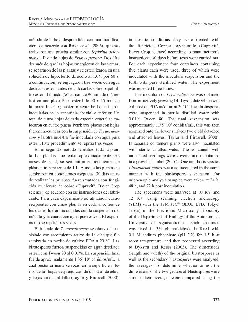
Publicación en línea, mayo 2019 322
Fully BilingualRevista Mexicana de FITOPATOLOGÍAMexican Journal of Phytopathology
método de la hoja desprendida, con una modifica-ción, de acuerdo con Rossi et al. (2006), quienes realizaron una prueba similar con Taphrina defor-mans utilizando hojas de Prunus persica. Dos días después de que las hojas emergieron de las yemas, se separaron de las plantas y se esterilizaron en una solución de hipoclorito de sodio al 1.0% por 60 s; a continuación, se enjuagaron tres veces con agua destilada estéril antes de colocarlas sobre papel fil-tro estéril húmedo (Whatman de 90 mm de diáme-tro) en una placa Petri estéril de 90 x 15 mm de la marca Interlux; posteriormente las hojas fueron inoculadas en la superficie abaxial o inferior. Un total de cinco hojas de cada especie vegetal se co-locaron en cuatro placas Petri; tres placas con hojas fueron inoculadas con la suspensión de T. caerules-cens y la otra muestra fue inoculada con agua pura estéril. Este procedimiento se repitió tres veces.
En el segundo método se utilizó toda la plan-ta. Las plantas, que tenían aproximadamente seis meses de edad, se sembraron en recipientes de plástico transparente de 1 L. Aunque las plantas se sembraron en condiciones asépticas, 30 días antes de realizar las pruebas, fueron tratadas con fungi-cida oxicloruro de cobre (Cupravit®, Bayer Crop science), de acuerdo con las instrucciones del fabri-cante. Para cada experimento se utilizaron cuatro recipientes con cinco plantas en cada uno, tres de los cuales fueron inoculados con la suspensión del inóculo y la cuarta con agua pura estéril. El experi-mento se repitió tres veces.
El inóculo de T. caerulescens se obtuvo de un aislado con crecimiento activo de 14 días que fue sembrado en medio de cultivo PDA a 20 °C. Las blastosporas fueron suspendidas en agua destilada estéril con Tween 80 al 0.01%. La suspensión final fue de aproximadamente 1.35’ 109 conidios/mL, la cual posteriormente se roció en la superficie infe-rior de las hojas desprendidas, de dos días de edad, y hojas unidas al tallo (Taylor y Birdwell, 2000).
in aseptic conditions they were treated with the fungicide Copper oxychloride (Cupravit®, Bayer Crop science) according to manufacturer’s instructions, 30 days before tests were carried out. For each experiment four containers containing five plants each were used, three of which were inoculated with the inoculum suspension and the forth with pure sterilized water. The experiment was repeated three times.
The inoculum of T. caerulescens was obtained from an actively growing 14-days isolate which was cultured on PDA médium at 20 °C. The blastospores were suspended in sterile distilled water with 0.01% Tween 80. The final suspension was approximately 1.35’ 109 conidia/mL, this was then atomized onto the lower surfaces two d old detached and attached leaves (Taylor and Birdwell, 2000). In separate containers plants were also inoculated with sterile distilled water. The containers with inoculated seedlings were covered and maintained in a growth chamber (20 °C). One non-hosts species Pittosporum tobira was also inoculated in the same manner with the blastospores suspension. For microscopic analysis samples were taken at 24 h, 48 h, and 72 h post inoculation.
The specimens were analysed at 10 KV and 12 KV using scanning electron microscopy (SEM) with the JSM-35C® (JEOL LTD, Tokyo, Japan) in the Electronic Microscopy laboratory of the Department of Biology of the Autonomous University of Aguascalientes. Each specimen was fixed in 3% glutaraldehyde buffered with 0.1 M sodium phosphate (pH 7.2) for 1.5 h at room temperature, and then processed according to Dykstra and Reuss (2003). The dimensions (length and width) of the original blastospores as well as the secondary blastospores were analysed, the averages. To determine whether or not the dimensions of the two groups of blastospores were similar their averages were compared using the

Publicación en línea, mayo 2019 323
Fully BilingualRevista Mexicana de FITOPATOLOGÍA
Mexican Journal of Phytopathology
Las plantas en recipientes separados también fue-ron inoculadas con agua destilada estéril. Los reci-pientes con las plántulas inoculadas fueron cubiertas y se mantuvieron en una cámara de crecimiento (20 °C). Asimismo, una especie no hospedera de Pittosporum tobira fue inoculada de la misma ma-nera con la suspensión de blastosporas. Para el aná-lisis microscópico se tomaron muestras a las 24 h, 48 h y 72 h después de la inoculación.
Los especímenes se analizaron a 10 KV y 12 KV usando un microscopio electrónico de barrido (MEB) modelo JSM-35C® (JEOL LTD, Tokio, Ja-pón) en el laboratorio de Microscopía Electrónica del Departamento de Biología de la Universidad Autónoma de Aguascalientes. Cada uno de los es-pecímenes se fijó en glutaraldehído al 3% en una solución amortiguadora de 0.1 M fosfato de sodio 0.1 M (pH 7.2) por 1.5 h a temperatura ambiente, y después se procesó de acuerdo con el método de Dykstra y Reuss (2003). Se analizaron las dimen-siones (largo y ancho) de las blastosporas originales y de las blastosporas secundarias, y se obtuvieron los promedios. Para determinar si las dimensiones de los dos grupos de blastosporas eran similares, se hizo una comparación de promedios utilizando la prueba t-Student. Se analizaron también la tasa de gemación de las blastosporas después de 24 h, 48 h y 72 h, y el crecimiento direccional de los tubos germinativos.
La microscopía electrónica de barrido reveló que las blastosporas en las hojas de Quercus spp. tuvieron gemación moderada 24 h después de la inoculación, y que en las hojas de Q. eduardii (Fi-guras 1 A-B) y de Q. potosina (Figuras 1 C-D) aumentó significativamente después de 48 h y 72 h. No se observaron cambios significativos en las hojas de la especie no hospedera e incluso después de 72 h (Figuras 1 E-F) no se observó gemación. Después de 48 h, se observaron nume-rosas blastosporas más pequeñas (Figura 2 A-B) de 0.87 – 1.77 µm × 0.191 – 0.485 µm en las ho-
t-Student test. The budding rate of the blastospores after 24 h, 48 h and 72 h and the directional growth of the germination tubes were also analysed.
Scanning electron microscopy revealed that the blastospores on Quercus spp., leaves exhibited moderate budding 24 h post inoculation and increased significantly after 48 h and 72 h on leaves of Q. eduardii (Figures 1 A-B). and Q. potosina (Figures 1 C-D). There were no significant changes observed on the leaves of the non-host species even after 72 h (Figures 1 E-F) no budding was observed. After 48 h, multiple smaller blastospores (Figure 2 A-B) with dimensions 0.87 – 1.77 µm × 0.191 – 0.485 µm were observed on the leaves of Q. eduardii. A descriptive analysis of the measurements produced the following scores (n=30): length: medium = 1.41 µm, s=0.05, SD (standard deviation) = 0.27 µm for the width the values were: medium = 0.34 µm, s=0.01, SD=0.08 µm.
The dimensions of these smaller blastospores were compared to the blastopores which were originally inoculated. The dimensions of the original blastospores were 3.06 – 5.49 µm × 1.09 – 3.01 µm, and a descriptive analysis of the measurements (n=30) were as follows, length: medium = 4.12 µm, s=0.12, SD=0.68 µm for the width the values were: medium = 1.94 µm, s=0.09, SD = 0.50 µm To compare the difference between the two sets of blastospores a t-Student test was carried out. On comparing the width of the secondary blastospores (medium = 0.34 µm) and the width of the original blastospores (medium = 1.94 µm), the difference between the two means was significant, t-Student test = -17.69, p < .005. There was also a statistically significant differences between the length of the secondary blastospores (medium = 1.41 µm) and the original blastospores (medium= 4.12 µm), t-Student test = -23.05, p < .005.
Once formed the secondary blastospores produced germ tubes. The formation of germ tubes
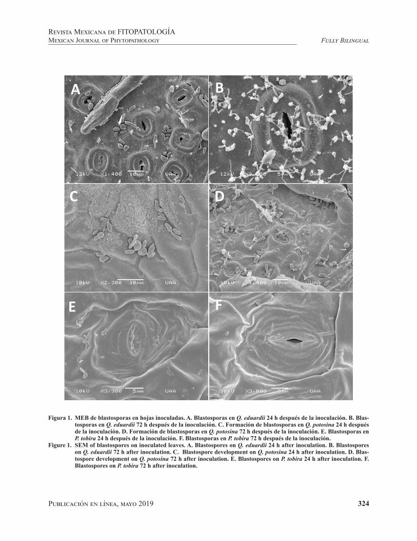
Publicación en línea, mayo 2019 324
Fully BilingualRevista Mexicana de FITOPATOLOGÍAMexican Journal of Phytopathology
Figura 1. MEB de blastosporas en hojas inoculadas. A. Blastosporas en Q. eduardii 24 h después de la inoculación. B. Blas-tosporas en Q. eduardii 72 h después de la inoculación. C. Formación de blastosporas en Q. potosina 24 h después de la inoculación. D. Formación de blastosporas en Q. potosina 72 h después de la inoculación. E. Blastosporas en P. tobira 24 h después de la inoculación. F. Blastosporas en P. tobira 72 h después de la inoculación.
Figure 1. SEM of blastospores on inoculated leaves. A. Blastospores on Q. eduardii 24 h after inoculation. B. Blastospores on Q. eduardii 72 h after inoculation. C. Blastospore development on Q. potosina 24 h after inoculation. D. Blas-tospore development on Q. potosina 72 h after inoculation. E. Blastospores on P. tobira 24 h after inoculation. F. Blastospores on P. tobira 72 h after inoculation.

Publicación en línea, mayo 2019 325
Fully BilingualRevista Mexicana de FITOPATOLOGÍA
Mexican Journal of Phytopathology
jas de Q. eduardii. Un análisis descriptivo de las me-diciones arrojó las siguientes medidas (n=30): largo: media = 1.41 µm, s=0.05, SD (desviación estándar) = 0.27 µm de largo, para el ancho los valores fueron: media = 0.34 µm, s = 0.01, SD = 0.08 µm.
Se hizo una comparación de las dimensiones de las blastosporas más pequeñas con las de las blastosporas originalmente inoculadas. Las dimen-siones de las blastosporas originales fueron 3.06 – 5.49 µm × 1.09 – 3.01 µm, y el análisis descriptivo
Figura 2. MEB del desarrollo de blastosporas secundarias. A-B Formación de múltiples blastosporas secundarias en la su-perficie del inóculo original (las flechas señalan las blastosporas secundarias). C. Tubos germinativos formados en el extremo apical de las blastosporas secundarias que crecen hacia los estomas. D. Tubos germinativos con patrón de crecimiento es al azar (las flechas señalan los tubos germinativos).
Figura 2. SEM of developing secondary blastospores. A-B Multiple secondary blastospores forming on the surface of the original inoculum (arrows showing secondary blastospores). C. Germination tubes formed on the apical end of the secondary blastospores growing towards the stomata. D. Germination tubes growing in a randomized pattern (arrows showing germination tubes).
was recorded 48 h after inoculation in Q. eduardii. Although many germ tubes grew towards the stomata (Figure 2C) many were observed growing very randomly (Figure 2D). On average the germ tubes measured 1.69 µm after 48 h and 5.18 µm after 72 h.
Although budding was observed on the surface of Q. potosina after 48 h post inoculation no secondary blastospores were observed there was also no formation of germ tubes. Disease symptoms

Publicación en línea, mayo 2019 326
Fully BilingualRevista Mexicana de FITOPATOLOGÍAMexican Journal of Phytopathology
de las mediciones (n= 30) fue el siguiente: media = 4.12 µm, s = 0.12, SD = 0.68 µm para el ancho los valores fueron: media = 1.94 µm, s = 0.09, SD = 0.50 µm. Para comparar la diferencia entre los dos grupos de blastosporas se realizó una prueba t-Student. Al comparar el ancho de las blastosporas secundarias (media = 0.34 µm) con el ancho de las dimensiones de las blastosporas originales (media = 1.94 µm), se observó una diferencia significati-va entre las dos medias, prueba t-Student = -17.69, p<.005. Asimismo, se encontraron diferencias sig-nificativas entre la longitud de las blastosporas se-cundarias (media = 1.41 µm) y la de las blastospo-ras originales (media = 4.12 µm), prueba t-Student = -23.05, p< .005.
Cuando terminaron de formarse, las blastos-poras secundarias produjeron tubos germinativos. La formación de los tubos germinativos se regis-tró 48 h después de la inoculación en Q. eduardii. Aunque muchos de los tubos germinativos crecie-ron hacia los estomas (Figura 2C), se observó que algunos crecieron al azar (Figura 2D). En prome-dio, los tubos germinativos midieron 1.69 µm des-pués de 48 h, y 5.18 µm después de 72 h.
Aunque hubo gemación en la superficie de Q. potosina 48 h después de la inoculación, no se ob-servaron blastosporas secundarias y tampoco hubo formación de tubos germinativos. Los síntomas de la enfermedad se manifestaron después de un mes únicamente en las hojas de Q. eduardii, en 30% de las plantas que fueron inoculadas (Figuras 3 A-D). Estos resultados son similares a los obtenidos por Taylor y Birdwell (2000).
Desde que Taylor y Birdwell publicaron los re-sultados de su estudio en 2000, no ha habido ningún otro estudio detallado de la interacción entre Quer-cus spp. y T. caerulescens. Una de las principales diferencias entre el presente estudio y el estudio de Taylor y Birdwell reside en que, en el presen-te estudio se utilizó un aislado de T. caerulescens
developed only on the leaves on Q. eduardii in 30% of the plant that were inoculated after one month (Figures 3 A-D), these results are similar to Taylor and Birdwell (2000).
Since the 2000 study of Taylor and Birdwell there has been no other comprehensive study into the interaction between Quercus spp. and T. caerulescens. One of the major differences between the present and the study of Taylor and Birdwell is that, in this present study an isolate of T. caerulescens obtained from Quercus eduardii (white oak) which is endemic to the Sierra Fria, Aguascalientes was used, whereas, the isolate used by Taylor and Birdwell in their experiments was derived from Quercus nigra (red oak) native to the eastern and south-central United States and can be found growing in all the coastal states. Taylor and Birdwell (2000) concluded that after 48 h germ tubes formed, this research confirms their observations that germ tubes formed after 48 h of contact with a suitable host. Many germ tubes grew towards the stomata; however, they were also observed growing arbitrarily (growing over and away from opened stomatal pores). This growth pattern was also described by Taylor and Birdwell (2000), they claimed that germ tubes frequently grew to extensive lengths and that the longer germ tubes growth appeared random rather than directional. The growth pattern exhibited by the germ tubes has led to the hypothesis that the orientation of the germ tubes is being affected by other factors in addition to chemotactic gradients emanating from the stomata (carbon dioxide concentration) however, because of the limited scope of this investigation it was not possible to determine the other factors involved.
Taylor and Birdwell (2000) stated the germ tubes formed from the apical ends of the original cells (conidia/blastospores) that were used to inoculate the leaves. This present study provides

Publicación en línea, mayo 2019 327
Fully BilingualRevista Mexicana de FITOPATOLOGÍA
Mexican Journal of Phytopathology
obtenido de Quercus eduardii (encino blanco), que es endémico de la Sierra Fría, Aguascalientes, en tanto que en sus experimentos, Taylor y Birdwell utilizaron uno derivado de Quercus nigra (encino rojo) originario de la región del este y sur-centro de Estados Unidos, que crece en los estados de la zona costera. Taylor y Birdwell (2000) llegaron a la con-clusión de que los tubos germinativos se formaron después de 48 h; el presente estudio confirma sus observaciones respecto a que los tubos germinati-vos se formaron 48 h después de entrar en contacto con un hospedante adecuado. Aunque muchos de los tubos germinativos crecieron hacia los estomas, se observó que también crecieron arbitrariamente, es decir, crecieron encima y fuera de los poros
Figura 3. Formación de ampollas en hojas de Q. eduardii 40 días después de haber sido inoculadas con T. caerulescens.Figure 3. Formation of leaf blisters on a leaves of Q. eduardii 40 days after being inoculated with T. caerulescens.
a stark contrast to their findings. The isolate from the Sierra Fria behaved in a very distinctive manner, the original conidia first produced a number of significantly smaller cells which were dubbed secondary blastospores. These secondary blastospores were the only cells capable of forming germ tubes, no germ tubes were ever observed originating from the larger blastospores. This investigation presents the first findings of these secondary blastospores.
Another finding of interest is that although the isolate used was obtained from Q. eduardii, it was capable of multiplying on the surface of Q. potosina, thereby weakening the argument that the T. caerulescens (at least this isolate) is ultra-specific

Publicación en línea, mayo 2019 328
Fully BilingualRevista Mexicana de FITOPATOLOGÍAMexican Journal of Phytopathology
estomáticos abiertos. Este patrón de crecimiento fue también descrito por Taylor y Birdwell (2000), quienes afirmaron que los tubos germinativos con frecuencia crecían hasta alcanzar grandes longi-tudes y que el crecimiento de los tubos germina-tivos más largos parecía ser al azar más bien que direccional. Debido al patrón de crecimiento de los tubos germinativos ha surgido la hipótesis de que la orientación de éstos está siendo afectada por otros factores además de los gradientes quimiotác-ticos que emanan de los estomas (concentración de dióxido de carbono). Sin embargo, dado el limitado alcance de esta investigación, no fue posible deter-minar los otros factores que intervienen.
Taylor y Birdwell (2000) mencionaron que los tubos germinativos se formaron desde los extre-mos apicales de las células originales (conidios/blastosporas) que se utilizaron para inocular las hojas. Este estudio presenta un marcado contraste con los resultados obtenidos por Taylor y Birdwell. El aislado de la Sierra Fría se comportó de manera muy diferente, ya que los primeros conidios origi-nales produjeron un número de células significa-tivamente más pequeñas que fueron consideradas como blastosporas secundarias. Las blastosporas secundarias fueron las únicas células que pudieron formar tubos germinativos; no se observó que las blastosporas más grandes produjeran tubos germi-nativos. En esta investigación se da a conocer, por primera vez, la formación de estas blastosporas se-cundarias.
Otro resultado de interés es que, aunque el ais-lado que se utilizó se obtuvo de Q. eduardii, éste tuvo la capacidad de multiplicarse en la superficie de Q. potosina y, por tanto, debilita el argumento de que T. caerulescens (al menos este aislado) es ultraespecífico y no tiene la capacidad de crecer en otra especie de Quercus además de las especies de las cuales se obtuvo el aislado (Taylor y Birdwell, 2000). Los investigadores atribuyeron este nuevo
and not capable of growth on another Quercus spp apart from the species that it was isolated from (Taylor and Birdwell, 2000). Researches attributed this new finding to the fact that in Mexico, there is a high degree of hybridization among the endemic species of Quercus spp. (Valencia, 2004) and it is possible that because of this similitude of the genetic information among these species this isolate can grow and possibly infect multiple Quercus spp.
In this study, it was determined that the blastospores of this isolate of T. caerulescens found infecting Q. eduardii of the Sierra Fria of Aguascalientes, were not capable of directly forming germination tubes, instead, the original inoculum formed numerous smaller blastospores which formed germination tubes. This research is the first time that these secondary blastospores have been observed in Taphrina spp. The study also showed that although the blastospores were capable of multiplying on Q. potosina no germination tubes were formed.
End of the English version
hallazgo al hecho de que en México existe un alto grado de hibridación entre las especies endémicas de Quercus spp. (Valencia, 2004), y que es posible que debido a la similitud de la información genéti-ca de estas especies este aislado se desarrolle y po-siblemente infecte múltiples especies de Quercus.
En el presente estudio se determinó que las blastosporas del aislado de T. caerulescens que in-fectaron la especie Q. eduardii de la Sierra Fría de Aguascalientes no tuvieron la capacidad de formar tubos germinativos directamente, y que, en lugar de esto, el inóculo original produjo numerosas blas-tosporas pequeñas que formaron tubos germinati-vos. En esta investigación se reporta por primera vez la observación de blastosporas secundarias en

Publicación en línea, mayo 2019 329
Fully BilingualRevista Mexicana de FITOPATOLOGÍA
Mexican Journal of Phytopathology
Taphrina spp. Asimismo, nuestro estudio muestra que, aunque las blastosporas tuvieron la capacidad de multiplicarse en Q. potosina, no hubo formación de tubos germinativos.
LITERATURA CITADA
Arriaga L, Espinoza JM, Aguilar C, Martínez E, Gómez L and Loa E. 2000. Regiones Terrestres Prioritarias de México. México. Comisión Nacional para el Conocimiento y Uso de la Biodiversidad (CONABIO). 609 p. http://bioteca.biodiversidad.gob.mx/janium/Documentos/1036.pdf
Dykstra MJ, and Reuss LE. 1993. Biological electron mi-croscopy: theory, techniques, and troubleshooting. 1st edition. Boston, MA. Springer. USA. 1–73. https://doi.org/10.1007/978-1-4684-0010-6.
Fonseca A and Rodrigues MG. 2011 Taphrina Fries (1832). Pp. 1291–1298. In: Kurtzman CP, Fell JW, Boekhout T (eds.) The Yeasts A Taxonomy Study. Elsevier. USA. 2354 p.
Horst RK. 2008. Westcott’s Plant Disease Handbook. 7th edn. Springer. New York, USA. 295–297. https://doi.org/10.1108/09504120910925832.
Lindsey BE, Rivero L, Calhoun CS, Grotewold E and Brkl-jacic J. 2017. Standardized Method for High-throughput Sterilization of Arabidopsis Seeds. Journal of Visualized Experiments128: 1–7. https://doi.org/10.3791/56587.
Moreno-Rico O, Marmolejo-Monsivais JG, Moreno-Manzano CE and Pérez-Hernández K. 2015. Primer reporte de Ta-phrina caerulescens en encinos (Quercus sp.) de la Sierra Fría de Aguascalientes. Congreso Nacional XVI e Interna-cional de Fitopatología. México. p. 4.
Nagao H and Katumoto K. 1998. Leaf blister of Quercus phi-llyraeoides caused by Taphrina caerulescens. Mycoscien-ce 39: 173–178. https://doi.org/10.1007/bf02464056.
Rodrigues MG and Fonseca A. 2003. Molecular systematic of the dimorphic ascomycete genus Taphrina. Internatio-nal Journal of Systematic and Evolutionary Microbiology 53:607–616. https://doi.org/10.1099/ijs.0.02437-0
Rossi V, Bolognesi M, Languasco L and Giosuè S. 2006. In-fluence of environmental conditions on infection of peach shoots by Taphrina deformans. Phytopathology 9: 155–163. https://doi.org/10.1094/phyto-96-0155
Rossi V and Languasco L. 2007. Influence of environmental conditions on spore production and budding in Taphrina deformans, the causal agent of peach leaf curl. Phytopa-thology 97: 359–365. https://doi.org/10.1094/phyto-97-3-0Taylor J and Birdwell DO. 2000. A scanning electron microscopic study of the Infection of Water Oak (Quercus nigra) by Taphrina caerulescens. Mycologia 92: 309–311. https://doi.org/10.2307/3761
Taylor J and Birdwell DO. 2000. A scanning electron micros-copic study of the Infection of Water Oak (Quercus nigra) by Taphrina caerulescens. Mycologia 92: 309–311. https://doi.org/10.2307/3761566.
Valencia S. 2004. Diversidad del género Quercus (Fagaceae) en México. Boletín de La Sociedad Botánica de México 45: 33–53. https://doi.org/10.17129/botsci.1692.

330Publicación en línea, mayo 2019
Ramírez-Salcedo HE, Barrientos-Ramírez L, Vargas-Radillo JJ, Rodríguez-Macías R, Ruíz-López MA and Virgen-Calleros G. Inhibition of Colletotrichum gloeosporioides and Botrytis cinerea by Guazuma ulmifolia Lam extracts. Mexican Journal of Phytopathology 37(2): 330-344.DOI: 10.18781/R.MEX.FIT.1812-1
Primera publicación DOI: 29 de Abril, 2019.First DOI publication: April 29, 2019.
Resumen. Guazuma ulmifolia (Malvaceae) es un árbol utilizado en la medicina popular de múltiples países para tratar diversas enfermedades. Se evaluó el efecto inhibitorio de los extractos de la corteza y fruto de esta planta, obtenidos con solventes de polaridad creciente, contra Colletotrichum gloeos-porioides y Botrytis cinerea. Se utilizó como cri-terio para elegir el solvente con mejor desempeño en la extracción, ensayos de cromatografía de capa fina (TLC) y el dato del rendimiento de extractos.
Inhibition of Colletotrichum gloeosporioides and Botrytis cinerea by Guazuma ulmifolia Lam extracts
Inhibición de Colletotrichum gloeosporioides y Botrytis cinerea con extractos de Guazuma ulmifolia Lam
Hilda Elisa Ramírez-Salcedo, estudiante del doctorado BEMARENA, CUCBA, Universidad de Guadalajara, México; Lucía Barrientos-Ramírez, J. Jesús Vargas-Radillo*, Departamento de Madera, Celulosa y Pa-pel, Cucei, Universidad de Gudalajara, Zapopan Jalisco México, CP 45010; Ramón Rodríguez-Macías, Ma-rio Alberto Ruíz-López, Departamento de Botánica, CUCBA, Universidad de Guadalajara, Zapopan Jalisco México, CP 45010; Gil Virgen-Calleros, Laboratorio de Fitopatología, CUCBA, Universidad de Guadalajara, Zapopan Jalisco México, CP 45010. *Autor para correspondencia: [email protected]
Recibido: 13 de Diciembre, 2018. Aceptado: 09 de Abril, 2019.
Abstract. Guazuma ulmifolia (Malvaceae) is a tree used in the popular medicine of many countries to treat various diseases. We evaluated the inhibitory activity of G ulmifolia bark and fruits extracts, obtained with solvents of increasing polarity, against Colletotrichum gloeosporioides and Botrytis cinerea. Thin-layer chromatography assay (TLC) and extract yield were the criteria used to establish the most efficient extraction solvent. Extracts were screened for their phytochemical components and their corresponding antifungal effect by thin-layer chromatography-Bioautography and Mycelial Growth Inhibition methods in a randomized design. Fruit extracts were more inhibitory than bark extracts, while C. gloeosporioides was more resistant than B. cinerea. At a dose of 10 mg·mL-1; fruit extracts inhibited 85.5 ± 2.1% of B. cinerea and 45.8 ± 10.0% of C. gloeosporioides mycelia growth. Bark extracts inhibited 58.0 ± 2.4% and 34.1 ± 2.9% of B. cinerea and C. gloeosporioides mycelial growth, respectively. Images obtained
Publicación en línea, mayo 2019 331
Fully BilingualRevista Mexicana de FITOPATOLOGÍA
Mexican Journal of Phytopathology
Posteriormente, se analizaron los extractos por me-dio de tamizaje fitoquímico y se evaluó la actividad antifúngica con los métodos de TLC-Bioautografia e inhibición de crecimiento micelial, aplicando un diseño experimental aleatorizado. Los extractos del fruto fueron más efectivos que los extractos de corteza, y por su parte, C. gloeosporioides fue más resistente que B. cinerea. A 10 mg mL-1, los extractos de fruto inhibieron 85.5 ± 2.1% el creci-miento micelial de B. cinerea y en 45.8 ± 10.0% el de C. gloeosporioides, mientras que los extractos de corteza inhibieron en 58.0 ± 2.4% y 34.1 ± 2.9% el crecimiento de B. cinerea y C. gloeosporioides, respectivamente. Imágenes obtenidas microscopia óptica muestran que los extractos alteraron la mor-fología original tanto de los micelios como de las esporas.
Palabras clave: Procianidinas, micelio, radical DPPH, cromatografía TLC
Las enfermedades fúngicas causan pérdidas considerables en la producción agrícola. Botrytis cinerea, causante de pudrición gris (moho gris), es un patógeno que puede infectar hasta 1,000 espe-cies de planta a través de hojas, peciolos, raíces, frutos, flores, semillas, y frutos postcosecha (Rupp et al., 2017). Colletotrichum gloeosporioides, de la familia de los Ascomicetos, es un fitopatógeno que causa antracnosis en plantas y frutos. Es uno de los más importante patógenos postcosecha ya que re-sulta en infecciones del fruto asintomáticas, laten-tes, o largamente inactivas, con síntomas que apare-cen hasta que el fruto madura (Rodríguez-López et al., 2009) cuando los conidios responden a señales químicas, ceras cuticulares y etileno, producidas por el hospedero. Se han probado diversos métodos para prevenir y controlar fitopatógenos, desde pro-ductos químicos, productos naturales renovables
with light microscopy showed that extracts altered the original mycelia and spore morphology.
Key words: Procyanidins, mycelium, DPPH radical, Chromatography TLC
Fungal diseases are responsible for considerable losses in agricultural production. Botrytis cinerea, which causes gray rot (gray mold), is a phytopathogen that can infect about 1,000 known plant species via leaves, petioles, roots, fruits, flowers, seeds, as well as postharvest fruits (Rupp et al., 2017). Colletotrichum gloeosporioides, of the family Ascomycetes, is a fungal disease that causes anthracnose in various parts of the plant and fruit. These fungi are one of the most relevant postharvest pathogens since its disease results in long quiescent or latent asymptomatic fruit infections with symptoms appearing during fruit ripening (Rodríguez-López et al., 2009) when the conidia respond to chemical signals, cuticle waxes and ethylene, produced by the host. Many methods have been tried to prevent and control phytopathogens, from the traditional use of chemicals, the development of renewable natural products with unique properties, including biocompatibility, biodegradability, antimicrobial activity, and safety, to the application of essential oils and plant extracts with microbicidal activity (Zhong et al., 2014). Guazuma ulmifolia (Malvaceae), commonly known as guacimo, mutamba, caulote or equiche, is a tropical tree of medium size distributed from northern Mexico to South America (Maurino dos Santos et al., 2018). For their antibacterial, antifungal, and anti-inflammatory properties, the bark, fruits, and leaves are traditionally used to treat various diseases (Maldini et al., 2013). In addition, the foliage and fruit are used as livestock and horses forage. Although bark, leaves and

Publicación en línea, mayo 2019 332
Fully BilingualRevista Mexicana de FITOPATOLOGÍAMexican Journal of Phytopathology
con atractivas propiedades como biocompatibili-dad, biodegradabilidad, actividad antimicrobiana y seguridad, hasta los aceites esenciales y extractos de plantas con actividad microbicida (Zhong et al., 2014). Guazuma ulmifolia (Malvaceae), conocida como guacimo, mutamba, caulote o equiche, es un árbol tropical de tamaño mediano distribuido desde el norte de México hasta América del Sur (Mau-rino dos Santos et al., 2018). Por sus propiedades antibacterianas, antifúngicas y antiinflamatorias, la corteza, los frutos y las hojas se usan para tra-tar diversas enfermedades (Maldini et al., 2013). Asimismo, el follaje y los frutos se utilizan como forraje para ganado y caballos. Aunque la corteza, las hojas y los extractos de fruto tienen propieda-des antimicrobianas, los informes científicos se han centrado en la corteza y sus propiedades medicina-les, atribuidas a su alto contenido de procianidinas (taninos condensados) (Lopes et al., 2009). Hay pocos reportes para control de fitopatógenos con extractos de G. ulmifolia, no obstante las restric-ciones en el uso de agroquímicos y el fomento del control biológico, que incluye extractos de plantas con propiedades fungicidas. Por ello, el objetivo de este estudio fue evaluar la actividad antimicrobiana de los extractos de fruto y corteza de G. ulmifolia contra B. cinerea y C. gloeosporioides.
Recolección de plantas. En abril de 2015 se re-colectó el material, corteza y frutos de árboles sil-vestres en el área “Bosque de la Primavera” en Tala Jalisco (20° 29’ 00” N de latitud y 103° 29’ 30” W longitud) a una altura de 1320 metros sobre el ni-vel del mar. El muestreo se efectuó sobre 5 árboles seleccionados al azar. De cada uno se tomó aproxi-madamente 3 kg de frutos maduros y 1 kg de cor-teza de diferentes partes del tallo, sin evidencia de enfermedades. Una muestra compuesta se secó al medio ambiente en el laboratorio a ~25 °C, se pulve-rizó en un molino de cuchillas (Retsch-GMBH) y
fruit extracts have antimicrobial properties, most scientific reports have focused on the bark and its medicinal properties, which are attributed to its high procyanidins content (condensed tannins) (Lopes et al., 2009); however, there are few reports on the use of G. ulmifolia extracts to control phytopathogens. Restrictions on the use of agrochemicals for the control of pests has led to the development of biological control methods, including biodegradable plant extracts with fungicidal properties. Therefore, the objective of this study was to evaluate the antimicrobial activity of G. ulmifolia fruit and bark extracts against B. cinerea and C. gloeosporioides.
Plant Collection. On April 2015, we collected bark and fruits from five randomly selected wild trees growing in the area known as “Bosque de la Primavera” in the town of Tala Jalisco (20° 29’ 00’’ N latitude and 103° 29’ 30’’ W longitude) at an altitude of 1320 meters above average sea level. From each tree, approximately 3 kg of mature fruits and 1 kg of bark from different parts of the stem, without evidence of diseases, were collected. A representative composite aliquot drawn from the original sample was dried at ~25 °C (in the laboratory), pulverized in a knife mill (Retsch-GMBH), and kept refrigerated until used. “Bosque de la Primavera” is a 30500 ha protected natural area located west of the metropolitan area of Guadalajara, Mexico comprised of oak, oak-pine, and tropical deciduous forest. The average annual rainfall fluctuates between 800 and 1000 mm, while the yearly average temperature is 20.6 ± 6.5 °C (Huerta-Martínez and Ibarra-Montoya, 2014).
Crude Extracts Preparation. The dry milled materials (100 g dry weight) were extracted at 4 °C with 500 mL of hexane, to remove the lipid material (lipids can interfere with the metabolites

Publicación en línea, mayo 2019 333
Fully BilingualRevista Mexicana de FITOPATOLOGÍA
Mexican Journal of Phytopathology
se mantuvo refrigerada hasta su uso. El “Bosque de la Primavera” es un área natural protegida ubica-da al oeste del área metropolitana de Guadalajara, México, de aproximadamente 30500 hectáreas de roble, roble-pino y bosque tropical caducifolio. La precipitación anual fluctúa entre 800 y 1000 mm y la temperatura anual de 20.6±6.5 °C (Huerta-Mar-tínez e Ibarra-Montoya, 2014).
Obtención de extractos crudos. Los materiales molidos y secos (100 g, peso seco) se extrajeron con 500 mL de hexano, para eliminar el material lipídico (interfiere con los análisis de metabolitos), por 2 horas con agitación en un matraz y baño de hielo (4 °C). Las pruebas de extracción fueron con solventes de diferentes polaridades (hexano, di-clorometano y metanol) para encontrar el más efi-ciente, auxiliándose de cromatografía en capa fina (TLC) para este fin. Los resultados indicaron que el diclorometano fue el mejor solvente de extracción. El fruto y corteza se extrajeron con diclorometano durante 48 horas y 28 °C con agitación continua (150 rpm), relación de disolvente a muestra seca de 5:1 (v/w), en incubadora (LabTech modelo LSI-3016R). El material se filtró con papel Whatman No. 4 y el material extraído se concentró en un ro-toevaporador Buchi a presión reducida y 40 °C. El rendimiento del extracto, en mg g-1, se determinó dividiendo los sólidos secos obtenidos entre el peso del material vegetal seco inicial.
Cultivo de patógenos y preparación de inóculos. Las cepas de C. gloeosporioides y B. cinerea fue-ron proporcionadas amablemente por el Laborato-rio de Fitopatología en el CUCBA de la Universi-dad de Guadalajara, donde se aislaron de papaya y fresa enfermas, respectivamente, y se identificaron por sus características morfológicas y fisiológicas según los protocolos correspondientes. Cultivos puros de estas cepas se dispersaron, por separado,
assays), for 2 hours with constant stirring in a flat bottom flask. Preliminary tests were carried out with solvents of different polarities (hexane, dichloromethane, and methanol) to find the most efficient extracting solvent as determined by a thin layer chromatography (TLC) test. The results of this screening indicated that dichloromethane was the best extracting solvent. Fruit and bark samples were extracted with dichloromethane for 48 h at 28 °C with continuous stirring (150 rpm) and a solution to dry sample ratio of 5:1 (v/w) in an incubator (LabTech model LSI-3016R). The slurry was filtered through Whatman paper No 4 and the solvent, containing the extracted material, concentrated until dryness in a rotary evaporator Buchi under reduced pressure and 40 °C. We used the total solids test to determined the extract yield and expressed as milligrams of dry solids in the solvent extract per gram of original dry plant material.
Pathogens Culture and Inoculum Preparation. CUCBA Phytopathology Laboratory of the University of Guadalajara kindly provided the C. gloeosporioides and B. cinerea strains isolated from diseased papaya and strawberry, respectively, and identified by morphology and physiological characteristics Pure cultures of these strains were transferred into separate glass tubes and dispersed in 5 mL of sterile saline solution (NaCl, 0.85% w/v) or distilled water, and the microorganisms concentration was adjusted with saline solution or water to 1 - 2.5 x 10⁶ cells mL-1, using for this a Neubauer cell-counting chamber (Moro et al., 2017).
Phytochemical Screening. Before the bioassay tests, the biological activity of the phytochemical compounds (e.g., flavonoids, tannins, terpenoids, alkaloids, saponins, glycosides, and so on) found

Publicación en línea, mayo 2019 334
Fully BilingualRevista Mexicana de FITOPATOLOGÍAMexican Journal of Phytopathology
en 5 mL de solución salina estéril (NaCl, 0,85% p/v) o agua destilada, y la concentración de los mi-croorganismos se ajustó con solución salina o agua a 1 - 2.5 x 10⁶ células mL-1, utilizando una cámara de recuento de células de Neubauer (Moro et al., 2017).
Tamizaje Fitoquímico. Antes de las pruebas de los bioensayos, se aplicaron pruebas para la identifi-cación cualitativa de los compuestos fitoquímicos con actividad biológica (flavonoides, taninos, ter-penoides, alcaloides, saponinas, glucósidos, etcéte-ra) presentes en los extractos, utilizando para ello procedimientos establecidos (Harbone, 1998).
TLC-Bioautografía (TLC-B). Los métodos de TLC-B son aquellos en los que se detectan cuali-tativamente compuestos bioactivos por medio de pruebas biológicas (antimicrobianas o enzimáticas) aplicadas sobre extractos químicos (Choma y Je-sionek, 2015). Los extractos de plantas se fraccio-naron por medio de TLC en sus diferentes consti-tuyentes fitoquímicos, para lo cual se aplicaron ~10 μL de extracto en una placa de TLC (10 x 10 cm, gel de sílice, Merk, 60F254), con una fase móvil de tolueno:acetato de etilo (85:15), visualizando la se-paración de los compuestos con luz UV (254 y 365 nm, Handfield UV Lamp, UVGL-58), y se registró el valor Rf (factor de retardo). Se utilizó la placa desarrollada para analizar la actividad biológica de los compuestos mediante bioautografía directa, usando extracto de té verde como control positivo. Las placas TLC se secaron y después se asperjaron con la suspensión fúngica correspondiente (1 x10⁶ células mL-1) hasta humedecerla, incubándolas du-rante 24 horas a 37 °C y después de esto se impreg-naron con violeta de p-yodonitrotetrazolio (Sigma, 2 mg mL-1, 6 h, 35 °C). La sal de tetrazolio (INT) es un aceptor de electrones; los microorganismos bio-lógicamente activos lo reducen a formazán, el cual
in the extracts were qualitatively identified using established procedures (Harbone, 1998).
TLC-Bioautography (TLC-B). TLC-B methods are those in which biological tests (antimicrobial or enzymatic) are applied on chemical extracts for the qualitative detection of bioactive compounds (Choma and Jesionek 2015). In our work, plant extracts were separated into their phytochemical constituents by TLC. For this, ~10 μL of extract was applied on a TLC plate (10 x10 cm, silica gel, Merk, 60F254) and developed with toluene:ethyl acetate (85:15) mobile phase. After this, the phytochemicals were visualized with UV light (254 and 365 nm, Handfield UV Lamp, UVGL-58) and their Rf value (Retardation factor) recorded. We then used the developed plates to assess the compounds antimicrobial activity by direct bioautography using a green tea extract as a positive control. The dried plates were sprayed with the corresponding fungal suspension (1 x10⁶ cells mL-1) until moist, incubated for 24 h at 37 °C, and then impregnated with p-iodonitrotetrazolium violet (Sigma, 2 mg mL-1, 6 h, 35 °C). Tetrazolium salt (INT) is an electron acceptor which the biologically active microorganisms reduce it to red-colored formazan becoming colorless when the fungi growth is inhibited (Eloff et al., 2008). Red-purple areas reveal active microbes and white areas indicate inhibition of fungal activity. To assess the antioxidant activity, we sprayed the TLC plates with a 0.2 % solution of the free radical compound DPPH (2, 2, diphenyl-1-picrylhydrazyl in methanol, Sigma®). After heating the TLC plates, the spots (compounds) with antioxidant activity turned to a yellowish white color over the DPPH purple background (Belaqziz et al., 2017).
Mycelial Growth Inhibition (MGI). Growth agar medium was prepared by blending (using
Publicación en línea, mayo 2019 335
Fully BilingualRevista Mexicana de FITOPATOLOGÍA
Mexican Journal of Phytopathology
es un compuesto de color rojo, y se vuelven incolo-ros cuando se inhibe el crecimiento de los hongos (Eloff et al., 2008). Las áreas de color rojo púrpura revelan microbios activos y las áreas blancas indi-can la inhibición de la actividad fúngica. Para la evaluación de la actividad antioxidante se asperja-ron las placas de TLC con una solución 0.2% del compuesto de DPPH (2, 2, difenilo-1-picrilhidra-zilo en metanol, Sigma®), que es un radical libre. Las placas de TLC se calentaron y los compuestos antioxidantes se visualizaron mediante el desarro-llo de manchas blancas amarillentas sobre el fondo púrpura del DPPH (Belaqziz et al., 2017).
Inhibición del Crecimiento Micelial (ICM). El agar de crecimiento fue preparado mediante la mezcla de Agar PDA líquido (auxiliándose de un homogeneizador marca DragonLab) con 1% (v/v), Tween 80 (surfactante para mejorar la emulsifica-ción) y suficiente extracto para alcanzar concen-traciones en el 1-20 mg mL-1 (rango amplio para encontrar varios datos de inhibición); el agar se depositó en cajas Petri (90 mmØ) y se dejó solidifi-car; luego se colocó un disco de 7 mm de diámetro impregnado con conidiósporas a una concentración controlada (1 x10⁶ células mL-1), en el centro de las placas. Se utilizaron PDA como control negativo y benomilo (1 mg mL-1) como control positivo. Be-nomilo [1-[(butilamino)carbonil]-1H-benzimida-zol-2-il] es un derivado azol comúnmente utilizado para el control químico de estos hongos. Las placas se incubaron a 28 °C y el crecimiento de la colo-nia se midió cada 24 h con la ayuda de una regla, hasta que el micelio en el control negativo alcanzó el borde de la placa. Las pruebas se realizaron por triplicado y el porcentaje de inhibición se calculó con la siguiente fórmula (Zhong et al., 2014): Crecimiento del - Crecimiento del micelio(control) micelio(tratamiento)% Inhibición micelial = x100
Crecimiento del micelio(control)
a DragonLab homogenizer) liquid PDA agar with 1% (v/v), Tween 80 (surfactant to improve emulsification) and enough extract to achieve concentrations in the 1-20 mg mL-1 range (this range is wide enough to detect inhibitory activity). Enough agar was deposited on Petri dishes (90 mmØ) and allowed to solidify; then a 7 mm diameter disc, impregnated with a fungus conidiospores solution containing 1 x10⁶ cells mL-1, was placed at the plates’ center. PDA and benomyl (1 mg mL-1) served as negative and positive controls, respectively. Benomyl [1-[(butylamino)carbonyl]- 1H-benzimidazol-2-yl], is an azole derivative commonly used for the chemical control of these fungi. The dishes were incubated at 28 °C and the colony growth measured every 24 h, with the aid of a ruler, until the mycelium in the negative control reached the dish’s edge. Tests were done in triplicate, and the percent mycelial inhibition calculated with the following formula (Zhong et al., 2014): Mycelial - Mycelial growth(control) growth(treatment)% Mycelia inhibition = x100
Mycelial growth(control)
The lethal concentrations, (LC50) and (LC95), were calculated from the curve of mycelial inhibition vs. extract concentration.
Microscopy study. Samples of fungi treated with bark and fruit guacimo extracts (10 mg mL-1), benomyl, and PDA were deposited on microscope slides and homogeneously dispersed with distilled water and viewed in an optical microscope (Zeiss model KF2) equipped with a camera (Axio Cam ERC5S).
Statistical Analyses. A randomized experimental design was set up with the factors: Plant part (Fruit and Bark); fungi (C. gloeosporioides and B.

Publicación en línea, mayo 2019 336
Fully BilingualRevista Mexicana de FITOPATOLOGÍAMexican Journal of Phytopathology
La concentración letal, (LC50) y (LC95) se calcu-ló a partir de la curva de % de inhibición micelial vs concentración de extracto.
Estudio de Microscopía. Se observaron las mues-tras de los hongos tratados con los extractos de cor-teza y fruto de guacimo (10 mg mL-1), benomilo y PDA, para lo cual se depositaron en portaobjetos, se dispersaron homogéneamente con agua destila-da, y fueron observados con un microscopio óptico (Zeiss modelo KF2) provisto de una cámara (Axio Cam ERC5S).
Análisis estadístico. Para el método de Inhibición del crecimiento micelial se aplicó un diseño aleato-rizado, con los factores experimentales: Parte de la planta (fruto y corteza); hongos (C. gloeosporioi-des y B. cinerea); tiempo de tratamiento (72 h, 144 h, 240 h, 312 h), y la concentración del extracto (1 mg mL-1, 10 mg mL-1, 20 mg mL-1). La variable de respuesta fue la inhibición del crecimiento ra-dial (%). Los datos se analizaron mediante ANO-VA multifactorial (nivel de confianza del 95%, P<0.05), para evaluar la significación estadística de los factores; y la prueba de Tukey para determinar los valores que difieren. Se empleó el software es-tadístico Statgraphics Centurion XVII (©2014 Sta-tpoint Technologies, Inc.) para el análisis de datos. En resultados, el rendimientos de los extractos para el fruto fue de 20.1 mg g-1 de material original base seca (bs) en hexano, 52.5 mg g-1 bs en diclorome-tano y 76.4 mg g-1 bs en metanol, mientras que para corteza fue de 56.7 mg g-1bs en hexano, 122.4 mg g-1 bs en diclorometano y 66.0 mg g-1bs en metanol. Estos valores son más altos que los 2 mg g-1 y 47 mg g-1 reportados para la corteza de guacimo extraídas por maceración con hexano y metanol, respectiva-mente (Camporese et al., 2003). El diclorometano fue el extractante más eficiente, según lo indicado por la cantidad de metabolitos evidenciados en la
cinerea); treatment time (72 h, 144 h, 240 h, 312 h), and extract concentration (1 mg mL-1, 10 mg mL-1, 20 mg mL-1). The response variable radial was growth inhibition (%). Data was analyzed by multifactorial ANOVA (95.0% confidence level, P < 0.05) to determine the statistical significance factors’ effect and Tukey’s test to establish the level of significance among values. We used the statistical software Statgraphics Centurion XVII (©2014 Statpoint Technologies, Inc.) for data analysis.
In results, the fruit extract yield in mg·g-1 dry weight (dw) for the different solvents were: 20.1 for hexane, 52.5 for dichloromethane, and 76.4 for methanol while those for the bark extract were 56.7 for hexane, 122.4 for dichloromethane, and 66.0 for methanol. These values are higher than the 2 mg g-1, and 47 mg g-1reported for macerated guacimo bark with hexane and methanol, respectively (Camporese et al., 2003). Dichloromethane was the most efficient solvent as indicated by the number of metabolites found in the TLC test. The screening attested the presence of alkaloids, flavonoids, tannins, triterpenes, saponins, and coumarins in the extracts, highlighting the presence of flavonoids, triterpenes, and coumarins, due to their biological activity. Our results agree with those of Singanan et al. (2018) who reported the presence of phenols, tannins, saponins, flavonoids, and terpenoids in G. ulmifolia fruit. Some of these biomolecules are characteristic of medicinal plants and have antioxidant, therapeutic, and medicinal properties that can inhibit or eliminate the growth of pathogens, with little or no toxicity to the host cells, and are good candidates for antimicrobial applications (Mujeeb et al., 2014). The TLC- Bioautography results for B. cinerea are included in Figure 1. Results for C. gloeosporioides and B. cinerea were similar, and for the sake of brevity, we only discuss those of the latter. TLC plate A in

Publicación en línea, mayo 2019 337
Fully BilingualRevista Mexicana de FITOPATOLOGÍA
Mexican Journal of Phytopathology
prueba TLC. El tamizaje evidenció la presencia en los extractos de guacimo de alcaloides, flavonoi-des, taninos, triterpenos, saponinas y cumarinas, destacando los flavonoides, triterpenos y cumari-nas, debido a su actividad biológica. Estos resulta-dos coinciden con los previamente reportados para fruto de G. ulmifolia, que mencionan la presencia de fenoles, taninos, saponinas, flavonoides y ter-penoides (Singanan et al., 2018). Algunas de estas biomoléculas son características de plantas medici-nales y tienen propiedades antioxidantes, terapéuti-cas y medicinales que pueden inhibir o eliminar el crecimiento de patógenos, con poca o ninguna toxi-cidad para las células hospederas, y son candidatos para aplicaciones antimicrobianas (Mujeeb et al., 2014). Los resultados de TLC-Bioautografía sobre B. cinerea se muestran en la Figura 1. El compor-
Figura 1. Pruebas biológicas cualitativas para extractos crudos de corteza y fruto de G. ulmifolia contra B. Cinerea. Fase móvil de tolueno/acetato de etilo (85/15). A) Placas de gel de sílice para TLC; B) TLC-DPPH. Actividad antioxi-dante de extractos de G. ulmifolia. Método de revelado: Vanillina; C) TLC-Bioautografía Directa. Actividad anti-fúngica de los extractos de G. ulmifolia y D) Te Verde- Bioautografía directa. Actividad Antifúngica de extractos de Té Verde (Comparativo).
Figure 1. Qualitative biological tests for crude extracts of G. ulmifolia’s bark and fruit against B. Cinerea. Mobile phase Toluene/Ethyl acetate (85/15). A) Silica gel TLC; B) TLC-DPPH. Antioxidant activity of G. ulmifolia extracts. De-tection method: Vanillin; C) TLC-Direct Bioautography. Antifungal activity of G. ulmifolia extracts and D) Green tea- Direct Bioautography. Antifungal activity of green tea extracts (Control).
Figure 1 shows the compounds present in guacimo bark and fruit extracts, shown as distinct round dots. Plate (B) shows the antioxidant activity of each of the spot found in Plate (A) against DPPH radical (TLC-DPPH); on this plate, a yellowish spot indicates antioxidant activity. Finally, plates C and D depict G. ulmifolia extracts and green tea, used as positive control, antimicrobial activity against B. cinerea (TLC-direct bioautography test), respectively. Clear spots indicate antimicrobial activity. As can be seen, the active zones found in plate C (G. ulmifolia extract) coincide with those of green tea, suggesting that both contain similar flavonoids and that this zone comprises the extracts antioxidant and antifungal activity. The Rf values for these spots are approximately 0.30-0.45. The size and intensity of the spots indicate the
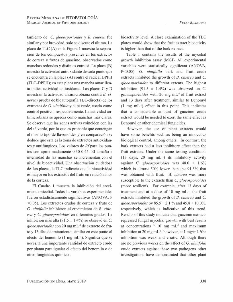
Publicación en línea, mayo 2019 338
Fully BilingualRevista Mexicana de FITOPATOLOGÍAMexican Journal of Phytopathology
tamiento de C. gloeosporioides y B. cinerea fue similar y por brevedad, solo se discute el último. La placa de TLC (A) en la Figura 1 muestra la separa-ción de los compuestos presentes en los extractos de corteza y frutos de guacimo, observados como manchas redondas y distintas entre sí. La placa (B) muestra la actividad antioxidante de cada punto que se encuentra en la placa (A) contra el radical DPPH (TLC-DPPH); en esta placa una mancha amarillen-ta indica actividad antioxidante. Las placas C y D muestran la actividad antimicrobiana contra B. ci-nerea (prueba de bioautografía TLC-directa) de los extractos de G. ulmifolia y el té verde, usado como control positivo, respectivamente. La actividad an-timicrobiana se aprecia como manchas más claras. Se observa que las zonas activas coinciden con las del té verde, por lo que es probable que contengan el mismo tipo de flavonoides y en comparación se deduce que esta es la zona de extractos antioxidan-tes y antifúngicos. Los valores de Rf para los pun-tos son aproximadamente 0.30-0.45. El tamaño e intensidad de las manchas se incrementan con el nivel de bioactividad. Una observación cuidadosa de las placas de TLC indicaría que la bioactividad es mayor en los extractos del fruto en relación a los de la corteza.
El Cuadro 1 muestra la inhibición del creci-miento micelial. Todas las variables experimentales fueron estadísticamente significativas (ANOVA, P <0.05). Los extractos crudos de corteza y fruto de G. ulmifolia inhibieron el crecimiento de B. cine-rea y C. gloeosporioides en diferentes grados. La inhibición más alta (91.5 ± 1.4%) se observó en C. gloeosporioides con 20 mg mL-1 de extracto de fru-to y 13 días de tratamiento, similar en este punto al efecto del benomilo (1 mg mL-1). Significa que se necesita una importante cantidad de extracto crudo por planta para igualar el efecto del benomilo o de otros fungicidas químicos.
bioactivity level. A close examination of the TLC plates would show that the fruit extract bioactivity is higher than that of the bark extract.
Table 1 contains the results of the mycelial growth inhibition assay (MGI). All experimental variables were statistically significant (ANOVA, P<0.05). G. ulmifolia bark and fruit crude extracts inhibited the growth of B. cinerea and C. gloeosporioides to different extents. The highest inhibition (91.5 ± 1.4%) was observed on C. gloeosporioides with 20 mg mL-1 of fruit extract and 13 days after treatment, similar to Benomyl (1 mg mL-1) effect in this point. This indicates that a considerable amount of guacimo crude extract would be needed to exert the same effect as Benomyl or other chemical fungicides.
However, the use of plant extracts would have some benefits such as being an innocuous biological control, among others. In contrast, the bark extracts had a less inhibitory effect than the fruit extracts. Under the same testing conditions (13 days, 20 mg mL-1) its inhibitory activity against C. gloeosporioides was 48.0 ± 1.6% which is almost 50% lower than the 91.5% that was obtained with fruit. B. cinerea was more susceptible to the extracts than C. gloeosporioides (more resilient). For example, after 13 days of treatment and at a dose of 10 mg mL-1, the fruit extracts inhibited the growth of B. cinerea and C. gloeosporioides by 85.5 ± 2.1 % and 45.8 ± 10.0%, respectively, which is indicative of this trend. Results of this study indicate that guacimo extracts repressed fungal mycelial growth with best results at concentrations ³ 10 mg mL-1 and maximum inhibition at 20 mg mL-1; however, at 1 mg·mL-1the inhibition was weak and erratic. Although there are no previous works on the effect of G. ulmifolia crude extracts against these two pathogens other investigations have demonstrated that other plant

Publicación en línea, mayo 2019 339
Fully BilingualRevista Mexicana de FITOPATOLOGÍA
Mexican Journal of Phytopathology
Sin embargo, el beneficio de usarlos consiste en un control biológico seguro e inocuo, entre otros. En contraste, los extractos de corteza inhibieron menos que los extracto de fruto. Bajo las mismas condiciones descritas (13 días, 20 mg mL-1) C. gloeosporioides fue inhibido en 48.0 ± 1.6%, por extractos de corteza, lo cual es casí 50% inferior al 91.5% que se obtuvo con fruto. Asimismo, B. ci-nerea fue más susceptible que C. gloeosporioides (más resistente) a los extractos; Por ejemplo, en 13
Cuadro 1. Inhibición del crecimiento micelial de B. cinerea y C. gloeosporioides con extractos de fruto y corteza de G. ulmifolia. Los valores representan x
_ ± sd de tres réplicas. Todas
las variables tuvieron un efecto estadísticamente significativo sobre el crecimiento del micelio (P <0.05): Pplant part = 0.0000, Pfungi = 0.0000, Pconcentration extracts = 0.0000, Ptreatment time = 0.0000 (ANOVA).
Table 1. Inhibition of B. cinerea and C. gloeosporioides mycelial growth by G. ulmifolia fruit and bark extracts. Values represent the x
_ ± sd of three replicates. All variables had a statis-
tically significant effect on mycelial growth (P<0.05): Pplant part = 0.0000, Pfungi = 0.0000, Pconcentration extracts = 0.0000, Ptreatment time = 0.0000 (ANOVA). x
Planta parte Hongo CE,
mg mL-1 Tiempo de Tratamiento, días
3 6 10 13
Inhibición de Crecimiento Micelial, %
Fruto
Cg
11020Benomilx
10.0±0.0a
13.3±4.7a
60.0±0.0c
80.0±0.0cd
9.5±2.1a
17.8±1.6ab
83.5±4.4cd
85.7±3.8cd
16.5±3.1ª37.2±3.3b
90.7±0.2d
90.6±2.2d
13.7±2.6ª45.8±10.0bc
91.5±1.4d
91.5±1.7d
Bc
110Benomil
36.5±12.3bc
44.8±5.4bc
80.0±0.0cd
6.6±20.3a
39.4±17.6b
87.6±3.8d
31.0±15.6bc
75.9±5.0cd
88.4±2.1d
28.0±18.7ab
85.5±2.1cd
91.5±1.6d
Corteza
Cg
11020Benomil
20.0±4.1ab
30.0±8.2b
60.0±0.0c
83.33±0.0cd
12.4±3.4a
34.2±1.5b
50.7±1.1bc
87.6±3.8d
18.1±2.4ab
44.2±1.3bc
42.6±3.0bc
89.4±2.2d
16.6±1.6ab
34.1±2.9b
48.0±1.6bc
91.5±1.7d
Bc
11020Benomil
19.2±1.5ab
58.5±1.8c
79.3±0.9cd
94.8±0.0d
18.6±1.2ab
57.3±1.7c
76.2±1.9cd
94.7±0.5d
20.4±1.6ab
59.4±2.3c
75.4±2.5cd
94.8±1.4d
15.5±2.3ab
58.0±2.4c
78.4±3.2cd
94.6±2.5d
CE=Concentración de Extractos; Cg= C. gloeosporioides; Bc= B. cinerea; x concentración Benomilo =1 mg mL-1 / CE = Concentration Extracts; Cg = C. gloeosporioides; Bc= B. cinerea; xBenomyl concentration = 1 mg mL-1
Los valores en la misma columna con letras diferentes indica que son significativamente diferentes, de acuerdo con la prueba de Tukey (P<0.05) / Values in the same column with different letters are significantly different according to Tukey´s test (P < 0.05).
extracts inhibit their growth. A methanol extract from Piscidia piscipula applied at 2 mg mL-1 resulted in a 52.3% C. gloeosporioides mycelial growth inhibition after five days (Peraza-Sanchez et al., 2005) which is higher than our results. In addition, the hydroalcoholic crude extracts of the soapberry (Sapindus saponaria) leaf 7 days after treatment resulted in a 70% C. gloeosporioides MGI when applied to 50 mg mL-1 and 80% MGI when applied at100 mg·mL-1, while concentrations

Publicación en línea, mayo 2019 340
Fully BilingualRevista Mexicana de FITOPATOLOGÍAMexican Journal of Phytopathology
días de tratamiento y 10 mg mL-1, los extractos de fruto inhibieron el crecimiento de B. cinerea y C. gloeosporioides en 85.5 ± 2.1% y 45.8 ± 10.0%, respectivamente, lo cual es indicativo de esta ten-dencia. Los resultados de este estudio indican que los extractos reprimieron el crecimiento del micelio fúngico, con los mejores efectos al aplicarlo a con-centraciones de ³10 mg mL-1, obteniendo inhibición máxima con 20 mg mL-1 (Cuadro 1). La inhibición fue débil y con comportamiento errático cuando se aplicó a 1 mg mL-1. No hay trabajos previos sobre el efecto de extractos crudos de G. ulmifolia contra estos patógenos; sin embargo, si hay informes so-bre los efectos de otros extractos de plantas contra ellos. El extracto de metanol de Piscidia piscipu-la aplicado a 2 mg mL-1 y 5 días ocasionó 52.3% ICM sobre C. gloeosporioides (Peraza-Sanchez et al., 2005), resultado mejor que el nuestro. También, los extractos crudos hidroalcohólicos de la hoja del jaboncillo (Sapindus saponaria), en 7 días de tra-tamiento inhibieron 70% el crecimiento micelial de C. gloeosporioides al aplicarlo a 50 mg mL-1 y 80% al aplicarlo a 100 mg mL-1, mientras que concentraciones menores de 50 mg mL-1 no tuvie-ron efecto (Passos Marinho et al., 2018). Extractos alcohólicos de las hojas de las vides silvestres (Vi-tis) aplicados a 12% de concentración ocasionaron 70% ICM de B. cinerea (Apolonio-Rodríguez et al., 2017), menor que nuestros resultados. La acti-vidad antimicrobiana y medicinal de los extractos de corteza, partes aéreas y frutos de G. ulmifolia se atribuye a la presencia de flavonoides del subgrupo flavan-3-ols (Lopes et al., 2009); compuestos tales como la catequina, epicatequina, flavanocoumari-nas y las procianidinas y proantocianidinas deriva-das de ellas, con propiedades antioxidantes y an-tiinflamatorias (Maldini et al., 2013). Los valores de concentración letal (CL, mg mL-1) se calcularon considerando los mejores datos de inhibición en el cuadro 1, y son los siguientes: 1. C. gloeosporioides:
lower than 50 mg mL-1 had no effect (Passos Marinho et al., 2018). Alcoholic extracts of wild grapevines (Vitis) leaves had a lower inhibitory effect than G. ulmifolia extracts. When applied at 12% wild grapevines (Vitis) leaves resulted in only 70% B. cinerea MGI (Apolonio-Rodríguez et al., 2017). The antimicrobial and medicinal activity of G. ulmifolia bark, aerial part, and fruits extracts are attributed to the presence of flavonoids of the subgroup flavan-3-ols (Lopes et al., 2009) with antioxidant and anti-inflammatory properties including catechin, epicatechin, flavanocoumarins, as the procyanidins and proanthocyanidins derived from them (Maldini et al., 2013). The Lethal Concentration (LC, mg mL-1) parameters calculated from the best inhibitory values shown in Table 1, are as follows: 1. C. gloeosporioides: Fruit (LC50 = 10.57, LC95 = 20.94); Bark (LC50 = 16.54, LC95 = 34.13), 2. B. cinerea: Fruit (LC50 = 5.24, LC9 5= 11.09); Bark (LC50 = 10.6, LC95 = 22.6). Results of the microscopy study are shown in Figure 2. Anatomy of C gloeosporioides and B. cinerea has been previously reported. Figure 2A shows of C gloeosporioides. Its main features (Rodríguez-López et al., 2009) are light gray to dark gray septate hyphae. Conidia are hyaline, cylindrical, and separated from the pedicel by a transverse septum. Also for B. cinerea (Figure 2B), it is established (Miclea Raluca Vasilica et al., 2012) septate and cylindrical hyphae, hyaline at the beginning and later become brown or black, which multiply by cytoplasmic division. Conidia are globose or elliptical, forming exogenous chains (Blastospores). Fungi treatment with guacimo extracts altered its morphology, including distorted, wavy, folded mycelia, with flat or empty portions (Xu et al., 2007); cytoplasmic coagulation exhibited by small vesicles in the mycelium (Figure 2C). Nonpolar phenolic compounds, such as those found in guacimo, could affect the cytoplasmic
Publicación en línea, mayo 2019 341
Fully BilingualRevista Mexicana de FITOPATOLOGÍA
Mexican Journal of Phytopathology
Fruto (CL50 = 10.57, CL95 = 20.94); Corteza (CL50
= 16.54, CL95 = 34.13), 2. B. cinerea: Fruto (CL50 = 5.24, CL95 = 11.09); Corteza (CL50 = 10.6, CL95 = 22.6). En la figura 2 se aprecian las característi-cas morfológicas de los hongos estudiados. La ana-tomía de C. gloeosporioides y B. cinerea ha sido previamente reportada. En la Figura 2A se muestra C. gloeosporioides. Sus características principales (Rodríguez-López et al., 2009) son hifas septadas cuyo color es de gris claro a gris oscuro. Los coni-dios son hialinos, cilíndricos y separados del pe-dicelo por un tabique transversal. Para B. cinerea (Figura 2B), se establece (Miclea Raluca Vasilica et al., 2012) hifas septadas y cilíndricas, hialina al principio y más tarde se vuelven marrón o negro. Se multiplican por división citoplásmica. Los coni-dios son globulares o elípticos, formando cadenas exógenas (blastosporas). El tratamiento de los hon-gos con extractos de guacimo ocasionó alteraciones en su morfología, a nivel de micelios deformados, ondulados, plegados, con porciones planas o vacías (Xu et al., 2007), con coagulación citoplásmica, que se muestran como pequeñas vesículas en el micelio (Figura 2C). Los compuestos fenólicos no polares, como los que se encuentran en el guacimo, podrían actuar sobre la membrana citoplasmática, causando su desintegración y pérdida de trifosfa-to de adenosina, lo que resulta en la muerte celu-lar (Hassani et al., 2012). Los conidios mostraron cambios en su forma original (Figura 2E). La pared celular de estos elementos puede sufrir daños, con numerosas protuberancias que eventualmente lle-van a su colapso, lo que podría inhibir su germina-ción (Sharma y Tripathi, 2008). De igual manera, C. gloeosporioides tratado con benomil (bencimi-dazol) mostró hifas vacías con grandes estructuras anómalas (Figura 2F). Benomilo es un fungicida comercial que se ha utilizado durante muchos años para controlar la antracnosis antes de la cosecha de frutos.
membrane, causing its disintegration loss of adenosine triphosphate and cell death (Hassani et al., 2012). The conidia showed changes in their original form. (Figure 2E). The conidias’ cell wall can also experience damage with numerous protrusions that eventually leads to their break down and inhibition of their germination (Sharma and Tripathi, 2008). In the same way, C. gloeosporioides treated with Benomyl (benzimidazole) showed empty hyphae with large anomalous structures (Figure 2F). Benomyl is a commercial fungicide that has been used for many years to control anthracnose before fruits harvesting.
In this study, the fruit extract was more effective on mycelial inhibition than the bark extract and could be a good source of biologically active compounds with therapeutic and fungicidal activity. Although the fruit is the most accessible part of the plant, which is edible, sweet, palatable, and consumed by the general population has not been extensively investigated. C. gloeosporioides was more resistant than B. cinerea; however, more specific tests and evaluation of pathogenicity and virulence are needed to confirm these results.
AcknowledgmentsWe like to thank the National Council for Science
and Technology (CONACYT), by awarding scholarship
no. 388048/256531, the BEMARENA-CUCBA doctorate
program and the Department of Wood Cellulose and Paper
(DMCyP)-CUCEI of the University of Guadalajara for the
support provided for this work.
End of the English version

Publicación en línea, mayo 2019 342
Fully BilingualRevista Mexicana de FITOPATOLOGÍAMexican Journal of Phytopathology
La tendencia observada en este trabajo fue que el extracto del fruto fue más efectivo que el extrac-to de corteza en la inhibición micelial y podría ser una buena fuente de compuestos biológicamente activos con posibles usos terapéuticos y fungicidas. Aunque el fruto es la parte más accesible de la
Figura 2. Cambios microscópicos observados en B. cinerea y C. gloeosporioides después de la exposición a extractos de fruto y corteza de G. ulmifolia. Microscopio de luz, 100x. A) Morfología de C. gloeosporioides; B) morfología B. cinerea; C) C. gloeosporioides tratado con extractos de fruto de G. ulmifolia: Observar hifas con estructura deformada y citoplasma coagulado; D) B. cinerea tratado con extracto de fruto de G. ulmifolia; E) C. gloeosporioides tratado con extracto de corteza de G. ulmifolia y F) C. gloeosporioides tratado con Benomilo.
Figure 2. Microscopic changes observed in B. cinerea and C. gloeosporioides after exposure to G. ulmifolia fruit and bark extracts. Light microscope, 100x. A) C. gloeosporioides morphology; B) B. cinereal morphology; C) C. gloeospori-oides treated with G. ulmifolia fruit extract: Observe hyphae with deformed structure and coagulated cytoplasm; D) B. cinerea treated with G. ulmifolia fruit extract; E) C. gloeosporioides treated with G. ulmifolia bark extract andF) C. gloeosporioides treated with Benomyl.
planta, comestible, dulce y apetecible y es consu-mida por la población, no ha sido ampliamente in-vestigada. Otra tendencia fue que C. gloeosporioi-des fue más resistente que B. cinérea, sin embargo, se necesitan pruebas más específicas y evaluación de la patogenicidad y/o virulencia para confirmar este desempeño.

Publicación en línea, mayo 2019 343
Fully BilingualRevista Mexicana de FITOPATOLOGÍA
Mexican Journal of Phytopathology
AgradecimientosAgradecemos el apoyo brindado a este proyecto por: el
Sistema Nacional de Ciencia y Tecnología (CONACYT), a
través de la beca escolar No. 388048/256531; al programa de
doctorado BEMARENA-CUCBA; y al Departamento de Ma-
dera, Celulosa y Papel (DMCyP)-CUCEI de la Universidad de
Guadalajara
LITERATURA CITADA
Apolonio-Rodríguez I, Franco-Mora O and Salgado-Siclán ML. 2017. Inhibición in vitro de Botrytis cinerea con extractos de hojas de vid silvestre (Vitis spp). Revis-ta Mexicana de Fitopatología 35: 170-185. https://doi.org/10.18781/R.MEX.FIT.1611-1
Belaqziz M, Tan SP, El-Abbassi A, Kiai H, Hafidi A, O’Donovan O and McLoughlin P. 2017. Assessment of the antioxidant and antibacterial activities of different oli-ve processing wastewaters. PLoS ONE 12(9): e0182622. https://doi.org/10.1371/journal.pone.0182622
Camporese A, Balick M, Arvigo R, Esposito RG, Morsellino N, Simone FD and Tubaro A. 2003. Screening of anti-bacterial activity of medicinal plants from Belize (Central America). Journal of Ethnopharmacology 87:103–107. https://doi.org/10.1016/S0378-8741(03)00115-6
Choma I and Jesionek W. 2015. TLC-Direct bioautography as a high throughput method for detection of antimicrobials in plants. Chromatography 2:225–238. https://doi.org/ 10.3390/chromatography2020225
Eloff JN, Katherine DR and Mcgaw LJ. 2008. The Biological activity and chemistry of the South African Combretaceae. Journal of Ethnopharmacology 1119:686–699.https://doi.org/10.1016/j.jep.2008.07.051
Harborne JB. 1998. Phytochemical Methods; A guide to mo-dern techniques of plant analysis. Third edition. Chap-man and Hall, London, 302 p.https://www.google.com.mx/url?sa=t&rct=j&q=&esrc=s&source=web&cd=6&ved=2ahUKEwjXoNCJn5jfAhUJPK0KHbZVBF8QFjAFegQIAxAC&url=https%3A%2F%2Ffaculty.psau.edu.sa%2Ffiledownload%2Fdoc-3-pdf-d71d9e49824fddce-b67a26b34e5bc770-original.pdf&usg=AOvVaw0ZVzhs9rixocQh6KWFFFqk
Hassani A, Fathi Z, Ghosta Y, Abdollahi A, Meshkatalsadat MH and Marandi RJ. 2012. Evaluation of plant essential oils for control of postharvest brown and gray mold rots on Apricot. Journal of Food Safety 32:94–101. https://doi.org/10.1111/j.1745-4565.2011.00353.x
Huerta-Martínez FM and Ibarra-Montoya JL. 2014. Wildfi-res at la primavera forest, Jalisco, Mexico: an approach to possible causes and consequences. CienciaUAT. 9(1): 23-32.www.scielo.org.mx/scielo.php?script=sci_arttext&pid=S2007-78582014000100023
Lopes GC, Rocha JCB, de Almeida GC and deMello JCP. 2009. Condensed tannins from the bark of Guazuma ulmifolia Lam.
(Sterculiaceae). Journal of the Brazilian Chemical So-ciety 20(6):1103-1109. http://dx.doi.org/10.1590/S0103-50532009000600016
Maldini M, Di Micco S, Montoro P, Darra E, Mariotto S, Bi-fulco G, Pizza C and Piacente S. 2013. Flavanocoumarins from Guazuma ulmifolia bark and evaluation of their af-finity for STAT1. Phytochemistry 86:64–71. https://doi.org/10.1016/j.phytochem.2012.10.011
Maurino dos Santos J, Monteiro Alfredo T, Ávila Antunes K, da Silva Melo da Cunha J, Almeida Costa EM and Silva Lima E. 2018. Guazuma ulmifolia Lam. Decreases Oxi-dative Stress in Blood Volume 2018: 16 pages. https://doi.org/10.1155/2018/2935051
Miclea Raluca V. Loredana Alexandra S and Carmen Emilia P. 2012. In vitro studies regarding the morphology of Bo-trytis Cinerea Pers. isolated from geranium Plants. ProEn-vironment 5(2012): 60–66. https://www.researchgate.net/publication/268816084
Moro IJ, Gabrielle Alexandre Gondo DG, Gonçalves Pierri E, Linharis Rodrigues Pietro RC, Pienna Soares C, Pergenti-no de Sousa D and Gonzaga dos Santos A. 2017. Evalua-tion of antimicrobial, cytotoxic and chemopreventive acti-vities of carvone and its derivatives 53(4):e00076. http://dx.doi.org/10.1590/s2175-97902017000400076
Mujeeb F, Bajpai P and Pathak N. 2014. Phytochemical eva-luation, antimicrobial activity, and determination of bioac-tive components from leaves of Aegle marmelos. BioMed Research International Volume 2014, Article ID 497606, 11 pages. http://dx.doi.org/10.1155/2014/497606
Passos Marinho GJ, Espellet Klein D and Siqueira Junior CL. 2018. Evaluation of soapberry (Sapindus saponaria L.) leaf extract against papaya anthracnose. Summa Phytopa-thology 44(2): 127-131. http://dx.doi.org/10.1590/0100-5405/175605
Peraza-Sánchez SR, Chan-Che EO and Ruiz-Sánchez E. 2005. Screening of Yucatecan plant extracts to control Colle-totrichum gloeosporioides and isolation of a new pima-rene from Acacia pennatula. Journal of Agricultural and Food Chemistry 53:2429–2432. https://doi.org/10.1021/jf040422i
Rodríguez-López ES, González-Prieto JM and Mayek-Pérez N. 2009. Infection of avocado (Persea america-na Mill.) by Colletotrichum gloeosporioides (Penz.) Penz. y Sacc.: Biochemical and genetics aspects. Re-vista Mexicana de Fitopatología 27:53–63. https://www.google.com.mx/url?sa=t&rct=j&q=&esrc=s&source=web&cd=1&cad=rja&uact=8&ved=2ahUKEwimj_nz05jfAhXMmq0KHWZpBVsQFjAAegQIChAB&url=http%3A%2F%2Fwww.scielo.org.mx%2Fscielo.php%3Fscript%3Dsci_arttext%26pid%3DS0185-009000100007&usg=AOvVaw2lvmZVIo7c0scZf7hpzViJ
Rupp S, Plesken C, Rumsey S, Dowling M, Schnabel G, We-ber RWS and Hahna M. 2017. Botrytis fragariae, a new species causing gray mold on Strawberries, shows high frequencies of specific and efflux-Based fungicide resis-tance. Applied and Environmental Microbiology 83(9): 1-16. https://doi.org/10.1128/AEM.00269-17
Sharma N and Tripathi A. 2008. Effects of Citrus sinensis (L.) Osbeck epicarp essential oil on growth and morpho-genesis of Aspergillus niger (L.) Van Tieghem. Microbiological
Publicación en línea, mayo 2019 344
Fully BilingualRevista Mexicana de FITOPATOLOGÍAMexican Journal of Phytopathology
Research 163:337–344. https://doi.org/10.1016/j.mi-cres.2006.06.009
Singanan M, Duraiswamy B and Varadarajan V. 2018. Physi-cochemical, phytochemicals and antioxidant evaluation of Guazuma ulmifolia fruit. International Journal of Pharma-cy and Pharmaceutical Sciences 10(9):87-91. http://dx.doi.org/10.22159/ijpps.2018v10i9.26778
Xu WT, Huang KL, Guo F, Qu W, Yang JJ, Liang ZH and Luo YB. 2007. Postharvest grapefruit seed extract and chitosan treatments of table grapes to control Botrytis cinerea. Pos-tharvest Biology and Technology 46:86–94. https://doi.org/10.1016/j.postharvbio.2007.03.019
Zhong Z, Aotegen B, Xu H and Zhao, S. 2014. The influen-ce of chemical structure on the antimicrobial activities of thiosemicarbazone-chitosan. Cellulose 21:105–114. https://doi.org/10.1007/s10570-013-0154-6
345Publicación en línea, mayo 2019
Rodríguez-Mendoza J, García-Ávila CJ, López-Buenfil JA, Araujo-Ruiz K, Quezada-Salinas A, Cambrón-Crisantos JM and Ochoa-Martínez DL. Identification from Tomato brown rugose fruit virus by RT-PCR from a coding region of replicase (RdRP). Mexican Journal of Phytopathology 37(2): 345-356.DOI: 10.18781/R.MEX.FIT.1902-6
Primera publicación DOI: 30 de Abril, 2019.First DOI publication: April 30, 2019.
Resumen. En la actualidad es de importancia estratégica contar con herramientas de diagnóstico rápido y preciso que permitan conocer si el material de interés está infectado con algún virus de importancia cuarentenaria o económica. Por tal motivo, en el presente trabajo se diseñó un par de oligonucleótidos específicos para la detección del ToBRFV por RT-PCR, considerando una región de la secuencia codificante de la RdRP localizada
Identification of Tomato brown rugose fruit virus by RT-PCR from a coding region of replicase (RdRP)
Identificación de Tomato brown rugose fruit virus por RT-PCR
de una región codificante de la replicasa (RdRP)
Johan Rodríguez-Mendoza, Clemente de Jesús García-Ávila, José Abel López-Buenfil, Karina Araujo-Ruiz, Andrés Quezada-Salinas, José Manuel Cambrón-Crisantos*, Dirección General de Sanidad Vegetal-Centro Nacional de Referencia Fitosanitaria. Carretera Federal México-Pachuca, Km 37.5, CP 55740, Tecámac, Estado de México, México; Daniel Leobardo Ochoa-Martínez, Postgrado en Fitosanidad-Fitopatología. Colegio de Postgraduados Km. 36.5 Carretera México-Texcoco, CP 56230, Montecillo, Texcoco, Estado de México. *Autor para correspondencia: [email protected].
Recibido: 27 de Febrero, 2019. Aceptado: 23 de Abril, 2019.
Abstract. Currently, it is of strategic importance to have fast and accurate diagnostic tools that allow us to know if the material of interest is infected with a quarantine or economic important virus. For this reason in the present work we designed a pair of specific oligonucleotides for the detection of ToBRFV by RT-PCR, considering a region of the coding sequence of the RdRP located in the ORF1 and the methodology to detect it was standardized. Additionally, the amplicons were cloned and sequenced, the products were used to predict the evolutionary relationship between ToMMV, ToMV, TMV and ToBRFV by the Maximum Likelihood method. The results indicated that the oligonucleotides designed in the present work allows the identification of fast and specific ToBRFV.
Key words: Solanaceae, Tobamovirus, specific oligonucleotides, standardization

Publicación en línea, mayo 2019 346
Fully BilingualRevista Mexicana de FITOPATOLOGÍAMexican Journal of Phytopathology
en el ORF1 y la metodología para detectarlo fue estandarizada. Adicionalmente, los amplicones fueron clonados y secuenciados, los productos fueron usados para predecir la relación evolutiva entre ToMMV, ToMV, TMV y ToBRFV por el método Maximum Likelihood. Los resultados indicaron que los oligonucleótidos diseñados en el presenté trabajo permiten la identificación de manera rápida y específica del ToBRFV.
Palabras clave: Solanaceae, Tobamovirus, oligonucleótidos específicos, estandarización
El cultivo de tomate (Solanum lycopersicum) y chile (Capsicum annuum), representan una activi-dad de importancia económica a nivel mundial (Li et al., 2017). Históricamente, plantas de las fami-lias Solanaceae y Cucurbitaceae se han visto afec-tadas por algunas especies del género Tobamovirus como: Tobacco mosaic virus (TMV) y Tomato mo-saic virus (ToMV) (Dombrovsky y Smith, 2017). En tomate, la resistencia a estos virus se introdujo por introgresión; sin embargo, la durabilidad de la resistencia se ve comprometida por la presión de selección de los patógenos (Dombrovsky y Smith, 2017; Maayan et al., 2018). La importancia de los tobamovirus se debe a su fácil dispersión, princi-palmente por medios mecánicos (Dombrovsky et al., 2017); también a través de semillas contamina-das, las partículas virales de los tobamovirus son extremadamente estables preservando su infecti-vidad durante varios años (Dombrovsky y Smith, 2017). Recientemente, se reportó la presencia del Tomato brown rugose fruit virus (ToBRFV) en México, afectando tomate y chile (Cambrón-Cri-santos et al., 2018). Las plantas enfermas desarro-llaron menor cantidad de flores y frutos; necrosis del pedúnculo y cáliz del fruto. Además, en frutos se observaron áreas amarillas, rugosidades y áreas necróticas (Salem et al., 2015).
Tomato (Solanum lycopersicum) and chili crops (Capsicum annuum) are an economically important activity worldwide (Li et al., 2017). Historically, plants of the Solanaceae and Cucurbitaceae families have been affected by some species of the Tobamovirus genus, such as Tobacco mosaic virus (TMV) and Tomato mosaic virus (ToMV) (Dombrovsky and Smith, 2017). Tomato resistance to these viruses was introduced by introgression. However, the durability of resistance is compromised by the pathogen’s selection pressure (Dombrovsky and Smith, 2017; Maayan et al., 2018). Tobamoviruses are important because they are easily dispersed, mainly by mechanical means or through contaminated seed (Dombrovsky et al., 2017); the viral particles of tobamoviruses are extremely stable and remain infectious for several years (Dombrovsky and Smith, 2017). Recently, the presence of the Tomato brown rugose fruit virus (ToBRFV) was reported in Mexico, which affects tomato and chili crops (Cambrón-Crisantos et al., 2018). The infected plants produced fewer flowers and fruits, and developed necrosis on the peduncle and fruit calyx. In addition, yellow and necrotic areas, as well as rugosity, were observed on fruits (Salem et al., 2015).
To identify tobamoviruses, the use of oligonucleotides has been reported for universal detection, which were designed using an alignment matrix from regions located in the ORF1 that encodes the RdRP of 32 tobamoviruses (Li et al., 2018), but currently there are no reports on specific oligonucleotides. Therefore, the objective of this study was to develop a specific method for detecting ToBRFV, including the design of a pair of specific oligonucleotides to amplify an encoding region of the ToBRFV’s viral RdRP (ORF1) through reverse transcription polymerase chain reaction (RT-PCR) specifically considering the nucleotidic regions of the structural or catalytic motifs, as well as standardize the detection method.

Publicación en línea, mayo 2019 347
Fully BilingualRevista Mexicana de FITOPATOLOGÍA
Mexican Journal of Phytopathology
Para la identificación de tobamovirus, se han reportado oligonucleótidos para su detección uni-versal, los cuales fueron diseñados tomando una matriz de alineamiento de regiones conservadas en los ORF1 que codifican la RdRP de 32 tobamovirus (Li et al., 2018); sin embargo, hasta el momento no hay reportes de oligonucleótidos específicos para la detección de ToBRFV; por lo que el objetivo del presente trabajo fue desarrollar un método de detección específico para ToBRFV, incluyendo el diseño de un par de oligonucleótidos específicos para la amplificación de una región codificante de la RdRP viral (ORF1) del ToBRFV por Reacción en Cadena de la Polimerasa con Transcripción Re-versa (RT-PCR), considerando, específicamente las regiones nucleotídicas de los motifs estructurales o catalíticos; así como, la estandarización del método de detección.
El diseño del par de oligonucleótidos específi-cos para ToBRFV consideró los genomas comple-tos de las especies más representativas del género Tobamovirus, reportados por el Centro Nacional de Información Biotecnológica, (National Center for Biotechnology Information, NCBI) (https://www.ncbi.nlm.nih.gov/), como: Tobacco mo-saic virus (TMV, Virgaviridae) (NC_001367.1, FR878069.1, HE818443.1 y V01408.1), Tomato mosaic virus (ToMV, Virgaviridae) (NC_002692.1, KY967227.1, MF002490.1 y AF332868.1), To-mato mottle mosaic virus (ToMMV, Virgaviri-dae) (NC_022230.1, FX898034.1, KR824950.1 y KF477193.1), adicionalmente, se consideraron los genomas de dos aislamientos de ToBRFV (Virgavi-ridae), Tomato brown rugose fruit virus aislado Is-raelí TBRFV-IL (KX619418.1) y Tomato brown ru-gose fruit virus aislado de Jordania (KT383474.1). Con el programa BioEdit versión 7.0.5.3 (Hall, 1999) se hizo un alineamiento global de los geno-mas utilizando el algoritmo Clustal W para generar la primer matriz e identificar regiones variables entre
To design the pair of specific oligonucleotides for ToBRFV the whole genome of the most representative species of the Tobamovirus genus reported by the National Center for Biotechnology Information (NCBI) (https://www.ncbi.nlm.nih.gov/) was considered, including Tobacco mosaic virus (TMV, Virgaviridae) (NC_001367.1, FR878069.1, HE818443.1 and V01408.1), Tomato mosaic virus (ToMV, Virgaviridae) (NC_002692.1, KY967227.1, MF002490.1 and AF332868.1), Tomato mottle mosaic virus (ToMMV, Virgaviridae) (NC_022230.1, FX898034.1, KR824950.1 and KF477193.1); the genomes of two ToBRFV isolates (Virgaviridae) were also considered: the Tomato brown rugose fruit virus, an isolate from Israel TBRFV-IL (KX619418.1), and the Tomato brown rugose fruit virus, an isolate from Jordan (KT383474.1). A global alignment of the genomes was carried out using the BioEdit program version 7.0.5.3 (Hall, 1999) and the Clustal W algorithm in order to create a matrix and identify variable regions between them (Figure 1). The selected region was used to obtain the oligonucleotides sequences, which were then analyzed in silico using the OligoAnalyzer server version 3.1 (https://www.idtdna.com/calc/Analyzer/Home/Instructions) to predict their physical and chemical properties (Annex 1). The oligonucleotides were synthesized by Integrated DNA Technologies, Inc. (IDT).
To standardize the process, seven tomato plants were used as a ToBRFV positive control, and one of chili with putative ToBRFV symptoms collected in the state of Michoacan (Cambrón-Crisantos et al., 2018), which were stored at -70 °C in the facilities of the National Center for Phytosanitary Reference (CNRF, for its acronym in Spanish), Plant Health General Directorate, National Service for Plant Health, Safety and Agri-Food Quality (SENASICA, for its acronym in Spanish). The central midrib of leaves (0.5 cm wide approximately) was taken to

Publicación en línea, mayo 2019 348
Fully BilingualRevista Mexicana de FITOPATOLOGÍAMexican Journal of Phytopathology
ellos (Figura 1). La región seleccionada se usó para obtener las secuencias de los oligonucleótidos, és-tas se analizaron in silico usando el servidor Oli-goAnalyzer versión 3.1 (https://www.idtdna.com/calc/Analyzer/Home/Instructions) para predecir sus propiedades fisicoquímicas (Anexo 1). Los oli-gonucleótidos fueron sintetizados por Integrated DNA Technologies, Inc. (IDT).
Para el proceso de estandarización se utilizaron como control positivo de ToBRFV, siete plantas de tomate y una de chile con síntomas putativos a ToBRFV, recolectadas en el Estado de Michoacán (Cambrón-Crisantos et al., 2018); resguardadas y almacenadas a -70 °C en las instalaciones del Cen-tro Nacional de Referencia Fitosanitaria (CNRF) de la Dirección General de Sanidad Vegetal, del Servicio Nacional de Sanidad, Inocuidad y Calidad Agroalimentaria. La nervadura central de las hojas fue separada de un ancho de 0.5 cm aproximada-mente para formar una muestra compuesta, la cual se picó finamente. La Extracción de RNA total, se realizó a partir de 100 mg de nervadura con el Kit SV Total RNA Isolation System Start-Up® (Prome-gaTM), siguiendo las especificaciones del fabrican-te; la pureza y concentración del RNA se cuantificó por espectrofotometría (Nano Drop 2000®, Thermo
prepare a composite sample that was then finely chopped. The total RNA extraction was performed using 100 mg of leaf midrib with the SV Total RNA Isolation System Start-Up® kit (PromegaTM), following the manufacturer’s specifications; RNA purity and concentration was quantified by spectrophotometry (Nano Drop 2000®, Thermo ScientificTM). We used as negative controls ten plants individually infected with the positive-sense single-stranded RNA genome virus: Rattail cactus necrosis-associated virus (RCNaV, Virgaviridae), Tobacco mosaic virus (TMV, Virgaviridae), Tomato mosaic virus (ToMV, Virgaviridae), Pepper mild mottle virus (PMMoV, Virgaviridae), Cucumber mosaic virus (CMV, Bromoviridae) and Papaya meleira virus (PMeV, Totiviridae), as well as with the DNA genome of Pepper golden mosaic virus (PepGMV, Geminiviridae) and Okra yellow mosaic Mexico virus (OYMMV, Geminiviridae).
The cDNA synthesis of the positive control was performed with random oligonucleotides (Random Hexamer, Invitrogen™), following the manufacturer’s instructions, under the following conditions: 1 cycle at 42 °C for 30 min, 1 cycle at 99 °C for 5 min, and finally at 12 °C for 5 min. For PCR, the specific oligonucleotides designed in
Figura 1. Región seleccionada para el diseño de los oligonucleótidos específicos para detectar a ToBRFV. A) Se consideraron los genomas de los Tobamovirus TMV, ToMV, ToMMV y ToBRFV.
Figure 1. Selected region to design the specific oligonucleotides to detect ToBRFV. A) The Tobamovirus genomes TMV, ToMV, ToMMV and ToBRFV were considered.
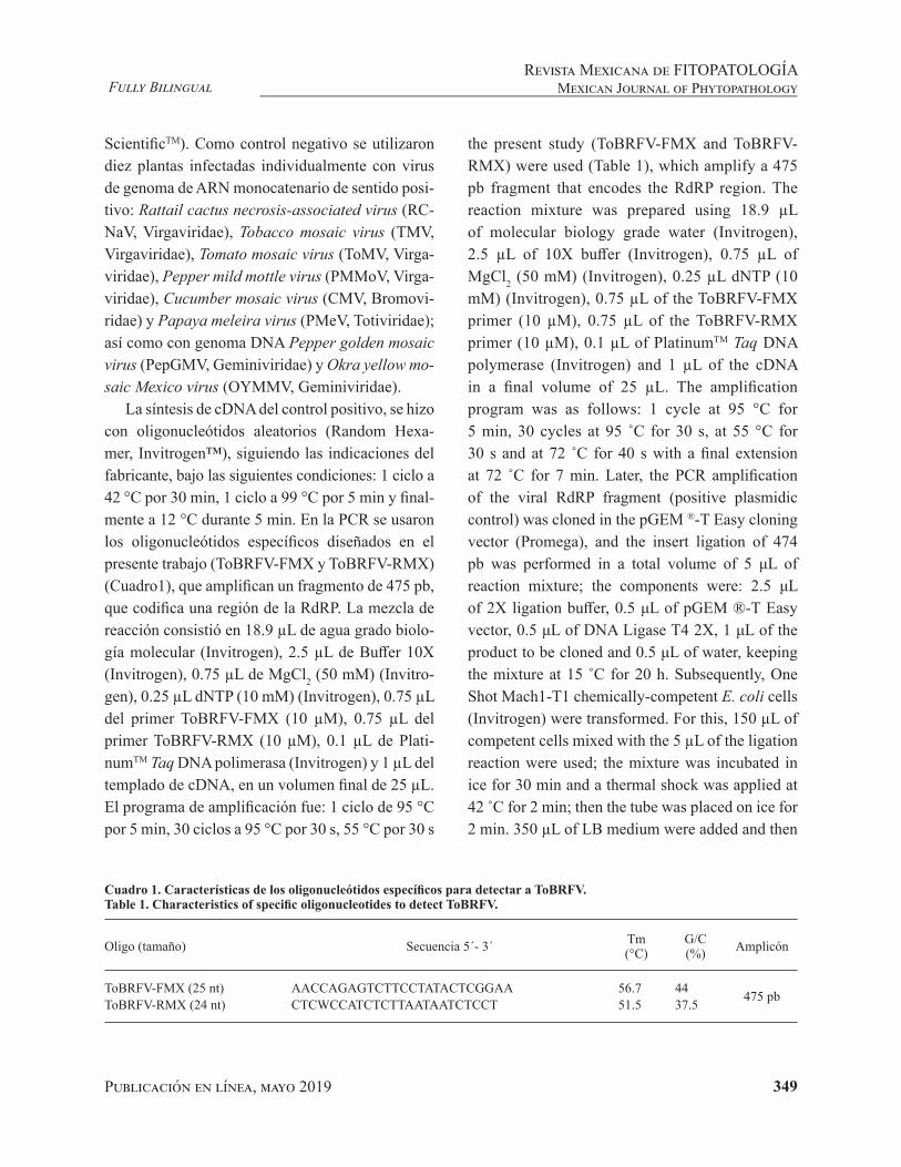
Publicación en línea, mayo 2019 349
Fully BilingualRevista Mexicana de FITOPATOLOGÍA
Mexican Journal of Phytopathology
ScientificTM). Como control negativo se utilizaron diez plantas infectadas individualmente con virus de genoma de ARN monocatenario de sentido posi-tivo: Rattail cactus necrosis-associated virus (RC-NaV, Virgaviridae), Tobacco mosaic virus (TMV, Virgaviridae), Tomato mosaic virus (ToMV, Virga-viridae), Pepper mild mottle virus (PMMoV, Virga-viridae), Cucumber mosaic virus (CMV, Bromovi-ridae) y Papaya meleira virus (PMeV, Totiviridae); así como con genoma DNA Pepper golden mosaic virus (PepGMV, Geminiviridae) y Okra yellow mo-saic Mexico virus (OYMMV, Geminiviridae).
La síntesis de cDNA del control positivo, se hizo con oligonucleótidos aleatorios (Random Hexa-mer, Invitrogen™), siguiendo las indicaciones del fabricante, bajo las siguientes condiciones: 1 ciclo a 42 °C por 30 min, 1 ciclo a 99 °C por 5 min y final-mente a 12 °C durante 5 min. En la PCR se usaron los oligonucleótidos específicos diseñados en el presente trabajo (ToBRFV-FMX y ToBRFV-RMX) (Cuadro1), que amplifican un fragmento de 475 pb, que codifica una región de la RdRP. La mezcla de reacción consistió en 18.9 µL de agua grado biolo-gía molecular (Invitrogen), 2.5 µL de Buffer 10X (Invitrogen), 0.75 µL de MgCl2 (50 mM) (Invitro-gen), 0.25 µL dNTP (10 mM) (Invitrogen), 0.75 µL del primer ToBRFV-FMX (10 µM), 0.75 µL del primer ToBRFV-RMX (10 µM), 0.1 µL de Plati-numTM Taq DNA polimerasa (Invitrogen) y 1 µL del templado de cDNA, en un volumen final de 25 µL. El programa de amplificación fue: 1 ciclo de 95 °C por 5 min, 30 ciclos a 95 °C por 30 s, 55 °C por 30 s
the present study (ToBRFV-FMX and ToBRFV-RMX) were used (Table 1), which amplify a 475 pb fragment that encodes the RdRP region. The reaction mixture was prepared using 18.9 µL of molecular biology grade water (Invitrogen), 2.5 µL of 10X buffer (Invitrogen), 0.75 µL of MgCl2 (50 mM) (Invitrogen), 0.25 µL dNTP (10 mM) (Invitrogen), 0.75 µL of the ToBRFV-FMX primer (10 µM), 0.75 µL of the ToBRFV-RMX primer (10 µM), 0.1 µL of PlatinumTM Taq DNA polymerase (Invitrogen) and 1 µL of the cDNA in a final volume of 25 µL. The amplification program was as follows: 1 cycle at 95 °C for 5 min, 30 cycles at 95 ˚C for 30 s, at 55 °C for 30 s and at 72 ˚C for 40 s with a final extension at 72 ˚C for 7 min. Later, the PCR amplification of the viral RdRP fragment (positive plasmidic control) was cloned in the pGEM ®-T Easy cloning vector (Promega), and the insert ligation of 474 pb was performed in a total volume of 5 μL of reaction mixture; the components were: 2.5 μL of 2X ligation buffer, 0.5 μL of pGEM ®-T Easy vector, 0.5 μL of DNA Ligase T4 2X, 1 μL of the product to be cloned and 0.5 μL of water, keeping the mixture at 15 ˚C for 20 h. Subsequently, One Shot Mach1-T1 chemically-competent E. coli cells (Invitrogen) were transformed. For this, 150 µL of competent cells mixed with the 5 µL of the ligation reaction were used; the mixture was incubated in ice for 30 min and a thermal shock was applied at 42 ˚C for 2 min; then the tube was placed on ice for 2 min. 350 µL of LB medium were added and then
Cuadro 1. Características de los oligonucleótidos específicos para detectar a ToBRFV.Table 1. Characteristics of specific oligonucleotides to detect ToBRFV.
Oligo (tamaño) Secuencia 5´- 3´ Tm(°C)
G/C(%) Amplicón
ToBRFV-FMX (25 nt) AACCAGAGTCTTCCTATACTCGGAA 56.7 44 475 pbToBRFV-RMX (24 nt) CTCWCCATCTCTTAATAATCTCCT 51.5 37.5

Publicación en línea, mayo 2019 350
Fully BilingualRevista Mexicana de FITOPATOLOGÍAMexican Journal of Phytopathology
y 72 °C por 40 s con una extensión final a 72 °C por 7 min. Posteriormente, el producto de la PCR del fragmento de la RdRP viral (Control plasmídico positivo) se clono en el vector de clonación pGEM ®-T Easy (Promega), la ligación del inserto de 474 pb se llevó acabo en un volumen total de 5 μL de mezcla de reacción, los componentes fueron: 2.5 μL de 2X Buffer de ligación, 0.5 μL vector pGEM ®-T Easy, 0.5 μL de DNA Ligasa T4 2X, 1 μL del pro-ducto a clonar y 0.5 μL de agua, manteniendo la mezcla a 15 °C durante 20 h. Posteriormente, se transformaron células One Shot Mach1-T1 E. coli (Invitrogen), quimiocompetentes. Para ello se uti-lizaron 150 µL de células competentes, mezcladas con los 5 µL de la reacción de ligación, esta se in-cubó en hielo por 30 min y se dio un choque térmico a 42 °C durante 2 min, después, el tubo se colocó en hielo durante 2 min. Se adicionaron 350 µL de medio LB y se incubó a 37 °C por 45 min. Fi-nalmente, se cultivó en placas con medio LB, 40 µL de ampicilina, 40 µL de IPTG y 40 µL de Xgal (20 mg mL-1) a 37 ̊ C durante 16 h. La construcción pGEM/RdRP-ToBRFV (CP-1, ~3490 nt) obtenida de esta clonación se usó como control positivo para la estandarización de la RT-PCR del fragmento de la RdRP. Posteriormente, los fragmentos clonados se secuenciaron por la metodología Sanger en el Laboratorio de Biología Molecular del CNRF, con el equipo Applied Biosystems modelo 3130.
La estandarización del método se realizó si-guiendo las condiciones de reacción señaladas pre-viamente. Para determinar la temperatura de ani-llamiento óptima, se analizaron seis temperaturas de anillamiento con diferencia de 4 °C (47, 51, 55, 59, 63 y 67 °C), a partir de seis controles a una sola concentración (100 ng µL-1): un control plas-mídico positivo a ToBRFV (CP-1), dos controles biológicos infectados con ToBRFV, To-P-100-2 y Ch-P-100-1, correspondientes a tomate y chi-le, respectivamente; un control matriz por cultivo
the tube was incubated at 37 °C for 45 min. Finally, the mixture was sown on plates containing LB medium, 40 µL of ampicillin, 40 µL of IPTG and 40 µL of Xgal (20 mg mL-1) at 37 °C for 16 h. The pGEM/RdRP-ToBRFV (CP-1, ~3490 nt) construct obtained from this cloning was used as a positive control for standardizing the RT-PCR of the RdRP fragment. Later, the cloned fragments were sequenced in the Molecular Biology Laboratory of CNRF using the Applied Biosystems equipment model 3130, following the Sanger’s methodology.
The method standardization was achieved under the reaction conditions previously described. To determine the optimal annealing temperature, six annealing temperatures with a 4 °C difference between them (47, 51, 55, 59, 63 and 67 °C) were analyzed using six controls in a single concentration (100 ng µL-1): one positive plasmidic control to ToBRFV (CP-1), two biological controls infected with ToBRFV, To-P-100-2 and Ch-P-100-1 corresponding to tomato and chili, respectively; one matrix control per crop (CM-Ch-100-3 and CM-To-100-1) and one of reagents (NTC). The working range was calculated and determined using a positive plasmidic control to ToBRFV (CP-1) at a 1000 ng µL-1 concentration, and then serial dilutions were made in order to obtain three work concentrations (100, 10 and 1 ng µL-1) (Figure 2A). The lower limit of detection was established using the last dilution of the working range (1 ng µL-1), and then serial dilutions were made in order to obtain three work concentrations (0.1, 0.01 and 0.001 ng µL-1) (Figure 2B). The specificity was verified using RCNaV, TMV, ToMV, PMMoV, CMV and PMeV negative controls, as well as DNA genome such as PepGMV and OYMMV (Figure 2C).
Standardization data were used to detect ToBRFV from the plant samples collected in the state of Michoacan and stored at -70 °C; the cDNA

Publicación en línea, mayo 2019 351
Fully BilingualRevista Mexicana de FITOPATOLOGÍA
Mexican Journal of Phytopathology
(CM-Ch-100-3 y CM-To-100-1) y uno de reactivos (NTC). El intervalo de trabajo se realizó y determi-nó a partir del control plasmídico positivo a ToBR-FV (CP-1) a una concentración de 1000 ng µL-1, se hicieron diluciones seriadas para obtener tres con-centraciones de trabajo (100, 10 y 1 ng µL-1) (Figu-ra 2A). El límite de detección inferior, se estableció a partir de la última dilución del intervalo de traba-jo (1 ng µL-1) se hicieron diluciones seriadas, para obtener tres concentraciones de trabajo (0.1, 0.01
synthesis was performed as previously described. The specific oligonucleotides designed in the present were used for PCR, which amplify a 475 pb fragment that encodes one RdPR region. The reaction mixture was prepared using 18.9 µL of molecular biology grade water (Invitrogen), 2.5 µL of 10X buffer (Invitrogen), 0.75 µL of MgCl2 (50 mM) (Invitrogen), 0.25 µL of dNTP (10 mM) (Invitrogen), 0.75 µL of the ToBRFV-FMX primer (10 µM), 0.75 µL of the ToBRFV-RMX
Figura 2. Estandarización de la RT-PCR para la detección específica de ToBRFV.A) Temperatura de anillamiento: M= Marcador molecular, 1= 47 °C, 2= 51 °C, 3= 55 °C, 4= 59 °C, 5= 63 °C, 6= 67 °C y 7= NTC.B) Intervalo de trabajo y límite de detección inferior: M= Marcador molecular, 1= 1 000 ng µL-1, 2= 100 ng µL-1, 3= 10 ng µL-1, 4= 1 ng µL-1, 5= NTC, 6= 0.1 ng µL-1, 7= 0.01 ng µL-1, 8= 0.001 ng µL-1 y 9= NTC.C) Especificidad: M= Marcador molecular, 1= RCNaV, 2= TMV, 3= CMV, 4= ToMV, 5= PMMoV, 6= PMeV, 7= PepGMV, 8= OYMMV, 9= NTC, 10= CP-1, 11= CM-Ch-100-3, 12= CM-To-100-1.Figure 2. Standardization of RT-PCR for the specific detection of ToBRFV.A) Annealing temperature: M= Molecular marker, 1= 47 °C, 2= 51 °C, 3= 55 °C, 4= 59 °C, 5= 63 °C, 6 = 77 °C and 7 = NTC.B) Working range and lower detection limit: M= Molecular marker, 1= 1 000 ng µL-1, 2= 100 ng µL-1, 3= 10 ng µL-1, 4= 1 ng µL-1, 5= NTC, 6= 0.1 ng µL-1, 7= 0.01 ng µL-1, 8= 0.001 ng µL-1 y 9= NTC.C) Specificity: M = Molecular marker, 1= RCNaV, 2= TMV, 3= CMV, 4= ToMV, 5= PMMoV, 6= PMeV, 7= PepGMV, 8= OYMMV, 9= NTC, 10= CP-1, 11= CM-Ch-100-3, 12= CM-To-100-1

Publicación en línea, mayo 2019 352
Fully BilingualRevista Mexicana de FITOPATOLOGÍAMexican Journal of Phytopathology
y 0.001 ng µL-1) (Figura 2B). La especificidad se verificó utilizando controles negativos de: RCNaV, TMV, ToMV, PMMoV, CMV, y PMeV, así como con genoma DNA como PepGMV y OYMMV (Fi-gura 2C).
Con los resultados de la estandarización se realizó la detección de ToBRFV a partir de las muestras vegetales recolectadas en el Estado de Michoacán y almacenadas a -70 °C; la síntesis de cDNA se realizó como se señaló anteriormente. En la PCR se usaron los oligonucleótidos específicos diseñados en el presente trabajo, los cuales am-plifican un fragmento de 475 pb que codifica una región de la RdRP. La mezcla de reacción consis-tió en 18.9 µL de agua grado biología molecular (Invitrogen), 2.5 µL de Buffer 10X (Invitrogen), 0.75 µL de MgCl2 (50 mM) (Invitrogen), 0.25 µL dNTP (10 mM) (Invitrogen), 0.75 µL del pri-mer ToBRFV-FMX (10 µM), 0.75 µL del primer ToBRFV-RMX (10 µM), 0.1 µL de PlatinumTM Taq DNA polimerasa (Invitrogen) y 1 µL del templado de cDNA, en un volumen final de 25 µL. La am-plificación se realizó a 95 °C por 5 min, seguida de 30 ciclos a 95 °C por 30 s, 55 °C por 30 s y 72 °C por 40 s con una extensión final a 72 °C por 7 min. Los ocho productos amplificados fueron visualiza-dos en un gel de agarosa (Invitrogen) al 1.5%, y se clonaron en el vector pGEM ®-T Easy. Los plás-midos con el inserto fueron secuenciados por la metodología Sanger en el Laboratorio de Biología Molecular del CNRF, utilizando el equipo Applied Biosystems modelo 3130; las secuencias obtenidas fueron ensambladas y editadas para obtener tama-ños de 400 pb y comparadas en la base de datos del NCBI (https://blast.ncbi.nlm.nih.gov/Blast.cgi). Para esta sección se utilizaron las ocho muestras utilizadas por Cambrón et al. (2018).
Finalmente, para confirmar la identidad del amplicón se realizó un análisis evolutivo, median-te la agrupación de las secuencias para hacer un
primer (10 µM), 0.1 µL of PlatinumTM Taq DNA polymerase (Invitrogen) and 1 µL of the cDNA in a final volume of 25 µL. The amplification was performed at 95 °C for 5 min, 30 cycles at 95 °C for 30 s, 55 °C for 30 s and 72 °C for 40 s with a final extension at 72 °C for 7 min. The eight amplified sequences were visualized in 1.5% agarose gel (Invitrogen) and cloned in the pGEM ®-T Easy vector. The plasmids with the insert were sequenced in the Molecular Biology Laboratory of CNRF using the Sanger’s methodology and the Applied Biosystems equipment model 3130. The obtained sequences were assembled and edited to obtain 400 pb sizes and compared with sequences in the NCBI database (https://blast.ncbi.nlm.nih.gov/Blast.cgi). For this section, we used the eight samples used by Cambrón et al. (2018).
Finally, to confirm the amplicon identity, an evolutionary analysis was conducted by sequence grouping in order to perform a local alignment using the MEGA program version 7.0.26 and the MUSCLE algorithm (Kumar et al., 2016). For the analysis, a matrix was created with sequences of Tobacco mosaic virus (TMV), Tomato mosaic virus (ToMV) and Tomato mottle mosaic virus (ToMMV). As external group to obtain the tree root, we used two sequences of Pepper mild mottle virus, an isolate from Huludao (PMMV) (MG515725.1) and Tobacco mild green mosaic virus, an isolate from Xiamen (ToMGMV) (JX534224.2). The Maximum Likelihood (ML) method based on the Tamura-Nei model with 1500 bootstrap replications was used. The initial trees for heuristic search were automatically obtained by applying the Neighbor-Join and BioNJ algorithms to a pairwise distance matrix using the Maximum Composite Likelihood (MCL) approach and selecting a topology with a higher log-likelihood value (Figure 3).
The in silico analysis of the ToBRFV-FMX and ToBRFV-RMX oligonucleotides showed to be

Publicación en línea, mayo 2019 353
Fully BilingualRevista Mexicana de FITOPATOLOGÍA
Mexican Journal of Phytopathology
alineamiento local utilizando el programa MEGA versión 7.0.26 y el algoritmo MUSCLE (Kumar et al., 2016). Para realizar este análisis, se constru-yó una matriz con secuencias de Tobacco mosaic virus (TMV), Tomato mosaic virus (ToMV) y To-mato mottle mosaic virus (ToMMV). Para obtener la raíz del árbol se usaron como grupo externo dos secuencias correspondientes a Pepper mild mottle virus aislado Huludao (PMMV) (MG515725.1) y Tobacco mild green mosaic virus aislado Xia-men (ToMGMV) (JX534224.2). Se usó el método Maximum Likelihood (ML) basado en el modelo de Tamura-Nei, con 1500 repeticiones de bootstrap. Los árboles iniciales para la búsqueda heurística se obtuvieron automáticamente aplicando los algorit-mos Neighbor-Join y BioNJ a una matriz de dis-tancias por pares estimadas utilizando el enfoque Maximum Composite Likelihood (MCL) y selec-cionando la topología con un valor de verosimilitud log superior (Figura 3).
El análisis in silico de los oligonucleótidos To-BRFV-FMX y ToBRFV-RMX mostró ser específi-co para ToBRFV, tomando como base los genomas de varios miembros del género Tobamovirus. De acuerdo a las propiedades fisicoquímicas predi-chas, los oligonucleótidos son estables a las condi-ciones de PCR. El amplicón obtenido de ~475 pb por RT-PCR con los oligonucleótidos diseñados en este trabajo, amplifican solamente en el control po-sitivo a ToBRFV y no en las muestras infectadas de virus con genoma de RNA monocatenario de senti-do positivo (incluidos varios tobamovirus) o virus de DNA, demostrando ser específicos. Por su parte, el análisis evolutivo de las secuencias nucleotídicas correspondientes al fragmento de la RdRP de las ocho muestras recolectadas en Michoacán, Méxi-co, sugiere la relación evolutiva entre TMV, ToMV y ToBRFV. Luria et al. (2017) mencionaron una relación evolutiva entre TMV y ToBRFV, los resul-tados del análisis en este trabajo con un fragmento
specific to ToBRFV, based on the genome of several members of the Tobamovirus genus. In keeping with the predicted physical and chemical properties, the oligonucleotides are stable under PCR conditions. The amplicon of ~475 pb obtained by RT-PCR using the oligonucleotides designed in this study amplifies only ToBRFV in the positive control but not in samples infected with positive-sense single-stranded RNA (including several tobamoviruses) or DNA viruses, thus demonstrating that they are specific. The evolutionary analysis of the nucleotidic sequences corresponding to the RdRP fragment of the eight samples collected in Michoacan, Mexico, suggests an evolutionary relationship among TMV, ToMV and ToBRFV. Luria et al. (2017) mentioned an evolutionary relationship between TMV and ToBRFV. The results of the analysis conducted in this study using an ORF1 fragment that encodes the RdRP are in agreement with the results reported by Cambrón-Crisantos et al. (2018), where a 1052 pb PCR product was used with the F-3666 and R-4718 oligonucleotides, reported by Luria et al. (2017), which showed high nodal values in the tree they created. The standardization in this study showed that 55 ˚C is the optimal annealing temperature of the designed oligonucleotides, given that only the 475 pb amplicon was obtained. It was established that the working range for this methodology ranges from 1 ng µL-1 to 1000 ng µL-1, and that the lower limit of detection was ranges from 0.001 ng µL-1 to 0.1 ng µL-1. The oligonucleotides designed in this study can specifically identify the presence of ToBRFV in plant material, thus providing a methodology for timely detection, in less time and more specifically, compared to using general oligonucleotides for the Tobamovirus genus, without producing false positives with other single-stranded RNA genome (+ssRNA) viruses.
End of the English version

Publicación en línea, mayo 2019 354
Fully BilingualRevista Mexicana de FITOPATOLOGÍAMexican Journal of Phytopathology
Figura 3. Análisis evolutivo de ToBRFV y virus relacionados con el género Tobamovirus basados en el alineamiento de la región correspondiente a RdRP viral. Las secuencias incluidas en el análisis se representan con el acrónimo del virus y los números de acceso a GenBank. Los valores nodales soportan la relación evolutiva de las secuencias analizadas.
Figure 3. Evolutionary analysis of ToBRFV and viruses related to the Tobamovirus genus based on the alignment of the region corresponding to viral RdRP. The sequences included in the analysis are represented by the acronym of the virus and GenBank access numbers. The nodal values support the evolutionary relationship of the analyzed sequences.
KX898034.1 Tomato mottle mosaic virus isolate CA16-01
MH128145.1 Tomato mottle mosaic virus isolate CpB1
KR824950.1 Tomato mottle mosaic virus isolate YYMLJ
KX898033.1 Tomato mottle mosaic virus isolate SC13-05
KT810183.1 Tomato mottle mosaic virus isolate NY-13
KF477193.1 Tomato mottle mosaic virus isolate MX5
KR824951.1 Tomato mottle mosaic virus isolate TiLhaLJ
KP202857.1 Tomato mottle mosaic virus isolate 10-100
ToMMV
KY967227.1 Tomato mosaic virus isolate YT
MF002490.1 Tomato mosaic virus strain JX
AF332868.1 Tomato mosaic virus isolate Queensland
X02144.1 Tomato mosaic virus strain OM
AB083196.1 Tomato mosaic virus strain L11A-Fukushima
KY949480.1 Tomato mosaic virus isolate Neimenggu
DQ873692.1 Tomato mosaic virus strain ToMV1-2
AB355139.1 Tomato mosaic virus strain L11Y
ToMV
MM3
MM8
MM7
KX619418.1 Tomato brown rugose fruit virus-israeli isolate TBRFV-IL
KT383474.1 Tomato brown rugose fruit virus isolate Tom1-Jo
MM1
MM2
MM4
MM5
MM6
ToBRFV
FR878069.1 Tobacco mosaic virus strain Ohio V
KY810785.1 Tobacco mosaic virus strain FERA 111011
HE818416.1 Tobacco mosaic virus isolate Fumeng
V01409.1 Tobacco mosaic virus (variant 2)
V01408.1 Tobacco mosaic virus genome (variant 1)
HE818443.1 Tobacco mosaic virus isolate Jimo
AB369276.1 Tobacco mosaic virus strain IM
JX993906.1 Tobacco mosaic virus isolate SXFQ
TMV
AB078435.1 Tobacco mild green mosaic strain Japanese
JX534224.2 Tobacco mild green mosaic virus isolate Xiamen100
78
98
100
89
100
Publicación en línea, mayo 2019 355
Fully BilingualRevista Mexicana de FITOPATOLOGÍA
Mexican Journal of Phytopathology
del ORF1 codificante de la RdRP, coinciden con lo reportado por Cambrón-Crisantos et al. (2018), donde se usó un producto de PCR de 1052 pb con los oligonucleótidos F-3666 y R-4718, reportados por Luria et al. (2017), mostrando altos valores no-dales en el árbol generado. La estandarización en este trabajo, mostró que 55 °C es la temperatura óptima de anillamiento de los oligonucleótidos di-señados, al obtener solo el amplicón de 475 pb. Se estableció para esta metodología que el intervalo de trabajo va de 1 ng µL-1 a 1000 ng µL-1, por su parte el límite de detección inferior va de 0.001 ng µL-1 a 0.1 ng µL-1.
Los oligonucleótidos diseñados en este trabajo, permiten identificar de manera específica la pre-sencia de ToBRFV en material vegetal, aportando así una metodología para la detección oportuna, en menor tiempo y con mayor especificidad, en com-paración con el uso de oligonucleótidos generales para el género Tobamovirus, sin generar falsos po-sitivos con otros virus de genoma de ARN monoca-tenario de cadena sencilla (+ssRNA).
LITERATURA CITADA
Cambrón-Crisantos JM, Rodríguez-Mendoza J, Valencia-Lu-na JB, Alcasio-Rangel S, García-Ávila CJ, López-Buenfil JA and Ochoa-Martínez DL. 2018. First report of Tomato brown rugose fruit virus (ToBRFV) in Michoacan, Mexi-co. Mexican Journal of Phytopathology, 37(1): 185–192. DOI: 10.18781/R.MEX.FIT.1810-5
Dombrovsky A and Smith E. 2017. Seed transmission of To-bamoviruses: Aspects of global disease distribution. Ad-vances in Seed Biology (pp. 234–260). DOI: 10.5772/intechopen.70244
Dombrovsky A, Tran-nguyen LTT, and Jones RAC. 2017. Cucumber green mottle mosaic virus: Rapidly Increasing Global Distribution, Etiology, Epidemiology, and Mana-gement. Review in Advance of Phytopathol, 55(10): 1–26. DOI: 10.1146/annurev-phyto-080516-035349
Hall TA. 1999. BioEdit: a user-friendly biological sequence alignment editor and analysis program for Windows 95/98/NT. Nucl. Acids. Symp. 41: 95–98.
Li Y, Tan G, Lan P, Zhang A, Liu Y, Li R y Li F. 2018. Detec-tion of Tobamoviruses by RT-PCR using a novel pair of degenerate primers. Journal of Virological Methods, 259: 122–128. DOI:10.1016/j.jviromet.2018.06.012
Luria N, Smith E, Reingold V, Bekelman I, Lapidot M, Levin I, Elad N, Tam Y, Sela N, Abu-Ras A, Erza N, Haberman A, Yitzhak L, Lachman O and Dombrovsky A. 2017. A new israeli Tobamovirus isolate infects tomato plants har-boring Tm-22 resistance genes. PLoS ONE, 12(1): 1–19. DOI: 10.1371/journal.pone.0170429
Maayan Y, Pandaranayaka EPJ, Srivastava DA, Lapidot M, Levin I, Dombrovsky A and Harel A. 2018. Using geno-mic analysis to identify tomato Tm-2 resistance-breaking mutations and their underlying evolutionary path in a new and emerging Tobamovirus. Archives of Virology, 163(7): 1863–1875. DOI: 10.1007/s00705-018-3819-5
Salem N, Mansour A, Ciuffo M, Falk BW and Turina M. 2015. A new Tobamovirus infecting tomato crops in Jordan. Archives of Virology, 161(2): 503–506. DOI: 10.1007/s00705-015-2677-7
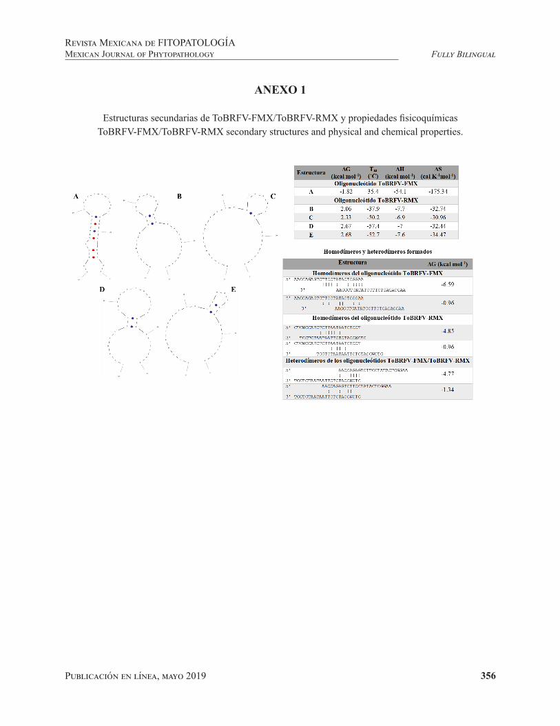
Publicación en línea, mayo 2019 356
Fully BilingualRevista Mexicana de FITOPATOLOGÍAMexican Journal of Phytopathology
ANEXO 1
Estructuras secundarias de ToBRFV-FMX/ToBRFV-RMX y propiedades fisicoquímicasToBRFV-FMX/ToBRFV-RMX secondary structures and physical and chemical properties.
357Publicación en línea, mayo 2019
Valadez-Ramírez P, Paz-Román J, Guzmán-González S, Buenrostro-Nava MT and Ochoa-Martínez DL. 2019. New records of Cucumber mosaic virus isolates and associated-satellite RNA in Colima, Mexico. Mexican Journal of Phytopathology 37(2): 357-364.DOI: 1018781/R.MEX.FIT.1901-2
Primera publicación DOI: 28 de Marzo, 2019.First DOI publication: March 28, 2019.
Resumen. El Cucumber mosaic virus (CMV) ocasiona una de las enfermedades virales más im-portantes a nivel mundial en plantas silvestres y cultivadas. En México son pocos los estudios que se han abordado con este virus, y dada su amplia gama de hospedantes e impacto económico, es ne-cesario contar con mayor información de su presen-cia y distribución en zonas de importancia agrícola como las del estado de Colima. En este trabajo, se reportan nuevos aislamientos del CMV identi-ficados por RT-PCR, secuenciación de DNA y su
New records of Cucumber mosaic virus isolates and associated-satellite RNA in Colima, Mexico
Nuevos registros de aislamientos del Cucumber mosaic virus y su RNA satélite en Colima, México
Pedro Valadez-Ramírez, Javier Paz-Román, Salvador Guzmán-González, Marco Tulio Buenrostro-Na-va, Facultad de Ciencias Biológicas y Agropecuarias, Universidad de Colima, Autopista Colima-Manzanillo Km 40, Tecomán, Colima, México, CP 28930; Daniel Leobardo Ochoa-Martínez*, Postgrado en Fitosanidad-Fitopatología, Colegio de Postgraduados, Km 36.5 Carretera México-Texcoco, Montecillo, Texcoco, Estado de México, CP 56230. *Autor para correspondencia: [email protected]
Recibido: 15 de Enero, 2019. Aceptado: 14 de Marzo, 2019.
Abstract. Cucumber mosaic virus (CMV) causes one of the most important viral diseases in wild and cultivated plants worldwide. Research on CMV in Mexico has been scarce, and due to the economic impact on plant health and wide host range, it is necessary to carry out studies on its presence and distribution in the agricultural geographical areas of the state of Colima. In this study, four CMV isolates identified by RT-PCR, DNA sequencing and phylogenetic analysis are reported: CMV-Vin from periwinkle (Catharanthus roseus), CMV-Chi from jalapeño pepper (Capsicum annuum), and CMV-Tom from saladette tomato (Solanum lycopersicum). In addition, the presence of CMV from cantaloupe melon (Cucumis melo) (CMV-Mel) was confirmed. The nucleotide sequences from CMV-Vin, CMV-Chi, and CMV-Mel clustered with CMV isolates from subgroup IB, while CMV-Tom clustered with members of subgroup IA. A satellite RNA sequence, without necrogenic domain, was detected only in CMV-Vin (satRNA Vin). This study reports for the first time the presence of CMV
Publicación en línea, mayo 2019 358
Fully BilingualRevista Mexicana de FITOPATOLOGÍAMexican Journal of Phytopathology
análisis filogenético: CMV-Vin en vinca (Catha-ranthus roseus), CMV-Chi en chile jalapeño (Cap-sicum annuum) y CMV-Tom en tomate saladette (Solanum lycopersicum). Se confirmó, además, la presencia del CMV en melón cantaloupe (Cucumis melo) (CMV-Mel). Los aislamientos CMV-Vin, CMV-Chi y CMV-Mel agruparon en el subgrupo IB, mientras que CMV-Tom agrupó en el subgrupo IA de CMV. De estos aislamientos, sólo CMV-Vin evidenció la presencia de un RNA satélite (satRNA Vin) sin dominio necrogénico. Este es el primer re-porte de la presencia del CMV en vinca, chile y tomate y de un RNA satélite en vinca en Colima, México.
Palabras clave: Caracterización de aislamientos de CMV, CARNA-5, RT-PCR, secuenciación, aná-lisis filogenéticos.
El Cucumber mosaic virus (CMV, Cucumovi-rus, Bromoviridae) es uno de los virus con mayor gama de hospedantes, incluyendo cucurbitáceas, solanáceas, cereales, leguminosas y ornamentales. Entre los síntomas que puede causar están: mosai-co, clorosis, necrosis, malformaciones foliares y enanismo, entre otros. También es posible que las plantas infectadas sean asintomáticas (Palukaitis et al., 1992). El CMV ha sido modelo de estudio de los diferentes mecanismos de interacción planta-patógeno, y es uno de los diez virus fitopatógenos de mayor importancia económica en el mundo (Ry-bicki, 2015).
La virosis por CMV es una limitante en la pro-ducción de cultivos en más de 18 estados de la Re-pública Mexicana y en algunos casos se reportan pérdidas superiores al 80% (Robles et al., 2010).
En el estado de Colima, el CMV se detectó en los cultivares de melón cantaloupe, ‘Durango’ y ‘Primo’ en el municipio de Tecomán (Orozco et al., 1994). Desde entonces se desconoce la existencia de
in periwinkle, pepper and tomato plants, and of an RNA satellite from periwinkle in Colima, Mexico.
Key words: CMV isolates, CARNA-5, RT-PCR, DNA sequencing, phylogenetic analyses.
The Cucumber mosaic virus (CMV, Cucumovirus, Bromoviridae) is one of the viruses with a wider host range that includes cucurbitaceae, solanaceae, cereals, legumes and ornamental plants. Some of the symptoms caused by CMV are mosaic, chlorosis, necrosis, foliar deformation and stunting, among others. Infected plants may also be asymptomatic (Palukaitis et al., 1992). CMV has been a study model of the different plant-pathogen interaction mechanisms, and one of the 10 most economically important phytopathogenic viruses worldwide (Rybicki, 2015).
Infections caused by CMV limit crop production in more than 18 states of Mexico, and in some cases losses of more than 80% have been reported (Robles et al., 2010).
CMV was detected in Durango and Primo cantaloupe melon cultivars grown in the municipality of Tecomán, state of Colima, Mexico (Orozco et al., 1994). Since then, no other hosts or subgroups of the virus are known nor its association with satellite RNAs (satRNA) in that location, so the objective of this study was to identify CMV isolates and their satRNA in plants in the state of Colima.
For that purpose, foliar tissue of cultivated plants and weeds with symptoms associated with CMV (i.e. mosaic, mottling, leaf filimorphism and deformation, as well as stunting, or a combination of them) was collected in the municipalities of Armería, Tecomán and Cuauhtémoc (Figures 1A and 1B). In the laboratory, the samples were washed with deionized water to remove impurities and then stored at -80 °C until their analysis.

Publicación en línea, mayo 2019 359
Fully BilingualRevista Mexicana de FITOPATOLOGÍA
Mexican Journal of Phytopathology
otros hospedantes o subgrupos del virus, así como su asociación con RNAs satélite (satRNA) en esta entidad federativa, por lo que el objetivo de este trabajo fue identificar aislamientos del CMV y su satRNA en plantas del estado de Colima, México.
Se recolectó tejido foliar de plantas cultivadas y malas hierbas con síntomas asociados al CMV (i.e. mosaico, moteado, filiformidad y deformación fo-liar, así como enanismo, o su mezcla) en los munici-pios de Armería, Tecomán y Cuauhtémoc (Figuras A y B). En el laboratorio, las muestras se lavaron con agua desionizada para eliminar las impurezas que pudieran estar presentes, y posteriormente se almacenaron a -80 °C hasta su procesamiento.
La extracción de RNA total se realizó a partir de 100 mg de tejido utilizando el Plant RNA Purifi-cation Reagent (Invitrogen, EE.UU.), siguiendo las recomendaciones del fabricante. El precipitado con el ácido ribonucleico se disolvió en 32 µL de agua tratada con 0.01% (v/v) de dietilpirocarbonato. La concentración de ácidos nucleicos se cuantificó con un espectrofotómetro NanoDrop 2000 (Thermo Scientific, EE.UU.) y se almacenó a -20 °C.
Posteriormente, se realizaron reacciones de transcripción inversa acoplada a reacción en cade-na de la polimerasa (RT-PCR) de punto final en un solo paso para detectar: a) un fragmento del gen endógeno 18S de aproximadamente 300 pb como indicativo de una adecuada calidad de los extractos con los oligonucleótidos descritos por Zamboni et al. (2008), b) un segmento del gen de la proteína de la cápside (CP) del CMV de ~500 pb con los oli-gonucleótidos reportados por Wylie et al. (1993), y c) satRNA con los oligonucleótidos reportados por Chen et al. (2011) que amplifican un fragmento de ~390 pb. Todas las reacciones de RT-PCR se lle-varon a cabo en volúmenes de 5 µL y consistieron de una mezcla del amortiguador de reacción 1X (0.2 mM de dNTPs y 1.2 mM de MgSO4), 0.5 µM de los oligonucleótidos correspondientes (sentido y
Total RNA extraction was performed using 100 mg of tissue with the Plant RNA Purification Reagent (Invitrogen, USA), following the manufacturer’s recommendations. The precipitate with ribonucleic acid was dissolved in 32 µL of treated water with 0.01% (v/v) diethylpyrocarbonate. The concentration of nucleic acids was quantified using a NanoDrop 2000 spectrophotometer (Thermo Scientific, USA) and stored at -20 °C.
Later on, reverse transcription reactions coupled to endpoint polymerase chain reaction (RT-PCR) were performed in one step to detect: a) a fragment of approximately 300 bp of the 18S endogenous gene which indicates that the extracts were of adequate quality with the oligonucleotides described by Zamboni et al. (2008); b) one ~500 bp segment of the CMV capsid protein gene (CP) with the oligonucleotides reported by Wylie et al. (1993); and c) satRNA with the oligonucleotides reported by Chen et al. (2011) that amplify a ~390 bp fragment. All the RT-PCR reactions were performed in 5 µL volumes and consisted of a mixture of 1X reaction buffer (0.2 mM of dNTPs and 1.2 mM of MgSO4), 0.5 µM of the corresponding oligonucleotides (sense and anti-sense), 0.6% (v/v) polyvinylpyrrolidone 40 (PVP40), 0.2 µL of the enzyme stock solution included in the SuperScript™ III One-Step RT-PCR System with Platinum Taq® High Fidelity kit (Invitrogen, USA) and an average concentration of 32 ng of each RNA extract.
The RT-PCRs were performed using a VeritiFast thermocycler (Applied Biosystems, USA) as follows: one 32-min cycle at 50 °C, one 2-min cycle at 94 °C, followed by 40 15-s cycles at 94 °C, 15-s cycles at 55 °C (for 18 S), 60 °C (for CMV CP) and 53 °C (for satRNA) and 15-s cycles at 68 °C; and one final 5-min cycle at 68 °C.
An extract of double-stranded RNA from Nicotiana glauca foliar tissue positive to CMV and

Publicación en línea, mayo 2019 360
Fully BilingualRevista Mexicana de FITOPATOLOGÍAMexican Journal of Phytopathology
antisentido), 0.6% (v/v) de polivinilpirrolidona 40 (PVP40), 0.2 µL de la solución madre de las enzimas contenidas en el kit de la SuperScript™ III One-Step RT-PCR System with Platinum Taq® High Fidelity (Invitrogen, EE.UU.) y una concentración prome-dio de 32 ng de cada extracto de RNA.
Cada RT-PCR se llevó a cabo en un termocicla-dor VeritiFast (Applied Biosystems, EE.UU.) con el siguiente programa: un ciclo de 32 min a 50 °C, un ciclo de 2 min a 94 °C, seguido de 40 ciclos de 15 s a 94 °C, 15 s a 55 °C (para 18 S), 60 °C (para CP del CMV) y 53 °C (para satRNA) y 15 s a 68 °C; y un ciclo final de 5 min a 68 °C.
Como control positivo para los diferentes en-sayos, se empleó un extracto de RNA bicatenario obtenido de tejido foliar de Nicotiana glauca po-sitivo a CMV y satRNA por RT-PCR y secuencia-ción. Por su parte, agua grado PCR y un extracto de RNA de tejido foliar de Catharanthus roseus, negativo a CMV y satRNA por RT-PCR, sirvieron como controles negativos.
Los productos de las RT-PCR se analizaron por electroforesis horizontal en geles de agarosa 1.5% (p/v) en amortiguador TAE 1X (tris-acetato 40 mM, Na2EDTA•2H2O 2 mM) a 80 V. El gel se tiñó con bromuro de etidio (0.5 µg/mL) y se expuso en luz UV, y la imagen resultante se obtuvo con un foto-documentador (MF-ChemiBIS-DNR Bio-Imaging Systems, Jerusalén, Israel).
Los amplicones correspondientes a la CP del CMV y satRNA de los distintos aislamientos ob-tenidos se purificaron con el Kit Wizard® SV Gel and PCR Clean-Up System (Promega, EE.UU.) siguiendo las instrucciones del fabricante, y su secuenciación se realizó por el método de Sanger en el Instituto de Neurobiología-Laboratorio de Proteogenómica de la Universidad Nacional Au-tónoma de México, Campus Juriquilla, Querétaro (http://www.inb.unam.mx/unidades/molecularya-nalitica/bios.html).
satRNA by RT-PCR and sequencing was used as a positive control in the different trials; as negative controls, PCR grade water and one RNA extract of Catharanthus roseus foliar tissue negative to CMV and satRNA by RT-PCR were used.
The RT-PCR amplifications were analyzed by horizontal electrophoresis in 1.5 % (w/v) agarose gels in a TAE 1X buffer (tris-acetate 40 mM, Na2EDTA•2H2O 2 mM) at 80 V. The gel was stained with ethidium bromide (0.5 µg/mL) and exposed to UV light; the resulting image was visualized in a photo-documentation system (MF-ChemiBIS-DNR Bio-Imaging Systems, Jerusalem, Israel).
The corresponding amplicons to CMV CP and satRNA of the different isolates obtained were purified with the Wizard® SV Gel and PCR Clean-Up System kit (Promega, USA), following the manufacturer’s instructions; the sequencing was performed in the Institute of Neurobiology’s Laboratory of Proteomics, Universidad Nacional Autónoma de México, Juriquilla campus at Querétaro (http://www.inb.unam.mx/unidades/molecularyanalitica/bios.html), using the Sanger’s method.
The sequences obtained in this study and other corresponding to the CP of different CMV subgroups (IA: U32859, U2281, D10538, Y18137, U43888, D00385, AJ276481 and AJ131623, IB: D42079, D28780, U20219 and X89652, and II: Y18138, U22822, Z12818, AB006813, AJ131620, AJ131621, L15336 and D00463) and satRNA (with necrogenic domain: D84389, D10039, U31660, D28559, DQ839133, X86421, X86425, D00542 and D10038; and without necrogenic domain: J02061, D10037, D42081, D42082, X86409, X86424, X54065 and X69136) reported worldwide, as well as the CP sequences of the Brome mosaic virus (BMV) (X58458) and the satRNA of the Peanut stunt virus (PSV) (NC_003855.1), which were used as external groups, were obtained from

Publicación en línea, mayo 2019 361
Fully BilingualRevista Mexicana de FITOPATOLOGÍA
Mexican Journal of Phytopathology
Las secuencias obtenidas en este estudio y otras correspondientes a la CP de diferentes subgru-pos del CMV (IA: U32859, U2281, D10538, Y18137, U43888, D00385, AJ276481 y AJ131623, IB: D42079, D28780, U20219 y X89652 y II: Y18138, U22822, Z12818, AB006813, AJ131620, AJ131621, L15336 y D00463) y satRNA (con do-minio necrogénico: D84389, D10039, U31660, D28559, DQ839133, X86421, X86425, D00542 y D10038 y sin dominio necrogénico: J02061, D10037, D42081, D42082, X86409, X86424, X54065 y X69136) reportados a nivel mundial, además de las secuencias de la CP del Brome mo-saic virus (BMV) (X58458) y RNA satélite del Peanut stunt virus (PSV) (NC_003855.1), las cua-les se usaron como grupos externos, se descargaron del GenBank-NCBI en formato fasta txt y se ali-nearon con Clustal W. La construcción de los árbo-les filogenéticos se realizó con MEGA6.0 (Tamura et al., 2013) usando el criterio óptimo de máxima verosimilitud con el modelo de sustitución T92, el cual se ajustó mejor a las secuencias, con 1,000 re-peticiones de bootstrap.
En total se recolectaron 30 muestras, 11 de ma-las hierbas: 2 de empanadilla (Commelina sp.), 2 de Cucurbita sp., 2 de amargosilla (Parthenium sp.), 2 de trébol (Trifolium sp.), 1 de malva silvestre (Mal-va sp.) y 2 de monacillo blanco (Herissantia sp.); 5 de plantas ornamentales: 3 de vinca (Catharanthus roseus) y 2 de azucena (Hippeastrum sp.); y 14 de plantas cultivadas: 3 de papayo (Carica papaya ‘CW-3’), 3 de tomate (Solanum lycopersicum Sala-det ‘Río Grande’), 3 de melón (Cucumis melo can-taloupe), 2 de chile (Capsicum annuum ‘Jalapeño’) y 2 de pepino (Cucumis sativus)]. De las 30 mues-tras, en 4 se amplificó la banda esperada de aproxi-madamente 500 pb, correspondiente a un segmento de la CP del CMV (Figura C). Dos aislamientos se obtuvieron de Tecomán: uno de melón (CMV-Mel) que mostraba mosaico en la localidad Caleras (19°
the GenBank-NCBI in fasta txt format and aligned with Clustal W. The phylogenetic trees were constructed using MEGA6.0 (Tamura et al., 2013) and the optimal maximum likelihood criterion and the T92 substitution model (which was best suited to the sequences) with 1,000 bootstrap repetitions.
A total of 30 samples were collected: 11 of weeds: 2 of empanadilla (Commelina sp.), 2 of Cucurbita sp., 2 of amargosilla (Parthenium sp.), 2 of clover (Trifolium sp.), 1 of common mallow (Malva sp.) and 2 of white bladdermallow (Herissantia sp.); 5 of ornamental plants: 3 of periwinkle (Catharanthus roseus) and 2 of white lily (Hippeastrum sp.); and 14 of cultivated plants: 3 of papaya (Carica papaya ‘CW-3’), 3 of tomato (Solanum lycopersicum Saladet ‘Río Grande’), 3 of melon (Cucumis melo cantaloupe), 2 of chili pepper (Capsicum annuum ‘Jalapeño’) and 2 of cucumber (Cucumis sativus)]. From the 30 samples, 4 amplified the expected band of approximately 500 bp corresponding to a segment of the CMV coat protein (CMV-CP) (Figure 1C). Two isolates were obtained in Tecomán: one of melon (CMV-Mel) showing mosaic (19° 02’ 40” N, 103° 54’ 04.9” W), and another of periwinkle (CMV-Vin) (18° 54’ 52” N, 103° 52’ 45.7” W) from plants showing mosaic and foliar deformation (Figures 1A and 1B), all collected in Caleras. The third isolate, detected on chili pepper (CMV-Chi), was collected in the municipality of Armería (18°55’36” N, 103°58’32.5’’ W) from foliar tissue showing yellowing, mosaic and foliar deformation. The fourth isolate was detected on tomato (CMV-Tom) in plants showing mosaic and foliar distortion collected in the municipality of Cuauhtémoc (19° 19’ 42” N, 103° 36’ 08.1” W).
The products size of the CP sequencing of the four CMV isolates obtained in this study ranged from 455 to 479 bp, with 94% and 97% nucleotidic

Publicación en línea, mayo 2019 362
Fully BilingualRevista Mexicana de FITOPATOLOGÍAMexican Journal of Phytopathology
02’ 40” N, 103° 54’ 04.9” O) y el otro de vinca (CMV-Vin) (18° 54’ 52” N, 103° 52’ 45.7” O) a partir de plantas con mosaico y deformación foliar (Figuras 1A y 1B). Un tercer aislamiento se iden-tificó de chile (CMV-Chi) en el municipio de Ar-
Figura 1. (A) Tejido foliar de melón cantaloupe y (B) vinca infectados con CMV y RNA satélite (en el caso de vinca) mostran-do mosaico y deformación foliar. (C) Producto amplificado de la CP del CMV y (D) RNA satélite por RT-PCR pun-to final en un solo paso. Carril 1: marcador de masa molecular de 100 pb (Promega®); Carril 2: control positivo (extracto de RNA bicatenario de Nicotiana glauca infectada con CMV y RNA satélite); Carril 3: control negativo (extracto de RNA de tejido foliar de Catharanthus roseus negativo a CMV y RNA satélite); Carril 4: control negati-vo (agua grado PCR); Carril 5: muestra de tomate (CMV-Tom); Carril 6: muestra de vinca (CMV-Vin); Carril 7: muestra de chile (CMV-Chi); Carril 8: muestra de melón (CMV-Mel). Análisis filogenético de (E) aislamientos de CMV y (F) RNA satélite obtenidos en Colima en comparación con secuencias nucleotídicas obtenidas de GenBank-NCBI.
Figure 1. (A) Cantaloupe melon and (B) periwinkle foliar tissue infected with CMV and satellite RNA; periwinkle foliar tis-sue showing mosaic and foliar deformation. (C) Amplified product of the CMV CP, and (D) satellite RNA by end-point one step RT-PCR. Lane 1: molecular weight marker of 100 bp (Promega®); Lane 2: positive control (extract of double-stranded RNA from Nicotiana glauca infected with CMV and satellite RNA); Lane 3: negative control (extract of RNA of Catharanthus roseus foliar tissue negative to CMV and satellite RNA); Lane 4: negative control (PCR grade water); Lane 5: tomato sample (CMV-Tom); Lane 6: periwinkle sample (CMV-Vin); Lane 7: chili pepper sample (CMV-Chi); Lane 8: melon sample (CMV-Mel). Phylogenetic analysis of (E) CMV isolates, and (F) satellite RNA collected in Colima compared with nucleotidic sequences obtained from the GenBank-NCBI.
identity with other isolates of the same virus reported in the GenBank database.
Three out of the four CMV isolates used in the present study showed a lower level of variability among them but were different from the tomato
F
E
A B C
D

Publicación en línea, mayo 2019 363
Fully BilingualRevista Mexicana de FITOPATOLOGÍA
Mexican Journal of Phytopathology
mería (18° 55’ 36” N, 103° 58’ 32.5’’ O) en tejido foliar con amarillamiento, mosaico y deformación. El cuarto aislamiento se detectó en tomate (CMV-Tom) procedente de Cuauhtémoc (19° 19’ 42” N, 103° 36’ 08.1” O), en plantas que mostraban mo-saico y distorsión foliar.
El tamaño de los productos de la secuenciación de la CP de los cuatro aislamientos del CMV obte-nidos en este estudio osciló de 455 a 479 pb, con porcentajes de identidad nucleotídica entre 94% y 97% con otros aislamientos reportados en la base de datos del GenBank de este mismo virus.
Tres de los cuatro aislamientos de CMV del presente estudio mostraron poca variabilidad entre sí, pero fueron diferentes del aislamiento de toma-te (CMV-Tom); es posible que se observe mayor variabilidad entre aislamientos si se incrementa el número de especies vegetales analizadas. De acuer-do al análisis filogenético, el CMV-Tom agrupó con aislamientos del subgrupo IA y más cercana-mente con un aislamiento de banano procedente de Colombia (U32859). CMV-Vin, CMV-Chi y CMV-Mel agruparon con el subgrupo IB (Figura E). Los aislamientos del subgrupo I se encuentran principalmente en zonas tropicales y subtropicales mientras que los del subgrupo II son prevalentes en zonas templadas (Palukaitis et al., 1992).
Sólo en el aislamiento CMV-Vin se detectó un RNA satélite (Figura D), con un tamaño estimado en 381 pb por secuenciación y con 91% de identi-dad nucleotídica con otros aislamientos del mun-do. Este satélite agrupó con otros aislamientos sin dominio necrogénico (X86424, X54065, X69136 y X86409) (Figura F). Este resultado muestra la necesidad de estudiar el comportamiento biológi-co del satRNA, particularmente la sintomatología que se observa en su presencia, así como su posible efecto en la transmisión del CMV por áfidos, entre otros aspectos.
isolates (CMV-Tom). A higher level of variability could be observed among isolates if the number of plant species analyzed increases. According to the phylogenetic analysis, the CMV-Tom was grouped with isolates of the IA subgroup, and more closely with a banana isolate from Colombia (U32859). CMV-Vin, CMV-Chi and CMV-Mel were grouped with the IB subgroup (Figure 1E). The subgroup I isolates are mainly found in tropical and subtropical zones, while the isolates from the subgroup II prevail in temperate zones (Palukaitis et al., 1992).
The CMV-Vin isolate was the only one containing a satRNA (Figure 1D) with an estimated size of 381 bp by sequencing and 91 % nucleotidic identity with other isolates of the world. The satellite was grouped with other isolates without necrogenic domain (X86424, X54065, X69136 and X86409) (Figure 1F). This result shows the need to study the satRNA’s biological behavior, particularly the symptoms observed when it is present, as well as its possible effect on CMV transmission by aphids, among other aspects.
Despite the low number of CMV isolates obtained and characterized in this study, our results confirmed the presence of CMV in the state of Colima and suggested that there is diversity among them. The present study suggest the need to continue analyzing a greater number of samples in order to deepen this observation.
In conclusion, three new CMV isolates are reported in the state of Colima, in chili pepper, tomato and periwinkle, the latter with a satellite RNA without necrogenic domain, as well as the presence of CMV in cantaloupe melon in the municipality of Tecomán.
End of the English version

Publicación en línea, mayo 2019 364
Fully BilingualRevista Mexicana de FITOPATOLOGÍAMexican Journal of Phytopathology
A pesar del bajo número de aislamientos del CMV obtenidos y caracterizados en este estudio, los resultados confirman la presencia de CMV en el estado de Colima y sugieren diversidad entre los mismos. El presente estudio sienta las bases para continuar con el análisis de un mayor número de muestras con la finalidad de profundizar sobre esta observación.
En conclusión, se reportan en el estado de Co-lima, tres nuevos aislamientos de CMV en chile, tomate y vinca, éste último con un RNA satélite sin dominio necrogénico. A su vez, se confirma la pre-sencia del CMV en melón cantaloupe en el munici-pio de Tecomán.
LITERATURA CITADA
Chen S, Gu H, Wang X, Chen J and Zhu W. 2011. Multiplex RT-PCR detection of Cucumber mosaic virus subgroups and Tobamoviruses infecting tomato using 18S rRNA as an internal control. Acta Biochimica et Biophysica Sinica 43:465-471. https://doi.org/10.1093/abbs/gmr031
Orozco SM, López AO, Pérez ZO and Delgadillo SF. 1994. Effect of transparent mulch, floating row covers and oil sprays on insect populations, virus diseases and yield of Cantaloup. Biological Agriculture and Horticulture 10:229-234. https://doi.org/10.1080/01448765.1994.9754675
Palukaitis P, Roossinck MJ, Dietzgen RG and Francki RIB. 1992. Cucumber mosaic virus. Advances in Virus Re-search 41:281-348. https://doi.org/10.1016/S0065-3527(08)60039-1
Robles HL, González FAC, Gill LEM, Pérez ML y López DJC. 2010. Virus fitopatógenos que afectan al cultivo de chile en México y análisis de las técnicas de detección. Tecnociencia Chihuahua 6:72-86. http://tecnociencia.uach.mx/numeros/.../numeros/v4n2/data/Virus_fitopato-genos_que_afectan_al_cultivo_del_chile_en_Mexico_y_analisis_de_las_tecnicas_de_deteccion.pdf
Rybicki EP. 2015. A top ten list for economically important plant viruses. Archives of Virology 160:17-20. https://doi.org/10.1007/s00705-014-2295-9
Tamura K, Stecher G, Peterson D, Filipski A and Kumar S. 2013. MEGA6: Molecular Evolutionary Genetics Analysis Version 6.0. Molecular Biology and Evolution 30:2715-2729. https://doi.org/10.1093/molbev/mst197
Wylie S, Wilson CR, Jones RAC and Jones MGK. 1993. A po-lymerase chain reaction assay for Cucumber mosaic virus in lupin seeds. Australian Journal of Agricultural Research 44:41-51. https://doi.org/10.1071/AR9930041
Zamboni A, Pierantoni L and De Franceshi P. 2008. Total RNA extraction from strawberry tree (Arbutus unedo) and se-veral other woody-plants. iForest 1:122-125. https://doi.org/10.3832/ifor0465-0010122