Revision Original Localisation in nervous system disorders...
11
61 Revision Original Neurosciences and History 2016; 4(2): 61-71 Localisation in nervous system disorders: examining the treatise written by Dr Xercavins in the late 19th century M. Balcells Department of Neurology. Hospital Universitari del Sagrat Cor, Barcelona, Spain. ABSTRACT In 1889, Dr Francisco de Paula Xercavins Rius (Sabadell, 1855 - Barcelona, 1937) published De la localización de las enfermedades del sistema nervioso, a painstaking review of the functions and localisations of the nervous system according to physiological knowledge at the time. Here, he examined sensitivity, motor function, and coordination at the levels of the spinal cord and brainstem, referring to the latter as the ‘spinal sub-apparatus’. The second part of the study, titled ‘the cortical sub-apparatus’, examined the function of the grey matter and its involvement in sensitivity, motor impulse, and intelligence. Xercavins showcased an exquisite knowledge of anatomy, especially that of the sensory and motor tracts of the central nervous system. He also included his own hypotheses and speculations regarding the seat of the higher functions. KEYWORDS Francisco de Paula Xercavins, history of neuroanatomy, nervous tracts, cerebral localisation, cerebral functions, 19th century Corresponding author: Dr Miquel Balcells E-mail: [email protected] Received: 28 June 2016 / Accepted: 15 July 2016 © 2016 Sociedad Española de Neurología Introduction Dr Francisco de Paula Xercavins Rius was born on 18 March, 1855, in Sabadell (Figure 1). He attended medical school in Barcelona and graduated in 1878. He earned his doctorate in 1882 in Madrid, with a dissertation on the nature and pathogenesis of puerperal processes. Xercavins worked in Hospital de la Santa Creu and in Casa de la Caridad, both in Barcelona. As a clinician, he treated both psychiatric and neurological entities, as was common in those days, and he showed interest in a variety of subjects, especially preventive medicine and the rehabilitation of the mentally ill. He founded an occupational therapy farm at the Institut Mental de la Santa Creu. Xercavins also published studies on preventing and treating the “social epidemics” affecting institutionalised mental patients and prisoners. In addition to the interests listed above, he was deeply involved in clinical neurology and neurological research. He organised a neurology clinic in which he used electrotherapy for different nervous diseases, and was one of Spain’s early proponents of ionisation and vibratory massage, both of which were typical treatments in those times. 1 His most distinguished works include La fisiología en los fenómenos psicológicos - Plan de distribución cerebral (1881) and La corea (mal de San Vito) en Barcelona y su provincia (1908). This article examines De la localización en las enfermedades del sistema nervioso. Sistemas medulares. Plan de distribución cerebral del autor – 1881. This treatise on the localisations of nervous diseases was presented at the International Congress of Medical Sciences held in 1881 in Barcelona. 2
Transcript of Revision Original Localisation in nervous system disorders...
61
Revision Neurosciences and History 2015; 3(3): 99-
Corresponding author: Dr Matthew LuedkeE-mail: [email protected]
Received: 6 marzo 2015 / Aceptado: 1 april 2015© 2015 Sociedad Española de Neurología
Original Neurosciences and History 2016; 4(2): 61-71
Localisation in nervous system disorders: examining the treatise written by Dr Xercavins
in the late 19th centuryM. Balcells Department of Neurology. Hospital Universitari del Sagrat Cor, Barcelona, Spain.
ABSTRACT
In 1889, Dr Francisco de Paula Xercavins Rius (Sabadell, 1855 - Barcelona, 1937) published De la localización delas enfermedades del sistema nervioso, a painstaking review of the functions and localisations of the nervous systemaccording to physiological knowledge at the time. Here, he examined sensitivity, motor function, and coordinationat the levels of the spinal cord and brainstem, referring to the latter as the ‘spinal sub-apparatus’. The second partof the study, titled ‘the cortical sub-apparatus’, examined the function of the grey matter and its involvement insensitivity, motor impulse, and intelligence. Xercavins showcased an exquisite knowledge of anatomy, especially that of the sensory and motor tracts of thecentral nervous system. He also included his own hypotheses and speculations regarding the seat of the higherfunctions.
KEYWORDSFrancisco de Paula Xercavins, history of neuroanatomy, nervous tracts, cerebral localisation, cerebral functions,19th century
Corresponding author: Dr Miquel BalcellsE-mail: [email protected]
Received: 28 June 2016 / Accepted: 15 July 2016© 2016 Sociedad Española de Neurología
Introduction
Dr Francisco de Paula Xercavins Rius was born on 18March, 1855, in Sabadell (Figure 1). He attended medicalschool in Barcelona and graduated in 1878. He earned hisdoctorate in 1882 in Madrid, with a dissertation on thenature and pathogenesis of puerperal processes.
Xercavins worked in Hospital de la Santa Creu and in Casade la Caridad, both in Barcelona. As a clinician, he treatedboth psychiatric and neurological entities, as was commonin those days, and he showed interest in a variety ofsubjects, especially preventive medicine and therehabilitation of the mentally ill. He founded anoccupational therapy farm at the Institut Mental de la SantaCreu. Xercavins also published studies on preventing andtreating the “social epidemics” affecting institutionalisedmental patients and prisoners.
In addition to the interests listed above, he was deeplyinvolved in clinical neurology and neurological research.He organised a neurology clinic in which he usedelectrotherapy for different nervous diseases, and was oneof Spain’s early proponents of ionisation and vibratorymassage, both of which were typical treatments in thosetimes.1
His most distinguished works include La fisiología en losfenómenos psicológicos - Plan de distribución cerebral(1881) and La corea (mal de San Vito) en Barcelona y suprovincia (1908). This article examines De la localizaciónen las enfermedades del sistema nervioso. Sistemasmedulares. Plan de distribución cerebral del autor – 1881.This treatise on the localisations of nervous diseases waspresented at the International Congress of MedicalSciences held in 1881 in Barcelona.2

62
The relationship between Jean-Martin Charcot (1825-1893) and Xercavins is well documented; upon readingXercavins’ book, Charcot sent him a letter congratulatinghim on his excellent description of anatomo-physiological correlations and urging him to use hisdiagrams as a teaching tool.1
Material and methods
This article examines Xercavins’ De la localización en lasenfermedades del sistema nervioso, published in 1889, aswell as contemporary original works to compare authors’anatomical knowledge.
M. Balcells
Figure 1. Francisco de P. Xercavins (1855-1937)
Results
Xercavins presents his manuscript in two principalsections: the ‘spinal sub-apparatus’ and the ‘cortical sub-apparatus’.
Spinal sub-apparatus
The first section presents the macroscopic anatomy ofthe spinal cord and brainstem in cross-sectional slices(Figure 2). The author states that slicing the spinal cordreveals the central grey matter with its anterior andposterior horns, and highlights the feature known as thecolumn of Clarke at the base of the posterior horn.
White matter rings these structures. Its most anteriorregion contains a small bundle of fibres. This is the directpyramidal tract, otherwise known as the bundle of Türck.Laterally, we observe the crossed pyramidal tract; the mostposterior area contains the direct cerebellar tract, or tractof Flechsig. Behind that, between the posterior horns, thewhite matter is divided into left and right sides by amedian sulcus. The more external white matter bundle isthe tract of Burdach, and the more internal bundle is thetract of Goll. The slice is also used to illustrate theradicular zones, which are linked to the 24 pairs of rootsemerging at the anterior horns of the spinal cord.
The slice at the level of the medulla oblongata reveals thepyramidal decussation, such grey matter structures as theolive, and nuclei of the lower cranial nerves. Differenttracts, extensions of those in the spinal cord, can beobserved in the white matter.
Grey matter in the pons corresponds to the nuclei ofcranial nerves. The white matter contains the pyramidaltracts and transverse fibres emanating from thecerebellum.
The slice at the level of the cerebral peduncles reveals thenucleus of the third cranial nerve and a grey matter formationdividing the peduncle in two parts: the anterior pespedunculi, and a superior portion, the head or tegmentum.
In this section, the author concludes that white matterabove this level is the continuation of that found in thecerebral peduncles. He also highlights the presence ofa series of central grey nuclei near the internal capsule,referring to the thalamus, lenticular nucleus, andcaudate nucleus.

Localisation in nervous system disorders
63
Next, we find the functions of the different bundleslocated in the spinal cord and brainstem. At brainstemlevel, the sensory pathways were recognised thanks toexperimental sectioning and to neuropathologicalfindings.
Xercavins states that unilateral destruction of the spinalcord, whether in an experimental model or caused bydisease, decreases motility on the same side as the lesion,and affects sensation on the opposite side; this is asummary of hemi-section lesion to the spinal cord,described by Brown-Séquard in his 1846 dissertation.3
Conservation of the vibratory and arthrokineticsensitivities was not mentioned. This was because Rumpfbegan his studies of vibratory sensitivity in 1889, afterXercavins had presented his lecture.4
Based on various studies, the author presented hisconclusions: “Sensory pathways are established in thespinal cord, from the posterior grey column to the centralgrey matter of the opposite side”.2(p14)
Since the tract of Goll degenerates in the ascendingdirection, it seems likely to have a sensory function.Nevertheless, clinical, histological, and experimental
Figure 2. Spinal cord. Transverse section, thoracic region. Drawn by Dr Xercavins
64
evidence refute this theory. The author’s conclusion wasthat “the tract of Goll is not responsible for transmittingsensory signals”.2(p14)
Turning to the outwardmost of the posterior columns,or tract of Burdach, which is directly connected to theposterior roots, the posterior horn, and the central greymatter, Xercavins linked it to altered sensitivity. Otherscholars disagreed; Dr Xercavins concluded thefollowing:
The tract named for Burdach allows for sensation tobe transmitted through its most external segment.It may link bundled grey matter cells with othershigher up, but it does not constitute the main sen-sory pathway toward the brain.2(p15)
He analysed the studies by Erb that showed howsensory signals travel along the posterior segment ofthe lateral column at the lumbar level; there is noevidence that this function exists at the level of thespinal segments in the upper thoracic and cervicalregion. The author concedes that “radicles thatpenetrate the posterior segment of the lateral columnenable sensory conduction, but they do not make thissegment the principal conductor”.2(p15)
On the other hand, studies by Vulpian state thathistological findings are necessary to demonstrate thatsensory stimuli are transmitted by the central greymatter of the spinal cord through the column of Clarke,which he regarded as the nucleus of the posterior rootsof the spinal cord. From the column of Clarke, thesensory stimulus would pass through the central greymatter to the opposite side of the lateral spinal tract,and along the posterior part of that tract in anascending direction.
Studies by such authors as Pierret, Vulpian, Brown-Séquard, Schiff, and others describe the transmission ofpain sensation through the posterior nuclei of the spinalcord and the lateral spinal bundle, but they do notconfirm that touch sensitivity follows the same path.Xercavins supported Wundt’s hypothesis that pain andother types of sensation are transmitted along the samecircuit.
Voroschilov’s experiments in slices of the posterior greymatter led to the conclusion that “transmission of spinalcord sensory currents can be verified through theposterior column; they reach the opposite side through
the connective central grey matter, and subsequentlyascend to the brain”.2(p16)
Sensitivity at the level of the medulla oblongata istransmitted by the gracile fasciculus and the cuneatefasciculus, which act as the continuation of the posteriorcolumns of the spinal cord. These fasciculi terminate inthe restiform bodies, which lead to the cerebellum. Basedon these macroscopic studies, the author deduces andaffirms the following: “The direct passage of sensationfrom the spinal cord to the brain cannot be verifiedthrough the restiform bodies”.2(p16)
In a later section, based on exclusion or on similarfindings in spinal cord slices, the author states that “thesensory fibres pass through the medulla oblongata bymeans of the external ventral grey matter on the floor ofthe fourth ventricle”.2(p17)
At the pontine level, the sensory conduction pathways arelocalised in posterior-exterior segments; the tractsbecome less compact as their fibres separate. Accordingto the text, “sensory fibres in the pons must be localisedin the posterior-exterior segments; somewhat separatedat the point of entry, they then condense to form thebundle of fibres at the other end known as the externalquarter of the cerebral peduncle”.2(p17)
The author observes the fibres that degenerate andcontinue ascending at the level of the cerebralpeduncles, and remarks that “the external or posteriorquarter of the foot of the cerebral peduncle isresponsible for sensory conduction ascending from theaxis of the spinal cord”.2(p17)
The study of the cerebral peduncles, and knowledge atthat time, led the author to conclude that thetegmentum contained the nucleus marking the originof the third cranial nerve. He also provides the locationof the corpora quadrigemina and the optic nerve nuclei;this nerve and the olfactory nerve were both consideredto feed into sensory pathways. Considering anatomicalknowledge at that time, the author was probablyunaware of the relationship between the second cranialnerve and the optic radiations, and of the intimal courseof the olfactory nerve.
The link between sensory pathways and ‘central nuclei’was clarified by the histological studies undertaken byMeynert and Flechsig, and by neuropathological studiesperformed by Vulpian and Charcot.
M. Balcells

Localisation in nervous system disorders
65
Xercavins concludes, “the posterior sixth of theinternal capsule, or lenticulo-optic segment, is aconduit for all the sensations from the opposite half ofthe body”.2(p18)
After examining studies by Meynert and Gratioletdescribing connections between the thalamus and thecerebral cortex, the author states,
Histologically speaking, the optical thalamus andthe direct posterior fasciculus should be consideredthe conduits for signals ascending from the spinalapparatus to the cortical apparatus.2(p19)
With additional mentions of authors including Cohn,Meynert, and Nothnagel, he concludes that “the opticalthalamus, according to clinical and experimentalfindings, does not form part of the motility system; thisis the central grey matter nucleus acting as anintermediary between the spinal sensory apparatus andthe cerebral grey layers”.2(p19) Xercavins concludes thathe does not know the function of sensory conductionin the tract of Goll. He does not specify the type ofsensation transmitted by the tract of Burdach, anderroneously states that the tract ascending from thelateral column (spinothalamic tract) is not the mainsensory path.
Motility system
Based on neuropathology studies providing evidence ofthe destruction of the brain’s central nuclei —the basalganglia— and the decussation of the pyramidal tracts inthe medulla, which carry motor impulses, Xercavinsstates that the motor pathway can be located by mappingdegeneration caused by lesions at the above sites in adescending direction.
He summarises findings by Charcot, Carville, and Duretas follows:
Common hemiplegia [the most frequent type] mayarise from total or partial lesion to the caudate orlenticular nucleus, or both at once; it may also resultfrom those located in the centrum ovale, or in thethree anterior sixths of the internal capsule and theinternal fourth of the pes pedunculi, always on theopposite side.2(p21-22)
Based on experiments and vivisections performed bysuch authors as Huguenin, Duret, Carville, and Hitzig, hededuced the presence of “a tract, known as the pyramidal
tract, which leaves the deep regions of the Hitzig zones,passes through the centrum ovale, and constitutes the 4thand 5th sixths of the internal capsule and the two middlefourths of the pes pedunculi; passing through the ponsand anterior pyramidal tracts of the medulla oblongata,it continues to become the lateral column of the spinalcord on the opposite side”.2(p23)
The author distinguishes between severe hemiplegia,causing extreme disability, exalted reflexes and delayedrigidity due to pyramidal tract lesion, and reversiblehemiplegia without rigidity owing to a lesion in the striatebodies.
At the spinal cord level, motor impulses are issued by theopposite side of the brainstem because of the medullarydecussation. In addition, we read that the motor impulseof the pyramidal tract is transmitted to the anteriorradicular zones.
The anterior internal column, known as the bundle ofTürck, degenerates as the result of lesion to thepyramidal tract before the level of the pyramidaldecussation; for this reason, Xercavins believed that thedirect pyramidal tract, a separate entity from the crossedpyramidal tract, had to contribute to the pyramid’smotor function.
The pyramidal tract is differentiated from the nerve rootzones by the lack of continuity between these structures;sectioning the pyramidal tract does not lead todegeneration of the root zones. As Xercavins concluded,referring to the pyramidal tract, “it does not transmitcerebral and spinal currents, but rather currents fromperipheral spinal nerves, to the peripheral nerve”.2(p25)
Examining the lateral column, located in the antero-lateral area of the spinal cord, shows that it becomesprogressively thinner until reaching the lumbar region;as such, its fibres terminate on the level of the rootsemerging from the anterior part of the spinal cord. Motorpathways therefore include cell bundles in the anteriorhorns of the spinal cord, from which the motor rootsoriginate.
The section on motility concludes with the statementthat “motility functions seem to be located in theantero-lateral segments of the spinal cord along its fulllength”.2(p27) Here, the author fails to mention theanterior horn, where the second motor neuron islocated.

66
The section on amyotrophy presents two postulates.Firstly, lesions to the cortex, centrum ovale, basalganglia, and pyramidal tract result in paralysis, notamyotrophy. Secondly, studies of progressive bulbarpalsy showed that paralysis was accompanied byamyotrophy due to lesion of the grey nuclei of motornerves located in the medulla oblongata.
Experiments by Hallopeau, Vulpian, Pitres, Hayem, andCharcot demonstrated that a spinal lesion accompaniedby muscle atrophy will always be located in the anteriorhorns of the spinal cord, on one of the anterior greycolumns.
This section ends with the summary statement that“every nerve involved in voluntary motility has its greymatter nucleus providing for the function and nutritionof the muscles to which it is distributed”.2(p26)
A lesion in the motor cortex, Hitzig zones, pyramidalbundle, and lateral column will give rise to musclecontracture in addition to paralysis, foot-clonus, orspinal epilepsy. Contracture is simply an exaggeratedform of physiological muscle tone.
The author cites the opinion of Vulpian and Charcot,who believed —but were not certain— that contracture(spasticity) originated in cells in the posterior spinalroots rather than in the pyramidal tract.
Since trismus and tetanus are caused by irritation ofsensory nerves, Xercavins states that “contracture is theexaggeration of physiological muscle tone resultingfrom a state of irritation that occurs in the sensoryportion of the diastaltic arches and affects the motorportions with which it joins, thus exciting the motorneurons of the spinal cord”.2(p26)
‘Diastaltic’ is a term coined by Cannon5 to describe thewave of intestinal contractions that is preceded by awave of inhibition.
A modulatory apparatus functions between the psychicimpulse and the muscle contraction: this is thecerebellum, which communicates with the thalamus,striate, and motor centres of the medulla oblongata.
Based on anatomoclinical findings by Hitzig, Meynert,Pierret, and others, our author concludes, “the cerebellarapparatus is responsible for overall coordination ofmotility...lesions, especially those affecting its peduncles,result in abnormal movements, whether of the eyes or
the entire body; and ataxic pronunciation [scanningspeech] in the absence of paralysis”.2(p34)
We read that when this modulatory system sustainsinjury, manifestations may affect the entire body, or elseisolated parts, and the author distinguishes betweencerebellar and spinal ataxia; ataxias of the latter typewere described by Charcot and Vulpian. In the author’s(translated) words, “the postero-exterior column, orcolumn of Burdach, is responsible for coordinating themovements of muscles innervated by the spinal cord;lesions to the internal segment in particular will resultin ataxia of the limbs”.2(p34-35) In the same paragraph, theauthor links the tract of Goll to arthrokinetic sensitivity,although this function was denied in earlier paragraphs.
Pierret, Charcot, and Vulpian expressed doubt as towhether the tract of Goll forms part of the cerebellum.As for the tract of Flechsig, both Xercavins and Vulpianbelieved it to originate in the column of Clarke.Experimental studies revealed that the tract of Flechsigdegenerates in the ascending direction and terminatesin the cerebellum. Its function is to contribute tocoordinating movement by transmitting informationfrom the muscles to the cerebellum.
Although his doubts remained, Xercavins accepted theopinions of the authors named above and stated that thecerebellum, with all of its connections, “should beregarded as the organ of motor coordination, andlesions to its components result in the numerous typesof ataxia”.2(p36)
With respect to localising the laterality of each function,the author explains the decussation of the sensorypathways at the level of the medulla oblongata, pons,cerebral peduncles, and internal capsule; thisconfiguration explains why a spinal lesion at any levelwill result in anaesthesia on the opposite side.
As for longitudinal localisation, the author describescases that match what are now known as alternatesyndromes; at the level of the cerebral peduncle, thelesion gives rise to third nerve palsy on the side of thelesion and hemiplegia on the opposite side, as Weberobserved.
Cortical sub-apparatus
Xercavins establishes the anatomical boundaries of thecortex by describing the grey basal ganglia as the
M. Balcells

Localisation in nervous system disorders
67
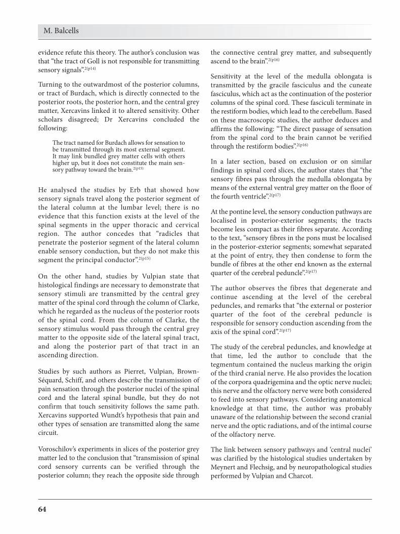
intermediate region between the spinal cord and thehigher cerebral functions (Figures 3 and 4).
Whereas spinal grey matter appears in nuclei andbundles, cerebral grey matter is arranged in layers. Thefunction of the grey matter in the cerebral cortex is toorganise and generate motor impulses, receive sensorystimuli, and establish their type or modality, and lastly,be responsible for intellectual activity.
Motor impulses pass through the bundle of Charcot (thepyramidal tract), and through the basal ganglia. Sensory
stimuli pass from the spinal cord to the thalamus, andfrom that location to the cortex.
The classic, rigid view of functional localisation holdsthat intelligence is located in the frontal lobe,sensitivity in the occipital lobe, and motility in theparietal lobe. In contrast with the above, new clinicalfindings and pathology studies show that the differentlobes of the brain each have multiple functions.Xercavins believed that the different lobes and gyricontributed, to a greater or lesser extent, to all of thebrain’s functions.
Figure 3. Antero-posterior view of the brain with its central nuclei and internal capsule
68
The teachings of his time established an analogy betweenthe columns of the spinal cord and the layers of the brain,stating that the more peripheral layers acted as thecontinuation of posterior columns of the spinal cord. Theanterior columns of the spinal cord extend to become thedeep layers of the brain.
As a newly qualified doctor in 1881, Xercavinssummarised the medical knowledge of his time in his“map of the distribution of cerebral functions” andsupported it with the latest clinical and experimentalfindings. His references are a testament to his curiosityand commitment to continuing education. In his laterwork, he modified that plan and presented his new ideasin two sections:
- The peripheral grey matter layers of the cerebral cortexconstitute the seat of sensitivity. They are also home to“common sense”. The deep layers of the cerebral cortexgenerate motility impulses, which are transmitted to thespinal cord through the bundle of Charcot (pyramidaltract) and the corticostriatal fibres. Intellectual activitytakes place across the length and breadth of theintermediate layers of the cortex.
- Both the peripheral layers, which receive sensoryinformation, and the deep layers which give rise tomotility are regarded as consisting of cell groups orterritories. The intermediate layers that are the seat of
intellectual functions are also laid out in small segmentsor ‘brainlets’.
My conclusions, based on the above, are as follows:1. Elements of sensitivity, intelligence, and motilityare to be found in all lobes of the brain.2. Elements of sensitivity, intelligence, and motilitymust be located in the different layers that make upthe cerebral cortex, from the most peripheral to thedeepest.3. The brain contains lobes and gyri, and its manylayers have been shown to be associated with fin-dings of sensitivity, intelligence, and motor impulsescorresponding to a specific organ or region of thebody. These regions are represented within the cere-bral cortex.2(p54)
Histological studies show that the posterior column of thespine reaches the internal capsule and continues throughthe direct posterior bundle of Gratiolet, which terminatesin the occipital lobe. The author believed, although notwithout many doubts, that this bundle transmitted visualstimuli, which is why other authors referred to the samestructure as the optical bundle.
Based on studies by Betz and Tarchinoff, he concludes:
Throughout the cortical grey matter, one finds sen-sory cells occupying the peripheral layers, but espe-cially the middle layers; these cells are also dominantin the post-Rolandic area, that is, the posterior lobesabove the anterior ones [here, the author refersmainly to the parietal lobe].2(p56)
Questions remained as to whether fibres from thepyramidal tract project to the striatum; Charcot hadshown that degeneration of the pyramidal tract reachedthe spinal cord with no alterations in the striate nucleus,but German authors, including Gudden, were doubtful.Dr Xercavins was more inclined to agree with Charcot’sversion.
Nevertheless, Flechsig’s studies were able to demonstratethat cortical fibres from all cerebral lobes reach the basalganglia, and that the latter transmit motor impulses to thespinal cord, although they did not specify how they mightbe related to the pyramidal tract.
At a later date, studies by Betz and Meinart showed thatmotor cells were found in all deep layers throughout thecerebral cortex, and especially in the ascending, frontal,parietal, and paracentral convolutions, otherwise knownas the Hitzig psychomotor centres. These cells are lessabundant in the deep post-Rolandic layers.
M. Balcells
Figure 4. Brain, internal surface

Localisation in nervous system disorders
69
On the subject of whether or not there may be cellsspecifically responsible for intellectual function, theauthor subscribes to Meynert’s hypothesis: the cerebralcortex is made up of five layers, and intellectual functionsreside in the intermediate layers.
Xercavins concludes the following:
Histology tends to assign the localisation of inte-llectual operations to the intermediate layers of thebrain’s entire surface; however, since the interme-diate layers are predominant in the frontal lobe,those operations may be more centred in thatlobe.2(p58)
Citing experimental studies by Kussmaul, Nothnagel, andGoltz, who employed fine cuts to lesion specific parts ofthe cortex, the author deduces that “there are no purelymotor gyri; the convolutions that are mainly involved inmotor function, or Hitzig centres, are also involved insensory processing”. Goltz had concluded that “thealterations that follow destruction of a specific sitedepend not on its location, but rather the degree or depthof the lesion”.2(p59)
His reading of experimental studies by differentresearchers led the author to conclude that there was nosuch thing as a pure motor or pure sensory convolution:“the patient will present motor or sensory deficitaccording to whether the lesion is deep or shallow”.
The psychic impulse that initiates movement, and inwhich both sensory and motor centres participate,logically begins in cells in the intermediate layers; inother words, “cells linked to intelligence must bebetween those for sensitivity and those for voluntaryimpulse”.
Clinical cases
The author’s review of clinical studies of cerebrovasculardisease allowed him to conclude that cortical lesionswere rare compared to lesions in the central nuclei[basal ganglia].
His conclusion reads as follows: “clinical studies confirmthat since pathological processes of different types affectthe cortical or deep tissue of the lobes of the brain, theyare also accompanied by alterations in sensitivity andmotility”.2(p60)
Studies of cerebral vascularisation had shown that thecortex is irrigated by short vessels, whereas the centrumovale and basal ganglia are irrigated by long vessels. Thisexplains why cortical haemorrhages are unusual, whereascerebral softening due to thrombosis or embolism iscommon; these cortical lesions appear as small wedges inthe area irrigated by capillaries.
Haemodynamic factors account for the low incidence ofdiseases affecting the anterior and posterior cerebralarteries; the middle cerebral artery is directly impactedby the systolic component, which results in a higherincidence of vascular accidents.
He also mentions the poor circulation in the distalsegments of the three cerebral arteries; in these outlyingareas, this means that cerebral softening manifests underconditions of atony of the circulatory system.
The section ends as follows:
Lesions in the striate bodies and corresponding por-tion of the internal capsule result in the commontype of apoplexy, or cerebral hemiplegia. Thoseaffecting the cortex are characterised by paralysesthat are sometimes partial but can become genera-lised as series of attacks occur; these coincide withspasms in the paralytic regions and cerebral epi-lepsy, followed later by delayed rigidity. However,intelligence is often spared, and rapid recoveries aresometimes seen.2(p62-3)
Histological, experimental, and clinical data
Using histology, experiments, and clinical practice as hisbasis, Xercavins puts forth the possibility of motor andintellectual functions coinciding in the same area of thebrain. After presenting this functional topography, hedescribes the brain as a confederation of smaller braincentres from which sensory, intellectual, and motorfunctions originate.
Results from experimental studies by authors includingHitzig and Ferrier, and from the autopsy studies carriedout by Wernher and Charcot, suggest that thepsychomotor centres are located in the ascending frontaland parietal gyri, and in the paracentral lobe. Theconclusion put forth is that “the centres of Hitzig shouldin a general sense be regarded as the cerebralpsychomotor localisations of the upper and lower limbson the opposite side of the body”.2(p65)
70
Experiments undertaken by Ferrier had shown thatpsychomotor functions could also exist in a morelimited form. One example cited by Xercavins focuseson the changes in eye movements due to occipital lobelesions. With this in mind, he concedes that “the Hitzigconvolutions are psychomotor centres correspondingnot to the body as a whole, but rather to specificregions. There must therefore be other brain centrescontrolling the remaining peripheral musclegroups”.2(p65)
Another confirmed localisation was Broca’s area forspeech, in the brain’s left hemisphere. Xercavinsmentions that aphasia may affect left-handedindividuals, with exceptions:
The centre of speech is firmly located in the oper-culum, that is, the foot of the third frontal gyrus.If the left hemisphere is the most functional, thecentre will reside on the left side...nevertheless, inthe left-handed or those with a dominant righthemisphere, speech function will be located in theright hemisphere.2(p68)
Broca’s area is specific only to speech and not othermodes of language; Trousseau, Laborde, and othersbelieve that there are distinct areas for written languageand sign language. This indicates that intelligencewould be present at other localisations. “Therefore, inagraphia, alexia, and amimia, the pathologicallocalisation is not that found in the centre of trueaphasia; rather, it is in the centres that innervate thearm, and nerves involved in vision and signlanguage”.2(p68) Here, the author is critical of the strictview of localisation of cerebral functions, and even ofthe idea of the dominant hemisphere.
At the same time, other modes of language resist theidea of strict functional partitioning within the brain;both alexia and amimia require a far more complexlevel of intellectual and motor organisation.
The evolution of the brain is cited to account for therole of hearing in language acquisition; the circuitpassing through the auditory centre is linked to thespeech centre, and the two centres are linked byintelligence that modulates and attaches meaning tospoken language.
Since the auditory centre is located in the first temporalgyrus, “the superior temporal gyrus is recognised as theseat of hearing, and the third frontal gyrus, with speech;the insula is associated with disposition in that it
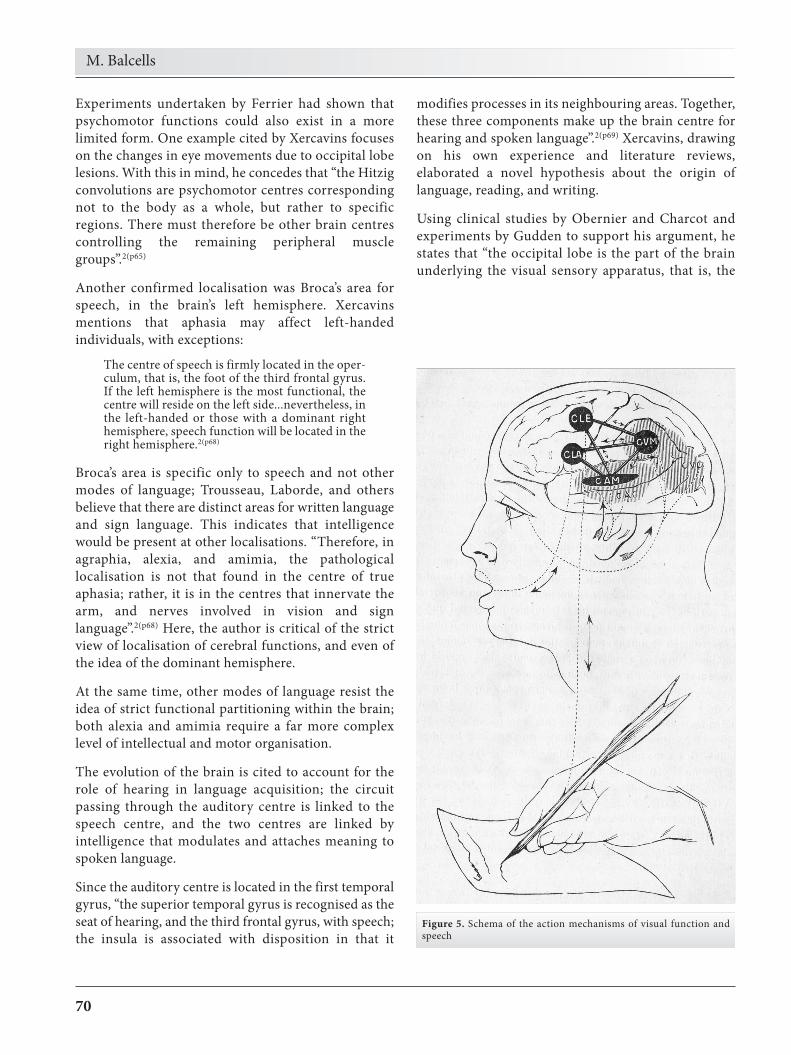
modifies processes in its neighbouring areas. Together,these three components make up the brain centre forhearing and spoken language”.2(p69) Xercavins, drawingon his own experience and literature reviews,elaborated a novel hypothesis about the origin oflanguage, reading, and writing.
Using clinical studies by Obernier and Charcot andexperiments by Gudden to support his argument, hestates that “the occipital lobe is the part of the brainunderlying the visual sensory apparatus, that is, the
M. Balcells
Figure 5. Schema of the action mechanisms of visual function andspeech

Localisation in nervous system disorders
71
intellectual disorder known as psychic blindness andits effects on related muscles”.2(p71)
The last paragraph of the study describes the completecycle of brain activity from a sensory stimulus to simplemotor responses such as those involved in musclereflexes, or even to higher activities such as interpretingvision or writing.
Using vision as an example, he presents an incompleteview of the visual pathways beginning with the stimulusto the retina and reaching the cortex of the occipitallobe after passing through the optical nerve, peduncles,internal capsule, posterior segment of the thalamus,bundle of Gratiolet, and occipital cortex.
The author does not mention the optic chiasm, visualpathway, corpora quadrigemina, or external geniculatebody. From this reading, it can be interpreted that thepathway crosses or connects to the peduncles and theinternal capsule.
Dr Xercavins once again presents his hypothesis onspeech and visual function, which was based on PierreMarie’s article reviewing aphasia and agraphiaaccording to the Charcot hypothesis.6 He includes a linedrawing of the action mechanism behind thesefunctions (Figure 5).
We posit that there is a common visual space thatdepends on a higher visual centre for words. An injury tothe first centre gives rise to word blindness (whenreading). An injury to the higher centre (interpretive)results in psychic blindness. The same concept can beapplied to auditory function: there is a common auditorycentre which is subordinate to a higher centre thatprocesses words. Lesions at these locations produce worddeafness and psychic deafness respectively; the latter termrefers to Wernicke or sensory aphasia.
The localisation for a lesion producing verbal ataxia,referring to Broca’s aphasia, is at the level of the insula.An interrupted connection between the psychic auditoryarea and the insula would produce transitional orrelational aphasia.
Alexia and agraphia are the product of a disconnectionin the higher intellectual centres. The visuospatial
centre is affected in alexia, whereas psychomotorcoordination is impaired in agraphia, although otherhand functions are preserved. The conclusion reads,“the occipito-temporal lobes display not only sensoryfunctions, but also intelligence, and the Hitzig centresdisplay functions of intelligence and volition inaddition to motility”.2(p73)
The study concludes that the cerebral localisation ofmultiple functions remains unknown; likewise, there aremany gyri not known to have specific functions.
Discussion
This treatise by Dr. Xercavins reveals his in-depthknowledge of anatomy, particularly of the sensory andmotor pathways of the central nervous system; the longlist of authors he cites is proof of his thorough and up-to-date knowledge of the literature published in his time.The author presents his own hypotheses and conjecturesregarding the seat of the higher functions, especiallylanguage, reading, and writing.
Conflicts of interest
The author has no conflicts of interest to declare.
References
1. Xercavins X. La obra del doctor Francisco de P. XercavinsRius 1855-1937 [dissertation]. Barcelona: Universidad deBarcelona; 1976.
2. Xercavins F. De la localización en las enfermedades del sis-tema nervioso. Sistemas medulares. Plan de distribucióncerebral del autor – 1881. Barcelona: Imp. J. Balmas; 1889.
3. Brown-Séquard CE. Recherches et expériences sur la physio-logie de la moelle épinière [dissertation]. Paris: Rignoux;1846.
4. Calne DB, Pallis CA. Vibratore sense: a critical review. Brain.1966;89:723-46.
5. Diccionario Médico. Barcelona: Salvat; 1972. Diastáltico; p.137.
6. Castells F. De la afasia en general y de la agrafia en particularsegún la enseñanza del profesor Charcot. Gaceta MédicaCatalana. 1889;288:365-71.