ReviewArticle - Semantic Scholar have been reports in the literature of patients ... Ginger ......
15
Hindawi Publishing Corporation Dermatology Research and Practice Volume 2013, Article ID 279289, 15 pages http://dx.doi.org/10.1155/2013/279289 Review Article Newer Hemostatic Agents Used in the Practice of Dermatologic Surgery Jill Henley 1 and Jerry D. Brewer 2 1 College of Osteopathic Medicine Glendale, Midwestern University, 13989 N59th Avenue, Glendale, AZ 85308, USA 2 Division of Dermatologic Surgery, Department of Dermatology Mayo Clinic, Mayo Clinic College of Medicine Rochester, 200 First Street SW, Rochester, MN 55905, USA Correspondence should be addressed to Jerry D. Brewer; [email protected] Received 7 June 2013; Accepted 7 July 2013 Academic Editor: Giuseppe Argenziano Copyright © 2013 J. Henley and J. D. Brewer. is is an open access article distributed under the Creative Commons Attribution License, which permits unrestricted use, distribution, and reproduction in any medium, provided the original work is properly cited. Minor postoperative bleeding is the most common complication of cutaneous surgery. Because of the commonality of this complication, hemostasis is an important concept to address when considering dermatologic procedures. Patients that have a bleeding diathesis, an inherited/acquired coagulopathy, or who are on anticoagulant/antiplatelet medications pose a greater risk for bleeding complications during the postoperative period. Knowledge of these conditions preoperatively is of the utmost importance, allowing for proper preparation and prevention. Also, it is important to be aware of the various hemostatic modalities available, including electrocoagulation, which is among the most effective and widely used techniques. Prompt recognition of hematoma formation and knowledge of postoperative wound care can prevent further complications such as wound dehiscence, infection, or skin-graſt necrosis, minimizing poor outcomes. 1. Introduction Dermatologists are estimated to perform over 3.9 million procedures each year [1]. Although the risks and complica- tions of dermatologic surgery are generally very low, even the most talented surgeon can experience complications related to hemostasis during both the intraoperative and postop- erative periods. Minor bleeding complications are the most frequently encountered complications of cutaneous surgery, which can predispose the patient to hematoma formation, increased risk of infection, skin graſt necrosis, and wound dehiscence. is chapter will highlight proper hemostasis technique to prevent complications. 2. Overview of Hemostasis By understanding the mechanism behind the physiologic clotting system, it is easier to understand how the different hemostatic agents work in the body. e body’s primary response to injury is reflex vasoconstriction of the blood vessels in the surrounding tissues, followed by formation of the platelet plug and activation of the fibrinolytic clotting cascade. ere are two separate pathways of the fibrinolytic clotting system, that lead to the final common pathway and formation of the insoluble fibrin clot. Function of the body’s hemostatic system can be monitored by various laboratory tests. ese tests can be helpful to assess the degree of anticoagulation in patients on antiplatelet and anticoagulant medications or who have inherited coagulopathies before proceeding with dermatologic procedures (see Table 1). 3. Preoperative Evaluation One of the most important steps that the dermatologist can take to prevent bleeding complications is to gain a thorough preoperative history of the patient before performing any kind of dermatologic procedure. is allows the physician to gain a better understanding of the patient’s overall health and should include a detailed history of the patient’s comor- bidities, prior surgeries including complications, current medications, social history, and family history, which can help reveal any potential bleeding diatheses.
Transcript of ReviewArticle - Semantic Scholar have been reports in the literature of patients ... Ginger ......

Hindawi Publishing CorporationDermatology Research and PracticeVolume 2013, Article ID 279289, 15 pageshttp://dx.doi.org/10.1155/2013/279289
Review ArticleNewer Hemostatic Agents Used in the Practice ofDermatologic Surgery
Jill Henley1 and Jerry D. Brewer2
1 College of Osteopathic Medicine Glendale, Midwestern University, 13989 N59th Avenue, Glendale, AZ 85308, USA2Division of Dermatologic Surgery, Department of Dermatology Mayo Clinic, Mayo Clinic College of Medicine Rochester,200 First Street SW, Rochester, MN 55905, USA
Correspondence should be addressed to Jerry D. Brewer; [email protected]
Received 7 June 2013; Accepted 7 July 2013
Academic Editor: Giuseppe Argenziano
Copyright © 2013 J. Henley and J. D. Brewer. This is an open access article distributed under the Creative Commons AttributionLicense, which permits unrestricted use, distribution, and reproduction in any medium, provided the original work is properlycited.
Minor postoperative bleeding is the most common complication of cutaneous surgery. Because of the commonality of thiscomplication, hemostasis is an important concept to address when considering dermatologic procedures. Patients that have ableeding diathesis, an inherited/acquired coagulopathy, or who are on anticoagulant/antiplatelet medications pose a greater risk forbleeding complications during the postoperative period. Knowledge of these conditions preoperatively is of the utmost importance,allowing for proper preparation and prevention. Also, it is important to be aware of the various hemostatic modalities available,including electrocoagulation, which is among the most effective and widely used techniques. Prompt recognition of hematomaformation and knowledge of postoperative wound care can prevent further complications such as wound dehiscence, infection, orskin-graft necrosis, minimizing poor outcomes.
1. Introduction
Dermatologists are estimated to perform over 3.9 millionprocedures each year [1]. Although the risks and complica-tions of dermatologic surgery are generally very low, even themost talented surgeon can experience complications relatedto hemostasis during both the intraoperative and postop-erative periods. Minor bleeding complications are the mostfrequently encountered complications of cutaneous surgery,which can predispose the patient to hematoma formation,increased risk of infection, skin graft necrosis, and wounddehiscence. This chapter will highlight proper hemostasistechnique to prevent complications.
2. Overview of Hemostasis
By understanding the mechanism behind the physiologicclotting system, it is easier to understand how the differenthemostatic agents work in the body. The body’s primaryresponse to injury is reflex vasoconstriction of the bloodvessels in the surrounding tissues, followed by formation of
the platelet plug and activation of the fibrinolytic clottingcascade. There are two separate pathways of the fibrinolyticclotting system, that lead to the final common pathway andformation of the insoluble fibrin clot. Function of the body’shemostatic system can be monitored by various laboratorytests. These tests can be helpful to assess the degree ofanticoagulation in patients on antiplatelet and anticoagulantmedications or who have inherited coagulopathies beforeproceeding with dermatologic procedures (see Table 1).
3. Preoperative Evaluation
One of the most important steps that the dermatologist cantake to prevent bleeding complications is to gain a thoroughpreoperative history of the patient before performing anykind of dermatologic procedure. This allows the physicianto gain a better understanding of the patient’s overall healthand should include a detailed history of the patient’s comor-bidities, prior surgeries including complications, currentmedications, social history, and family history, which canhelp reveal any potential bleeding diatheses.

2 Dermatology Research and Practice
Table 1: Overview of hemostasis.
Stages of hemostasis Physiology MonitoringPrimary hemostasis
Formation of the platelet plug
Platelets first adhere to the exposed collagen and von Willebrand’s factor on thesubendothelium. Then, circulating stimuli activate the platelets, causing shapechanges in the platelets [2]. Upon activation, platelet receptors get transferred to thesurface, allowing for platelet aggregation. Platelets then release granules thatstimulate further platelet aggregation and vasoconstriction [2, 3].
BT, PFA-100analysis
Secondary hemostasis
Intrinsic pathwayPlasma proteins get activated in contact with negatively charged surfaces, leading toactivation of factor XII and other clotting factors, ultimately leading to the finalcommon pathway and formation of the fibrin clot [4].
aPTT
Extrinsic pathwayDamaged endothelium exposes tissue factor, activating the extrinsic pathway,leading to thrombin production, and activation of other clotting factors, ultimatelyleading to the final common pathway and formation of the fibrin clot [2, 4].
PT
Final common pathwayBoth pathways lead to activation of factor X, which converts prothrombin intothrombin. Thrombin leads to formation of the insoluble fibrin clot, by convertingfibrinogen into fibrin [5]. The clot is then stabilized by factor XIII [6].
Abbreviations: ADP: adenosine diphosphate, aPTT: activated partial thromboplastin time, BT: bleeding time, PFA-100 analysis: platelet function analysis, PT:prothrombin time, and TXA2 : thromboxane A2.
Specifically, questions regarding comorbid illnesses thatcan lead to coagulopathies, such as chronic renal and liverdisease, hematologic disorders, and malignancies, should beasked, in addition to prior diagnoses of inherited bleedingdisorders such as von Willebrand’s disease and hemophilia[7]. Many mild forms of bleeding disorders go undiag-nosed until later in life and should be investigated bythorough questioning of bleeding complications in priorminor surgical procedures (dental/oral surgery), prolongedepisodes of epistaxis, menorrhagia, bruising, history ofprior blood transfusions, and any family history of bleed-ing disorders. On physical exam, it is important to lookfor any indications of hemostatic abnormalities such asincreased bruising or petechiae. For more details on howto manage a patient with a bleeding disorder, please seethe section titled, “Approach to the Patient with a BleedingDisorder.” It is also important to investigate the patient’scardiac history including the presence of a pacemaker orimplantable cardiac defibrillator (estimated that 4% of Mohspatients are estimated to have one), because the use ofcertain electrosurgical agents used for hemostatic purposesmay be prohibited in these patients [8]. Also, alcohol is anatural anticoagulant, and obtaining information regardingconsumption could be beneficial as part of the preoperativeevaluation.
4. Pharmacologic Agents and TheirEffects on Anticoagulation
Many patients that are going to have dermatologic surgery areon anticoagulant and antiplatelet medications. One questionmany healthcare providers face prior to cutaneous surgeryis whether to continue anticoagulation medications prior tosurgery. The discrepancy lies between keeping the patient
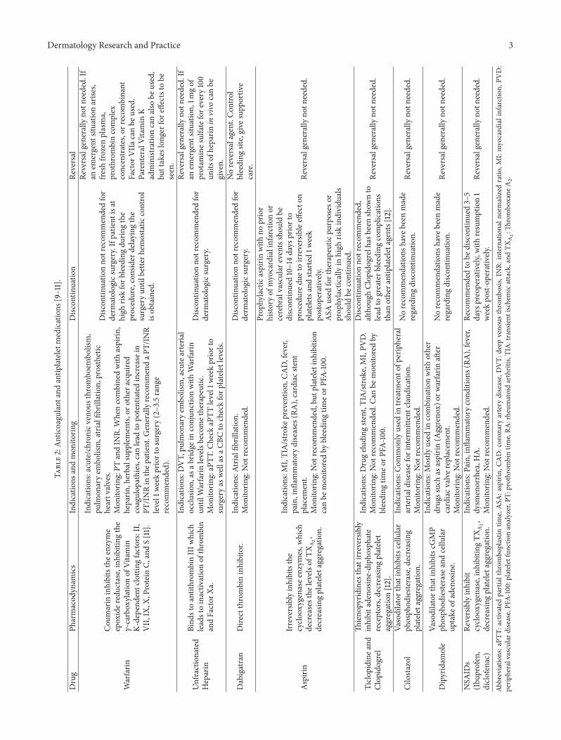
on their current medication regimen, potentially increasingthe patient’s risk for bleeding complications during the peri-operative period, or discontinuing their medication, whichhas now been proven to increase the patient’s risk of life-threatening thromboembolic events during the postopera-tive period. For a list of some of the most widely usedanticoagulant and antiplatelet medications today, includingpharmacodynamics of the different medications, and variousrecommendations regarding usage during the perioperativeperiod see Table 2.
Due to several clinical studies conducted in the past tenyears, the general consensus between dermatologic surgeonshas been to continue patients on their anticoagulant medica-tions preoperatively, because the benefit of these medicationssignificantly outweighs the risk of bleeding complicationsduring or after procedure [1, 9, 12–18]. The overall risk ofhemorrhagic complications in cutaneous surgeries, such ascontinuous bleeding or hematoma formation in a patient whois not on anticoagulant medications, is very low (1.4%) [18].
In a 2005 nationwide survey of Mohs surgeons, 66%were found to continue Warfarin during the perioperativeperiod [19]. Some studies have shown that there is anincrease in minor bleeding complications for patients takingWarfarin chronically, which includes:minor bleeding definedas bleeding less than 24 hours postoperatively, hematomaformation, bleeding that is controlled in the office setting,and bleeding controlled with manual compression [1, 20].For patients taking Warfarin, checking the internationalnormalized ratio (INR) within 48 hours to one week prior tothe surgery can give the surgeon a better idea of the currentmagnitude of anticoagulation. It is generally recommendedthat the INR level be within the therapeutic range of 2–3.5 preoperatively [21]. There is a suggestion that the higherthe INR level (especially >3.5), the higher the risk forhemorrhagic complications [15]. Because minor bleeding can
Dermatology Research and Practice 3Ta
ble2:Anticoagu
lant
andantip
lateletmedications
[9–11].
Drug
Pharmacod
ynam
ics
Indicatio
nsandmon
itorin
gDisc
ontin
uatio
nRe
versal
Warfarin
Cou
marin
inhibitsthee
nzym
eepoxider
eductase,inh
ibiting
the
𝛾-carbo
xylatio
nof
Vitamin
K-depend
entclotting
factors:II,
VII,IX,
X,ProteinC,
andS[11].
Indicatio
ns:acute/chron
icveno
usthrombo
embo
lism,
pulm
onaryem
bolism,atrialfi
brillation,
prosthetic
heartvalves.
Mon
itorin
g:PT
andIN
R.Whencombinedwith
aspirin
,heparin
,herbalsup
plem
ents,
orothera
cquired
coagulop
athies,can
lead
topo
tentiatedincrease
inPT
/INRin
thep
atient.G
enerallyrecommendaP
T/IN
Rlevel1
weekpriortosurgery(2–3.5range
recommended).
Disc
ontin
uatio
nno
trecom
mendedfor
derm
atologicsurgery.Ifpatie
ntisat
high
riskforb
leedingdu
ringthe
procedure,consider
delay
ingthe
surgeryun
tilbette
rhem
ostatic
control
isob
tained.
Reversalgenerally
notn
eeded.If
anem
ergent
situatio
narise
s,fre
shfro
zenplasma,
prothrom
bincomplex
concentrates,orrecom
binant
Factor
VIIa
canbe
used.
ParenteralVitamin
Kadministratio
ncanalso
beused,
buttakes
longer
fore
ffectstobe
seen.
Unfractionated
Heparin
Bind
stoantithrom
binIIIw
hich
leadstoinactiv
ationof
thrombin
andFactor
Xa.
Indicatio
ns:D
VT,pu
lmon
aryem
bolism,acutearteria
locclu
sion,
asab
ridge
inconjun
ctionwith
Warfarin
until
Warfarin
levelsbecometherapeutic.
Mon
itorin
g:aP
TT.C
heck
aPTT
level1
weekpriorto
surgeryas
wellasa
CBCto
checkforp
lateletlevels
.
Disc
ontin
uatio
nno
trecom
mendedfor
derm
atologicsurgery.
Reversalgenerally
notn
eeded.If
anem
ergent
situatio
n,1m
gof
protam
ines
ulfatefore
very
100
units
ofheparin
invivo
canbe
given.
Dabigatran
Dire
ctthrombininhibitor.
Indicatio
ns:A
trialfi
brillation.
Mon
itorin
g:Not
recommended.
Disc
ontin
uatio
nno
trecom
mendedfor
derm
atologicsurgery.
Noreversalagent.Con
trol
bleeding
site,give
supp
ortiv
ecare.
Aspirin
Irreversiblyinhibitsthe
cyclo
oxygenasee
nzym
es,w
hich
decreasesthe
levelsof
TXA2,
decreasin
gplateletaggregation.
Indicatio
ns:M
I,TIA/stro
kepreventio
n,CA
D,fever,
pain,infl
ammatorydiseases
(RA),cardiacs
tent
placem
ent.
Mon
itorin
g:Not
recommended,bu
tplatele
tinh
ibition
canbe
mon
itoredby
bleeding
timeo
rPFA
-100.
Prop
hylacticaspirin
with
noprior
histo
ryof
myocardialinfarctionor
cerebralvascular
eventsshou
ldbe
discon
tinued10–14days
priorto
procedured
ueto
irreversib
leeffecto
nplatele
tsandsta
rted
1week
posto
perativ
ely.
ASA
used
fortherapeuticpu
rposes
orprop
hylacticallyin
high
riskindividu
als
shou
ldbe
continued.
Reversalgenerally
notn
eeded.
Ticlo
pidine
and
Clop
idogrel
Thieno
pyrid
ines
thatirr
eversib
lyinhibitadeno
sine-diph
osph
ate
receptors,decreasin
gplatele
taggregation[12].
Indicatio
ns:D
rugelu
ding
stent,T
IA/stro
ke,M
I,PV
D.
Mon
itorin
g:Not
recommended.Ca
nbe
mon
itoredby
bleeding
timeo
rPFA
-100.
Disc
ontin
uatio
nno
trecom
mended,
althou
ghClop
idogrelh
asbeen
show
nto
lead
togreaterb
leedingcomplications
than
othera
ntiplateletagents[12].
Reversalgenerally
notn
eeded.
Cilostazol
Vasodilatorthatinh
ibits
cellu
lar
phosph
odieste
rase,decreasing
plateletaggregation.
Indicatio
ns:C
ommon
lyused
intre
atmento
fperipheral
arteria
ldise
asefor
interm
ittentclaud
ication.
Mon
itorin
g:Not
recommended.
Norecommendatio
nshave
been
made
regardingdiscon
tinuatio
n.Re
versalgenerally
notn
eeded.
Dipyridam
ole
Vasodilator
thatinhibitscG
MP
phosph
odieste
rase
andcellu
lar
uptake
ofadenosine.
Indicatio
ns:M
ostly
used
incombinatio
nwith
other
drugssuchas
aspirin
(Aggreno
x)or
warfarin
after
cardiacv
alve
replacem
ent.
Mon
itorin
g:Not
recommended.
Norecommendatio
nshave
been
made
regardingdiscon
tinuatio
n.Re
versalgenerally
notn
eeded.
NSA
IDs
(Ibu
profen,
diclo
fenac)
Reversiblyinhibit
cyclo
oxygenase,inhibitin
gTX
A2,
decreasin
gplateletaggregation.
Indicatio
ns:Pain,
inflammatorycond
ition
s(RA
),fever,
dysm
enorrhea,H
A.
Mon
itorin
g:Not
recommended.
Recommendedto
bediscon
tinued3–5
days
preoperativ
ely,w
ithresumption1
weekpo
st-op
eratively
.Re
versalgenerally
notn
eeded.
Abbreviatio
ns:aPT
T:activ
ated
partialthrom
boplastin
time,ASA
:aspirin,
CAD:coron
aryartery
disease,DVT:
deep
veno
usthrombo
sis,INR:
internationaln
ormalized
ratio
,MI:myocardialinfarction,
PVD:
perip
heralvasculard
isease,PF
A-100:plateletfunctio
nanalyzer,P
T:prothrom
bintim
e,RA
:rheum
atoidarthritis,
TIA:transient
ischemicattack,and
TXA2:Th
rombo
xane
A2.

4 Dermatology Research and Practice
be psychologically disturbing, an elevated INR level abovethe therapeutic range can be an indication for postponing theprocedure depending on the urgency of the surgery.
In comparison to Warfarin, the 2005 survey of Mohssurgeons found that amajority of surgeons (87%) discontinueprophylactic (not medically necessary) aspirin use 7–10 daysprior to surgery, with a majority (77%) continuing medicallynecessary aspirin [19]. There is a consensus that the benefitsof continuing medically necessary aspirin outweigh the riskof discontinuation. Patients taking another antiplatelet agent,Clopidogrel, have been found to be at increased risk ofbleeding complications during cutaneous surgery. In a recentstudy conducted at Mayo Clinic, patients were found to betwenty eight times more likely to have a severe complication(defined as bleeding for <1 hour, bleeding not stopped withpressure, acute hematoma formation, flap or graft necrosis,or wound dehiscence >2mm) with Clopidogrel use duringsurgery [13]. Patients were also found to be eight times morelikely to experience a severe complication when taking Clopi-dogrel in combinationwith aspirin than aspirinmonotherapy[13]. Although anatomical site has been speculated as apossible risk factor for postoperative bleeding complications,it appears that flaps and grafts are the biggest associated risk[13]. There have been reports in the literature of patientson anticoagulation medications who unfortunately devel-oped either a thromboembolic stroke or acute myocardialinfraction after stopping their anticoagulation medicationsto undergo cutaneous surgery [17]. Thus it is the currentconsensus of dermatologic surgeons in the United States, andthe opinion of the authors, no matter what the increased riskof a postoperative bleeding complication might be whetherinfluenced by anatomic site or anticoagulation, that theseanticoagulation medications should not be stopped prior tocutaneous surgery regardless of the anatomic site or antici-pated complicated nature of the cutaneous surgery. Althoughbleeding risk has been found to be significantly increasedin these patients, continuation of the medication duringsurgery is still recommended due to the increased risk of life-threatening thromboembolic events that can accompany thediscontinuation of these medications.
Many patients are not just on one type of anticoagulationor antiplatelet agent but a combination. Certain combinationsof anticoagulation medications, especially with Clopidogrel,have been shown to have a more profound effect on bleedingcomplications. Distinctly, the combination of Warfarin andClopidogrel is 40 times more likely to lead to increased peri-operative and postoperative bleeding complications, includ-ing hematoma formation in comparison to other antico-agulant agents [1]. Patients with recent drug eluding stentplacement are advised to remain on dual antiplatelet therapywith Clopidogrel and aspirin for six months to one year andare highly advised to continue both of these medications inthe perioperative period due to high risk of stent restenosis[22, 23].
Although most studies support continuation of anticoag-ulant medications perioperatively, a 2005 survey of 271 Mohssurgeons found that 37% still discontinued medically neces-sary aspirin and 44% still discontinued Warfarin [19]. Dis-continuation of thesemedications can lead to life-threatening
thromboembolic events such as deep venous thromboses,pulmonary embolism, myocardial infarctions, cerebrovascu-lar accidents, cardiac stent thrombosis, or clotted prostheticheart valves [17, 23]. Alam and Goldberg presented two casesthat led to pulmonary embolus and a clotted prosthetic heartvalvewithin 36 hours after operative cutaneous surgery due tothe patients’ antiplatelet and anticoagulantmedications beingdiscontinued [17]. It appears that prophylactic aspirin use canbe discontinued 7–10 days prior to the procedure withoutsignificant risk of thrombotic events.
5. Alternative Medicine andHerbal Supplements
According to the 2007 National Health Interview Survey, 4out of 10 adults in the United States were found to have usedsome form of complementary alternative medicine duringthe past year. It has been shown that up to 70% of peopledo not tell their physicians that they are taking a herbalsupplement [24]. Many popular alternative supplementscontain a dietary ingredient, such as garlic, ginkgo biloba,feverfew, ginseng, and ginger [25]. Although it has becomeincreasingly common for patients to be using alternativetherapies such as those mentioned above, they are unlikelyto volunteer this information to their physician [26]. Alongwithwestern pharmacotherapy, alternative therapies can havedose-related antiplatelet side effects especially in combinationwith other anticoagulant/antiplatelet pharmacologic agents(see Table 3).
6. Approach to the Patient witha Bleeding Disorder
Whether discovered upon taking a thorough preoperativehistory or a previously diagnosed condition, knowledge of aninherited or acquired bleeding disorder is of great importancebefore proceeding with dermatologic surgery. This sectiondiscusses how to approach patients with acquired disorders ofcoagulation due to chronic illnesses such as uremia secondaryto chronic renal failure, severe liver cirrhosis, and the mostcommonly encountered hereditary bleeding disorders suchas von Willebrand’s disease and hemophilia A/B.
The rates of chronic illnesses such as chronic renal failureare on the rise in the United States, with an increase in pop-ulation longevity and chronic debilitating illnesses such ashypertension and type II diabetesmellitus. Uremia secondaryto chronic renal failure causes a qualitative platelet defectthat can lead to a bleeding diathesis and can be monitoredby checking a bleeding time or PFA-100. Knowledge of thiscondition can help prevent bleeding complications, and byworking in conjunction with the patient’s nephrologist, theplatelet defects can often be improved with hemodialysisor desmopressin prior to the procedure [31]. Desmopressinimproves platelet defects, providing improvement of thebleeding time for up to 24 hours [32]. Severe liver cirrhosiscan also cause a coagulopathy, leading to increased risk forbleeding complications. Liver damage decreases productionof the clotting factors decreasing the body’s ability to form

Dermatology Research and Practice 5
Table 3: Dietary supplements and anticoagulant properties.
Type of supplement Mechanism of action Comments
GarlicAllicin, adenosine, and paraffinic sulfide in garlicinhibit platelet aggregation, increasing bleeding time[26, 27].
Should be used in caution in conjunction with otheranticoagulants such as Coumadin and heparin [27].
Ginkgo-bilobaInhibits platelet activating factor [26]. Plateletaggregation thought to be inhibited by terpeneginkgolide B [24, 28].
Discontinue 36 hours before surgery [27]. One energy drinkcontains more than recommended dosage [28]. Cautionshould be used when combining with Cilostazil [28]. Somestudies have shown no increase in bleeding when comparedto a placebo [29].
Ginseng Inhibits platelet aggregation by altering inhibitingthromboxane function [24, 27]. Large ingredient in energy drinks.
GingerGingerol in ginger inhibits platelet function byinhibiting platelet activation also decreases synthesisof thromboxane [24, 27].
Has not shown to interact with NSAIDs or warfarin. Morestudies need to be performed on the extent of ginger’santicoagulant properties.
Vitamin E Decreased platelet adhesion and aggregation [24].Anticoagulant properties are dosedependent. Because it is afat soluble vitamin, large doses can be stored in the bodycausing toxicity as well as increased propensity to bleed [27].
Omega-3-fish oil Decreased platelet adhesion and aggregation [24].Has not been shown to increase bleeding complications inspinal surgery [30]. In conjunction with other anticoagulantmedications, may lead to increased effect [27].
a fibrin clot, and many patients will exhibit concurrent portalhypertension causing splenic sequestration of platelets andthrombocytopenia. The patient’s risk for bleeding can bemonitored preoperatively by the PT, aPTT, platelet count,and bleeding time. The patient’s gastroenterologist shouldbe consulted prior to the operation, and administrationof recombinant tissue factor VIIa, fresh frozen plasma, orprothrombin complex concentrates may need to be givenpre/postoperatively to help manage bleeding [33].
Although relatively rare in general, von Willebrand’sdisease is the most common inherited bleeding disorderaffecting up to 1% of the population [34]. With the propermanagement of these conditions both pre- and postop-eratively in collaboration with the patient’s hematologist,the patient’s risk for bleeding complications decreases sub-stantially. The severity of the inherited defect (amount ofclotting factor absent) corresponds to the amount of pre-operative preparation needed. For minor surgeries, suchas dermatologic surgery, it is generally recommended thatcoagulation factor levels approach 40–50% of normal serumlevels before proceeding with the operation, with continuedfactor replacement 5–7 days postoperatively [34].
Although vonWillebrand’s disease comprises up to 1% ofthe population, significant bleeding has been shown to occurin only 10% of the affected patients [34]. Desmopressin iscommonly given to patients with this disease preoperativelyto help increase release of vWF from the endothelial cells[34]. Severely affected patients also exhibit decreased FactorVIII levels (20%) and can be given Factor VIII concentratespre/postoperatively [34]. Hemophilia A is more commonthan hemophilia B, and this is due to a decrease or absenceof Factor VIII (Hemophilia B has decreased Factor IX) andis most often discovered in childhood due to greater riskfor developing deeper hemorrhages such as: hemarthroses,CNS bleeds, hematomas, or hematuria [31]. In conjunction
with the patient’s hematologist, factor VIII and IX con-centrates can be given to the patient preoperatively andshould be continued for up to 5–7 days postoperatively [34].Hematoma formation is the most common complicationfor hemophiliacs, even with factor replacement, so patientsshould be monitored closely during the postoperative period(see Table 4).
The importance of the preoperative history and physicalexam cannot be underestimated. If there is any suspicionby the surgeon that a bleeding diathesis is present, thepatient should undergo further laboratory testing to assesscoagulation status.
7. Introduction to Hemostatic Agents
There are many different hemostatic modalities that can beimplemented during surgical procedures. The specific typesof modalities used depend upon the surgeon’s preference,the efficacy and ease of use of the products, expense, andthe bleeding risks of the particular patient at hand. The nextfew sections explore the various hemostatic techniques usedtoday to provide optimal outcomes for the patient.
8. Anesthetic TechniquesPromoting Hemostasis
The anesthetic agent chosen for the operation can providehemostatic benefits to the patient when applicable. Anes-thetic agents can lead to vasodilation of blood vessels increas-ing blood loss. Thus, the addition of a vasoconstrictive agentsuch as epinephrine or norepinephrine can improve intra-operative hemostasis. Not only do vasoconstrictive agentsdecrease bleeding, but they also increase the duration ofaction of the anesthetics, leading to decreased anesthetics
6 Dermatology Research and Practice
Table 4: Acquired and inherited coagulopathies and management.
Mechanism Monitoring TreatmentAcquired coagulopathies
Uremia (chronic renal failure) Qualitative defect in platelets with anormal platelet count. BT or PFA-100 DDAVP; per patients nephrologist,
hemodialysis, or peritoneal dialysis [31, 32].
Liver cirrhosis
Decreased production of the clottingfactors; coincident splenomegaly canlead to sequestration of platelets andthrombocytopenia.
PT, aPTT, BT,and platelet count
Vitamin K, FFP, recombinant Factor VIIa,Cryoprecipitate, Platelet transfusions,Prothrombin complex concentrates, andDesmopressin [33].
Inherited coagulopathies
Von-Willebrand’s diseaseDecreased production ofvon-Willebrand’s factor and factorVIII.
BT, aPTT DDAVP, factor VIII concentrates,Cryoprecipitate [34].
Hemophilia A Decreased Factor VIII. aPTT Factor VIII concentrates, DDAVP [34].Hemophilia B Decreased factor IX. aPTT Factor IX concentrates [34].
Abbreviations: aPTT: activated partial thromboplastin time, BT: bleeding time, DDAVP: desmopressin, FFP: fresh frozen plasma, PFA-100: platelet functionanalyzer, and PT: prothrombin time.
required and prolonged anesthetic effect after procedure.Premixed concentrations of epinephrine in 1 : 100,000 or1 : 200,000 are generally considered safe and effective [10].Caution should be taken in pregnant and breastfeedingpatients because epinephrine is considered Category C andcan be secreted in breast milk [10]. Epinephrine infusedanesthetic agents should also be used cautiously in patientswho have vascular compromise or who take beta blockers.In patients on beta blockers, epinephrine leads to unop-posed alpha
1-receptor stimulation which can lead to life-
threatening increases in blood pressure [11]. Epinephrine isabsolutely contraindicated in patients with severe cardiovas-cular disease, severe hypertension, pheochromocytoma, orsevere hyperthyroidism [10]. In the past, it was generallyrecommended that epinephrine be avoided in proceduresinvolving the nose, ear lobes, fingers, toes, or genitals,including the penis, but recent studies have shown thatcertain concentrations of anesthetic with epinephrine (0.5%lidocaine with 1 : 200,000 epinephrine for example) show noevidence of ischemia or necrosis when injected into the digits[35]. Caution, however, should be used when operating onthese special sites in patients with peripheral vascular diseaseand compromised circulation.
Another anesthetic technique that provides excellenthemostasis is tumescent anesthesia. This technique is oftenused in liposuction surgery or in areas of the body asso-ciated with increased risk for bleeding. The technique isaccomplished by injecting extremely dilute concentrationsof lidocaine (0.1%) and epinephrine (1 : 1,000,000) subcuta-neously into the tissues providing anesthesia to the superficialand deeper tissues while vasoconstricting the surroundingblood vessels [10, 36]. Dr. Jeffrey Klein, the innovator ofthe tumescent anesthesia technique, created the originalformula, adding 1,000mg of lidocaine and 1 amp of 1 : 1,000epinephrine to one liter of normal saline, creating the con-centration of 0.1% lidocaine with 1 : 1,000,000 epinephrine[10]. The large amount of fluid injected leads to swelling andinduration of the tissues, placing pressure on the surrounding
nerves and vascular structures, providing anesthetic andhemostatic effects on the tissue [36, 38]. For full hemo-static effect, the surgeon should wait 20–30 minutes aftertumescent anesthesia is started before beginning with theprocedure; however most of the time, the anesthetic effectof tumescence anesthesia is almost instantaneous, especiallywhen it involves the superficial layers of the skin [10]. Thistechnique provides prolonged anesthesia to the patient post-procedurally for up to 48 hours [37].
9. Electrosurgery
Electrosurgery is by far the most common hemostatic tech-nique used in cutaneous surgery due to its accessibility,multi-functionality, ease of use, low expense, and effectiveness.There are different types of electrosurgical units dependingon the surgeon’s desired use of the product. The electro-surgical unit can be monoterminal or biterminal dependingon the number of electrodes. The biterminal electrosurgicalunit works by producing a high frequency, low voltageelectrical current that is transmitted from an electrosurgicalgenerator through an active electrode to the patient’s tissues,and then back to the generator through a return electrode[10, 39]. The electrical current can be transmitted throughthe active electrode to the skin through one tissue contactpoint (monopolar) or through two tissue contact points(bipolar). An example of bipolar electrocoagulation would bethe use of tissue forceps. Some studies suggest that the use ofbipolar electrocoagulation produces less surrounding tissuedamage, due to the ability of the forceps to grasp the specifichemorrhagic vessel providing hemostasis to a localized area[40]. The waveforms that are transmitted to the tissues canbe categorized as damped or undamped. Damped waveformsprovide the best hemostasis by generating heat to the tissues,leading to sealing of blood vessels; however these techniquescan be more destructive [8].
The most common types of electrosurgery used bydermatologic surgeons are electrodesiccation, electrosection,
Dermatology Research and Practice 7
electrocoagulation, and electrocautery [8]. A unit such as theBovie, capable of conducting electrosection and electrocoag-ulation, provides the physician the ability to cut through thetissues and simultaneously provide hemostasis in a biterminalfashion. The unit produces high amplitude, low voltagecurrents that can be damped or undamped depending onthe desire to cut or provide coagulation. Electrocoagulationproduces dampedwaveforms providing excellent hemostasis,while electrosection produces mainly undamped waveformsthat slice through tissue layers [8]. Because these methodsgenerate an electrical current, they can alter implantablecardioverter defibrillators and pacemakers, potentially pro-ducing premature firing of the devices, generating arrhyth-mias, or causing asystole. Amethod called electrocautery canbe used in this select group of patients, because it worksby generating heat from a high resistance wire instead ofproducing electricity [8]. Less heat can be generated in areasof increased blood flow; therefore, electrocautery is generallyonly used for hemostasis of small cutaneous vessels. Poolingof blood in areas of increased blood flow not only hinders thevisualization of the specific bleeding vessels to be cauterizedbut also decreases the effectiveness of the cauterization bydecreasing the conduction of electrical current to the tissues.This problem can be prevented by dabbing the site with gauzeor a cotton tipped applicator followed by quick cauterization.
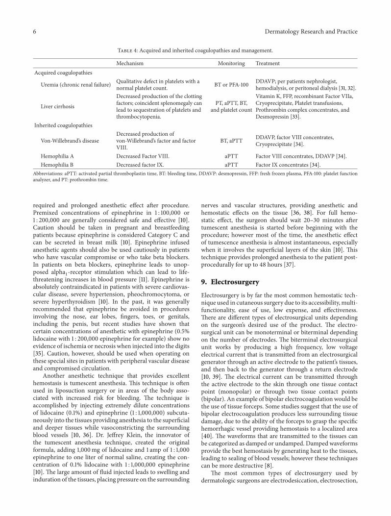
Electrosurgery can produce thermal damage to the sur-rounding healthy tissues during the procedure [41]. Excesscharring of the tissues can lead to decreased wound healingand slower recovery of the tissues postoperatively. This side-effect can be prevented by using the lowest power settingfor the shortest amount of time during electrocoagulation orby touching a hemostat with the tip of a monopolar unit toproduce pin-point coagulation (see Figure 1). Another poten-tially worrisome complication of electrosurgery is the risk offire and electrical shock. This can be prevented by ensuringthat the surgical area is not prepped with ethanol basedproducts and through the use of insulated disposable tipson the electrosurgical device [10]. 35% aluminum chloride,a popular topical hemostatic agent used for shave biopsies,should not be used in conjunction with electrosurgery aswell, because of the risk of fire. Also, the active electrodetransmitting the electrical current to the tissue has an insu-lated shaft and base preventing electrical shock to the patientand surgeon [39]. It is important to keep in mind whenremoving lesions caused by human papillomavirus and otherviral pathogens in the skin or when operating on patientswho are suffering from a concomitant viral illness such asHIV or hepatitis, that these infections have the potential tobe transmitted through the smoke plume [42]. Because ofthis, proper protection is warranted such as protective eyewear, masks, and gloves along with a smoke evacuator, and itis generally recommended that the smoke evacuator be heldwithin 2 cm of the area being cauterized [42].
10. Physical Hemostatic Techniques
Among the cheapest andmost accessible of all the hemostaticmodalities is that of manual compression. This is by far one
Figure 1: Minimizing the amount of surrounding tissue damageby using a monopolar electrocoagulation device applied to tissueforceps.
of the most basic techniques and has been used throughouthistory to stop bleeding and enhance coagulation. Downwardpressure should be applied firmly to the affected area for 15–20minutes depending on the extent of bleeding to tamponadethe vessel(s). If the bleeding is severe, applying pressure tothe supplying artery further upstream in addition to thewound area can help decrease blood flow to the affectedarea. This pressure applied allows time for platelets to adhereand initiation of the clotting cascade, as well as time togather additional hemostatic agents to aid in the process.Sterile gauze pads and cotton tipped applicators can facilitatethe process by soaking up the excess blood allowing bettervisualization of the surgical field and by providing counterpressure to aid in hemostasis. Cotton tipped applicators,(applicators with 8-inch handles and oversized cotton tips),are often used in nasal procedures, providing counterforceto the surgical field when entered in the nares for providingstabilization and hemostasis to the tissues [43]. Surgicalinstruments, chalazion clamp, can also provide hemostasis bymanually compressing tissue during surgery [44, 45].
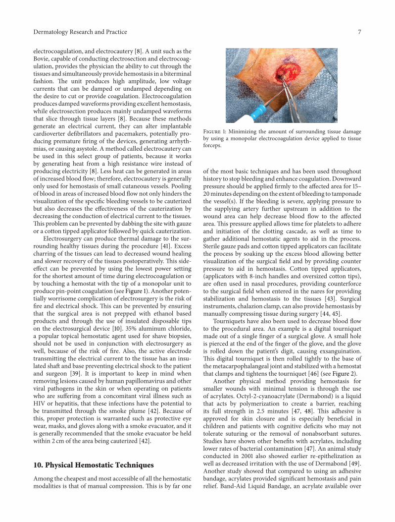
Tourniquets have also been used to decrease blood flowto the procedural area. An example is a digital tourniquetmade out of a single finger of a surgical glove. A small holeis pierced at the end of the finger of the glove, and the gloveis rolled down the patient’s digit, causing exsanguination.This digital tourniquet is then rolled tightly to the base ofthemetacarpophalangeal joint and stabilized with a hemostatthat clamps and tightens the tourniquet [46] (see Figure 2).
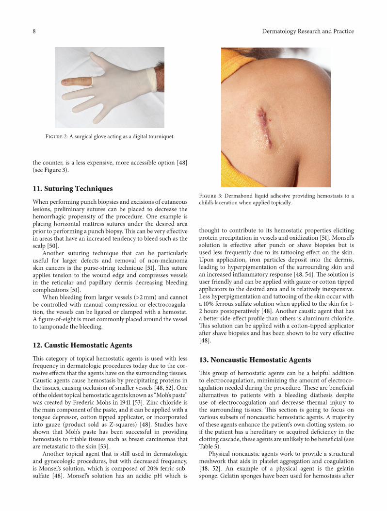
Another physical method providing hemostasis forsmaller wounds with minimal tension is through the useof acrylates. Octyl-2-cyanoacrylate (Dermabond) is a liquidthat acts by polymerization to create a barrier, reachingits full strength in 2.5 minutes [47, 48]. This adhesive isapproved for skin closure and is especially beneficial inchildren and patients with cognitive deficits who may nottolerate suturing or the removal of nonabsorbant sutures.Studies have shown other benefits with acrylates, includinglower rates of bacterial contamination [47]. An animal studyconducted in 2001 also showed earlier re-epithelization aswell as decreased irritation with the use of Dermabond [49].Another study showed that compared to using an adhesivebandage, acrylates provided significant hemostasis and painrelief. Band-Aid Liquid Bandage, an acrylate available over
8 Dermatology Research and Practice
Figure 2: A surgical glove acting as a digital tourniquet.
the counter, is a less expensive, more accessible option [48](see Figure 3).
11. Suturing Techniques
When performing punch biopsies and excisions of cutaneouslesions, preliminary sutures can be placed to decrease thehemorrhagic propensity of the procedure. One example isplacing horizontal mattress sutures under the desired areaprior to performing a punch biopsy.This can be very effectivein areas that have an increased tendency to bleed such as thescalp [50].
Another suturing technique that can be particularlyuseful for larger defects and removal of non-melanomaskin cancers is the purse-string technique [51]. This sutureapplies tension to the wound edge and compresses vesselsin the reticular and papillary dermis decreasing bleedingcomplications [51].
When bleeding from larger vessels (>2mm) and cannotbe controlled with manual compression or electrocoagula-tion, the vessels can be ligated or clamped with a hemostat.A figure-of-eight is most commonly placed around the vesselto tamponade the bleeding.
12. Caustic Hemostatic Agents
This category of topical hemostatic agents is used with lessfrequency in dermatologic procedures today due to the cor-rosive effects that the agents have on the surrounding tissues.Caustic agents cause hemostasis by precipitating proteins inthe tissues, causing occlusion of smaller vessels [48, 52]. Oneof the oldest topical hemostatic agents known as “Moh’s paste”was created by Frederic Mohs in 1941 [53]. Zinc chloride isthe main component of the paste, and it can be applied with atongue depressor, cotton tipped applicator, or incorporatedinto gauze (product sold as Z-squares) [48]. Studies haveshown that Moh’s paste has been successful in providinghemostasis to friable tissues such as breast carcinomas thatare metastatic to the skin [53].
Another topical agent that is still used in dermatologicand gynecologic procedures, but with decreased frequency,is Monsel’s solution, which is composed of 20% ferric sub-sulfate [48]. Monsel’s solution has an acidic pH which is
Figure 3: Dermabond liquid adhesive providing hemostasis to achild’s laceration when applied topically.
thought to contribute to its hemostatic properties elicitingprotein precipitation in vessels and oxidization [51]. Monsel’ssolution is effective after punch or shave biopsies but isused less frequently due to its tattooing effect on the skin.Upon application, iron particles deposit into the dermis,leading to hyperpigmentation of the surrounding skin andan increased inflammatory response [48, 54]. The solution isuser friendly and can be applied with gauze or cotton tippedapplicators to the desired area and is relatively inexpensive.Less hyperpigmentation and tattooing of the skin occur witha 10% ferrous sulfate solution when applied to the skin for 1-2 hours postoperatively [48]. Another caustic agent that hasa better side-effect profile than others is aluminum chloride.This solution can be applied with a cotton-tipped applicatorafter shave biopsies and has been shown to be very effective[48].
13. Noncaustic Hemostatic Agents
This group of hemostatic agents can be a helpful additionto electrocoagulation, minimizing the amount of electroco-agulation needed during the procedure. These are beneficialalternatives to patients with a bleeding diathesis despiteuse of electrocoagulation and decrease thermal injury tothe surrounding tissues. This section is going to focus onvarious subsets of noncaustic hemostatic agents. A majorityof these agents enhance the patient’s own clotting system, soif the patient has a hereditary or acquired deficiency in theclotting cascade, these agents are unlikely to be beneficial (seeTable 5).
Physical noncaustic agents work to provide a structuralmeshwork that aids in platelet aggregation and coagulation[48, 52]. An example of a physical agent is the gelatinsponge. Gelatin sponges have been used for hemostasis after
Dermatology Research and Practice 9
Table5:Hem
ostatic
agents.
Hem
ostatic
agent
Prod
uctinformation
Mechanism
ofactio
nPo
tentialsidee
ffects
Caustic
agents
Zinc
chlorid
e(M
oh’spaste
)
Paste
thatcanbe
appliedtopically.U
sed
infre
quently,but
iseffectiv
einproviding
hemostasis
tometastatic
cutaneou
swou
nds[53].
Precipitatesp
roteinsc
ausin
gcoagulationof
small
vessels
[48].L
efton
foru
pto
48ho
urs.
Can
bevery
painfuland
irritatin
gto
thep
atient
[53].
Ferricsubsulfate(M
onsel’s
solutio
n)Solutio
ncanbe
appliedwith
acottontip
ped
applicator
orgauzep
ad[48,54].
Precipitatesp
roteinsintravascularlyandoxidizes
tissues.L
esse
xpensiv
e,morea
ccessib
le,andeasy
toapply[48,54].
Beingused
lessdu
etointradermalferrug
inou
sdepo
sitsc
ausin
gtatto
oing
ofthes
kinaft
eruse
[48,54].
Aluminum
chlorid
eSolutio
ncanbe
appliedwith
acottontip
ped
applicator
after
shaveb
iopsy[48].
Precipitatesp
roteinsc
ausin
gcoagulationof
vessels
[48].E
asily
accessibleandeasy
toapply.
Can
bepainfuland
irritatin
gto
thep
atient
[48].
Non-caustic
agents
Gelatin
(Gelfoam,
Surgifo
am)
Gelfoam
Plus
(Gelatin
combinedwith
human
thrombin)
Com
esin
asterilep
owdero
rspo
nge.Po
rcine
deriv
ed[48].
Theg
elatin
isableto
absorb
morethan45xits
weight,providingam
atrix
forthe
clotting
cascade
inadditio
nto
providingap
hysic
albarrier.
Absorbed
bytheb
odyin
4–6weeks
[48,55,56].
Caninterfe
rewith
healingof
wou
ndedges,
generally
notrecom
mendedforu
sein
skin
incisio
ns.C
anfacilitateb
acteria
lgrowth
leadingto
infectionor
leadingto
foreignbo
dyreactio
nswhenleftin
thetissue.C
anincrease
insiz
eleading
tocompressio
nof
surrou
nding
structures,including
nerved
amage.When
combinedwith
thrombincanlead
toallergic/anaph
ylactic
reactio
ns[48,55].
Polyethylene
glycol
Hydrogel(CoSeal)
Liqu
idcompo
sedof
twoPE
Gpo
lymersthat
polymerizea
ndcross-lin
kin
thetissue
[57,58].
Increasesp
latele
tadh
erence,providing
quick
hemostasis
[57].
Swellsup
to4x
itssiz
epotentia
llycausing
damagetothes
urroun
ding
tissues
[58].
Micropo
rous
polysaccharid
esph
eres
(Aris
ta)
Com
esin
awhitepo
wderthatis100%plant
based.Fo
rmed
bycross-lin
king
ofpu
rified
plant
starch[52,58,59].
Dehydratesthe
bloo
d,concentratingRB
C’s,
platele
ts,proteins,promotingadherencetotheg
elmatrix
.Also
causes
aphysic
albarrierinthetiss
ue[52,58,59].
Causesimmediatesw
ellin
g,hasthe
potentialto
caused
amagetosurrou
ndingstr
uctures.Use
cautiouslyin
diabeticsd
ueto
potentialto
increase
glucoseload[58].
Microfib
rillarc
ollagen
(Avitene,H
elistat)
Bovine
collagenform
edinto
flour,sheets,or
spon
ges[48].
Collagenfram
eworkprom
otes
platele
taggregationandcoagulationcascade[
48].
Side
effectsarer
are.Allergicandforeignbo
dyreactio
nshave
occurred
[48].
Cellulose
(Surgicel,Oxycel)
Oxidizedcellu
lose
arranged
into
sheets,
gauze,or
smallerstrips.Ca
ndu
rablybe
placed
inthetissue
[48,60].
Physicallyactsto
tampo
nade
thev
esselsand
providea
meshw
orkforthe
fibrin
olyticcascadeto
occur.Be
comes
gelatin
ousin24–4
8hrsandis
absorbed
bytheb
odyby
1–6weeks.R
elatively
inexpensive[48].
Can
causeg
ranu
lomatou
sreactions
andshou
ldbe
used
carefully
inclo
sedspaces
dueto
increasedris
kof
swellingin
thetissues,can
causec
ompressio
nof
surrou
ndingstr
uctures
[60].
ProQRpo
wder
Com
binatio
nof
ahydroph
ilicp
olym
erand
potassium
saltpackaged
into
apow
der[48,51].
Availableo
verthe
coun
ter,relatively
inexpensive
andeasy
toapply.
Form
saneschar
inbo
dyin<60
second
s,du
eto
thep
olym
ersd
ehydratin
gtheb
lood
andthe
potassium
saltbind
ingto
thep
ositivelycharged
redbloo
dcells
[48,52].
Fewsid
eeffectsreported.
Thrombin(Th
rombin-JM
I,Re
cothromb,Ev
ithrom,
Floseal)
Canbe
bovine
deriv
edor
human
recombinant
thrombin.
Com
esin
apow
dero
rsolution.
Floseal
iscompo
sedof
agelatin
matrix
andthrombinand
comes
intwoseparatecompartmentsthataren
otmixed
until
timeo
fuse
[48,60].
Prom
otes
body’sph
ysiologicc
lotting
cascadeb
yactiv
elyconvertin
gfib
rinogen
into
fibrin
.Sho
uld
notb
eusedin
patie
ntsthath
aved
ecreased
fibrin
ogen
levels[48,57,60].
Bovine
thrombinhasb
eenshow
nto
cause
coagulop
athy
weeks
after
use,du
etoantib
odies
form
ingagainstfactorV
.Hum
anthrombin
althou
ghcle
ansedthorou
ghly,
hasthe
potential
totransm
itviruses[48,57,60].
10 Dermatology Research and Practice
Table5:Con
tinued.
Hem
ostatic
agent
Prod
uctinformation
Mechanism
ofactio
nPo
tentialsidee
ffects
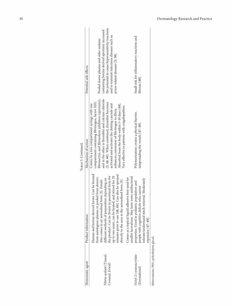
Fibrin
sealant(Tisseal,
Crosseal,E
vicel)
Hum
anandbo
vine
deriv
edform
s.Ca
nbe
form
edfro
mautologous
plasmao
rpoo
ledfro
mdo
nors.
Also
comes
inan
aerosolized
form
[5].En
tails
different
metho
dsof
preparationdepend
ingon
thep
rodu
ct.C
anbe
frozenin
prem
ixed
form
for
upto
twoyears,canbe
heated,and
stirred
for2
0minutes
priortouse[48,60].C
analso
besprayed
directlyto
thea
reainthea
erosolized
form
[5].
Com
esin
atwocompartmentsyringe
with
one
compartmentcon
tainingfib
rinogen,factorX
III,
fibronectin,and
fibrin
olysisinhibitors(aprotinin),
andin
theo
ther
isthrombinandcalcium
chlorid
e[5,48,60].Whencombined,thrombinbecomes
activ
ated
andactiv
ates
thec
lotting
cascadea
ndenhances
conversio
nof
fibrin
ogen
tofib
rin.
Absorbed
from
theb
odywith
in5–10
days
[48].
Very
effectiv
einpatie
ntsw
ithcoagulop
athies.
Pooled
dono
rplasm
aand
oldersealants
containing
bovine
deriv
edaprotin
inincreased
thep
otentia
ltocauseh
ypersensitivityreactio
nsandto
transm
itinfectious
diseases
such
asprion-related
diseases
[5,58].
Octyl-2-cyano
acrylate
(Dermabon
d)
Com
esas
atop
icalliq
uidadhesiv
ebestu
sedfor
smallerlacerations.M
ayhave
somea
ntibacteria
lprop
ertie
s.Goo
din
pediatric
popu
latio
nand
peop
lewith
cogn
itive
deficits
who
cann
ottolerate/und
ersta
ndstitchremoval.M
oderately
expensive[47–4
9].
Polymerizationcreatesa
physicalbarrier,
tampo
nading
thev
essels[47–49].
Smallrisk
forinfl
ammatoryreactio
nsand
fibrosis
[48].
Abbreviatio
ns:P
EG:polyethyleneg
lycol.

Dermatology Research and Practice 11
punch biopsies of the skin and may be more effective inareas such as cartilage or periosteum, which have a hardertime forming granulation tissue [48]. One study showedthat wounds treated with gelatin sponges and left to healby secondary intention led to increased granulation tissueformation and an overall better appearance of thewound [55].Absorbable gelatin sponges soaked in aluminum chloridehave also been shown to provide quick hemostasis after nailpunch biopsies when left in the wound for two weeks [56].Another physical agent, polyethylene glycol hydrogel, is asynthetic, biodegradable hemostat that polymerizes quickly,providing hemostasis in less than 60 seconds. Caution shouldbe taken with closed wounds because it can swell up tofour times its own size, potentially leading to surroundingtissue damage [57, 58]. Another physical agent, collagen, hasbeen shown to be a superior hemostat to other products,because not only platelets adhere more readily to the collagenmatrix, but also they are stimulated to degranulate enhancingplatelet aggregation [57]. This product is less effective inpatients with thrombocytopenia [58]. The product is bovinederived, with the potential to cause allergic and foreignbody reactions. Other hemostatic agents containing oxidizedcellulose (Surgicel or Oxycel), arranged into sheets or gauze,can be durably placed into bleeding tissues, causing hemosta-sis by tamponading vessels and by providing a physicalmeshwork for the clotting cascade to occur [48, 51]. Althoughrelatively inexpensive and easy to use, they can potentiallycause granulomatous reactions and increased swelling tissues[48, 60]. Two more physical agents, Urgent QR powder andmicroporous polysaccharide hemispheres, can be sprinkledtopically on wounds to enhance hemostasis. Urgent QRpowder is a hydrophilic polymer combined with potassiumsalt, used only on wounds left open to heal by secondaryintention, because of the body’s inability to metabolize thesubstance [52]. It hemostatically forms an eschar in the bodyin less than a minute, due to the polymers dehydrating theblood and the potassium salt binding to the positively chargedred blood cells [48, 52]. It is sold over the counter and isless expensive than some of the other hemostatic agents[52]. Another physical agent, composed of purified potatostarch powder, is microporous polysaccharide hemispheres.This agent can be degraded by enzymes in the body (alpha-amylase and pyrase), allowing use in closed wounds [52]. Itaccelerates the clotting process, by dehydrating the blood,causing concentration of the platelets and clotting factors[52, 58, 59]. This product can be sprinkled topically as apowder or incorporated into wound dressings. This agenthas been shown to be less hemostatically effective and moreexpensive than electrocoagulation but is a good alternative inindividuals that have contraindication to electrocoagulation[59]. This product should be used cautiously in diabetics,because it has the potential to increase glucose loads [58].
Physiologic agents are other hemostatic agents, whichpotentiate the body’s own physiologic clotting mechanisms.For example, thrombin products have been created toenhance the fibrinolytic cascade and the final conversion offibrinogen into fibrin. Topical bovine thrombin and humanrecombinant thrombin have been shown to be effectivehemostatic agents in areas where there is diffuse bleeding
(the specific vessel cannot be identified) and from directbleeding from bone [61]. Studies have shown an increasedrisk for postoperative coagulopathies with bovine-derivedthrombin, because antibodies formed against the thrombinare cross-reactive against human factor V [48, 57, 61]. Humanrecombinant thrombin has less antigenic effects, decreasingthe risk for developing a postoperative coagulopathy, butcarries a small risk for viral transmission to the patient [48,57, 58, 61].
Another class of hemostatic agents gaining in popularityis the fibrin sealants. Whereas other products may rely onthe patient’s own platelet and clotting factors for hemostaticactivation, fibrin sealants do not. This may be beneficial insituations where the patient has an inherited or acquiredcoagulation abnormality [62]. In order to be activated, thetwo compartments full of thrombin and fibrinogen must bemixed together (which can lead to clotting, if mixed prema-turely) [48, 62]. When combined, thrombin (from one com-partment) converts fibrinogen (from the other compartment)into insoluble fibrin in the presence of calcium [5, 48]. Theamount of thrombin contained in the fibrin sealant is thoughtto contribute to the rapidity of clot formation, whereas theamount of fibrinogen contributes to the mechanical strengthof the clot [58]. A 2009 Cochrane review showed that fibrinsealants lead to an average reduction of blood loss of 161mLper procedure and argued that the benefits of using theproduct must outweigh the potential side effects of its use[5]. Potential side effects of fibrin sealants are infectiousdisease transmission, hypersensitivity reactions, and neuro-toxicity [5, 48]. The newest fibrin sealant, Evicel, is humanderived and does not contain tranexamic acid, decreasingthe risk for neurotoxicity with use [58]. Newer fibrin sealantformulations are derived from autologous plasma or frompooled plasma donors, leading to decreased hypersensitivityreactions with the autologous sealants [5, 58].
The authors recommend intraoperative hemostasis thatis done with precision via electrosurgery as the mainstayof hemostasis in routine cutaneous surgeries. For smallerbiopsies, where electrosurgery is not necessary, aluminumchloride is cheap and easily accessible and should alsobe considered as a mainstay. For postoperative bleedingcomplications, the other measures discussed in this sectioncan be considered if physical hemostatic techniques (manualpressure) are inadequate.
14. Postoperative Recommendationsand Complications
Patients that have increased intraoperative bleeding are atgreater risk for postoperative bleeding complications, withcomplications most likely to occur within the first 48 hoursfollowing the procedure [10, 63]. A pressure dressing shouldbe applied to the operative area for at least 24 hours post-operatively to ensure adequate compression of the tissues.The typical wound dressing normally consists of a topicalantibiotic ointment, a Telfa pad cut to conform to theoperative area, and a layer of gauze secured and compressedto the skin by adhesive tape [64]. It is generally recommended
12 Dermatology Research and Practice
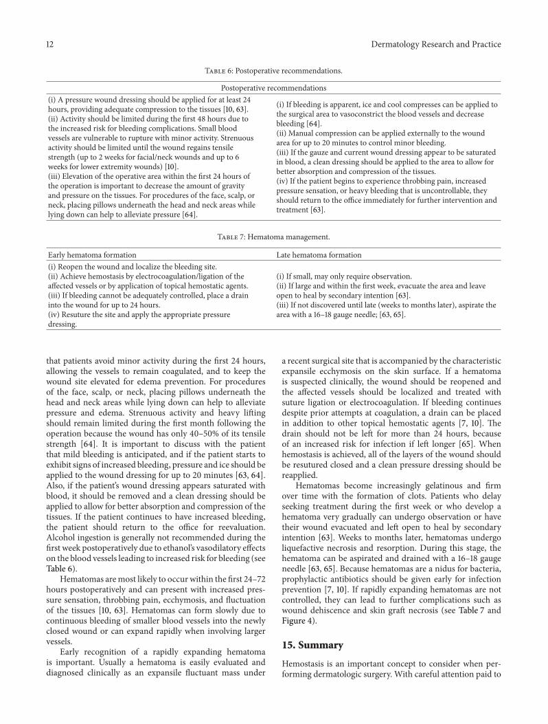
Table 6: Postoperative recommendations.
Postoperative recommendations(i) A pressure wound dressing should be applied for at least 24hours, providing adequate compression to the tissues [10, 63].(ii) Activity should be limited during the first 48 hours due tothe increased risk for bleeding complications. Small bloodvessels are vulnerable to rupture with minor activity. Strenuousactivity should be limited until the wound regains tensilestrength (up to 2 weeks for facial/neck wounds and up to 6weeks for lower extremity wounds) [10].(iii) Elevation of the operative area within the first 24 hours ofthe operation is important to decrease the amount of gravityand pressure on the tissues. For procedures of the face, scalp, orneck, placing pillows underneath the head and neck areas whilelying down can help to alleviate pressure [64].
(i) If bleeding is apparent, ice and cool compresses can be applied tothe surgical area to vasoconstrict the blood vessels and decreasebleeding [64].(ii) Manual compression can be applied externally to the woundarea for up to 20 minutes to control minor bleeding.(iii) If the gauze and current wound dressing appear to be saturatedin blood, a clean dressing should be applied to the area to allow forbetter absorption and compression of the tissues.(iv) If the patient begins to experience throbbing pain, increasedpressure sensation, or heavy bleeding that is uncontrollable, theyshould return to the office immediately for further intervention andtreatment [63].
Table 7: Hematoma management.
Early hematoma formation Late hematoma formation(i) Reopen the wound and localize the bleeding site.(ii) Achieve hemostasis by electrocoagulation/ligation of theaffected vessels or by application of topical hemostatic agents.(iii) If bleeding cannot be adequately controlled, place a draininto the wound for up to 24 hours.(iv) Resuture the site and apply the appropriate pressuredressing.
(i) If small, may only require observation.(ii) If large and within the first week, evacuate the area and leaveopen to heal by secondary intention [63].(iii) If not discovered until late (weeks to months later), aspirate thearea with a 16–18 gauge needle; [63, 65].
that patients avoid minor activity during the first 24 hours,allowing the vessels to remain coagulated, and to keep thewound site elevated for edema prevention. For proceduresof the face, scalp, or neck, placing pillows underneath thehead and neck areas while lying down can help to alleviatepressure and edema. Strenuous activity and heavy liftingshould remain limited during the first month following theoperation because the wound has only 40–50% of its tensilestrength [64]. It is important to discuss with the patientthat mild bleeding is anticipated, and if the patient starts toexhibit signs of increased bleeding, pressure and ice should beapplied to the wound dressing for up to 20 minutes [63, 64].Also, if the patient’s wound dressing appears saturated withblood, it should be removed and a clean dressing should beapplied to allow for better absorption and compression of thetissues. If the patient continues to have increased bleeding,the patient should return to the office for reevaluation.Alcohol ingestion is generally not recommended during thefirst week postoperatively due to ethanol’s vasodilatory effectson the blood vessels leading to increased risk for bleeding (seeTable 6).
Hematomas aremost likely to occur within the first 24–72hours postoperatively and can present with increased pres-sure sensation, throbbing pain, ecchymosis, and fluctuationof the tissues [10, 63]. Hematomas can form slowly due tocontinuous bleeding of smaller blood vessels into the newlyclosed wound or can expand rapidly when involving largervessels.
Early recognition of a rapidly expanding hematomais important. Usually a hematoma is easily evaluated anddiagnosed clinically as an expansile fluctuant mass under
a recent surgical site that is accompanied by the characteristicexpansile ecchymosis on the skin surface. If a hematomais suspected clinically, the wound should be reopened andthe affected vessels should be localized and treated withsuture ligation or electrocoagulation. If bleeding continuesdespite prior attempts at coagulation, a drain can be placedin addition to other topical hemostatic agents [7, 10]. Thedrain should not be left for more than 24 hours, becauseof an increased risk for infection if left longer [65]. Whenhemostasis is achieved, all of the layers of the wound shouldbe resutured closed and a clean pressure dressing should bereapplied.
Hematomas become increasingly gelatinous and firmover time with the formation of clots. Patients who delayseeking treatment during the first week or who develop ahematoma very gradually can undergo observation or havetheir wound evacuated and left open to heal by secondaryintention [63]. Weeks to months later, hematomas undergoliquefactive necrosis and resorption. During this stage, thehematoma can be aspirated and drained with a 16–18 gaugeneedle [63, 65]. Because hematomas are a nidus for bacteria,prophylactic antibiotics should be given early for infectionprevention [7, 10]. If rapidly expanding hematomas are notcontrolled, they can lead to further complications such aswound dehiscence and skin graft necrosis (see Table 7 andFigure 4).
15. Summary
Hemostasis is an important concept to consider when per-forming dermatologic surgery. With careful attention paid to
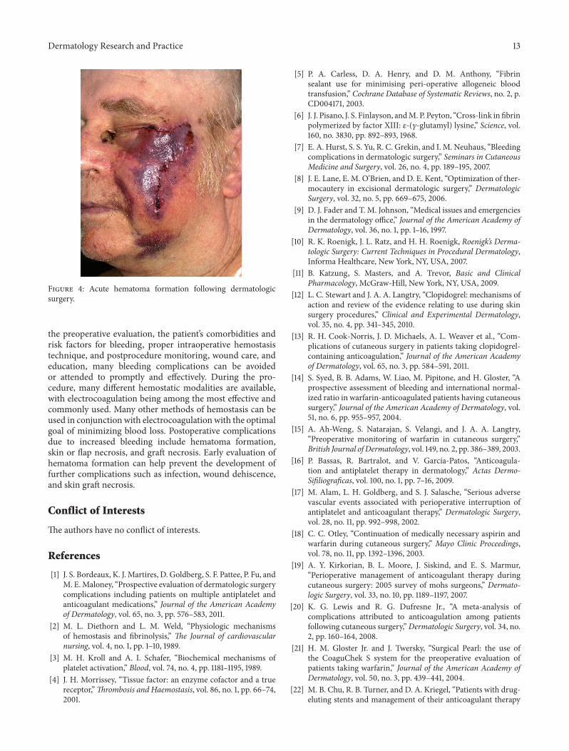
Dermatology Research and Practice 13
Figure 4: Acute hematoma formation following dermatologicsurgery.
the preoperative evaluation, the patient’s comorbidities andrisk factors for bleeding, proper intraoperative hemostasistechnique, and postprocedure monitoring, wound care, andeducation, many bleeding complications can be avoidedor attended to promptly and effectively. During the pro-cedure, many different hemostatic modalities are available,with electrocoagulation being among the most effective andcommonly used. Many other methods of hemostasis can beused in conjunction with electrocoagulation with the optimalgoal of minimizing blood loss. Postoperative complicationsdue to increased bleeding include hematoma formation,skin or flap necrosis, and graft necrosis. Early evaluation ofhematoma formation can help prevent the development offurther complications such as infection, wound dehiscence,and skin graft necrosis.
Conflict of Interests
The authors have no conflict of interests.
References
[1] J. S. Bordeaux, K. J.Martires, D. Goldberg, S. F. Pattee, P. Fu, andM. E.Maloney, “Prospective evaluation of dermatologic surgerycomplications including patients on multiple antiplatelet andanticoagulant medications,” Journal of the American Academyof Dermatology, vol. 65, no. 3, pp. 576–583, 2011.
[2] M. L. Diethorn and L. M. Weld, “Physiologic mechanismsof hemostasis and fibrinolysis,” The Journal of cardiovascularnursing, vol. 4, no. 1, pp. 1–10, 1989.
[3] M. H. Kroll and A. I. Schafer, “Biochemical mechanisms ofplatelet activation,” Blood, vol. 74, no. 4, pp. 1181–1195, 1989.
[4] J. H. Morrissey, “Tissue factor: an enzyme cofactor and a truereceptor,”Thrombosis and Haemostasis, vol. 86, no. 1, pp. 66–74,2001.
[5] P. A. Carless, D. A. Henry, and D. M. Anthony, “Fibrinsealant use for minimising peri-operative allogeneic bloodtransfusion,” Cochrane Database of Systematic Reviews, no. 2, p.CD004171, 2003.
[6] J. J. Pisano, J. S. Finlayson, andM. P. Peyton, “Cross-link in fibrinpolymerized by factor XIII: 𝜀-(𝛾-glutamyl) lysine,” Science, vol.160, no. 3830, pp. 892–893, 1968.
[7] E. A. Hurst, S. S. Yu, R. C. Grekin, and I. M. Neuhaus, “Bleedingcomplications in dermatologic surgery,” Seminars in CutaneousMedicine and Surgery, vol. 26, no. 4, pp. 189–195, 2007.
[8] J. E. Lane, E. M. O’Brien, and D. E. Kent, “Optimization of ther-mocautery in excisional dermatologic surgery,” DermatologicSurgery, vol. 32, no. 5, pp. 669–675, 2006.
[9] D. J. Fader and T. M. Johnson, “Medical issues and emergenciesin the dermatology office,” Journal of the American Academy ofDermatology, vol. 36, no. 1, pp. 1–16, 1997.
[10] R. K. Roenigk, J. L. Ratz, and H. H. Roenigk, Roenigk’s Derma-tologic Surgery: Current Techniques in Procedural Dermatology,Informa Healthcare, New York, NY, USA, 2007.
[11] B. Katzung, S. Masters, and A. Trevor, Basic and ClinicalPharmacology, McGraw-Hill, New York, NY, USA, 2009.
[12] L. C. Stewart and J. A. A. Langtry, “Clopidogrel: mechanisms ofaction and review of the evidence relating to use during skinsurgery procedures,” Clinical and Experimental Dermatology,vol. 35, no. 4, pp. 341–345, 2010.
[13] R. H. Cook-Norris, J. D. Michaels, A. L. Weaver et al., “Com-plications of cutaneous surgery in patients taking clopidogrel-containing anticoagulation,” Journal of the American Academyof Dermatology, vol. 65, no. 3, pp. 584–591, 2011.
[14] S. Syed, B. B. Adams, W. Liao, M. Pipitone, and H. Gloster, “Aprospective assessment of bleeding and international normal-ized ratio in warfarin-anticoagulated patients having cutaneoussurgery,” Journal of the American Academy of Dermatology, vol.51, no. 6, pp. 955–957, 2004.
[15] A. Ah-Weng, S. Natarajan, S. Velangi, and J. A. A. Langtry,“Preoperative monitoring of warfarin in cutaneous surgery,”British Journal of Dermatology, vol. 149, no. 2, pp. 386–389, 2003.
[16] P. Bassas, R. Bartralot, and V. Garcıa-Patos, “Anticoagula-tion and antiplatelet therapy in dermatology,” Actas Dermo-Sifiliograficas, vol. 100, no. 1, pp. 7–16, 2009.
[17] M. Alam, L. H. Goldberg, and S. J. Salasche, “Serious adversevascular events associated with perioperative interruption ofantiplatelet and anticoagulant therapy,” Dermatologic Surgery,vol. 28, no. 11, pp. 992–998, 2002.
[18] C. C. Otley, “Continuation of medically necessary aspirin andwarfarin during cutaneous surgery,” Mayo Clinic Proceedings,vol. 78, no. 11, pp. 1392–1396, 2003.
[19] A. Y. Kirkorian, B. L. Moore, J. Siskind, and E. S. Marmur,“Perioperative management of anticoagulant therapy duringcutaneous surgery: 2005 survey of mohs surgeons,” Dermato-logic Surgery, vol. 33, no. 10, pp. 1189–1197, 2007.
[20] K. G. Lewis and R. G. Dufresne Jr., “A meta-analysis ofcomplications attributed to anticoagulation among patientsfollowing cutaneous surgery,”Dermatologic Surgery, vol. 34, no.2, pp. 160–164, 2008.
[21] H. M. Gloster Jr. and J. Twersky, “Surgical Pearl: the use ofthe CoaguChek S system for the preoperative evaluation ofpatients taking warfarin,” Journal of the American Academy ofDermatology, vol. 50, no. 3, pp. 439–441, 2004.
[22] M. B. Chu, R. B. Turner, and D. A. Kriegel, “Patients with drug-eluting stents and management of their anticoagulant therapy
14 Dermatology Research and Practice
in cutaneous surgery,” Journal of the American Academy ofDermatology, vol. 64, no. 3, pp. 553–558, 2011.
[23] D. G. Rizik and K. J. Klassen, “Assessing the landscape ofstent thrombosis: the drug-eluting versus bare-metal stentcontroversy,” American Journal of Cardiology, vol. 102, no. 9, pp.4J–11J, 2008.
[24] S. C. Collins and R. G. Dufresne Jr., “Dietary supplements inthe setting of Mohs surgery,” Dermatologic Surgery, vol. 28, no.6, pp. 447–452, 2002.
[25] P. M. Barnes, B. Bloom, and R. L. Nahin, “Complementary andalternative medicine use among adults and children: UnitedStates, 2007,” National Health Statistics Reports, no. 12, pp. 1–23,2009.
[26] S. M. Dinehart and L. Henry, “Dietary supplements: alteredcoagulation and effects on bruising,”Dermatologic Surgery, vol.31, no. 7, pp. 819–826, 2005.
[27] J. Heller, J. S. Gabbay, K. Ghadjar et al., “Top-10 list of herbaland supplemental medicines used by cosmetic patients: Whatthe plastic surgeon needs to know,” Plastic and ReconstructiveSurgery, vol. 117, no. 2, pp. 436–445, 2006.
[28] M. J. Stanger, L. A. Thompson, A. J. Young, and H. R. Lieber-man, “Anticoagulant activity of select dietary supplements,”Nutrition Reviews, vol. 70, no. 2, pp. 107–117, 2012.
[29] A. J. Kellermann and C. Kloft, “Is there a risk of bleedingassociated with standardized Ginkgo biloba extract therapy? Asystematic review andmeta-analysis,” Pharmacotherapy, vol. 31,no. 5, pp. 490–502, 2011.
[30] C. K. Kepler, R. C. Huang, D. Meredith, J.-H. Kim, andA. K. Sharma, “Omega-3 and fish oil supplements do notcause increased bleeding during spinal decompression surgery,”Journal of Spinal Disorders & Techniques, vol. 25, no. 3, pp. 129–132, 2011.
[31] R. E. Taylor and P. M. Blatt, “Clinical evaluation of the patientwith bruising and bleeding,” Journal of the American Academyof Dermatology, vol. 4, no. 3, pp. 348–368, 1981.
[32] H. K. Lee, Y. J. Kim, J. U. Jeong, J. S. Park, H. S. Chi, and S. B.Kim, “Desmopressin improves platelet dysfunction measuredby in vitro closure time in uremic patients,” Nephron ClinicalPractice, vol. 114, no. 4, pp. c248–c252, 2010.
[33] D. E. Bernstein, L. Jeffers, E. Erhardtsen et al., “Recombinantfactor VIIa corrects prothrombin time in cirrhotic patients: apreliminary study,” Gastroenterology, vol. 113, no. 6, pp. 1930–1937, 1997.
[34] S. R. Peterson and A. K. Joseph, “Inherited bleeding disordersin dermatologic surgery,” Dermatologic Surgery, vol. 27, no. 10,pp. 885–889, 2001.
[35] B. Firoz, N. Davis, and L. H. Goldberg, “Local anesthesia usingbuffered 0.5% lidocaine with 1:200,000 epinephrine for tumorsof the digits treated with Mohs micrographic surgery,” Journalof the American Academy of Dermatology, vol. 61, no. 4, pp. 639–643, 2009.
[36] P. Davila and I. Garcia-Doval, “Tumescent anesthesia in derma-tologic surgery,” Actas Dermo-Sifiliograficas, vol. 103, no. 4, pp.285–287, 2012.
[37] J. A. Klein, “Tumescent technique for regional anesthesiapermits lidocaine doses of 35 mg/kg for liposuction,” Journal ofDermatologic Surgery and Oncology, vol. 16, no. 3, pp. 248–263,1990.
[38] D. Balducci, O. Morandi, S. Mazzetti, M. Tonni, A. Becchetti,and R. Pancaldi, “Ambulatory saphenectomy: 80 operated casesusing tumescent anesthesia,” Chirurgia Italiana, vol. 54, no. 1,pp. 77–82, 2002.
[39] D. G. Ferris, S. Saxena, B. L. Hainer, J. R. Searle, J. L. Powell, andJ. N. Gay, “Gynecologic and dermatologic electrosurgical units:a comparative review,” Journal of Family Practice, vol. 39, no. 2,pp. 160–169, 1994.
[40] B. Bergdahl and B. Stenquist, “An automatic computerizedbipolar coagulator for dermatologic surgery,” Journal of Derma-tologic Surgery and Oncology, vol. 19, no. 3, pp. 225–227, 1993.
[41] R. Hambley, P. A. Hebda, E. Abell, B. A. Cohen, and B. V.Jegasothy, “Wound healing of skin incisions produced by ultra-sonically vibrating knife, scalpel, electrosurgery, and carbondioxide laser,” Journal of Dermatologic Surgery and Oncology,vol. 14, no. 11, pp. 1213–1217, 1988.
[42] W. S. Sawchuk, P. J. Weber, D. R. Lowy, and L. M. Dzubow,“Infectious papillomavirus in the vapor of warts treated withcarbon dioxide laser or electrocoagulation: detection and pro-tection,” Journal of the American Academy of Dermatology, vol.21, no. 1, pp. 41–49, 1989.
[43] K. Nouri and C. J. Ballard, “Cotton-tipped applicators used insurgery of the nose,” Dermatologic Surgery, vol. 31, no. 11, pp.1440–1441, 2005.
[44] I. Amir, E. S. Marmur, and D. A. Kriegel, “Bloodless nasalalar surgery: another innovative use of the chalazion clamp,”Dermatologic Surgery, vol. 35, no. 5, pp. 843–844, 2009.
[45] E. Cigna, E. M. Buccheri, C. Monarca, and N. Scuderi,“Hemostasis in skin surgery,” Aesthetic Plastic Surgery, vol. 32,no. 4, p. 702, 2008.
[46] A. B. Aksakal and E. AdiSen, “Method for facilitating theapplication of digital tourniquets,” Dermatologic Surgery, vol.35, no. 9, p. 1389, 2009.
[47] D. M. Toriumi and A. A. Bagal, “Cyanoacrylate tissue adhesivesfor skin closure in the outpatient setting,” OtolaryngologicClinics of North America, vol. 35, no. 1, pp. 103–118, 2002.
[48] M. D. Palm and J. S. Altman, “Topical hemostatic agents: areview,” Dermatologic Surgery, vol. 34, no. 4, pp. 431–445, 2008.
[49] W.H. Eaglstein, T. P. Sullivan, P. A. Giordano, and B.M.Miskin,“A liquid adhesive bandage for the treatment of minor cuts andabrasions,” Dermatologic Surgery, vol. 28, no. 3, pp. 263–267,2002.
[50] A. A. Ingraffea, “Use of a preliminary horizontal mattress sutureon scalp biopsies to achieve rapid hemostasis,” DermatologicSurgery, vol. 36, no. 8, p. 1312, 2010.
[51] P. R. Cohen, P. T. Martinelli, K. E. Schulze, and B. R. Nelson,“The cuticular purse string suture: a modified purse stringsuture for the partial closure of round postoperative wounds,”International Journal of Dermatology, vol. 46, no. 7, pp. 746–753,2007.
[52] J. Ho and G. Hruza, “Hydrophilic polymers with potassium saltandmicroporous polysaccharides for use as hemostatic agents,”Dermatologic Surgery, vol. 33, no. 12, pp. 1430–1433, 2007.
[53] M. Kakimoto, H. Tokita, T. Okamura, and K. Yoshino, “Achemical hemostatic technique for bleeding from malignantwounds,” Journal of Palliative Medicine, vol. 13, no. 1, pp. 11–13,2010.
[54] R. B. Armstrong, J. Nichols, and J. Pachance, “Punch biopsywounds treated with Monsel’s solution or a collagen matrix. Acomparison of healing,” Archives of Dermatology, vol. 122, no. 5,pp. 546–549, 1986.
[55] P. P. Rullan, C. Vallbona, J. M. Rullan, J. N. Mansbridge,and V. B. Morhenn, “Use of gelatin sponges in Mohs micro-graphic surgery defects and stagedmelanoma excisions: a novelapproach to secondary wound healing,” Journal of Drugs inDermatology, vol. 10, no. 1, pp. 68–73, 2011.
Dermatology Research and Practice 15
[56] C. Hwa, O. I. Kovich, and J. A. Stein, “Achieving hemostasisafter nail biopsy using absorbable gelatin sponge saturated inaluminum chloride,” Dermatologic Surgery, vol. 37, no. 3, pp.368–369, 2011.
[57] F. I. Broekema,W. Van Oeveren, J. Zuidema, S. H. Visscher, andR. R.M. Bos, “In vitro analysis of polyurethane foam as a topicalhemostatic agent,” Journal ofMaterials Science, vol. 22, no. 4, pp.1081–1086, 2011.
[58] S. Dhillon, M. T. De Boer, E. N. Papacharalabous, and M.Schwartz, “Fibrin sealant (Evicel [quixil/crosseal]): a reviewof its use as supportive treatment for haemostasis in surgery,”Drugs, vol. 71, no. 14, pp. 1893–1915, 2011.
[59] S. R. Tan and W. D. Tope, “Effectiveness of microporouspolysaccharide hemospheres for achieving hemostasis in Mohsmicrographic surgery,” Dermatologic Surgery, vol. 30, no. 6, pp.908–914, 2004.
[60] M. A. Schreiber and D. J. Neveleff, “Achieving hemostasiswith topical hemostats: making clinically and economicallyappropriate decisions in the surgical and trauma settings,”AORN Journal, vol. 94, no. 5, pp. S1–S20, 2011.
[61] W. C. Chapman, N. Singla, Y. Genyk et al., “A phase 3,randomized, double-blind comparative study of the efficacyand safety of topical recombinant human thrombin and bovinethrombin in surgical hemostasis,” Journal of the AmericanCollege of Surgeons, vol. 205, no. 2, pp. 256–265, 2007.
[62] L. K. Krishnan, M. Mohanty, P. R. Umashankar, and A. VijayanLal, “Comparative evaluation of absorbable hemostats: advan-tages of fibrin-based sheets,” Biomaterials, vol. 25, no. 24, pp.5557–5563, 2004.
[63] A. Delaney, S. Diamantis, and V. J. Marks, “Complications oftissue ischemia in dermatologic surgery,”DermatologicTherapy,vol. 24, no. 6, pp. 551–557, 2011.
[64] C. Y. Cho and J. S. Lo, “Dressing the part,”Dermatologic Clinics,vol. 16, no. 1, pp. 25–47, 1998.
[65] S. J. Salasche, “Acute surgical complications: cause, prevention,and treatment,” Journal of the American Academy of Dermatol-ogy, vol. 15, no. 6, pp. 1163–1185, 1986.