ReviewArticle … ObstetricThromboprophylaxis:TheSwedishGuidelines ... 1Department of Obstetrics and...
7
Hindawi Publishing Corporation Advances in Hematology Volume 2011, Article ID 157483, 6 pages doi:10.1155/2011/157483 Review Article Obstetric Thromboprophylaxis: The Swedish Guidelines Pelle G. Lindqvist 1 and Margareta Hellgren 2, 3 1 Department of Obstetrics and Gynecology, Karolinska University Hospital, 14186 Stockholm, Sweden 2 Department of Obstetrics and Gynecology, Sahlgrenska University Hospital Gothenburg, 41345 Gothenburg, Sweden 3 Department of Antenatal Care, Primary Care South Bohusl¨ an, 43102 Molndal, Sweden Correspondence should be addressed to Pelle G. Lindqvist, [email protected] Received 28 April 2011; Accepted 5 October 2011 Academic Editor: Thomas Kickler Copyright © 2011 P. G. Lindqvist and M. Hellgren. This is an open access article distributed under the Creative Commons Attribution License, which permits unrestricted use, distribution, and reproduction in any medium, provided the original work is properly cited. Obstetric thromboprophylaxis is difficult. Since 10 years Swedish obstetricians have used a combined risk estimation model and recommendations concerning to whom, at what dose, when, and for how long thromboprophylaxis is to be administrated based on a weighted risk score. In this paper we describe the background and validation of the Swedish guidelines for obstetric thromboprophylaxis in women with moderate-high risk of VTE, that is, at similar or higher risk as the antepartum risk among women with history of thrombosis. The risk score is based on major risk factors (i.e., 5-fold increased risk of thromboembolism). We present data on the efficacy of the model, the cost-effectiveness, and the lifestyle advice that is given. We believe that the Swedish guidelines for obstetric thromboprophylaxis aid clinicians in providing women at increased risk of VTE with effective and appropriate thromboprophylaxis, thus avoiding both over- and under-treatment. 1. Introduction The incidence of obstetric venous thromboembolism (VTE) in the Nordic countries is estimated at 10 to 13 cases per 10000 pregnancies, half of them diagnosed during the first six weeks after birth [1, 2]. VTE is one of the most common causes of maternal death [3, 4] and leads to morbidity in the form of postthrombotic syndrome in up to 50–60% [5, 6]. Several factors are known to increase the risk of obstetric VTE, such as personal or family history of VTE, thrombo- philia, older age, high body mass index (BMI), immobiliza- tion, surgery, smoking, nulliparity, and cancer [1, 2]. Thromboprophylaxis during pregnancy usually consists of daily subcutaneous injections of low molecular weight heparin (LMWH), in combination with compression stock- ings [7–10]. There are several recommendations concerning how to identify women at high risk of VTE during pregnancy and the puerperium. Some of them divide women into low- medium and high-risk groups [7, 8] and others are based on weighted risk scores [9, 10]. Risk assessment and man- agement of obstetric thromboprophylaxis differ in different countries. The aim of this paper is to describe the background and validation of the Swedish guidelines for obstetric thrombo- prophylaxis in women with moderate-high risk of VTE. A weighted risk score for estimation of obstetric VTE risk has been used in Sweden for around 10 years. Recommendations concerning to whom, at what dose, when, and for how long thromboprophylaxis is to be administered are based on this risk score. A small number of special cases (women with antithrombin deficiency, antiphospholipid syndrome (APS) with prior VTE or prior multiple VTE, as well as those on chronic warfarin treatment) cannot be managed according to the risk score. These women are estimated to be at very high, that is, 15% or higher, risk of obstetric VTE. They are recommended “two-dose” thromboprophylaxis, which is not included in this paper. Thromboprophylaxis in relation to legal abortion, miscarriage, or ectopic pregnancy is also excluded from this paper. 2. Methods The available literature from 1996 to 2000 on risk factors for obstetric VTE was reviewed. Two different risk models
Transcript of ReviewArticle … ObstetricThromboprophylaxis:TheSwedishGuidelines ... 1Department of Obstetrics and...

Hindawi Publishing CorporationAdvances in HematologyVolume 2011, Article ID 157483, 6 pagesdoi:10.1155/2011/157483
Review Article
Obstetric Thromboprophylaxis: The Swedish Guidelines
Pelle G. Lindqvist1 and Margareta Hellgren2, 3
1 Department of Obstetrics and Gynecology, Karolinska University Hospital, 14186 Stockholm, Sweden2 Department of Obstetrics and Gynecology, Sahlgrenska University Hospital Gothenburg, 41345 Gothenburg, Sweden3 Department of Antenatal Care, Primary Care South Bohuslan, 43102 Molndal, Sweden
Correspondence should be addressed to Pelle G. Lindqvist, [email protected]
Received 28 April 2011; Accepted 5 October 2011
Academic Editor: Thomas Kickler
Copyright © 2011 P. G. Lindqvist and M. Hellgren. This is an open access article distributed under the Creative CommonsAttribution License, which permits unrestricted use, distribution, and reproduction in any medium, provided the original work isproperly cited.
Obstetric thromboprophylaxis is difficult. Since 10 years Swedish obstetricians have used a combined risk estimation modeland recommendations concerning to whom, at what dose, when, and for how long thromboprophylaxis is to be administratedbased on a weighted risk score. In this paper we describe the background and validation of the Swedish guidelines for obstetricthromboprophylaxis in women with moderate-high risk of VTE, that is, at similar or higher risk as the antepartum risk amongwomen with history of thrombosis. The risk score is based on major risk factors (i.e., 5-fold increased risk of thromboembolism).We present data on the efficacy of the model, the cost-effectiveness, and the lifestyle advice that is given. We believe that theSwedish guidelines for obstetric thromboprophylaxis aid clinicians in providing women at increased risk of VTE with effective andappropriate thromboprophylaxis, thus avoiding both over- and under-treatment.
1. Introduction
The incidence of obstetric venous thromboembolism (VTE)in the Nordic countries is estimated at 10 to 13 cases per10000 pregnancies, half of them diagnosed during the firstsix weeks after birth [1, 2]. VTE is one of the most commoncauses of maternal death [3, 4] and leads to morbidity in theform of postthrombotic syndrome in up to 50–60% [5, 6].Several factors are known to increase the risk of obstetricVTE, such as personal or family history of VTE, thrombo-philia, older age, high body mass index (BMI), immobiliza-tion, surgery, smoking, nulliparity, and cancer [1, 2].
Thromboprophylaxis during pregnancy usually consistsof daily subcutaneous injections of low molecular weightheparin (LMWH), in combination with compression stock-ings [7–10]. There are several recommendations concerninghow to identify women at high risk of VTE during pregnancyand the puerperium. Some of them divide women into low-medium and high-risk groups [7, 8] and others are basedon weighted risk scores [9, 10]. Risk assessment and man-agement of obstetric thromboprophylaxis differ in differentcountries.
The aim of this paper is to describe the background andvalidation of the Swedish guidelines for obstetric thrombo-prophylaxis in women with moderate-high risk of VTE. Aweighted risk score for estimation of obstetric VTE risk hasbeen used in Sweden for around 10 years. Recommendationsconcerning to whom, at what dose, when, and for how longthromboprophylaxis is to be administered are based on thisrisk score. A small number of special cases (women withantithrombin deficiency, antiphospholipid syndrome (APS)with prior VTE or prior multiple VTE, as well as those onchronic warfarin treatment) cannot be managed accordingto the risk score. These women are estimated to be at veryhigh, that is, 15% or higher, risk of obstetric VTE. Theyare recommended “two-dose” thromboprophylaxis, which isnot included in this paper. Thromboprophylaxis in relationto legal abortion, miscarriage, or ectopic pregnancy is alsoexcluded from this paper.
2. Methods
The available literature from 1996 to 2000 on risk factorsfor obstetric VTE was reviewed. Two different risk models

2 Advances in Hematology
Table 1: Risk factors and their odds ratios (ORs) for VTE (1,2).
Prevalence (%) Pregnancy OR Postpartum OR
Overweight (BMI > 28)1
>28 BMI 12.8 5 5
Familial thrombosis (first-degree relatives)
yes 5.1 5 5
Age
<20 2.5 1.0 2.5
≥20–<35 85.2 1.0 1
≥35 12.4 1.0 1.2
Smoking
yes 21.0 1.2 1.2
Parity
Primapara 41.3 2.3 1.1
1 birth 35.4 1 1
2 births 15.8 1.3 1.7
>2 births 7.3 2.6 1.8
Preeclampsia
Yes 2.0 3–5
Cesarean section
Yes 11.0 4.9
FV Leiden
Noncarrier 89.1 1 1
Heterozygotes 10.6 5 5
Homozygotes 0.3 25–100 25–100
Protein S or protein C deficiency 0.1 5–25 5–25
Prothrombin gene mutation 2.0 5 5
Hyperhomocysteinemia 2–5 2–51BMI > mean + 1SD in early pregnancy is regarded as overweight.
were constructed, one a weighted risk score based on majorrisk factors associated with a five-fold increased risk or amultiple thereof [11, 12], and the other an individualizedcomputerized risk assessment using and including estimatesof the absolute risk [11]. Hem-ARG, the Swedish Associationof Obstetricians and Gynecologists (SFOG) reference andworking group on hemostatic disturbances in obstetrics andgynecology, chosed the weighted risk score model. Swedishguidelines based on this model were published in 2004 [12],and established as a National Guideline by SFOG in 2009.The guidelines were revised in 2010-2011, resulting in theversion presented here.
2.1. Risk Assessment of Obstetric VTE. To facilitate the assess-ment of VTE risk during pregnancy and the puerperium, ascoring system was created by adding the weight of variousrisk factors (Table 1) for obstetric VTE [1]. An approximatelyfive-fold increased risk of VTE during pregnancy, shown bythe odds ratio (OR) for a number of risk factors for VTE,yields one risk score point (Table 2). Three conditions, cesa-rean section (CS), preeclampsia, and abruptio placentae, areonly considered to be risk factors during the puerperium.Since all risk factors cannot be included in an algorithm, thevariable “other major risk factor” is included (Table 2) and
may be used, according to the clinician’s decision, in casesof malignancy, extreme obesity, and so forth. The woman’srisk score is calculated as the sum of the points. Women withrisk score 1 are at a five-fold increased risk, risk score 2 (twovariables with 1 point or one variable with 2 points) are ata 25-fold increased risk and risk score 3 entails a 125-foldincreased risk, and so forth, compared to risk score 0, whichmeans no increased risk (Table 2). Women with a previousVTE or APS without VTE are given 4 risk points, regardlessof other risk factors. The total risk score is the basis fordetermining when, how, and at what dose thromboprophy-laxis should be administered (Table 3).
During pregnancy it is mainly women with prior VTEthat reach the risk score level for thromboprophylaxis. A het-erozygous carrier of Factor V Leiden (FVL) without anyother risk factors has 1 risk point and would not be recom-mended ante- or postpartum prophylaxis. However, if shealso has first-degree heredity for VTE, one more risk pointis added, resulting in a risk score of 2, leading to a rec-ommended one week of postpartum thromboprophylaxis.If she also undergoes CS, she is given 3 risk points (FVL,heredity, and CS), resulting in a risk score of 3, and she wouldbe recommended six weeks of postpartum thromboprophy-laxis.
Advances in Hematology 3
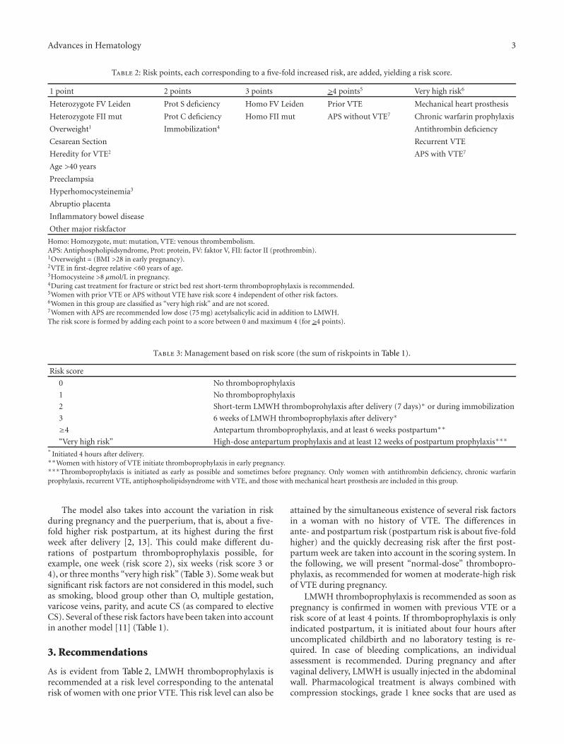
Table 2: Risk points, each corresponding to a five-fold increased risk, are added, yielding a risk score.
1 point 2 points 3 points >4 points5 Very high risk6
Heterozygote FV Leiden Prot S deficiency Homo FV Leiden Prior VTE Mechanical heart prosthesis
Heterozygote FII mut Prot C deficiency Homo FII mut APS without VTE7 Chronic warfarin prophylaxis
Overweight1 Immobilization4 Antithrombin deficiency
Cesarean Section Recurrent VTE
Heredity for VTE2 APS with VTE7
Age >40 years
Preeclampsia
Hyperhomocysteinemia3
Abruptio placenta
Inflammatory bowel disease
Other major riskfactor
Homo: Homozygote, mut: mutation, VTE: venous thrombembolism.APS: Antiphospholipidsyndrome, Prot: protein, FV: faktor V, FII: factor II (prothrombin).1Overweight = (BMI >28 in early pregnancy).2VTE in first-degree relative <60 years of age.3Homocysteine >8 µmol/L in pregnancy.4During cast treatment for fracture or strict bed rest short-term thromboprophylaxis is recommended.5Women with prior VTE or APS without VTE have risk score 4 independent of other risk factors.6Women in this group are classified as “very high risk” and are not scored.7Women with APS are recommended low dose (75 mg) acetylsalicylic acid in addition to LMWH.The risk score is formed by adding each point to a score between 0 and maximum 4 (for >4 points).
Table 3: Management based on risk score (the sum of riskpoints in Table 1).
Risk score
0 No thromboprophylaxis
1 No thromboprophylaxis
2 Short-term LMWH thromboprohylaxis after delivery (7 days)∗ or during immobilization
3 6 weeks of LMWH thromboprophylaxis after delivery∗
≥4 Antepartum thromboprophylaxis, and at least 6 weeks postpartum∗∗
“Very high risk” High-dose antepartum prophylaxis and at least 12 weeks of postpartum prophylaxis∗∗∗∗
Initiated 4 hours after delivery.∗∗Women with history of VTE initiate thromboprophylaxis in early pregnancy.∗∗∗Thromboprophylaxis is initiated as early as possible and sometimes before pregnancy. Only women with antithrombin deficiency, chronic warfarinprophylaxis, recurrent VTE, antiphospholipidsyndrome with VTE, and those with mechanical heart prosthesis are included in this group.
The model also takes into account the variation in riskduring pregnancy and the puerperium, that is, about a five-fold higher risk postpartum, at its highest during the firstweek after delivery [2, 13]. This could make different du-rations of postpartum thromboprophylaxis possible, forexample, one week (risk score 2), six weeks (risk score 3 or4), or three months “very high risk” (Table 3). Some weak butsignificant risk factors are not considered in this model, suchas smoking, blood group other than O, multiple gestation,varicose veins, parity, and acute CS (as compared to electiveCS). Several of these risk factors have been taken into accountin another model [11] (Table 1).
3. Recommendations
As is evident from Table 2, LMWH thromboprophylaxis isrecommended at a risk level corresponding to the antenatalrisk of women with one prior VTE. This risk level can also be
attained by the simultaneous existence of several risk factorsin a woman with no history of VTE. The differences inante- and postpartum risk (postpartum risk is about five-foldhigher) and the quickly decreasing risk after the first post-partum week are taken into account in the scoring system. Inthe following, we will present “normal-dose” thrombopro-phylaxis, as recommended for women at moderate-high riskof VTE during pregnancy.
LMWH thromboprophylaxis is recommended as soon aspregnancy is confirmed in women with previous VTE or arisk score of at least 4 points. If thromboprophylaxis is onlyindicated postpartum, it is initiated about four hours afteruncomplicated childbirth and no laboratory testing is re-quired. In case of bleeding complications, an individualassessment is recommended. During pregnancy and aftervaginal delivery, LMWH is usually injected in the abdominalwall. Pharmacological treatment is always combined withcompression stockings, grade 1 knee socks that are used as

4 Advances in Hematology
Table 4: Distribution of risk score∗ in a pregnant population, at delivery [17], among women with postpartum VTE, or postpartumpulmonary embolism [19].
Riskscore∗
Reference group during Reference group at Post partum VTE Post partum pulmonary
pregnancy delivery group embolism
(n = 2384) (n = 2384) (n = 37) (n = 11)
n % n % n % n %
0 1940 81.4 1758 74.0 10 27.0 2 18.2
1 414 17.4 515 22.0 9 24.3 1 9.1
2 22 0.9 96 4.0 10 27.0 4 36.4
3 0 0 7 0.3 3 8.1 2 18.2
≥4 8 0.3 8 0.3 5 13.5 1 9.1∗
Risk score based on anamnestic variables.
early as possible during pregnancy and continued at least 12weeks postpartum. In the case of postthrombotic syndrome,grade 2 stockings are recommended.
3.1. “Normal Dose” Thromboprophylaxis during Pregnancy.Before the initiation of thromboprophylaxis, APTT, PT(INR), and platelet count are checked. Platelet count is re-peated after two weeks of thromboprophylaxis to rule outheparin-induced thrombocytopenia (HIT). LMWH throm-boprophylaxis is administered at the “normal dose” (Table 5)once daily throughout pregnancy without monitoring anti-coagulant effect to women with a VTE risk score of at least4 and body weight ≤90 kg (in early pregnancy). A higherinitiation dose is recommended (Table 5) for women with abody weight exceeding 90 kg; they should also test anti-FXaactivity three hours postinjection, about 2 weeks after theinitiation or dose change. An anti-FXa activity between 0.20and 0.45 U/mL is the goal. If necessary, the dose is adjusted,reduced, or increased by half the “normal dose.” If theinitial anti-FXa activity is appropriate, no further verificationis required, in the absence of abnormal weight gain orobstetric complications. The recommendations of differentdoses according to weight are based on pharmacokineticstudies and clinical experience of recurrences [14, 15].
In addition to the usual visits to the midwife, women withprior VTE are usually scheduled for two extra visits to anobstetrician: one at the beginning of pregnancy for planningand initiation of thromboprophylaxis and one at about 34weeks of gestation for planning of thromboprophylaxis inrelation to delivery; lifestyle information is also given (seebelow).
Short-term thromboprophylaxis is recommended duringsituations entailing temporary risk increases (e.g., fractureswith casts, strict bed rest). Women with ovarian hyperstimu-lation syndrome (OHSS) after in vitro fertilization are given“normal-dose” thromboprophylaxis during hospitalizationand for six weeks after leaving hospital.
3.2. “Normal Dose” Thromboprophylaxis during Delivery andPostpartum. On arrival at the maternity ward, APTT, PT(INR), and platelet count are assessed and the time of the lastLMWH injection is recorded. Spontaneous vaginal deliveryis usually planned. LMWH is discontinued during active
labor and the next injection is administered about four hoursafter birth. Thromboprophylaxis is initiated about four hoursafter CS in cases with risk score >2. The first week afterCS, the injection is given in the thigh in order to avoidhematomas in the abdominal wall.
After delivery, the same dose of LMWH is given as duringpregnancy and the thromboprophylaxis is continued for one(risk score 2) or six weeks (risk score 3 or 4), dependingon the risk score. After complicated delivery, individualizedinitiation of thromboprophylaxis is mandatory. Cessation ofbleeding must always take precedence before thrombopro-phylaxis, but it is important to start thromboprophylaxisagain as soon as possible after the condition is stabi-lized. LMWH thromboprophylaxis is usually recommendedpostpartum but sometimes the women want to change towarfarin in order to avoid injections. Warfarin thrombo-prophylaxis makes it even more important to be vigilantfor bleeding complications postpartum if abnormal bleedingoccurs. The effect of warfarin may vary considerably; higherdoses and more monitoring are necessary in the immediatepostpartum period, compared to non-pregnant conditions[16]. If warfarin is being considered for women with proteinC or S deficiency, it is initiated at a lower dose than normaland LMWH administration should continue simultaneouslyfor at least one week.
4. Discussion
4.1. Thromboprophylaxis at Different Risk Levels. The riskscore is a clinical aid for avoiding over- and under-treatment.The proportion of women with risk score 2 in a pregnantpopulation is 0.9% during pregnancy and 4.0% postpartum(Table 4) [17]. The reason for this discrepancy is that CS,preeclampsia, and abruptio placentae only raise the risk ofVTE postpartum (Table 2). If the risk score is the basisfor clinical management, less than 5% of all women willreceive LMWH thromboprophylaxis, that is, less than 0.5%of the population will receive ante- and postpartum throm-boprophylaxis and 4–4.5 % will only be given postpartumthromboprophylaxis.
4.2. Efficacy of LMWH Thromboprophylaxis. Without throm-boprophylaxis, VTE recurs during pregnancy in about 10%

Advances in Hematology 5
Table 5: LMWH dose.
Body weight∗
(Kg)Dalteparins.c. U/24 h
Tinzaparins.c. U/24 h
Enoxaparinmg/24 h
Risk score = 2 to 4∗∗∗
“Normal-dose” thromboprophylaxis<90 5000 4500 40
>90 7500∗∗ 75 U/kg∗∗ 60∗∗
“Very high risk” of VTE∗∗∗∗
<50 2500 × 2∗∗ 20 × 2∗∗
“High-dose” thromboprophylaxis 50–90 5000 × 2∗∗ 175 U/kg∗∗ 40 × 2∗∗
>90 7500 × 2∗∗ 60 × 2∗∗∗
Initial maternal weight at the antenatal care unit.∗∗Initial doses.∗∗∗Maximum score is 4.∗∗∗∗measurable anti-FXa activity during the whole day, that is, >0.1 U/mL plasma before next dose.
(5% during pregnancy and 5% postpartum) among womenwith a prior VTE [18]. Thromboprophylaxis according toour guidelines prevents 88% of VTE in women with one pre-vious episode [18]. However, these women are still at a six-fold increased risk of both ante- and postpartum VTE [18].The highest risk is during the post-treatment period, 43–100days after delivery and termination of thromboprophylaxis[18]. During the postpartum period, there are many womenwithout VTE history at the same risk as those with priorVTE during the antepartum period. Theoretically, the riskscore should be able to reduce the number of postpartumpulmonary embolisms by two thirds [19]. Since two thirdsof lethal pulmonary embolisms occur postpartum and 1/6of maternal mortality is caused by pulmonary embolism, therisk of maternal mortality may theoretically be lowered by1/12 if the algorithm is followed [19].
Increased maternal age is an established risk factor forVTE. A five-fold increased risk is more associated with ma-ternal age >40 than with the commonly quoted age of >35[1]. Regarding thrombophilias, we adapted the algorithmaccording to practice in Sweden. Protein C and protein S defi-ciencies were given 2 points each. Almost all women withthrombophilia, but without history of VTE, were testedbecause of family history of VTE (defined as first-degreerelative(s) with onset before age 60), yielding a risk score 2 +1 = 3. They would usually be recommended at least six weeksof postpartum thromboprophylaxis. Homozygous FVL wasgiven 3 risk points and these women would always berecommended at least six weeks of postpartum thrombopro-phylaxis.
5. Cost-Effectiveness
Following the guidelines has been shown to be cost-effective,especially when it comes to postpartum thromboprophylaxisfor those with two and three risk points, for whom the cost ofthromboprophylaxis is between 25% and 50% of the cost ofthrombotic complications [19]. It is important to rememberthat the cost-effectiveness data is only valid if the guidelinesare followed strictly.
5.1. Lifestyle Advice. Fertile women with history of VTE havean approximately 1–3% annual risk of recurrence [18, 20].All women with prior VTE should be given lifestyle advice.Regular exercise cuts the recurrence risk by half [21]. Exercisemay be recommended in the form of walking briskly foraround 30 minutes a day, in addition to normal everydayphysical activity. Swimming and water aerobics are suitableright up until birth during uncomplicated pregnancy. Therisk of VTE is 50% higher during the winter months thanthe rest of the year [22]. This may be an effect of vitamin Ddeficiency in a large proportion of the population, especiallyduring winter [22]. Women with habitual sun exposure areat 30% lower risk of VTE as compared to those avoidingsun exposure [22]. Smoking increases the risk of VTE andsmokers should be advised to quit [1, 23]. Women withBMI <25 are at a 30% lower risk of VTE than overweight(BMI 25–30) women and at three-fold lower risk than obese(BMI 30+) women [1, 23]. By maintaining normal weight,preferably through exercise, the risk of blood clots is kept low[1].
5.2. Amendments. In the revised (2011) version of the rec-ommendations, inflammatory bowel disease was included asa risk factor [24, 25] and abruptio placentae was added as arisk factor postpartum [2, 26]. Presently, profuse blood lossand placenta previa are being assessed as potential major riskfactors. In the prior version of the guidelines, women with aprovoked VTE after a temporary situation, such as orthope-dic surgery, initiated their prophylaxis at 20 weeks gestation.However, after several recurrences before 20 weeks, we con-sidered that the difference in risk between women sufferinga VTE after a temporary and a permanent risk factor wasnot sufficient to lead to differing clinical management. Allwomen with prior VTE are recommended thromboprophy-laxis from early pregnancy, which also makes the guidelineseasier to follow.
We conclude that the Swedish guidelines for obstetricthromboprophylaxis aid clinicians in providing women atincreased risk of VTE with effective and appropriate throm-boprophylaxis, thus avoiding both over-and under-treat-ment.

6 Advances in Hematology
Conflict of Interests
The authors have no conflict of interests.
References
[1] P. Lindqvist, B. Dahlback, and K. Marsal, “Thrombotic riskduring pregnancy: a population study,” Obstetrics and Gyne-cology, vol. 94, no. 4, pp. 595–599, 1999.
[2] A. F. Jacobsen, F. E. Skjeldestad, and P. M. Sandset, “Incidenceand risk patterns of venous thromboembolism in pregnancyand puerperium-a register-based case-control study,” Ameri-can Journal of Obstetrics and Gynecology, vol. 198, no. 2, pp.e233–e237, 2008.
[3] M. Knight, “Antenatal pulmonary embolism: risk factors,management and outcomes,” An International Journal ofObstetrics & Gynaecology, vol. 115, no. 4, pp. 453–461, 2008.
[4] P. E. Marik and L. A. Plante, “Venous thromboembolic diseaseand pregnancy,” The New England Journal of Medicine, vol.359, no. 19, pp. 2025–2033, 2008.
[5] S. Rosfors, A. Noren, R. Hjertberg, L. Persson, K. Lillthors, andS. Torngren, “A 16-year haemodynamic follow-up of womenwith pregnancy-related medically treated iliofemoral deepvenous thrombosis,” European Journal of Vascular and Endo-vascular Surgery, vol. 22, no. 5, pp. 448–455, 2001.
[6] M. D. McColl, J. Ellison, I. A. Greer, R. C. Tait, and I. D. Walker,“Prevalence of the post-thrombotic syndrome in youngwomen with previous venous thromboembolism,” BritishJournal of Haematology, vol. 108, no. 2, pp. 272–274, 2000.
[7] C. Nelson-Piercy, P. MacCallum, and L. Mackillop, “Reducingthe risk of thrombosis and embolism during pregnancy andthe puerperium,” in RCOG Green-Top Guideline No 37a, RoyalCollege of Obstetricians and Gynecologists, 2009.
[8] S. M. Bates, I. A. Greer, I. Pabinger, S. Sofaer, and J. Hirsh,“Venous thromboembolism, thrombophilia, antithrombotictherapy, and pregnancy: American college of chest physiciansevidence-based clinical practice guidelines,” Chest, vol. 133,no. 6, pp. S844–S886, 2008.
[9] Y. Dargaud, L. Rugeri, M. C. Vergnes et al., “A risk score forthe management of pregnant women with increased risk ofvenous thromboembolism: a multicentre prospective study,”British Journal of Haematology, vol. 145, no. 6, pp. 825–835,2009.
[10] C. Chauleur, S. Quenet, M. N. Varlet et al., “Feasibility ofan easy-to-use risk score in the prevention of venous throm-boembolism and placental vascular complications in pregnantwomen: a prospective cohort of 2736 women,” ThrombosisResearch, vol. 122, no. 4, pp. 478–484, 2008.
[11] P. G. Lindqvist, M. Kublikas, and B. Dahlback, “Individual riskassessment of thrombosis in pregnancy,” Acta Obstetricia etGynecologica Scandinavica, vol. 81, no. 5, pp. 412–416, 2002.
[12] Hem-ARG, Hemostasrubbningar Inom Obstetrik och Gynek-ologi, Arbets och Referensgruppen for Hemostasrubbningar,Stockholm, Sweden, 2004.
[13] H. Salonen Ros, P. Lichtenstein, R. Bellocco, G. Petersson, andS. Cnattingius, “Increased risks of circulatory diseases in latepregnancy and puerperium,” Epidemiology, vol. 12, no. 4, pp.456–460, 2001.
[14] M. Blomback, K. Bremme, M. Hellgren, and H. Lindberg, “Apharmacokinetic study of dalteparin (Fragmin�) during latepregnancy,” Blood Coagulation and Fibrinolysis, vol. 9, no. 4,pp. 343–350, 1998.
[15] L. A. Norris, J. Bonnar, M. P. Smith, P. J. Steer, and G. Savidge,“Low molecular weight heparin (tinzaparin) therapy for mod-erate risk thromboprophylaxis during pregnancy. A pharma-cokinetic study,” Thrombosis and Haemostasis, vol. 92, no. 4,pp. 791–796, 2004.
[16] C. Brooks, J. M. Rutherford, J. Gould, M. M. Ramsay, and D. K.James, “Warfarin dosage in postpartum women: a case-controlstudy,” An International Journal of Obstetrics & Gynaecology,vol. 109, no. 2, pp. 187–190, 2002.
[17] P. G. Lindqvist, P. Olofsson, and B. Dahlback, “Use of selectivefactor V Leiden screening in pregnancy to identify candidatesfor anticoagulants,” Obstetrics and Gynecology, vol. 100, no. 2,pp. 332–336, 2002.
[18] P. G. Lindqvist, K. Bremme, and M. Hellgren, “Efficacy ofobstetric thromboprophylaxis and long-term risk of recur-rence of venous thromboembolism,” Acta Obstetricia et Gyne-cologica Scandinavica, vol. 90, no. 6, pp. 648–653, 2011.
[19] P. G. Lindqvist, J. Torsson, A. Almqvist, and O. Bjorgell, “Post-partum thromboembolism: severe events might be pre-ventable using a new risk score model,” Vascular Health andRisk Management, vol. 4, no. 5, pp. 1081–1087, 2008.
[20] S. C. Christiansen, W. M. Lijfering, F. M. Helmerhorst, F. R.Rosendaal, and S. C. Cannegieter, “Sex difference in risk ofrecurrent venous thrombosis and the risk profile for a secondevent,” Journal of Thrombosis and Haemostasis, vol. 8, no. 10,pp. 2159–2168, 2010.
[21] P. G. Lindqvist, E. Epstein, and H. Olsson, “The relation-ship between lifestyle factors and venous thromboembolismamong women: a report from the MISS study,” British Journalof Haematology, vol. 144, no. 2, pp. 234–240, 2009.
[22] P. G. Lindqvist, E. Epstein, and H. Olsson, “Does an active sunexposure habit lower the risk of venous thrombotic events? AD-lightful hypothesis,” Journal of Thrombosis and Haemostasis,vol. 7, no. 4, pp. 605–610, 2009.
[23] A. F. Jacobsen, F. E. Skjeldestad, and P. M. Sandset, “Ante- andpostnatal risk factors of venous thrombosis: a hospital-basedcase-control study,” Journal of Thrombosis and Haemostasis,vol. 6, no. 6, pp. 905–912, 2008.
[24] C. N. Bernstein, J. F. Blanchard, D. S. Houston, and A. Wajda,“The incidence of deep venous thrombosis and pulmonaryembolism among patients with inflammatory bowel disease: apopulation-based cohort study,” Thrombosis and Haemostasis,vol. 85, no. 3, pp. 430–434, 2001.
[25] M. J. Grainge, J. West, and T. R. Card, “Venous thromboem-bolism during active disease and remission in inflammatorybowel disease: a cohort study,” The Lancet, vol. 375, no. 9715,pp. 657–663, 2010.
[26] M. Prochazka, C. Happach, K. Marsal, B. Dahlback, and P.G. Lindqvist, “Factor V Leiden in pregnancies complicated byplacental abruption,” An International Journal of Obstetrics &Gynaecology, vol. 110, no. 5, pp. 462–466, 2003.

Submit your manuscripts athttp://www.hindawi.com
Stem CellsInternational
Hindawi Publishing Corporationhttp://www.hindawi.com Volume 2014
Hindawi Publishing Corporationhttp://www.hindawi.com Volume 2014
MEDIATORSINFLAMMATION
of
Hindawi Publishing Corporationhttp://www.hindawi.com Volume 2014
Behavioural Neurology
EndocrinologyInternational Journal of
Hindawi Publishing Corporationhttp://www.hindawi.com Volume 2014
Hindawi Publishing Corporationhttp://www.hindawi.com Volume 2014
Disease Markers
Hindawi Publishing Corporationhttp://www.hindawi.com Volume 2014
BioMed Research International
OncologyJournal of
Hindawi Publishing Corporationhttp://www.hindawi.com Volume 2014
Hindawi Publishing Corporationhttp://www.hindawi.com Volume 2014
Oxidative Medicine and Cellular Longevity
Hindawi Publishing Corporationhttp://www.hindawi.com Volume 2014
PPAR Research
The Scientific World JournalHindawi Publishing Corporation http://www.hindawi.com Volume 2014
Immunology ResearchHindawi Publishing Corporationhttp://www.hindawi.com Volume 2014
Journal of
ObesityJournal of
Hindawi Publishing Corporationhttp://www.hindawi.com Volume 2014
Hindawi Publishing Corporationhttp://www.hindawi.com Volume 2014
Computational and Mathematical Methods in Medicine
OphthalmologyJournal of
Hindawi Publishing Corporationhttp://www.hindawi.com Volume 2014
Diabetes ResearchJournal of
Hindawi Publishing Corporationhttp://www.hindawi.com Volume 2014
Hindawi Publishing Corporationhttp://www.hindawi.com Volume 2014
Research and TreatmentAIDS
Hindawi Publishing Corporationhttp://www.hindawi.com Volume 2014
Gastroenterology Research and Practice
Hindawi Publishing Corporationhttp://www.hindawi.com Volume 2014
Parkinson’s Disease
Evidence-Based Complementary and Alternative Medicine
Volume 2014Hindawi Publishing Corporationhttp://www.hindawi.com